original article n-terminal domain of the spike protein of
TRANSCRIPT

1/9https://immunenetwork.org
ABSTRACTPorcine epidemic diarrhea virus (PEDV) is a contagious coronavirus infecting pigs that leads to significant economic losses in the swine industry. Given that PEDV infection occurs in gut epithelial cells mainly via the fecal-oral route, induction of PEDV-specific immune responses in the mucosal compartment is required for protective immunity against viral infection. However, an effective mucosal vaccine against the currently prevalent PEDV strain is not available. In this study, we demonstrated that the N-terminal domain (NTD) of the spike (S) protein of PEDV represents a new vaccine candidate molecule to be applied via the mucosal route. We first established an Escherichia coli expression system producing the partial NTD (NTD231–501) of the PEDV S protein. Orally administered NTD231–501 protein specifically interacted with the apical area of M cells in the follicle-associated epithelium of Peyer's patch. Additionally, the NTD protein induced antigen-specific immune responses in both the systemic and mucosal immune compartments when administered orally. Collectively, we propose the NTD of the PEDV S protein to be a candidate mucosal vaccine molecule.
Keywords: Mucosal immunity; N-terminal domain; Porcine epidemic diarrhea virus; Vaccine
INTRODUCTION
Porcine epidemic diarrhea virus (PEDV), a causative agent of PED, was first described as a devastating enteric disease of pigs in the United Kingdom in 1971 (1). PEDV is a member of the Alphacoronavirus genus that has a single-stranded positive-sense RNA genome (2). PEDV infection induces diarrhea, causing high mortality in suckling piglets and weight loss in older hogs, which have led to significant economic losses in the swine industry (3). The PEDV genome consists of seven open reading frames encoding four structural proteins, the spike (S) glycoprotein, membrane protein, envelope protein, and nucleocapsid protein (4). Among the structural proteins, the S glycoprotein consists of the S1 and S2 domains. The C-terminal domain (CTD) of the S1 protein has been considered a major target Ag inducing neutralizing Ab production, because it interacts with aminopeptidase N (APN), a functional receptor involved in PEDV infection, on the host cell membrane (5). Based on the observation
Immune Netw. 2018 Jun;18(3):e21https://doi.org/10.4110/in.2018.18.e21pISSN 1598-2629·eISSN 2092-6685
Original Article
Received: Apr 9, 2018Revised: Jun 11, 2018Accepted: Jun 12, 2018
*Correspondence toYong-Suk JangDepartment of Molecular Biology and the Institute for Molecular Biology and Genetics, Chonbuk National University, 567 Baekje-daero, Deokjin-gu, Jeonju 54896, Korea.E-mail: [email protected]
Copyright © 2018. The Korean Association of ImmunologistsThis is an Open Access article distributed under the terms of the Creative Commons Attribution Non-Commercial License (https://creativecommons.org/licenses/by-nc/4.0/) which permits unrestricted non-commercial use, distribution, and reproduction in any medium, provided the original work is properly cited.
Conflict of InterestThe authors declare no potential conflict of interest.
AbbreviationsAP, alkaline phosphatase; APN, aminopeptidase N; CT, cholera toxin; CTD, C-terminal domain; E. coli, Escherichia coli; GST, glutathione S-transferase; HRP, horseradish peroxidase; LP, lamina propria; M, microfold; NTD, N-terminal domain; PEDV, porcine epidemic diarrhea virus; PP, Peyer's patch; S, spike; WGA, wheat-germ agglutinin
Sae-Hae Kim1, Byeol-Hee Cho2, Kyung-Yeol Lee3, Yong-Suk Jang1,2,*
1 Department of Molecular Biology and the Institute for Molecular Biology and Genetics, Chonbuk National University, Jeonju 54896, Korea
2 Department of Bioactive Material Sciences and Research Center of Bioactive Materials, Chonbuk National University, Jeonju 54896, Korea
3 Department of Oral Microbiology and Institute of Oral Bioscience, Chonbuk National University, Jeonju 54896, Korea
N-terminal Domain of the Spike Protein of Porcine Epidemic Diarrhea Virus as a New Candidate Molecule for a Mucosal Vaccine

Author ContributionsConceptualization: Jang YS, Lee KY; Data curation: Kim SH, Cho BH, Lee KY, Jang YS; Formal analysis: Jang YS, Kim SH; Funding acquisition: Jang YS, Lee KY; Investigation: Kim SH, Cho BH, Lee KY, Jang YS; Methodology: Kim SH, Cho BH; Project administration: Jang YS; Supervision: Jang YS; Validation: Jang YS, Lee KY; Visualization: Kim SH; Writing - original draft: Kim SH; Writing - review & editing: Jang YS.
that PEDV infection occurs mainly in epithelial cells of the small intestine via the fecal-oral route, and gross lesions are detected in the gastrointestinal tract of PEDV-infected animals, the establishment of mucosal immunity is required to induce protective immunity against PEDV infection (6,7). However, a mucosal PEDV vaccine is not available, and inducing mucosal immunity is difficult with current PEDV vaccine materials, which are administered intramuscularly, because the gut mucosa has a unique immune system, into which Ags enter only from the intestinal lumen due to the lack of afferent lymphatic vessels in mucosal immune-inductive site, compared with that of the systemic immune system. Interestingly, recent reports suggest that the N-terminal domain (NTD) of S protein also interacts with 5-N-acetylneuraminic acid, a possible sugar co-receptor, expressed on epithelial cells and aids in PEDV infection in the host (8). Consequently, use of the NTD as a mucosal vaccine material is a plausible vaccination strategy to protect against PEDV infection.
The gut mucosal immune system consists of immune-inductive site, such as Peyer's patches (PPs), where Ag-specific immune induction occurs, and immune-effector site, such as the lamina propria (LP), where Ag-specific IgA is produced (9). PPs do not have afferent lymphatic vessels; therefore, the main gateway of Ags into PPs is via microfold (M) cells, specialized epithelial cells mainly found in the follicle-associated epithelium of PPs in the gut (10). M cells are distinguished from enterocytes by their unique morphological features, such as a thin glycocalyx, short and irregular microvilli, and an internal pocket containing Ag-presenting cells (APCs) (11). Luminal Ags transported across the interior of M cells interact with APCs, resulting in induction of Ag-specific IgA+ B cells. Dimeric IgA secreted from plasma B cells of the LP plays a major role as a mucosal defender through immune exclusion, intracellular neutralization, and Ag excretion (12). Consequently, it is required to induce pathogen-specific mucosal immunity to protect the host against pathogen infection in the mucosa.
To induce gut mucosal immunity against pathogen infection, targeting vaccine materials to M cells is the ideal strategy, because the uptake of luminal Ags is tightly regulated in gut epithelial cells (13). It has been suggested that targeting Ags to M cells can be enhanced by facilitating the interactions of Ag with M cell-specific molecules, such as GP2 protein, complement 5a receptor, and α (1,2)-fucose-containing carbohydrate moiety (14-16). For example, tetanus toxoid can be targeted to M cells by complexing with an M cell-specific Ab (NKM 16-2-4), which interacts with α (1,2)-fucose-containing carbohydrate moiety, and oral administration of the complex with the mucosal adjuvant cholera toxin (CT) effectively induces tetanus toxoid-specific immune responses in the systemic and mucosal compartments (16). In that sense, it is presumed that the NTD of the S protein of PEDV interacts with the mucosal surface based on its ability to interact specifically with 5-N-acetylneuraminic acid. In this study, we demonstrate that the partial NTD (NTD231–501) of the PEDV S1 protein effectively interacts with M cells, and oral immunization of recombinant NTD231–501 alone or together with the mucosal adjuvant CT induces Ag-specific immune responses in both the systemic and mucosal compartments. Consequently, we believe that recombinant NTD231–501 of the PEDV S1 protein is a good candidate molecule for application in a mucosal vaccine against PEDV infection.
MATERIALS AND METHODS
Experimental materialsAll reagents used in this study were purchased from Sigma-Aldrich Chemical Co. (St. Louis, MO, USA) unless otherwise specified. BALB/c mice (6 weeks old) were purchased from the
2/9https://doi.org/10.4110/in.2018.18.e21
N-terminal Domain of PEDV Spike Protein as a Mucosal Vaccine
https://immunenetwork.org
Koatech Laboratory Animal Center (Pyeongtaek, Korea). Experimental procedures involving laboratory animals were approved by the Institutional Animal Care and Use Committee of Chonbuk National University (approval numbers: CBU 2011-0061 and CBU 2015-0004) and carried out in accordance with the guidelines suggested by the committee.
Construction of recombinant partial NTD protein of the PEDV S1 proteinThe partial NTD gene (encoding amino acids 231–501) was amplified from the S1 protein gene of the PEDV BM1 strain, which was synthesized by GeneScript (Piscataway, NJ, USA) using a specific primer set (F: 5′-CGC GGA TCC ACA GCT AAT TGC ATT-3′, R: 5′-CCG CTC GAG TCA AAA AGA AAT TGG CTG-3′). The amplified NTD231–501 gene was cloned into the pGEX 4T-1 expression vector (GE Healthcare Life Sciences, Buckinghamshire, UK) or the pET SUMO vector (Thermo Fisher Scientific, Waltham, VA, USA). The recombinant NTD231–501 protein expressed in the Escherichia coli (E. coli) BL21 (DE3) strain was purified using a glutathione- or Ni-NTA-based affinity purification system (GE Healthcare Life Sciences and Thermo Fischer Scientific, respectively) according to the manufacturers' instructions. The recombinant NTD231–501 protein containing a glutathione S-transferase (GST) tag was expressed from the pGEX 4T-1 vector, and the recombinant NTD231–501 protein without a GST tag was expressed from the pSUMO vector; these constructs were used as the immunization and coating Ags, respectively, in subsequent experiments.
Western blot analysisFor western blot analysis, the protein lysate was treated with or without isopropyl β-D-1-thiogalactopyranoside, subjected to 10% sodium dodecyl sulfate polyacrylamide gel electrophoresis, and transferred to polyvinylidene fluoride membranes. Immunoblotting was performed using an anti-GST Ab (Santa Cruz Biotechnology, Dallas, TX, USA). For visualization, a horseradish peroxidase (HRP)-conjugated secondary Ab was added, and bands were detected using an enhanced chemiluminescence detection kit (GE Healthcare Life Sciences).
Immunofluorescence analysisWhole-mount PPs prepared from the small intestines of BALB/c mice were incubated with recombinant NTD231–501 protein (1 μg) for 2 h, fixed with 4% paraformaldehyde for 48 h, blocked with 3% bovine serum albumin (BSA) and 0.1% glycine in PBS, and then stained with anti-M cell Ab (NKM 16-2-4) followed by Alexa 488-conjugated anti-rat Ab, anti-GST Ab followed by Alexa 594-conjugated anti-rabbit Ab, or Alexa 405-conjugated wheat-germ agglutinin (WGA). Specimens were analyzed by confocal laser scanning microscopy (LSM 510 META; Carl Zeiss, Oberkochen, Germany).
Mucin-binding assayThe mucin-binding activity of NTD231–501 of the PEDV S1 protein was measured as described previously (17). Briefly, for ELISA, bovine mucin (60 μg/mL in PBS) was coated in 96-well MaxiSorp plates (Nunc, Rochester, NY, USA). After blocking with 3% BSA in PBS, the wells were incubated with the indicated amounts of recombinant NTD231–501 or control proteins at 37ºC for 2 h. Then, the plates were washed with PBS and incubated with an anti-GST Ab (Abcam, Cambridge, UK) followed by the HRP-conjugated secondary Ab. Finally, 3,3′,5,5′ tetramethylbenzidine substrate reagent was added, and enzyme activity was measured on a microplate reader (BMG Labtech, Ortenberg, Germany).
ELISA and ELISPOT assaysA total of 50 μg recombinant NTD231–501 protein was orally administered with or without CT to female BALB/c mice once a week for 3 wk. To measure the NTD-specific serum IgG
3/9https://doi.org/10.4110/in.2018.18.e21
N-terminal Domain of PEDV Spike Protein as a Mucosal Vaccine
https://immunenetwork.org

isotype, at 4 wk after the last oral immunization, sera were prepared and analyzed by ISO-2 (Sigma-Aldrich) according to the manufacturer's instructions. Briefly, wells were coated with recombinant NTD231–501 protein and incubated with sera. After washing, each indicated IgG isotype-specific Ab and alkaline phosphatase (AP)-conjugated secondary Ab were added sequentially to the plates. Finally, AP substrate was added, and enzyme activity was measured on a microplate reader (BMG Labtech).
To measure the number of NTD-specific IgA-secreting cells in the LP, LP lymphocytes were prepared using enzyme digestion buffer containing DNase I (Sigma-Aldrich) and collagenase D (Roche, Basel, Switzerland) and added to an ELISPOT plate coated with recombinant NTD231–501 Ag. The plate was cultured for 24 h and incubated with AP-conjugated anti-IgA Ab after removing the cells. IgA Ab-secreting cells were visualized by the addition of nitro-blue tetrazolium and 5-bromo-4-chloro-3′-indolyphosphate substrate solution.
Statistical analysesStatistical analyses were performed using Prism 7 (GraphPad Software, La Jolla, CA, USA). Data are presented as means±standard error of repeated experiments unless otherwise specified. Differences in the means of multiple independent variables were compared between the control and treatment groups using 1-way analysis of variance, followed by Tukey's post hoc test. Differences were considered significant at p<0.05.
RESULTS
Expression of recombinant NTD231-501 protein in E. coliThe prototype Belgian CV777 strain (genogroup 1) of PEDV was first identified in 1978 (2). The genomic sequence of the currently prevalent PEDV strain spreading rapidly in South Korea, the United States, and China differs from that of the CV777 strain (18). The BM1 strain of PEDV isolated from South Korea during October 2013 to June 2014 belongs to subgroup 2a, genogroup 2 of PEDV, and its genomic sequence is 99.2% similar to those of North American strains of PEDV (19). The S gene of PEDV BM1 strain consists of the S1 region (residues 21–793) encoding the receptor-binding domain and the S2 region (residues 794–1385), involved in viral fusion to host cells. The S1 region in the PEDV BM1 strain consists of an NTD (residues 231–501) with sugar-binding ability and a CTD (residues 502–780) containing a pAPN-binding site (Fig. 1). Previous studies have suggested that the NTD has sugar-binding ability and aids in host receptor binding of PEDV (8). However, the role of the domain as a mucosal vaccine Ag has been poorly studied. We first attempted to express recombinant NTD231–501 of the PEDV S1 protein and constructed pGEX 4T-1 encoding the NTD231–501 gene with an N-terminal GST tag (Fig. 2A). Recombinant NTD231–501 of the PEDV S1 protein was produced in E. coli, and western blot analysis of the bacterial cell lysate using the anti-GST Ab revealed a specific 55 kDa band (Fig. 2B). The soluble fraction of recombinant NTD231–501 protein was purified by glutathione Sepharose chromatography (Fig. 2C).
4/9https://doi.org/10.4110/in.2018.18.e21
N-terminal Domain of PEDV Spike Protein as a Mucosal Vaccine
https://immunenetwork.org
2311 501 641 780
NTD231–501 CTD
Sugar-binding domain APN-binding domain
Figure 1. The NTD231–501 of the PEDV S1 protein contains a sugar-binding domain. Schematic diagram shows the CTD and the NTD of the S protein encompassing amino acids 231–501.

Recombinant NTD231–501 of the PEDV S1 protein interacts with the M cells of PPsThe NTD231–501 of the PEDV S1 protein has been shown to exhibit sugar-binding capability, as determined using a mucin-binding assay (8). We also monitored the mucin-binding ability of our recombinant NTD231–501 protein, because it was purified under denaturing conditions and contains a GST tag (Fig. 3A). Recombinant NTD231–501 exhibited significantly (p<0.05) higher mucin binding ability compared with the control (an unrelated protein: a recombinant non-structural 3 protein of dengue virus). Consequently, we speculated that recombinant NTD231–501 of the PEDV S1 protein has mucin-binding ability and may induce PEDV S1 Ag-specific immune responses after immunization.
In the gut mucosa, M cells have a unique α (1,2)-fucose-containing carbohydrate moiety compared with enterocytes that can be detected using an M cell-specific Ab (NKM 16-2-4) (16). By contrast, WGA binds to enterocytes but not to M cells. Based on these observations, we assumed that the NTD231–501 interacts with specific glycan motifs expressed on M cells such
5/9https://doi.org/10.4110/in.2018.18.e21
N-terminal Domain of PEDV Spike Protein as a Mucosal Vaccine
https://immunenetwork.org
C
(kDa)
180130
100
70
55
40
35
25
Wash ElutionFlow
through
B
(kDa)180130
100
70
55
40
35
25
Beforeinduction
Afterinduction
Beforeinduction
Afterinduction
SDS-PAGE Western blot
A
pGEX-NTD231-501
(5,782 bp)
NTD231-501
GST
Amp
Figure 2. Expression of the NTD231–501 of the PEDV S1 protein in E. coli. (A) The gene region encoding NTD231–501 was cloned into the pGEX 4T-1 vector containing a GST tag. (B) Recombinant NTD231–501 of the PEDV S1 protein was expressed by isopropyl β-D-1-thiogalactopyranoside induction and confirmed by Western blot analysis using an anti-GST Ab. (C) Recombinant NTD231–501 of the PEDV S1 protein was purified by glutathione Sepharose chromatography.
Mucin-binding assay
O.D
. 430
µg
125250 62.5 31.3 15.6 0500
0.05
0.10
0.15
0.20 NTDControl*
A B
NKM-16-2-4 NTD231–501 WGA Merge
Figure 3. Recombinant NTD231–501 of the PEDV S1 protein exhibited mucin-binding activity in a mucin-binding assay. (A) Recombinant NTD231–501 of the PEDV S1 protein purified by GST affinity chromatography was incubated on an ELISA plate coated with mucin. Binding activity was determined using an HRP-conjugated anti-GST Ab followed by the addition of tetramethyl benzidine substrate. Numbers on the X-axis indicate the amounts of NTD or control proteins. (B) Whole PP incubated with recombinant NTD231–501 of the PEDV S1 protein purified by GST affinity chromatography was stained with NKM 16-2-4 anti-M cell Ab (Alexa 488, green), anti-GST Ab (Alexa 594, red), or WGA (Alexa 405, blue). Samples were examined by confocal laser scanning microscopy. Dotted lines depict the M cells, and scale bars represent 20 μm. *p<0.05 indicates a significant difference between the NTD231–501 protein and control recombinant protein.
as M cell-specific NKM 16-2-4 Ab. To this end, we aimed to determine whether recombinant NTD231–501 of the PEDV S1 protein binds on the apical area of M cells. Therefore, recombinant NTD231–501 was incubated with whole-mount PPs to monitor potential specific binding of the protein to the apical area of M cells (Fig. 3B). Recombinant NTD231–501 localized to the apical area of M cells, which stained with NKM 16-2-4 but not with WGA. Therefore, we hypothesize that recombinant NTD231–501 of the PEDV S1 protein functions as a mucosal Ag capable of targeting itself to M cells, which are involved in mucosal Ag uptake.
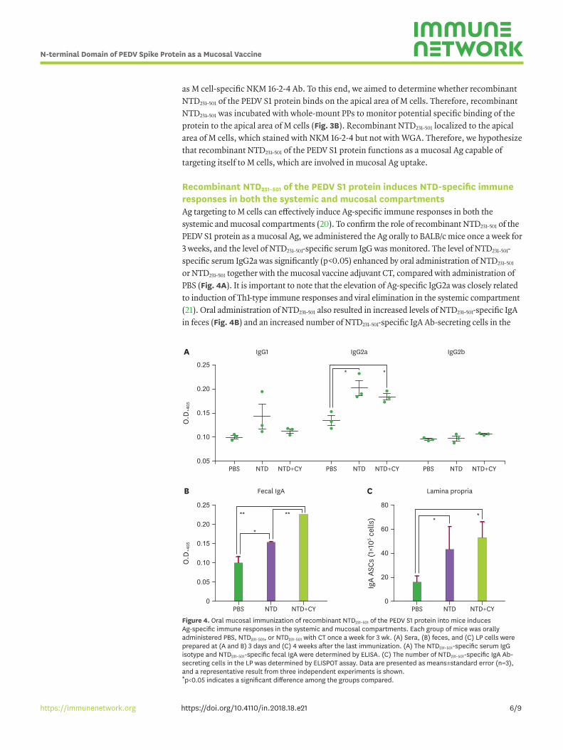
Recombinant NTD231–501 of the PEDV S1 protein induces NTD-specific immune responses in both the systemic and mucosal compartmentsAg targeting to M cells can effectively induce Ag-specific immune responses in both the systemic and mucosal compartments (20). To confirm the role of recombinant NTD231–501 of the PEDV S1 protein as a mucosal Ag, we administered the Ag orally to BALB/c mice once a week for 3 weeks, and the level of NTD231–501-specific serum IgG was monitored. The level of NTD231–501-specific serum IgG2a was significantly (p<0.05) enhanced by oral administration of NTD231–501 or NTD231–501 together with the mucosal vaccine adjuvant CT, compared with administration of PBS (Fig. 4A). It is important to note that the elevation of Ag-specific IgG2a was closely related to induction of Th1-type immune responses and viral elimination in the systemic compartment (21). Oral administration of NTD231–501 also resulted in increased levels of NTD231–501-specific IgA in feces (Fig. 4B) and an increased number of NTD231–501-specific IgA Ab-secreting cells in the
6/9https://doi.org/10.4110/in.2018.18.e21
N-terminal Domain of PEDV Spike Protein as a Mucosal Vaccine
https://immunenetwork.org
A IgG1 IgG2a IgG2b
PBS NTD NTD+CY PBS NTD NTD+CY PBS NTD NTD+CY
0.25
0.20
0.15
0.10
0.05
O.D
. 405
* *
C80
60
40
20
0
IgA
ASCs
(1×1
05 cel
ls)
PBS NTD
Lamina propria
NTD+CY
**
B0.25
0.20
0.15
0.10
0.05
0
O.D
. 405
PBS NTD
Fecal IgA
NTD+CY
*
****
Figure 4. Oral mucosal immunization of recombinant NTD231–501 of the PEDV S1 protein into mice induces Ag-specific immune responses in the systemic and mucosal compartments. Each group of mice was orally administered PBS, NTD231–501, or NTD231–501 with CT once a week for 3 wk. (A) Sera, (B) feces, and (C) LP cells were prepared at (A and B) 3 days and (C) 4 weeks after the last immunization. (A) The NTD231–501-specific serum IgG isotype and NTD231–501-specific fecal IgA were determined by ELISA. (C) The number of NTD231–501-specific IgA Ab-secreting cells in the LP was determined by ELISPOT assay. Data are presented as means±standard error (n=3), and a representative result from three independent experiments is shown. *p<0.05 indicates a significant difference among the groups compared.
LP, a mucosal immune effector tissue related to the level of mucosal secretory IgA (Fig. 4C). Collectively, we conclude that recombinant NTD231–501 of the PEDV S1 protein is a candidate molecule for application in a mucosal vaccine, because its oral administration induces Ag-specific immune responses in both the systemic and mucosal compartments.
DISCUSSION
The current use of PEDV-attenuated vaccines is associated with several problems, including inefficient protective immune induction against the currently prevalent PEDV strains and poor immune induction in the mucosal compartment, which includes the gastrointestinal tract, the main site of PEDV infection (22). Consequently, application of an oral mucosal vaccine using a recombinant Ag against the currently prevalent PEDV strains should be considered in the development of effective PEDV vaccines. One candidate Ag to be used as a recombinant PEDV vaccine is the S1 protein, although it is clear that the production of recombinant PEDV S1 protein in a prokaryotic expression system is not plausible. Herein, we addressed these issues and discussed the potential use of NTD231–501 of the S1 protein from the PEDV BM1 strain, a currently prevalent strain in South Korea, as a mucosal vaccine candidate Ag.
The GST gene, which is a part of the pGEX expression vector, originates from eukaryotic organisms, and GST aids in the stable expression of fusion proteins, because it rapidly folds into a stable conformation during gene translation (23). The NTD231–501 gene of the PEDV S1 protein was inserted into the pGEX 4T-1 expression vector containing a thrombin recognition site for cleaving the target protein. After expressing the construct in E. coli (BL21 Star (DE3)), the recombinant NTD231–501 protein yield was 20 mg/L. Although host infection by PEDV begins with the interaction between the PEDV S1 CTD and APN of host cells, recognition of the sugar moiety by the PEDV S1 NTD is also critical in PEDV infection in host cells (8). Additionally, it was recently reported that knockout of porcine APN in swine testicle (ST) cells did not inhibit PEDV infection (24). Consequently, we were interested in determining the function of the NTD of the PEDV S protein. We found that the recombinant NTD231–501 protein produced in this study specifically binds to mucin and to the apical area of M cells.
Oral delivery of Ag can induce Ag-specific secretory IgA generation in the mucosal lining of the body, including the gastrointestinal tract, salivary glands, and mammary glands (25). Therefore, we assumed that oral administration of Ag would be a suitable strategy to induce protective immunity against PEDV infection. However, a currently licensed oral mucosal subunit vaccine does not exist due to the lack of a mucosal adjuvant that can potentiate delivery of the Ag into M cells and enhance the immune response. In that sense, Ag, which is capable of targeting the Ag itself to M cells, is an attractive target for developing an oral mucosal vaccine against PEDV infection. For example, when fed orally, the rice-based cholera vaccine MucoRice-cholera toxin B subunit was taken up by M cells as a result of its M cell-targeting ability and efficiently induced cholera toxin B subunit-specific immune responses in both the systemic and mucosal compartments (26). Therefore, our finding that the NTD231–501 of the PEDV S protein specifically targets itself to M cells suggests that NTD231–501 is a promising Ag candidate for an oral mucosal vaccine against PEDV infection. More importantly, the Ag-specific IgG2a isotype, which was efficiently induced by oral administration of NTD231–501, is closely related to the induction of protective immunity against a number of viruses, including influenza virus, Ebola virus, and yellow fever virus (1,12,27). In addition, the Fc region of IgG2a activates Fc receptors with a high affinity by interacting with
7/9https://doi.org/10.4110/in.2018.18.e21
N-terminal Domain of PEDV Spike Protein as a Mucosal Vaccine
https://immunenetwork.org
complement components, resulting in efficient induction of Fc receptor-mediated effector functions, such as Ab-dependent cell-mediated cytotoxicity and opsonophagocytosis by macrophages (28). Moreover, IgG2a production is closely associated with the induction of Th1-type immune responses (21). Consequently, we believe that Ag-specific IgG2a induction by oral administration of NTD231–501 of the PEDV S protein is a promising strategy to develop an oral mucosal vaccine against PEDV infection.
ACKNOWLEDGEMENTS
This study was supported by a grant from the Next-Generation BioGreen 21 Program (project No. PJ011801), Rural Development Administration of Korea and by the Basic Science Research Programs (2016R1A2B2010096), through the National Research Foundation (NRF), funded by the Korean Ministry of Science, ICT & Future Planning. Ms. BH Cho was supported by the BK21 PLUS Program in the Department of Bioactive Materials. Experiment for Confocal laser scanning microscopy was performed with the instrument installed in the Center for University-Wide Research Facilities (CURF) at Chonbuk National University.
REFERENCES
1. Wood EN. An apparently new syndrome of porcine epidemic diarrhoea. Vet Rec 1977;100:243-244. PUBMED | CROSSREF
2. Pensaert MB, de Bouck P. A new coronavirus-like particle associated with diarrhea in swine. Arch Virol 1978;58:243-247. PUBMED | CROSSREF
3. Deng F, Ye G, Liu Q, Navid MT, Zhong X, Li Y, Wan C, Xiao S, He Q, Fu ZF, et al. Identification and comparison of receptor binding characteristics of the spike protein of two porcine epidemic diarrhea virus strains. Viruses 2016;8:55. PUBMED | CROSSREF
4. Kocherhans R, Bridgen A, Ackermann M, Tobler K. Completion of the porcine epidemic diarrhoea coronavirus (PEDV) genome sequence. Virus Genes 2001;23:137-144. PUBMED | CROSSREF
5. Li BX, Ge JW, Li YJ. Porcine aminopeptidase N is a functional receptor for the PEDV coronavirus. Virology 2007;365:166-172. PUBMED | CROSSREF
6. Alonso C, Goede DP, Morrison RB, Davies PR, Rovira A, Marthaler DG, Torremorell M. Evidence of infectivity of airborne porcine epidemic diarrhea virus and detection of airborne viral RNA at long distances from infected herds. Vet Res (Faisalabad) 2014;45:73. PUBMED | CROSSREF
7. de Arriba ML, Carvajal A, Pozo J, Rubio P. Mucosal and systemic isotype-specific antibody responses and protection in conventional pigs exposed to virulent or attenuated porcine epidemic diarrhoea virus. Vet Immunol Immunopathol 2002;85:85-97. PUBMED | CROSSREF
8. Liu C, Tang J, Ma Y, Liang X, Yang Y, Peng G, Qi Q, Jiang S, Li J, Du L, et al. Receptor usage and cell entry of porcine epidemic diarrhea coronavirus. J Virol 2015;89:6121-6125. PUBMED | CROSSREF
9. Brandtzaeg P, Kiyono H, Pabst R, Russell MW. Terminology: nomenclature of mucosa-associated lymphoid tissue. Mucosal Immunol 2008;1:31-37. PUBMED | CROSSREF
10. Brandtzaeg P, Pabst R. Let's go mucosal: communication on slippery ground. Trends Immunol 2004;25:570-577. PUBMED | CROSSREF
11. Kucharzik T, Lügering N, Rautenberg K, Lügering A, Schmidt MA, Stoll R, Domschke W. Role of M cells in intestinal barrier function. Ann N Y Acad Sci 2000;915:171-183. PUBMED | CROSSREF
8/9https://doi.org/10.4110/in.2018.18.e21
N-terminal Domain of PEDV Spike Protein as a Mucosal Vaccine
https://immunenetwork.org
12. Strugnell RA, Wijburg OL. The role of secretory antibodies in infection immunity. Nat Rev Microbiol 2010;8:656-667. PUBMED | CROSSREF
13. Frey A, Neutra MR. Targeting of mucosal vaccines to Peyer's patch M cells. Behring Inst Mitt 1997;98:376-389.PUBMED
14. Hase K, Kawano K, Nochi T, Pontes GS, Fukuda S, Ebisawa M, Kadokura K, Tobe T, Fujimura Y, Kawano S, et al. Uptake through glycoprotein 2 of FimH+ bacteria by M cells initiates mucosal immune response. Nature 2009;462:226-230. PUBMED | CROSSREF
15. Kim SH, Jung DI, Yang IY, Kim J, Lee KY, Nochi T, Kiyono H, Jang YS. M cells expressing the complement C5a receptor are efficient targets for mucosal vaccine delivery. Eur J Immunol 2011;41:3219-3229. PUBMED | CROSSREF
16. Nochi T, Yuki Y, Matsumura A, Mejima M, Terahara K, Kim DY, Fukuyama S, Iwatsuki-Horimoto K, Kawaoka Y, Kohda T, et al. A novel M cell-specific carbohydrate-targeted mucosal vaccine effectively induces antigen-specific immune responses. J Exp Med 2007;204:2789-2796. PUBMED | CROSSREF
17. Li X, Chen H. Evaluation of the porcine gastric mucin binding assay for high-pressure-inactivation studies using murine norovirus and tulane virus. Appl Environ Microbiol 2015;81:515-521. PUBMED | CROSSREF
18. Chung HC, Lee JH, Nguyen VG, Huynh TM, Lee GE, Moon HJ, Park SJ, Kim HK, Park BK. New emergence pattern with variant porcine epidemic diarrhea viruses, South Korea, 2012–2015. Virus Res 2016;226:14-19. PUBMED | CROSSREF
19. Chung HC, Nguyen VG, Moon HJ, Lee JH, Park SJ, Lee GE, Kim HK, Noh YS, Lee CH, Goede D, et al. Isolation of porcine epidemic diarrhea virus during outbreaks in South Korea, 2013–2014. Emerg Infect Dis 2015;21:2238-2240. PUBMED | CROSSREF
20. Kim SH, Lee KY, Jang YS. Mucosal immune system and M cell-targeting strategies for oral mucosal vaccination. Immune Netw 2012;12:165-175. PUBMED | CROSSREF
21. Gracie JA, Bradley JA. Interleukin-12 induces interferon-gamma-dependent switching of IgG alloantibody subclass. Eur J Immunol 1996;26:1217-1221. PUBMED | CROSSREF
22. Gerdts V, Zakhartchouk A. Vaccines for porcine epidemic diarrhea virus and other swine coronaviruses. Vet Microbiol 2017;206:45-51. PUBMED | CROSSREF
23. Harper S, Speicher DW. Expression and purification of GST fusion proteins. Curr Protoc Protein Sci 2008;Chapter 6:Unit 6.6. PUBMED | CROSSREF
24. Li W, Luo R, He Q, van Kuppeveld FJ, Rottier PJ, Bosch BJ. Aminopeptidase N is not required for porcine epidemic diarrhea virus cell entry. Virus Res 2017;235:6-13. PUBMED | CROSSREF
25. Lycke N. Recent progress in mucosal vaccine development: potential and limitations. Nat Rev Immunol 2012;12:592-605. PUBMED | CROSSREF
26. Nochi T, Yuki Y, Katakai Y, Shibata H, Tokuhara D, Mejima M, Kurokawa S, Takahashi Y, Nakanishi U, Ono F, et al. A rice-based oral cholera vaccine induces macaque-specific systemic neutralizing antibodies but does not influence pre-existing intestinal immunity. J Immunol 2009;183:6538-6544. PUBMED | CROSSREF
27. Huber VC, McKeon RM, Brackin MN, Miller LA, Keating R, Brown SA, Makarova N, Perez DR, Macdonald GH, McCullers JA. Distinct contributions of vaccine-induced immunoglobulin G1 (IgG1) and IgG2a antibodies to protective immunity against influenza. Clin Vaccine Immunol 2006;13:981-990. PUBMED | CROSSREF
28. Huber VC, Lynch JM, Bucher DJ, Le J, Metzger DW. Fc receptor-mediated phagocytosis makes a significant contribution to clearance of influenza virus infections. J Immunol 2001;166:7381-7388. PUBMED | CROSSREF
9/9https://doi.org/10.4110/in.2018.18.e21
N-terminal Domain of PEDV Spike Protein as a Mucosal Vaccine
https://immunenetwork.org