original article: in vitro and in vivo anticancer … journal 2017;16:448-463 – issn 1611-2156...
TRANSCRIPT

EXCLI Journal 2017;16:448-463 – ISSN 1611-2156 Received: September 15, 2016, accepted: March 13, 2017, published: April 03, 2017
448
Original article:
IN VITRO AND IN VIVO ANTICANCER STUDIES OF 2′-HYDROXY CHALCONE DERIVATIVES EXHIBIT APOPTOSIS IN COLON
CANCER CELLS BY HDAC INHIBITION AND CELL CYCLE ARREST
Aditya Narayan Pande1#, Subhankar Biswas1#, Neetinkumar D. Reddy1, B.S. Jayashree2, Nitesh Kumar1, C. Mallikarjuna Rao1,*
1 Department of Pharmacology, Manipal College of Pharmaceutical Sciences, Manipal University, Manipal-576104, Karnataka, India
2 Department of Pharmaceutical Chemistry, Manipal College of Pharmaceutical Sciences, Manipal University, Manipal-576104, Karnataka, India
# Both authors contributed equally to this work. * Corresponding author: Dr. C. Mallikarjuna Rao, Principal and Professor of Pharmacology,
Manipal College of Pharmaceutical Sciences, Manipal University, Manipal - 576104, Karnataka, India, Phone: +91 0820 2922482, E-mail: [email protected], [email protected]
http://dx.doi.org/10.17179/excli2016-643
This is an Open Access article distributed under the terms of the Creative Commons Attribution License (http://creativecommons.org/licenses/by/4.0/).
ABSTRACT
Considering the therapeutic values of bioflavonoids in colon cancer treatment, six 2′-hydroxy chalcones (C1-C6) were synthesized, characterized and screened for in vitro cytotoxicity on human colon carcinoma (HCT116) and African green monkey kidney epithelial cells (Vero). Only C5 showed selective cytotoxicity against HCT116 cells. Other potent cytotoxic compounds were C1, C2 and C3. Further screening included enzyme inhibition studies on histone deacetylase (HDAC) enzyme where C1 showed lowest IC50 value (105.03 µM). Based on cytotoxicity data C1, C2 and C3 were selected for further in vitro mechanistic studies, namely apoptotic studies (Acridine or-ange/Ethidium bromide (AO/EB) and Annexin V), cell cycle analysis using propidium iodide (PI) stain and in vivo anticancer efficacy in 1,2-dimethyl hydrazine (DMH) induced colorectal carcinoma in Wistar rats. The com-pounds induced apoptosis in more than 30 % cells in AO/EB and Annexin V staining. They also showed cell cycle arrest in G2/M phase with PI staining. They showed a significant reduction in aberrant crypt foci formation and adenocarcinoma count along with a significant (p<0.05) reduction in TNF-α levels as compared to DMH control at 100 mg/kg dose. Thus, it can be concluded that the synthesized 2′-hydroxychalcones were effective against colon adenocarcinoma in in vitro and in vivo studies. Keywords: chalcones, apoptosis, cell cycle, HDAC, DMH, colon cancer
INTRODUCTION
Cancer is a multifactorial disease charac-terized by uncontrolled and abnormal cellular growth. Unlike normal cells, cancer cells con-tinue to grow and divide eventually replicat-ing exponentially into harmful cells (Pisani et al., 1999). It is undoubtedly a life threatening
disease. However, there is a misconception about its cure. Much of the efforts in cancer research carried out during the last few dec-ades prompted the researchers and cell biolo-gist in understanding its pathophysiology, sig-naling mechanism and developing strategic therapies that can effectively treat cancer so

EXCLI Journal 2017;16:448-463 – ISSN 1611-2156 Received: September 15, 2016, accepted: March 13, 2017, published: April 03, 2017
449
as to eliminate or slow the impact of disease on patients’ lives. The 5-year relative survival rate for all cancers diagnosed in 2004-2010 was 68 %, up from 49 % in 1975-1977 (Siegel et al., 2015).
Among the various types of cancer diag-nosed, it is worth mentioning that colorectal cancer (CRC) is the third most fatal malig-nancy affecting both men and women (Siegel et al., 2014). Most CRC develops slowly as non-cancerous polyp (adenomas) and show harmful effects over years evolving into inva-sive cancer (Schulmann et al., 2002). Several risk factors including age, familial history of adenomatous polyps, diet and heavy alcohol consumption have been implicated in the pathogenesis of CRC (Haggar and Boushey, 2009). Apart from these factors, recent re-search has also highlighted the role of epige-netics in CRC (Goel and Boland, 2012). Among the epigenetic factors, histone deacetylases (HDACs) a group of enzymes involved in silencing gene expression are re-ported to be over-expressed in CRC (Mariadason, 2008). Amid the various modal-ities of treatment strategies available for CRC, surgery is the most preferred choice de-pending on the stage of CRC. However, a per-son’s general health plays a crucial role for better outcome of surgery. Furthermore, sur-gery along with chemotherapy is recom-mended in many cases for long term survival and prevention of reoccurrence (Giacchetti et al., 1999). Although there are many chemo-therapeutic drugs for CRC, side effects asso-ciated with them limits their usage, providing researchers to develop newer molecules lead-ing to better therapeutic outcome.
The current drug discovery program has explored natural products such as bio flavo-noids, polyphenols, chalcones for their anti-oxidant and cytotoxic properties that have given insight to the medicinal chemists to use them as potential anti-cancer agents. Chal-cones are precursors of flavonoids that are present in various parts of a plant having anti-inflammatory and anti-tumor activity (Jeon et al., 2012; Zhang et al., 2013). It is noteworthy to mention that quercetin, a cyclized chalcone
and curcumin have been explored as potential anti-cancer agents. However, a few detailed studies have been carried out on the anti-tu-mor properties of chalcones against colon ad-enocarcinoma. They are considered to have fewer side effects when compared with chemotherapeutic agents (Syam et al., 2012). In addition, there is an increasing arousal of interest in flavonols and other dietary poly phenols owing to their HDAC inhibitory ac-tivity in cancer cells (Rajendran et al., 2011). Thus, chalcones and flavonols could be most appropriate candidates to be evaluated for their HDAC inhibitory potential. Hence, our study was aimed at developing different sub-stituted chalcones and assessing their anti-cancer activity against colon adenocarci-noma.
MATERIALS AND METHODS
Chemicals and instruments Chemicals used for synthesis were pro-
cured from Sigma-Aldrich Co. LLC, St. Louis, MO, USA; Merck Specialities Pvt. Ltd, Mumbai, India; Spectrochem Pvt. Ltd., Mumbai, MH, India. Melting point of the syn-thesized compounds were determined using capillary melting point apparatus from Tosh-niwal Systems and Instruments Pvt. Ltd., Chennai, TN, India. Thin layer chromatog-raphy was carried out on pre-coated silica gel plates procured from Merck # 60F254 using the following developing system: Hexane/ Ethyl acetate (8:2, v/v) and the spots were vis-ualized under UV lamp (254 or 366 nm) and/or iodine vapor. The IR spectra were rec-orded using IR spectrometer (Model FTIR-8300, Shimadzu Co., Kyoto, Japan) using KBr pellets. Mass spectra were recorded us-ing LC-MS (ESI) (Model LCMS-2010A, Shi-madzu Co., Kyoto, Japan). 1H and 13C NMR were recorded at 400 MHz (Model Ascend 400, Bruker Biosciences Corporation, Biller-ica, MA, USA) using DMSO (D6) as solvent. Chemical shifts are reported in δ values (ppm). Signal multiplicities are represented by s (singlet), d (doublet), m (multiplet). All tested compounds possess a purity of not less than 95 %.
EXCLI Journal 2017;16:448-463 – ISSN 1611-2156 Received: September 15, 2016, accepted: March 13, 2017, published: April 03, 2017
450
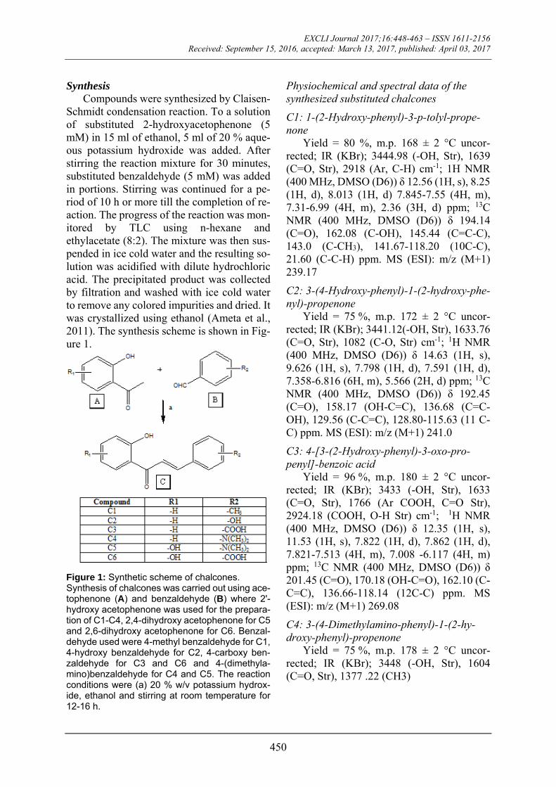
Synthesis Compounds were synthesized by Claisen-
Schmidt condensation reaction. To a solution of substituted 2-hydroxyacetophenone (5 mM) in 15 ml of ethanol, 5 ml of 20 % aque-ous potassium hydroxide was added. After stirring the reaction mixture for 30 minutes, substituted benzaldehyde (5 mM) was added in portions. Stirring was continued for a pe-riod of 10 h or more till the completion of re-action. The progress of the reaction was mon-itored by TLC using n-hexane and ethylacetate (8:2). The mixture was then sus-pended in ice cold water and the resulting so-lution was acidified with dilute hydrochloric acid. The precipitated product was collected by filtration and washed with ice cold water to remove any colored impurities and dried. It was crystallized using ethanol (Ameta et al., 2011). The synthesis scheme is shown in Fig-ure 1.
Figure 1: Synthetic scheme of chalcones. Synthesis of chalcones was carried out using ace-tophenone (A) and benzaldehyde (B) where 2ʹ-hydroxy acetophenone was used for the prepara-tion of C1-C4, 2,4-dihydroxy acetophenone for C5 and 2,6-dihydroxy acetophenone for C6. Benzal-dehyde used were 4-methyl benzaldehyde for C1, 4-hydroxy benzaldehyde for C2, 4-carboxy ben-zaldehyde for C3 and C6 and 4-(dimethyla-mino)benzaldehyde for C4 and C5. The reaction conditions were (a) 20 % w/v potassium hydrox-ide, ethanol and stirring at room temperature for 12-16 h.
Physiochemical and spectral data of the synthesized substituted chalcones
C1: 1-(2-Hydroxy-phenyl)-3-p-tolyl-prope-none
Yield = 80 %, m.p. 168 ± 2 °C uncor-rected; IR (KBr); 3444.98 (-OH, Str), 1639 (C=O, Str), 2918 (Ar, C-H) cm-1; 1H NMR (400 MHz, DMSO (D6)) δ 12.56 (1H, s), 8.25 (1H, d), 8.013 (1H, d) 7.845-7.55 (4H, m), 7.31-6.99 (4H, m), 2.36 (3H, d) ppm; 13C NMR (400 MHz, DMSO (D6)) δ 194.14 (C=O), 162.08 (C-OH), 145.44 (C=C-C), 143.0 (C-CH3), 141.67-118.20 (10C-C), 21.60 (C-C-H) ppm. MS (ESI): m/z (M+1) 239.17
C2: 3-(4-Hydroxy-phenyl)-1-(2-hydroxy-phe-nyl)-propenone
Yield = 75 %, m.p. 172 ± 2 °C uncor-rected; IR (KBr); 3441.12(-OH, Str), 1633.76 (C=O, Str), 1082 (C-O, Str) cm-1; 1H NMR (400 MHz, DMSO (D6)) δ 14.63 (1H, s), 9.626 (1H, s), 7.798 (1H, d), 7.591 (1H, d), 7.358-6.816 (6H, m), 5.566 (2H, d) ppm; 13C NMR (400 MHz, DMSO (D6)) δ 192.45 (C=O), 158.17 (OH-C=C), 136.68 (C=C-OH), 129.56 (C-C=C), 128.80-115.63 (11 C-C) ppm. MS (ESI): m/z (M+1) 241.0
C3: 4-[3-(2-Hydroxy-phenyl)-3-oxo-pro-penyl]-benzoic acid
Yield = 96 %, m.p. 180 ± 2 °C uncor-rected; IR (KBr); 3433 (-OH, Str), 1633 (C=O, Str), 1766 (Ar COOH, C=O Str), 2924.18 (COOH, O-H Str) cm-1; 1H NMR (400 MHz, DMSO (D6)) δ 12.35 (1H, s), 11.53 (1H, s), 7.822 (1H, d), 7.862 (1H, d), 7.821-7.513 (4H, m), 7.008 -6.117 (4H, m) ppm; 13C NMR (400 MHz, DMSO (D6)) δ 201.45 (C=O), 170.18 (OH-C=O), 162.10 (C-C=C), 136.66-118.14 (12C-C) ppm. MS (ESI): m/z (M+1) 269.08
C4: 3-(4-Dimethylamino-phenyl)-1-(2-hy-droxy-phenyl)-propenone
Yield = 75 %, m.p. 178 ± 2 °C uncor-rected; IR (KBr); 3448 (-OH, Str), 1604 (C=O, Str), 1377 .22 (CH3)

EXCLI Journal 2017;16:448-463 – ISSN 1611-2156 Received: September 15, 2016, accepted: March 13, 2017, published: April 03, 2017
451
C5: 1-(2-Hydroxy-4-methyl-phenyl)-3-p-tolyl-propenone Yield = 80 %, m.p. 153 ± 2 °C uncorrected; IR (KBr); 3443.05 (-OH, Str), 1597 (C=O, Str)
C6: 4-[3-(2,6-Dihydroxy-phenyl)-3-oxo-pro-penyl]-benzoic acid
Yield = 65 %, m.p. 186 ± 2 °C uncor-rected; IR (KBr); 3070.78 (-OH, Str), 1616.40 (C=O, Str), 1707.06 (Ar COOH, C=O Str), 2652.21 (COOH, O-H Str).
In vitro studies
Cell lines and their maintenance Human colorectal carcinoma (HCT116)
and African green monkey kidney epithelial cells (VERO) were procured from the Na-tional Centre for Cell Science, Pune, MH, In-dia. The cells were maintained in Dulbecco’s Modified Eagle’s Medium (DMEM) (Sigma-Aldrich Co. LLC, ST. Louis, Mo, USA) sup-plemented with 10 % Fetal Bovine Serum (FBS) (HiMedia Laboratories, Mumbai, In-dia) and 1 × Penicillin/Streptomycin at 37 °C in CO2 incubator (NU-5501 E/G, NuAire Inc., Plymoth, MN, USA) in humidified atmos-phere of 5 % CO2 and 95 % air. The cells were maintained by routine sub-culturing in 25 cm2 tissue culture flasks.
Cytotoxicity assay Cytotoxic potential of the test compounds
were assessed using MTT assay (Kumar et al., 2016). In brief, HCT116 and Vero cells were harvested from confluent flask and seeded (5 × 10⁴ cells/well) in 96 well plates. After 24 h of incubation, the cells were exposed to dif-ferent concentration of the test compounds for 48 h. Further, 50 µl of MTT reagent (HiMedia Laboratories, Mumbai, India) (2 mg/ml in sterile PBS) was added into each well after 48 h and incubated for 3 more h. The formazan crystals formed were solubilized using 100 % DMSO and the optical density was measured at 540 nm using micro plate reader (ELx800, BioTek Instruments Inc., Winooski, VT, USA).
Whole cell HDAC enzyme assay HCT116 cells were harvested and seeded
in 96 well sterile, black well plates at 2 × 10⁴ cells/well and incubated overnight. Further, the cells were treated with different concen-tration of the test compounds for a period of 18 h. Then, 15 mM Boc-Lys(Ac)-AMC sub-strate (Sigma Aldrich # SCP0168) was added and incubated for 1 h. The reaction was termi-nated by the addition of 50 µL of stop solution (trypsin 2 mg/ml, 1 % NP40, 1 µl SAHA) in HDAC assay buffer (25 mM Tris-HCl (pH 8.0), 137 mM NaCl, 2.7 mM KCl, 1 mM MgCl2). The reaction was then allowed to proceed for 15 min at 37 °C after which, the fluorescence was measured at 360 nm excita-tion and 460 nm emission using fluorescence micro plate reader (FLx800, BioTek Instru-ments Inc., Winooski, VT, USA) (Reddy et al., 2015).
Acridine orange/Ethidium bromide (AO/EB) staining
AO/EB dual staining was performed in or-der to determine the apoptosis inducing po-tential of the selected test compounds in HCT116 cell line with few modifications (Kumar et al., 2016). In brief, 5 × 10⁵ cells were seeded in 6 wells plate containing 2 ml of medium and incubated for 24 h. After 24 h, cells were treated with the test compounds and incubated for 48 h. After 48 h of incuba-tion, the wells were washed with phosphate buffer saline (PBS) and cells were fixed with ice cold ethanol (70 %) for 30 min. Ethanol was then removed and cells were washed again with PBS followed by the addition of 200 µl of AO/EB (20/30 µg/ml) stain in each well. The plate was later kept in the dark for 20 min. The excess stain was further washed thrice with PBS and observed for their fluo-rescence under a fluorescent microscope (Eclipse TS100-F, Nikon Instruments Inc., Melville, NY, USA).
Annexin V assay Apoptosis determination by Annexin V
staining was carried out using Muse Cell An-alyzer with the kit provided by the manufac-turer Merck Millipore. In brief, 1 × 10⁶ cells

EXCLI Journal 2017;16:448-463 – ISSN 1611-2156 Received: September 15, 2016, accepted: March 13, 2017, published: April 03, 2017
452
were seeded in 60 mm tissue culture dish and after overnight adherence test compounds were added and incubated. After 48 h the cells were detached by trypsinization, centrifuged and resuspended. 100 µl of cell suspension was added with 100 µl of Annexin V reagent and incubated for 20 min at room temperature following which the cells were analyzed for apoptosis.
Cell cycle analysis
The ability of the test compounds to arrest any phase of the cell cycle was determined us-ing the described procedure (Reddy et al., 2015). In this method, HCT116 cells were harvested and seeded at a density of 1 × 10⁶ cell in 60 mm petri plate and incubated for 24 h. After 24 h cells were treated with test com-pounds for 48 h. Then the cells were washed with PBS, trypsinized and centrifuged. The cell pellets were later fixed in 70 % ice cold ethanol and stored at -20 °C for 24 h. After fixing, the pellet was dislodged in PBS and stained with propidium iodide solution. The cells were then analyzed using Accuri C6 flow cytometer with the threshold levels ad-justed to remove the debris (BD Biosciences, San Jose, CA, USA) and data analysis were performed using BD Accuri™ C6 software.
In vivo studies
Animals Male Wistar rats inbred at the Central An-
imal Research Facility, Manipal University, were used in our study. The animal care and handling were carried out in accordance with the guidelines issued by the Institutional Ani-mal Ethics Committee (IAEC), Manipal. Af-ter obtaining research proposal approval (IAEC/KMC/16/2015) the animals were ac-climatized to the experimental room having temperature of 23 ± 2 °C, humidity (50 ± 5 %) and 12 h light and dark cycles. Rats were housed in sterile polypropylene cages con-taining sterile paddy.
Acute toxicity studies Acute toxicity study was carried out to de-
termine the safe dose using Organization for
Economic Cooperation and Development (OECD) – 425 guideline. Limit test was per-formed using 2000 mg/kg dose of the test compounds in 6 h fasted rats. Animals were observed for any signs of toxicity for the first 4 h continuously and then daily for 14 days.
Preparation of the test compound and standard drug
Test compound: Three test compounds, namely C1, C2 & C3 were suspended in 0.25 % sodium carboxy methyl cellulose (CMC) and were administered orally (p.o) with a dosing volume of 10 ml/kg.
Standard drug: 5-Fluorouracil (5-FU) was used as standard. It was procured in the form of injection and administered intraperitone-ally (i.p) with a dosing volume of 10 ml/kg.
DMH (1,2-dimethyl hydrazine) induced colon cancer in Wistar rats
Induction of colon cancer was achieved using DMH according to a previously de-scribed procedure with few modifications (Perše and Cerar, 2005). DMH at a dose of 30 mg/kg was administered i.p once a week for 20 weeks. The incidence of aberrant crypt foci (ACFs) and adenocarcinoma were con-firmed by sacrificing a few of the animals af-ter 20 weeks confirming the induction of co-lon cancer in experimental animals. Finally, the animals were randomized into five exper-imental groups based on their body weight.
Experimental groups Group 1 (normal control): Animals (n=6) were administered 0.25 % CMC in water p.o. Group 2 (DMH control): Animals (n=6) were administered DMH Group 3 (standard drug): Animals (n=6) re-ceived 5-FU 10 mg/kg i.p. for 21 days of study period Group 4 (C1): Animals (n=6) received C1 at 100 mg/kg p.o for 21 days Group 5 (C2): Animals (n=6) received C2 at 100 mg/kg p.o. for 21 days. Group 6 (C3): Animals (n=6) received C3 at 100 mg/kg p.o. for 21 days.

EXCLI Journal 2017;16:448-463 – ISSN 1611-2156 Received: September 15, 2016, accepted: March 13, 2017, published: April 03, 2017
453
Parameters assessed in the experimental animals
ACF formation and adenocarcinoma incidence
The distal part of the colon, which was re-moved from the experimental animals after the study period was cut open and placed flat on a filter paper and fixed with 10 % buffered formalin for 12 h. Further, it was stained with 0.1 % methylene blue in PBS for 5 min. Spec-imens were later observed under microscope for ACF formation and were calculated as the number of counts/5 cm2 in colon tissue. The entire colon was considered to study the inci-dence of adenocarcinoma. The growth was critically observed and the count and size were noted from each animal.
TNF-α level The levels of TNF-α were estimated in the
colon of experimental animals, for which 10 % homogenate of colon tissue was pre-pared in tissue lysis buffer. The homogenate was centrifuged and the supernatant was col-lected to measure TNF-α levels using com-mercially available ELISA kits of rat TNF-α (# KRC3011, Invitrogen).
Colon length/weight ratio and organ index Length of the isolated colon was meas-
ured in centimeters and weight was measured in g. The colon length/weight (L/W) ratio was then calculated. Isolated spleen, kidney and heart of the experimental animals were also weighed in g and respective index was calcu-lated.
Histopathology of colon Histopathology was carried out according
to the described procedure (Reddy et al., 2015). The stained slides were then analyzed under a microscope for any anatomical changes.
Statistical analysis All the values were expressed as mean ±
SEM of 6 animals. Data were analyzed using one-way ANOVA followed by Tukey’s mul-tiple comparison tests using Prism 5.03 (Graph Pad Software Inc., La Jolla, CA,
USA). Values of p < 0.05 were considered to be significant.
RESULTS
In vitro studies
MTT assay MTT assay was carried out to evaluate the
cytotoxic potential of the synthesized com-pounds on HCT116 and Vero cell line. Com-pounds C1, C2, C3 and C5 were found to be cytotoxic against colon cancer cell line after 48 h of treatment. Among these compounds, C1 was found to be most cytotoxic with an IC50 value of 37.07 µM on HCT116 cells. Fur-thermore, in Vero cell lines C1, C2 and C3 exhibited potential cytotoxicity compared with the remaining three synthesized com-pounds. Moreover C1 displayed greater cyto-toxicity compared with C2 and C3. Table 1 provides the IC50 value of the synthesized compounds on both the cell lines tested.
Table 1: In vitro cytotoxicity screening in human colon cancer and normal cells
COMPOUND IC50 Value (µM/ml)
HCT116 VERO
C1 37.07 26.28 C2 81.41 88.26
C3 116.9 177.8
C4 >500 >500 C5 261.2 >500 C6 >500 >500
The IC50 values were calculated using nonlinear regression analysis. All values are represented as mean of the experi-ments carried out in triplicate.
Whole cell HDAC assay The whole cell HDAC enzyme inhibition
assay was performed to determine the effect of test compounds on the epigenetic machin-ery of HCT116 cells. Dose dependent HDAC enzyme inhibition was observed in all treat-ment groups, with C1 as the most potent com-pound with an IC50 value of 105 ± 10 µM. The other potent compound was found to be C5

EXCLI Journal 2017;16:448-463 – ISSN 1611-2156 Received: September 15, 2016, accepted: March 13, 2017, published: April 03, 2017
454
with IC50 value below 200 µM i.e, 160.4 ± 15.5 µM. The IC50 values for the remaining compounds were 394.3 ± 12.9, 928.7 ± 56.6, 470.4 ± 11 and 826.1 ± 58.1 µM respectively for C2, C3, C4 and C6. SAHA, a non-specific HDAC inhibitor, was used as standard and was found to have an IC50 value of 3.6 ± 0.2 µM. Figure 2 shows the histogram plot of whole cell HDAC assay.
AO/EB staining To determine the mechanism of cell death
induced by test compounds, fluorescent dye based apoptosis assay was carried out. In the present study Acridine Orange/Ethidium Bro-
mide stain was used. Green stained unfrag-mented nuclei were observed in untreated cells, indicating non-apoptotic cells. Where-as, the presence of highly condensed chromo-some was visible in cells treated with test compounds appearing as green fragmented nuclei. Treatment with 5-FU showed 45 ± 0.87 % apoptotic cells in HCT116 cells. Al-ternatively, % apoptotic cells in C1, C2 and C3 treated groups were found to be 42.4 ± 2.25 %, 40 ± 2.5 % and 38.7 ± 0.94 % respec-tively. Figure 3 shows the % apoptotic cells represented as histogram and Figure 4 shows the fluorescent images.
Figure 2: Whole cell HDAC inhibition assay. The histogram plot represents the IC50 value of whole cell HDAC enzyme inhibition assay carried out in HCT116 cell line. All values are represented as mean ± SEM and the experiments were carried out in triplicate.
Figure 3: Percentage apoptotic cells in HCT116 cell line. Percentage apoptotic cells in HCT116 cell line determined by AO/EB staining. All values are mean ± SEM of 100 cells. ap< 0.05 vs. control.

EXCLI Journal 2017;16:448-463 – ISSN 1611-2156 Received: September 15, 2016, accepted: March 13, 2017, published: April 03, 2017
455
Figure 4: AO/EB staining in HCT116 cell line. The images indicate induction of apoptosis in HCT116 cell line by various treatment groups after 48 hrs. (A) Normal control, (B) 5-FU, (C) C1, (D) C2, (E) C3. Arrows indicate condensed nucleus. Magnification 40X.
Apoptosis detection study During apoptosis, phosphatidylserine
which is predominantly situated along the cy-tosolic side of the plasma membrane translo-cates to the extracellular side. After transloca-tion, it is detectable by the family of calcium-dependent phospholipid binding proteins called Annexin. More than 30 % of apoptotic cells were observed after 48h of incubation in the various treatment groups. Total apoptotic cells in normal, 5-FU, C1, C2 and C3 was found to be 6.10 %, 38.08 %, 37.70 %, 36.60 %, and 31.80 % of total cells respec-tively. Early apoptotic events were more prominent in the treatment of 5-FU and C2 with 15.86 % and 18 % of total cells, respec-tively, while it was less in C1 and C4 treat-ment 5.85 % and 4.55 %, respectively (Figure 5).
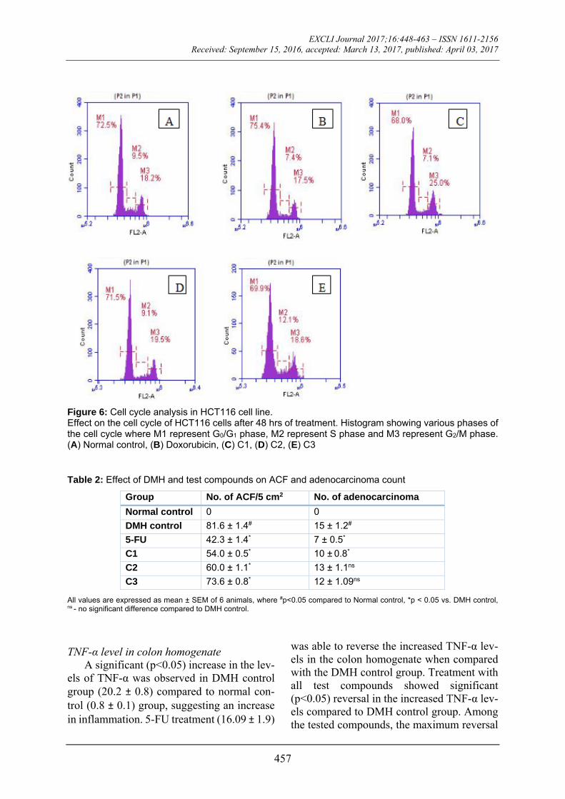
Cell cycle analysis Cell cycle analysis revealed that cells in
normal control contained 72.5 %, 9.5 % and 18.2 % cells in G0/G1, S and G2/M phase re-spectively. The treatments with C1 and C2 showed a notable cell cycle arrest in G2/M phase by increasing the percentage cell counts compared to normal control i.e., 25 % and 19.5 % cells respectively. These results sug-gested potentials of C1 and C2 as a G2/M phase blocker. Furthermore an increase in the percentage of cells in S phase (12 %) was ob-served in C3 treatment compared to normal control (Figure 6).

EXCLI Journal 2017;16:448-463 – ISSN 1611-2156 Received: September 15, 2016, accepted: March 13, 2017, published: April 03, 2017
456
Figure 5: Determination of apoptosis by Annexin V staining. Effect of binding of Annexin V on the surface of HCT116 cell after 48 hrs of treatment. Apoptosis was determined as % live, early and late apoptosis/dead cells by MUSE cell analyzer. In vivo studies
In vivo toxicity studies No signs of toxicity were observed in the
experimental animals treated with the test compounds at 2000 mg/kg dose. Further stud-ies were carried out using 1/20th of the admin-istered dose.
DMH (1,2-dimethyl hydrazine) induced colon cancer in Wistar rats
ACF formation and adenocarcinoma incidence
The incidence of colon carcinoma is com-monly detected with the presence of aberrant crypt foci and the incidence of adenocarci-noma. The ACF formation in the DMH
treated control group were 81.6 ± 1.4/5 cm2 of colon tissue, whereas no such incidences were observed in normal control group. The ACF count was found to be significantly (p<0.05) lower in the all treatment groups compared to DMH control (Table 2).
Adenocarcinoma formation was observed in the entire colon region of DMH treated control group (15 ± 1.2). Treatment with 5-FU and C1 showed significant decrease in the number of adenocarcinoma compared to DMH control group (7 ± 0.5 and 10 ± 0.8 re-spectively). However no significant decrease in adenocarcinoma was observed in C2 and C3 treatment compared to DMH control group (Table 2).
EXCLI Journal 2017;16:448-463 – ISSN 1611-2156 Received: September 15, 2016, accepted: March 13, 2017, published: April 03, 2017
457
Figure 6: Cell cycle analysis in HCT116 cell line. Effect on the cell cycle of HCT116 cells after 48 hrs of treatment. Histogram showing various phases of the cell cycle where M1 represent G0/G1 phase, M2 represent S phase and M3 represent G2/M phase. (A) Normal control, (B) Doxorubicin, (C) C1, (D) C2, (E) C3 Table 2: Effect of DMH and test compounds on ACF and adenocarcinoma count
Group No. of ACF/5 cm2 No. of adenocarcinoma
Normal control 0 0
DMH control 81.6 ± 1.4# 15 ± 1.2#
5-FU 42.3 ± 1.4* 7 ± 0.5*
C1 54.0 ± 0.5* 10 ± 0.8*
C2 60.0 ± 1.1* 13 ± 1.1ns
C3 73.6 ± 0.8* 12 ± 1.09ns
All values are expressed as mean ± SEM of 6 animals, where #p<0.05 compared to Normal control, *p < 0.05 vs. DMH control, ns - no significant difference compared to DMH control.
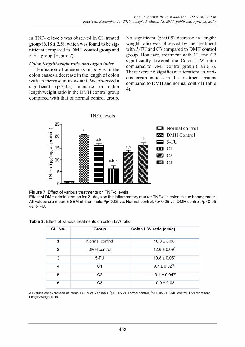
TNF-α level in colon homogenate A significant (p<0.05) increase in the lev-
els of TNF-α was observed in DMH control group (20.2 ± 0.8) compared to normal con-trol (0.8 ± 0.1) group, suggesting an increase in inflammation. 5-FU treatment (16.09 ± 1.9)
was able to reverse the increased TNF-α lev-els in the colon homogenate when compared with the DMH control group. Treatment with all test compounds showed significant (p<0.05) reversal in the increased TNF-α lev-els compared to DMH control group. Among the tested compounds, the maximum reversal
EXCLI Journal 2017;16:448-463 – ISSN 1611-2156 Received: September 15, 2016, accepted: March 13, 2017, published: April 03, 2017
458
in TNF- α levels was observed in C1 treated group (6.18 ± 2.5), which was found to be sig-nificant compared to DMH control group and 5-FU group (Figure 7).
Colon length/weight ratio and organ index Formation of adenomas or polyps in the
colon causes a decrease in the length of colon with an increase in its weight. We observed a significant (p<0.05) increase in colon length/weight ratio in the DMH control group compared with that of normal control group.
No significant (p<0.05) decrease in length/ weight ratio was observed by the treatment with 5-FU and C3 compared to DMH control group. However, treatment with C1 and C2 significantly lowered the Colon L/W ratio compared to DMH control group (Table 3). There were no significant alterations in vari-ous organ indices in the treatment groups compared to DMH and normal control (Table 4).
TNF levels
0
5
10
15
20
25Normal controlDMH Control5-FUC1C2C3
a
b
b,
b
b
TNF-
(pg
/mg
of p
rote
in)
a,
a,
a,
a,
c
Figure 7: Effect of various treatments on TNF-α levels. Effect of DMH administration for 21 days on the inflammatory marker TNF-α in colon tissue homogenate. All values are mean ± SEM of 6 animals. ap<0.05 vs. Normal control, bp<0.05 vs. DMH control, cp<0.05 vs. 5-FU. Table 3: Effect of various treatments on colon L/W ratio
SL. No. Group Colon L/W ratio (cm/g)
1 Normal control 10.8 ± 0.06
2 DMH control 12.6 ± 0.09*
3 5-FU 10.8 ± 0.05*
4 C1 9.7 ± 0.02*#
5 C2 10.1 ± 0.04*#
6 C3 10.9 ± 0.08
All values are expressed as mean ± SEM of 6 animals. *p< 0.05 vs. normal control, #p< 0.05 vs. DMH control. L/W represent Length/Weight ratio.

EXCLI Journal 2017;16:448-463 – ISSN 1611-2156 Received: September 15, 2016, accepted: March 13, 2017, published: April 03, 2017
459
Table 4: Effect of test compounds on organ index
Group Spleen index Kidney index Heart index
Normal control 0.3 ± 0.04 0.6 ± 0.03 0.3 ± 0.01
DMH control 0.3 ± 0.02ns 0.6 ± 0.02ns 0.3 ± 0.01ns
5-FU 0.4 ± 0.02ns 0.7 ± 0.02ns 0.2 ± 0.01ns
C1 0.4 ± 0.01ns 0.76 ± 0.02ns 0.3 ± 0.1ns
C2 0.4 ± 0.01ns 0.6 ± 0.02ns 0.3 ± 0.0ns
C3 0.4 ± 0.01ns 0.7 ± 0.03ns 0.3 ± 0.01ns
All values are expressed as mean ± SEM of six animals. ns - no significant difference between normal control.
Histopathology of colon Observation of the section of colon in the
normal control group displayed a normal ar-chitecture with finger like mucosal projec-tions called villi and the presence of crypts in between them. No sign of dysplasia or crypt abscess was observed in the normal control
group. However, sections of colon in DMH control group had a distorted morphology with the formation of crypt abscess and aber-rant crypt foci. Treatment with various com-pounds showed a restoration in the morphol-ogy of the colon along with a reduction in the formation of crypt abscess (Figure 8).
Figure 8: Effect of test compounds on the histopathology of colon. Photomicrographs of histological changes in the colon of experimental animals at 400 x magnification. (A) Normal Control, (B) DMH control, (C) 5-FU, (D) C1, (E) C2, (F) C3. The arrows indicate aberrant crypt foci.

EXCLI Journal 2017;16:448-463 – ISSN 1611-2156 Received: September 15, 2016, accepted: March 13, 2017, published: April 03, 2017
460
DISCUSSION In the present study, six substituted 2′hy-
droxy chalcones were synthesized and their anticancer potential was evaluated using in vitro mechanistic and target specific studies in human colon cancer cell line. Furthermore, the efficacy of the test compounds was as-sessed in in vivo model of colon adenocarci-noma. Since, various factors (including genet-ics, epigenetics and environmental) are in-volved in understanding the pathophysiology of cancer, it is regarded as a multifactorial dis-ease (Liu et al., 2008). Several targets are val-idated among these factors which play a criti-cal role in this complex disease. HDACs, a group of enzymes, are involved in altering tu-mor suppressor gene expression and regulat-ing the stages of apoptosis and cell cycle pro-gression (Minucci and Pelicci, 2006). Numer-ous studies have demonstrated the increased expression of HDAC in colon cancer (Nakagawa et al., 2007), based on which we explored the ability of the synthesized com-pounds to inhibit this target.
Cytotoxicity assays are frequently used to screen various compounds for their ability to inhibit cell proliferation and viability. One co-lon cancer cell line, namely HCT116 and one normal cell line, i.e., Vero cells were used to evaluate the cytotoxic potential of these syn-thesized compounds. On HCT 116, C1, C2, C3 showed IC50 values below 200 µM. Ex-cept C5, none of the tested compounds showed selective cytotoxicity to cancer cells.
Further HDAC inhibition study was per-formed for all the synthesized compounds to evaluate their potential to modulate the epige-netic pathways leading to the expression of apoptosis inducing genes. It is well docu-mented that cancer development is not limited to genetic changes rather it involves interplay between genetic and epigenetic regulation (You and Jones, 2012). The insight on the ep-igenetic regulation provides a platform to un-derstand the rationale for the development of drug candidates that could target the epige-nome of a cell. Further, the chances of inter-action between histone and DNA increases owing to the deacetylation of histone resulting
in chromatin compaction and repression of genes involved in apoptosis and cell cycle progression (Ropero and Esteller, 2007). Moreover, an imbalance between the enzyme histone acetyltransferases (HAT) and histone deacetylases (HDAC) is observed in cancer with the balance shifting towards HDAC over activity. Thus, the present study was designed to identify the dose dependent inhibitory re-sponse of the synthesized test compounds on HDAC enzymes. Four compounds namely C1, C2, C4 and C5 showed inhibition of HDAC with IC50 value below 500. Thus tak-ing mainly cytotoxicity data into considera-tion along with HDAC inhibition data three molecules were selected for further evalua-tion of their mechanistic and efficacy study.
Attempts were made to understand the Structure Activity Relationship (SAR) of the chalcones synthesized. Although we could not clearly arrive at the SAR of the synthe-sized compounds, some deductions from the structural modifications that might have di-rectly or indirectly contributed for eliciting the anti-cancer activity could be drawn. Out of the six test compounds synthesized and screened for cytotoxicity compounds C1, C2 and C3 with 2ʹ-hydroxy group and methyl, hydroxyl and carboxy substitution at the 4th position of the B ring, respectively, were found to exhibit anti-cancer potential at an IC50 value of less than 200 µM when com-pared with the other synthesized compounds. The 2ʹ-hydroxyl group seems to have played a significant role in establishing the structural activity of chalcones with respect to mostly stabilizing them by forming hydrogen bond. Further, the 2ʹ-hydroxy group might also play a crucial role in chalcone-flavonone equilib-rium (Avila et al., 2008). For this reason, the 2ʹ-hydroxy group might be considered as an important functional group contributing to-wards the activity.
Many reports available on chalcones showing anti-cancer potential bears a substi-tution at the para position on the phenyl ring. Thus, the position 4 of B-ring plays an im-portant role in determining the anti-cancer ac-tivity. In our study, we observed that when
EXCLI Journal 2017;16:448-463 – ISSN 1611-2156 Received: September 15, 2016, accepted: March 13, 2017, published: April 03, 2017
461
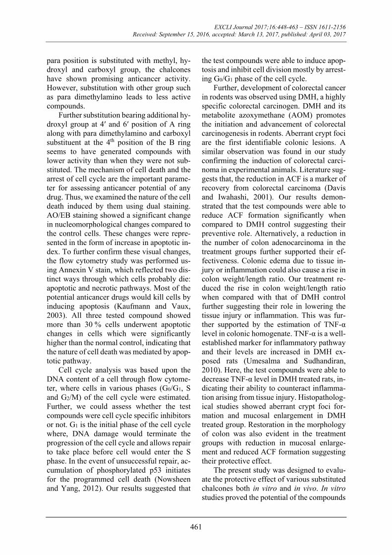
para position is substituted with methyl, hy-droxyl and carboxyl group, the chalcones have shown promising anticancer activity. However, substitution with other group such as para dimethylamino leads to less active compounds.
Further substitution bearing additional hy-droxyl group at 4ʹ and 6ʹ position of A ring along with para dimethylamino and carboxyl substituent at the 4th position of the B ring seems to have generated compounds with lower activity than when they were not sub-stituted. The mechanism of cell death and the arrest of cell cycle are the important parame-ter for assessing anticancer potential of any drug. Thus, we examined the nature of the cell death induced by them using dual staining. AO/EB staining showed a significant change in nucleomorphological changes compared to the control cells. These changes were repre-sented in the form of increase in apoptotic in-dex. To further confirm these visual changes, the flow cytometry study was performed us-ing Annexin V stain, which reflected two dis-tinct ways through which cells probably die: apoptotic and necrotic pathways. Most of the potential anticancer drugs would kill cells by inducing apoptosis (Kaufmann and Vaux, 2003). All three tested compound showed more than 30 % cells underwent apoptotic changes in cells which were significantly higher than the normal control, indicating that the nature of cell death was mediated by apop-totic pathway.
Cell cycle analysis was based upon the DNA content of a cell through flow cytome-ter, where cells in various phases (G0/G1, S and G2/M) of the cell cycle were estimated. Further, we could assess whether the test compounds were cell cycle specific inhibitors or not. G1 is the initial phase of the cell cycle where, DNA damage would terminate the progression of the cell cycle and allows repair to take place before cell would enter the S phase. In the event of unsuccessful repair, ac-cumulation of phosphorylated p53 initiates for the programmed cell death (Nowsheen and Yang, 2012). Our results suggested that
the test compounds were able to induce apop-tosis and inhibit cell division mostly by arrest-ing G0/G1 phase of the cell cycle.
Further, development of colorectal cancer in rodents was observed using DMH, a highly specific colorectal carcinogen. DMH and its metabolite azoxymethane (AOM) promotes the initiation and advancement of colorectal carcinogenesis in rodents. Aberrant crypt foci are the first identifiable colonic lesions. A similar observation was found in our study confirming the induction of colorectal carci-noma in experimental animals. Literature sug-gests that, the reduction in ACF is a marker of recovery from colorectal carcinoma (Davis and Iwahashi, 2001). Our results demon-strated that the test compounds were able to reduce ACF formation significantly when compared to DMH control suggesting their preventive role. Alternatively, a reduction in the number of colon adenocarcinoma in the treatment groups further supported their ef-fectiveness. Colonic edema due to tissue in-jury or inflammation could also cause a rise in colon weight/length ratio. Our treatment re-duced the rise in colon weight/length ratio when compared with that of DMH control further suggesting their role in lowering the tissue injury or inflammation. This was fur-ther supported by the estimation of TNF-α level in colonic homogenate. TNF-α is a well-established marker for inflammatory pathway and their levels are increased in DMH ex-posed rats (Umesalma and Sudhandiran, 2010). Here, the test compounds were able to decrease TNF-α level in DMH treated rats, in-dicating their ability to counteract inflamma-tion arising from tissue injury. Histopatholog-ical studies showed aberrant crypt foci for-mation and mucosal enlargement in DMH treated group. Restoration in the morphology of colon was also evident in the treatment groups with reduction in mucosal enlarge-ment and reduced ACF formation suggesting their protective effect.
The present study was designed to evalu-ate the protective effect of various substituted chalcones both in vitro and in vivo. In vitro studies proved the potential of the compounds
EXCLI Journal 2017;16:448-463 – ISSN 1611-2156 Received: September 15, 2016, accepted: March 13, 2017, published: April 03, 2017
462
to induce apoptosis and arrest G0/G1 phase of cell cycle in human colon cancer cell line. We also found that they were able to alter the ep-igenetic pathways evident from HDAC inhi-bition. In addition, we observed a reduction in ACF and adenocarcinoma formation in the colon of animals treated with the test com-pounds. Furthermore, they were able to lower the increased levels of TNF-α suggesting their role in inflammation. These results demon-strated the efficacy of the compounds against colon adenocarcinoma, providing a potential lead for anticancer drug development.
Acknowledgements
The work was supported by a Grant SR/SO/HS-0282/2012 obtained from the Sci-ence and Engineering Research Board, De-partment of Science and Technology (DST-SERB). The authors would like to acknowledge All India Council for Technical Education (AICTE) for providing funds for the procurement of Flow cytometer used in the study. We thank the Department of Phar-macology, Manipal College of Pharmaceuti-cal Sciences, Manipal University, India for providing necessary facilities to carry out the present work.
Conflict of interest
The authors declare that they have no con-flict of interest to disclose.
REFERENCES
Ameta K, Rathore NS, Kumar B. Synthesis of some novel chalcones and their facile one-pot conversion to 2-aminobenzene-1, 3-dicarbonitriles using malono-nitrile. Analele Universitatii Bucuresti Chimie. 2011; 20:15-24.
Avila HP, Smânia Ede F, Monache FD, Smânia A Jr. Structure-activity relationship of antibacterial chal-cones. Bioorg Med Chem. 2008;16:9790-4.
Davis PA, Iwahashi CK. Whole almonds and almond fractions reduce aberrant crypt foci in a rat model of colon carcinogenesis. Cancer Lett. 2001;165:27-33.
Giacchetti S, Itzhaki M, Gruia G, Adam R, Zidani R, Kunstlinger F, et al. Long-term survival of patients with unresectable colorectal cancer liver metastases following infusional chemotherapy with 5-fluoroura-cil, leucovorin, oxaliplatin and surgery. Ann Oncol. 1999;10:663-9.
Goel A, Boland CR. Epigenetics of colorectal cancer. Gastroenterology. 2012;143:1442-60.e1
Haggar FA, Boushey RP. Colorectal cancer epidemiol-ogy: incidence, mortality, survival, and risk factors. Clin Colon Rectal Surg. 2009;22:191-7.
Jeon J-H, Kim S-J, Kim C-G, Kim J-K, Jun J-G. Syn-thesis of biologically active chalcones and their anti-inflammatory effects. Bull Korean Chem Soc. 2012; 33:953-7.
Kaufmann SH, Vaux DL. Alterations in the apoptotic machinery and their potential role in anticancer drug resistance. Oncogene. 2003;22:7414-30.
Kumar H, Savaliya M, Biswas S, Nayak PG, Maliyak-kal N, Manjunath Setty M, et al. Assessment of the in vitro cytotoxicity and in vivo anti-tumor activity of the alcoholic stem bark extract/fractions of Mimusops elengi Linn. Cytotechnology. 2016;68:861-77.
Liu L, Li Y, Tollefsbol TO. Gene-environment interac-tions and epigenetic basis of human diseases. Curr Is-sues Mol Biol. 2008;10:25-36.
Mariadason JM. HDACs and HDAC inhibitors in co-lon cancer. Epigenetics. 2008;3:28-37.
Minucci S, Pelicci PG. Histone deacetylase inhibitors and the promise of epigenetic (and more) treatments for cancer. Nat Rev Cancer. 2006;6:38-51.
Nakagawa M, Oda Y, Eguchi T, Aishima S, Yao T, Hosoi F, et al. Expression profile of class I histone deacetylases in human cancer tissues. Oncol Rep. 2007;18:769-74.
Nowsheen S, Yang E. The intersection between DNA damage response and cell death pathways. Exp Oncol. 2012;34:243-54.
Perše M, Cerar A.The dimethylhydrazine induced col-orectal tumours in rat-experimental colorectal carcino-genesis. Radiol Oncol. 2005;39:61-70.
Pisani P, Parkin DM, Bray F, Ferlay J. Estimates of the worldwide mortality from 25 cancers in 1990. Int J Cancer. 1999;83:18-29.
Rajendran P, Ho E, Williams DE, Dashwood RH. Die-tary phytochemicals, HDAC inhibition, and DNA damage/repair defects in cancer cells. Clin Epigenetics. 2011;3(1):4.

EXCLI Journal 2017;16:448-463 – ISSN 1611-2156 Received: September 15, 2016, accepted: March 13, 2017, published: April 03, 2017
463
Reddy ND, Shoja MH, Jayashree BS, Nayak PG, Ku-mar N, Prasad VG, et al. In vitro and in vivo evaluation of novel cinnamyl sulfonamide hydroxamate deriva-tive against colon adenocarcinoma. Chem Biol Inter-act. 2015;233:81-94.
Ropero S, Esteller M. The role of histone deacetylases (HDACs) in human cancer. Mol Oncol. 2007;1:19-25.
Schulmann K, Reiser M, Schmiegel W. Colonic cancer and polyps. Best Pract Res Clin Gastroenterol. 2002; 16:91-114.
Siegel R, DeSantis C, Jemal A. Colorectal cancer sta-tistics, 2014. CA Cancer J Clin. 2014;64:104-17.
Siegel RL, Miller KD, Jemal A. Cancer statistics, 2015. CA Cancer J Clin. 2015;65:5-29.
Syam S, Abdelwahab SI, Al-Mamary MA, Mohan S. Synthesis of chalcones with anticancer activities. Mol-ecules. 2012;17:6179-95.
Umesalma S, Sudhandiran G. Differential inhibitory effects of the polyphenol ellagic acid on inflammatory mediators NF-kappaB, iNOS, COX-2, TNF-alpha, and IL-6 in 1,2-dimethylhydrazine-induced rat colon car-cinogenesis. Basic Clin Pharmacol Toxicol. 2010;107: 650-5.
You JS, Jones PA. Cancer genetics and epigenetics: two sides of the same coin? Cancer Cell. 2012;22:9-20.
Zhang EH, Wang RF, Guo SZ, Liu B. An update on antitumor activity of naturally occurring chalcones Evid Based Complement Alternat Med. 2013;2013: 815621.