original article: drosophila melanogaster – an … · excli journal 2014;13:1239-1253 – issn...
TRANSCRIPT

EXCLI Journal 2014;13:1239-1253 – ISSN 1611-2156 Received: July 24, 2014, accepted: September 25, 2014, published: November 21, 2014
1239
Original article:
DROSOPHILA MELANOGASTER – AN EMBRYONIC MODEL FOR STUDYING BEHAVIORAL AND BIOCHEMICAL EFFECTS OF
MANGANESE EXPOSURE
Ana Paula Lausmann Ternes1, Ana Paula Zemolin2, Litiele Cezar da Cruz2, Gustavo Felipe da Silva1, Ana Paula Fleig Saidelles1, Mariane Trindade de Paula3, Caroline Wagner4, Ronaldo Medeiros Golombieski5, Érico Marlon de Moraes Flores2, Rochele Sogari Picoloto2, Antônio Batista Pereira1, Jeferson Luis Franco1, Thaís Posser1* 1 Centro Interdisciplinar de Pesquisa em Biotecnologia (CIP/BIOTEC), Universidade Federal
do Pampa, Campus São Gabriel, RS, 97300 000, Brasil 2 Departamento de Química, Centro de Ciências Naturais e Exatas, Universidade Federal de
Santa Maria, Santa Maria, RS, 97105-900, Brasil 3 Programa de Pós Graduação em Bioquímica, Universidade Federal do Pampa, Campus
Uruguaiana, CEP 97500-970 4 Universidade Federal do Pampa, Campus Caçapava do Sul, RS, CEP 96570-000 Brasil 5 Laboratório de Biologia Molecular de Drosophila e Sequenciamento (LabDros), Univer-
sidade Federal de Santa Maria, Santa Maria, RS, 97300 000. * Corresponding author: Thaís Posser, Universidade Federal do Pampa Campus São Gabriel,
Centro Interdisciplinar de Pesquisa em Biotecnologia (CIP/BIOTEC), Av. Antônio Trilha, 1847 Brasil. CEP: 97300-000; Tel: +555532326075, E-mail: [email protected]
ABSTRACT
Embryonic animals are especially susceptible to metal exposure. Manganese (Mn) is an es-sential element, but in excess it can induce toxicity. In this study we used Drosophila mela-nogaster as an embryonic model to investigate biochemical and behavioral alterations due to Mn exposure. Flies were treated with standard medium supplemented with MnCl2 at 0.1 mM, 0.5 mM or 1 mM from the egg to the adult stage. At 0.5 mM and 1 mM Mn, newly ecloded flies showed significantly enhanced locomotor activity when assessed by negative geotaxis behavior. In addition, a significant increase in Mn levels (p < 0.0001) was observed, while Ca, Fe, Cu, Zn and S levels were significantly decreased. A significant drop in cell viability oc-curred in flies exposed to 1 mM Mn. There was also an induction of reactive oxygen species at 0.5 mM and 1 mM Mn (p < 0.05). At 1 mM, Mn increased Catalase (p < 0.005), Superox-ide Dismutase (p < 0.005) and Hsp83 (p < 0.0001) mRNA expression, without altering Cata-lase or Superoxide Dismutase activity; the activity of Thioredoxin reductase and Glutatione-S-transferase enzymes was increased. Mn treatment did not alter ERK or JNK1/2 phosphory-lation, but at 1 mM caused an inhibition of p38MAPK phosphorylation. Together these data suggest mechanisms of adaptation in the fly response to Mn exposure in embryonic life.
Keywords: Manganese, Drosophila melanogaster, MAPK, oxidative stress, Thioredoxin re-ductase
EXCLI Journal 2014;13:1239-1253 – ISSN 1611-2156 Received: July 24, 2014, accepted: September 25, 2014, published: November 21, 2014
1240
INTRODUCTION
Manganese (Mn) is an essential element for living organisms. It is the twelfth most abundant element in earth´s crust and is pre-sent in rocks, water, soil and food, normally associated with other elements (Santamaria, 2008; Farina et al., 2013). Environmental and occupational exposure to Mn may occur by contact with fungicides, such as Maneb, Manconzeb, methylcyclopentadienyl man-ganese tricarbonyl (MTT)-an anti-knock agent in gasoline, Mn-ore mining, Mn alloy production and dry alkaline battery manufac-ture (Mergler and Baldwin, 1997; Mergler, 1999).
Dietary ingestion is the main source of Mn for humans and Mn absorption takes place mostly in the gastrointestinal tract where it is homeostatically controlled in the intestinal wall (Au et al., 2008). The brain is especially susceptible to metal intoxication during embryonic development, when it is known that Mn is able to cross the placenta and to be excreted in the maternal milk (Betharia and Maher, 2012). Mn absorption is increased during the neonatal period, when biliary excretion is poorly developed, leading to elevated concentrations of Mn in the brain and other tissues (Aschner and Aschner, 2005). In children, Mn exposure is associat-ed with alterations in psychomotor and cog-nitive development; furthermore a positive correlation exists between Mn exposure and hyperactivity (Menezes-Filho et al., 2011; Roels et al., 2012; Torres-Agustín et al., 2013).
Exposure to high levels of Mn can lead to pathological conditions, including neuro-degeneration (Mergler et al., 1994). The mechanisms mediating Mn toxicity are com-plex and not completely understood. Some of them include: (1) Mn accumulation in astrocytes leading to disruption of their ability to promote neu-ronal differentiation and decreasing gluta-mate uptake by astrocytes (Erikson and Aschner, 2003; Giordano et al., 2009); (2) Mn induced- loss of dopaminergic neu-rons (Stanwood et al., 2009);
(3) Inhibition of respiratory chain complexes and induction of reactive oxygen species (ROS) (Zhang et al., 2004; Sriram et al., 2010).
The use of alternative models in toxico-logical studies has been growing over the years. The fruit fly Drosophila melanogaster has served as a unique and powerful model for studies on human genetics and diseases. Although humans and flies are only distantly related, almost 75 % of disease related genes in humans have functional orthologs in the fly (Deepa et al., 2009; Pandey and Nichols, 2011). Moreover, the fast and external de-velopmental cycle of this organism enable the study of toxicological effects of com-pounds during the developmental period. All these advantages make flies an appropriate model for studies related with metal toxicity (Bonilla-Ramirez et al., 2011; Paula et al., 2012) and human neurodegeneration (Hirth, 2010).
As the embryonic development period is particularly sensitive to Mn exposure, in this paper we aimed to investigate the behavior and biochemical alterations caused by Mn exposure during the embryonic development of Drosophila melanogaster, focusing on ad-aptations in the antioxidant systems and MAPK signaling pathways. The levels of Mn and major essential elements were also determined.
MATERIALS AND METHODS
Reagents Anti-phospho-p38MAPK, anti-phospho JNK,
anti-phospho ERK, anti ERK and ß-actin an-tibodies were purchased from Cell Signaling Technology (Danvers, MA, United States). EDTA (CAS 60-00-4), glycine (CAS 56-40-6), tris(hydroxymethyl) aminomethane (CAS 77-86-1) and ammonium persulfate (CAS 7727-54-0) were purchased from Serva (Heidelberg, Germany). L-Glutathione re-duced (CAS 70-18-8), 1-chloro-2,4-dinitro-benzene (CAS 97-00-7), sodium orthovana-date (CAS 13721-39-6), manganese (II) chloride tetrahydrate (CAS 13446-34-9), ß-mercaptoethanol (CAS 60-24-2), methanol

EXCLI Journal 2014;13:1239-1253 – ISSN 1611-2156 Received: July 24, 2014, accepted: September 25, 2014, published: November 21, 2014
1241
(CAS 67-56-1), tween 20 (CAS 9005-64-5), potassium phosphate dibasic (CAS 7758-11-4), potassium phosphate monobasic (CAS 7778-77-0), potassium bicarbonate (CAS 298-14-6) and anti-rabbit imunoglobulin an-tibody, N,N,N′,N′-Tetramethylethylenedia-mine (CAS 110-18-9), quercetin (CAS 117-39-5), protease inhibitor cocktail for use with mammalian cell and tissue extracts, 5,5´di-thiobis(2-nitrobenzoic acid) (CAS 69-78-3), 2´,7´-dichlorofluorescein diacetate (CAS 2044-85-1), glycerol (CAS 56-81-5), resaz-urin sodium salt (CAS 62758-13-8), triton x-100 (CAS 9002-93-1), sodium chloride (CAS 7647-14-5), albumin from bovine se-rum (CAS 9048-46-8), HEPES (CAS 7365-45-9), ß-nicotinamide adenine dinucleotide 2´-phosphate reduced tetrasodium salt were obtained from Sigma Aldrich (St. Louis, MO, United States). Bis-acrylamide, hybond nitrocellulose, acrylamide (CAS 79-06-1), sodium dodecyl sulfate (CAS 151-21-3), bo-ric acid (CAS 10043-35-3) were purchased from GE Healthcare Bio-Sciences AB (Upp-sala, Sweden). All other reagents were com-mercial products of the highest purity grade available.
Animals
Drosophila melanogaster (Harwich strain) was obtained from the National Spe-cies Stock Center, Bowling Green, OH, USA. The flies were maintained at 25 °C on 12 h light/dark cycle in glass bottles contain-ing 10 mL of standard medium (mixture of 39 % coarse and 32 % medium corn flour, 10 % wheat germ, 14 % sugar, 2 % milk powder, 1 % salt, 1 % soybean flour, 1 % rye flour, a pinch of methyl paraben (99-76-3) and lyophilized yeast. All experiments were performed with the same strain, and both genders were used at random.
Animal treatment
Adults flies were placed in 10 mL of standard medium supplemented with 3 mL of a fresh solution (0.1 mM, 0.5 mM or 1 mM) of manganese chloride (MnCl2). In the control group the standard medium were
supplemented with 3 mL of ultrapure water. After ten days laying eggs the adult flies were removed. When eggs were newly ecloded, 1 to 3 day old flies were used for all analyses. The MnCl2 concentrations were chosen based on previous studies (Bonilla-Ramirez et al., 2011).
Locomotor assay
Locomotor activity was determined using the negative geotaxis assay as described by Bland et al. (2009) with minor modifications. Briefly, for each assay, individual flies (1-3 days old) were immobilized on ice and placed separately in a glass tube; this method of immobilization does not affect fly neurol-ogy (Deepa et al., 2009). After 15 minutes the flies were gently tapped to the bottom of the tube and the time required to climb up 8 cm of the tube wall was recorded. Each fly was tested 4 times at 1 minute intervals. For each experiment, the climbing mean was cal-culated.
Metal content
Two hundred flies per group were washed three times in ultrapure water and then dried on a filter paper in the incubator at 37 °C for 90 minutes. Flies were digested in closed vessels according to the procedure de-scribed previously by Bizzi et al. (2010). Flies (~ 70 mg) were transferred to quartz vessels with 6 mL of nitric acid 3 mol L-1. After closing and capping the rotor, the ves-sels were pressurized with 7.5 bar of oxygen using the valve originally designed for pres-sure release after conventional acid sample digestion. Then, the rotor was placed inside a microwave oven (Multiwave 3000 Micro-wave Sample Preparation System, Anton Paar, Graz, Austria). The system was equipped with eight high-pressure quartz vessels (volume of 80 mL, maximum opera-tional temperature and pressure of 280 °C and 80 bar, respectively). Pressure was mo-nitored in each vessel during all the runs. Microwave heating program was as follows: (1) 1000 W, with a ramp of 5 min; (2) 1000 W for 10 min; and
EXCLI Journal 2014;13:1239-1253 – ISSN 1611-2156 Received: July 24, 2014, accepted: September 25, 2014, published: November 21, 2014
1242
(3) 0 W for 20 min (cooling step). After digestion, the pressure in each ves-
sel was carefully released. The resulting so-lutions were transferred to polypropylene vi-als and diluted to 25 mL with water. Deter-mination of calcium (Ca), iron (Fe), potassi-um (K), magnesium (Mg), manganese (Mn), sodium (Na), phosphorus (P), sulfur (S), and zinc (Zn) was performed using an inductive-ly coupled plasma optical emission spec-trometer (Optima 4300 DV, PerkinElmer, Shelton, USA) with axial view configura-tion. A concentric nebulizer and cyclonic spray chamber were used. Argon 99.996 % (White Martins, São Paulo, Brazil) was used for plasma generation, nebulization and as auxiliary gas. The instrumental parameters were carried out in according with previous work (Pereira et al., 2013). Two readings were averaged to give one value per biologi-cal replicate and expressed as a mean (±) standard deviation of the mean (SD). Metals levels were expressed relative to the weight of flies used for analysis (μg metal/g of dried weight tissue).
Cellular viability
Cellular viability was measured by two different methods. Firstly, cellular viability was measured using 3-(4,5-dimethylthiazol-2-yl)-2,5-diphenyltetrazolium bromide (MTT) reduction assay as described by Su-dati et al. (2013) with minor modifications. The analysis was performed on the whole body of female flies. The flies were incubat-ed in MTT for 60 min (37 °C), after MTT was removed the sample was incubated in DMSO for 30 min (37 °C). The absorbance from formazan dissolution by addition of DMSO was monitored in an EnsPireR mul-timode plate reader (PerkinElmer, USA) at 540 nm.
The second method used was the resaz-urin reduction assay. The method uses the indicator resazurin to measure the metabolic capacity of cells. Viable cells reduce resaz-urin into resorufin, a fluorescent compound (Franco et al., 2009). Groups of 40 flies were mechanically homogenized in 1 mL 20 mM
Tris buffer (pH 7.0) and centrifuged at 1,000 RPM for 10 min at 4 °C. The supernatant was incubated in Elisa plates with 20 mM Tris buffer (pH 7.0) and resazurin for two hours. The fluorescence was recorded using EnsPireR multimode plate reader (Perkin Elmer, USA) at ex579nm and em584nm.
DCF-DA oxidation assay
Groups of 20 flies were mechanically homogenized in 1 mL 20 mM Tris buffer (pH 7.0), and centrifuged at 1,000 RPM for 10 min, 4 °C. The supernatant was used to quantify 2’-7’-dichlorofluorescein diacetate (DCF-DA) oxidation as a general index of oxidative stress as described by Perez-Severiano et al. (2004). The fluorescence emission of DCF resulting from DCF-DA oxidation was monitored at regular intervals (ex488nm and em530nm) in an EnsPireR mul-timode plate reader (PerkinElmer, USA).
Determination of gene expression by real-time quantitative PCR (qPCR)
Real-time quantitative PCR (qRT-PCR) was performed according to the method de-scribed by Paula et al. (2012). The primers utilized are shown in Table 1. All samples were analyzed as technical and biological triplicates with a negative control. Threshold and baselines were manually determined us-ing the StepOne Software v2.0 (Applied Bi-osystems, NY). SYBR fluorescence was ana-lyzed by StepOne software version 2.0 (Ap-plied Biosystems, NY), and the CT (cycle threshold) value for each sample was calcu-lated and reported using the 2-∆∆CT method (Livak and Schmittgen, 2001). The GPDH gene was used as an endogenous reference showing no alterations in response to the treatment. For each well, analyzed in quad-ruplicate, a ∆CT value was obtained by sub-tracting the GPDH CT value from the CT value of the interest gene (sequences of test-ed genes are represented in Table 1). The ∆CT mean value obtained from the control group of each gene was used to calculate the ∆∆CT of the respective gene (2-∆∆CT).
EXCLI Journal 2014;13:1239-1253 – ISSN 1611-2156 Received: July 24, 2014, accepted: September 25, 2014, published: November 21, 2014
1243
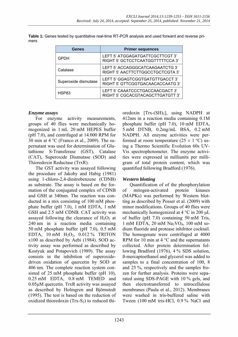
Table 1: Genes tested by quantitative real-time RT-PCR analysis and used forward and reverse pri-mers
Genes Primer sequences
GPDH LEFT 5’ ATGGAGATGATTCGCTTCGT 3’ RIGHT 5’ GCTCCTCAATGGTTTTTCCA 3’
Catalase LEFT 5’ ACCAGGGCATCAAGAATCTG 3’ RIGHT 5’ AACTTCTTGGCCTGCTCGTA 3’
Superoxide dismutase LEFT 5’ GGAGTCGGTGATGTTGACCT 3’ RIGHT 5’ GTTCGGTGACAACACCAATG 3’
HSP83 LEFT 5’ CAAATCCCTGACCAACGACT 3’ RIGHT 5’ CGCACGTACAGCTTGATGTT 3’
Enzyme assays
For enzyme activity measurements, groups of 40 flies were mechanically ho-mogenized in 1 mL 20 mM HEPES buffer (pH 7.0), and centrifuged at 14.000 RPM for 30 min at 4 °C (Franco et al., 2009). The su-pernatant was used for determination of Glu-tathione S-Transferase (GST), Catalase (CAT), Superoxide Dismutase (SOD) and Thioredoxin Reductase (TrxR).
The GST activity was assayed following the procedure of Jakoby and Habig (1981) using 1-chloro-2,4-dinitrobenzene (CDNB) as substrate. The assay is based on the for-mation of the conjugated complex of CDNB and GSH at 340nm. The reaction was con-ducted in a mix consisting of 100 mM phos-phate buffer (pH 7.0), 1 mM EDTA, 1 mM GSH and 2.5 mM CDNB. CAT activity was assayed following the clearance of H2O2 at 240 nm in a reaction media containing 50 mM phosphate buffer (pH 7.0), 0.5 mM EDTA, 10 mM H2O2, 0.012 % TRITON x100 as described by Aebi (1984). SOD ac-tivity assay was performed as described by Kostyuk and Potapovich (1989). The assay consists in the inhibition of superoxide-driven oxidation of quercetin by SOD at 406 nm. The complete reaction system con-sisted of 25 mM phosphate buffer (pH 10), 0.25 mM EDTA, 0.8 mM TEMED and 0.05μM quercetin. TrxR activity was assayed as described by Holmgren and Björnstedt (1995). The test is based on the reduction of oxidized thioredoxin (Trx-S2) to reduced thi-
oredoxin [Trx-(SH)2], using NADPH at 412nm in a reaction media containing 0.1M phosphate buffer (pH 7.0), 10 mM EDTA, 5 mM DTNB, 0.2mg/mL BSA, 0.2 mM NADPH. All enzyme activities were per-formed at room temperature (25 ± 1 °C) us-ing a Thermo Scientific Evolution 60s UV-Vis spectrophotometer. The enzyme activi-ties were expressed in milliunits per milli-gram of total protein content, which was quantified following Bradford (1976).
Western blotting
Quantification of of the phosphorylation of mitogen-activated protein kinases (MAPKs) was performed by Western blot-ting as described by Posser et al. (2009) with minor modifications. Groups of 40 flies were mechanically homogenized at 4 °C in 200 μL of buffer (pH 7.0) containing 50 mM Tris, 1 mM EDTA, 20 mM Na3VO4, 100 mM so-dium fluoride and protease inhibitor cocktail. The homogenate were centrifuged at 4000 RPM for 10 min at 4 °C and the supernatants collected. After protein determination fol-lowing Bradford (1976), 4 % SDS solution, ß-mercaptoethanol and glycerol was added to samples to a final concentration of 100, 8 and 25 %, respectively and the samples fro-zen for further analysis. Proteins were sepa-rated using SDS-PAGE with 10 % gels, and then electrotransferred to nitrocellulose membranes (Paula et al., 2012). Membranes were washed in tris-buffered saline with Tween (100 mM tris-HCl, 0.9 % NaCl and
EXCLI Journal 2014;13:1239-1253 – ISSN 1611-2156 Received: July 24, 2014, accepted: September 25, 2014, published: November 21, 2014
1244
0.1 % Tween-20, pH 7.5) and incubated overnight at 4 °C with specific primary anti-bodies (anti-phospho-p38MAPK, anti-phospho JNK, anti-phospho ERK, anti ERK and anti ß-actin). Following incubation, membranes were washed in tris-buffered saline with Tween and incubated for 1 h at 25 °C with anti rabbit-IgG secondary specific antibod-ies. Antibody binding was visualized using the ECL Western Blotting substrate Kit (Promega). Band staining density was quan-tified using the Scion Image software (Scion Image for Windows) and expressed as the percentage (%) of the control group (mean ± standard deviation of the mean). The values were normalized using total proteins (total ERK and ß-actin).
Statistical analysis
Statistical analysis was performed using one-way ANOVA followed by Tukey´s post hoc test. Pearson’s correlation test was ap-plied for detection of significant statistical differences among the metals. Differences were considered statistically significant when p < 0.05. GraphPad Prism 5 Software was used for artwork creation.
RESULTS
Exposure to Mn causes hyperactive behaviors and alters metal levels in Drosophila melanogaster
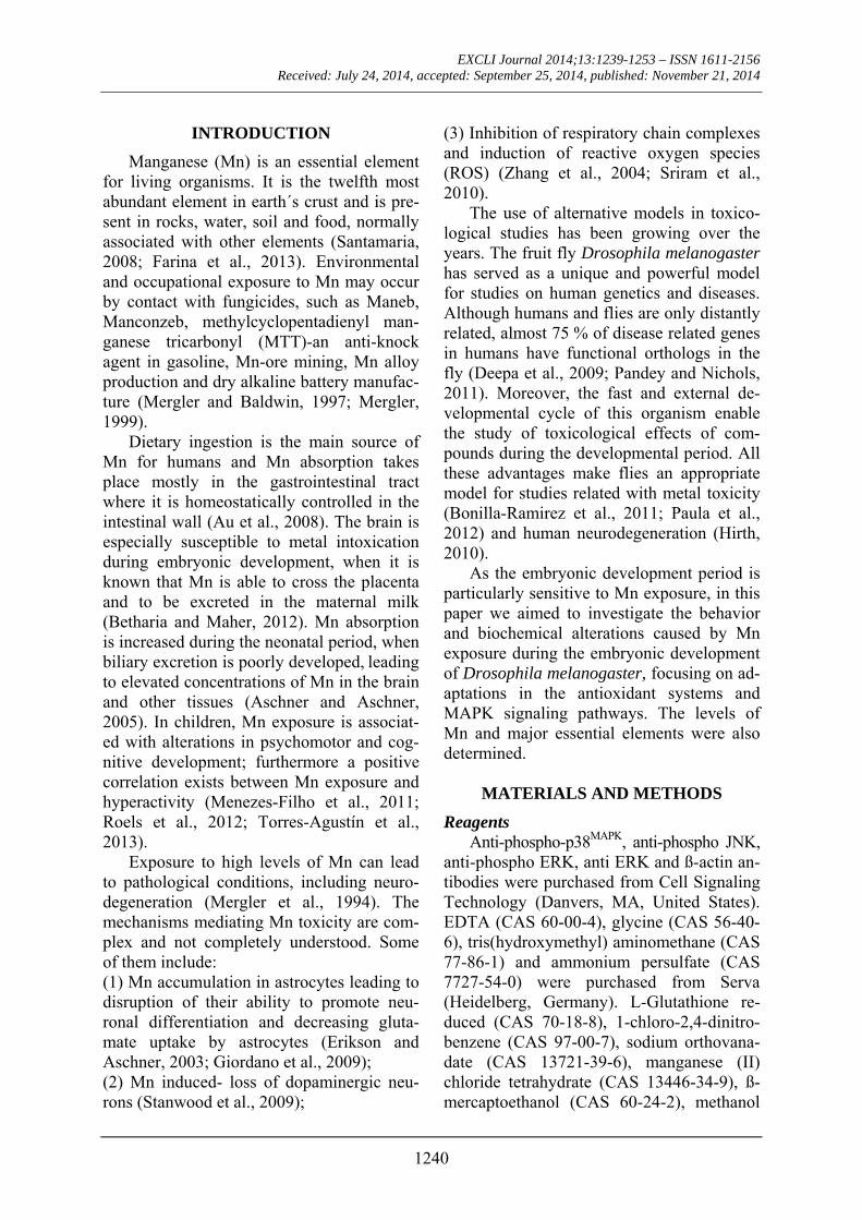
The evaluation of climbing behavior per-formance by negative geotaxis showed that flies exposed to 0.5 mM and 1 mM of Mn
reached the limit of columns significantly (p < 0.005) faster than controls (Figure 1).
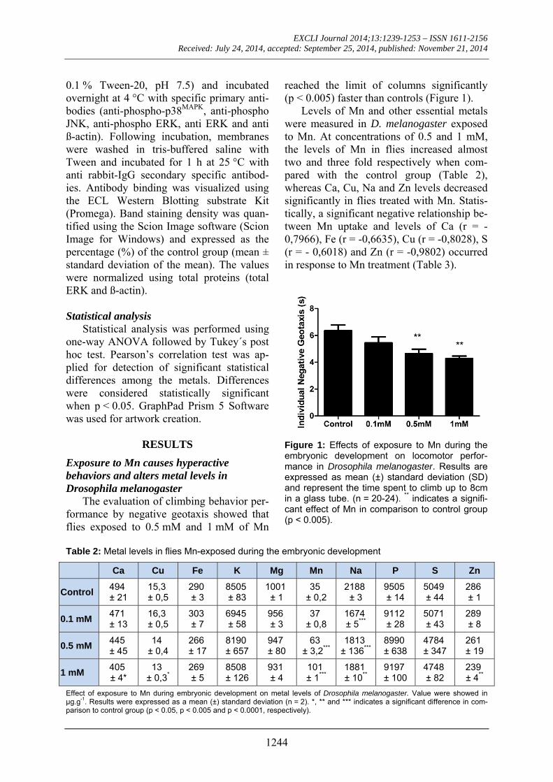
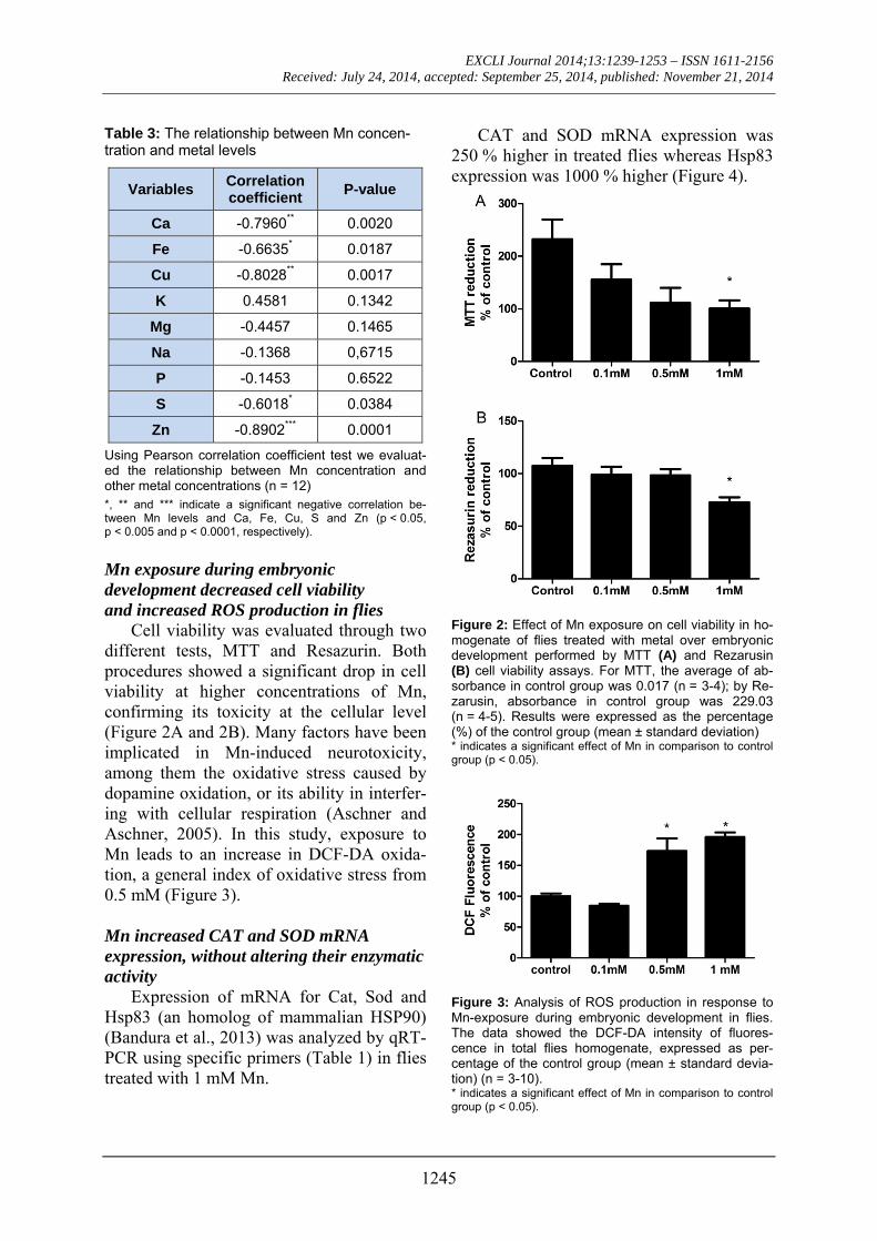
Levels of Mn and other essential metals were measured in D. melanogaster exposed to Mn. At concentrations of 0.5 and 1 mM, the levels of Mn in flies increased almost two and three fold respectively when com-pared with the control group (Table 2), whereas Ca, Cu, Na and Zn levels decreased significantly in flies treated with Mn. Statis-tically, a significant negative relationship be-tween Mn uptake and levels of Ca (r = -0,7966), Fe (r = -0,6635), Cu (r = -0,8028), S (r = - 0,6018) and Zn (r = -0,9802) occurred in response to Mn treatment (Table 3).
Figure 1: Effects of exposure to Mn during the embryonic development on locomotor perfor-mance in Drosophila melanogaster. Results are expressed as mean (±) standard deviation (SD) and represent the time spent to climb up to 8cm in a glass tube. (n = 20-24). ** indicates a signifi-cant effect of Mn in comparison to control group (p < 0.005).
Table 2: Metal levels in flies Mn-exposed during the embryonic development
Ca Cu Fe K Mg Mn Na P S Zn
Control 494 ± 21
15,3 ± 0,5
290 ± 3
8505 ± 83
1001 ± 1
35 ± 0,2
2188 ± 3
9505 ± 14
5049 ± 44
286 ± 1
0.1 mM 471 ± 13
16,3 ± 0,5
303 ± 7
6945 ± 58
956 ± 3
37 ± 0,8
1674 ± 5***
9112 ± 28
5071 ± 43
289 ± 8
0.5 mM 445 ± 45
14 ± 0,4
266 ± 17
8190 ± 657
947 ± 80
63 ± 3,2***
1813 ± 136***
8990 ± 638
4784 ± 347
261 ± 19
1 mM 405 ± 4*
13 ± 0,3*
269 ± 5
8508 ± 126
931 ± 4
101 ± 1***
1881 ± 10**
9197 ± 100
4748 ± 82
239 ± 4**
Effect of exposure to Mn during embryonic development on metal levels of Drosophila melanogaster. Value were showed in μg.g-1. Results were expressed as a mean (±) standard deviation (n = 2). *, ** and *** indicates a significant difference in com-parison to control group (p < 0.05, p < 0.005 and p < 0.0001, respectively).
EXCLI Journal 2014;13:1239-1253 – ISSN 1611-2156 Received: July 24, 2014, accepted: September 25, 2014, published: November 21, 2014
1245
Table 3: The relationship between Mn concen-tration and metal levels
Variables Correlation coefficient
P-value
Ca -0.7960** 0.0020
Fe -0.6635* 0.0187
Cu -0.8028** 0.0017
K 0.4581 0.1342
Mg -0.4457 0.1465
Na -0.1368 0,6715
P -0.1453 0.6522
S -0.6018* 0.0384
Zn -0.8902*** 0.0001
Using Pearson correlation coefficient test we evaluat-ed the relationship between Mn concentration and other metal concentrations (n = 12) *, ** and *** indicate a significant negative correlation be-tween Mn levels and Ca, Fe, Cu, S and Zn (p < 0.05, p < 0.005 and p < 0.0001, respectively).
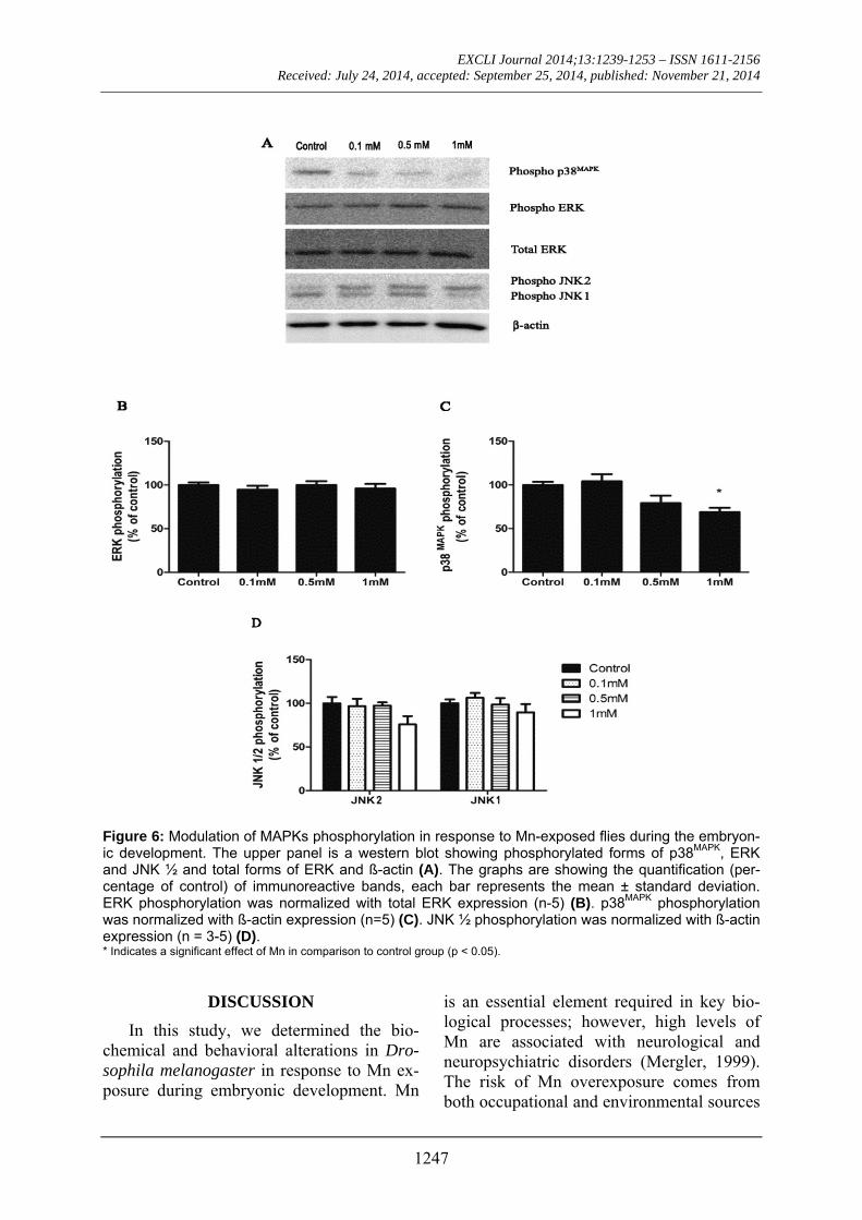
Mn exposure during embryonic development decreased cell viability and increased ROS production in flies
Cell viability was evaluated through two different tests, MTT and Resazurin. Both procedures showed a significant drop in cell viability at higher concentrations of Mn, confirming its toxicity at the cellular level (Figure 2A and 2B). Many factors have been implicated in Mn-induced neurotoxicity, among them the oxidative stress caused by dopamine oxidation, or its ability in interfer-ing with cellular respiration (Aschner and Aschner, 2005). In this study, exposure to Mn leads to an increase in DCF-DA oxida-tion, a general index of oxidative stress from 0.5 mM (Figure 3).
Mn increased CAT and SOD mRNA expression, without altering their enzymatic activity
Expression of mRNA for Cat, Sod and Hsp83 (an homolog of mammalian HSP90) (Bandura et al., 2013) was analyzed by qRT-PCR using specific primers (Table 1) in flies treated with 1 mM Mn.
CAT and SOD mRNA expression was 250 % higher in treated flies whereas Hsp83 expression was 1000 % higher (Figure 4).
Figure 2: Effect of Mn exposure on cell viability in ho-mogenate of flies treated with metal over embryonic development performed by MTT (A) and Rezarusin (B) cell viability assays. For MTT, the average of ab-sorbance in control group was 0.017 (n = 3-4); by Re-zarusin, absorbance in control group was 229.03 (n = 4-5). Results were expressed as the percentage (%) of the control group (mean ± standard deviation) * indicates a significant effect of Mn in comparison to control group (p < 0.05).
Figure 3: Analysis of ROS production in response to Mn-exposure during embryonic development in flies. The data showed the DCF-DA intensity of fluores-cence in total flies homogenate, expressed as per-centage of the control group (mean ± standard devia-tion) (n = 3-10). * indicates a significant effect of Mn in comparison to control group (p < 0.05).

EXCLI Journal 2014;13:1239-1253 – ISSN 1611-2156 Received: July 24, 2014, accepted: September 25, 2014, published: November 21, 2014
1246
The levels of the antioxidant enzymes ac-tivity TrxR, GST, SOD and CAT were de-termined TrxR and GST activity were in-creased at concentrations of 0.5 mM and 1 mM (Figure 5), while CAT and SOD activ-ity showed no significant differences.
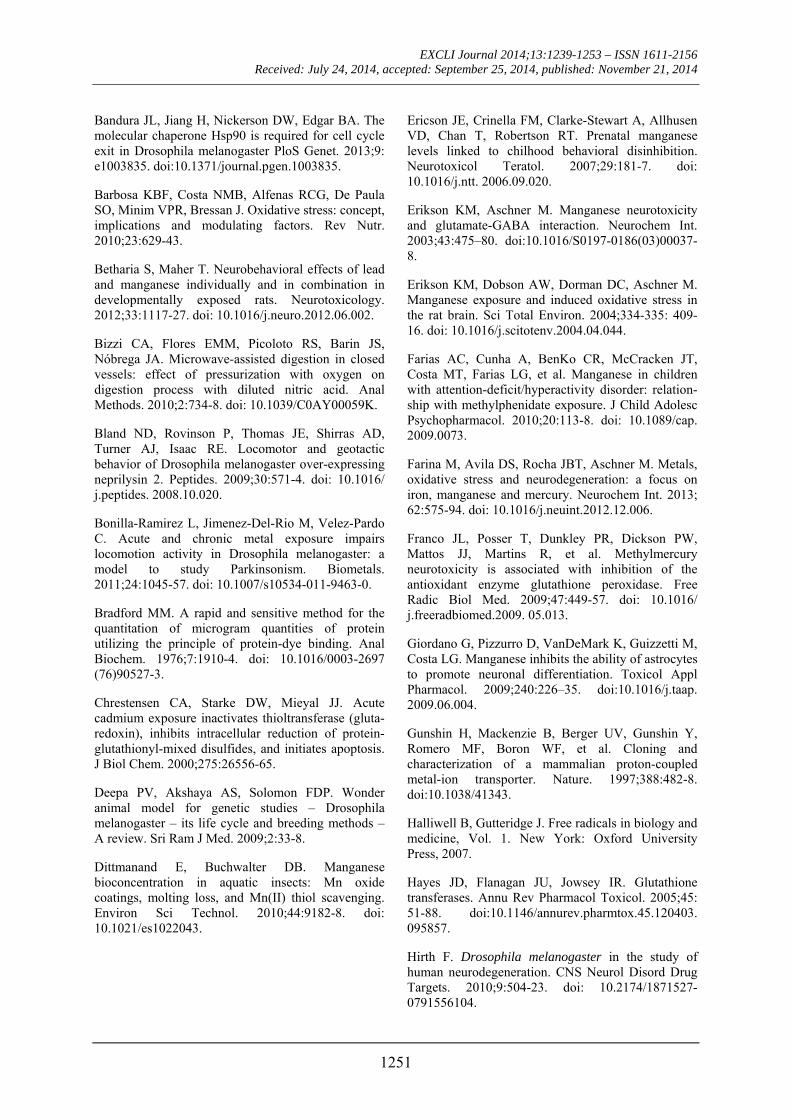
Mn exposure inhibited p38MAPK phosphory-lation
MAPKs phosphorylation levels were in-vestigated in flies exposed to Mn. There was a 40 % inhibition of p38MAPK phosphoryla-tion in flies exposed to Mn at 1 mM, while the phosphorylation level of extracellular signal-regulated kinases (ERK) was unal-tered. C-Jun-N-terminal Kinases 2 (JNK2) phosphorylation was not statistically differ-ent from controls (Figure 6).
Figure 4: Quantitative real time PCR (qRT-PCR) analysis of CAT, SOD and HSP83 mRNA in flies Mn-exposed at 1 mM. We used qRT-PCR to quantify lev-els of mRNA, relative to respective controls, after ex-posure. The data were normalized against GPDH transcript levels and each bar represents the mean ± standard deviation expressed as percent of its respec-tive control (n = 3-4) ** and *** indicate a significant effect of Mn in comparison to control group (p < 0.005 and p < 0.0001, respectively).
Figure 5: Effects observed on enzymatic activities in flies Mn-exposed during embryonic develop-ment. TrxR activity (n = 5-7) (A), GST activity (n = 3-4) (B), SOD activity (n = 6-8) (C) and CAT activity (n = 5-6) (D). The data shows the enzymatic activities in flies homogenate expressed as mean (mU/mg protein) ± standard deviation. * and *** indicate a significant effect of Mn in comparison to control group (p < 0.05 and p < 0.0001, respectively).
EXCLI Journal 2014;13:1239-1253 – ISSN 1611-2156 Received: July 24, 2014, accepted: September 25, 2014, published: November 21, 2014
1247
Figure 6: Modulation of MAPKs phosphorylation in response to Mn-exposed flies during the embryon-ic development. The upper panel is a western blot showing phosphorylated forms of p38MAPK, ERK and JNK ½ and total forms of ERK and ß-actin (A). The graphs are showing the quantification (per-centage of control) of immunoreactive bands, each bar represents the mean ± standard deviation. ERK phosphorylation was normalized with total ERK expression (n-5) (B). p38MAPK phosphorylation was normalized with ß-actin expression (n=5) (C). JNK ½ phosphorylation was normalized with ß-actin expression (n = 3-5) (D). * Indicates a significant effect of Mn in comparison to control group (p < 0.05).
DISCUSSION
In this study, we determined the bio-chemical and behavioral alterations in Dro-sophila melanogaster in response to Mn ex-posure during embryonic development. Mn
is an essential element required in key bio-logical processes; however, high levels of Mn are associated with neurological and neuropsychiatric disorders (Mergler, 1999). The risk of Mn overexposure comes from both occupational and environmental sources

EXCLI Journal 2014;13:1239-1253 – ISSN 1611-2156 Received: July 24, 2014, accepted: September 25, 2014, published: November 21, 2014
1248
(Mergler and Baldwin, 1997). Mn intoxica-tion, a syndrome known as Manganism, is characterized by an extrapyramidal dysfunc-tion and neuropsychiatric symptomatology and is associated with prolonged occupation-al exposure to high concentrations of this metal. Classical symptoms include irritabil-ity, intellectual deficits, compulsive behav-iors, tremors and cock-like walk (Mergler, 1999; Roth, 2006).
In rodents, Krishna et al. (2014) showed that adult mice exposed to Mn through the drinking water presented neurobehavioral deficits and glial activation related with Mn deposition in brain. Moreover, others studies demonstrated that Mn toxicity in rats is ac-companied by increased cholesterol biosyn-thesis and impairments in neuronal function of the hippocampus, which is involved in learning and memory (Öner and Sentürk, 1995; Sentürk and Öner, 1996). It has been shown that Mn supplementation during the neonatal period of rats resulted in increased Mn concentrations in tissues leading to ad-verse effects on motor development and be-havior (Tran et al., 2002).
Mn uptake is increased during the neona-tal period as biliary excretion, which has been suggested as a pathway for Mn elimina-tion from the body, is poorly developed at this stage (Aschner and Aschner, 2005). Ex-posure to Mn during the embryonic and early postnatal periods may result in increased levels of Mn in the brain and other tissues including bone, liver, pancreas and kidney (Aschner and Aschner, 2005; Roels et al., 2012). Higher levels of Mn retention in utero may affect children´s psychomotor develop-ment (Takser et al., 2003). Possible adverse effects of Mn exposure on children´s health include cognitive deficits and hyperactive behaviors (Menezes-Filho et al., 2009; Tor-res-Agustín et al., 2013). Children exposed to high levels of Mn during the fetal period were more impulsive, inattentive, agressive, defiant, disobedient, destructive and hyper-active (Ericson et al., 2007). It is recognized that factors such as the source and the dura-tion of exposure, as well as nutritional status,
can interfere in the intensity and incidence of neurological symptoms associated with Mn exposure in humans. Chronic consumption of drinking-water containing Mn at levels ranging from 81 to 2300 µg/l was associated with progressively higher prevalence of neu-rological symtoms (Kondakis et al., 1989). The concentrations used in this study were from 0.1 mM of Mn in food (corresponding to 19 mg/L in the medium). Despite the use of relatively elevated concentrations, body levels of Mn were not altered at 0.1 mM. In previous studies, adult flies were acutely ex-posed to Mn (0.5-20 mM) diluted in sucrose, as the only source of food and liquid, which lead to significant locomotor deficits (Bonil-la-Ramirez et al., 2011). Our study is the first where Mn was provided as a cereal based diet over all the embryonic period. Thus, more studies are necessary to under-stand the rate of Mn uptake from diet in flies and how it may affect neurological behav-iour.
In our study, flies exposed to Mn at 0.5 mM and 1 mM showed increased loco-motor speed in the locomotor behavior test (assayed as negative geotaxis behavior), pointing to a hyperactive-like behavior in Drosophila melanogaster. Furthermore, Mn levels were substantially increased in treated flies, while Ca, Cu, Zn, Fe and S levels were all decreased. This relationship may be in part associated with a competition of the metals for the same mechanism of transport into the flies cells. Facilitated diffusion, ac-tive transport, divalent metal transport 1 (DMT1), ZIP8 and transferrin (Tf)-depen-dent transport mechanisms are all involved in cellular Mn transportation (Aschner et al., 2007). Among these metal transport systems, DMT1 has a very broad substrate specificity and is likely to be the major transmembrane protein responsible for the uptake of a varie-ty of divalent cations, including Mn2+, Cd2+, Zn2+, Co2+, Ni2+, Cu2+ and Pb2+ (Gunshin et al., 1997). In the flies, many proteins in-volved in the metabolism of biometals such as ferritin, transferrin, iron regulatory pro-teins, divalent metal transporter are ex-

EXCLI Journal 2014;13:1239-1253 – ISSN 1611-2156 Received: July 24, 2014, accepted: September 25, 2014, published: November 21, 2014
1249
pressed (Bonilla-Ramirez et al., 2011). In this context, Mn uptake is less frequently studied in comparison with other metals and the mechanisms related to Mn transport are considerably more complex, occurring in most part in the divalent (II) and oxide forms (Tebo et al., 2004). Mn has the capacity to interact and /or compete with Ca (Dittman and Buchwalter, 2010). In a study performed in the aquatic insect Hydropsyche sparna, Mn exposure decreased cadmium (Cd) and Zn accumulation. Furthermore, increased Ca concentrations significantly reduced Mn ac-cumulation in the insect (Poteat et al., 2012). Dittmanand and Buchwalter (2010) suggest-ed that Mn is also absorbed by Ca transport-ers in aquatic insects, where increasing am-bient Ca concentrations decrease Mn accu-mulation.
There was also a negative correlation be-tween Mn and Fe levels. Iron deficiency has been suggested as a possible contributing cause of attention deficit and hyperactivity disorder (ADHD) in children (Konofal et al., 2008). Concomitantly, children with ADHD showed elevated serum Mn concentrations (Konofal et al., 2008). Recent studies have suggested that Mn accumulates in dopamin-ergic neurons via the presynaptic dopamine transporter (DAT) and an altered functioning of the dopaminergic system has been well established in the etiology of ADHD (Farias et al., 2010).
Our results showed decreased cell viabil-ity using two different methodologies and increased ROS generation in flies exposed to Mn during development. Previous work by our group in PC12 cells, demonstrated that Mn leads to increased production of hydro-gen peroxide (H2O2) (Posser et al., 2009). H2O2 is a highly permeable and reactive molecule being able to react with metals such as Fe, thus generating hydroxyl radicals (Jiménez Del Río M and Vélez-Pardo, 2004; Barbosa et al., 2010) resulting in a propaga-tion of oxidative damage in cells.
Tissues can respond to oxidative stress by modulating antioxidant defenses (Halli-well and Gutteridge, 2007). We measured
the gene expression of Hsp83, CAT and SOD in Drosophila melangaster. Earlier studies showed that cellular stress may in-duce heat shock proteins in parallel with ROS production (Kim et al., 2004; Paula et al., 2012). Our results showed a significant increase in Hsp83 mRNA levels in Mn treat-ed flies. Previously, our group demonstrated that exposure of flies to heavy metals such as mercury causes increases in the expression of Hsp83 (Paula et al., 2012). CAT and SOD mRNA levels were significantly increased by Mn, but the enzymatic activity of these proteins was unchanged. The antioxidant ezyme SOD converts superoxide radicals (O2
º-) to H2O2 and CAT catalyzes the conver-sion of H2O2 to oxygen (O2) and water (Bar-bosa et al., 2010), thus neutralizing these re-active species. Considering that both SOD and CAT are crucial in the cell defense against oxidative stress (Halliwell and Gut-teridge, 2007), it might be expected that a posttranscriptional regulation mechanisms could maintain adequate levels of these pro-teins, however, further studies are necessary to elucidate this. Our results also showed that the activity of TrxR and GST was enhanced in Mn exposed flies. These two enzymes play an important role in protection against oxidative stress (Mustacich and Powis, 2000). GST is a complex group of phase II detoxifying enzymes that participate in the metabolism of electrophilic substances, in-cluding carcinogenic, mutagenic and toxic compounds (Hayes et al., 2005). TrxR is a dimeric FAD-containing enzyme that cata-lyzes the NADPH-dependent reduction of the active-site disulfide in Trx-S2 to give a dithiol in Trx-(SH)2 (Zhao et al., 2002). Thi-oredoxin consists in one of the major redox-regulating proteins displaying a number of biological activities, including cytoprotection against ROS, protein repairing and protein disulfide reduction and modulation of signal-ing pathways (Yan et al., 2012). Our data suggest that the increase in the levels of Mn and TrxR activity could represent a response to oxidation of thioredoxin in response to Mn-induced oxidative stress.

EXCLI Journal 2014;13:1239-1253 – ISSN 1611-2156 Received: July 24, 2014, accepted: September 25, 2014, published: November 21, 2014
1250
MAPKs regulate the activity of a range of transcription factors thereby controlling gene expression and cellular function. The three most-studied MAPKs are ERK1/2, JNK1/2 and p38MAPK (Ichijo, 1999). ASK1 (Apoptosis Signaling Kinase 1) is a member of mitogen activated protein kinase kinase family (MAPKK) and an upstream activator of MAPK signaling pathway (Yan et al., 2012). The redox state of thioredoxin regulates ASK1. Under normal conditions, ASK1 is bound to and inhibited by thioredoxin and when thioredoxin is oxidized, ASK1 can be activated and apoptotic signaling through the p38
MAPK/JNK1/2 MAPKs initiated (Ichijo et al., 1997). Studies conducted by Yan et al. (2012) in a pancreatic carcinoma cell line, showed inhibition of TrxR by indolequinones resulting in a change of Thioredoxin-1 redox state to an oxidized form and activation of p38MAPK /JNK1/2. Similarly, Cd treatment activated ASK1 and its downstream MAPK in neuronal cells (Kim et al., 2005), and inhibits components of thioredoxin system (Chrestensen et al., 2000), while that Liedhegner et al. (2011) demonstrated that knockdown of ASK1 as well as chemical inhibition of p38MAPK and JNK played protective effects against L-DOPA induced apoptosis. We show that Mn induced TrxR activity while p38MAPK
/JNK1/2 phosphorylation were inhibited. This suggests an involvement of thioredoxin system in the mechanism of Mn induced toxicity. Augmented TrxR activity may represent a cellular response to high levels of ROS induced by Mn exposure, thus preventing the oxidation of thioredoxin and its dissociation of ASK1. This could contribute to diminished activation of p38MAPK pathway upstream kinases resulting in lower levels of phosphorylation of this MAPK thus minimizing apoptotic cell death.
In summary, our study demonstrate for a first time that developmental exposure to Mn leads to hyperactive-like behavior and accumulation of this metal in Drosophila melanogaster. The observed raise in Mn
levels is negatively correlated with levels of other essential metals. This result fits with previous studies showing that Mn accumulation and Fe deficiency are associated with hyperactive behavior in children (Ericson et al., 2007; Konofal et al., 2008). The induction of stress responsive genes and antioxidant enzyme activity associated with inhibition of p38MAPK phosphorylation at higher concentrations of Mn may represent an adaptive response to oxidative stress generated by this metal, in an attempt to avoid exacerbated cellular damage.
ACKNOWLEDGEMENTS
The authors thank to Universidade Federal do Pampa, Conselho Nacional de Desenvolvimento Científico e Tecnológico (CNPq 482313/2013-7), Fundação de Amparo à Pesquisa do Estado do Rio Grande do Sul (Fapergs 1954/2551/13-7), FAPERGS/PRONEM, FINEP and INCT-APA for financial support. The authors thank Professor Peter R. Dunkley for critical review of the manuscript.
CONFLICT OF INTEREST
The authors declare that they have no conflict of interest.
REFERENCES
Aebi H. Catalase in vitro. Methods Enzymol. 1984; 105:121-6.
Aschner JL, Aschner M. Nutritional aspects of manganese homeostasis. Mol Aspects Med. 2005;26: 353-62. doi: 10.1016/j.mam.2005.07.003.
Aschner M, Guilarte TR, Schneider JS, Zheng W. Manganese: recent advances in understanding its transport and neurotoxicity. Toxicol Appl Pharmacol. 2007;221:131-47. doi: 10.1016/j.taap.2007.03.001.
Au C, Benedetto A, Aschner M. Manganese transport in eukaryotes: The role of DMT1. Neurotoxicology. 2008;29:569–76. doi: 10.1016/j.neuro.2008.04.022.
EXCLI Journal 2014;13:1239-1253 – ISSN 1611-2156 Received: July 24, 2014, accepted: September 25, 2014, published: November 21, 2014
1251
Bandura JL, Jiang H, Nickerson DW, Edgar BA. The molecular chaperone Hsp90 is required for cell cycle exit in Drosophila melanogaster PloS Genet. 2013;9: e1003835. doi:10.1371/journal.pgen.1003835.
Barbosa KBF, Costa NMB, Alfenas RCG, De Paula SO, Minim VPR, Bressan J. Oxidative stress: concept, implications and modulating factors. Rev Nutr. 2010;23:629-43.
Betharia S, Maher T. Neurobehavioral effects of lead and manganese individually and in combination in developmentally exposed rats. Neurotoxicology. 2012;33:1117-27. doi: 10.1016/j.neuro.2012.06.002.
Bizzi CA, Flores EMM, Picoloto RS, Barin JS, Nóbrega JA. Microwave-assisted digestion in closed vessels: effect of pressurization with oxygen on digestion process with diluted nitric acid. Anal Methods. 2010;2:734-8. doi: 10.1039/C0AY00059K.
Bland ND, Rovinson P, Thomas JE, Shirras AD, Turner AJ, Isaac RE. Locomotor and geotactic behavior of Drosophila melanogaster over-expressing neprilysin 2. Peptides. 2009;30:571-4. doi: 10.1016/ j.peptides. 2008.10.020.
Bonilla-Ramirez L, Jimenez-Del-Rio M, Velez-Pardo C. Acute and chronic metal exposure impairs locomotion activity in Drosophila melanogaster: a model to study Parkinsonism. Biometals. 2011;24:1045-57. doi: 10.1007/s10534-011-9463-0.
Bradford MM. A rapid and sensitive method for the quantitation of microgram quantities of protein utilizing the principle of protein-dye binding. Anal Biochem. 1976;7:1910-4. doi: 10.1016/0003-2697 (76)90527-3.
Chrestensen CA, Starke DW, Mieyal JJ. Acute cadmium exposure inactivates thioltransferase (gluta-redoxin), inhibits intracellular reduction of protein-glutathionyl-mixed disulfides, and initiates apoptosis. J Biol Chem. 2000;275:26556-65.
Deepa PV, Akshaya AS, Solomon FDP. Wonder animal model for genetic studies – Drosophila melanogaster – its life cycle and breeding methods – A review. Sri Ram J Med. 2009;2:33-8.
Dittmanand E, Buchwalter DB. Manganese bioconcentration in aquatic insects: Mn oxide coatings, molting loss, and Mn(II) thiol scavenging. Environ Sci Technol. 2010;44:9182-8. doi: 10.1021/es1022043.
Ericson JE, Crinella FM, Clarke-Stewart A, Allhusen VD, Chan T, Robertson RT. Prenatal manganese levels linked to chilhood behavioral disinhibition. Neurotoxicol Teratol. 2007;29:181-7. doi: 10.1016/j.ntt. 2006.09.020.
Erikson KM, Aschner M. Manganese neurotoxicity and glutamate-GABA interaction. Neurochem Int. 2003;43:475–80. doi:10.1016/S0197-0186(03)00037-8.
Erikson KM, Dobson AW, Dorman DC, Aschner M. Manganese exposure and induced oxidative stress in the rat brain. Sci Total Environ. 2004;334-335: 409-16. doi: 10.1016/j.scitotenv.2004.04.044.
Farias AC, Cunha A, BenKo CR, McCracken JT, Costa MT, Farias LG, et al. Manganese in children with attention-deficit/hyperactivity disorder: relation-ship with methylphenidate exposure. J Child Adolesc Psychopharmacol. 2010;20:113-8. doi: 10.1089/cap. 2009.0073.
Farina M, Avila DS, Rocha JBT, Aschner M. Metals, oxidative stress and neurodegeneration: a focus on iron, manganese and mercury. Neurochem Int. 2013; 62:575-94. doi: 10.1016/j.neuint.2012.12.006.
Franco JL, Posser T, Dunkley PR, Dickson PW, Mattos JJ, Martins R, et al. Methylmercury neurotoxicity is associated with inhibition of the antioxidant enzyme glutathione peroxidase. Free Radic Biol Med. 2009;47:449-57. doi: 10.1016/ j.freeradbiomed.2009. 05.013.
Giordano G, Pizzurro D, VanDeMark K, Guizzetti M, Costa LG. Manganese inhibits the ability of astrocytes to promote neuronal differentiation. Toxicol Appl Pharmacol. 2009;240:226–35. doi:10.1016/j.taap. 2009.06.004.
Gunshin H, Mackenzie B, Berger UV, Gunshin Y, Romero MF, Boron WF, et al. Cloning and characterization of a mammalian proton-coupled metal-ion transporter. Nature. 1997;388:482-8. doi:10.1038/41343.
Halliwell B, Gutteridge J. Free radicals in biology and medicine, Vol. 1. New York: Oxford University Press, 2007.
Hayes JD, Flanagan JU, Jowsey IR. Glutathione transferases. Annu Rev Pharmacol Toxicol. 2005;45: 51-88. doi:10.1146/annurev.pharmtox.45.120403. 095857.
Hirth F. Drosophila melanogaster in the study of human neurodegeneration. CNS Neurol Disord Drug Targets. 2010;9:504-23. doi: 10.2174/1871527-0791556104.

EXCLI Journal 2014;13:1239-1253 – ISSN 1611-2156 Received: July 24, 2014, accepted: September 25, 2014, published: November 21, 2014
1252
Holmgren A, Björnstedt M. Thioredoxin and thioredoxin reductase. Methods Enzymol. 1995;252:199-208. doi: 10.1016/0076-6879(95)52023-6.
Ichijo H. From receptors to stress-activated MAP kinases. Oncogene. 1999;18:6087-93. doi:10.1038/ sj.onc.1203129.
Ichijo H, Nishida E, Irie K, ten Dijke P, Saitoh M, Moriguchi T, et al. Induction of apoptosis by ASK1, a mammalian MAPKKK that activates SAPK/JNK and p38 signaling pathways. Science. 1997;275:90-4. doi: 10.1126/science.275.5296.90.
Jakoby WB, Habig WH. Glutathione S-transferases (rat and human). Methods Enzymol. 1981;77:218-31.
Jiménez Del Río M, Vélez-Pardo C. Transition metal-induced apoptosis in lymphocytes via hydroxyl radical generation, mitochondria dysfunction, and caspase-3 activation: An In vitro model for neurodegeneration. Arch Med Res. 2004;35:185–93. doi:10.1016/j.arcmed. 2004.01.001.
Kim YM, Kim HJ, Song EJ, Lee KJ. Glucuronic acid is a novel inducer of heat shock response. Mol Cell Biochem. 2004;259:23-33.
Kim SD, Moon CK, Eun SY, Ryu PD, Jo SA. Identification of ASK1, MKK4, JNK, c-Jun, and caspase-3 as a signaling cascade involved in cadmium-induced neuronal cell apoptosis. Biochem Biophys Res Commun. 2005;328:326-34.
Kondakis XG, Makris N, Leotsinidis M, Prinou M, Papapetropoulos T. Possible health effects of high manganese concentration in drinking water. Arch Environ Health. 1989;44:175-8.
Konofal E, Lecendreux M, Deron J, Marchand M, Cortese S, Zaïm M, et al. Effects of iron supplement-ation on attention deficit hyperactivity disorder in children. Pediatr Neurol. 2008;38:20-6. doi:10.1016/ j.pediatrneurol.2007.08.014.
Kostyuk VA, Potapovich AI. Superoxide-driven oxidation of quercetin and a simple sensitive assay for determination of superoxide dismutase. Biochem Int. 1989;19:1117-24.
Krishna S, Dodd CA, Hekmatyar SK, Filipov NM. Brain deposition and neurotoxicity of manganese in adult mice exposed via the drinking water. Arch Toxicol. 2014;88:47-64. doi:10.1007/s00204-013-1088-3.
Liedhegner EA, Steller KM, Mieyal JJ. Levodopa activates apoptosis signaling kinase (ASK1) and promotes apoptosis in a neuronal model: implications for the treatment of Parkinson’s disease. Chem Res Toxicol. 2011;24:1644-52.
Livak KJ, Schmittgen TD. Analysis of relative gene expression data using real-time quantitative PCR and the 2(-Delta Delta C(T)) method. Methods. 2001;25: 402-8. doi:10.1006/meth.2001.1262.
Menezes-Filho JA, Bouchard M, Sarcinelli PN, Moreira JC. Manganese exposure and the neuro-psychological effect on children and adolescents: a review. Pan Am J Public Health. 2009;26:541-8. doi:10. 1590/S1020-49892009001200010.
Menezes-Filho JA, Novaes CO, Moreira JC, Sarcinelli PN, Mergler D. Elevated manganese and cognitive performance in school-aged children and their mothers. Environ Res. 2011;111:156–63. doi:10. 1016/j.envres.2010.09.006.
Mergler D. Neurotoxic effects of low level exposure to manganese in human populations. Environ Res. 1999;80:99-102. doi:10.1006/enrs.1998.3902.
Mergler D, Baldwin M. Early manifestations of manganese neurotoxicity in humans: an update. Environ Res. 1997;73:92-100. doi:10.1006/enrs. 1997.3710.
Mergler D, Huel G, Bowler R, Iregren A, Bélanger S, Baldwin M, et al. Nervous system dysfunction among workers with long-term exposure to manganese. Environ Res. 1994;64:151-80. doi:10.1006/enrs. 1994.1013.
Mustacich D, Powis G. Thioredoxin reductase. Biochem J. 2000;346:1-8.
Öner G, Sentürk ÜK. Reversibility of manganese-induced learning defect in rats. Food Chem Toxicol. 1995;33:559-63. doi: 10.1016/0278-6915(95)00020-3.
Pandey UB, Nichols CD. Human disease models in Drosophila melanogaster and the role of the fly in therapeutic drug discovery. Pharmacol Rev. 2011;63: 411-36.
Paula MT, Zemolin AP, Vargas AP, Golombieski RM, Loreto ELS, Saidelles AP, et al. Effects of Hg(II) exposure on MAPK phosphorylation and antioxidant system in D. Melanogaster. Environ Toxicol. 2012;29:621-30. doi: 10.1002/tox.21788.
Pereira JSF, Pereira LSF, Schmidt L, Moreira CM, Barin JS, Flores EMM. Metals determination in milk powder samples for adult and infant nutrition after focused-microwave induced combustion. Microchem J. 2013;109:29-35. doi:10.1016/j.microc.2012.05.010.

EXCLI Journal 2014;13:1239-1253 – ISSN 1611-2156 Received: July 24, 2014, accepted: September 25, 2014, published: November 21, 2014
1253
Perez-Severiano F, Rodriguez-Perez M, Pedraza-Chaverri J, Maldonado PD, Medina-Campos ON, Ortiz-Plata A. S-Allylcysteine, a garlic-derived antioxidant, ameliorates quinolinic acid-induced neurotoxicity and oxidative damage in rats. Neurochem Int. 2004;45:1175-83. doi: 10.1016/ j.neuint.2004.06.008.
Posser T, Franco JL, Bobrovskaya L, Leal RB, Dickson PW, Dunkley PR. Manganese induces sustained Ser40 phosphorylation and activation of tyrosine hydroxylase in PC12 cells. J Neurochem. 2009;110:848-56. doi: 10.1111/j.1471-4159.2009. 06185.x.
Poteat MD, Díaz-Jaramillo M, Buchwalter DB. Divalent metal (Ca, Cd, Mn, Zn) uptake and interactions in the aquatic insect Hydropsyche sparna. J Exp Biol. 2012;215:1575-83. doi:10.1242/ jeb.063412.
Roels HA, Bowler RM, Kim Y, Henn BC, Mergler D, Hoet P, et al. Manganese exposure and cognitive deficits: A growing concern for manganese neurotoxicity. Neurotoxicology. 2012;33:872-80. doi: 10.1016/j. neuro.2012.03.009.
Roth JA. Homeostatic and toxic mechanisms regulating manganese uptake, retention, and elimination. Biol Res. 2006;39:45-57. doi:10.4067/ S0716-97602006 000100006.
Santamaria AB. Manganese exposure, essentiality & toxicity. Indian J Med Res. 2008;128:484-500.
Sentürk Ü, Öner G. The effect of manganese-induced hypercholesterolemia on learning in Rats. Biol Trace Elem Res. 1996;51:249-57. doi:10.1007/BF02784079.
Sriram K, Lin GX, Jefferson AM, Roberts JR, Wirth O, Hayashi Y, et al. Mitochondrial dysfunction and loss of Parkinson´s disease-linked proteins contribute to neurotoxicity of manganese-containing welding fumes. FASEB J. 2010;24:4989-5002. doi: 10.1096/ fj.10-163964.
Stanwood GD, Leich DB, Savchenko V, Wu J, Fitsa-nakis VA, Anderson DJ, et al. Manganese exposure is cytotoxic and alters dopaminergic and GABAergic neurons within the basal ganglia. J Neurochem. 2009;110:378–89. doi:10.1111/j.1471-4159. 2009. 06145.x.
Sudati JH, Vieira FA, Pavin SS, Dias GR, Seeger RL, Golombieski R, et al. Valeriana officinalis attenuates the rotenone-induced toxicity in Drosophila melanogaster. Neurotoxicology. 2013;37:118-26. doi:10. 1016/j.neuro.2013.04.006.
Takser L, Mergler D, Hellier G, Sahuquillo J, Huel G. Manganese, monoamine metabolite levels at birth, and child psychomotor development. Neurotoxicol-ogy. 2003;24:667–74. doi: 10.1016/S0161-813X (03) 00058-5.
Tebo BM, Bargar JR, Clement BG, Dick GJ, Murray KJ, Parker D, et al. Biogenic manganese oxides: properties and mechanisms of formation. Annu Rev Earth Planet Sci. 2004;32:287-328. doi:10.1146/ annurev.earth.32.101802.120213.
Torres-Agustín R, Rodríguez-Agudelo Y, Schilmann A, Solís-Vivanco R, Montes S, Riojas-Rodríguez H, et al. Effect of environmental manganese exposure on verbal learning and memory in Mexican children. Environ Res. 2013;121:39-44. doi:10.1016/j.envres. 2012.10.007.
Tran TT, Chowanadisai W, Crinella FM, Chicz-DeMet A, Lönnerdal B. Effect of high dietary manganese intake of neonatal rats on tissue mineral accumulation, striatal dopamine levels, and neuro-developmental status. Neurotoxicology. 2002;23: 635-43. doi:10.1016/S0161-813X(02)00091-8.
Yan C, Siegel D, Newsome J, Chilloux A, Moody CJ, Ross D. Antitumor indolequinones induced apoptosis in human pancreatic cancer cells via inhibition of thioredoxin reductase and activation of redox signaling. Mol Pharmacol. 2012;81:401–10. doi:10.1124/mol. 111.076091.
Zhang S, Fu J, Zhou Z. In vitro effect of manganese chloride exposure on reactive oxygen species generation and respiratory chain complexes activities of mitochondria isolated from rat brain. Toxicol In Vitro. 2004;18:71-7. doi:10.1016/j.tiv.2003.09.002.
Zhao R, Masayasu H, Holmgren A. Ebselen: a substrate for human thioredoxin reductase strongly stimulating its hydroperoxide reductase activity and a superfast thioredoxin oxidant. Proc Natl Acad Sci USA. 2002;99:8579-84. doi:10.1073/pnas.122061399.