original article alleviation of doxorubicin-induced ... · hhd inhibited dox-induced increase in...
TRANSCRIPT

Int J Clin Exp Med 2018;11(10):10574-10584www.ijcem.com /ISSN:1940-5901/IJCEM0073028
Original ArticleAlleviation of doxorubicin-induced cardiotoxicity by Hong Huang decoction may involve a reduction in myocardial oxidative stress and activation of Akt/FoxO3a pathways
Jing-Xian Xue, Xiao-Qing Zhang, Wei-He Bian, Chang Yao
The Affiliated Hospital of Nanjing University of Chinese Medicine, Nanjing, Jiangsu, P. R. China
Received January 18, 2018; Accepted June 26, 2018; Epub October 15, 2018; Published October 30, 2018
Abstract: Doxorubicin (DOX)-induced cardiotoxicity limits the use of DOX as an antitumor drug. This study investigat-ed whether Hong Huang decoction (HHD) inhibits DOX-induced cardiotoxicity. Thirty Balb/c mice were divided into three groups: control, DOX (3 mg/kg i.p., 30 days), and HHD+DOX (DOX plus 9 g/kg HHD by oral gavage). Myocardial enzyme levels, histologic changes, apoptosis (TUNEL assay), and protein expression of cleaved (c)-caspase-3, cas-pase-3, and Bcl-2 (Western blotting) were assessed. H9c2 cells were assigned to control, DOX, HHD, and HHD+DOX groups. H9c2 cell growth (MTT assay), apoptosis (TUNEL assay), reactive oxygen species (ROS) levels, and expres-sion of c-caspase-3, caspase-3, Bcl-2, p-FoxO3a, and p-Akt (Western blotting) were measured. Regarding in vivo ex-periments, the DOX+HHD group had lower levels of myocardial enzymes (P < 0.05), less marked histologic changes, lower apoptosis rates, and higher c-caspase-3 levels than the DOX group (P < 0.01). IC50 for inhibition of H9c2 cell growth by DOX in the absence and presence of HHD was 3.03 ± 0.11 µM and 7.07 ± 0.48 μM, respectively. In H9c2 cells, HHD prevented DOX-induced increase of c-caspase-3/caspase-3 ratio and decrease in Bcl-2 levels (P < 0.01). H9c2 cell apoptosis rates were lower in the DOX+HHD group than in the DOX group (30.2 ± 2.5% vs. 42.6 ± 5.2%, P < 0.05). HHD inhibited DOX-induced increase in ROS levels in H9c2 cells (P < 0.01). DOX-induced reductions in p-FoxO3a and p-Akt (S473) protein levels were attenuated by HHD and by the antioxidant, NaHS (P < 0.05). HHD protected against DOX-induced cardiotoxicity, in vitro and in vivo, possibly by reducing oxidative stress and activating Akt/FoxO3a signaling.
Keywords: Hong Huang decoction (HHD), doxorubicin, cardiotoxicity, oxidative stress, Akt, FoxO3a
Introduction
Cancer and cardiovascular disease are the two main causes of mortality worldwide [1]. Doxo- rubicin (DOX) is an anthracycline-based anti- tumor drug used in many clinical chemothera- py regimens [2]. However, cumulative doses of DOX can cause myocardial damage, including cardiomyopathy and heart failure. This dam- age is associated with poor prognosis [3-5]. Development of an effective drug to prevent and treat cardiac toxicity induced by DOX would greatly benefit patients undergoing chemother-apy for cancer.
The anticancer effects of DOX are thought to include intercalation into DNA to interfere with protein synthesis and inhibition of topoisomer-ase II to impair DNA repair. These actions are
also believed to contribute to the cardiotoxici- ty of DOX [3, 6]. For example, topoisomerase II-beta has been implicated in mediating DOX-induced DNA double-strand breaks in cardiac cells [7] and activating DNA response genes that initiate apoptosis pathways [8]. However, an additional mechanism thought to play an important role in DOX-induced cardiotoxicity is the production of reactive oxygen species (ROS) [8, 9]. Oxidative stress occurs when a cellular imbalance develops between levels of ROS (such as free radicals) and antioxidant mecha-nisms. Oxidative stress causes myocardial inju-ry and has been implicated in several cardio-vascular diseases [9-14]. DOX can be activated by mitochondrial complex I into a more reactive semiquinone derivative that increases oxida-tive stress, causing oxidative damage to mito-chondria [9]. Indeed, DOX has been shown to

HHD alleviates doxorubicin-induced cardiotoxicity
10575 Int J Clin Exp Med 2018;11(10):10574-10584
enhance lipid peroxidation (thus damaging cell membranes), cause lesions in mitochondrial DNA, and induce free-radical-associated mito-chondrial dysfunction [15]. The heart may be particularly susceptible to oxidative stress be- cause normal myocardium contains antioxidant enzymes, such as superoxide dismutase and glutathione peroxidase, at lower levels than those found in other tissues [9, 16, 17].
Forkhead homeobox type O (FoxO) transcrip-tion factors, including FoxO1, FoxO3a and Fox- O4, are critical mediators of cellular responses to oxidative stress. FoxO3a is a particularly important transcription factor because it is involved in not only the response to oxidative stress but also regulation of apoptosis-relat- ed genes [18, 19]. Phosphorylation of FoxO3a plays an important role in the modulation of many cellular functions. If phosphorylation of FoxO3a is reduced, nuclear localization of Fox- O3a is promoted, leading to cell apoptosis [20]. FoxO3a can be phosphorylated by a variety of kinases, including protein kinase B (Akt) [21]. Activation of Akt can enhance phosphorylation of FoxO3a (at site S253), which retains FoxO3a in the cytoplasm and reduces its transcription-al activity, thereby inhibiting apoptosis. DOX has been demonstrated to downregulate Fox- O3a levels in mouse hearts [22] and reduce levels of phosphorylated FoxO3a (p-FoxO3a) in H9c2 cardiac cells [23]. Furthermore, DOX has also been shown to enhance nuclear ex- pression of FoxO3a in H9c2 cells [24]. Inter- estingly, exogenous hydrogen sulfide (H2S) has been reported to attenuate DOX-induced toxic-ity in H9c2 cardiac cells through activation of phosphoinositide 3-kinase (PI3K)/Akt/FoxO3a pathways [25]. In addition, vitexin (an extract from hawthorn leaves) has been demonstrat- ed to suppress DOX-induced oxidative stress, inflammation, apoptosis, and myocardial dam-age via a mechanism that may involve increas- ed cellular expression of p-FoxO3a [26]. Thus, the Akt/FoxO3a signaling pathway may be a novel target for development of drugs to reduce DOX-induced cardiotoxicity.
Hong Huang decoction (HHD), a Chinese herb formula, contains Astragalus membranaceus, rhubarb, turmeric, and Rhodiola rosea. A previ-ous study found that HHD exerted a protective effect in patients with breast cancer during DOX-based chemotherapy [27]. Of the various constituents of HHD, Astragalus polysaccha-
rides have been demonstrated to exert antio- xidant effects and protect cells from apopto- sis caused by oxidative stress [28]. Astragalus polysaccharides have also been shown to at- tenuate DOX-induced ROS production and car-diomyocyte apoptosis via a mechanism that involves reduced activation of p38 signaling pathways [29]. Furthermore, Astragaloside IV has been found to inhibit DOX-induced mito-chondrial ROS production, mitochondrial dam-age, and activation of the mitochondrial apop-totic pathway via a mechanism that involves stimulation of PI3K/Akt pathways [30]. Sali- droside, an active component of Rhodiola, has been reported to protect heart cells against DOX-induced toxicity by attenuating oxidative stress and inhibiting apoptosis [31]. Curcumin, a component of turmeric, has been suggest- ed to protect against DOX-induced cardiotoxi- city [32, 33], although not all studies have agreed [34].
This present study hypothesized that HHD pro-tects against DOX-induced cardiotoxicity by in- hibiting oxidative stress and activating Akt/FoxO3a pathways. Therefore, the aim of the pr- esent study was to investigate whether HHD attenuated DOX-induced cardiotoxicity, in vitro (H9c2 cells) and in vivo (in mice), and to exam-ine whether any such effects were associat- ed with reduced levels of oxidative stress and enhanced expression of p-Akt and p-FoxO3a.
Materials and methods
Mice
Protocols for all animal experiments were re- viewed and approved by the Institutional Re- view Board of Animal Research at the Affiliat- ed Hospital of Nanjing University of Tradition- al Chinese Medicine, Nanjing, China. Female BALB/c mice (6-8 weeks old, weighing 20-25 g) were purchased from the Pharmacology Labo- ratory at the Affiliated Hospital of Nanjing University of Traditional Chinese Medicine. Th- ese animals were maintained in the housing facility under controlled environmental condi-tions (12/12-hour light/dark cycle).
Preparation of HHD
Astragalus membranaceus (30 g), rhubarb (6 g), turmeric (10 g), and Rhodiola (10 g) were acquired from Jiangsu Province Hospital of Traditional Chinese Medicine (Nanjing, China).

HHD alleviates doxorubicin-induced cardiotoxicity
10576 Int J Clin Exp Med 2018;11(10):10574-10584
These herbs were blended in 1000 mL of dou-ble-distilled water (1:10, w/v) for 1 hour and then heated to 100°C for 2 hours to concen-trate the sample to a final volume of 50 mL (0.92 g/mL) [35, 36]. After cooling, HHD was stored at 4°C.
In vivo experiments
Mice were randomly divided into three groups (n = 10 per group): control, DOX, and DOX+HHD. Mice in the control group received daily oral gavage of normal saline for 25 days and intra-peritoneal injections of normal saline on days 5, 10, 15, 20 and 25. Animals in the DOX group received intraperitoneal injections of DOX (3 mg/kg, dissolved in normal saline; no: NSC- 123127, Selleck Chemicals, Houston, TX, USA) on days 5, 10, 15, 20 and 25 to obtain a cumu-lative dose of 15 mg/kg over 25 days [37]. Mice in the DOX+HHD group received intraperi-toneal injections of DOX (as with the DOX group) plus daily oral gavage of HHD (9 g/kg). The ani-mals were weighed every 7 days.
Measurement of plasma levels of myocardial enzymes
Blood samples were obtained from the orbit of each mouse on day 30. Blood was centri-fuged at 5000 rpm for 3 minutes to separate the plasma. Plasma levels of aspartate amino-transferase (AST), creatine kinase (CK), cre-atine kinase isoenzyme MB (CK-MB), and lac-tate dehydrogenase (LDH) were measured with a biochemical analyzer (P800, KeyGen Biotech, Nanjing, China).
Histology
Mice were killed by cervical dislocation on day 30 and the hearts were removed and weigh- ed. The left ventricle of the heart was isolated by dissection, fixed in 4% paraformaldehyde at 4°C overnight, embedded in paraffin, and sec-tioned. Sections were stained with hematoxylin and eosin (H&E), using standard techniques, and viewed using light microscopy [38, 39].
Culture of rat embryonic H9c2 cardiomyocytes
H9c2 cells (KeyGen Biotech) were maintain- ed in complete Dulbecco’s Modified Eagle Medium with 10% (v/v) heat-inactivated fetal bovine serum. All cells were maintained at
37°C in a humidified atmosphere of 95% air and 5% CO2.
Cell growth assay
The effects of HHD on cell growth were deter-mined using 3-(4,5-dimethylthiazol-2-yl)-2,5-di-phenyltetrazolium bromide (MTT) Cell Proli- feration and Cytotoxicity Assay Kit (KeyGen Biotech). Briefly, H9c2 cells in 96-well plates (5,000 cells/well) were incubated for 24 hours with HHD (0-5 µM) in the absence and pres-ence of DOX (2 mg/mL). Next, 10 µL MTT was added to each well and the plates were incu-bated for 4 hours at 37°C in 5% CO2 and in the dark. Dimethyl sulfoxide was added to dissol- ve the formazan crystals. Absorbance at 570 nm was measured using a microplate reader (BioTek Instruments, Winooski, VT, USA).
ROS assay
H9c2 cells (105 cells/well) were cultured for 24 hours in the presence of HHD (2 mg/mL) and/or DOX (1 µM, chosen based on the MTT assay experiments). They were then stained using a ROS kit (Beyotime Institute of Biotechnology, Shanghai, China), in accordance with manufac-turer protocol. The cells were imaged using a laser confocal microscope (excitation wave-length, 488 nm; emission wavelength, 525 nm). ROS levels were calculated from the aver-age fluorescence intensity using ImageJ soft-ware (National Institutes of Health, Rockville, MD, USA).
Measurement of cell apoptosis
Cell apoptosis in mouse left ventricles and H9c2 cells was measured using TUNEL (ter- minal deoxynucleotidyltransferase-mediated dUTP-biotin nick end labeling) assay. TUNEL staining of sections of mouse left ventricles (prepared as described above for H&E stain- ing) was carried out using the TUNEL Cell Apoptosis Detection Kit (Beyotime Institute of Biotechnology). Degree of apoptosis was calcu-lated from 10-15 random fields (400 × magnifi-cation). H9c2 cells were fixed in 4% paraformal-dehyde, rinsed with phosphate-buffered saline (PBS), and permeabilized with 0.1% Triton X-100 for FITC end-labeling of fragmented DNA of apoptotic H9c2 cells using the TUNEL Cell Apoptosis Detection Kit (Beyotime Institute of Biotechnology). After washing twice with PBS,

HHD alleviates doxorubicin-induced cardiotoxicity
10577 Int J Clin Exp Med 2018;11(10):10574-10584
cells were stained with 30 µL 4’,6-diamidi- no-2-phenylindole (DAPI). TUNEL-positive cells were imaged under a fluorescence 228 mic- roscope at excitation and emission wavele- ngths of 488 and 530 nm, respectively.
Western blot analysis
After removal of the left ventricle (used for his-tology and TUNEL assay), the remaining mouse cardiac tissue was homogenized in radioimmu-noprecipitation assay buffer (Thermo Fisher Scientific China, Shanghai, China) for extraction of proteins. Proteins (20 μg/lane) were resolved by 10% sodium dodecyl sulfate polyacrylamide gel electrophoresis (SDS-PAGE) and electro-phoretically transferred to polyvinylidene difluo-ride membranes. Membranes were blocked with 5% bovine serum albumin in Tris-buffered saline-Tween 20 (TBST, 20 mM Tris-HCl, pH 7.6, 150 mM NaCl, and 0.1% Tween-20) for 1 hour at room temperature and incubated with pri-mary antibodies overnight at 4°C. Primary anti-bodies (Cell Signaling Technology, Beverly, CA, USA) used were: rabbit cleaved caspase-3 (c-caspase-3; 1:1000, lot No.: 9664), rabbit caspase-3 (1:1000, lot No.: 9665), and rabbit anti-mouse glyceraldehyde 3-phosphate dehy-
drogenase (GAPDH; 1:2,000, lot No.: 5174). Membranes were then washed three times wi- th TBST for 5 minutes and incubated with ho- rseradish peroxidase (HRP)-conjugated anti-rabbit secondary antibody (1:1,000). After wa- shing four times with TBST for 5 minutes, the bands were visualized using an electrochemi- luminescence detection kit (Millipore, Billerica, MA, USA). GAPDH served as a loading control for densitometry normalization. Intensities of the Western blot bands were analyzed using ImageJ software.
H9c2 cells were administered 2 mg/mL HHD or/and 1 µM DOX for 24 hours and cell lysates were then prepared. Western blotting was per-formed, as above, using the following primary antibodies (Cell Signaling Technology): rabbit p-FoxO3a (S253) (1:1000, lot No.: 9466), rabbit FoxO3a (1:1000, lot No.: 12829), rabbit p-Akt (s-473, lot No.: 4046), rabbit Akt (lot No.: 4685), rabbit c-caspase-3 (1:1000, lot No.: 9664), rab-bit caspase-3 (1:1000, lot No.: 9665), and rabbitβ-actin (1:2,000, lot No.: 4970). The sec-ondary antibody was an HRP-conjugated anti-rabbit secondary antibody (1:1,000). β-actin served as the loading control.
Figure 1. Hong Huang decoction (HHD) attenuated doxorubicin (DOX)-induced cardiomyopathy in vivo. A. Mean body weight of the mice in each group (n = 10 per group). B. Scatter plot showing the heart weights (expressed as % body weight) of all the mice in each group (n = 10 per group). *P < 0.01 vs. DOX group. C. Serum levels of aspartate aminotransferase (AST), creatine kinase (CK), creatine kinase isoenzyme MB (CK-MB), and lactate dehydrogenase (LDH). #P < 0.05 vs. control group; *P < 0.01 vs. DOX group. D. Representative images for each group showing sec-tions of the left ventricle stained with hematoxylin and eosin (× 200). The DOX group showed focal myocytolysis and necrosis (arrows); note the congested blood vessels (double-headed arrow). The DOX+HHD group exhibited focal myocytolysis (arrow) to a much smaller extent.

HHD alleviates doxorubicin-induced cardiotoxicity
10578 Int J Clin Exp Med 2018;11(10):10574-10584
Statistical analysis
Data were analyzed using SPSS 16.0 (SPSS Inc., Chicago, IL, USA) and are expressed as mean ± standard error of the mean (SEM) or standard deviation (SD). Statistical compari-sons were made using one-way analysis of va- riance (ANOVA) and Fisher’s least significant difference (LSD) post-hoc test. P < 0.05 is con-sidered statistically significant.
Results
HHD reduced doxorubicin-induced cardiomy-opathy in vivo
Over the course of the 30-day experimental period, the average weight of mice increased progressively from 21.00 ± 1.67 g to 23.00 ± 1.26 g in the control group but decreased slightly in the DOX group from 21.63 ± 1.06 g to 21.25 ± 1.28 g (Figure 1A). However, mice in the DOX+HHD group exhibited an increase in
1852 ± 322 U/L; P < 0.01), and CK-MB (567 ± 233 vs. 795 ± 121 U/L; P < 0.01). In the control group, sections of left ventricles stained with H&E showed no evidence of inflammation or myocardial fibrosis (Figure 1D). In contrast, the DOX group exhibited myocardial necrosis, in- flammatory cell infiltration, myocardial edema, and hemorrhaging in the ventricular wall (Figure 1D). Although a small amount of inflammatory cell infiltration was observed in the DOX+HHD group, there was no myocardial necrosis, myo-cardial edema, or hemorrhaging (Figure 1D). Taken together, these data suggest that HHD alleviated DOX-induced cardiomyopathy.
HHD inhibited DOX-induced myocardial apop-tosis in vivo
TUNEL assay was applied to sections of mouse myocardium to detect apoptosis (Figure 2A). Rates of apoptosis were significantly higher in the DOX group than the control group (P < 0.05; Figure 2B), consistent with DOX-induced car-
Figure 2. Hong Huang decoction (HHD) reduced doxorubicin (DOX)-induced myocardial apoptosis in vivo. A. Representative images for each group show-ing sections of myocardium subjected to the TUNEL assay (× 400). Arrows: myocardial apoptosis. B. The proportion of TUNEL-positive nuclei in each group. #P < 0.05 vs. control group; *P < 0.01 vs. DOX group. C. Western blot images showing protein expression of caspase-3, cleaved caspase-3 (c-caspase-3), Bcl-2, and glyceraldehyde 3-phosphate dehydrogenase (GAPDH, used as the internal control). D. Mean data quantified from Western blot experiments showing c-caspase-3/caspase-3 ratio. and Bcl-2 level (relative to that of GAPDH) in each experimental group. #P < 0.05 vs. control group; *P < 0.01 vs. DOX group.
weight from 21.75 ± 1.66 g to 22.75 ± 1.28 g (Figure 1A), indicating that HHD was able to attenuate the detri-mental effects of DOX on ani-mal growth. Thirty days after initiation of treatment, heart weight (as a % of body weig- ht) was numerically higher in the DOX group than the con-trol group (Figure 1B). Furth- ermore, heart weight was sig-nificantly lower in the DOX+ HHD group than in the DOX group (P < 0.05; Figure 1B). To confirm whether DOX induc- ed myocardial damage, ser- um levels of myocardial en- zymes were measured. The DOX group exhibited signifi-cantly elevated levels of AST, CK, LDH, and CK-MB com-pared with control mice (all P < 0.05; Figure 1C). Howev- er, compared with the DOX group, DOX+HHD group show- ed significantly lower levels of AST (166 ± 44 vs. 212 ± 23 U/L; P < 0.01), CK (1480 ± 355 vs. 1823 ± 232 U/L, P < 0.01), LDH (1256 ± 233 vs.

HHD alleviates doxorubicin-induced cardiotoxicity
10579 Int J Clin Exp Med 2018;11(10):10574-10584
diomyopathy. Notably, apoptosis rates were sig-nificantly lower in the DOX+HHD group than the DOX group (P < 0.01; Figure 2B). Cleaved cas-pase-3 has been thought to carry out most of the proteolysis that occurs during apoptosis and detection of c-caspase-3 has been con- sidered a reliable marker of cells undergoing apoptosis [40]. Protein levels of c-caspase-3 in mouse myocardium were significantly elevat- ed in the DOX group compared with controls (P < 0.05), but this elevation was significantly attenuated by HHD (P < 0.01; Figure 2C, 2D). Compared with the control group, DOX also downregulated expression of Bcl-2 (P < 0.05; Figure 2C, 2D), a protein known to inhibit ap- optosis [41]. This reduction in Bcl-2 protein expression appeared to be less marked in the DOX+HHD group (Figure 2C, 2D), although th- ere were no significant differences between
the DOX and DOX+HHD groups. These data pro-vide good evidence that HHD inhibited DOX-induced myocardial apoptosis in vivo.
HHD reduced DOX-induced apoptosis in H9c2 cells in vitro
MTT assay was used to measure inhibition of H9c2 cell growth by a range of DOX concentra-tions (0-5 µM). The effects of HHD (2 mg/mL) were determined. In the absence of HHD, the concentration of DOX causing 50% inhibition of cell growth (IC50 value) at 24 hours was 3.03 ± 0.11 µM. However, in the presence of HHD, the IC50 value for DOX was 7.07 ± 0.48 µM. (Figure 3A), implying that HHD attenuated DOX-induced reduction in H9c2 cell viability. Based on these results, the concentration of DOX used for sub-sequent experiments was 1 µM.
Figure 3. Hong Huang decoction (HHD) reduced doxorubicin (DOX)-induced myocardial apoptosis in H9c2 cells. A. Concentration-dependent inhibition of H9c2 cell growth (assessed using the MTT assay) by DOX in the absence and presence of HHD (2 mg/mL). B. Representative Western blots showing the protein expression of caspase-3, cleaved caspase-3 (c-caspase-3), Bcl-2, and β-actin (used as the internal control) in each experimental group (DOX, 1 µM; HHD, 2 mg/mL; for 24 h). C. Mean data quantified from Western blot experiments showing c-caspase-3/caspase-3 ratio, and Bcl-2 level (relative to that of β-actin) in each experimental group. #P < 0.05 vs. control group; *P < 0.01 vs. DOX group. D. Left: representative fluorescence images of H9c2 cells stained using the TUNEL assay (green) to show apoptosis and DAPI (blue) to identify the nuclei (DOX, 1 µM; HHD, 2 mg/mL; for 24 h). Right: quantification of apoptosis rate from the TUNEL assay. #P < 0.05 vs. control group; *P < 0.01 vs. DOX group.
HHD alleviates doxorubicin-induced cardiotoxicity
10580 Int J Clin Exp Med 2018;11(10):10574-10584
Western blotting was employed to measure lev-els of apoptosis-related proteins in H9c2 cells after treatment for 24 hours with DOX (1 µM), HHD (2 mg/mL), or their combination (Figure 3B). Compared with the control group, the DOX group showed an increase in c-caspase-3/cas-pase-3 ratio and a decrease in Bcl-2 levels (P < 0.05, Figure 3C). These effects of DOX were prevented by co-administration of HHD (P < 0.01, Figure 3C). HHD alone was without signifi-cant effect (Figure 3C). In further experiments, rates of H9c2 cell apoptosis were measured by TUNEL assay. Apoptotic cell rates were signifi-cantly lower in the DOX group than the DOX+HHD group (30.2 ± 2.5% vs. 42.6 ± 5.2%; P < 0.05; Figure 3D). HHD alone had little or no effect (Figure 3D). These data indicate that HHD co- uld protect against DOX-induced apoptosis of H9c2 cells.
HHD reduced oxidative stress caused by DOX in H9c2 cells in vitro
Representative confocal microscopy images from each experimental group showing ROS
ducted using sodium hydrogen sulfide (NaHS), an antioxidant reported to reduce DOX-induced apoptosis of H9c2 cells [42]. NaHS was found to exert similar effects to HHD on DOX-induced levels of p-FoxO3a and p-Akt (S473) (Figure 5B). These data raise the possibility that the protective effects of HHD on DOX-induced car-diotoxicity may be due, at least in part, to a reduction in oxidative stress and activation of Akt/FoxO3a signaling.
Discussion
One previous study found that HHD exerted protective effects in patients with breast can-cer during DOX-based chemotherapy [27]. A notable finding of the present study was that HHD was able to attenuate DOX-induced car-diotoxicity in mice, in vivo, as evidenced from histologic observations and from measure-ments of serum myocardial enzymes levels, apoptosis rates, and c-caspase-3 levels. In H9c2 cells, the IC50 value for growth inhibition by DOX was higher in the presence of HHD than in the absence of HHD. Moreover, HHD prevent-
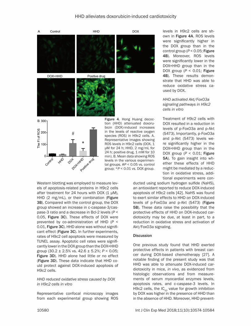
Figure 4. Hong Huang decoc-tion (HHD) attenuated doxoru-bicin (DOX)-induced increases in the levels of reactive oxygen species (ROS) in H9c2 cells. A. Representative images showing ROS levels in H9c2 cells (DOX, 1 µM for 24 h; HHD, 2 mg/mL for 24 h; positive drug, 1 mM for 10 min). B. Mean data showing ROS levels in the various experimen-tal groups. #P < 0.05 vs. control group; *P < 0.01 vs. DOX group.
levels in H9c2 cells are sh- own in Figure 4A. ROS levels were significantly higher in the DOX group than in the control group (P < 0.05; Figure 4B). Moreover, ROS levels were significantly lower in the DOX+HHD group than in the DOX group (P < 0.01; Figure 4B). These results demon-strate that HHD was able to reduce oxidative stress ca- used by DOX.
HHD activated Akt/FoxO3a signaling pathways in H9c2 cells in vitro
Treatment of H9c2 cells with DOX resulted in a reduction in levels of p-FoxO3a and p-Akt (S473). Importantly, p-FoxO3a and p-Akt (S473) levels we- re significantly higher in the DOX+HHD group than in the DOX group (P < 0.01; Figure 5A). To gain insight into wh- ether these effects of HHD might be mediated by a reduc-tion in oxidative stress, addi-tional experiments were con-

HHD alleviates doxorubicin-induced cardiotoxicity
10581 Int J Clin Exp Med 2018;11(10):10574-10584
ed DOX-induced apoptosis of H9c2 cells and inhibited DOX-induced increase in ROS levels in these cells. Importantly, DOX-induced reduc-tions in p-FoxO3a and p-Akt (S473) protein lev-els in H9c2 cells were attenuated by HHD and the effects of HHD were mimicked by the anti-oxidant, NaHS. Taken together, these findings suggest that HHD can protect against DOX-induced cardiotoxicity, in vitro and in vivo, pos-sibly by reducing oxidative stress and activat- ing Akt/FoxO3a signaling pathways.
Although DOX is an effective anticancer drug, the associated cardiotoxicity is a limiting fac- tor in its clinical use. In vivo experiments, in the present study, demonstrated that DOX caused histologic changes in the heart (consis-tent with myocardial damage) and increased serum cardiac enzyme levels, myocardial c-cas-pase-3 protein levels, and cardiomyocyte apop-tosis rates. In H9c2 cells, DOX also increased c-caspase-3 protein levels and apoptosis rates. These findings are consistent with numerous previous studies of DOX-induced cardiotoxicity, in vivo and in vitro [22-26, 29-34, 42]. Thus, the
with a reduction in ROS levels, suggesting that the beneficial effects of HHD on cell survival may be mediated, at least in part, by suppres-sion of DOX-induced oxidative stress. In agree-ment with the present data, previous studies of vitexin [26], cilostazol [43], vitamin C [44], allicin [45], Zingiber officinale extract [46], and various components of HHD, including Ast- ragalus polysaccharides [28, 29], salidroside [31], and curcumin [32, 33], have found that inhibition of oxidative stress may protect ag- ainst DOX-induced toxicity. Therefore, based on present findings, this study proposes that inhibition of ROS levels may be an important mechanism contributing to beneficial effects of HHD against DOX-induced cardiotoxicity. Al- though the present study did not identify spe-cific components of HHD exerting antioxidant effects, it is likely that multiple constituents contribute [28, 29, 31-33].
Several mechanisms have been suggested to underlie the protective effects of agents that reduce oxidative stress in cardiomyocytes ex- posed to DOX. For example, the beneficial ef-
Figure 5. Hong Huang decoction (HHD) activated Akt/FoxO3a signaling path-ways in H9c2 cells treated with doxorubicin (DOX). A. Top: representative Western blots showing protein expression of FoxO3a, phosphorylated FoxO3a (p-FoxO3a), Akt, p-Akt, and β-actin (used as the internal control) in H9c2 cells from each experimental group (DOX, 1 µM; HHD, 2 mg/mL; for 24 h). Bottom: mean data quantified from Western blot experiments showing p-FoxO3a and p-Akt levels (relative to that of β-actin) in each experimental group. B. Top: representative Western blots showing the protein expression of FoxO3a, p-FoxO3a, Akt, p-Akt, and β-actin in H9c2 cells from each experimental group (DOX, 1 µM; NaHS, 100 µM; for 24 h). Bottom: mean data quantified from the Western blot experiments showing p-FoxO3a and p-Akt levels (relative to that of β-actin) in each experimental group. *P < 0.05 vs. DOX group.
model systems used in the present study are reproduc-ible and suitable for studies concerning DOX-associated cardiotoxicity.
It has been recognized that induction of oxidative stress is an important mechanism contributing to the adverse cardiac effects of DOX [9, 15]. This study observed that treatment of cultured H9c2 cells with DOX resulted in a substantial elevation in cellu-lar levels of ROS, in agree-ment with previous studies [25, 29-31, 33, 42]. H2O2, known to impose oxidative stress on cells, has been sh- own to mimic DOX and pro-mote cardiomyocyte apopto-sis [25]. Furthermore, the an- tioxidant H2S has been repo- rted to inhibit the detriment- al actions of DOX on heart cells [25, 42]. The present study found that inhibition of DOX-induced H9c2 cell apop-tosis by HHD was associated

HHD alleviates doxorubicin-induced cardiotoxicity
10582 Int J Clin Exp Med 2018;11(10):10574-10584
fects of H2S in DOX-treated H9c2 cells have been attributed to suppression of p38 mitogen-activated protein kinase (MAPK)/extracellular signal-regulated kinase (ERK) 1/2 signaling [25, 47], inhibition of downstream nuclear fac-tor-kappa-B (NF-κB) pathways [47], and sup-pression of calreticulin expression [48]. Inte- restingly, reduced activation of p38 signaling has also been implicated in mediating the pro-tective effects of Astragalus polysaccharides against DOX-induced ROS production and ap- optosis [29]. However, Akt signaling is another pathway that plays a key role in cell growth and apoptosis. Akt activation has been thought to promote cardiac cell survival by inhibiting apop-tosis [49]. Akt can phosphorylate and regulate FoxO3a, a multifunctional transcription factor involved in regulation of differentiation, devel-opment, proliferation, apoptosis, and necrosis [50]. FoxO3a can transduce a variety of cellular stimuli, including oxidative stress, resulting in either apoptosis or protective cell growth arrest [51, 52]. DOX has been shown to reduce levels of FoxO3a in mouse hearts [22], decrease lev-els of p-FoxO3a in H9c2 cells [23], and upregu-late nuclear expression of FoxO3a in H9c2 cells [24]. Furthermore, H2S and vitexin have been suggested to attenuate DOX-induced toxicity in H9c2 cardiac cells by activating FoxO3a path-ways [25, 26]. This study found that HHD increased levels of p-Akt and p-FoxO3a, consis-tent with stimulation of Akt/FoxO3a signaling. Therefore, this present study proposes that inhibition of oxidative stress and activation of the Akt/FoxO3a pathway may be a mechanism that contributes to the protective effects of HHD against DOX-induced cardiotoxicity. Inte- restingly, the beneficial effects of Astragalosi- de IV (a component of HHD) have also been attributed to stimulation of PI3K/Akt signaling [30]. Additional studies are necessary to iden-tify the specific components of HHD that inhibit ROS levels and stimulate Akt/FoxO3a signaling in cardiomyocytes treated with DOX.
In conclusion, this study has shown that HHD, a Chinese herbal medicine, protects against DOX-induced cardiotoxicity, in vitro and in vivo. Furthermore, these actions may be mediated, at least in part, by a reduction in oxidative stress and activation of Akt/FoxO3a signaling pathways. Further studies are necessary to characterize the mechanisms in more detail and establish, definitively, whether HHD is clini-cally useful as an inhibitor of DOX-induced cardiotoxicity.
Acknowledgements
This research was funded by The Project (Gr- ant No. 81473676), supported by National Natural Science Foundation of China, Phase II Project Funded by the Priority Academic Pr- ogram Development of Jiangsu Higher Edu- cation Institutions (Grant No. 012062003010), and the Developing Program for High-level Academic Talent in Jiangsu Hospital of TCM (Grant No. y2018rc11).
Disclosure of conflict of interest
None.
Address correspondence to: Chang Yao, The Af- filiated Hospital of Nanjing University of Chinese Medicine, 155 Hanzhong Road, Nanjing 210029, Jiangsu, P. R. China. Tel: +86-18013966854; E-mail: [email protected]
References
[1] GBD 2013 Mortality and Causes of Death Col-laborators. Global, regional, and national age-sex specific all-cause and cause-specific mor-tality for 240 causes of death, 1990-2013: a systematic analysis for the global burden of disease study 2013. Lancet 2015; 385: 117-171.
[2] Rivankar S. An overview of doxorubicin formu-lations in cancer therapy. J Cancer Res Ther 2014; 10: 853-858.
[3] Bloom MW, Hamo CE, Cardinale D, Ky B, Nohria A, Baer L, Skopicki H, Lenihan DJ, Ghe-orghiade M, Lyon AR and Butler J. Cancer ther-apy-related cardiac dysfunction and heart fail-ure: part 1: definitions, pathophysiology, risk factors, and imaging. Circ Heart Fail 2016; 9: e002661.
[4] Xie Y, Collins WJ, Audeh MW, Shiao SL, Gottlieb RA, Goodman MT, Merz CN and Mehta PK. Breast cancer survivorship and cardiovascular disease: emerging approaches in cardio-oncol-ogy. Curr Treat Options Cardiovasc Med 2015; 17: 60.
[5] Yamaguchi N, Fujii T, Aoi S, Kozuch PS, Horto-bagyi GN and Blum RH. Comparison of cardiac events associated with liposomal doxorubicin, epirubicin and doxorubicin in breast cancer: a Bayesian network meta-analysis. Eur J Cancer 2015; 51: 2314-2320.
[6] Tacar O, Sriamornsak P and Dass CR. Doxoru-bicin: an update on anticancer molecular ac-tion, toxicity and novel drug delivery systems. J Pharm Pharmacol 2013; 65: 157-170.
[7] Lyu YL, Kerrigan JE, Lin CP, Azarova AM, Tsai YC, Ban Y and Liu LF. Topoisomerase IIbeta

HHD alleviates doxorubicin-induced cardiotoxicity
10583 Int J Clin Exp Med 2018;11(10):10574-10584
mediated DNA double-strand breaks: implica-tions in doxorubicin cardiotoxicity and preven-tion by dexrazoxane. Cancer Res 2007; 67: 8839-8846.
[8] Varga ZV, Ferdinandy P, Liaudet L and Pacher P. Drug-induced mitochondrial dysfunction and cardiotoxicity. Am J Physiol Heart Circ Physiol 2015; 309: H1453-1467.
[9] Carvalho FS, Burgeiro A, Garcia R, Moreno AJ, Carvalho RA and Oliveira PJ. Doxorubicin-in-duced cardiotoxicity: from bioenergetic failure and cell death to cardiomyopathy. Med Res Rev 2014; 34: 106-135.
[10] Kanaan GN and Harper ME. Cellular redox dys-function in the development of cardiovascular diseases. Biochim Biophys Acta 2017; 1861: 2822-2829.
[11] Damiani RM, Moura DJ, Viau CM, Caceres RA, Henriques JA and Saffi J. Pathways of cardiac toxicity: comparison between chemotherapeu-tic drugs doxorubicin and mitoxantrone. Arch Toxicol 2016; 90: 2063-2076.
[12] Belcastro E, Wu W, Fries-Raeth I, Corti A, Pom-pella A, Leroy P, Lartaud I and Gaucher C. Oxi-dative stress enhances and modulates protein S-nitrosation in smooth muscle cells exposed to S-nitrosoglutathione. Nitric Oxide 2017; 69: 10-21.
[13] Farias JG, Molina VM, Carrasco RA, Zepeda AB, Figueroa E, Letelier P and Castillo RL. Antioxi-dant therapeutic strategies for cardiovascular conditions associated with oxidative stress. Nutrients 2017; 9.
[14] Moris D, Spartalis M, Spartalis E, Karachaliou GS, Karaolanis GI, Tsourouflis G, Tsilimigras DI, Tzatzaki E and Theocharis S. The role of reac-tive oxygen species in the pathophysiology of cardiovascular diseases and the clinical signifi-cance of myocardial redox. Ann Transl Med 2017; 5: 326.
[15] Lebrecht D, Kokkori A, Ketelsen UP, Setzer B and Walker UA. Tissue-specific mtDNA lesions and radical-associated mitochondrial dysfunc-tion in human hearts exposed to doxorubicin. J Pathol 2005; 207: 436-444.
[16] Rochette L, Guenancia C, Gudjoncik A, Hachet O, Zeller M, Cottin Y and Vergely C. Anthracy-clines/trastuzumab: new aspects of cardiotox-icity and molecular mechanisms. Trends Phar-macol Sci 2015; 36: 326-348.
[17] Kalyanaraman B. Iron signaling and oxidant damage. Cardiovasc Toxicol 2007; 7: 92-94.
[18] Peng S, Zhao S, Yan F, Cheng J, Huang L, Chen H, Liu Q, Ji X and Yuan Z. HDAC2 selectively regulates FOXO3a-mediated gene transcrip-tion during oxidative stress-induced neuronal cell death. J Neurosci 2015; 35: 1250-1259.
[19] Storz P. Forkhead homeobox type O transcrip-tion factors in the responses to oxidative
stress. Antioxid Redox Signal 2011; 14: 593-605.
[20] Sengupta A, Molkentin JD, Paik JH, DePinho RA and Yutzey KE. FoxO transcription factors promote cardiomyocyte survival upon induc-tion of oxidative stress. J Biol Chem 2011; 286: 7468-7478.
[21] Huang H and Tindall DJ. FOXO factors: a matter of life and death. Future Oncol 2006; 2: 83-89.
[22] Zhou L, Li R, Liu C, Sun T, Htet Aung LH, Chen C, Gao J, Zhao Y and Wang K. Foxo3a inhibits mitochondrial fission and protects against doxorubicin-induced cardiotoxicity by sup-pressing MIEF2. Free Radic Biol Med 2017; 104: 360-370.
[23] Liu MH, Zhang Y, He J, Tan TP, Wu SJ, Fu HY, Chen YD, Liu J, Le QF, Hu HJ, Yuan C and Lin XL. Upregulation of peroxiredoxin III in doxorubicin-induced cytotoxicity and the FoxO3a-depen-dent expression in H9c2 cardiac cells. Exp Ther Med 2015; 10: 1515-1520.
[24] Sampaio SF, Branco AF, Wojtala A, Vega-Nare-do I, Wieckowski MR and Oliveira PJ. p66Shc signaling is involved in stress responses elicit-ed by anthracycline treatment of rat cardio-myoblasts. Arch Toxicol 2016; 90: 1669-1684.
[25] Liu MH, Lin XL, Zhang Y, He J, Tan TP, Wu SJ, Liu J, Tian W, Chen L, Yu S, Li J and Yuan C. Hydrogen sulfide attenuates doxorubicin-in-duced cardiotoxicity by inhibiting reactive oxy-gen species-activated extracellular signal-reg-ulated kinase 1/2 in H9c2 cardiac myocytes. Mol Med Rep 2015; 12: 6841-6848.
[26] Sun Z, Yan B, Yu WY, Yao X, Ma X, Sheng G and Ma Q. Vitexin attenuates acute doxorubicin cardiotoxicity in rats via the suppression of oxi-dative stress, inflammation and apoptosis and the activation of FOXO3a. Exp Ther Med 2016; 12: 1879-1884.
[27] Yue YZ, Bian WH, Yao C, Ren XM, Zhang XQ, Guo MM, Ying Y and Chen L. Clinical study of self-made hong-huang antioxidant inhibiting oxidative stress reaction during chemotherapy in patients with breast cancer. Chinese Journal of Information on Traditional Chinese Medicine 2014; 21: 11-14.
[28] Huang YF, Lu L, Zhu DJ, Wang M, Yin Y, Chen DX and Wei LB. Effects of astragalus polysac-charides on dysfunction of mitochondrial dy-namics induced by oxidative stress. Oxid Med Cell Longev 2016; 2016: 9573291.
[29] Zhou L, Chen L, Wang J and Deng Y. Astragalus polysaccharide improves cardiac function in doxorubicin-induced cardiomyopathy through ROS-p38 signaling. Int J Clin Exp Med 2015; 8: 21839-21848.
[30] Jia Y, Zuo D, Li Z, Liu H, Dai Z, Cai J, Pang L and Wu Y. Astragaloside IV inhibits doxorubicin-in-duced cardiomyocyte apoptosis mediated by mitochondrial apoptotic pathway via activat-

HHD alleviates doxorubicin-induced cardiotoxicity
10584 Int J Clin Exp Med 2018;11(10):10574-10584
ing the PI3K/Akt pathway. Chem Pharm Bull (Tokyo) 2014; 62: 45-53.
[31] Wang XL, Wang X, Xiong LL, Zhu Y, Chen HL, Chen JX, Wang XX, Li RL, Guo ZY, Li P and Jiang W. Salidroside improves doxorubicin-induced cardiac dysfunction by suppression of exces-sive oxidative stress and cardiomyocyte apop-tosis. J Cardiovasc Pharmacol 2013; 62: 512-523.
[32] Swamy AV, Gulliaya S, Thippeswamy A, Koti BC and Manjula DV. Cardioprotective effect of cur-cumin against doxorubicin-induced myocardial toxicity in albino rats. Indian J Pharmacol 2012; 44: 73-77.
[33] Junkun L, Erfu C, Tony H, Xin L, Sudeep KC, Mingliang Z, Yanqin W and XiangQian Q. Cur-cumin downregulates phosphate carrier and protects against doxorubicin induced cardio-myocyte apoptosis. Biomed Res Int 2016; 2016: 1980763.
[34] Hosseinzadeh L, Behravan J, Mosaffa F, Bah-rami G, Bahrami A and Karimi G. Curcumin po-tentiates doxorubicin-induced apoptosis in H9c2 cardiac muscle cells through generation of reactive oxygen species. Food Chem Toxicol 2011; 49: 1102-1109.
[35] Deng P, Li X, Wei Y, Liu J, Chen M, Xu Y, Dong B, Zhu L and Chai L. The herbal decoction modi-fied Danggui Buxue Tang attenuates immune-mediated bone marrow failure by regulating the differentiation of T lymphocytes in an im-mune-induced aplastic anemia mouse model. PLoS One 2017; 12: e0180417.
[36] Qin L, Lan Y, Sun J, Zhang B, Xie P, Zheng J and Ma S. A Chinese herbal medicine (Modified Guomin Decoction) influences the differentia-tion of CD4+ T-cell subsets in OVA-induced asthmatic mice. Neuro Endocrinol Lett 2017; 38: 187-198.
[37] Gao Y, Xu Y, Hua S, Zhou S and Wang K. ALDH2 attenuates Dox-induced cardiotoxicity by inhib-iting cardiac apoptosis and oxidative stress. Int J Clin Exp Med 2015; 8: 6794-6803.
[38] Wu X, Qi X, Lu Y, Lin C, Yuan Y, Zhu Q, Yin Q, Li W, Li Y and Bian H. Liguzinediol protects against cardiac fibrosis in rats in vivo and in vitro. Biomed Pharmacother 2016; 80: 260-267.
[39] Xu HL, Mao KL, Huang YP, Yang JJ, Xu J, Chen PP, Fan ZL, Zou S, Gao ZZ, Yin JY, Xiao J, Lu CT, Zhang BL and Zhao YZ. Glioma-targeted super-paramagnetic iron oxide nanoparticles as drug-carrying vehicles for theranostic effects. Nanoscale 2016; 8: 14222-14236.
[40] Crowley LC and Waterhouse NJ. Detecting cleaved caspase-3 in apoptotic cells by flow cy-tometry. Cold Spring Harb Protoc 2016; 2016.
[41] Willis S, Day CL, Hinds MG and Huang DC. The Bcl-2-regulated apoptotic pathway. J Cell Sci 2003; 116: 4053-4056.
[42] Liu MH, Zhang Y, He J, Tan TP, Wu SJ, Guo DM, He H, Peng J, Tang ZH and Jiang ZS. Hydrogen sulfide protects H9c2 cardiac cells against doxorubicin-induced cytotoxicity through the PI3K/Akt/FoxO3a pathway. Int J Mol Med 2016; 37: 1661-1668.
[43] Koh JS, Yi CO, Heo RW, Ahn JW, Park JR, Lee JE, Kim JH, Hwang JY and Roh GS. Protective ef-fect of cilostazol against doxorubicin-induced cardiomyopathy in mice. Free Radic Biol Med 2015; 89: 54-61.
[44] Akolkar G, da Silva Dias D, Ayyappan P, Bagchi AK, Jassal DS, Salemi VMC, Irigoyen MC, De Angelis K and Singal PK. Vitamin C mitigates oxidative/nitrosative stress and inflammation in doxorubicin-induced cardiomyopathy. Am J Physiol Heart Circ Physiol 2017; 313: H795-h809.
[45] Abdel-Daim MM, Kilany OE, Khalifa HA and Ahmed AAM. Allicin ameliorates doxorubicin-induced cardiotoxicity in rats via suppression of oxidative stress, inflammation and apopto-sis. Cancer Chemother Pharmacol 2017; 80: 745-753.
[46] Ajith TA, Hema U and Aswathi S. Zingiber offici-nale Roscoe ameliorates anticancer antibiotic doxorubicin-induced acute cardiotoxicity in rat. J Exp Ther Oncol 2016; 11: 171-175.
[47] Guo R, Wu K, Chen J, Mo L, Hua X, Zheng D, Chen P, Chen G, Xu W and Feng J. Exogenous hydrogen sulfide protects against doxorubicin-induced inflammation and cytotoxicity by inhib-iting p38MAPK/NFkappaB pathway in H9c2 cardiac cells. Cell Physiol Biochem 2013; 32: 1668-1680.
[48] Liu MH, Zhang Y, Lin XL, He J, Tan TP, Wu SJ, Yu S, Chen L, Chen YD, Fu HY, Yuan C and Li J. Hydrogen sulfide attenuates doxorubicinin-duced cardiotoxicity by inhibiting calreticulin expression in H9c2 cells. Mol Med Rep 2015; 12: 5197-5202.
[49] Czubaty A and Piekielko-Witkowska A. Protein kinases that phosphorylate splicing factors: roles in cancer development, progression and possible therapeutic options. Int J Biochem Cell Biol 2017; 91: 102-115.
[50] Furukawa-Hibi Y, Kobayashi Y, Chen C and Mo-toyama N. FOXO transcription factors in cell-cycle regulation and the response to oxidative stress. Antioxid Redox Signal 2005; 7: 752-760.
[51] Lam EW, Brosens JJ, Gomes AR and Koo CY. Forkhead box proteins: tuning forks for tran-scriptional harmony. Nat Rev Cancer 2013; 13: 482-495.
[52] Zhao Y, Wang Y and Zhu WG. Applications of post-translational modifications of FoxO family proteins in biological functions. J Mol Cell Biol 2011; 3: 276-282.