suppression of isoproterenol-induced apoptosis in h9c2 ... · this study investigated the...
TRANSCRIPT
Chinese Journal of Physiology 59(6): 323-330, 2016 323DOI: 10.4077/CJP.2016.BAE393
Corresponding author: Chih-Yang Huang, PhD, Graduate Institute of Basic Medical Science, China Medical University and Hospital, No. 91 Hsueh-Shih Rd., Taichung 40402, Taiwan, R.O.C. Tel: +886-4-22053366 ext. 3313, Fax: +886-4-22333641, E-mail: [email protected]#Contributed equally to this work.Received: July 27, 2015; Revised (Final Version): May 6, 2016; Accepted: June 29, 2016.2016 by The Chinese Physiological Society and Airiti Press Inc. ISSN : 0304-4920. http://www.cps.org.tw
Suppression of Isoproterenol-Induced Apoptosis in H9c2 Cardiomyoblast Cells by Daidzein
through Activation of Akt
Wei-Syun Hu1, 2, Yueh-Min Lin3, 4, Wei-Wen Kuo5, Lung-Fa Pan6, 7, Yu-Lan Yeh3, 4, Yi-Hui Li8, Chia-Hua Kuo9, Ray-Jade Chen10, V. Vijaya Padma11, Tung-Sheng Chen8, 12, #,
and Chih-Yang Huang8, 13, 14, #
1School of Medicine, College of Medicine, China Medical University, Taichung 40402, Taiwan, R.O.C.2Division of Cardiovascular Medicine, Department of Medicine, China Medical University Hospital,
Taichung 40447, Taiwan, R.O.C.3Department of Pathology, Changhua Christian Hospital, Changhua 50006, Taiwan, R.O.C.
4Jen-Teh Junior College of Medicine, Nursing and Management, Miaoli 35664, Taiwan, R.O.C.5Department of Biological Science and Technology, China Medical University, Taichung 40402,
Taiwan, R.O.C.6Division of Cardiology, Armed Force Taichung General Hospital, Taichung 41152, Taiwan, R.O.C.
7Department of Medical Imaging and Radiological Sciences of Central Taiwan University of Science and Technology, Taichung 40601, Taiwan, R.O.C.
8Graduate Institute of Basic Medical Science, China Medical University, Taichung 40402, Taiwan, R.O.C.
9Department of Sports Sciences, University of Taipei, Taipei 10048, Taiwan, R.O.C.10Department of Surgery, School of Medicine, College of Medicine, Taipei Medical University,
Taipei 11031, Taiwan, R.O.C.11Department of Biotechnology, Bharathiar University, Coimbatore-641 046, India,
12Biomaterials Translational Research Center, China Medical University Hospital, Taichung 40402, Taiwan, R.O.C.
13Graduate Institute of Chinese Medical Science, China Medical University, Taichung 40402, Taiwan, R.O.C.
and14Department of Health and Nutrition Biotechnology, Asia University, Taichung 41354, Taiwan,
Republic of China
Abstract
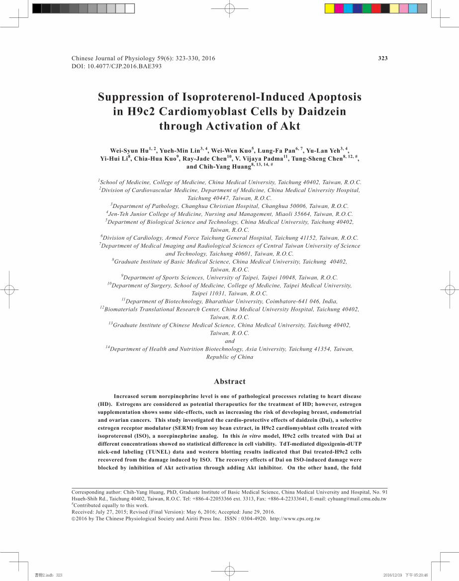
Increased serum norepinephrine level is one of pathological processes relating to heart disease (HD). Estrogens are considered as potential therapeutics for the treatment of HD; however, estrogen supplementation shows some side-effects, such as increasing the risk of developing breast, endometrial and ovarian cancers. This study investigated the cardio-protective effects of daidzein (Dai), a selective estrogen receptor modulator (SERM) from soy bean extract, in H9c2 cardiomyoblast cells treated with isoproterenol (ISO), a norepinephrine analog. In this in vitro model, H9c2 cells treated with Dai at different concentrations showed no statistical difference in cell viability. TdT-mediated digoxigenin-dUTP nick-end labeling (TUNEL) data and western blotting results indicated that Dai treated-H9c2 cells recovered from the damage induced by ISO. The recovery effects of Dai on ISO-induced damage were blocked by inhibition of Akt activation through adding Akt inhibitor. On the other hand, the fold
書冊2.indb 323 2016/12/19 下午 05:20:46

324 Hu, Lin, Kuo, Pan, Yeh, Li, Kuo, Chen, Padma, Chen and Huang
Introduction
Heart disease (HD) is a multiple cardio-dysfunc-tion process and a major cause of death worldwide. Several cardio-dysfunctions are associated with HD, including inflammation, hypertrophy, apoptosis and fibrosis (2, 11, 22). According to epidemiological reports, HD risk is lower in premenopausal women than in men of the same age (13). On the other hand, HD risk is increased in women with menopause. These evidences indicate that female hormones may partially play an important role in the cardio-dysfunction process (13).
Among the female hormones, estrogens show a cardiac protective role in in vitro and in vivo studies (1, 5). Estrogens are the major sex steroids existing in three natural forms, including estrone (E1), 17β-estradiol (E2) and estriol (E3). Among these estrogens, E2 is most important due to its abundance and potency. Experimental results have indicated that estrogens regulate expression and activation of proteins related to cell survival, apoptosis suppression and other com-pensatory effects, leading to cardio-protection (23). Therefore, estrogen replacement therapy (ERT) in meno-pausal women has been considered as a therapeutic strategy for reducing HD risk (7, 18). Some reports stated that high-dose or long-term estrogen supple-mentation might activate cell proliferation and metasta-sis of breast, endometrial and ovarian cancers (8, 20). As a result, adequate supplementation of natural, botanical estrogen-like compounds that mimic E2 in reducing HD without adverse effects should be consid-ered as an alternative to HRT in menopausal women.
Neurotransmitters, such as norepinephrine, can regulate heart function (4). Increased norepinephrine levels activate beta-adrenergic receptors and induce cellular signaling pathways activate apoptosis and suppress cell survival, leading to cardiomyocyte dys-function (12, 15). Shizukuda et al. (21) and Saito et al. (19) reported that norepinephrine regulates calcineu-rine expression in heart cells, which then dephos-phorylates the mitochondrial protein Bad, leading to mitochondrial membrane instability and the release of cytochrome C. This induces caspase 3 activation, resulting in cardiomyocyte programmed cell death. Furthermore, several papers reported cardiac protective effects of natural estrogen-like compounds on cardio-myocyte damage induced by isoproterenol (ISO) (3,
17), a norepinephrine analog which mimics hypertension in patients. Hu et al. (10) showed that genistein ame-liorates cardiomyoblast hypertrophy induced by ISO through the p38-Erk1/2-JNK-NKκB signaling pathway. Maulik et al. (14) pointed out that ISO induces cardiac hypertrophy in rats through inducible nitric oxide synthase (iNOS) expression, and that cardiac hy-pertrophy can be suppressed by genistein treatment by inhibition of iNOS. These findings suggest that genistein shows cardiac protective potentials in cardiac dysfunction induced by ISO. Taken together, nor-epinephrine exhibits pro-apoptotic effects on cardio-myocytes, and estrogens, including phytoestrogens, show cardio-protection both in vitro and in vivo. At present, the cardiac protective effects of daidzein (Dai), another natural estrogen-like compound from soy bean extract, has not been studied. Dai has as similar chemical structure as E2 and genistein (Fig. 1). The cardio-protective role of Dai in cardiac cells under ISO stress needs to be clarified.
We hypothesize that Dai protects cardiomyoblast cells from ISO-induced cell death (14) by activating pro-survival signaling pathways similar to E2 and genistein. To test our hypothesis, activation of pro- apoptotic and anti-apoptotic proteins was first inves-tigated in H9c2 cardiomyoblasts treated with both Dai and ISO. We also determined which signaling path-way was responsible for the cardio-protective effects of Dai in the H9c2 cell model.
Materials and Methods
Cell Culture
H9c2 cardiomyoblasts, a subclone of the original clonal cell line derived from embryonic BD1X rat heart tissues, obtained from the American Type Culture Collection (ATCC, CRL-1446) (Rockville, MD, USA)
DaidzeinEstradiol
OH
HO
OH
HO
O
O
Fig. 1. Chemical structures of estradiol and daidzein (Dai).
changes of phosphorylated Akt (p-Akt)/Akt normalized with the control for con, 0.25, 0.5, 1, 3 and 24 h of treatment were 1, 2, 5, 13, 11 and 10, respectively. In conclusion, Dai ameliorates apoptosis of cardio- myoblasts induced by ISO through Akt signaling pathway.
Key Words: apoptosis, daidzein, estrogen, heart disease, isoproterenol
書冊2.indb 324 2016/12/19 下午 05:20:46

Cardioprotection of Daidzein 325
were cultured in 10-cm culture dishes containing Dulbecco’s modified Eagle’s medium (DMEM) sup-plemented with 100 μg/ml streptomycin, 100 μg/ml penicillin, 1 mM HEPS buffer, 2 mM glutamine and 10% Clontech fetal bovine serum in humidified air (5% CO2) at 37°C. Before treatment with indicated agents, H9c2 cardiomyoblasts, passage 32 to passage 40, were incubated overnight in serum-free essential medium.
Cell Viability Assay
MTT (3-(4,5-Dimethylthiazol-2-yl)-2,5-di-phenyltetrazolium bromide) assays were applied for measuring cell viability. Briefly, H9c2 cells (1 × 10⁵ cells per well) were plated in 24-well plates. Different dosages of Dai were added to the wells and incubated for 24 h. The culture medium was then removed and 200 μl MTT solution (0.5 mg/ml) was added to each well. After 4 h incubation at 37°C, the MTT solution was removed and 150 μl dimethyl sulfoxide (DMSO) was added to each well. The absorbance was mea-sured at 550 nm using an automated micro-plate reader.
TUNEL (TdT-Mediated Digoxigenin-dUTP Nick-End Labeling) Assay
The TUNEL method was carried out by using a commercial kit (Roche Molecular Biochemicals, Mannheim, Germany). To explore the Dai effects on the ISO-induced apoptosis, staining was performed according to the manufacturer’s protocol. The TUNEL assay was performed in a 96-well plate with 5,000 cells per well. TUNEL-positive cells were identified with a fluorescence microscope using an excitation wave-length in the 450–500 nm range and a detection wave-length in the 515–565 nm range (green). After TUNEL imaging, the specimen was stained with 4’,6- diamidino-2-phenylindole (DAPI) based on manufac-turer’s protocol. Specimen was then read by using excitation wavelength of 358 nm and a detection wave- length of 461 nm (blue). The percentage of apoptotic cells was calculated by dividing the number of TUNEL- positive cells by the number of DAPI-positive cells visualized in the same field. Three independent ex- periments were then averaged and statistically analyzed.
Western Blotting
H9c2 cells with a cell density of 5 × 10⁵ cells were plated onto 10-cm dish and incubated overnight at 37°C. Dosage (10-8 M) of Dai and 1 µM kinase inhibitors including SP600125 (SP, JNK inhibitor), QZN (NFκB inhibitor), U0126 (Erk1/2 inhibitor), SB203508 (SB, p38 inhibitor) and LY294002 (LY, Akt inhibitor) were added to the dishes and pre-incubated
for 1 h before ISO (50 µM) treatment. H9c2 cells were washed with cold PBS and resuspended in lysis buffer (50 mM Tris, pH 7.5, 0.5 M NaCl, 1 mM ethylenedi- aminetetraacetic acid (EDTA), pH 7.5, 10% glycerol, 1 mM β-mercaptoethanol (BME), 1% IGEPAL-630) after treating ISO for 24 h with a proteinase inhibitor cocktail (Roche Molecular Biochemicals, New York, NY, USA) to extract total protein. After incubation for 30 min on ice, the supernatant was collected by cen- trifugation at 12,000 × g for 15 min at 4°C. The protein concentration was determined using the Bradford method. Samples containing 40 μg proteins were loaded to each well and analyzed using western blot analysis. Briefly, protein samples were separated by 12% SDS-PAGE and transferred onto polyvinylidene difluoride (PVDF) membrane (Millipore, Belford, MA, USA). Membranes were then blocked with a blocking buffer (20 mM Tris–HCl, 5% non-fat dry milk, pH 7.6, 0.1% Tween 20 and 150 mM NaCl) for at least 1 h at room temperature. Membranes were then incubated overnight with primary antibodies against Akt, phosphorylated Akt (p-Akt), Bad, phosphorylated Bad (p-Bad), Erk1/2, phosphorylated Erk1/2 (p-Erk1/2), caspase 3, caspase 9 and α-tubulin (all purchased from Santa Cruz Biotechnology, Inc., Paso Robles, CA, USA) in the above solution on an orbit shaker at 4°C. Fol-lowing incubation with primary antibodies, mem-branes were incubated with appropriate horserad-ish peroxidase-linked secondary antibodies (Santa Cruz Biotechnology, Inc.), and the membranes were observed under a cooled charge-coupled device (CCD) camera for detection of enzyme-based chemilumines-cence in the membranes. Gels for western blots were analyzed by using ImageJ software (National institutes of health (NIH), Bethesda, MD, USA). In addition to Akt, Erk1/2 and Bad phosphorylation normalized with the respective total forms, bands in other western blots were normalized with an internal control, α- tubulin), and expressed as fold change.
Statistical Analysis
Data for TUNEL analysis and cell viability were
Dai (M)
Cel
l Via
bilit
y (%
)
10-100
50
100
150
10-9 10-8 10-7 10-6 10-5
Fig. 2. Cell viability test for H9c2 cells treated with different dosages of Dai.
書冊2.indb 325 2016/12/19 下午 05:20:46

326 Hu, Lin, Kuo, Pan, Yeh, Li, Kuo, Chen, Padma, Chen and Huang
expressed as mean ± SD (n = 3) and analyzed by using one-way ANOVA. Student’s t-test was applied for post- hoc test and statistical significance was considered at the level of P < 0.05. Correlation analysis was performed in Fig. 4 and two tailed F-test was applied for calcu-lating significance. Statistical significance was con-sidered at the level of P < 0.05.
Results
H9c2 Cell Viability in the Presence of Dai
Viability of H9c2 cardiomyoblasts in the presence of Dai is shown in Fig. 2. Compared to the control (H9c2 cells in the absence of Dai), no significant changes were observed in the presence of different Dai con-
ISO (50 µM)
Dai (M)
TU
NE
L P
ositi
ve (
%)
0
5
10
15
10-810-910-10 10-7 10-6 10-5
+ + + + + + +
****
**
# # ###
B
−
− −
Dai 10-9 M Dai 10-8 M Dai 10-7 M Dai 10-6 M Dai 10-5 M
ISO 50 µM
ISO 50 µM
Control DMSO DMSO
TUNELTUNEL
TUNELTUNELTUNELTUNEL TUNEL
TUNELTUNELTUNEL
DAPI DAPI DAPI DAPI DAPI
DAPIDAPIDAPIDAPIDAPI
Dai 10-10 M
A
Fig. 3. TUNEL and DAPI analyses of H9c2 cells on treatment with ISO (50 μM) and Dai. Representative images of TUNEL and DAPI staining (A) and quantification of TUNEL data expressed as mean ± SD (n = 3) (B) are shown. **P < 0.01 compared to control; #P < 0.05 compared to ISO; ##P < 0.01 compared to ISO.
書冊2.indb 326 2016/12/19 下午 05:20:47
Cardioprotection of Daidzein 327
centrations (ranging from 10-10 to 10-5 M, P > 0.05). The cell viability results exhibited that treatment Dai was not cytotoxic to the H9c2 cells at the concen-trations tested. The Dai treatment dosage for H9c2 cells used in subsequent experiments in this study was 10-8 M.
TUNEL Analysis of H9c2 Cells in the Presence of Dai
and ISO
The TUNEL assay results to investigate H9c2 cell apoptosis in the presence of Dai or ISO are shown in Fig. 3. Compared to the control (upper panel, first column), the TUNEL-positive signal (apoptotic bodies, shown as green fluorescence) for ISO-treated H9c2 cells (upper panel, third column) increased significantly.
Con 24310.50.25
Treatment Time (h)
0
0.5
1.5
1
2
p-E
rk2/
Erk
2
Con 24310.50.25
Treatment Time (h)
0
0.2
0.4
0.6
0.8
1
1.2
p-E
rk1/
Erk
1
Con 24310.50.25
Treatment Time (h)
0
2
4
8
6
12
10
14
p-A
kt/A
kt
y = -1 + 2.2857X
R2 = 0.7256
P = 0.031
0
21
43
65
87
109
Con 24310.50.25
Treatment Time (h)
p-B
ad/B
ady = 0.1333 + 1.2X
R2 = 0.7132
P = 0.034
Dai 10-8 M
60 kDa
60 kDa
23 kDa
25 kDa
57 kDaα-Tubulin
Fold Changep-ERK2/ERK2
Fold Changep-ERK1/ERK1
Fold Changep-Bad/Bad
Fold Changep-Akt/Akt
42/44 kDa
42/44 kDa
1
1 3 5 4 4 9
2 5 13 11 101
1/4 1/2 1 3 24Con
1
ERK1/2
P-ERK1/2
p-Bad
Bad
p-Akt
Akt
0.8
0.9 0.5 0.5 0.1 0.07
0.050.6 0.5 0.3
46 kDa
Dai 10-8 M
ISO 50 mM
17 kDa
54 kDaα-Tubulin
Caspase 3
Caspase 9
Con
A
B
C
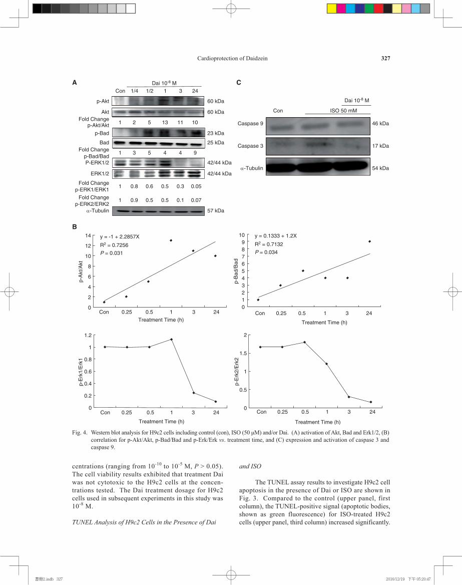
Fig. 4. Western blot analysis for H9c2 cells including control (con), ISO (50 μM) and/or Dai. (A) activation of Akt, Bad and Erk1/2, (B) correlation for p-Akt/Akt, p-Bad/Bad and p-Erk/Erk vs. treatment time, and (C) expression and activation of caspase 3 and caspase 9.
書冊2.indb 327 2016/12/19 下午 05:20:47

328 Hu, Lin, Kuo, Pan, Yeh, Li, Kuo, Chen, Padma, Chen and Huang
In contrast, the TUNEL-positive signals were signifi-cantly decreased when different concentrations of Dai were added to the H9c2 cells in the presence of ISO challenge (Fig. 3A). The quantified results of Fig. 3A are presented in Fig. 3B, which showed statistically significant differences (P < 0.01) for ISO, ISO + Dai (10-10 M) and ISO + Dai (10-9 M) when compared to the control. Further significance could be observed in groups ISO + Dai (10-8 M) (P < 0.05), ISO + Dai (10-7 M) (P < 0.05), ISO + Dai (10-6 M) (P < 0.05) and ISO + Dai (10-5 M) (P < 0.01) when compared to the ISO treatment group.
Protein Activation in H9c2 Cells in the Presence of Dai and ISO
Western blotting approach was performed for investigating the levels of Akt, Erk1/2 and Bad ac-tivation in H9c2 cells in the presence of both ISO and Dai treatments, as represented by phosphorylation of these proteins. Expression levels of p-Akt, Akt, p-Bad, p-Erk1/2 and Erk1/2 were found to be in-creased in H9c2 cells treated with Dai for 1 h when compared to the control (con) (Fig. 4A). After 1 h, most of the protein bands decreased in intensities except for Erk1/2. The fold changes of p-Akt/Akt, p-Bad/Bad, p-Erk1/Erk1 and p-Erk2/Erk2 normal-ized with controls are shown in the Fig. 4A. In addition, correlation analysis between p-Akt/Akt, p-Bad/Bad, p-Erk1/Erk1 and p-Erk2/Erk2 vs. treat-
ment time showed that p-Erk1/Erk1 and p-Erk2/Erk2 correlations were significantly (P < 0.05) (Fig. 4B). It was further observed that some protein levels were increased in ISO-treated H9c2 cells, including the active form of caspase 9 and caspase 3 when compared to the control (Fig. 4C, columns 1 and 2). By contrast, the caspase 9 and caspase 3 protein levels were suppressed after addition of Dai (10-8 M) into ISO-treated H9c2 cells (Fig. 4C, column 3). The fold change for caspase 9 and 3 for control, ISO and ISO + Dai were 1, 1, 0.7, and 1, 1.5 and 0.7, respectively.
Caspase Activation in H9c2 Cells in the Presence of Dai, ISO and Kinase Inhibitors
In order to investigate which kinase pathways are involved in the Dai protective effects in ISO-treated H9c2 cells, inhibitors of signal pathways were tested (Fig. 5). The fold changes were expressed in the Figs. 5, A and B.
Discussion
We investigated the activation of pro- and anti- apoptotic proteins by Dai. Our data showed that compared to the ISO-treatment, the number of apoptotic cells was significantly decreased after treating the H9c2 cells with various Dai concentrations. These TUNEL images illustrate that Dai treatment decreased ISO-induced apoptosis in H9c2 cells in a dose-dependent manner, leading to cardiomyoblasts protection under stress.
Our results further indicated that the activation of Akt was increased in a time-dependent manner in H9c2 cells after treatment with Dai, while Bad was inactivated (Figs. 4, A and B). Furthermore, activation of Erk1 and Erk2 (increased p-Erk1/Erk1 ratio) was observed in 1 h and then decreased; p-Erk2/Erk2 was activated in 30 min before decrease. These findings illustrate that Dai is capable of activating survival pro-teins, but inhibiting pro-apoptotic proteins in H9c2 cells. ISO induced activation of pro-apoptotic caspases 9 and 3, which was reversed by Dai treatment (Fig. 4B). Furthermore, we found that such protective effects of Dai no longer existed by adding the Akt inhibitor. This finding suggests that Akt activation plays a central role in mediating cardio-protection exerted by Dai against ISO-induced myocardial injury (Fig. 5).
Fan et al. (6) pointed out that lipopolysaccharide (LPS) treatment induces inflammation in H9c2 cells through expression of toll-like receptor 4 (TLR4). Be-sides, upregulation of TLR4 accompanies suppression of survival markers, such as insulin-like growth factor 1 (IGF-1), phosphoinositide 3-kinase (PI3K) and Akt, leading to cardiac cell apoptosis. Estrodiol/estrogen receptor α (E2/ERα) treatment is capable of suppressing LPS-induced TLR4 expression, resulting in reduction
Dai 10-8 M
ISO 50 mM
SB SP QZN
54 kDa
1
ConCaspase 3
Fold Change(17kDa/35kDa)
α-Tubulin
B
0.60.70.90.22
17 kDa20 kDa35 kDa
Dai 10-8 M
ISO 50 mM
35 kDa
Con
Caspase 3
Fold Change
α-Tubulin
1
A
54 kDa
U Ly
1.41.3 0.5 0.8
Fig. 5. Caspase activation of H9c2 cells on treatment with ISO (50 μM) and/or Dai in the presence of (A) U0126 (U, Erk1/1 inhibitor), LY294002 (LY, Akt inhibitor), (B) SB203508 (SB, p38 inhibitor), SP600125 (SP, JNK inhibitor) and QZN (NFκB inhibitor).
書冊2.indb 328 2016/12/19 下午 05:20:48

Cardioprotection of Daidzein 329
of inflammatory as well as apoptotic responses. Further- more, Hsieh et al. (9) stated that induction of apoptosis in cardiomyocytes by hypoxia is mediated through the hypoxia-inducible factor 1α (HIF-1α)/BCL2/adenovirus E1B interacting protein 3 (BNIP3)/IGF binding protein-3 (IGFBP3) signaling pathway. Ac-tivation of this pathway suppresses IGF1 survival signaling and Akt activation, leading to apoptosis of cardiomyocytes. Moreover, treatment of E2/ERβ is capable of blocking activation of the HIF1α/BNIP3/ IGFBP3 signaling pathway, leading to expression of Akt and cell survival. These in vitro studies indicate that E2/ER can rescue cardiac cell damage through expression of survival markers, such as IGF1 and Akt. In addition to in vitro models, Nguyen et al. (16) per- formed an animal model to illustrate the beneficial effects of genistein, a soy phytoestrogen, in cardio-protection. This paper provides further evidence to support that supplementation of genistein can enhance cardiac IGF1 gene expression in ovariectomized rats, leading to expression of down-stream proteins, including PI3K and Akt. Taken together, ER activation can ameliorate cardiomyocyte damage by upregulation of survival markers.
Genistein treatment improves heart function by upregulation of survival genes. In this study, Dai was shown to upregulate Akt activation, and this finding is consistent with previous studies. It is speculated here that like E2 or genistein, Dai may serve as an inducer to activate upstream regulators of survival pathways in cardiomyocytes, such as IGF1. However, cardioprotective roles induced by genistein and Dai are different. Hu et al. (10) showed that genistein acts mainly through activation of caspase 8 and NFκB, not through Akt activation. Activation of NFκB (p-NFκB) results in expression of several downstream proteins, including tumor necrosis factor α (TNFα), which is capable of inducing extrinsic cytokines mediating apoptosis through activation of caspase 8. Thus, we speculate that genistein treatment can decrease activation of NFκB, resulting in reduction of pro-apoptotic cytokines, such as TNFα, and caspase 8 in ISO-damaged H9c2 cells. By contrast, Dai treatment increases activation of Akt,
leading to increase of p-Bad and decrease of intrin-sic apoptosis in ISO-damaged H9c2 cells.
The role of Akt expression is confirmed by using kinase inhibitors in this study. Although Akt ex-pression plays a central role in Dai cardio-protection for H9c2 cardiomyoblasts in the presence of ISO stress, siRNA should be used to confirm the role of Akt in this in vitro model because it is unclear if LY is specific to inhibition of Akt expression. Further-more, the relationship between Dai and ER should be investigated, and an animal model should be de-signed in a future study.
The protective role of Dai in ISO-treated H9c2 cells can be summarized as follows: [1] ISO triggers programmed cell death by mediating activation of caspase 9 and 3. [2] Dai upregulates the activation of Akt, then inactivates Bad, leading to suppressed apoptosis signaling induced by the Bad-caspase 9-caspase 3 axis. The key findings in this study are summarized in Fig. 6. Other signaling pathways, such as cardiac hypertrophy, fibrosis or inflammation, can also be investigated in the future to further understand the mechanism of Dai in the cardioprotective effects.
Acknowledgments
This study was supported in part by Taiwan Ministry of Health and Welfare Clinical Trial and Research Center of Excellence (MOHW105-TDU-B-212-133019).
References
1. Bell, J.R., Bernasochi, G.B., Varma, U., Raaijmakers, A.J. and Delbridge, L.M. Sex and sex hormones in cardiac stress--mech-anistic insights. J. Steroid Biochem. Mol. Biol. 137: 124-135, 2013.
2. Chang, Y.M., Velmurugan, B.K., Kuo, W.W., Chen, Y.S., Ho, T.J., Tsai, C.T., Ye, C.X., Tsai, C.H., Tsai, F.J. and Huang, C.Y. Inhibitory effect of alpinate Oxyphyllae fructus extracts on Ang II-induced cardiac pathological remodeling-related pathways in H9c2 cardiomyoblast cells. BioMedicine 3: 148-152, 2013.
3. Chen, X.J., Meng, D., Feng, L., Bian, Y.Y., Li, P., Yang, D., Cao, K.J. and Zhang, J.N. Protective effect of astragalosides on myocardial injury by isoproterenol in SD rats. Am. J. Chinese Med. 34: 1015-1025, 2006.
4. Communal, C., Singh, K., Pimentel, D.R. and Colucci, W.S. Nor-epinephrine stimulates apoptosis in adult rat ventricular myocytes by activation of the beta-adrenergic pathway. Circulation 98: 1329-1334, 1998.
5. Deschamps, A.M., Murphy, E. and Sun, J. Estrogen receptor activation and cardioprotection in ischemia reperfusion injury. Trends Cardiovasc. Med. 20: 73-78, 2010.
6. Fan, M.J., Huang-Liu, R., Shen, C.Y., Ju, D.T., Lin, Y.M., Pai, P., Huang, P.Y., Ho, T.J., Tsai, F.J., Tsai, C.H. and Huang, C.Y. Re-duction of TLR4 mRNA stability and protein expressions through inhibiting cytoplasmic translocation of HuR transcription factor by E2 and/or ERα in LPS-treated H9c2 cardiomyoblast cells. Chinese J. Physiol. 57: 8-18, 2014.
7. Gerhard-Herman, M., Hamburg, N. and Ganz, P. Hormone re-
Dai
p-Bad
Bad
p-Akt
Caspase 9Isoproterenol
Apoptosis
Caspase 3
Fig. 6. Proposed mechanism for cardioprotective effects of daidzein against isoproterenol-induced cardiac dam-age in H9c2 cells. ISO induces H9c2 cell apoptosis by activation of Bad/Caspase 9/Caspase 3 signalings. Dai blocks the apoptotic signalings by activation of p-Akt/p-Bad survival proteins.
書冊2.indb 329 2016/12/19 下午 05:20:48

330 Hu, Lin, Kuo, Pan, Yeh, Li, Kuo, Chen, Padma, Chen and Huang
placement therapy and cardiovascular risk. Curr. Cardio. Rep. 2: 288-292, 2000.
8. Hinds, L. and Price, J. Menopause, hormone replacement and gynaecological cancers. Menopause Int. 16: 89-93, 2010.
9. Hsieh, D.J., Kuo, W.W., Lai, Y.P., Shibu, M.A., Shen, C.Y., Pai, P., Yeh, Y.L., Lin, J.Y., Viswanadha, V.P. and Huang, C.Y. 17β-estradiol and/or estrogen receptor β attenuate the autophagic and apoptotic effects induced by prolonged hypoxia through HIF-1α-mediated BNIP3 and IGFBP-3 signaling blockage. Cell Physiol. Biochem. 36: 274-284, 2015.
10. Hu, W.S., Lin, Y.M., Ho, T.J., Chen, .RJ., Li, Y.H., Tsai, F.J., Tsai, C.H., Day, C.H., Chen, T.S. and Huang, C.Y. Genistein suppress-es the isoproterenol-treated H9c2 cardiomyoblast cell apoptosis associated with P-38, Erk1/2, JNK, and NFκB signaling protein activation. Am. J. Chin. Med. 41: 1125-1136, 2013.
11. Huang, C.Y. and Lee, S.D. Possible pathophysiology of heart failure in obesity: Cardiac apoptosis. BioMedicine 2: 36-40, 2012.
12. Kaye, D.M., Lefkovits, J., Jennings, G.L., Bergin, P., Broughton, A. and Esler, M.D. Adverse consequences of high sympathetic nervous activity in the failing human heart. J. Am. Coll. Cardiol. 26: 1257-1263, 1995.
13. Masood, D.E., Roach, E.C., Beauregard, K.G. and Khalil, R.A. Impact of sex hormone metabolism on the vascular effects of menopausal hormone therapy in cardiovascular disease. Curr. Drug Metab. 11: 693-714, 2010.
14. Maulik, S.K., Prabhakar, P., Dinda, A.K. and Seth, S. Genistein prevents isoproterenol-induced cardiac hypertrophy in rats. Can. J. Physiol. Pharmacol. 90: 1117-1125, 2012.
15. Meredith, I.T., Eisenhofer, G., Lambert, G.W., Dewar, E.M., Jen-nings, G.L. and Esler, M.D. Cardiac sympathetic nervous activity in congestive heart failure: Evidence for increased neuronal nor-epinephine release and preserved neuronal uptake. Circulation 88: 136-145, 1993.
16. Nguyen, B.T., Kararigas, G. and Jarry, H. Dose-dependent effects of a genistein-enriched diet in the heart of ovariectomized mice. Genes Nutr. 8: 383-390, 2013.
17. Punithavathi, V.R. and Prince, P.S.M. Combined effects of quer-cetin and alpha-tocopherol on lipids and glycoprotein compo-nents in isoproterenol induced myocardial infarcted Wistar rats. Chem. Biol. Interact. 181: 322-327, 2009.
18. Rosano, G.M., Vitale, C. and Fini, M. Hormone replacement therapy and cardioprotection: what is good and what is bad for the cardiovascular system? Ann. N. Y. Acad. Sci. 1092: 341-348, 2006.
19. Saito, S., Hiroi, Y., Zou, Y., Aikawa, R., Toko, H., Shibasaki, F., Yazaki, Y., Nagai, R. and Komuro, I. ß-Adrenergic pathway induces apoptosis through calcineurin activation in cardiac myo-cytes. J. Biol. Chem. 275: 34528-34533, 2000.
20. Schramek, D., Sigl, V. and Penninger, J.M. ANKL and RANK in sex hormone-induced breast cancer and breast cancer metastasis. Trends Endocrinol. Metas. 22: 188-194, 2011.
21. Shahbaz, A.U., Zhao, T., Zhao, W., Johnson, P.L., Ahokas, R.A., Bhattacharya, S.K., Sun, Y., Gerling, I.C. and Weber, K.T. Calci-um and zinc dyshomeostasis during isoproterenol-induced acute stressor state. Am. J. Physiol. Heart Circ. Physiol. 300: H636-H644, 2011.
22. Watanabe, K., Sukumaran, V., Veeraveedu, P.T., Thandavarayan, R.A., Gurusamy, N., Ma, M., Arozal, W., Sari, F.R., Lakshmanan, A.P., Arumugam, S., Soetikno, V., Rajavel, V. and Suzuki, K. Regulation of inflammation and myocardial fibrosis in experi-mental autoimmune myocarditis. Inflamm. Allergy Drug Targets 10: 218-225, 2011.
23. Yang, X.P. and Reckelhoff, J.F. Estrogen, hormonal replacement therapy and cardiovascular disease. Curr. Opin. Nephrol. Hyper-tens. 20: 133-138, 2011.
書冊2.indb 330 2016/12/19 下午 05:20:48