original article aberrant differentiation of urothelial ... · original article aberrant...
TRANSCRIPT
Int J Clin Exp Pathol 2014;7(9):5837-5845www.ijcep.com /ISSN:1936-2625/IJCEP0001433
Original ArticleAberrant differentiation of urothelial cells in patients with ureteropelvic junction obstruction
Teng Hou1,2*, Xiong Yang1*, Bo Hai1, Bing Li1, Wencheng Li1, Feng Pan1, Min Chen1, Fuqing Zeng1, Xiaomin Han1
1Department of Urology, Union Hospital, Tongji Medical College of Huazhong University of Science and Technology, Wuhan 430022, China; 2State Key Laboratory of Oncology in South China, Sun Yat-sen University Cancer Center, Collaborative Innovation Center for Cancer Medicine, Guangzhou, GD 510060, China. *Equal contributors.
Received July 16, 2014; Accepted August 23, 2014; Epub August 15, 2014; Published September 1, 2014
Abstract: Aim: To investigate the urothelial changes in the pathogenesis of ureteropelvic junction obstruction (UPJ-O). Methods: A total of 12 patients of UPJ-O were respectively studied. The expression of Annexin A7, Annexin A11, EGFR, Keratin 5, uroplakin III, and SMA in the urothelium of obstructed UPJ segment and of the normal ure-ter below the obstructed segment were determined by immunofluorescence. Transmission electron microscopy was used to determine the morphological changes in UPJ epithelium in compared to normal ureteral epithelium. Results: We found that Annexin A7, Annexin A11, EGFR, Keratin 5, and SMA were upregulated, while uroplakin III was downregulated in the urothelium of UPJ-O patients. Furthermore, ultrastructural analyses showed that intercel-lular spaces between urothelial cells were dilated and the number of microvilli on superficial cells was increased in UPJ-O patients. Conclusions: We propose that a disrupted urothelial barrier in UPJ-O may results in urothelial inflammatory response and truncated differentiated urothelial cells, which may play an important role in the devel-opment and pathogenesis of UPJO.
Keywords: Differentiation, proliferation, urothelial cells, UPJO
Introduction
Ureteropelvic junction obstruction (UPJ-O) is a congenital defect of the urinary 8tract that causes a blockage where the ureter and renal pelvis meet. It is the most common urinary tract obstruction in children, occurring in 1/1000 to 1/2000 newborns [1] and could be caused by intrinsic disorganization or extrinsic compression from crossing vessels [2].
The exact pathophysiology of UPJ obstruction is still unknown. Previous studies have implicated that the histological alterations described for UPJ-O are defective innervations [1], increased collagen and elastin [3], local inflammation and fibrosis [4], and the decreased density of C-kit positive interstitial cells of Cajal [5]. It has also been revealed that the underlying mechanism of UPJ obstruction is highly associated with smooth muscle structural derangement. However, abnormality concerning the quantita-tive amount of smooth muscle in the obstruc-tive segment compared with that in normal UPJ remains controversial. Kajbafzadeh et al. repor- ted that smooth muscle apoptosis index and
the content of elastin fibers were significantly increased at the site of ureteropelvic junction obstruction [6]. Murakumo et al. also reported the atrophy of muscle fibers and an increase of collagen fibers in the muscle layers of obstruct-ed UPJs [7]. In contrast to these findings, Starr et al. indicated a increased proportion of sm- ooth muscle cells in the stenotic portion [8].
Although histological studies of UPJ obstruction mostly focus on the changes in the intermuscu-lar and intramuscular connective tissue, atypi-cal changes in the urothelium were frequently observed in the obstructed segment in UPJ-O patients. Tadros et al. observed cytokine altera-tions in the hyperplastic urothelial cells of UPJ-O samples [9]. Chiou et al. reported infiltration of urothelial cells, as well as urothelial hyperplasia in UPJ-O segment [10]. Ruiz-Deya et al. sugges- ted that NF-kB may participate in inflammatory responses in UPJ obstruction [4]. Takeyama et al. detected irregular mucosal folds character-ized by fibroepithelial polyps projected into the lumen in the stenotic segment [11]. However, urothelial inflammatory in the pathogenesis of UPJ-O has not been well addressed. In addition,
Aberrant differentiated urothelium in UPJO
5838 Int J Clin Exp Pathol 2014;7(9):5837-5845
whether the urothelial cells in the obstructive segment undergo abnormal differentiation has not been documented.
In the present study, we examined the inflam-matory and differentiative changes in the uro-thelium of obstructed UPJs. We observed a truncated differentiation of the urothelial cells in the obstructive segments by uroplakin immu-nostaining and electron microscopy. We there-fore hypothesized that the aberrant differentia-tion of urothelial cells might contribute to uro-thelial inflammatory responses by positive feedback loops in UPJ-O. Our study may shed light on the pathogenesis of ureteropelvic junc-tion obstruction.
Materials and methods
Patients and tissue specimens
This study was conducted on a total of 12 par-affin-embedded UPJ-O samples, which were collected immediately after surgical resection at Wuhan Union Hospital from 2012 to 2013. Samples used in this study were approved by the committes for ethical review of research involving human subjects at Wuhan Union Hospital. Clinical information of the samples is summarized in Table 1. The 12 patients includ-
ed 10 males and 2 females from 4 to 82 months (mean, 21.4 months). Preoperative urine cultures samples were collected to con-firm the absence of UTIs. The UPJ tissues from all patients were divided into two parts: the obstructed UPJ, and the normal ureter below the obstructed segment as a control.
Immunofluorescence
Immunofluorescence analysis was performed to study altered protein expression in 12 paraf-fin-embedded UPJ-O samples. The procedures were carried out according to manufacturer’s instructions. In brief, paraffin-embedded speci-mens were cut into 4 lm sections and baked at 65 C for 30 min. The sections were deparaf-finized with xylenes and rehydrated. Sections were submerged into ethylenediaminetetraace-tic acid antigenic retrieval buffer and micro-waved for antigenic retrieval. The sections were treated with 3% hydrogen peroxide in methanol to quench the endogenous peroxidase activity, followed by incubation with 1% fish skin gelatin to block the non-specific binding. Tissue sec-tions were incubated overnight with polyclonal rabbit antibody against EGFR (Santa Cruz Biotechnology, USA; 1:100), polyclonal goat antibody against Annexin 7 (Santa Cruz Biotechnology, USA; 1:50), polyclonal goat anti-body against Annexin 11 (Santa Cruz Bio-technology, USA; 1:50), polyclonal rabbit anti-body against Keratin 5 (Convance, USA; 1:500), monoclonal rabbit antibody against uroplakin III (Abcam, USA; 1:200), monoclonal mouse antibody against SMA (Abcam, USA; 1:200). After washing, sections were incubated for 30 min at room temperature with fluorochrome-coupled secondary antibodies (Alexa Fluor, 1:200). Finally, washed slides were cover-slipped with DAPI (Invitrogen).
Transmission electron microscopy
For transmission electron microscopy, ureter samples were cut into small pieces (<1 mm2), fixed with 2.5% glutaraldehyde in 0.1 M sodium caco-dylate buffer, pH 7.4, post-fixed with 2% (wt/vol) osmium tetroxide, and embedded in Epon 812 (Polysciences, Inc.).
Results
Immunofluorescence features
To investigate the differentiation status of the urothelial cells in UPJ-O patients, immunofluo-rescence was performed in 12 UPJ-O samples.
Table 1. Clinical and immunofluorescence fe- atures of the uretopelvic junction obstruction (UPJO) patients enrolled in the studyPatient NO./Sex
Age (ms)
ANX7 U/N
ANX11 U/N
EGFR U/N
KRT5 U/N
UPKIII U/N
1/M 6 +/- +/- ++/+ ++/+ -/++2/M 13 ++/- +/- ++/++ ++/+ ++/++3/M 28 ++/- ++/- ++/+ ++/+ -/++4/F 10 +/- +/+ ++/- ++/+ +/++5/M 7 -/- ++/+ ++/- +/+ +/++6/M 24 ++/+ +/+ +/+ ++/+ -/++7/M 15 +/- ++/- ++/+ ++/+ +/++8/M 35 ++/+ ++/- +/- +/+ ++/++9/F 18 +/- ++/- ++/+ ++/+ -/++10/M 82 +/- +/+ ++/- ++/+ -/++11/M 15 +/- ++/- ++/+ ++/+ -/++12/M 4 ++/- +/- +/+ ++/+ +/++ANX7: annexin a7, ANX11: annexin a11, KRT5: keratin 5, UP-KIII: uroplakin III; For ANX7, ANX11, EGFR, UPKIII: ++, most superficial cells labelled; +, about half of superficial cells la-belled; –, no labelled cells; for KRT5: ++, most basal cells labelled; +, about half of basal cells labelled; –, no labelled cells.
Aberrant differentiated urothelium in UPJO
5839 Int J Clin Exp Pathol 2014;7(9):5837-5845
Aberrant differentiated urothelium in UPJO
5840 Int J Clin Exp Pathol 2014;7(9):5837-5845
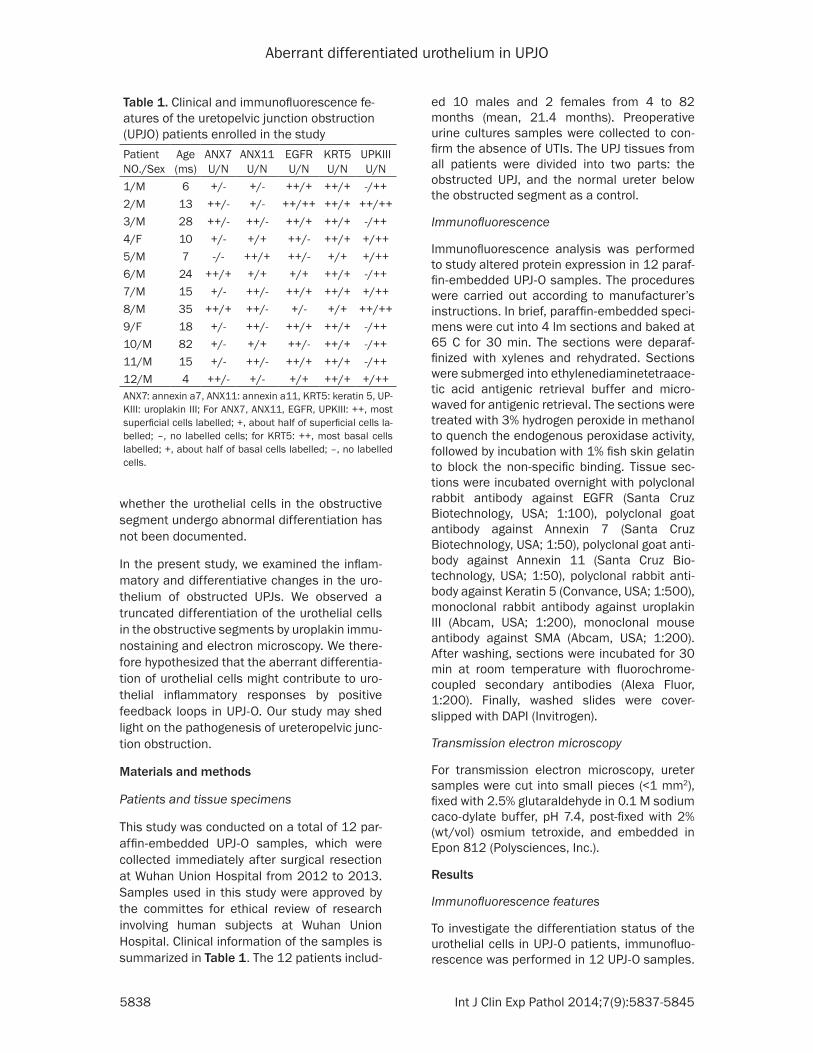
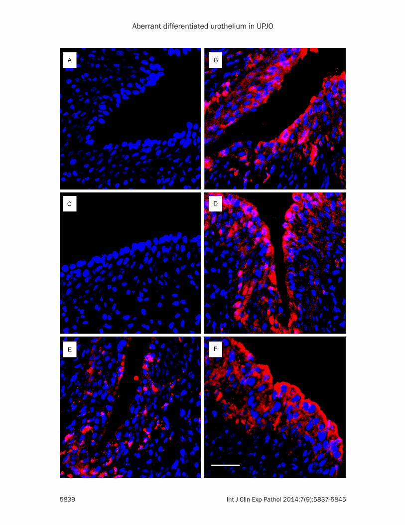
Figure 1. Representative images of Annexin 7 (A, B), Annexin 11 (C, D), and EGFR (E, F) from immunohistochemistry assays in the urothelium of obstructed UPJ segment (A, C, E) and of the normal ureter below the obstructed segment (B, D, F). Bar, 50 um.
Aberrant differentiated urothelium in UPJO
5841 Int J Clin Exp Pathol 2014;7(9):5837-5845
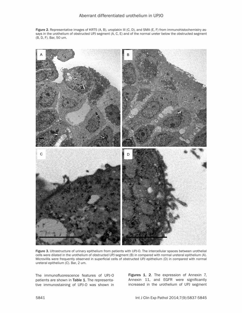
Figure 2. Representative images of KRT5 (A, B), uroplakin III (C, D), and SMA (E, F) from immunohistochemistry as-says in the urothelium of obstructed UPJ segment (A, C, E) and of the normal ureter below the obstructed segment (B, D, F). Bar, 50 um.
Figure 3. Ultrastructure of urinary epithelium from patients with UPJ-O. The intercellular spaces between urothelial cells were dilated in the urothelium of obstructed UPJ segment (B) in compared with normal ureteral epithelium (A). Microvillis were frequently observed in superficial cells of obstructed UPJ epithelium (D) in compared with normal ureteral epithelium (C). Bar, 2 um.
The immunofluorescence features of UPJ-O patients are shown in Table 1. The representa-tive immunostaining of UPJ-O was shown in
Figures 1, 2. The expression of Annexin 7, Annexin 11, and EGFR were significantly increased in the urothelium of UPJ segment
Aberrant differentiated urothelium in UPJO
5842 Int J Clin Exp Pathol 2014;7(9):5837-5845
compared with that of the paired normal ureter (Figure 1A-F). Keratin 5 was strongly expressed in the basal and intermediate layer of UPJ epi-thelium, while it was diffusely expressed in the basal layer of normal ureteral epithelium (Figure 2A, 2B). The superficial urothelial cells or normal ureter showed uroplakin staining, whereas loss of expression was observed in UPJ urothelium (Figure 2C, 2D). Expression of SMA in mesenchymal layers of ureter was ober-served to increase in UPJ segment in compared to normal ureter (Figure 2E, 2F).
Abnormal urothelial apical surface in UPJ-O ureter
The ureteral epithelium of patients with UPJ-O frequently showed dilatations of intercellular spaces that connected the neighbouring cells (Figure 3A, 3B). Moreover, the UPJ-O patients showed numerous relatively undifferentiated superficial cells covered with microvilli, which were not frequently observed in normal termi-nally differentiated cells in the ureter below the obstructed segment (Figure 3C, 3D).
Discussion
At present, the role of urothelial inflammatry in the development of UPJ-O is unclear. In this study, we report that Annexin A7 (ANX7), Annexin A11 (ANX11), EGFR, Keratin 5 (KRT5), and smooth muscle antigen (SMA) were overex-pressed, while the expression of uroplakin III was decreased in the urothelium of UPJ-O patients. Moreover, ultrastructural analyses showed dilated intercellular spaces and
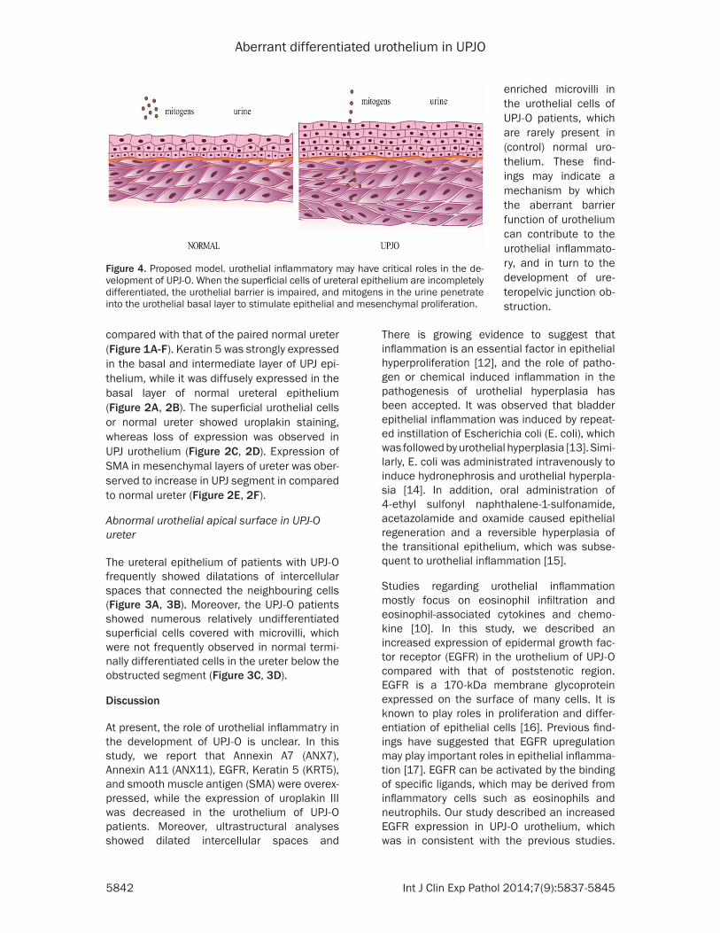
Figure 4. Proposed model. urothelial inflammatory may have critical roles in the de-velopment of UPJ-O. When the superficial cells of ureteral epithelium are incompletely differentiated, the urothelial barrier is impaired, and mitogens in the urine penetrate into the urothelial basal layer to stimulate epithelial and mesenchymal proliferation.
enriched microvilli in the urothelial cells of UPJ-O patients, which are rarely present in (control) normal uro-thelium. These find-ings may indicate a mechanism by which the aberrant barrier function of urothelium can contribute to the urothelial inflammato-ry, and in turn to the development of ure-teropelvic junction ob- struction.
There is growing evidence to suggest that inflammation is an essential factor in epithelial hyperproliferation [12], and the role of patho-gen or chemical induced inflammation in the pathogenesis of urothelial hyperplasia has been accepted. It was observed that bladder epithelial inflammation was induced by repeat-ed instillation of Escherichia coli (E. coli), which was followed by urothelial hyperplasia [13]. Simi- larly, E. coli was administrated intravenously to induce hydronephrosis and urothelial hyperpla-sia [14]. In addition, oral administration of 4-ethyl sulfonyl naphthalene-1-sulfonamide, acetazolamide and oxamide caused epithelial regeneration and a reversible hyperplasia of the transitional epithelium, which was subse-quent to urothelial inflammation [15].
Studies regarding urothelial inflammation mostly focus on eosinophil infiltration and eosinophil-associated cytokines and chemo-kine [10]. In this study, we described an increased expression of epidermal growth fac-tor receptor (EGFR) in the urothelium of UPJ-O compared with that of poststenotic region. EGFR is a 170-kDa membrane glycoprotein expressed on the surface of many cells. It is known to play roles in proliferation and differ-entiation of epithelial cells [16]. Previous find-ings have suggested that EGFR upregulation may play important roles in epithelial inflamma-tion [17]. EGFR can be activated by the binding of specific ligands, which may be derived from inflammatory cells such as eosinophils and neutrophils. Our study described an increased EGFR expression in UPJ-O urothelium, which was in consistent with the previous studies.
Aberrant differentiated urothelium in UPJO
5843 Int J Clin Exp Pathol 2014;7(9):5837-5845
Although several EGFR ligands have been found to be increased in UPJ-O, and the increase has been thought to be responsible for the patho-genesis of UPJ-O [18], the exact role and impor-tance of EGFR and its ligands in modulating the urothelial inflammation need to be explored.
Annexins are a unique class of proteins that provide a link between Ca2+ signaling and membrane functions and that have pivotal roles in the regulation of many cellular process-es in all eukaryotic organisms. Annexin A7, the first annexin to be described, was described in regulation of cell growth and differentiation. The ANX7 gene acts as a tumor suppressor. Expression studies demonstrated that ANX7 is highly correlated with late stage of prostate cancer and with poor cellular differentiation of gastric cancer [19]. Whether ANX7 is involved in the pathogenesis of the urothelial infl- ammatory process need further investigation.
The calcium-dependent phospholipid-binding protein annexin ANX11 is expressed in a wide range of tissues. Anti-ANX11 antibodies have been found in association with a number of chronic inflammatory diseases including Sjogren syndrome, polymyositis, and rheuma-toid arthritis [20]. A recent study has implicated annexin ANX11 as a highly associated suscep-tibility locus for sarcoidosis, which is a systemic immune disorder characterized by abnormal collections of chronic inflammatory cells form as nodules in various organs [21]. In addition, ANX11 is likely to play an important role in the regulation of cell apoptosis, proliferation and differentiation [22]. We have demonstrated a significant increase in ANX7 and ANX11 expres-sion in the urothelium of UPJ-O segment, which indicates their involvement in the pathogenesis of UPJ-O. In addition, Annexin A1 and A6 are thought to be important regulators of EGFR sig-naling pathway in the regulation of critical phys-iological processes including proliferation, dif-ferentiation, inflammation and cell migration [23]. It would be interesting to investigate whether ANX7 and ANX11 participate in the crosstalk with the EGFR signaling pathway in the future.
The intermediate filament protein KRT5 is a basal cell marker of keratinocytes. It is expressed in mitotically active keratinocytes of all stratified squamous epithelium. Normally it is expressed at a low level in normal urotheli-
um, while it is upregulated in hyperplastic uro-thelium [24]. We observed an appreciable increase of KRT5 expression in both basal and intermediate layers of urothelium in UPJ-O seg-ments, which indicates that the urothelium of UPJ-O segments exhibited substantial hyper-plasia. In the light of the above information, it’s possible that urothelial hyperproliferation in the UPJ-O segment may arise from urotheial inflammatory reactions regulated by interacting proteins such as annexins and EGFR ligands.
The major function of urothelium is to provide an impermeable barrier, which is maintained in part by terminally differentiated umbrella cells in the outermost layer. No relationship between the pathogenesis of the obstructed portion in UPJ-O and urothelial differentiation has been shown in the literature previously. In this study, we found that patients with UPJ-O had only a few of their superficial urothelial cells stained positively with uroplakins antibodies, suggest-ing a significant defect in urothelial differentia-tion. Moreover, we found that increased inter-cellular spaces and microvilli-enriched superfi-cial cells in UPJ-O epithelium, indicating a defective urothelial barrier. Our study was sup-ported by Romih et al., who proposed that the exposure of undifferentiated cells to the lumi-nal surface may contribute to defective urothe-lial permeability [25]. It is possible that the high concentrations of EGF-related mitogens in the urine penetrate into the urothelial basal layer to stimulate cell proliferation [26]. Besides, the fully assembled urothelial plaques that are known to trigger certain growth-inhibiting sig-nals are now absent in the uroplakin-deficient urothelium, which may lead to urothelial hyper-plasia as well. Thus, we speculate that in patients with UPJ-O the barrier is disrupted, fol-lowed by a inflammatory response resulting in urothelial hyperproliferation and incompletely differentiated superficial cells, which amplify the urothelial inflammatory response in a posi-tive feedback loop (Figure 4).
Although urothelial layers are seperated from stroma by basement membrane which directly contact them, urothelial dynamics are strictly connected to the underlying stromal phenome-na. Early study showed that urotheial-mesen-chymal interactions are necessary in the devel-opment of bladder [27]. It has also been sug-gested that urotheial-mesenchymal interac-tions may be important in ureteric bud morpho-
Aberrant differentiated urothelium in UPJO
5844 Int J Clin Exp Pathol 2014;7(9):5837-5845
genesis and collagen synthesis, as well as in smooth muscle proliferation [28]. Urothelium-derived factors or signals were presumed to play vital roles in mediating detrusor smooth muscle contractility [29], and in promoting mesenchymal proliferation and smooth muscle differentiation [2]. In contrast, urothelial prolif-eration and differentiation might also be regu-lated by stromal cells-derived factors [30]. In the present study, we also observed that SMA was overexpressed in UPJ-O patients. Taken together, we speculate that the urothelial-mes-enchymal interactions become active and trig-ger stromal remodeling and smooth muscle proliferation in the pathogenesis of UPJ-O.
In conclusion, we describe a previously unre-ported association of abnormal differentiation of urothelial cells and UPJ-O. Further studies are necessary to define the biological process-es and the molecular mechanisms responsible for this pathology. Our finding may shed lights on a better understanding of the pathology of UPJ-O.
Disclosure of conflict of interest
None.
Address correspondence to: Xiaomin Han, Depar- tment of Urology, Union Hospital, Tongji Medical College of Huazhong University of Science and Technology, Wuhan 430022, China. Tel: +86 27-8575-1625; Fax: +86 27-8577-6343; E-mail: [email protected]
References
[1] Wang Y, Puri P, Hassan J, Miyakita H and Reen DJ. Abnormal innervation and altered nerve growth factor messenger ribonucleic acid ex-pression in ureteropelvic junction obstruction. J Urol 1995; 154: 679-683.
[2] Yiee JH, Johnson-Welch S, Baker LA and Wilcox DT. Histologic differences between extrinsic and intrinsic ureteropelvic junction obstruc-tion. Urology 2010; 76: 181-184.
[3] Kaya C, Bogaert G, de Ridder D, Schwentner C, Fritsch H, Oswald J and Radmayr C. Extracellular matrix degradation and reduced neural densi-ty in children with intrinsic ureteropelvic junc-tion obstruction. Urology 2010; 76: 185-189.
[4] Ruiz-Deya G, Sikka SC, Thomas R and Abdel-Mageed AB. Potential role for the nuclear tran-scription factor NF-kappa B in the pathogene-sis of ureteropelvic junction obstruction. J Endourol 2002; 16: 611-615.
[5] Solari V, Piotrowska AP and Puri P. Altered ex-pression of interstitial cells of Cajal in congeni-
tal ureteropelvic junction obstruction. J Urol 2003; 170: 2420-2422.
[6] Kajbafzadeh AM, Payabvash S, Salmasi AH, Monajemzadeh M and Tavangar SM. Smooth muscle cell apoptosis and defective neural de-velopment in congenital ureteropelvic junction obstruction. J Urol 2006; 176: 718-723; dis-cussion 723.
[7] Murakumo M, Nonomura K, Yamashita T, Ushiki T, Abe K and Koyanagi T. Structural changes of collagen components and diminu-tion of nerves in congenital ureteropelvic junc-tion obstruction. J Urol 1997; 157: 1963-1968.
[8] Starr NT, Maizels M, Chou P, Brannigan R and Shapiro E. Microanatomy and morphometry of the hydronephrotic “obstructed” renal pelvis in asymptomatic infants. J Urol 1992; 148: 519-524.
[9] Tadros Y, Ruiz-Deya G, Crawford BE, Thomas R and Abdel-Mageed AB. In vivo proteomic analy-sis of cytokine expression in laser capture-mi-crodissected urothelial cells of obstructed ure-teropelvic junction procured by laparoscopic dismembered pyeloplasty. J Endourol 2003; 17: 333-336.
[10] Chiou YY, Shieh CC and Cheng HL. In situ ex-pression of T-helper type 2 immunoregulatory response in myofibroblast of a pediatric blad-der inflammatory pseudotumor. Pathol Int 2006; 56: 154-157.
[11] Takeyama J and Sakai K. Mucosal abnormali-ties in congenital ureteropelvic junction ob-struction. Histopathology 2007; 51: 716-717.
[12] Elkahwaji JE, Zhong W, Hopkins WJ and Bushman W. Chronic bacterial infection and inflammation incite reactive hyperplasia in a mouse model of chronic prostatitis. Prostate 2007; 67: 14-21.
[13] Uchida K, Samma S, Rinsho K, Warren JR and Oyasu R. Stimulation of epithelial hyperplasia in rat urinary bladder by Escherichia coli cysti-tis. J Urol 1989; 142: 1122-1126.
[14] Baumgart P, Muller KM and Lison AE. Epithelial abnormalities in the renal pelvis in experimen-tal hydronephrosis and pyelonephritis. Pathol Res Pract 1983; 176: 185-195.
[15] Frith CH, West RW, Stanley JW and Jackson CD. Urothelial lesions in mice given 4-ethylsulfonyl-naphthalene-1-sulfonamide, acetazolamide, and oxamide. J Environ Pathol Toxicol Oncol 1984; 5: 25-38.
[16] Pawankar R, Holgate ST and Rosenwasser LJ. Allergy frontiers. New York: Springer; 2009.
[17] Brandl K, Sun L, Neppl C, Siggs OM, Le Gall SM, Tomisato W, Li X, Du X, Maennel DN, Blobel CP and Beutler B. MyD88 signaling in nonhematopoietic cells protects mice against induced colitis by regulating specific EGF re-ceptor ligands. Proc Natl Acad Sci U S A 2010; 107: 19967-19972.
Aberrant differentiated urothelium in UPJO
5845 Int J Clin Exp Pathol 2014;7(9):5837-5845
[24] Sun TT. Altered phenotype of cultured urotheli-al and other stratified epithelial cells: implica-tions for wound healing. Am J Physiol Renal Physiol 2006; 291: F9-21.
[25] Romih R, Korosec P, Jezernik K, Sedmak B, Trsinar B, Deng FM, Liang FX and Sun TT. Inverse expression of uroplakins and inducible nitric oxide synthase in the urothelium of pa-tients with bladder outlet obstruction. BJU Int 2003; 91: 507-512.
[26] Kong XT, Deng FM, Hu P, Liang FX, Zhou G, Auerbach AB, Genieser N, Nelson PK, Robbins ES, Shapiro E, Kachar B and Sun TT. Roles of uroplakins in plaque formation, umbrella cell enlargement, and urinary tract diseases. J Cell Biol 2004; 167: 1195-1204.
[27] DiSandro MJ, Li Y, Baskin LS, Hayward S and Cunha G. Mesenchymal-epithelial interactions in bladder smooth muscle development: epi-thelial specificity. J Urol 1998; 160: 1040-1046; discussion 1079.
[28] Chiou YY, Shieh CC, Cheng HL and Tang MJ. Intrinsic expression of Th2 cytokines in urothe-lium of congenital ureteropelvic junction ob-struction. Kidney Int 2005; 67: 638-646.
[29] Santoso AG, Sonarno IA, Arsad NA and Liang W. The role of the urothelium and ATP in medi-ating detrusor smooth muscle contractility. Urology 2010; 76: 1267.
[30] Mudge CS and Klumpp DJ. Induction of the urothelial differentiation program in the ab-sence of stromal cues. J Urol 2005; 174: 380-385.
[18] Ozel SK, Emir H, Dervisoglu S, Akpolat N, Senel B, Kazez A, Soylet Y, Cetin G, Danismend N and Buyukunal SN. The roles of extracellular matrix proteins, apoptosis and c-kit positive cells in the pathogenesis of ureteropelvic junction ob-struction. J Pediatr Urol 2010; 6: 125-9.
[19] Hsu PI, Huang MS, Chen HC, Hsu PN, Lai TC, Wang JL, Lo GH, Lai KH, Tseng CJ and Hsiao M. The significance of ANXA7 expression and its correlation with poor cellular differentiation and enhanced metastatic potential of gastric cancer. J Surg Oncol 2008; 97: 609-614.
[20] Misaki Y, Van Venrooij WJ and Pruijn GJ. Prevalence and characteristics of anti-56K/annexin XI autoantibodies in systemic autoim-mune diseases. J Rheumatol 1995; 22: 97-102.
[21] Hofmann S, Franke A, Fischer A, Jacobs G, Nothnagel M, Gaede KI, Schurmann M, Muller-Quernheim J, Krawczak M, Rosenstiel P and Schreiber S. Genome-wide association study identifies ANXA11 as a new susceptibility locus for sarcoidosis. Nat Genet 2008; 40: 1103-1106.
[22] Song J, Shih Ie M, Chan DW and Zhang Z. Suppression of annexin A11 in ovarian cancer: implications in chemoresistance. Neoplasia 2009; 11: 605-614.
[23] Radke S, Austermann J, Russo-Marie F, Gerke V and Rescher U. Specific association of an-nexin 1 with plasma membrane-resident and internalized EGF receptors mediated through the protein core domain. FEBS Lett 2004; 578: 95-98.