organs composed of 2 or more tissue types. duct sweat, saliva ductless hormones epithelium?...
Post on 22-Dec-2015
222 views
TRANSCRIPT
Organs
Composed of 2 or more tissue types
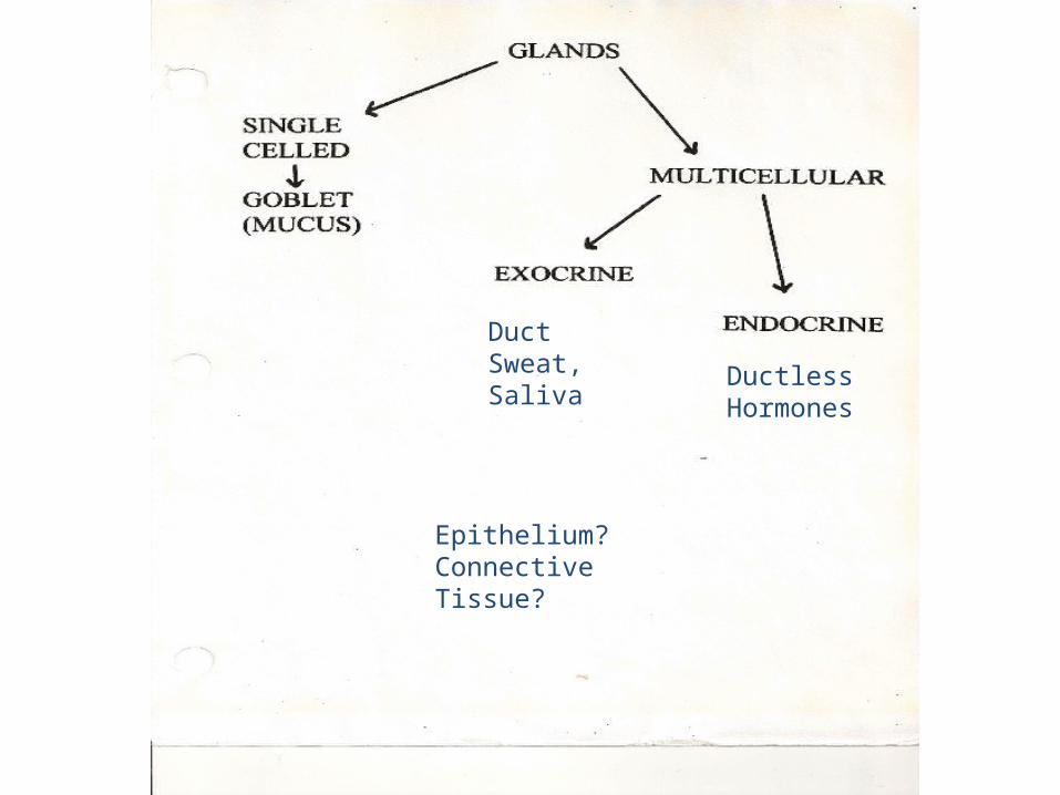
DuctSweat, Saliva Ductless
Hormones
Epithelium?Connective Tissue?
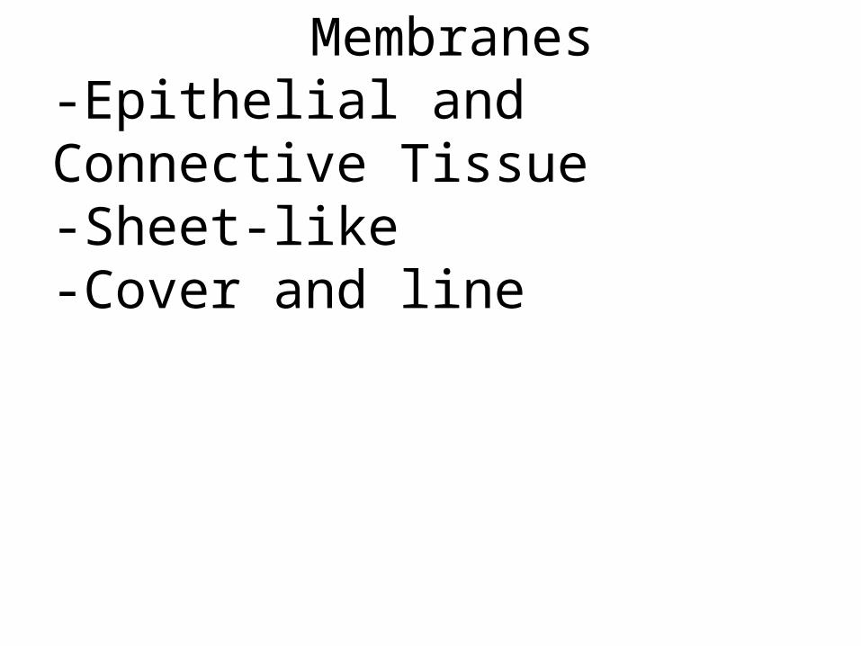
Membranes-Epithelial and Connective Tissue-Sheet-like-Cover and line

Serous Membranes: line closed cavities, covers internal organs
1. What kind of epithelium?
Mucous Membranes: Line open tracts1. What kind of epithelium?
Cutaneous Membrane (Integument, Skin): Covers the body
1. What kind of epithelium?

Serous Membranes-Covers the outside of internal organs-Lines the inside of closed cavities-Simple squamous secretes a serous fluid for lubrication
(Mesothelium)
Layer of areolar(Sub-serous Fascia)

Copyright © 2010 Pearson Education, Inc.
Figure 1.10 Serous membrane relationships.
Outer balloon wall(comparable to parietal serosa) Peripheral
Air (comparable to serous cavity)
Inner balloon wall(comparable to visceral serosa)
Heart
Parietalpericardium
Pericardialspace with serous fluid (reduces friction)
Visceralpericardium
(a) A fist thrust into a flaccid balloon demonstratesthe relationship between the parietal and visceralserous membrane layers.
(b) The serosae associated with the heart.
Pg 19
Copyright © 2010 Pearson Education, Inc.
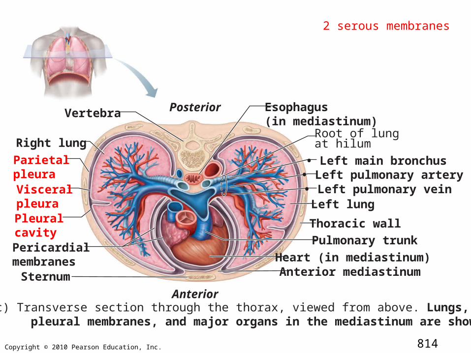
Esophagus(in mediastinum)
Right lung
Parietal pleuraVisceralpleura Pleural cavityPericardial membranesSternum
Anterior
Posterior
Root of lungat hilum
Left lung
Thoracic wall
Pulmonary trunk
Heart (in mediastinum)Anterior mediastinum
(c) Transverse section through the thorax, viewed from above. Lungs, pleural membranes, and major organs in the mediastinum are shown.
• Left main bronchus• Left pulmonary artery• Left pulmonary vein
Vertebra
814
2 serous membranes

Copyright © 2010 Pearson Education, Inc.
(d)
Pancreas
LiverLesser omentum
Stomach
Duodenum
Transversemesocolon
Greater omentumMesentery
Jejunum
Visceral peritoneum
Urinary bladder
Transverse colon
Ileum
Parietal peritoneum
Rectum889

Mucous Membranes (mucosa)-Found lining the inside of our open systems (tracts)-4 locations?-Epithelium may make mucous (lubrication, protection, absorption)
lumen
Epithelium
Basement Membrane
Lamina Propria(areolar CT)
Muscularis Mucosa ?(smooth muscle)
Copyright © 2010 Pearson Education, Inc.
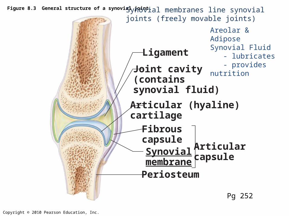
Figure 8.3 General structure of a synovial joint.
Periosteum
Ligament
FibrouscapsuleSynovialmembrane
Joint cavity(containssynovial fluid)
Articular (hyaline)cartilage
Articularcapsule
Pg 252
Areolar & AdiposeSynovial Fluid - lubricates - provides nutrition
Synovial membranes line synovial joints (freely movable joints)
Copyright © 2010 Pearson Education, Inc.
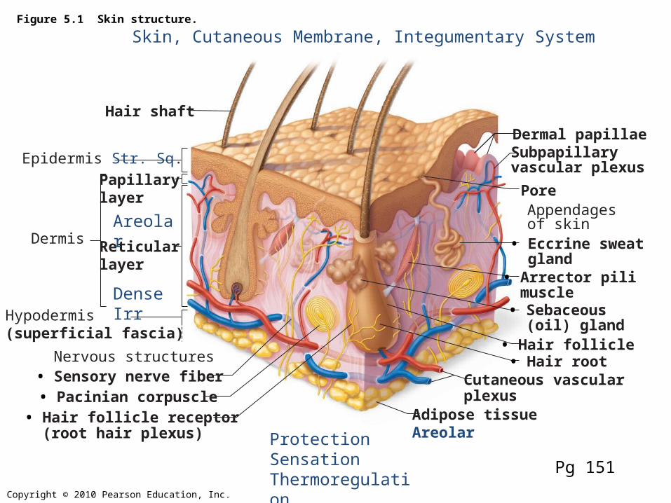
Figure 5.1 Skin structure.
Epidermis Str. Sq.
Hair shaft
Dermis Reticularlayer
Papillarylayer
Hypodermis(superficial fascia)
Dermal papillae
Pore
Subpapillaryvascular plexus
Appendagesof skin • Eccrine sweat gland• Arrector pili muscle• Sebaceous (oil) gland• Hair follicle• Hair rootNervous structures
• Sensory nerve fiber• Pacinian corpuscle• Hair follicle receptor (root hair plexus)
Cutaneous vascularplexus
Adipose tissueAreolar
Pg 151
Skin, Cutaneous Membrane, Integumentary System
Dense Irr
Areolar
ProtectionSensationThermoregulation

Copyright © 2010 Pearson Education, Inc.
Figure 5.2 The main structural features of the skin epidermis.
Melanocyte
Melanin granule
Tactile(Merkel)cellSensorynerve ending
Epidermaldendritic cell
Dermis
Dermis
Keratinocytes
Desmosomes
(b)
(a)
Stratum corneumMost superficial layer; 20–30 layers of dead cells represented only by flat membranous sacs filled with keratin. Glycolipids in extracellular space.
Stratum granulosum (3-5)Three to five layers of flattened cells, organelles deteriorating; cytoplasm full of lamellated gran-ules (release lipids) and keratohyaline granules.
Stratum spinosum (8-10)Several layers of keratinocytes unified by desmosomes. Cells contain thick bundles of intermediate filaments made of pre-keratin.
Stratum basaleDeepest epidermal layer; one row of actively mitotic stem cells; some newly formed cells become part of the more superficial layers. See occasional melanocytes and epidermal dendritic cells.
Pg 153
Stratum Lucidum?
Stratum Germinativum
Langerhan’s Cell

Copyright © 2010 Pearson Education, Inc.
Figure 5.1 Skin structure.
Epidermis Str. Sq.
Hair shaft
Dermis Reticularlayer
Papillarylayer
Hypodermis(superficial fascia)
Dermal papillae
Pore
Subpapillaryvascular plexus
Appendagesof skin • Eccrine sweat gland• Arrector pili muscle• Sebaceous (oil) gland• Hair follicle• Hair rootNervous structures
• Sensory nerve fiber• Pacinian corpuscle• Hair follicle receptor (root hair plexus)
Cutaneous vascularplexus
Adipose tissueAreolar
Pg 151
Skin, Cutaneous Membrane, Integumentary System
Dense Irr
Areolar
ProtectionSensationThermoregulation

Copyright © 2010 Pearson Education, Inc.
Figure 5.3 The two regions of the dermis.
Dermis
(a) Light micrograph of thick skin identifying the extent of the dermis, (50x)
(b) Papillary layer of dermis, SEM (22,700x)
(c) Reticular layer of dermis, SEM (38,500x)
FingerprintsAreolarVessels, receptors
80%Dense Irr.Tension linesStrength, binds waterSkin
Superficial FasciaDeep Fascia
154
© 2013 Pearson Education, Inc.
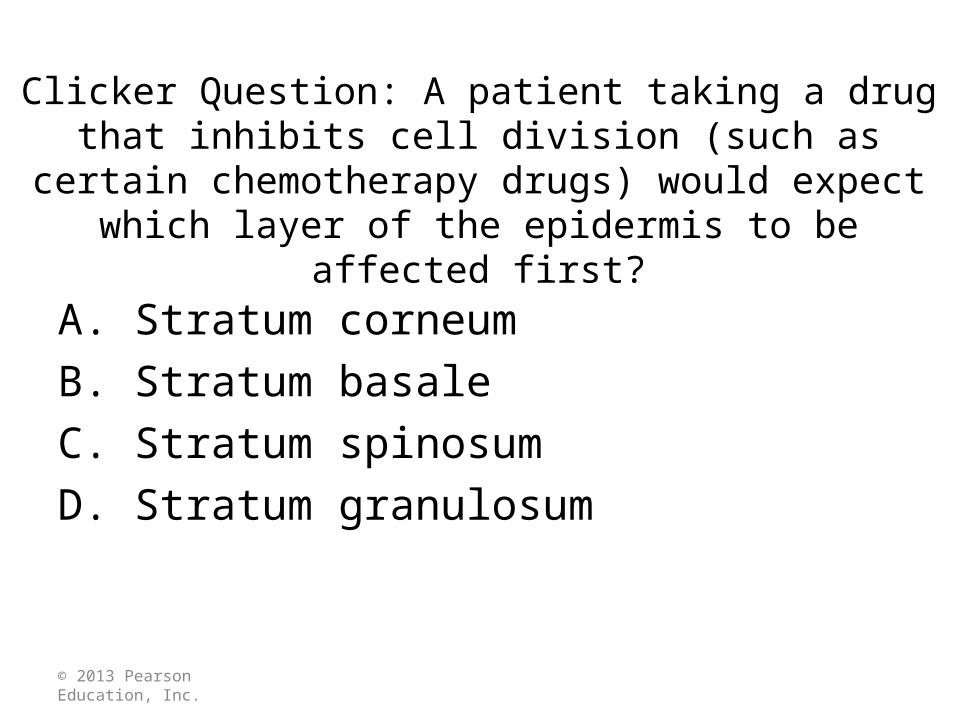
Clicker Question: A patient taking a drug that inhibits cell division (such as certain chemotherapy drugs) would expect
which layer of the epidermis to be affected first?
A. Stratum corneumB. Stratum basaleC. Stratum spinosumD. Stratum granulosum