epithelial tissue (epithelium) - masarykova univerzita · epithelial tissue (epithelium) general...
TRANSCRIPT

Epithelial tissue (epithelium)
General characteristics of epithelium
Is avascular tissue (without blood supply – cells receive nourishment by
diffusion from a highly vascular area of loose connective tissue just below
the basement membrane called the lamina propria )
is highly cellular tissue – cells are arranged to form cohesive sheet or
groups with no or little extracellular matrix
displays a free surface – usualy luminal surface (turned to the lumen)
opposite (basal) surface adheres to extracellular basement membrane or
lamina basalis
epithelial cells display polarity – apical (luminal), lateral and basal
surfaces with structural specialization
epithelial cells are specialised for absorption, secretion or to act as barrier
lateral surfaces display junctional complexes for intercellular cohesion
and communication
One type of epithelium may change into another type – metaplasia (examples:
pseudostratified ep. of respiratory passages transforms into stratified squamous
ep. on the surface of epiglottis and soft palate)
Membrane specializations of epithelia Lateral surface
Specialised structures are present in epithelia which link individual cells together. Two
main adhesion types are distinguished:
1. Cell membrane proteins acting as specialised cell adhesion molecules (CAMs)
2. Specialised areas of the cell membrane incorporated into cell junctions.
Three types are recognized: occluding junctions, anchoring or adherence junctions and
communicating junctions.
o Occluding junctions bind cell together to form an impermeable barrier
Zonula occludens or tight junction
o Anchoring junctions link the cytoskeleton of cells to each other and two
underlying tissues
Zonula adherens provides mechanical strength
Macula adherens or desmosomes provides mechanical strength in
tissues where there are tensile or shearing stresses, eg skin
o Communications junctions allow direct cell-cell communication
Gap junction or nexus allow rapid communication for
coordinated action
Luminal (free, apical) surface
Microvilli – short finger-like projection of the cell membrane to increased
surface area (regularly arranged microvilli in intestines – striated border, in
kidney tubules – brush border)
Cilia – hair-like surface projections of cells involved in transport
Glycocalyx – thin extracellular layer consisting of protein glycoprotein and
sugar residues; stains PAS positive; can act as enzyme, CAM or for cell
recognition
Basal surface
Basal invaginations or folds – greatly enhance surface area; folded membrane with
ions pumps + mitochondria form basal labyrinth in kidney tubules.
Basal lamina – basement membrane
Epithelial tissues are physically separated from underlying connective tissues by
a basement membrane or basal lamina. The portion of an epithelial cell
attached to the basement membrane is called its basal surface. The opposite side
- facing the external environment, or lumen of a body cavity, is its apical
surface. Basement membranes are composed of a special type of collagen and a
substance called laminin (see below). The basement membrane helps epithelial
cells orient themselves in relation to other tissues. After epithelial injury (e.g., an
abrasion), the basement membrane serves as a scaffolding upon which new cells
attach themselves during healing.
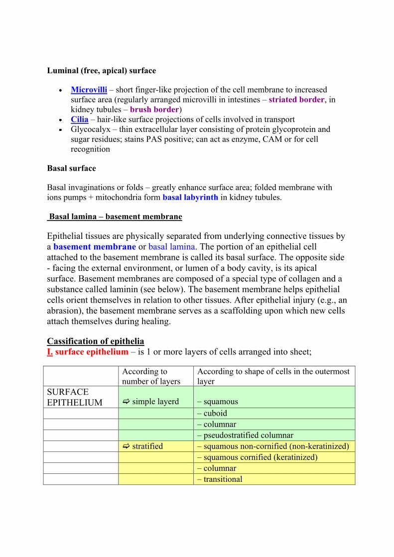
Cassification of epithelia
I. surface epithelium – is 1 or more layers of cells arranged into sheet;
According to
number of layers
According to shape of cells in the outermost
layer
SURFACE
EPITHELIUM
simple layerd
– squamous
– cuboid
– columnar
– pseudostratified columnar
stratified – squamous non-cornified (non-keratinized)
– squamous cornified (keratinized)
– columnar
– transitional
SIMPLE EPITHELIA – only 1 (single) layer on basement membrane
Squamous – single layer of flattened thin cells with little cytoplasm and prominent nucleus.
(In the smallest tubules and ducts of different organs, Henle´s loop or Bowman´s capsule in
kidney) Endothelium – squamous epithelium in
cardiovascular system. Mesothelium - squamous epithelium of mesodermal origin lining
serous membranes and cavities.
Cuboidal - cell height, width and depth are the same, round centrally placed nucleus.
(In renal tubules and small glanular ducts)
Columnar - cell height greater than width, nucleus elliptical or cigar shaped.
(In the intestines, in the oviduct)
Pseudostratified – single layer but nuclei situated at different levels in the cell. All cells are in
contact with the basement membrane, but not all cells reach the apical surface. Both
conditions create the illusion of several cell layers. (In the respiratory passages – nasal cavity,
larynx, trachea, bronchi)
STRATIFIED EPITHELIUM – consists of basal layer on basement membrane,
several layers of polyhedral cells and surface layer. According to the shape of
cells in this layer the epithelium is named (squamous, columnar, transitional)
Stratified squamous – resiting to mechanical influences (press)
– non-keratinised (mouth cavity, vocal cords, vagina, anus)
– keratinised (epidermis of the skin) – the cells are released continously from the surface
Stratified columnar – 2 or more layers of cells, columnar cells form the upper layer (two-
layered in ductus epididymis and ductus deferens, more-layered male urethra, conjunctive)
Transitional - stratified, top layer dome or umbrella shaped. (only in some urinary passages –
renal pelvis, ureter and urinary bladder). Epithelium change the shape of cells and number of
layers according to wall conditions of urinary passages – distansion or contraction

Epithelia with special functions:
resorptive, sensory, respiratory, myoepithelial cells
glandular epithelium – multicellular epithelial structures that specialize in
synthesizing and secreting complex molecules.
CLASSIFICATION OF GLANDS
GLANDULAR
EPITHELIUM
unicellular Single cells in coverinrg epithelium –
(Paneth cells, goblet cells,
enteroendocrine cells, Leydig cells)
multicellular Accordin of mechanism of secretion
endocrine
exocrine – merocrine, apokrine,holocrine
According to loclalization
intraepithelial extraepithelial
According to arrangement of ducts
simple branched compound
According to type of secretory portions
tubular alveolar (acinar) tuboalveolar
According to product properties
mucous serous mixed
According of mechanism of secretion
endocrine – glands withou ducts, product is released into the blood through the
wall of capilareis
exocrine – secretory cells of exocrine glands release their products into ducts in
three different ways:
merocrine apocrine holocrine
- membrane-bound secretory granules are moved to the apical surface where they coalesce with the membrane on to release the product.
- the apical portions of cells are pinched off and lost during the secretory process.
- secretory cell degenerates and as it breaks apart, the contents of the cell become the secretory product.
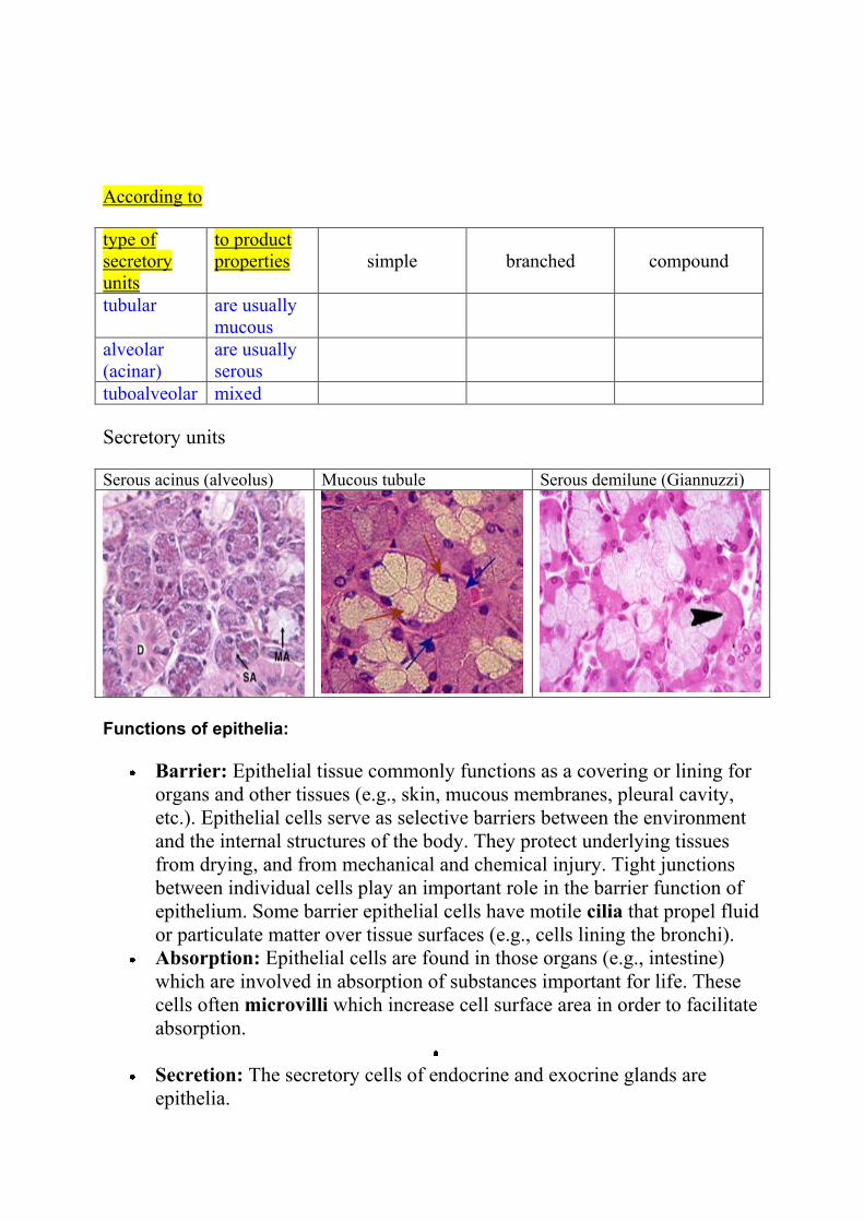
According to
type of
secretory
units
to product
properties
simple
branched
compound
tubular are usually
mucous
alveolar
(acinar)
are usually
serous
tuboalveolar mixed
Secretory units
Serous acinus (alveolus) Mucous tubule Serous demilune (Giannuzzi)
Functions of epithelia:
Barrier: Epithelial tissue commonly functions as a covering or lining for
organs and other tissues (e.g., skin, mucous membranes, pleural cavity,
etc.). Epithelial cells serve as selective barriers between the environment
and the internal structures of the body. They protect underlying tissues
from drying, and from mechanical and chemical injury. Tight junctions
between individual cells play an important role in the barrier function of
epithelium. Some barrier epithelial cells have motile cilia that propel fluid
or particulate matter over tissue surfaces (e.g., cells lining the bronchi).
Absorption: Epithelial cells are found in those organs (e.g., intestine)
which are involved in absorption of substances important for life. These
cells often microvilli which increase cell surface area in order to facilitate
absorption.
Secretion: The secretory cells of endocrine and exocrine glands are
epithelia.
Sensory: Many of the more complex sensory receptors of the nervous
system are derived from specialized epithelia called neuroepithelia (e.g.,
the rods and cones of the retina, olfactory receptors of the nose, taste
receptors on the tongue, etc.). Sensory receptors function by converting
mechanical, chemical, or electromagnetic signals from the environment
into nerve impulses which can be processed by the nervous system.
Contractility: Some very specialized epithelial cells (myoepithelia)
contain the contractile proteins myosin and actin similar to muscle.
Myoepithelia are associated with the ducts of sweat, salivary, lacrimal,
amd mammary glands and assist in the secretory process.
Origin: Epithelial tissues are derived from all three primary germ
cell layers.
Ectoderm: The epithelial cells of the skin and oral cavity (epidermis) are
derived from ectoderm. Epithelial cells covering the cornea and lens, as
well as sensory receptors of the eyes, ears, and nose, are also ectodermal
in origin.
Mesoderm: The epithelial lining of blood vessels (endothelium) is
derived from mesoderm. The epithelial lining of the pleural and peritoneal
cavities (mesothelium) also originate from mesodermal cells.
Endoderm: The epithelial lining of the respiratory system and digestive
tracts - as well as the functional cells (parenchyma) of the liver, pancreas,
gallbladder, thyroid, and parathyroid, are derived from endoderm.
Muscle Tissue
Muscle tissue is characterized on the basis of a functional property:
- the ability of muscle cells to contract.
- the bulk of the cytoplasmic volume consists of the contractile protein
filaments actin and myosin.
- muscle is responsible for movement of the body and changes in the size
and shape of internal organs.
Three types of muscle tissue can be identified histologically:
- skeletal muscle
- cardiac muscle
- smooth muscle
The fibres of skeletal muscle and cardiomyocytes (cells of cardiac muscle)
exhibit cross striations at the light microscope level and they are both referred to
as striated muscle.
Terms:
Plasmamembrane of muscle cells – sarcolemma
Cytoplasm of muscle cells – sarcoplasm
Smooth endoplasmic reticulum – sarcoplasmic reticulum
Muscles consist of muscle cells and connective tissue:
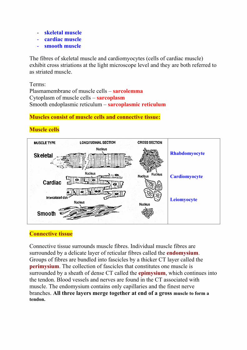
Muscle cells
Rhabdomyocyte
Cardiomyocyte
Leiomyocyte
Connective tissue
Connective tissue surrounds muscle fibres. Individual muscle fibres are
surrounded by a delicate layer of reticular fibres called the endomysium.
Groups of fibres are bundled into fascicles by a thicker CT layer called the
perimysium. The collection of fascicles that constitutes one muscle is
surrounded by a sheath of dense CT called the epimysium, which continues into
the tendon. Blood vessels and nerves are found in the CT associated with
muscle. The endomysium contains only capillaries and the finest nerve
branches. All three layers merge together at end of a gross muscle to form a
tendon.
Three basic layers
o endomysium - surrounds each cell (fiber)
o perimysium - surrounds a group of cells forms a fascicle
o epimysium - surrounds entire gross muscle
Endomysium
o thin delicate connective tissue
o blends with muscle cell membrane
Perimysium
o ordinary loose connective tissue
o divides groups of muscle cells into bundles within the gross
muscle
Epimysium
o capsule = dense irregular connective tissue
I. Skeletal Muscle
General Features
Called skeletal muscles or somatic (body) musculature
Rapid contractions
Voluntary innervation
Striations visible with light microscope (LM)
Skeletal muscle is attached to the skeleton and controls motor movements and posture. There
are a few instances where this type of muscle is restricted to soft tissues: the tongue, pharynx,
diaphragm and upper part of the esophagus.
Skeletal muscle fiber structure
Striated skeletal muscle cells = muscle fibers = a multinucleated syncytium
formed by the fusion of individual small muscle cells precursors – myoblasts,
during development.
Striated skeletal muscle cell = fiber (syncytium)
Cylindrical with tapered ends
Length – several cm, thickness - about 10-100 µm in diameter
Multinucleated – nuclei are located peripherally, immediately under
the plasma membrane (sarcolemma)
Striations - alternating light and dark bands - visible with LM
A part of sarcoplasm of muscle fiber:
Sarcolemma with invaginations: T-tubules.
Sarcoplasm: nuclei, small GA, mitochondria, glycogen; sarcoplasmic reticulum
network surrounds myofibrils and arround Z-lines forms terminal cisternae.
T-tubule + 2 terminal cisternae = TRIAD at the level of Z-line
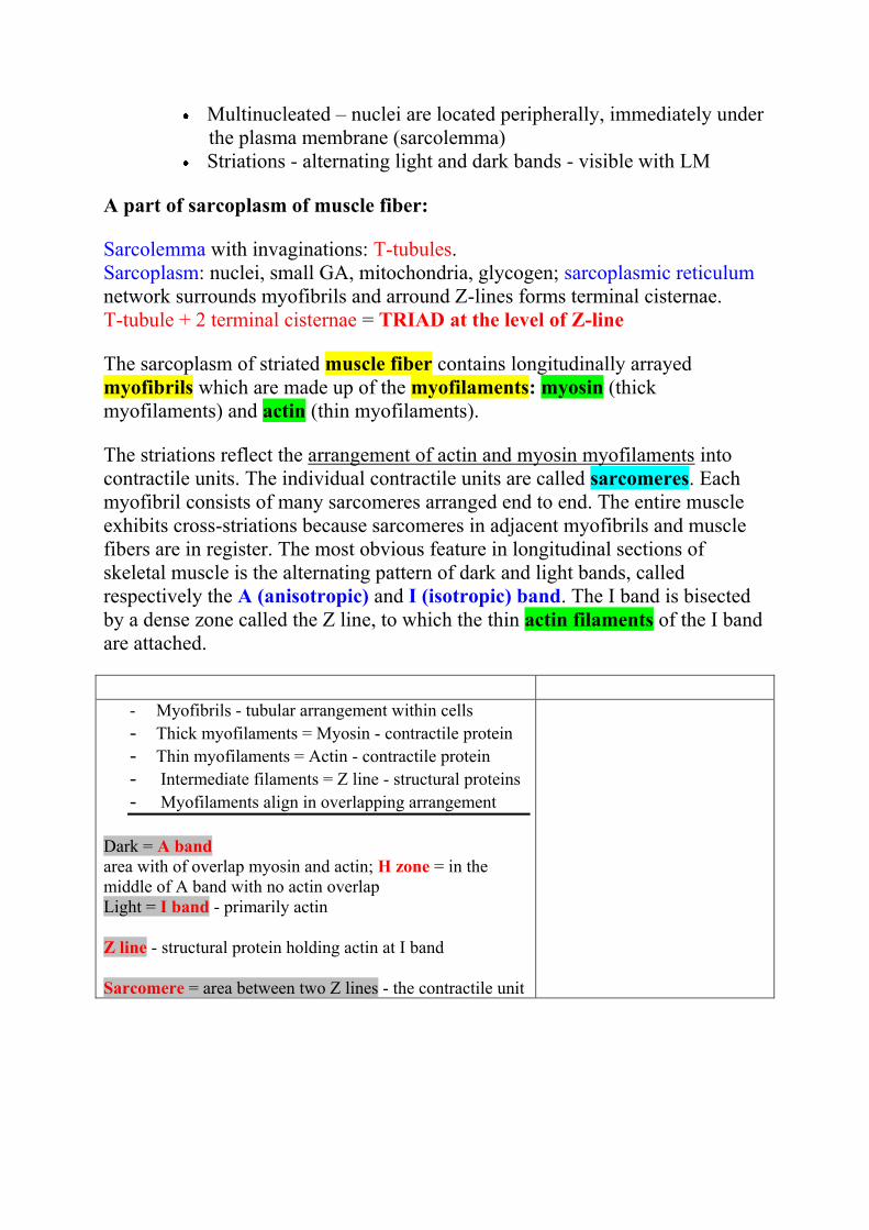
The sarcoplasm of striated muscle fiber contains longitudinally arrayed
myofibrils which are made up of the myofilaments: myosin (thick
myofilaments) and actin (thin myofilaments).
The striations reflect the arrangement of actin and myosin myofilaments into
contractile units. The individual contractile units are called sarcomeres. Each
myofibril consists of many sarcomeres arranged end to end. The entire muscle
exhibits cross-striations because sarcomeres in adjacent myofibrils and muscle
fibers are in register. The most obvious feature in longitudinal sections of
skeletal muscle is the alternating pattern of dark and light bands, called
respectively the A (anisotropic) and I (isotropic) band. The I band is bisected
by a dense zone called the Z line, to which the thin actin filaments of the I band
are attached.
- Myofibrils - tubular arrangement within cells
- Thick myofilaments = Myosin - contractile protein - Thin myofilaments = Actin - contractile protein - Intermediate filaments = Z line - structural proteins - Myofilaments align in overlapping arrangement
Dark = A band
area with of overlap myosin and actin; H zone = in the
middle of A band with no actin overlap
Light = I band - primarily actin
Z line - structural protein holding actin at I band
Sarcomere = area between two Z lines - the contractile unit
II. Cardiacl Muscle
General Features
Wall of heart
Contracts rapidly
Autonomic (involuntary) innervation - cardiac muscle is regulated
by autonomic and hormonal stimuli
Lacks residual stem cells and therefore cannot regenerate after
damage
Cardiac muscle exhibits striations because it also has actin and
myosin myofilaments arranged into sarcomeres. Generally these
striations do not appear as well-defined as in skeletal muscle.
Cardiac muscle cells = cardiomyocytes
- Cells are columnar, the end(s) can be branched
-Cells are end-to-end arranged into fibers
- Fibers branch and anastomose
- Moderate length, about 100 µm
- Moderate diameter, 10-50 µm
- Striation is present and visible with LM
- Single nucleus (rarely 2 nuclei) - centrally placed
- Intercalated discs join ends of cells together –
desmosomes and gap junctions (nexuses)
A number of features distinguish cardiac from skeletal muscle:
- cardiac muscle cells have only one or two nuclei, which are centrally located
- myofibrils separate to pass around the nucleus, leaving a perinuclear clear area
- 1 T-tubule + 1 terminal cisterna of sarcoplasmic reticulum = DIAD et the level
of border between A and I band, there are no triads (1 T-tubule – 2 cisternae)
as in skeletal muscle fibers
As in skeletal muscle, individual muscle fibres are surrounded by delicate
connective tissue. Numerous capillaries of coronary circulation are found in the
connective tissue around cardiac muscle fibres.
Cardiac muscle cells are joined to one another in a linear array. The boundary
between two cells abutting one another is called an intercalated disc.
Intercalated discs consist of several types of cells junctions whose purpose is to
facilitate the passage of an electrical impulse from cell to cell and to keep the
cells bound together during constant contractile activity.
Specialized fibres, called Purkinje fibres, arise from the atrioventricular node
and travel along the interventricular septum toward the apex of the heart,
sending branches into the ventricular tissue. Purkinje fibres are of larger
diameter than ordinary cardiac fibres, with fewer myofibrils and an extensive,
well-defined clear area around the nucleus. They conduct impulses at a rate
about four times faster than that of ordinary cardiac fibres and serve to
coordinate the contraction of the atria and ventricles.
III. Smooth Muscle
General Features
Walls of hollow viscera
Contracts slowly
often prolonged sustained contractions
Autonomic (involuntary) innervation
Smooth muscle is the intrinsic muscle of the internal organs and blood vessels. It
is also found in the iris and ciliary body of the eye and associated with hair
follicles (arrector pili). No striations are present in smooth muscle due to the
different arrangement of actin and myosin filaments. Like cardiac muscle,
smooth muscle fibres are intrinsically contractile but responsive to autonomic
and hormonal stimuli. They are specialized for slow, prolonged contraction.
Each csll is fusiform in shape with a thicker central portion and tapered at both
ends. The single nucleus is located in the central part of the cell. Cells range
enormously in size, from 20 (in wall of small blood vessels) to 500 (in wall of
uterus during pregnancy) micrometers. Smooth muscle cells lie over one another
in a staggered fashion (tapered part of one cell over thicker part of another).
One distinguishing physiological feature of smooth muscle is its ability to secrete connective
tissue matrix. In the walls of blood vessels and the uterus in particular, smooth muscle fibres
secrete large amounts of collagen and elastin.
Smooth muscle cells = leiomyocytes
- Spindle shape cells - elongated and tapered
- Moderate length - about 100-200 µm
- Thin diameter - about 5-10 µm
- Single nucleus - centrally placed
- NO striations – aktin and myosin
myofilaments are not arranged into
myofibrils, aktin filaments are attached to
darc bodies (analogic to Z-lines)
- Cells are typically arranged in bundles or
sheets
- intercellular junctions: desmosomes,
nexuses
Mechanism of contraction of striated muscles
The sarcoplasmic reticulum (SR), a specialized form of the smooth
endoplasmic reticulum, is system of structurally and functionally specialized for
the rapid release of calcium ions under appropriate conditions. SR forms a
network of tubules and cisternae around the myofibrils. Flattened cisternae also
called terminal cisternae, are in contact with an invagination of the
sarcomalemma called the Transverse tubule. The T-tubule and two adjacent
terminal cisternae constitute a triad which lie over the middle of the I-band (at
the Z-line) in skeletal muscle, while T-tubule and one terminal cisterna form
diad at the border between I and A band in cardiac muscle.
The wave of depolarization conducted along the sarcolemma and to the interior
of the fiber by the T-tubule causes the SA to release calcium ions, which can
then diffuse among the myofilaments and initiate contraction by binding to
troponin. This binding then causes the tropomyosin to change its association
with actin so as to expose reactive sites capable of interacting with myosin
heads. The myosin and actin filaments of a sarcomere overlap with the same
relative polarity on either side of the midline. The actin filaments are anchored
to the Z disc and the myosin filaments are bipolar. During contraction, the actin
and myosin filaments slide past each other without shortening. The sliding
motion is driven by the myosin heads walking toward the actin filament. All
sarcomeres in myofibrils are shortened (distance between Z-lines shortens)
during contraction.
The contraction needs energy (ADP ATP, glycogen).
After contraction, the calcium moves back into the cisternae of the sarcoplasmic
reticulum. Tropomyosin then covers the reactive sites on the actin myofilaments
(blocks the actin site) and relaxation occurs.
Nervous Tissue (NT)
- highly specialized tissue
- forms, receives and sorts signals (irritability)
- transmits electrical impulses (conductivity)
Functions of Nerve Tissue
Nervous tissue allows an organism to sense stimuli in both the internal and external
environment.
The stimuli are analysed and integrated to provide appropriate, co-ordinated
responses in various organs.
The afferent or sensory neurons conduct nerve impulses from the sense organs and
receptors to the central nervous system.
Internuncial or connector neurons supply the connection between the afferent and
efferent neurons as well as different parts of the central nervous system.
Efferent or somatic motor neurons transmit the impulse from the central nervous
system to a muscle (the effector organ) which then react to the initial stimulus.
Autonomic motor or efferent neurons transmit impulses to the involuntary muscles
and glands.
- NT forms central and peripheral nerve system (CNS and PNS) - NT consists of nerve cells = NEURONS and associated supporting cells =
NEUROGLIA; neurons are specifically designed to transmit electrical impulses and
to receive and process information; neuroglial cells are non-conducting cells that are
in intimate physical contact with neurons. They provide physical support, electrical
insulation and metabolic exchange with the vascular system. - NT originates from ectoderm
NEURON
Nerve cells are very variable in appearance, shape and size, but all neurons have a cell body,
also called soma or perikarion, and processes extending from the nerve cell to communicate
with other cells. There are two types of processes: dendrites that receive impulses and axons
(neurits) that transmit impulses. All nerve cells have one axon, which is usually the longest
process that extends from the cell and one or more (hundreds) dendrites, these are generally
shorter and thicker than the axon. The junction where a nerve cell communicates with another
nerve cell or an effector cell (eg. muscle fibre) is called a synapse, which can be chemical or
electric. The terminal part of the axon with chemical synapses releases substances called a
neurotransmitter which acts on the membrane of the other cell.
Cell body – PERIKARION: contains nucleus and most cytoplasm with organelles:
- nucleus – round or oval, very light, with prominent nucleolus
- rough ER (called Nissl´ substance) – involved in synthesis of proteins
(neurotransmitters)
- other usual organelles (mitochondria, Golgi apparatus, lysosomes)
- neurofibrils – neurofilaments and neurotubules
- pigment lipofuscin
DENDRITES – input structure – receive signals; number of dendrites: one – several
hundreds
short, branched processes with structure similar to perikarion (cytoplasm + organelles
+ neurofibrils)
incoming signals summate to initiate action potential highly branched tree structure
Classification of neurons according to number of processes (dendrites):
1. Multipolar neuron – several dendrites extend from body found in brain &
spinal cord
2. Bipolar neuron – one dendrite and one axon (in retina of eye)
3. Unipolar neuron – one process only, link to axon (sensory neurons)
4. Pseudounipolar neuron – one short process divides later into dendrite and axon
(spinal ganglia)
AXON – only one
no protein synthesis here
Trigger zone - where nerve impulses arise
Axon hillock – the cone-shaped base of the axon, its cytoplasm is free of rER (Nissl
substance)
Axons terminal - end with fine branching with „terminal boutons“ – mitochondria and
synaptic vesicles containing neurotransmitters
Axon hillock and terminal are not covered with oligodendrocytes (in CNS) or
Schwann cells (in PNS)
Serves for impulses transmission and for axonal transport of neurotransmitters and
nutrients
Classification of neurons according to length of axon:
1. Golgi type I – long axon (up to 1 m) – somatic motor neurons
2. Golgi type II - short axon (in μm)
Classification of neurons according to function:
1. sensitive neurons – (afferent) conduct informations from receptors to CNS
2. motor neurons – (efferent) conduct infirmations from CNS to effector cells:
somatomotor to skeletal muscle and visceromotor to smooth muscle cells,
cardiomyocytes or glandular cells
3. interneurons (97 %)
Sheaths of axons:
Schwann sheath (neurilemma) – Schwann
cells surround the axon (gray fibers)
Myelin sheath – lipoprotein product of
Schwann cells in PNS and oligodendrocytes in
CNS
- electrically insulates axon
- inreases speed of nerve impulse
- wraps around one axon many times and has a
lamellar appearance
Many axons are wrapped in a lipid-rich covering called myelin. This myelin sheath insulates
the axon from the surrounding extracellular component and increases the rate of electrical
conduction. The myelin sheath is discontinuous at intervals called the nodes of Ranvier. The
area covered with myelin is called internodal area (internodium). In myelinated axons, the
voltage reversal (that is, the impulse propagation) can occur only at the nodes, and the
impulse "jumps" from node to node. This is called saltatory conduction. In unmyelinated
axons, the impulse is conducted more slowly, moving as a wave of voltage reversal along the
axon.
Synapses
- NEURON – NEURON
Presynaptic neuron - conducts signal to a synapse // synaptic vesicles with
neurotransmiter
Synaptic cleft (20-30 nm thick)
Postsynaptic neuron - conducts signal from a synapse // receptors on cell membrane
- Axodendritic (1)
- Axosomatic (2)
- Axoaxonal (3)
- Dendrodendritic synapses
- NEURON – EFFECTOR CELL
Effector cells – muscle cells (in smooth muscle = neuromuscular spindle, in skeletal muscle
= motor-end-plate), cardiomyocytes, glandular cells
Chemical Synapses
Presynaptic cell releases neurotransmitters from synaptic vesicles
Act on the postsynaptic cell (help initiate AP)
Neurotransmitters can excite or inhibit
Neurotransmitters (acetylcholine, serotonin, norepinepherine and epinephrin,
dopamine, GABA, …)
Neurotransmiter must be removed to prevent continual firing of neurons
Enzymatically - acetylcholineresterase
Many pharmaceuticals and drugs modulate this effect
Cocaine block removal of dopamine
Electrical Synapses
Without synaptic vesicles; synaptic cleft – only 2 nm thick
Depolarizating wave continues from presynaptic to postsynaptic membrane
Morphologically (in electron microscope) it looks like communicatin intercellular
connection: gap junction (nexus)
SUPPORT CELLS PLAY A VITAL ROLE
Support cells are essential to the function and survival of nerve cells. The CNS and PNS each
have their own specific types of support cells.
Support cells in the CNS:
The general term for support cells in the CNS is glia or neuroglia (glial cells, neuroglial
cells). There are four types of neuroglial cells. (1) Oligodendrocytes, the myelin-secreting
cells of the CNS. (2) Astrocytes, which provide physical and metabolic support for nerve
cells. (3) Microglia, (microglial cells), which are the phagocytes of the CNS. (4) Ependyma
(ependymal cells) lining brain cavities and central canal in spinal cord.
Oligodendrocytes. As their name implies, oligodendrocytes have few processes. They are
often found in rows between axons. The myelin sheath around axons is formed by concentric
layers of oligodendrocytes plasma membrane. Each oligodendrocyte gives off several tongue-
like processes that find their way to the axon, where each process wraps itself around a
portion of the axon, forming an internodal segment of myelin. Each process appears to spiral
around its segment of the axon in a centripetal manner, with the continued insinuation of the
leading edge between the inner surface of newly formed myelin and the axon. One
oligodendrocyte may myelinate one axon or several. The nucleus-containing region may be at
some distance from the axon(s) it is myelinating. In the CNS, nodes of Ranvier (between
myelinated regions) are larger than those of the PNS, and the larger amount of exposed
axolemma makes saltatory conduction more efficient.
Unmyelinated axons in the CNS are truly bare, that is they are not embedded in any glial cell
process. (In contrast to the situation in the PNS, described below.)
Astrocytes. Astrocytes are the largest of the neuroglial cells. They have elaborate processes
that extend between neurons and blood vessels. The ends of the processes expand to form end
feet, which cover large areas of the outer surface of the blood vessel or axolemma. Astrocytes
are believed to play a role in the movement of metabolites and wastes to and from neurons,
and in regulating ionic concentrations within the neurons. They may be involved in regulating
the tight junctions in the capillaries that form the blood-brain barrier. Astrocytes also cover
the bare areas of neurons, at nodes of Ranvier and synapses. They may act to confine
neurotransmitters to the synaptic cleft and to remove excess neurotransmitters.
Two kinds of astrocytes are identified, protoplasmic and fibrous astrocytes. Both types
contain prominent bundles of intermediate filaments, but the filaments are more numerous in
fibrous astrocytes. Fibrous astrocytes are more prevalent in white matter, protoplasmic ones in
grey matter.
Microglia. These are the smallest of the glial cells, with short twisted processes. They are the
phagocytes of the CNS, considered part of the mononuclear phagocytic system (see pg 110 in
Ross et al.). They are believed to originate in bone marrow and enter the CNS from the blood.
In the adult CNS, they are present only in small numbers, but proliferate and become actively
phagocytic in disease and injury. Their alternate name, mesoglia, reflects their embyonic
origin from mesoderm (the rest of the nervous system, including the other glial cells, is of
neuroectodermal or neural crest origin).
Ependymal cells. Cuboidal to columnar cells in one layer lining the fluid-filled brain
ventricles and central canal (canalis centralis) in spinal cord. Ependyma is involved in
cerebrospinal fluid production in som regions (choroid plexus).
Support cells in the PNS:
The support cells of the PNS are called satellite cells and Schwann cells.
Satellite cells. Satellite cells surround the cell bodies of the neurons in ganglia (ganglion
cells). These small cuboidal cells form a complete layer around the nerve cell body, but only
their nuclei are visible in routine preparations. They help maintain a controlled
microenvironment around the nerve cell body, providing electrical insulation and a pathway
for metabolic exchange. In paravertebral and peripheral ganglia, nerve cell processes must
penetrate between satellite cells to establish a synapse.
Satelite cells – nutrition and isolation of neurons in ganglia
Schwann cells. Schwann cells are responsible for the myelination of axons in the PNS. A
Schwann cell wraps itself, jelly roll-fashion, in a spiral around a short segment of an axon.
During the wrapping, cytoplasm is squeezed out of the Schwann cell and the leaflets of
plasma membrance of the concentric layers of the Schwann cell fuse, forming the layers of
the myelin sheath. An axon's myelin sheath is segmented because it is formed by numerous
Schwann cells arrayed in sequence along the axon. The junction where two Schwann cells
meet has no myelin and is called the node of Ranvier (the areas covered by Schwann cells
being the internodal regions).
The lack of Schwann cell cytoplasm in the concentric rings of the myelin sheath is what
makes it lipid-rich. Schwann cell cytoplasm is however found in several locations. There is an
inner collar of Schwann cell cytoplasm between the axon and the myelin, and an outer collar
around the myelin. The outer collar is also called the sheath of Schwann or neurilemma, and
contains the nucleus and most of the organelles of the Schwann cell. The node of Ranvier is
also covered with Schwann cell cytoplasm, and this is the area where the plasma membranes
of adjacent Schwann cells meet. (These adjacent plasma membranes are not tightly apposed at
the node, so that extracellular fluid has free acess to the neuronal plasma membrane.) Finally,
small islands of Schwann cell cytoplasm persist within successive layers of the myelin sheath,
these islands are called Schmidt-Lanterman clefts.
Myelination (development of myelin sheath):
Not all nerve fibres is the PNS are covered with myelin, some axons are unmyelinated. In
contrast to the situation in the CNS, unmyelinated fibres in the PNS are not completely bare,
but are enveloped in Schwann cell cytoplasm. The Schwann cells are elongated in parallel to
the long axis of the axons, which fit into grooves on the surface of the Schwann cell. One
axon or a group of axons may be enclosed in a single groove. Schwann cells may have only
one or up to twenty grooves. Single grooves are more common in the autonomic nervous
system.