organophosphate mechanism of action and … mechanism of action and potential biomarker studies...
TRANSCRIPT

Organophosphate mechanism of action and potential
biomarker studies
Oksana LockridgeUniversity of Nebraska Medical Center
Omaha, NE, [email protected]
Topics
1. A new motif of organophosphorus agent (OP) binding to tyrosine in proteins that have no active site serine
2. OP binding to Tubulin in brain as a possible explanation for OP neurotoxicity
3. Antibodies for diagnosis of OP exposure and for isolation of OP-labeled proteins
Appendix Presentation 4 - Lockridge
RAC-GWVI Meeting Minutes November 2-3, 2009 Page 89 of 269

Background• Inhibition of acetylcholinesterase
explains the acute toxicity of organophosphorus agents (OP) but does not explain low dose toxicity.
• Epidemiologist have linked low dose exposure to Parkinson’s disease, neurologic disorders, and depression.
Mechanism of low dose toxicity is unknown
• These illnesses result from OP doses that do not inhibit acetylcholinesterase activity.
• A mechanism to explain low dose toxicity is needed.
Appendix Presentation 4 - Lockridge
RAC-GWVI Meeting Minutes November 2-3, 2009 Page 90 of 269

Hypothesis• Our hypothesis is that unknown
proteins are modified by organophosphorus poisons and that the modification disrupts the function of the nervous system.
Mass spectrometry to test our hypothesis
• We are using mass spectrometry to identify proteins covalently modified by organophosphorus agents and to identify the modified amino acid.
Appendix Presentation 4 - Lockridge
RAC-GWVI Meeting Minutes November 2-3, 2009 Page 91 of 269
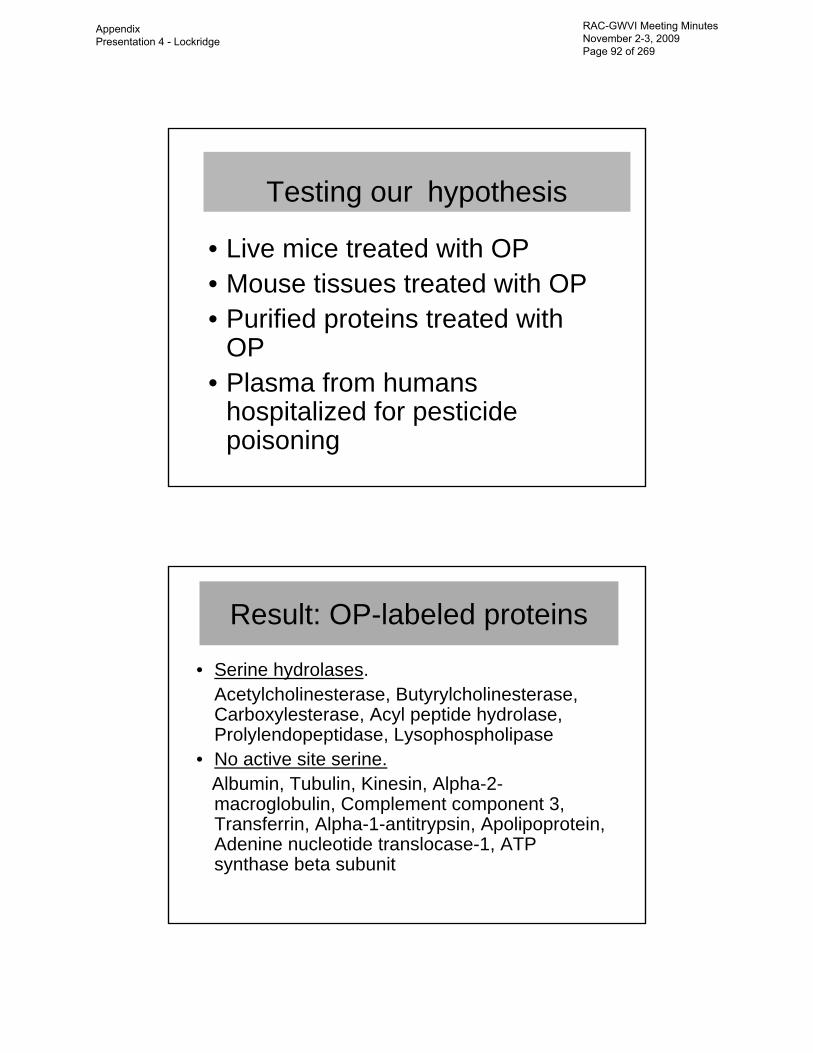
Testing our hypothesis
• Live mice treated with OP• Mouse tissues treated with OP• Purified proteins treated with
OP• Plasma from humans
hospitalized for pesticide poisoning
Result: OP-labeled proteins
• Serine hydrolases.Acetylcholinesterase, Butyrylcholinesterase, Carboxylesterase, Acyl peptide hydrolase, Prolylendopeptidase, Lysophospholipase
• No active site serine.Albumin, Tubulin, Kinesin, Alpha-2-macroglobulin, Complement component 3, Transferrin, Alpha-1-antitrypsin, Apolipoprotein, Adenine nucleotide translocase-1, ATP synthase beta subunit
Appendix Presentation 4 - Lockridge
RAC-GWVI Meeting Minutes November 2-3, 2009 Page 92 of 269

Covalent binding to Tyrosine is a new OP binding motif
• The OP attachment site in albumin, tubulin, and in other proteins is tyrosine.
• We conclude that OP binding to tyrosine is a new motif for OP modification of proteins that have no active site serine.
Testing plasma from people exposed to OP
• Plasma was obtained from the Poison Center in Omaha, Nebraska and from the Paris police.
• Methods were developed to identify OP adducts on butyrylcholinesterase and on albumin in human plasma.
Appendix Presentation 4 - Lockridge
RAC-GWVI Meeting Minutes November 2-3, 2009 Page 93 of 269
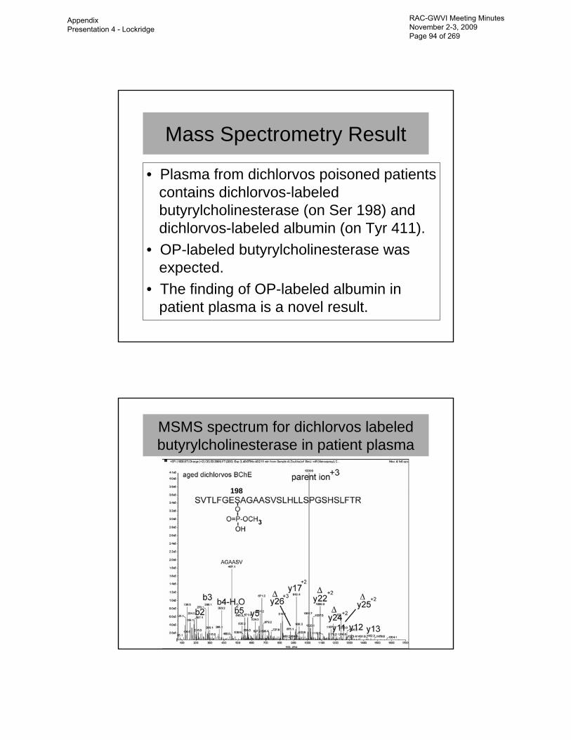
Mass Spectrometry Result
• Plasma from dichlorvos poisoned patients contains dichlorvos-labeled butyrylcholinesterase (on Ser 198) and dichlorvos-labeled albumin (on Tyr 411).
• OP-labeled butyrylcholinesterase was expected.
• The finding of OP-labeled albumin in patient plasma is a novel result.
MSMS spectrum for dichlorvos labeled butyrylcholinesterase in patient plasma
198
Appendix Presentation 4 - Lockridge
RAC-GWVI Meeting Minutes November 2-3, 2009 Page 94 of 269

MSMS spectrum for dichlorvos labeled albumin from patient
411
Significance (Take home point)
• The presence of OP-labeled albumin in humans suggests that other proteins can also become labeled on tyrosine by OP.
• OP-labeled albumin does not explain neurotoxicity.
• The search for proteins involved in OP toxicity is not limited to proteins in the serine hydrolase family.
Appendix Presentation 4 - Lockridge
RAC-GWVI Meeting Minutes November 2-3, 2009 Page 95 of 269

Topic 2.Tubulin in brain
could be involved in OP neurotoxicity
Focus on tubulin• Tubulin was selected
for detailed study because tubulin is modified by OP on tyrosine when mouse brain extracts are treated with OP. Tubulin polymer makes
hollow microtubules
Appendix Presentation 4 - Lockridge
RAC-GWVI Meeting Minutes November 2-3, 2009 Page 96 of 269

Focus on tubulin• Tubulin has important functions in neurons.
– Tubulin polymerizes to form microtubules. Microtubules are the highway for transport of cell components from the cell body to the nerve axons.
– Disruption of microtubule function results in cell death.
Methods:
chlorpyrifos oxon3.0 mg/kg i.p.
vehiclecontrol
24 hours
Purify tubulin from brainPolymerize tubulin to make microtubules
microtubules
Nanoimaging SDS-PAGE In gel digestion
Mass spectrometry
Appendix Presentation 4 - Lockridge
RAC-GWVI Meeting Minutes November 2-3, 2009 Page 97 of 269

Result: Nanoimages of microtubules
Chlorpyrifos oxon Control
Microtubule height and width were measured for 150 fibers at 4 to 5 positions along the fiber (a total of 700 measurements)
Microtubule width and height comparison
Appendix Presentation 4 - Lockridge
RAC-GWVI Meeting Minutes November 2-3, 2009 Page 98 of 269

Nanoimaging results
• Tubulin from OP treated mice assembles into abnormally thin microtubules.
• The microtubules from OP treated mice have fewer microtubule-associated proteins.
Microtubule-associated proteins• Microtubule-associated proteins are
essential for microtubule function– Facilitate polymerization and stabilize
microtubule structure– Transport cargo to the nerve endings by
traveling along the microtubules
www.cytochemistry.net/cell-biology/microt2.gif
Appendix Presentation 4 - Lockridge
RAC-GWVI Meeting Minutes November 2-3, 2009 Page 99 of 269

Proteins are selectively depleted from microtubules of chlorpyrifos oxon treated
mice• Microtubule-associated protein 2 isoform 1• Tau• Myosin Va• Cytoskeleton associated protein 5• Heat shock protein 84 kDa• Dynein• Alpha-internexin• Plectin 1 isoform 4
ConclusionExposure to chlorpyrifos oxon in vivo:
- Disrupts the structure of microtubules.
- Depletes microtubules of the normal complement of microtubule-associated proteins.
Appendix Presentation 4 - Lockridge
RAC-GWVI Meeting Minutes November 2-3, 2009 Page 100 of 269

Impact (Take home point)These results suggest microtubule
function is impaired by OP exposure.
Dysfunctional microtubules could be part of the explanation for the neurotoxicity associated with exposure to OP.
Impact (Take home point)Proteins not yet identified could also be
involved in OP neurotoxicity.If these unidentified proteins are in low
abundance, the OP-labeled versions will be difficult to detect by mass spectrometry.
The task of finding them will be easier if they can be immunoprecipitated with anantibody specific for OP-tyrosine.
Appendix Presentation 4 - Lockridge
RAC-GWVI Meeting Minutes November 2-3, 2009 Page 101 of 269

Antibodies for diagnosis of OP exposure
and for isolation of OP-labeled proteins
Topic 3
Background• To date no one has made an antibody
that detects nerve agent exposure. • Attempts by others have focused on an
antibody to nerve agent-modified cholinesterase. This strategy has failed because adducts on the active site serine of cholinesterase are unstable.
• We have a new strategy and we have preliminary results to suggest our new strategy works.
Appendix Presentation 4 - Lockridge
RAC-GWVI Meeting Minutes November 2-3, 2009 Page 102 of 269

New strategy• Our new strategy is based on our mass
spectrometry results. We have found that OP make a bond not only with the active site serine of cholinesterase, but also with tyrosine in albumin and tyrosine in other proteins.
• OP modified tyrosine is very stable.• Animal studies have shown that nerve agent-
labeled albumin can be detected as late as 24 days after exposure, at a time when no nerve agent modified cholinesterase is detectable.
Polyclonal antibody results• Polyclonal antibodies were made in
rabbit to a small peptide labeled on tyrosine with soman.
• The antiserum was tested for binding specificity on Western blots.
• Binding affinity was tested in a competition ELISA assay.
Appendix Presentation 4 - Lockridge
RAC-GWVI Meeting Minutes November 2-3, 2009 Page 103 of 269

Western blot result
The polyclonal antibody recognizes soman-labeled albumin and soman-labeled transferrin. It does not recognize unlabeled proteins.This result shows high binding specificity for soman-tyrosine adducts.
Binding affinity result
Binding affinity was tested in a competition assay between soman-albumin and soman-peptide RYGRK for binding to the polyclonal antibody.
The binding affinity of 10 -11.5 M is a very promising result.
The high binding affinity is a very promising result.
Appendix Presentation 4 - Lockridge
RAC-GWVI Meeting Minutes November 2-3, 2009 Page 104 of 269

Limit of detection
Western blotLanes were loaded in duplicate with 0.5 to 0.005 µg of soman-labeled albumin. The blot was hybridized with polyclonal antibody diluted 1:2000.
The limit of detection was 0.01 micrograms of soman-labeled human albumin.
Human plasma has 40,000 micrograms of albumin per milliliter.
Human saliva has 35 micrograms albumin per milliliter.
A limit of detection of 0.01 micrograms means that low exposure levels will be detectable.
Appendix Presentation 4 - Lockridge
RAC-GWVI Meeting Minutes November 2-3, 2009 Page 105 of 269

Plan• The plan is to make monoclonal antibodies to a
variety of OP-tyrosine.• Each OP will have a specific antibody for that
OP.• Reasons for making monoclonals rather than
polyclonals– monoclonals can be made in unlimited quantities – monoclonals have higher specificity– monoclonals can be labeled with a fluorescent tag to
increase detection sensitivity
Potential uses of the monoclonals• Monoclonal antibodies will detect low
dose exposure to OP. The antibodies can be used in hand-held devices.
• Pesticide workers, war-fighters, and the worried-well can benefit.
• Monoclonal antibodies can be used to isolate OP-labeled proteins and thus lead to an understanding of the mechanism of low dose neurotoxicity.
Appendix Presentation 4 - Lockridge
RAC-GWVI Meeting Minutes November 2-3, 2009 Page 106 of 269

Advantages of an antibody test for exposure
• Simple• Rapid• Inexpensive• Portable
Conclusion
• Monoclonal antibodies for detection of OP exposure are expected to be specific and highly sensitive.
• Exposure levels too low to cause toxic symptoms should be detectable with the monoclonal antibodies.
Appendix Presentation 4 - Lockridge
RAC-GWVI Meeting Minutes November 2-3, 2009 Page 107 of 269

Conclusion (Take home point)The novelty of our approach is that
antibodies will be made against OP adducts on tyrosine.
The strengths are the solid evidence from mass spectrometry that OP make a covalent bond with tyrosine in proteins, and our demonstration that antibodies against soman-labeled tyrosine show high affinity and specificity.
AcknowledgementsAcknowledgements
Bin LiPatrick MassonWei JiangMariya LiyasovaEllen DuysenLawrence SchopferLuda ShlyakhtenkoHasmik Grigoryan
Core facilities at UNMC for mass spectrometry and for nanoimaging.
Appendix Presentation 4 - Lockridge
RAC-GWVI Meeting Minutes November 2-3, 2009 Page 108 of 269