oral abstract presentations abstracts 464, 216, 471, 211, 308 will … · 2017-07-10 · oral...
TRANSCRIPT

Oral Abstract Presentations
• Abstracts 464, 216, 471, 211, 308 will be presented during the Early Career Pre-Conference Session
• Abstracts 1-17 will be presented Monday - Wednesday Poster Abstract Presentations
• Abstracts 20–174 will be presented on Monday
• Abstracts 185-335 will be presented on Tuesday
• Abstracts 336-484 will be presented on Wednesday

BCVS 2017 Scientific Sessions - Oral Abstracts Presented in the Early Career Pre-Session Conference
These abstracts are also being presented as posters
464
Two Different Microdomains of β1-adrenoreceptor Signaling Revealed by Live Cell Imaging
Alexander Froese, Viacheslav O Nikolaev, Univ Medical Ctr Hamburg, Hamburg, Germany
Background: 3’,5’-cyclic adenosine monophosphate (cAMP) is an ubiquitous second messenger and a crucial regulator of cardiac function and disease. In cardiomyocytes, it is produced predominantly after activation of β1-adrenergic receptors (β1-ARs) by catecholamines and acts intracellularly in discrete functionally relevant microdomains formed, for example, around calcium-handling proteins. Previously, we reported that β1-ARs are distributed across various cardiomyocyte membrane areas, including transverse (T)-tubules and cell crests. However, it is unknown whether these two β1-AR pools contribute differentially to the regulation of cardiac contractility and gene expression. Methods and Results: To directly visualize receptor-microdomain communication in cardiomyocytes, we established a combination of scanning ion conductance microscopy (SICM) with transgenically expressed targeted Förster resonance energy transfer (FRET)-based biosensors. Using this approach, we measured local cAMP responses in distinct microdomains of mouse ventricular cardiomyocytes (such as plasma membrane, cytosol and nucleus) after localized stimulation of β1-AR on different membrane structures of healthy and diseased cardiomyocytes. Using a plasma membrane targeted cAMP biosensor, we found that β1-AR stimulation at the crest induced stronger cAMP signals compared to β1-AR stimulated in the T-tubuli where cAMP was highly confined by PDE3. This difference was abolished in a pressure overload hypertrophy model due to submembrane redistribution of PDEs. Interestingly, crest β1-AR signals could propagate deeper inside the cell, inducing higher nuclear cAMP responses than recorded from receptors stimulated in the T-tubules. Conclusions: in the present study, we have demonstrated that β1-ARs located in T-tubuli and cell crests form two differentially regulated cAMP microdomains, each having its typical PDE repertoire and generating distinct second messenger signals. More detailed understanding of these two microdomains at different subsarcolemmal locations may contribute to new therapeutic strategies including more specific β-blockers.
A. Froese: None. V.O. Nikolaev: None.
216
Causal Role of Oxidized Lipids in Pulmonary Hypertension Development
Gregoire Ruffenach, Soban Umar, Mylene Vaillancourt, Victor Grijalva, Ellen I. O’Connor, Shayan Moazeni, Christine Cunningham, Abbas Ardehali, Aman Mahajan, Srininivasa T. Reddy, Mansoureh Eghbali, Univ of California, Los Angeles, CA
Pulmonary arterial hypertension (PAH) is a deadly disease characterized by increased pulmonary arterial pressure and pulmonary vascular occlusion. Recently, we and others demonstrated a robust increase in oxidized lipids, including 15-hydroxyeicosatetraenoic acids (15-HETE), in the lungs and plasma of PAH patients and animal models of pulmonary hypertension (PH). We hypothesized that diets rich in 15-HETE are sufficient to cause PH in wild type mice. We also examined whether 15-HETE or its metabolites are required to cause PH by comparing the effect of 15-HETE with 15-HETE methyl ester, which is a stable form of 15HETE that is not easily metabolized. C57BL/6 male mice were fed for 3 weeks with 15-HETE diet (5μg/day), 15-HETE methyl ester (15-HETE-ME, 5μg/day), or regular chow diet (n=8-21 mice/group). PH development was followed via weekly serial echocardiography. Right ventricular systolic pressure (RVSP) was measured via direct heart catheterization. RV hypertrophy index (RV/[IVS+LV]) was measured. Lung morphology and lipid accumulation were assessed using H&E and Oil red O staining.

Echocardiography revealed the first sign of PH in mice on 15HETE diet as early as one week and a significant decrease in the pulmonary arterial acceleration time after 2 weeks of treatment (16.6±1.9 vs. 21.2±1.4 msec, p<0.05). Mice on 15HETE diet also had significantly higher RVSP (31.3±1.1 vs. 38.4±2.3 mmHg, p<0.05). Increase in RVSP was concomitant with significantly higher RV hypertrophy index (0.26 ± 0.02 vs. 0.33 ±0.02, p<0.05). Pulmonary arteriolar thickness was also significantly increased in mice on 15-HETE diet compared to regular diet (35.1±0.8 vs 53.4±1, p<0.05). Our new model of PH is not a model of atherosclerosis as there was no detectable plaque in aorta of the mice on 15-HETE diet. Finally, mice on 15-HETE-ME diet also developed PH as RVSP was significantly higher compared to control (31.3±1.1 vs. 39±3 mmHg, p<0.05). The severity of PH was similar in 15HETE-ME and 15HETE, confirming 15HETE itself and not its metabolites is sufficient to cause PH in wild type mice. We have developed a new and physiologically relevant animal model to study PH as a consequence of oxidized lipids overload as it occurs in humans with PAH.
G. Ruffenach: None. S. Umar: None. M. Vaillancourt: None. V. Grijalva: None. E.I. O’Connor: None. S. Moazeni: None. C. Cunningham: None. A. Ardehali: None. A. Mahajan: None. S.T. Reddy: None. M. Eghbali: None.
This Research has received full or partial funding support from the American Heart Association.
471
Kinase-independent Function of PI3Kγ Enables ERK Activation
Maradumane L Mohan, Arunachal Chatterjee, Swetha Ganapathy, Sromona Mukherjee, Sathyamangla V Naga Prasad, Cleveland Clinic Fndn, Cleveland, OH
Phosphoinositide 3-kinase (PI3K) enzymes are critical in many cellular processes including cell survival. PI3Kγ, a member of the PI3K family, is activated in response to G-protein coupled receptor (GPCR) stimulation leading to extracellular regulated kinase (ERK) signal transduction cascade, a cell survival pathway. However, less is known about the underlying mechanisms of PI3Kγ-directed ERK activation. Knockdown of PI3Kγ showed that PI3Kγ not only regulates ERK phosphorylation in response to GPCR stimulation but also to receptor tyrosine kinase activation in HEK 293 cells. The key role of PI3Kγ in ERK activation was further validated by loss of insulin-stimulated ERK phosphorylation in PI3Kγ-knockout (KO) mouse embryonic fibroblasts (MEFs). Surprisingly, ERK activation in KO MEFs post-insulin stimulation was completely rescued by expression of kinase-dead PI3Kγ mutant in KO MEFs demonstrating a kinase-independent role of PI3Kγ in regulating ERK function. Mechanistic studies showed that PI3Kγ regulates ERK activation by inhibiting ERK dephosphorylation following stimulation thereby, sustaining ERK phosphorylation and activation. Critically, PI3Kγ regulates ERK dephosphorylating phosphatase PP2A by interacting and sequestering PP2A from ERK maintaining ERK phosphorylation, which is evidenced by increased PP2A association with ERK in KO MEFs. Consistently, ERK activation was completely abolished in KO MEFs following carvedilol or insulin suggesting an essential role for PI3Kγ in ERK activation pathway. Correspondingly, primary cardiac fibroblasts isolated from KO mice showed complete loss of insulin-stimulated ERK phosphorylation compared to WT mice. This is intriguing given that GSK3 phosphorylation and not ERK phosphorylation is regulated by inhibition of PP2A through kinase-independent mechanism of PI3Kγ in the total cardiac lysates. Even though GSK3 and ERK are substrates for PP2A, our findings that ERK is regulated by kinase-independent function PI3Kγ suggest the existence of this unique regulation in fibroblasts and not in cardiomyocytes. Thus, kinase activity of PI3Kγ may contribute to cardiac-pathology while kinase-independent function could be beneficial and will be discussed in presentation.
M.L. Mohan: None. A. Chatterjee: None. S. Ganapathy: None. S. Mukherjee: None. S.V. Naga Prasad: None.
This Research has received full or partial funding support from the American Heart Association.

211
Myocardial Hypertrophy and Circulating RNAs
Ravi V Shah, Ravi Shah, Newburyport, MA; Olivia Ziegler, Massachusetts General Hosp, Boston, MA; Kahraman Tanriverdi, Univ of Mass-Worcester, Worcester, MA; Jian Rong, Martin Larson, Framingham Heart Study, Framingham, MA; Alexander Pico, UCSF, San Francisco, CA; Daniel Levy, Framingham Heart Study, Framingham, MA; Ramachandran S Vasan, Boston Univ, Boston, MA; Saumya V Das, Massachusetts General Hosp, Boston, MA; Jane Freedman, Univ of Mass-Worcester, Worcester, MA
While increased left ventricular mass (LVM) is strongly associated with incident heart failure (HF), events during transition from increased LVM to HF remain unclear. Extracellular non-coding RNAs (ex-RNAs) have been implicated in cardiac hypertrophy, though whether these ex-RNAs reflect important pathways in HF in humans is underexplored. In >2,000 individuals with concomitant M-mode echocardiography and ex-RNA measurements in the Framingham Heart Study, we found that lower circulating concentrations of three ex-RNAs—miR-20a-5p, miR-106b-5p, miR-17-5p—were associated with (1) greater LVM (+ one other pre-clinical phenotype, e.g., left atrial dimension or LVEDV) and (2) greater incident HF risk over a median follow-up 7.7 years (Fig. A). These 3 miRNAs were members of a tight cluster, regulating 883 mRNAs in common, associated with “hypertension” (OMIM) and biological process relevant to HF, including TGF-β signaling. We observed an increase in myocardial expression of these miRNAs during different phases of hypertrophy/HF development (Fig. C, D). Using gain and loss of function in vitro, our preliminary results suggest up-regulation of cardiomyocyte miR-106b expression abrogates expression of pathologic hypertrophy markers (ANP and BNP) during phenylephrine treatment, consistent with in silico results suggesting broad connections between miR-106b targets and natriuretic peptide signaling (Fig. B, E-F). These results provide translational evidence that circulating miRNAs associated with hypertrophy in patients may be protective in the transition from hypertrophy to HF at the molecular level.
R.V. Shah: None. O. Ziegler: None. K. Tanriverdi: None. J. Rong: None. M. Larson: None. A. Pico: None. D. Levy: None. R.S. Vasan: None. S.V. Das: None. J. Freedman: None.
This Research has received full or partial funding support from the American Heart Association.

308
CTRP9 Regulates the Fate of Implanted Mesenchymal Stem Cells and Mobilizes Their Protective Effects Against Ischemic Heart Injury via Multiple Novel Signaling Pathways
Wenjun Yan, Wayne Lau, Theodore Christopher, Bernard Lopez, Thomas Jefferson Univ, Philadelphia, PA; Erhe Gao, Walter Koch, Temple Univ, Philadelphia, PA; Yajing Wang, Xinliang (Xin) Ma, Thomas Jefferson Univ, Philadelphia, PA
Cell therapy remains the most promising approach against ischemic heart failure. However, the poor survival of engrafted stem cells in the ischemic environment limits their therapeutic efficacy for cardiac repair post-MI. CTRP9 is a novel pro-survival cardiokine with significantly downregulated expression after MI. Here, we tested a hypothesis that CTRP9 might be a cardiokine required for a healthy microenvironment promoting stem cell survival and cardioprotection. Mice were subjected to MI and treated with adipose-derived mesenchymal stem cells (ADSCs, intramyocardial transplantation), CTRP9, or their combination. Administration of ADSCs alone failed to exert significant cardioprotection. However, administration of ADSCs in addition to CTRP9 further enhanced the cardioprotective effect of CTRP9 (P<0.05 vs. CTRP9 alone), suggesting a synergistic effect. CTRP9 significantly increased ADSCs survival and migration after implantation. Conversely, the number of engrafted ADSCs was significantly reduced in the CTRP9KO heart. CTRP9 promoted ADSCs proliferation and migration in vitro, and protected ADSCs against hydrogen peroxide-induced cellular death. Discovery-drive approaches followed by cause-effect analysis identified that CTRP9 enhances ADSCs proliferation/migration by ERK1/2-MMP-9 signaling. CTRP9 promotes anti-apoptotic/cell survival via ERK-Nrf2/anti-oxidative protein expression. Mass spectrometry, immunocytochemistry, and immunoprecipitation identified N-cadherin as the novel CTRP9 binding partner on ADSC. N-cadherin knockdown completely abolished the above noted CTRP9 biological effects. Finally, CTRP9 promotes Sod-3 expression and secretion from ADSCs, protecting cardiomyocytes against oxidative stress-induced cell death. We provide the first evidence that CTRP9 promotes ADSCs proliferation/survival, stimulates ADSCs migration, and attenuates cardiomyocyte cell death by previously unrecognized signaling mechanisms (N-cadherin-ERK/MMP-9 and N-cadherin-ERK/Nrf2-SOD). These results suggest that CTRP9 is a cardiokine critical in maintaining a healthy microenvironment facilitating stem cell engraftment in infarcted myocardial tissue, thereby enhancing stem cell therapeutic efficacy.
W. Yan: None. W. Lau: None. T. Christopher: None. B. Lopez: None. E. Gao: None. W. Koch: None. Y. Wang: None. X. Ma: None.

BCVS 2017 Scientific Sessions - Oral Abstracts
1
Sprr2b Drives Proliferation of Cardiac Fibroblasts by Relieving p53-mediated Cell Cycle Inhibition
Ryan M Burke, Janet K Lighthouse, Pearl J Quijada, Ronald Dirkx, Michael A. Trembley, Eric M. Small, Univ of Rochester, Rochester, NY
Pathological cardiac remodeling is initially a compensatory attempt to increase cardiac output, but ultimately leads to the development of fibrosis, a form of scarring that contributes to heart failure (HF). In contrast, physiological cardiac remodeling in response to exercise is not associated with the development of fibrosis and typically remains compensatory. Understanding how cardiac fibroblasts (CF), the primary source of extracellular matrix in the heart, respond to pathological and physiological cues might lead to novel approaches to limit the maladaptive effects of pathological cardiac remodeling. We performed RNA sequencing to define genes that are differentially regulated in CF during physiological (swimming) or pathological (pressure overload) remodeling. This study revealed that cardiac expression of the small proline rich 2b (Sprr2b) gene is restricted to CFs and is significantly elevated in disease and lost in exercise. We demonstrate that SPRR2B drives CF proliferation, but not myofibroblast differentiation, in response to pathological cues. SPRR2B facilitates an interaction between MDM2 and USP7, a nuclear deubiquitinase that leads to proteasomal degradation of p53. SPRR2B-USP7-MDM2 complex formation and p53 degradation is at least partially dependent upon phosphorylation of SPRR2B by Src-family NRTKs. SPRR2B thus relieves p53-mediated constraints on cell cycle progression in response to Src-dependent signaling, leading to CF accumulation. Importantly, SPRR2B expression is elevated in cardiac tissue from human HF patients relative to individuals without heart disease and positively correlates with a proliferative, activated gene expression profile in HF patient CF. Treatment of human HF fibroblasts with IGF-1/H2O2 to mimic physiological cues significantly abrogated SPRR2B expression and increased expression of p53-dependent cell cycle checkpoint genes, which correlated with a less activated phenotype. Taken together, this study defines a unique tissue-specific role of Sprr2b in driving pathological CF cell cycle progression that may underlie the development of cardiac fibrosis.
R.M. Burke: None. J.K. Lighthouse: None. P.J. Quijada: None. R. Dirkx: None. M.A. Trembley: None. E.M. Small: None.
2
MANF, A Structurally Unique Redox-Sensitive Chaperone, Restores ER Protein Folding in the Ischemic Heart.
Adrian Arrieta, Erik A. Blackwood, Winston T. Stauffer, Michelle Santo Domingo, Amber N. Pentoney, Donna J. Thuerauf, San Diego State Univ, San Diego, CA; Shirin Doroudgar, Dept of Cardiology, Angiology, and Pneumology, Univ Hosp Heidelberg, Germany, San Diego, CA; Christopher C. Glembotski, San Diego State Univ, San Diego, CA
Rationale: In cardiomyocytes, most secreted and membrane proteins are synthesized and folded in the sarcoplasmic/endoplasmic reticulum (SR/ER). We previously showed that during myocardial ischemia, decreased oxygen creates a reducing environment in the SR/ER, preventing protein disulfide isomerases (PDIs) from forming disulfide bonds in nascent proteins, causing ER stress, i.e. the toxic accumulation of unfolded proteins which contributes to cardiomyocyte death. In response to ER stress, the transcription factor, ATF6 induces chaperones that restore SR/ER protein folding. We found that ATF6 also induces mesencephalic astrocyte-derived neurotrophic factor (MANF), a recently identified protein of unknown function. MANF is structurally unique, so its function cannot be inferred from other proteins. Since MANF is induced by ATF6, is ER-localized, and contains a conserved redox-sensitive motif found in PDIs, we hypothesized that MANF is a redox-sensitive chaperone that optimizes cardiomyocyte viability during ischemia. Methods: The redox status of MANF during reductive ER stress and the ability of MANF to bind

misfolded proteins during ischemia were assessed in neonatal rat ventricular myocytes (NRVM). The ability of recombinant MANF to suppress aggregation of misfolded proteins was examined in an in vitro chaperone assay. Finally, the effects of MANF loss-of-function in the ischemic heart, in vivo, were determined by generating a transgenic mouse model that expresses a cardiomyocyte-specific MANF-targeted microRNA. Results: In NRVM subjected to ER stress MANF was as sensitive to changes in ER redox status as the sentinel PDI, PDIA1. Moreover, MANF formed disulfide-linked complexes with misfolded proteins during ischemia-mediated ER stress. Under reducing conditions, recombinant MANF suppressed aggregation of model misfolded proteins, in vitro. MANF knockdown in the heart, in vivo, increased damage from myocardial infarction, and an AAV9-based gene therapy approach rescued the effects of MANF deficiency, in vivo. Conclusions: MANF is a redox-sensitive SR/ER-resident chaperone that is a critical contributor to SR/ER protein folding during the adaptive ER stress response and decreases tissue damage in the ischemic heart.
A. Arrieta: None. E.A. Blackwood: None. W.T. Stauffer: None. M. Santo Domingo: None. A.N. Pentoney: None. D.J. Thuerauf: None. S. Doroudgar: None. C.C. Glembotski: None.
3
Loss of Type 9 Adenylyl Cyclase Triggers Reduced Phosphorylation of Hsp20 and Diastolic Dysfunction
Tanya A Baldwin, Yong Li, Yan Wang, Cameron S Brand, Carmen W Dessauer, McGovern Medical Sch at Univ of Texas Health Science Ctr at Houston (UTHealth), Houston, TX
Adenylyl cyclase type 9 (AC9) is found tightly associated with the scaffolding protein Yotiao and the IKs ion channel in heart. But apart from potential IKs regulation, physiological roles for AC9 are unknown. Utilizing a gene-trap mouse line that disrupts expression of AC9, we show that loss of AC9 reduces less than 2% of total AC activity in heart but eliminates Yotiao-associated AC activity. AC9-/- mice exhibit no structural abnormalities but show a significant bradycardia. Global changes in PKA phosphorylation patters are not altered in AC9-/- heart, however basal phosphorylation of heat shock protein 20 (Hsp20) is significantly decreased. AC9 binds Hsp20 in a Yotiao-independent manner, while deletion of AC9 decreases Hsp20-associated AC activity in heart, consistent with and an AC9-Hsp20 complex. Phosphorylation of Hsp20 occurs largely in ventricles and is vital for the cardioprotective effects of Hsp20. Decreased Hsp20 phosphorylation suggests a potential baseline ventricular defect for AC9-/-. Doppler echocardiography of AC9-/- mice displays a decrease in the early ventricular filling velocity and ventricular filling ratio (E/A), indicative of grade 1 diastolic dysfunction. Our findings unveil potential new roles for AC9 in cardiac function and emphasize the importance of local cAMP production in the context of macromolecular complexes.
T.A. Baldwin: None. Y. Li: None. Y. Wang: None. C.S. Brand: None. C.W. Dessauer: None.
4
Regulation of Mitochondrial Complex I Assembly
Edward Owusu-Ansah, Columbia Univ, New York, NY
Mitochondrial CI (NADH: ubiquinone oxidoreductase) is the first and largest of the electron transport chain (ETC) complexes involved in oxidative phosphorylation, and has a molecular mass approaching 1 MDa. Mitochondrial Complex I (CI) is composed of 44 distinct subunits, but only 14 of these subunits are directly required for catalysis. A fundamental question in mitochondrial biology is to elucidate the roles of the additional 30 or so accessory subunits. In addition, CI can form different supercomplexes with other ETC complexes. It is unclear how these supercomplexes are assembled in the human heart, or any organism. We have established Drosophila flight muscles as a suitable system for studying CI assembly as well as the assembly of CI-containing supercomplexes. We show that many of the 30 accessory subunits regulate specific steps of CI assembly in vivo; and that CI biogenesis in flight muscles proceeds via the formation of ~315-, ~370-, ~550-, and ~815 kDa CI assembly intermediates as has been reported in mammalian systems. A specific accessory subunit (dNDUFA5) is required for the formation or

stabilization of the ~315 kDa assembly intermediate. Additionally, we define a specific role for the CX9C-containing accessory subunit (dNDUFS5); by showing that it is required for converting a transient CI assembly intermediate (an ~700 kDa assembly intermediate) into the ~815 kDa assembly intermediate. Finally, we are performing genetic screens to identify genes that specifically regulate supercomplex assembly. Our findings highlight the potential of studies of CI biogenesis in Drosophila to uncover novel mechanisms of CI assembly in a living organism, and establish Drosophila as a suitable model organism for addressing questions relevant to CI biogenesis in humans.
E. Owusu-Ansah: None.
This Research has received full or partial funding support from the American Heart Association.
5
Restoring Nitroso-Redox Balance as a Therapeutic Approach for Cardiovascular Disease
Vikram Shettigar, Honglan Wang, Bo Zhang, Steve R Roof, Paul M Janssen, Jonathan P Davis, Ohio State Univ, Columbus, OH; Richard J Gumina, Vanderbilt Univ Medical Ctr, Nashville, TN; Brandon J Biesiadecki, Frederick A Villamena, Mark T Ziolo, Ohio State Univ, Columbus, OH
Anti-oxidant therapy has been an immense clinical disappointment for the treatment of cardiomyopathies. Concurrently with the increase in reactive oxygen species (ROS), there is also a decrease in cardiac levels of nitric oxide (NO), resulting in a nitroso-redox imbalance not addressed by anti-oxidant treatment alone. Key modulators of cardiac function are sensitive to the nitroso-redox balance such as kinases and phosphatases. Thus, along with changes in protein oxidation and/or S-nitrosylation levels, the nitroso-redox imbalance also alters protein phosphorylation. We developed a unique and novel compound (EMEPO) that can correct the nitroso-redox imbalance by simultaneously scavenging ROS and producing NO. We hypothesized that EMEPO is a novel agent that will ameliorate cardiac dysfunction by reestablishing the proper protein post-translational modifications. We demonstrated the efficacy of EMEPO in two cardiac models of nitroso-redox imbalance; a genetic model (NOS1-/-) and a disease model- murine myocardial infraction (MI). Both models displayed nitroso-redox imbalance with systolic and diastolic dysfunction. EMEPO treatment had a much greater effect than anti-oxidant treatment alone in palliating the cardiac dysfunction. A major contributor to these dysfunctions observed in cardiomyopathies is altered ryanodine receptor (RyR) activity. EMEPO was able to restore RyR activity and each aberrant post-translational modification (oxidation, S-nitrosylation, and phosphorylation). We believe these EMEPO-induced changes in RyR will occur in many proteins that orchestrate signaling networks and function. Our data highly suggest that simultaneously restoring both ROS and NO levels (i.e., correcting the nitroso-redox imbalance) is a promising therapeutic approach for MI and heart failure patients. Our first designed nitroso-redox balancer, EMEPO, shows great potential as a novel strategy for the treatment of heart disease.
V. Shettigar: None. H. Wang: None. B. Zhang: None. S.R. Roof: None. P.M. Janssen: None. J.P. Davis: None. R.J. Gumina: None. B.J. Biesiadecki: None. F.A. Villamena: None. M.T. Ziolo: 2. Research Grant; Significant; American Heart Assocation.
This Research has received full or partial funding support from the American Heart Association.
6
Restoration of Impaired Diastolic Function in Hypertrophic Cardiomyopathy Induced Pluripotent Stem Cell-derived Cardiomyocytes by Re-balancing the Calcium Homeostasis
Haodi Wu, Huaxiao Yang, Joe Zhang, Chi Keung Lam, June-Wha Rhee, Timon Seeger, Karim Sallam, Ning Ma, Joseph Wu, Stanford Univ, Stanford, CA
Background:Diastolic dysfunction is commonly seen in hypertrophic cardiomyopathy (HCM). However, the cellular mechanism is not fully understood, and no effective treatment so far has been developed. We hypothesize here that HCM patient-specific induced pluripotent stem cell-derived cardiomyocytes (iPSC-

CMs) can recapitulate the cellular mechanism, and provide us a platform for mechanistic study and for drug screening of diastolic dysfunctions in HCM. Methods and Results:We generated beating iPSC-CMs from healthy individuals and HCM patients carrying familial mutations (MYH7 R663H (n=2 lines) and MYBPC3 R943ter (n=2 lines)). Sarcomere shortening measurement in patterned iPSC-CMs with live cell confocal imaging showed significantly prolonged diastolic phase and slower relaxation velocity in HCM iPSC-CMs compared to WT cells. To elucidate the cellular mechanism, Fura-2 AM ratiometric calcium imaging showed marked elevation of resting calcium level and increased abnormal calcium handlings in HCM iPSC-CMs, which were exaggerated by β-adrenergic activation with isoproterenol. By applying calcium transient and contractile force simultaneous recording, we defined a “risk index of diastolic dysfunction” (measured as transient-contraction gain factor), which was significantly increased in HCM iPSC-CMs. Thus, both elevated basal calcium level and increased calcium sensitivity of myofilament contribute to the abnormal diastolic function in HCM iPSC-CMs. Gene expression profiling of HCM and WT iPSC-CMs indicated that increased calcium channels may underlie the increased basal calcium concentration in HCM cells. Indeed, partially blocking the calcium influx by calcium blockers reset the basal calcium level, attenuated calcium mishandling, and restored the diastolic function in HCM iPSC-CMs. Moreover, re-balancing calcium homeostasis significantly improved long-term survival rate of HCM iPSC-CMs at both basal level and under β-adrenergic stress. Conclusion: The iPSC-CM models carrying patient-specific HCM mutations recapitulated diastolic dysfunction on single cell level. Future studies using these platform may reveal additional novel cellular mechanisms and therapeutic targets of diastolic dysfunction in HCM disease.
H. Wu: None. H. Yang: None. J. Zhang: None. C. Lam: None. J. Rhee: None. T. Seeger: None. K. Sallam: None. N. Ma: None. J. Wu: None.
This Research has received full or partial funding support from the American Heart Association.
7
Chromatin Organization in Diseased and Healthy Mouse Heart
Chukwuemeka George Anene Nzelu, Dominic Lee, Wilson Tan, Zenia Tiang, Matias Autio Ilmaris, Peter Li, Melissa Fullwood, Roger Foo, Natl Univ of Singapore, Singapore, Singapore
Background The three-dimensional chromatin structure regulates transcription by bringing regulatory elements such as enhancers in close spatial proximity with target genes. This arrangement helps to ensure cell type-specific gene expression profiles. We employed Hi-C, Chip-seq and CRISPR knock out to provide a detailed genome-wide view of the 3D chromatin structure in healthy and diseased mouse heart tissue and also to link regulatory elements with their target genes. Results The 3D chromatin organization of Sham and TAC adult ventricular cardiomyocytes (CMs) as well as atrial-origin HL1 cardiomyocytes showed high degree of similarity. While topological associated domains are stable across all cell types, the A and B genomic compartments showed important variations, which correlated well with differential gene expression profiles. Analysis of the interactions in regions enriched with the histone enhancer mark H3K27ac identified putative regulatory elements responsible for cell-type specific gene expression. In addition, CRISPR deletion of a regulatory region upstream of Nppb and Nppa loci led to a downregulation of these genes in HL1 cells, confirming results obtained via Hi-C and 4C Conclusion We show evidence of the relationship between genome organization and gene expression by comparing atrial and Sham and TAC ventricular cardiac cells. Importantly, this is the first detailed report of genome-wide 3D chromatin interactions in cardiac cells, revealing cardiac-relevant genomic compartments that associate with cardiac-specific gene expression.
C. Anene Nzelu: None. D. Lee: None. W. Tan: None. Z. Tiang: None. M.A. Ilmaris: None. P. Li: None. M. Fullwood: None. R. Foo: None.

8
Effectiveness of Combination Allogeneic Stem Cells in a Novel Large Animal Model of Chronic Kidney Disease-induced Heart Failure With Preserved Ejection Fraction (HFpEF)
Angela Castellanos Rieger, Bryon A. Tompkins, Makoto Natsumeda, Victoria Florea, Kevin Collon, Jose Rodriguez, Marcus Rosado, Wayne Balkan, Joshua M Hare 30136, Ivonne H Schulman, Univ of Miami, Miami, FL
Background: Chronic Kidney Disease (CKD) is an independent risk factor for cardiovascular morbidity and mortality. Left ventricular (LV) hypertrophy and heart failure with preserved ejection fraction (HFpEF) are the primary manifestations of the cardiorenal syndrome in 60 to 80% of CKD patients. Therapies that improve morbidity and mortality in HFpEF are lacking. Stem cell therapy reduces fibrosis, increases neovascularization, and promotes cardiac repair in ischemic and non-ischemic cardiomyopathies. We hypothesized that stem cell treatment ameliorates HFpEF in a CKD model. Methods: Yorkshires pigs (n=27) underwent 5/6 nephrectomy via renal artery embolization and 4-weeks later received either: allogeneic (allo-) MSC (10×106), allo-kidney c-kit+ cells (c-kit; 10×106), combination (MSC+c-kit; 1:1 ratio [5×10⁶ each]), or placebo (each n=5). Cell therapy was delivered via the patent renal artery. Kidney function, renal and cardiac MRI, and PV loops were measured at baseline, and at 4- and 12-weeks (euthanasia) post-embolization. Results: The CKD model was confirmed by increased creatinine and BUN and decreased GFR. Mean arterial pressure (MAP) was not different between groups from baseline to 4 weeks (p=0.7). HFpEF was demonstrated at 4 weeks by increased LV mass (20.3%; p= 0.0001), wall thickening (p<0.008), EDP (p=0.01), EDPVR (p=0.005), and arterial elastance (p=0.03), with no change in EF. Diffuse intramyocardial fibrosis was evident in histological analysis and delayed enhancement MRI imaging. After 12 weeks, there was a significant difference in MAP between groups (p=0.04), with an increase in the placebo group (19.97± 8.65 mmHg, p=0.08). GFR significantly improved in the combination group (p=0.033). EDV increased in the placebo (p=0.009) and c-kit (p=0.004) groups. ESV increased most in the placebo group (7.14±1.62ml; p=0.022). EF, wall thickness, and LV mass did not differ between groups at 12 weeks. Conclusion: A CKD large animal model manifests the characteristics of HFpEF. Intra-renal artery allogeneic cell therapy was safe. A beneficial effect of cell therapy was observed in the combination and MSC groups. These findings have important implications on the use of cell therapy for HFpEF and cardiorenal syndrome.
A. Castellanos Rieger: None. B.A. Tompkins: None. M. Natsumeda: None. V. Florea: None. K. Collon: None. J. Rodriguez: None. M. Rosado: None. W. Balkan: None. J.M. Hare: 8. Consultant/Advisory Board; Modest; Starr Foundation and the Soffer Family Foundation (grants). He holds equity in Vestion Inc; and maintains a professional relationship with Vestion as a consultant and member of the Board. I.H. Schulman: None.
9
Cardiac BIN1 Improves Dyad Organization and Calcium Transient in Cardiomyocytes
Ying Fu, Kang Zhou, Yan Liu, Sosse Agvanian, Bing Xu, Robin M Shaw, TingTing Hong, Cedars-Sinai Medical Ctr, Los Angeles, CA
BIN1 (bridging integrator 1) is a membrane scaffolding protein that forms microfolds at t-tubules and organizes dyad microdomains essential for a normal calcium transient. Reduction of cBIN1 has emerged as a new hallmark in heart failure and is associated with impaired calcium transient and increased risk of arrhythmias. We hypothesized that gene transfer of cardiac BIN1 (cBIN1) in vivo can improve calcium transients in disease models. We introduced V5-tagged cBIN1 or GFP through AAV9-mediated expression in adult mice. Confocal imaging of isolated cardiomyocytes was used to assess organization of dyad microdomain. Calcium transients in response to acute administration of isoproterenol (ISO) was measured as functional readout of excitation-contraction (EC) coupling. Here we report that cBIN1 significantly enhanced both basal and ISO-induced increase in calcium transient compared to cells expressing GFP. Immunofluorescent labeling revealed that cBIN1 expression increased both LTCC and RyR at t-tubule. In addition, in a mouse model of ischemic cardiomyopathy, permanent LAD ligation resulted in a marked reduction in ISO-induced increment in calcium transient, indicating blunted beta-adrenergic responsiveness and impaired regulation of EC coupling machinery. In comparison, an ISO-
induced increment in calcium transient was largely maintained in cBIN1-expressing cells. These results demonstrate that cBIN1 increases calcium transients by upregulating and organizing dyad proteins at t-tubule microfolds. More importantly, cBIN1 rescues diminished beta-adrenergic responsiveness in ischemic cardiomyopathy and improves EC coupling of viable cardiomyocytes. This work suggests that cBIN1 provides a therapeutic opportunity to improve cardiac function in ischemic cardiomyopathy and heart failure.
Y. Fu: None. K. Zhou: None. Y. Liu: None. S. Agvanian: None. B. Xu: None. R.M. Shaw: None. T. Hong: None.
This Research has received full or partial funding support from the American Heart Association.
10
Increasing Cardiac Fatty Acid Oxidation Protects Against High Fat Diet Induced Cardiomyopathy in Mice
Dan Shao, Stephen C. Kolwicz Jr, Nathan Roe, Outi Villet, Loreta De Tomasi, Alyssa Noriko Braun, Rong Tian, Univ of Washington, Seattle, WA
In the obese and diabetic heart, an imbalance between fatty acid uptake and fatty acid oxidation (FAO) promotes the development of cardiac lipotoxicity. We previously demonstrated that cardiac-specific deletion of ACC2 in adult mice was effective in increasing myocardial FAO while maintaining normal cardiac function and energetics. In this study, we tested the hypothesis that ACC2 deletion in an adult heart would prevent the cardiac lipotoxic phenotype in a mouse model of diet-induced obesity. ACC2 flox/flox (CON) and ACC2 flox/flox-MerCreMer+ (iKO) were injected with tamoxifen and subjected to a high fat diet (HFD) for 24 weeks. HFD induced similar body weight gain and glucose intolerance in CON and iKO. In isolated Langendorff-perfused heart experiments, HFD feeding increased FAO 1.6-fold in CON mice which was further increased to 1.9-fold in iKO mice compared with CON on chow diet. HFD induced systolic and diastolic dysfunction was abolished in iKO mice compared with CON mice (Fractional shortening 32.8±2.8% (CON) vs. 39.2±3.2% (iKO), E’/A’ ratio 0.91±0.09 (CON) vs. 1.11±0.08 (iKO), p< 0.05, n=5-6). Heart weight /Tibia length ratio was significantly higher in CON than iKO mice after HFD feeding (7.19±0.22 vs. 6.47±0.28, p<0.05, n=6). These data indicate that elevated myocardial FAO per se does not cause the development of cardiac dysfunction in obese animals. In fact, enhancing FAO via ACC2 deletion prevents HFD induced cardiac dysfunction and attenuates pathological hypertrophy. Molecular markers for ER stress such as p-PERK (1.5 fold) and p-JNK (2 fold) was elevated in CON-HFD hearts, which was completely attenuated in iKO-HFD hearts. Impairment of autophagy was also observed in CON-HFD hearts evidenced by decreases in LC3 II (60%) and increases in P62 (75%) level, while no difference in autophagy were observed in iKO-HFD hearts compared to iKO-chow. Therefore, the beneficial effect for enhancing cardiac fatty oxidation in HFD induced obesity model may be mediated, in part, by maintenance of cellular homeostasis and survival through regulating ER stress and autophagy. Taken together, our findings suggest that promoting cardiac FAO is an effective strategy to resist the development of cardiac lipotoxicity during diet-induced obesity.
D. Shao: None. S.C. Kolwicz Jr: None. N. Roe: None. O. Villet: None. L.D. Tomasi: None. A.N. Braun: None. R. Tian: None.
11
Lamin A/C Mutations Epigenetically Dysregulate Scn5a Gene Expression, Perturbing Action Potential Properties in IPSC-derived Cardiomyocytes
Silvia Crasto, Humanitas Res Hosp, Rozzano (Milan), Italy; Nicolò Salvarani, Inst of Genetic and Biomedical Res (IRGB), Natl Res Council of Italy, Milan, Italy; Michele Miragoli, Dept of Clinical and Experimental Med, Univ of Parma, Parma, Italy; Marianna Paulis, Inst of Genetic and Biomedical Res (IRGB), Natl Res Council of Italy, Milan, Italy; Paolo Kunderfranco, Humanitas Res Hosp, Rozzano (Milan), Italy; Pierluigi Carullo, Inst of Genetic and Biomedical Res (IRGB), Natl Res Council of Italy, Milan, Italy; Alberto Forni, Giuseppe Faggian, Div of Cardiac Surgery, Univ of Verona, Verona, Italy; Gianluigi Condorelli, Humanitas Univ, Rozzano, Milan), Italy; Elisa Di Pasquale, Inst of Genetic and Biomedical Res (IRGB), Natl Res Council of Italy, Milan, Italy

Mutations of the LMNA gene, encoding the nuclear lamina proteins Lamin A/C, are a common cause of dilated cardiomyopathy, typically manifesting in association with cardiac conduction defects. LaminA/C regulate various nuclear activities, including maintenance of the nuclear structure, gene transcription and chromatin organization. Most studies on the consequences of Lamin A/C defects were conducted on fibroblasts, while studies on human cardiomyocytes (CMs) are scarce. We therefore generated a cardiac model of laminopathy obtained by differentiation of CMs from induced pluripotent stem cells (iPSCs) of patients carrying the K219T Lamin A/C mutation. In vitro, these cells recapitulate the morphological features of dilated cardiomyopathy, specifically sarcomeric disorganization and increased size. Using this model, we performed a comprehensive analysis of the electrophysiological properties of LMNA-CMs both at single cell level and in a multi-cellular setting. Using patch-clamp technique, results revealed significant changes in maximal upstroke velocity (dV/dtmax), action potential amplitude (APA) and overshoot (OV) in LMNA-CMs compared to those obtained from family-matched healthy controls (CNTR); these defects were associated with a reduction of the peak sodium currents and a diminished conduction velocity, measured in strands of electrically-coupled CMs. Biochemical studies showed a significant reduction of both the sodium channel Nav1.5 protein and its transcript in LMNA-CMs, accompanied by an increased binding of LaminA/C to the promoter of its coding gene, SCN5A. Binding of the Polycomb group protein SUZ12 and of the H3K27me3 histone repressive mark was also increased. Consistently, 3D-FISH experiments also indicated a preferential localization of SCN5A genomic loci at the nuclear periphery in LMNA-CMs. As a whole, our findings support a model in which mutated Lamin A/C perturb SCN5A gene expression by favouring PRC2 (Polycomb Repressive Complex 2) binding to its promoter, leading to decreased sodium current peak and slower conduction velocity. This mechanism may eventually sustain the conduction abnormalities inevitably occurring in patients with LMNA-cardiomyopathy.
S. Crasto: None. N. Salvarani: None. M. Miragoli: None. M. Paulis: None. P. Kunderfranco: None. P. Carullo: None. A. Forni: None. G. Faggian: None. G. Condorelli: None. E. Di Pasquale: None.
This Research has received full or partial funding support from the American Heart Association.
12
M6A Modification in RNA Regulates Cardiomyocyte and Cardiac Function in Heart Failure
Prabhu Mathiyalagan, Yaxuan Liang, Yassine Sassi, Erik Kohlbrenner, Jiqiu Chen, Djamel Lebeche, Maria Giovanna Trivieri, Kiyotake Ishikawa, Kenneth Fish, Roger J Hajjar, Susmita Sahoo, Icahn Sch of Med at Mount Sinai, New York, NY
Background: Adenosine in RNA is a substrate for addition or removal of methyl group. Reported five decades ago, methylated adenosine (m6A), the most abundant and functionally relevant chemical modification in RNA, whose transcriptome-wide mapping became possible only recently due to next generation sequencing (NGS). Coupled with NGS, m6A-methylated RNA capture (MeRIP-seq) identified widespread m6A distribution in ~8000 mRNA and ~1000 lncRNA transcripts in human and mouse transcriptome. Methods and Results: In a first of its kind approach, we examined m6A RNA methylation in both failing and non-failing hearts. We discovered that global m6A RNA methylation is significantly higher in left ventricles (LV) of failing human, swine and mouse hearts as compared to non-failing controls. Increase in m6A was associated with significantly lower expression of one of the key m6A demethylases, FTO, in the ischemic heart. siRNA-mediated silencing of FTO resulted in significant arrhythmias, loss of Ca2+ dynamics such as Ca2+ transient decay (Tau) and cardiomyocyte relaxation time in isolated adult rat cardiomyocytes. Conversely, FTO gene transfer reduced m6A and improved Ca2+ transients and contractile function in primary cardiomyocytes under hypoxia. In a mouse model of MI, AAV-mediated gene transfer of FTO significantly improved cardiac function post-MI. We identified transcriptome-wide m6A distribution signatures and conserved methylated sites of several mRNAs and lncRNAs using MeRIP-seq in both human and mouse failing and non-failing LV. Detailed MeRIP map of individual transcripts identified differentially methylated 3'-UTR, 5'-UTR and exon sites within several cardiac mRNAs that are important for cardiac function. Conclusion: Our data provide first evidence that m6A modification in RNA is a regulator of cardiomyocyte Ca2+ dynamics and cardiac function. Our findings on the dynamic nature of the cardiac m6A-epitranscriptome will add another portfolio to mRNA and lncRNA regulation of cardiac remodeling.

P. Mathiyalagan: None. Y. Liang: None. Y. Sassi: None. E. Kohlbrenner: None. J. Chen: None. D. Lebeche: None. M. Giovanna Trivieri: None. K. Ishikawa: None. K. Fish: None. R.J. Hajjar: None. S. Sahoo: None.
This Research has received full or partial funding support from the American Heart Association.
13
Suppressing Microtubule Detyrosination Reduces Stiffness and Improves Contractility in Human Cardiomyocytes
Benjamin L Prosser, Upenn Sch of Med, Philadelphia, PA; Kenneth Bedi, Hosp of the Univ of Pennsylvania, Philadelphia, PA; Matthew Caporizzo, Christina Chen, Patrick Robison, Upenn Sch of Med, Philadelphia, PA; Kenneth Margulies, Hosp of the Univ of Pennsylvania, Philadelphia, PA
The microtubule contribution to myocyte mechanics has been a controversial topic over the years. Utilizing high-speed, super-resolution imaging, we were recently able to directly observe microtubule behavior in working myocytes (Robison et al., Science 2016). Strikingly, we found that microtubules buckle like springs between sarcomeric attachment points, providing a mechanical resistance that limits sarcomere shortening and stretch. Further, we identified that post-translational “detyrosination” of microtubules regulates their attachment to the sarcomere, and thus the microtubule contribution to both passive and active mechanics. Here we present new data identifying microtubule detyrosination as a compelling therapeutic target for the treatment of human heart failure. Using quantitative mass spectrometry, we have probed the cytoskeletal changes that occur during the progression of human heart failure in over 40 patient samples at different stages and etiologies of disease. We find that progressive upregulation and stabilization of the structural cytoskeleton, particularly microtubules and intermediate filaments, is a robust hallmark of human heart failure. Next, we have performed detailed biophysical studies on isolated myocytes from explanted failing and non-failing human hearts. Using advanced imaging, single myocyte tensile tests and atomic force microscopy (e.g. Prosser et al., Science 2011; Robison et al. Science 2016), we have interrogated the contribution of detyrosinated microtubules to the active and passive mechanics of human myocytes. We find that by reducing microtubule detyrosination, we can robustly improve contractile function. Suppressing detyrosination significantly lowers passive stiffness at physiologic rates, while robustly improving contraction velocity, fractional shortening, and relaxation speed. Of note, the improvement in mechanics correlates with the severity of disease, as myocytes from end-stage patients show greater benefits than those from non-failing or compensated hypertrophic hearts. In conclusion, our work demonstrates pre-clinical efficacy for suppressing detyrosinated microtubules to improve myocyte mechanics in human heart failure.
B.L. Prosser: 2. Research Grant; Modest; Sanofi - iAward partially supports this work. 2. Research Grant; Significant; Current NIH R01 funding this work.. K. Bedi: None. M. Caporizzo: None. C. Chen: None. P. Robison: None. K. Margulies: 2. Research Grant; Modest; Sanofi - iAward partially supports this work.
This Research has received full or partial funding support from the American Heart Association.
14
Identification of a Major Role for Cytochrome B5 Reductase 3 in Cardiomyocyte Metabolism and Function
Adam C Straub, Nolan T Carew, Helene M Altmann, Joesph C Galley, Scott Hahn, Megan P Miller, Sruti Shiva, Dennis McNamara, Univ of Pittsburgh, Pittsburgh, PA
Cytochrome B 5 Reductase 3 (Cyb5R3) also known as methemoglobin reductase regulates redox signaling in erythrocytes and endothelial cells by maintaining heme iron in the reduced (Fe2+) state. Knowing the importance of highly regulated redox signaling in other hematopoietic and somatic lineages, we conducted a pharmacological inhibition study using a CyB5R3 inhibitor in mice. Inhibition resulted in

dilated cardiomyopathy (DCM) after 2 weeks. To determine the potential for human relevance, a high frequency point mutation (T117S) in African American populations was studied and served as a model to understand the impact of mutated CyB5R3 in human heart failure. We found T117S individuals associated with accelerated time to first acute cardiac events and time to death. With this evidence and the unknown function of CyB5R3 in cardiomyocytes, we created the first CyB5R3, cardiomyocyte specific inducible knockout (i-cKO) (Myh6-CreERT2 - flox/flox) and we observed >50% lethality 15 days post-last tamoxifen injection. These mice recapitulated a DCM phenotype similar to the pharmacological study. Hemodynamic measurements showed increased left and right ventricular stroke volumes and decreased ejection fractions. Histology displayed myocardial inflammation and early stage fibrosis while electron microscopy revealed myofibril dystrophy. With these results, we hypothesized CyB5R3 i-cKO mice develop impaired metabolism and bioenergetics due to loss of CyB5R3 mediated heme reduction. I-cKO animals had smaller mitochondrial size, a 30% loss of total ATP and a rise in lactate production, indicating glycolytic shift from oxidative phosphorylation. RNAseq analysis showed decreased transcription in mitochondrial complexes I, II and IV and decreased complex IV activity. Since oxygen is the final electron acceptor for complex IV, we hypothesized the loss of CyB5R3 impaired oxygen delivery to the mitochondria. Therefore, a “psuedohypoxic state” was created, which was supported via Hypoxyprobe staining, by labeling cardiomyocytes specifically with low pO2. Collectively, the results provide an important breakthrough in cardiomyocyte biology by identifying Cyb5R3 as the first heme iron reductase critical for regulating cardiac metabolism.
A.C. Straub: None. N.T. Carew: None. H.M. Altmann: None. J.C. Galley: None. S. Hahn: None. M.P. Miller: None. S. Shiva: None. D. McNamara: None.
This Research has received full or partial funding support from the American Heart Association.
15
Glucose-Mediated Remodeling of Cardiac DNA Methylation
Mark E Pepin, David K Crossman, Joseph P Barchue, Salpy V Pamboukian, Steven M Pogwizd, Adam R Wende, Univ of Alabama at Birmingham, Birmingham, AL
To identify the role of glucose in the development of diabetic cardiomyopathy, we had directly assessed glucose delivery to the intact heart on alterations of DNA methylation and gene expression using both an inducible heart-specific transgene (glucose transporter 4; mG4H) and streptozotocin-induced diabetes (STZ) mouse models. We aimed to determine whether long-lasting diabetic complications arise from prior transient exposure to hyperglycemia via a process termed “glycemic memory.” We had identified DNA methylation changes associated with significant gene expression regulation. Comparing our results from STZ, mG4H, and the modifications which persist following transgene silencing, we now provide evidence for cardiac DNA methylation as a persistent epigenetic mark contributing to glycemic memory. To begin to determine which changes contribute to human heart failure, we measured both RNA transcript levels and whole-genome DNA methylation in heart failure biopsy samples (n = 12) from male patients collected at left ventricular assist device placement using RNA-sequencing and Methylation450 assay, respectively. We hypothesized that epigenetic changes such as DNA methylation distinguish between heart failure etiologies. Our findings demonstrated that type 2 diabetic heart failure patients (n = 6) had an overall signature of hypomethylation, whereas patients listed as ischemic (n = 5) had a distinct hypermethylation signature for regulated transcripts. The focus of this initial analysis was on promoter-associated CpG islands with inverse changes in gene transcript levels, from which diabetes (14 genes; e.g. IGFBP4) and ischemic (12 genes; e.g. PFKFB3) specific targets emerged with significant regulation of both measures. By combining our mouse and human molecular analyses, we provide evidence that diabetes mellitus governs direct regulation of cellular function by DNA methylation and the corresponding gene expression in diabetic mouse and human hearts. Importantly, many of the changes seen in either mouse type 1 diabetes or human type 2 diabetes were similar supporting a consistent mechanism of regulation. These studies are some of the first steps at defining mechanisms of epigenetic regulation in diabetic cardiomyopathy.
M.E. Pepin: None. D.K. Crossman: None. J.P. Barchue: None. S.V. Pamboukian: None. S.M. Pogwizd: None. A.R. Wende: 2. Research Grant; Modest; NIH Grant Support.
16
A Minipig Genetic Model of Hypertrophic Cardiomyopathy
Eric M Green, MYOKARDIA, South San Francisco, CA; Robert M Weiss, Abhay Divekar, Univ of Iowa, Iowa City, IA; Sadie R Bartholomew Ingle, Marcus Henze, Raja Kawas, MYOKARDIA, South San Francisco, CA; Lindsey Gifford, Melissa K Davis, Univ of Iowa, Iowa City, IA; Frank Rohret, Exemplar Genetics, Sioux Center, IA; Daniel R Thedens, Univ of Iowa, Iowa City, IA; Hector M Rodriguez, Marc J Evanchik, Robert L Anderson, MYOKARDIA, South San Francisco, CA; Jessica Sieren, Univ of Iowa, Iowa City, IA; Christopher S Rogers, Exemplar Genetics, Sioux Center, IA; David K Meyerholz, Ferhaan Ahmad, Univ of Iowa, Iowa City, IA
Introduction: Hypertrophic cardiomyopathy (HCM) is a heritable disease of heart muscle associated with increased risk of heart failure and sudden death. Mutations in genes encoding sarcomere proteins are commonly associated with HCM. However, the mechanisms by which these mutations lead to molecular, cellular and organ-level pathophysiology are uncertain, partly because of the lack of model systems amenable to integrated translational studies. Methods: Using homologous recombination and somatic cell nuclear transfer, we generated Yucatan minipigs with a heterozygous knock-in of the R403Q mutation in MYH7, a well-characterized human HCM mutation. We conducted deep phenotyping with biomechanical studies of myocardial tissue samples, circulating biomarker analysis, cardiac imaging and histologic and multi-omic analysis of LV biopsy samples. Results: We followed a cohort of 22 R403Q pigs and 6 WT herdmates. Juvenile animals (3 months) showed early signs of HCM with elevated serum troponin I, increased myocardial contractility in muscle fibers and hearts and interstitial fibrosis and myocyte disarray. At late adolescence (9 months), disarray and fibrosis had progressed, but contractility had normalized with some pigs progressing to systolic dysfunction. Across the cohort, end-diastolic pressure was increased with evidence of diastolic dysfunction and elevation in B-type natriuretic peptide. Transcriptomic analysis at both 3 and 9 months showed dysregulation of metabolic modules and an upregulation of pro-fibrotic pathways. By one year of age, 11 of 22 R403Q pigs had suffered sudden cardiac death, whereas all wildtype pigs survived. Conclusions: We have developed the first large-animal genetic model of HCM. Young pigs with the MYH7 R403Q mutation show functional and histologic features of the preclinical human phenotype, and late adolescent animals have signs of advanced disease with an increased rate of sudden cardiac death. These data suggest that our minipig model may yield insights throughout the natural history of HCM from preclinical to end-stage disease. This model will thus be invaluable for advancing understanding of HCM and for the development of novel therapeutics.
E.M. Green: 1. Employment; Significant; MyoKardia. 7. Ownership Interest; Significant; MyoKardia. R.M. Weiss: 2. Research Grant; Modest; MyoKardia. A. Divekar: 2. Research Grant; Modest; MyoKardia. S.R. Bartholomew Ingle: 1. Employment; Significant; MyoKardia. 7. Ownership Interest; Significant; MyoKardia. M. Henze: 1. Employment; Significant; MyoKardia. 7. Ownership Interest; Significant; MyoKardia. R. Kawas: 7. Ownership Interest; Significant; MyoKardia. L. Gifford: None. M.K. Davis: None. F. Rohret: 1. Employment; Significant; Exemplar Genetics. D.R. Thedens: None. H.M. Rodriguez: 1. Employment; Significant; MyoKardia. 7. Ownership Interest; Significant; MyoKardia. M.J. Evanchik: 1. Employment; Significant; MyoKardia. 7. Ownership Interest; Significant; MyoKardia. R.L. Anderson: 1. Employment; Significant; MyoKardia. 7. Ownership Interest; Significant; MyoKardia. J. Sieren: 2. Research Grant; Modest; MyoKardia. C.S. Rogers: 1. Employment; Significant; Exemplar Genetics. D.K. Meyerholz: 2. Research Grant; Modest; MyoKardia. F. Ahmad: 2. Research Grant; Modest; MyoKardia.

17
A Critical Role of TRAF2 in Myocardial Survival and Homeostasis by Suppressing Necroptosis
Xiaoyun Guo, Haifeng Yin, Lei Li, Yi Chen, Jing Li, Jessica Doan, Rachel Steinmetz, Qinghang Liu, Univ of Washington, Seattle, WA
Programed cell death, including apoptosis and necroptosis, is critically involved in ischemic cardiac injury, pathological cardiac remodeling, and heart failure progression. Whereas apoptosis signaling is well established, the regulatory mechanisms of necroptosis and its significance in the pathogenesis of heart failure remain largely unkown. Here we identified TNF receptor-associated factor 2 (Traf2) as a key suppressor of myocardial necroptosis, which critically regulates myocardial survival and homeostasis. It has been shown that transgenic expression of Traf2 protects the heart against ischemia-reperfusion injury, but the underlying mechanisms remain unclear. Moreover, the role of Traf2 in myocardial necroptosis and pathological remodeling has not been investigated in a loss-of-function approach. By generating cardiac-specific Traf2 knockout mice, we found that ablation of Traf2 in the heart induced pathological remodeling and heart failure by promoting necroptotic myocyte death. Importantly, plasma TNFα level was significantly elevated in Traf2-deficient mice, and genetic ablation of TNFR1 (TNF receptor-1) largely abrogated pathological cardiac remodeling and dysfunction associated with Traf2 deficiency. Mechanistically, our data revealed that Traf2 critically regulates RIP1-RIP3-MLKL necroptosis signaling, with the adaptor protein TRADD (TNFR1-associated death domain protein) as an upstream regulator and TAK1 (TGFβ-activated kinase-1) as a downstream effector. Moreover, Traf2 prevents the degradation of key pro-survival signaling proteins TAK1, FLIP, cIAPs, and NFκB-p65 via the ubiquitin-proteasome pathway. Lastly, genetic deletion of RIP3 largely rescued the cardiac phenotype triggered by Traf2 deletion, further validating a critical role of necroptosis in regulating pathological remodeling and heart failure propensity. Taken together, these results identify an important Traf2-mediated, NFκB-independent, pro-survival pathway in the heart by suppressing necroptosis signaling, which may serve as a new therapeutic target for pathological remodeling and heart failure.
X. Guo: None. H. Yin: None. L. Li: None. Y. Chen: None. J. Li: None. J. Doan: None. R. Steinmetz: None. Q. Liu: None.

2017 Scientific Sessions - Poster Abstracts
20
Caridomoyocyte Biology Revealed by Fluorescence Ubiquitination-based Cell-cycle Indicators
Roberto Alvarez Jr., Pearl J. Quijada, Bingyan J. Wang, Lilian Amheh, Natalie Navarrette, Maya Shaytrit, Thi Ho, Natalie A. Gude, Mark A. Sussman, San Diego State Univ, San Diego, CA
Existing myocyte contribution to new myocyte formation remains an active area of investigation. Novel experimental methodology is needed to faithfully label cardiomyocyte cell-cycle activity after birth and following injury. The Fluorescence Ubiquitination Cell Cycle Indicator (FUCCI) system can be used to aid visualization of cell cycle activity and progression by monitoring the inverse oscillation dynamics of fluorescently tagged cell cycle fusion proteins AzG-hGeminin and mKO2-hCdt1. Using this system, we hypothesize that cardiomyocytes retain the capacity to cycle throughout postnatal development and re-enter the cell cycle following acute myocardial infarction injury (MI). A novel cardiac specific FUCCI transgenic mouse model, αMHC-FUCCI, was developed to study cell-cycle dynamics of cardiomyocytes. αMHC-FUCCI hearts were collected throughout postnatal (PN) development to examine cardiomyocyte cell-cycle. Similarly, adult αMHC-FUCCI mice were subjected to MI, injected daily with BrdU and harvested at 3, 7, 10, 14 and 21 days post-MI for further analysis. Peak incidence of single mKO2-hCdt1 (7%, G1) and AzG-hGem (2%, S/G2/M) fluorescence in cardiomyocytes occurs at PN7 and decreases over time as as confirmed by colocalization with BrdU and/or mitotic marker phospho-histone 3. Interestingly, continued mitotic activity exists at PN14 as observed by AzG+/pH3+ myocytes and concurrent mKO2+/AzG+ fluorescence is observed in 60% of adult myocardium by at one month. Together, these results indicate cardiac myocytes remain active at least two weeks after birth and transition into a G1/S phase as opposed to a mitotic exit (G0) as adults. Intriguingly, BrdU+ label is only detected in the non-myocyte interstitial population in and around the border zone through the first two weeks post-MI. BrdU+/AzG+ and or/mKO+ myocytes are detectable at 21days post-MI, indicating a lag in cardiomyocyte cell cycle re-entry. These results suggest myocytes retain the ability to re-enter the cell cycle at low levels three weeks post-MI. Future studies will analyze cardiomyocyte cell-cycle biology in response to diffuse injury and will further elucidate the mechanism behind myocardial regeneration.
R. Alvarez: None. P.J. Quijada: None. B.J. Wang: None. L. Amheh: None. N. Navarrette: None. M. Shaytrit: None. T. Ho: None. N.A. Gude: None. M.A. Sussman: None.
21
Ploidy Alteration of Murine Cardiac Progenitor Cells in Response to Infarction Injury
Kathleen M Broughton, Bingyan J. Wang, Taeyong Kim, San Diego State Univ, San Diego, CA; Sadia Mohsin, Temple Univ, Philadelphia, PA; Dieter Kubli, Pearl Quijada, Megan Monsanto, Natalie Gude, Tiffany Khieu, Nicky Nguyen, Michael Rosa, Mark A Sussman, San Diego State Univ, San Diego, CA
Introduction: Discovery of endogenous cardiac progenitor cells (CPC) prompted intense research efforts in multiple experimental animal models and clinical trials for heart failure treatment. Our lab identified a fundamental difference in ploidy content between rodent (rat, mouse) CPCs possessing mononuclear tetraploid (4n) chromosome content versus large mammal (human, swine) CPCs with mononuclear diploid (2n) content. Ploidy differences raise provocative questions regarding translational applicability of myocardial regeneration in rodents as polyplodization often correlates with enhanced regenerative potential. Hypothesis: Mononuclear chromatin duplication in CPCs improves regenerative capacity of the heart through higher stress resistance and overriding senescence cell-cycle arrest. Methods and Results: Ploidy of cultured CPCs is consistent and stable ploidy content over increased passages with samples from eight humans, two swine strains, six mouse strains, and seven rat clonal lines as determined by karyotype, confocal microscope and flow cytometry analyses. In situ ploidy

analysis of CPCs reveals diploid content in human tissue and a mixture of mononuclear diploid and tetraploid nuclei in mouse, confirmed using freshly isolated Lin- c-kit+ CPCs. Tetraploid nuclear phenotype of murine CPCs is markedly different from predominantly diploid (2n) murine c-kit+ cells located in other tissues such as intestine and bone marrow. Higher ploidy content concurrent with expansion of the CPC pool are evident in the border zone at seven days post-infarction in adult FVB mice compared to age and gender matched non-injured hearts. Conclusion: Tetraploid c-kit+ cells found within the rodent heart may contribute to species-specific characteristics of stem cells and myocardial regenerative capacity. Future studies will focus upon the biological properties of diploid versus tetraploid CPCs and advantages of polyploid content for mediating myocardial regeneration.
K.M. Broughton: None. B.J. Wang: None. T. Kim: None. S. Mohsin: None. D. Kubli: None. P. Quijada: None. M. Monsanto: None. N. Gude: None. T. Khieu: None. N. Nguyen: None. M. Rosa: None. M.A. Sussman: None.
This Research has received full or partial funding support from the American Heart Association.
23
Exosome Inhibition Improved Blood Perfusion in Ischemic Hindlimb of db/db Diabetic Mice
Zhongjian Cheng, Venkata Naga Srikanth Garikipati, Maria Cimini, Chunlin Wang, May Trungcao, Yan Tang, Yujia Ye, Cindy Benedict, David Goukassian, Suresh K Verma, Raj Kishore, MEBR983, Philadelphia, PA
Background-Critical limb ischemia (CLI), a life-threatening condition characterized by pain at rest and tissue loss with ulcer and gangrene, imposes a major public healthy burden, resulting in high mortality and disability. The occurrence of CLI in patients with diabetes mellitus is very frequent. However, the effective therapy for CLI in diabetic patients is absent. Recent studies demonstrated that exosome from diabetic animals/cells has detrimental effects on the post-injury cardiovascular repair. Here, we tested the hypotheses that exosome inhibition in vivo improves blood flow recovery and protects skeletal muscle in ischemic hindlimbs of diabetic db/db mice following surgical ischemia. Methods and Results-Exosomes were isolated from bone-marrow derived progenitor cells or plasma in non-diabetic db/+ and diabetic db/db mice by ultracentrifugation. Diabetic exosome (5 ug/ml) inhibited tube formation of human cardiac microvascular endothelial cells. Unilateral hindlimb ischemia surgery was conducted by ligation of left femoral artery in 12-week old, male db/+ and db/db mice. Exosome inhibitor GW4869 (2 µg/g body weight) was given by intraperitoneal injection every other day for 4 weeks starting from one week before the HLI surgery. HLI mice injected with vehicle served as controls. Mice were divided into four groups: 1) db/+ + vehicle; 2) db/db+ vehicle; 3) db/+ GW4869; 4) db/db + GW4869. GW4869 decreased necrosis and loss of toe/toenail, improved blood flow, enhanced capillary/arterial density, skeletal muscle architecture and cell survival in ischemic hindlimb of diabetic db/db mice 21 days post-ligation. Conclusions-Although preliminary, our experiments suggest that therapeutic targeting of dysfunctional exosome secretion could represent a new avenue for the prevention and treatment of ischemic injury in diabetic patients.
Z. Cheng: None. V. Garikipati: None. M. Cimini: None. C. Wang: None. M. Trungcao: None. Y. Tang: None. Y. Ye: None. C. Benedict: None. D. Goukassian: None. S. Verma: None. R. Kishore: None.
This Research has received full or partial funding support from the American Heart Association.

24
Graphene Oxide Containing Thermosensitive Hydrogel Improves the Survival of Transplanted Stem Cells in Ischemic Myocardium
Sekaran Saravanan, Ejlal Abu Al-Rub, Niketa Sareen, Glen Lester Sequiera, Meenal Moudgil, Sanjiv Dhingra, St. Boniface Gen Hosp Res Cen, Winnipeg, MB, Canada
Bone marrow derived mesenchymal stem cell (MSC) therapy has the potential to preserve cardiac function and prevent heart failure following a myocardial infarction (MI). However, in our recent studies, long term survival of transplanted stem cells was not detected, which was associated with deterioration of cardiac function. Therefore, strategies to enhance survival of transplanted cells would preserve the benefits of MSCs therapy for heart repair. Recently, graphene oxide (GO) nanosheets are being widely investigated for their ability to promote adherence and homing of transplanted MSCs to the extra cellular matrix in the heart. However, the presence of reactive oxygen functional groups (-OH, -COOH) in the structure of GO increases oxidative stress and activate apoptotic pathways in MSCs, which impairs the benefits of GO based biomaterials. Therefore, to nullify these side effects of GO, we synthesized a novel thermosensitive hydrogel by conjugating GO with chitosan (CS). Our Fourier transformed infrared spectrum (FTIR) analysis revealed an interaction between the oxygen functional groups of GO and amino (-NH2) groups of chitosan. Our data also demonstrate that CS masked the reactive functional groups of GO and prevented GO mediated ROS induction. This novel chitosan-graphene oxide (CS-GO) composite exhibited optimal porosity for cell conjugation and retention. Furthermore, this novel biomaterial was cyto-compatible as it exerted negligible toxicity to encapsulated MSCs. Upon exposure to hypoxic/ischemic environment, we observed a significant increase in apoptosis in MSCs as evidenced by an increase in the Bax/Bcl-xl ratio and caspase 3 activation. However, conjugation of MSCs to CS-GO polymer prevented hypoxia induced apoptosis. Therefore, coupling of GO with chitosan prevented GO mediated oxidative stress and downregulated hypoxia induced apoptosis of MSCs. Therefore, the outcome of this study will provide a novel delivery system for MSCs to the heart and improve their long term survival in the infarcted heart.
S. Saravanan: None. E. Abu Al-Rub: None. N. Sareen: None. G. Sequiera: None. M. Moudgil: None. S. Dhingra: None.
25
P2Y14 Purinergic Receptor Overexpression: Letting Blind Cardiac Progenitor Cells 'See' Again
Farid G Khalafalla, Waqas Kayani, Arwa Kassab, Kelli Ilves, Roberto Jr. Alvarez, Monica Chavarria, Benjamin Norman, Mark A Sussman, San Diego State Univ, San Diego, CA
Heart failure (HF) is a leading cause of death due to limited regenerative capacity of adult mammalian heart following injury. Autologous stem cell therapy holds promise for promoting cardiac regeneration. However, stem cells derived from aged/diseased organs exhibit poor growth and survival capabilities. Empowering cardiac progenitor cells (CPC) with prosurvival genes has been attempted. Nonetheless, the molecular mechanisms by which stem cells initially detect stress signals to stimulate appropriate regenerative responses are poorly understood. This study aims to explore the physiological responses mediated by purinergic receptors, which represent a major detector for extracellular nucleotides released during injury/stress, with a focus on P2Y14 nucleotide receptor (P2Y14R) activated by extracellular UDP-conjugated sugars. P2Y14R mediates proliferation of keratinocytes and chemotaxis of neutrophils and hematopoietic stem cells (HSCs). In addition, P2Y14R enhances HSC resistance to stress-induced senescence and maintains regenerative capacity after injury. However, the physiological roles of P2Y14R in CPCs are largely unknown. Preliminary data show striking correlations between P2Y14R expression in human CPCs derived from HF patients (hCPCs) and patients’ ejection fraction (EF), where low EF corresponds to low P2Y14R expression hCPCs. Moreover, hCPCs with relatively slower growth kinetics and enhanced senescence exhibit dramatic decreases in P2Y14R expression compared to fast-growing hCPCs. P2Y14R overexpression improves hCPC proliferation, migration, survival under stress stimuli and reverses senescent-associated phenotypes. Furthermore, P2Y14R-overexpressing hCPCs show remarkable upregulation in the expression of paracrine factors critical for cardiac repair. Preliminary studies will be extended in vivo to assess whether P2Y14R overexpression in hCPCs improves their

reparative potential for injured mouse myocardium. Overall, this study introduces a novel interventional molecular approach to improve the therapeutic outcome of hCPCs by enhancing their capability to detect stress-induced extracellular nucleotides and initiate proper regenerative responses through augmenting P2Y14R expression.
F.G. Khalafalla: None. W. Kayani: None. A. Kassab: None. K. Ilves: None. R.J. Alvarez: None. M. Chavarria: None. B. Norman: None. M.A. Sussman: 7. Ownership Interest; Significant; CardioCreate Company.
This Research has received full or partial funding support from the American Heart Association.
26
Tcf7l1 Intrinsically Contributes to Cardiac Lineage Establishment
Rui Liang, Yu Liu, Univ of Houston, Houston, TX
T-cell factor/lymphoid enhancer-binding factor (TCF/LEF) proteins are critical downstream effectors of the canonical Wnt pathway. Tcf7l1 is the most abundant among the four TCF/LEF proteins and acts as a key regulator of self-renewal in ESCs. It negatively modulates the expression level of pluripotent genes, and prepares the epiblast for transition to lineage specification. Tcf7l1-/- embryos show a range of defects related to axis misinduction. Some severely affected embryos lack the heart and somites. Because of defective axis induction, whether Tcf7l1 intrinsically contributes to cardiac development is unknown. Earlier work in our lab established that TCF/LEF proteins cooperate with Oct4 in driving the transcription of the cardiac mesoderm factor Mesp1, but the responsible TCF/LEF protein is not identified. Here, we have built ESC systems to conditionally manipulate Tcf7l1 expression in a Tcf7l1-/- background. A combination of tTA/tet-O-Tcf7l1 transgenes allows us to temporally “knockout” Tcf7l1 with doxycycline, while a combination of rtTA/tet-O-Tcf7l1 transgenes allows us to temporally activate Tcf7l1 expression with doxycycline. We found that “knockout” of Tcf7l1 on day 4 during ESC differentiation caused decreased expression in cardiac genes including Tbx5, Nkx2-5, and α-MHC, without affecting the expression of Goosecoid, Eomes, and Brachyury, markers of mesendoderm development. Tcf7l1-/- ESCs showed slower progression toward mesoderm formation, but temporally activated Tcf7l1 induced cardiac gene expression after mesoderm was formed. Tcf7l1 ChIP-PCR demonstrated that Tcf7l1 bound to cis-regulatory regions of core cardiac transcription factor genes. Our data suggest that Tcf7l1 intrinsically contributes to cardiac lineage development through activating Mesp1 and downstream cardiac transcription factors.
R. Liang: None. Y. Liu: None.
This Research has received full or partial funding support from the American Heart Association.
27
Loss of GATA4 in Kit Lineage-derived Endothelium Leads to Vascular Permeability and Increased Heterotypic Fusion
Bryan D Maliken, Onur Kanisicak, Jason Karch, Jeffery D Molkentin, Cincinnati Children's Hosp Medical Ctr, Cincinnati, OH
The treatment of cardiac ischemic injury using heart-derived c-Kit stem cells showed promising results in clinical trials with scar reduction and improved ejection fraction following coronary infusion. We previously developed a c-Kit lineage tracing mouse model showing that these cells very rarely convert into de novo cardiomyocytes and most lineage-derived cardiomyocytes are due to fusion. To potentially reduce this de novo rate to zero, and unequivocally evaluate the cellular fusion process in vivo, we deleted the essential cardiomyogenic transcription factor Gata4 in c-Kit cells using a Cre-loxP approach (Kit-Gata4 KO). Mice with the tamoxifen inducible Kit-MerCreMer allele crossed into a Gata4 homozygous LoxP targeted background with the Rosa26-eGFP reporter were fed tamoxifen at weaning to delete Gata4 in all c-Kit expressing cells and show them and their progeny as eGFP+. Unexpectedly, we observed a greater than

10-fold increase in Kit lineage-traced cardiomyocytes in Kit-Gata4 KO mice compared to Kit only controls with up to 4 months of tamoxifen treatment (90% v. 81% fusion in Kit-Gata4 KO v. controls). A 6-fold increase in leukocyte (CD45+) infiltration was observed suggesting the possibility for greater rates of fusion in the heart. However, bone marrow transplant studies revealed that Kit-Gata4 KO bone marrow does not exhibit increased cardiac infiltration or heterotypic fusion with cardiomyocytes. The reciprocal experiment, donating control bone marrow to recipient mice with Kit-Gata4 KO hearts, showed a 4-fold increase in fusion of control bone marrow showing that increases in fusion was intrinsic to the cells within the heart. Analysis of cardiac endothelial cells in Kit-Gata4 KO mice revealed that total CD31+ cells were increased by 30% with 2 months of tamoxifen treatment with a parallel increase in endothelial progenitor cells (CD133+) in the bone marrow. This endothelial phenotype was corroborated using endothelial-specific deletion of Gata4 in a Tie2-CreER mouse which showed a 5-fold increase in both CD31+ cells and vascular permeability after 1 month of tamoxifen. Currently, studies are underway to assess the mechanism of altered endothelial integrity and expansion in the absence of Gata4 in c-kit hemoangioblast progenitors.
B.D. Maliken: None. O. Kanisicak: None. J. Karch: None. J.D. Molkentin: None.
29
Isolation of Cardiomyocytes Undergoing Mitosis with Complete Cytokinesis
Hsiaoyun Y Milliron, Matthew Weiland, Megan Bowman, Stefan Jovinge, Van Andel Res Inst, Grand Rapids, MI
Adult cardiomyocytes (CMs) fail to exit the cell cycle and result in polyploidization and multi-nucleation. This extremely limited renewal potential is a key barrier for cardiac regeneration after heart injury. The use of traditional cell cycle assays such as Ki67 or purine analogue administration does not allow for isolation of viable CMs in late-M phase. We hypothesize that a molecular beacon (MB)-based method could be developed to isolate a highly pure population of hiPSC-derived CMs capable of completing mitosis and cytokinesis. We first differentiated human PSCs into cardiomyocytes using the small molecules CHIR and IWR and then applied MBs targeting CDC20 mRNAs, followed by fluorescence-activated cell sorting (FACS). Validation of cell cycle-specific and mitosis-associated gene expressions of cell fractions sorted from G2 and M-phase were performed by RT-qPCR prior to transcriptional profiling by single-cell RNA sequencing. Gene set enrichment analysis (GSEA) confirmed the observation of the gene ontology (GO) analysis. The ‘GO_cytokinesis’ and the ‘GO_cell separation after cytokinesis’ gene sets were highly enriched and predominantly upregulated in the CDC20(+) (M-phase) population compared with the CDC20(-) (G2-phase) population. Furthermore, using these data sets we expect to develop robust surface marker based flow cytometry protocols at a number and purity allowing for statistically significant genome-wide integrative transcriptome and epigenome analysis. In conclusion, the isolation of CMs using CDC20 MB and DNA content dye proved to enrich for cells that were confirmed by GSEA to be CMs in cytokinesis.
H.Y. Milliron: None. M. Weiland: None. M. Bowman: None. S. Jovinge: None.

30
miR34a Affects the Functional Potential of Human Cardiac Progenitor Cells
Rachana Mishra, Sudhish Sharma, Wen C Cheng, Tami J Kingsbury, Progyaparamita Saha, Curt I Civin, Sunjay Kaushal, Univ of Maryland, Baltimore, MD
Background: miR-34a expression is increased in adult hearts and promotes age- associated cardiomyocyte cell death. Here we explored the effect of miR-34a on the secretome of cardiac progenitor cells (CPCs) and their potential to recover the myocardial functional and tissue regeneration in response to injury. Hypothesis: We hypothesize that miR-34a/HSF1 pathway is directly involved in controlling the functional activity of CPCs during chronological aging. METHODS: Human specimens were obtained during routine cardiac surgical procedures from right atrial appendage tissue discarded from 2 age groups: neonates and adults patients. We developed a reproducible isolation method that generated c-kit+Cd45- cells using immune-activated magnetic bead selection, regardless of the initial weight or age. Results and Conclusion: Here we are reporting a novel finding that knock down of miR-34a in adult-derived CPCs (aCPCs) significantly increases their cell survival, paracrine secretion that directly correlated with significantly improved LV function in the myocardial infarcted (MI) rodent model in comparison to aCPCs. Previously, we have showed that heat shock factor 1 (HSF1) is the key chaperone pathway that affects CPC’s growth potential and functional activity. Here we are showing that up-regulation of miR-34a in CPCs are attributed towards down regulation of HSF1 and their paracrine secretion. Data showed not only novel insights into the effects of miR34a/heat shock pathway on CPCs survival and controlling their paracrine behavior but it is also paramount to characterize and dissect the mechanism of cell therapy in order to generate the most optimally designed clinical trial in adult heart -failure patients.
R. Mishra: None. S. Sharma: None. W.C. Cheng: None. T.J. Kingsbury: None. P. Saha: None. C.I. Civin: None. S. Kaushal: None.
31
Nifedipine Increases Vascular Formations in Skin Wounds
Dinaldo Cavalcanti Oliveira, Federal Univ of Pernambuco, Recife, Brazil; Augusto Brasileiro, Lais Dantas, Faculdade de Medicina Nova Esperança (FAMENE), João Pessoa, Brazil; Danielle Oliveira, Federal Univ of Pernambuco, Recife, Brazil; Vitor Nunes, Maria Isabel Gadelha, Hosp Ilha do Leite. HAPVIDA, Recife, Brazil; Jessica Amorim, Federal Univ of Pernambuco, Recife, Brazil
Introduction: Loss of skin integrity can result in a wound. Wounds are associated with high cost and disability for patients. Peripheral arterial disease may be the cause of wounds that infect and lead to amputations. Our objective was to evaluate whether the use of nifedipine in wounds is associated with greater amounts of new vascular formations and collagen when compared to placebo. Hypothesis: The new vascular formation in wounds is greater in those treated with nifedipine than in those treated with placebo. Methods: We performed 32 wounds in pigs and locally applied placebo or nifedipine (1%, 10% or 20%). Wounds were evaluated macro and microscopically at 6 different moments. A logistic longitudinal model of mixed effects was applied. For this purpose, the response to moderate or marked polymorphonuclear cells was considered as a dependend variable, with the comparison groups (placebo, nifedipine 1%, 10% and 20%) as the explanatory variable. Based on the placebo group, the OR was estimated with its respective confidence interval for the nifedipine groups at the different doses. Value of p <0.05 was considered significant. This study was approved by the animal research ethics committee. Results: The figure below shows the comparison of new vascular formation and collagen between groups at six different times. Conclusions: The use of nifedipine in wounds was associated with greater vascular formation when compared to placebo. There were differences in the production of collagen between the groups evaluated (in the groups of nifedipine 10% and 20% less amount of collagen) . Nifedipine may have a positive impact on the healing of skin wounds.

D.C. Oliveira: None. A. Brasileiro: None. L. Dantas: None. D. Oliveira: None. V. Nunes: None. M. Gadelha: None. J. Amorim: None.
32
The Role of Cyclin A2 in Adult Human Cardiomyocyte Plasticity
Amaresh Ranjan, Sangeetha Vadakke Madathil, Hina Chaudhry, Icahn Sch of Med at Mount S, New York, NY
The adult mammalian heart is known to have a very low abundance of progenitor cells which can take part in active cycling and regeneration after damage. Cardiomyocytes exit the cell cycle soon after birth coincident with the silencing of cyclin A2 (CCNA2). In our previous studies, we demonstrated that viral delivery of Ccna2 induces cardiac regeneration in infarcted hearts of small and large animal models. However, the molecular mechanism whereby Ccna2 induces cardiac regeneration and increase in cardiac function deserves further study. To explore further, we isolated adult mouse cardiomyocytes and induced Ccna2 expression by using adenovirus transfection and cultured them for 3 weeks. Co-expression of the mature cardiac marker troponin Tc with the immature cardiac marker non-muscle myosin IIB was observed. Additionally, expression of epithelial to mesenchymal transition markers (vimentin and FSP1) was observed. Also, decreased expression of mature cardiac markers α-MHC, ckmt2 and cTnT was noted. To study the factors responsible for human cardiomyocyte plasticity and cell division, we have optimized a novel method for culturing adult human cardiomyocytes in our laboratory. We cultured cardiomyocytes isolated from heart tissue obtained from a 55 yr old male patient. After transfection with CCNA2 adenovirus made for human use (cTnT promoter driving human CCNA2 cDNA), they were co-transfected with two more adenoviruses (1) cTnT-GFP to label cardiomyocytes (green) and (2) CMV-α-actinin-m-Cherry to label the sarcomere (red). Time lapse live epifluorescence microscopy was carried out for 70 hrs and time lapse movies were prepared (please refer the youtube link to see a representative time lapse movie https://youtu.be/OBrJGCq7YCA). Movies were analyzed to calculate the cytokinesis index in samples transfected with (Test) and without (Null) CCNA2 adenoviruses. We observed a significantly higher cytokinesis index in CCNA2 samples versus Null. We are further investigating the role of cyclin A2 in dedifferentiation of adult human cardiomyocytes to generate immature or progenitor cardiac cells and their contractile status, which could be utilized for regeneration and functional restoration of damaged adult heart tissue.
A. Ranjan: None. S. Vadakke Madathil: None. H. Chaudhry: 7. Ownership Interest; Significant; Ventrinova Inc.
33
Combination of Allogeneic Mesenchymal and Kidney-derived Stem Cells Promotes Kidney Repair in a Swine Model of Chronic Kidney Disease
Angela Castellanos Rieger, Bryon A. Tompkins, Makoto Natsumeda, Victoria Florea, Jose Rodriguez, Marcus Rosado, Wayne Balkan, Joshua M Hare, Ivonne H Schulman, Univ of Miami, Miami, FL
Background: Chronic kidney disease (CKD) has a high prevalence (~14% of the population) and is associated with a significantly increased risk of cardiovascular disease and a 15-fold higher rate of mortality than the general population. Current therapies slow disease progression but do not repair organ damage, leading to end-stage renal disease. Stem cell therapy has the potential to promote repair via neovascularization and antifibrotic effects. We tested the renal reparative capacity of allogeneic mesenchymal stem cells (MSCs) and kidney ckit+ stem cells (c-kit) in an established swine model of CKD. Methods: Yorkshires pigs (n=27) underwent 5/6 nephrectomy via renal artery embolization and 4-weeks later received either: MSC (10×106), c-kit (10×106), combination (MSC+c-kit; 1:1 ratio [5×10⁶ each]), or placebo (each n=5). Allogeneic cell therapy was delivered via the patent renal artery of the remnant kidney. Kidney functional parameters and renal MRI were measured at baseline, and at 4- and 12-weeks (euthanasia) post-embolization. Results: The CKD model was validated from baseline to 4 weeks by an increased creatinine: (Δ1.1 ± 0.15 mg/dl; p<0.0001), BUN (Δ13.50 ± 2.99mg/dl; p=0.0003), and urine protein/creatinine ratio (Δ0.311mg/g; p=0.018), and decreased GFR (Δ49.82 ±6.41 ml/min; p=0.0002). Mean arterial pressure (MAP) was not different between groups from baseline to 4 weeks. After 12 weeks, there was a significant difference in MAP between groups (p=0.04), with an increase in the placebo group (19.97± 8.65 mmHg, p=0.08). BUN and creatinine levels improved in all of the groups from 4-12 weeks. GFR also improved in all the groups, but with the greatest effect in the combination group (76± 23.83ml/min; p= 0.03) from 4-12 weeks. Urine protein/creatinine ratio did not change in placebo but decreased in cell treated groups. There was no evidence of immune rejection as evaluated in a complete body necropsy. Conclusion: Allogeneic MSCs and kidney-derived stem cells are safe in a CKD swine model. The combination of stem cells was shown to be more efficacious in improving kidney function. These novel findings have important implications for the advancement of cell therapy for CKD.
A.C. Rieger: None. B.A. Tompkins: None. M. Natsumeda: None. V. Florea: None. J. Rodriguez: None. M. Rosado: None. W. Balkan: None. J.M. Hare: 8. Consultant/Advisory Board; Modest; He holds equity in Vestion Inc; and maintains a professional relationship with Vestion as a consultant and member of the board. I.H. Schulman: None.
34
Acetylation of GATA 4 Enhanced Direct Cardiac Reprogramming of Induced Cardiomyocytes
Vivek P Singh, Megumi Mathison, Jaya Pratap Pinnamaneni, Deepthi Sanagasetti, Narasimhaswamy S Belaguli, Sonal Somvanshi, Jacqueline K Olive, Jianchang Yang, Todd K Rosengart, Baylor Coll Med, Houston, TX
Objective: Direct reprogramming of fibroblasts into induced cardiomyocytes (iCMs) by forced expression of cardiomyogenic factors, GMT (GATA4, Mef2c and Tbx5), has recently been demonstrated, suggesting a promising statregy for cardiac regeneration. However, the efficiency of direct reprogramming is usually relatively low and requires extensive epigenetic redesigning, although the underlying mechanism are largely unknown. Methods: In a recent study, we created a novel mutation in rat GATA 4 by replacing lysine residue with glutamine at position 299 i.e. (K299Q), to mimic constitutive acetylation and examined whether constitutive acetylation of GATA4, when compared with wild type GATA4, further enhance GMT-mediated direct reprogramming efficiency of induced cardiomyocytes in vitro and accordingly ventricular function after myocardial infarction in rat, in vivo. Results: We found that acetylated GATA 4 (K299Q), in the presence of Mef2c and Tbx5 upregulated cardiac-specific markers, suppressed fibroblast genes, in rat cardiac fibroblasts (RCFs) more efficiently when compared with Mef2c, Tbx5 plus wild type GATA4. FACS analyses revealed that G(K299Q) MT induced significantly more cardiomyocyte marker cardiac troponin T (cTnT) expression compared with GMT alone. Mechanistic studies demonstrated that the K299Q substitution, resulting in enriched p300 occupancy at the GATA 4 promoter, induced acetylation of Histine 3, decreased HDAC expression. In addition, substitution augmented the increase in an acetylated

form of GATA-4 and its DNA binding and transcriptional activity, compared with wildtype GATA 4. In agreement with upregulated cTNT gene expression in vitro, echocardiographic analysis demonstrate that the acetylated G(K299Q) MT vectors have improved effect in enhancing ventricular function than GMT vectors from postinfarct baselines as compared to negative control [G(K299Q) MT, 15.6% ± 2.7%; G(WT)MT, 12.8% ± 1.7%; GFP, -2.3% ± 1.1%]. Conclusions: Collectivily, these data indicate that acetylated GATA4 (K299Q) significantly increases reprogramming efficiency of induced cardiomyocytes (iCMs), in vitro and in vivo, and provide new insight into the molecular mechanism underlying cardiac regeneration.
V.P. Singh: None. M. Mathison: None. J.P. Pinnamaneni: None. D. Sanagasetti: None. N.S. Belaguli: None. S. Somvanshi: None. J.K. Olive: None. J. Yang: None. T.K. Rosengart: None.
35
Transendocardial Mesenchymal Stem Cell Injection Demonstrates Reverse Remodeling Effects of Global LV Volumes and Enhanced Lateral Papillary Muscle Shortening
Louis Vincent, Makoto Natsumeda, Angela C. Rieger, Victoria Florea, Bryon A. Tompkins, Wayne Balkan, Lionel Leroux, Joshua M. Hare, Univ of Miami Miller Sch of Med, Miami, FL
Rationale: Secondary mitral regurgitation (MR) carries a poor prognosis despite improvements in surgical and transcatheter interventions. Mesenchymal stem cell (MSC) therapy for heart disease reduces infarct size and left ventricle (LV) dilatation, reverses remodeling, and improves regional contractility and functional capacity. However, it is unknown if the benefits of MSC therapy on LV structure and function apply to lateral papillary muscle shortening, an important predictor of secondary MR severity. Hypothesis: Test the hypothesis that administration of MSCs promotes interpapillary muscle distance (IPMD) shortening. Methods/Results: This retrospective analysis draws on results from autologous or allogeneic MSC injection therapies in a Göttingen swine model of chronic ischemic cardiomyopathy (ICM). MRI was used to measure end-diastolic volume (EDV), end-systolic volume (ESV), diastolic/systolic IPMD, and IPMD shortening. NOGA mapping and angiographic tracings of left ventriculography allowed for assessment of the effect of injection localized to papillary muscles (defined as injection within cardiac segments 4, 6, 10 and 12 in the 16-segment model). Three months after stem cell injection, EDV increased in both placebo- (12.2±3.6 mL; p=0.002) and MSC- (10.2±2.6 mL; p=0.03) treated swine. ESV increased only in placebo- (7.1±2.2 mL; p=0.003) but not MSC- treated swine. Systolic IPMD was maintained with MSC therapy (1.20±0.74 mm; p=0.33) but increased in placebo (1.83±0.60 mm; p=0.01). Systolic IPMD was preserved whether MSC injection was localized to papillary muscle (0.53±0.49 mm; p=0.44) or not (0.22±0.40 mm; p=0.24). Notably, IPMD shortening was significantly greater in MSC- (8.1±5.6%; p=0.02) but not placebo-injected (4.7±5.0%; p=0.69) swine. There were no between group differences in IPMD shortening (p=0.08). Conclusion: This study is the first to show that transendocardial MSC injections significantly enhanced IPMD shortening and lateral interpapillary muscle contraction in a model of chronic ICM. This effect was independent of injection site.
L. Vincent: None. M. Natsumeda: None. A.C. Rieger: None. V. Florea: None. B.A. Tompkins: None. W. Balkan: None. L. Leroux: None. J.M. Hare: 7. Ownership Interest; Modest; Vestion Inc.. 8. Consultant/Advisory Board; Modest; Vestion Inc., Longeveron LLC.
36
Cardiac Progenitor Cell Lineage Tracing During Embryonic Cardiomyogenesis
Bingyan J Wang, Alvin Muliono, Roberto Alvarez Jr., Mark A Sussman, San Diego State Univ, San Diego, CA
Introduction: Stem cell therapy represents great promises to myocardium regeneration. Multipotent c-Kitpos cardiac progenitor cells (CPCs) are able to differentiate into endothelial cells, smooth muscle cells, and cardiomyocytes. However, fundamental knowledge of CPC biology remains incomplete. Studies in rodent myocardial infarction model revealed that CPCs have poor long-term survival and engraftment after adoptive transfer, perhaps due to the severely damaged host environment. Therefore, it is critical to understand how CPCs interface with the recipient environment following transfer in order to enhance their

true regenerative potentials. Hypothesis: Adoptively transferred stem cells are thought to survive and engraft best in an environment closely resembling their original habitat. Thus, we hypothesized that the embryonic environment provides the optimal spatiotemporal conditions to promote CPCs engraftment and commitment to cardiac fate. Methods: CPCs isolated from adult mouse hearts were expanded, fluorescence-tagged, and injected into blastocysts at E3.75 and in utero at E15.5. Embryos were analyzed following cardiogenesis by immunofluorescence for presence of CPC-derived tissues. Additionally, CPCs were injected intramyocardially at various stages from P0 to P7, to follow long-term adoptive transfer and assess CPCs lineage commitment. Results and Conclusions: At 48 hours post injection, donor CPCs were found anchoring in blastocoel and trophoblasts at E5.5, and were detected within the host myocardium at E17.5 predominantly at perivascular regions (n=4). Interestingly, CPCs also integrated into aminochorionic sac, indicating a novel non-cardiogenic fate of CPCs (n=5). CPCs injected at P3 stably engrafted into left ventricular myocardium by 14 days post injection (n=4), sharing gap junction proteins (ZO-1, Connexin-43) with neighboring cells. In conclusion, this study provides vivid evidence for the first time of CPC engraftment and survival in vivo under homeostasis during cardiogenesis. Future studies will assess the permissive environmental conditions, which may optimize their use in therapeutic applications, and the cardiogenic potential of CPCs in order to provide fundamental insights on CPCs biology.
B.J. Wang: None. A. Muliono: None. R. Alvarez: None. M.A. Sussman: None.
37
Interleukin-10 Deficiency Can Alter Contents and Further Impair Functions of Exosomes Derived From Endothelial Progenitor Cells
Yujia Yue, Venkata N S Garikipati, Suresh Kumar Verma, David Goukassian, Cindy Benedict, Raj Kishore, Temple Univ, Philadelphia, PA
Endothelial progenitor cell (EPC) based therapy has been shown to have an immense potential to promote cardiac neovascularization and to attenuate ischemic injury. Recent research revealed that the therapeutic performance of stem cells is largely due to paracrine effects, in which exosomes (Exo) play an essential role. However, the autologous transplantation of EPCs in patients with systemic inflammation, which is a common symptom in patients with ischemic heart diseases, yield modest results, suggesting the compromised cell function and altered Exo performance. So we hypothesized that EPCs under inflammatory stress might change Exo contents, which eventually compromises their repair ability in ischemic heart disease. Whether modulation of identified targets like specific microRNAs in Exo cargo can rescue and/or enhance their reparative properties of dysfunctional Exo is not known. We have previously shown in IL-10 KO mice (model mimicking systemic inflammation) that loss of Interleukin-10 (IL-10) impairs EPCs functions via miR-375. After cell expansion, we isolated Exo from these two groups and compared their functions in terms of cell survival, proliferation, migration and angiogenic capacity in vitro. Our studies revealed that WT-EPC-Exo treatment enhanced endothelial cells proliferation and tube formation and inhibited apoptosis; whereas IL-10 KO-Exo exhibited opposite effects, suggesting that reparative capacity of WT-EPC Exo is lost in Exo derived from IL-10-KO-EPCs. The deep sequencing and proteomic analysis between WT and IL-10KO-Exo revealed drastically altered Exosomal cargo. Importantly, IL-10KO-EPC-Exos were highly enriched in microRNAs and proteins related to pro-inflammation, cell apoptosis and anti-angiogenesis. Through modulation of specific target (here as miR-375), we partially rescued IL-10KO-EPC-Exo dysfunction. Taken together, our study revealed that Exo display impaired function under inflammatory stimulus via changing Exo contents, and the dysfunction can be rescued by modulation specific target packed in Exo.
Y. Yue: None. V.N. Garikipati: None. S.K. Verma: None. D. Goukassian: None. C. Benedict: None. R. Kishore: None.
This Research has received full or partial funding support from the American Heart Association.

38
Bioreactor Based Approach for Valve Tissue Engineering: Novel Application of Decellularized Porcine Pericardium
Francesco Amadeo, Ctr Cardiologico Monzino, Milano, Italy; Gianluca Polvani, Marco Agrifoglio, Univ degli Studi di Milano, Milano, Italy; Federica Boschetti, Politecnico di Milano, Milano, Italy; Maurizio Pesce, Rosaria Santoro, Ctr Cardiologico Monzino, Milano, Italy
Background Aldehyde-fixed pericardium is commonly used in valve implant manufacturing. Despite its wide employment, this tissue undergoes chronic rejection that limits implant performance and durability. In this work, we employed a method to engineer a leaflet-like tissue by seeding aortic valve interstitial cells (VIC) into fixative-free decellularized pericardium using a bioreactor based approach. Methods Following treatment with hypotonic buffer (Tris-HCl) to induce cell lysis, porcine pericardium was incubated with TritonX-100, to remove adipose tissue and then treated with sodium dodecylsulfate to wash cellular debris. DNA was removed by incubation into a DNAse I solution. Pericardium permeability was measured on samples before and after decellularization (pressure from 735 Pa to 2200 Pa). A direct perfusion bioreactor was employed to seed (3days, 3ml/min) porcine VICs (6.5E+5 cell/scaffold) into decellularized pericardium patches (6mm diameters) and perform long-term culture (up to 14 days, 0.03ml/min). Cell seeding efficiency (Day 3) and cell proliferation (Day 7-14) were evaluated by 3-(4,5-Dimethylthiazol-2-yl)-2,5-Diphenyltetrazolium Bromide (MTT) staining, histological analyses and fluorescence staining for quiescent/activated VIC markers (DAPI, vimentin, α-smooth muscle actin). Results Permeability tests revealed a significant increase in decellularized samples (1-way ANOVA p< 0.05), thus supporting its use in a direct perfusion bioreactor system. MTT staining revealed homogeneous cell seeding distribution, supported by DAPI staining, showing efficient cellularization through the whole patch volume. Computer-based cell nuclei counting revealed a significant cell increase from day 3 to 7 and 14 (p<0.05 1-way ANOVA). Immunofluorescence showed a marked reduction of αSMA in cells populating the inner layers. Conclusions Our data show, for the first time, the capability to seed and culture VICs into a cell/fixative free pericardium with a direct perfusion system. Moreover, the use of pericardium treated with our decellularization procedure and recellularized under dynamic conditions supports a more physiological growth of VIC, as suggested by downregulation of αSMA in the inner pericardium layer.
F. Amadeo: None. G. Polvani: None. M. Agrifoglio: None. F. Boschetti: None. M. Pesce: None. R. Santoro: None.
40
Investigation into Heart Aging
Kai Chen, Shirley Wang, Jianglei Chen, Wilton Sun, Fnu Mujib Ullha, Zhongjie Sun, Univ of Oklahoma Health Sciences Ctr, Oklahoma City, OK
Background: Aging is a well-known contributing factor for heart failure, which is the leading cause of death world-wide. However, the underlying mechanisms of heart aging are less clear. Methods and Results: We found that the overall heart size, heart weight, and cardiomyocyte size were increased in aged mice (24 months). Cardiac fibrosis was also found in old mice. Therefore, aging induced cardiac hypertrophy and remodeling. Concomitantly, heart function declined significantly in aged mice, as evidenced by decreases in fraction shorting, ejection fraction, left ventricular stroke volume, and cardiac output. Renal and circulating Klotho levels were dramatically decreased, suggesting that there may be association between a decrease in Klotho and heart failure in aged mice. Using Klotho mutant homozygous (KL-/-) mice, we found that Klotho deficiency caused cardiac hypertrophy and heart failure. Interestingly, treatment with secreted Klotho prevented cardiac hypertrophy, remodeling and heart failure in aged mice and KL (-/-) mice. Secreted Klotho treatment attenuated excessive cardiac oxidative stress and apoptosis in old mice and KL (-/-) mice. In this study, all KL (-/-) mice were fed with low-phosphate diet which maintained serum phosphate at the normal level, suggesting that the cardioprotective effects of secreted Klotho were independent of phosphate metabolism. Mechanistically, Klotho deficiency suppressed glutathione reductase (GR) expression and activity in the heart through downregulation of a transcription factor Nrf2. Furthermore, heart-specific overexpression of GR prevented aging- and Klotho
deficiency-induced heart failure, excessive cardiac oxidative stress and apoptosis in old mice and KL (-/-) mice. Conclusions: Klotho deficiency plays an important role in aging-related heart failure. Secreted Klotho protects against heart aging via activation of glutathione reductase which attenuates cardiac oxidative stress, apoptosis and fibrosis.
K. Chen: None. S. Wang: None. J. Chen: None. W. Sun: None. F.M. Ullha: None. Z. Sun: None.
41
Cardiomyocyte-specific Conditional Deletion of GSK-3β Leads to Cardiac Dysfunction in a High Fat Diet Induced Obesity Model
Manisha Gupte, Samvruta Tumuluru, Anand Prakash Singh, Prachi Umbarkar, Qinkun Zhang, Firdos Ahmad, Shan Parikh, Thomas Force, Hind Lal, Vanderbilt Univ Medical Cente, Nashville, TN
Introduction: Previous studies from our group have demonstrated that cardiac myocyte glycogen synthase kinase-3’s (GSK-3) are required to maintain normal cardiac physiology. Adult mice lacking both isoforms of GSK-3 (α and β) in cardiac myocytes exhibit excessive dilatative remodeling and ventricular dysfunction ultimately leading to death. While high fat diet (HFD) induced obesity is associated with increased risk of cardiovascular disease, the specific role of cardiac GSK-3α or GSK-3β in obesity-associated cardiac dysfunction is unknown. Objective: The primary goal of the present study was to investigate the role of cardiomyocyte GSK-3β in cardiac homeostasis in HFD-induced chronic obesity model. Method: Cardiomyocyte specific-GSK-3β knock out (CM-GSK-3βKO) and wild type (WT) mice were fed either a chow (11.5% calories from fat) or high-fat (60% calories from fat) for 24 weeks. Cardiac function was accessed by non-invasive transthoracic echocardiography. Results: HFD significantly increased body weight, lean and fat mass in the WT and CM-GSK-3βKO compared to chow. However, there was no difference in body weights, lean and fat mass between the two genotypes fed either a chow or HFD. Furthermore, ventricular chamber dimensions and cardiac function were comparable between the WT and CM-GSK-3βKO mice fed a chow diet. In contrast, high fat fed CM-GSK-3βKO hearts exhibit significant cardiac hypertrophy (heart weight/tibia length ratio) and ventricular dysfunction (reduced ejection fraction (EF) and fractional shortening (FS)) compared to the WT. Interestingly cardiomyocytes from HF fed CM-GSK-3βKO exhibit structural abnormalities and increased expression of pro-apoptotic protein Bax and reduced expression of Bcl-2, an anti-apoptotic protein. Conclusion: In summary, these data suggests that cardiac GSK-3β is important in the setting of HFD-induced chronic obesity to maintain cardiac function. In the absence of GSK-3β, cardiomyocytes undergo morphometric abnormalities, excessive fat infiltration and apoptosis leading to cardiac dysfunction.
M. Gupte: None. S. Tumuluru: None. A.P. Singh: None. P. Umbarkar: None. Q. Zhang: None. F. Ahmad: None. S. Parikh: None. T. Force: None. H. Lal: None.
This Research has received full or partial funding support from the American Heart Association.
42
Lysosomal Membrane Permeabilization Contributes to Hyperglycemia-Induced Cardiomyocytes Dysfunction
Satoru Kobayashi, Yuan Huang, Tamayo Kobayashi, Qiangrong Liang, New York Inst of Technology, Old Westbury, NY
Hyperglycemia is an independent risk factor for diabetic heart failure. However, the mechanisms that mediate hyperglycemia-induced cardiac damage remain poorly understood. Previous studies have shown an association between lysosomal dysfunction and diabetic heart failure. Lysosomal membrane permeabilization (LMP) is a prominent feature of lysosomal dysfunction. We examined if high glucose (HG) induces LMP in cardiomyocyte. Immunofluorescent microscopy showed the release of cathepsin D (CTSD) from lysosome under HG, suggesting that HG induces LMP. To determine whether LMP and the ensuing CTSD expression play a role in HG-induced cardiomyocyte injury, we treated cells with siRNA

targeting CTSD or low dose of CTSD inhibitor pepstatinA (0.5ng/mL). Remarkably, both CTSD down-regulation approaches reduced HG-induced myocyte injury, as shown by elevated cleavage of Caspase3 and by propidium iodide staining. These results suggest that LMP and the ensuing CTSD expression mediate HG-induced cardiotoxicity. In addition, by using LMP/lysophagy reporter tfGalectin3, we found that HG-induced LMP was accompanied by impaired lysophagy, suggesting a failed elimination of damaged lysosomes by lysophagy, which may exacerbate HG-induced LMP. Indeed, the lysophagy inducer L-leucyl-L-leucine methyl ester (LLOMe) stimulated lysophagy flux, attenuated HG-induced LMP, and restored the beating frequency of HG-treated cardiomuyocytes. Together, these findings demonstrate a novel mechanism by which high glucose impairs lysophagy, induces LMP and triggers the release of CTSD, which collectively contribute to hyperglycemic cardiotoxicity.
S. Kobayashi: None. Y. Huang: None. T. Kobayashi: None. Q. Liang: None.
This Research has received full or partial funding support from the American Heart Association.
43
A20 Plays as a Regulator in Tnfα-induced Injury of Human Umbilical Vein Endothelial Cells Through Tak1-dependent Pathway
Lei Li, Jiafeng Lin, Maoping Chu, Kangting Ji, Lianpin Wu, Wenzhou Medical Univ, Wenzhou, China
A20, a negative regulator of nuclear factor κB signaling, has been shown to attenuate atherosclerotic events. Transforming growth factor beta-activated kinase 1 (TAK1) plays a critical role in TNFα-induced atherosclerosis via endothelial nitric oxide (NO) synthase (eNOS) uncoupling and NO reduction. Aims:In the study, we investigated the hypothesis that A20 protected endothelial cells induced by TNFα through
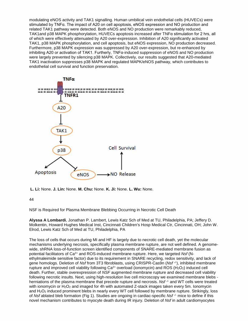
modulating eNOS activity and TAK1 signalling. Human umbilical vein endothelial cells (HUVECs) were stimulated by TNFα. The impact of A20 on cell apoptosis, eNOS expression and NO production and related TAK1 pathway were detected. Both eNOS and NO production were remarkably reduced, TAK1and p38 MAPK phosphorylation, HUVECs apoptosis increased after TNFα stimulation for 2 hrs, all of which were effectively attenuated by A20 over-expression. Inhibition of A20 significantly activated TAK1, p38 MAPK phosphorylation, and cell apoptosis, but eNOS expression, NO production decreased. Furthermore, p38 MAPK expression was suppressed by A20 over-expression, but re-enhanced by inhibiting A20 or activation of TAK1. Furtherly, TNFα-induced suppression of eNOS and NO production were largely prevented by silencing p38 MAPK. Collectively, our results suggested that A20-mediated TAK1 inactivation suppresses p38 MAPK and regulated MAPK/eNOS pathway, which contributes to endothelial cell survival and function preservation.
L. Li: None. J. Lin: None. M. Chu: None. K. Ji: None. L. Wu: None.
44
NSF is Required for Plasma Membrane Blebbing Occurring in Necrotic Cell Death
Alyssa A Lombardi, Jonathan P. Lambert, Lewis Katz Sch of Med at TU, Philadelphia, PA; Jeffery D. Molkentin, Howard Hughes Medical Inst, Cincinnati Children’s Hosp Medical Ctr, Cincinnati, OH; John W. Elrod, Lewis Katz Sch of Med at TU, Philadelphia, PA
The loss of cells that occurs during MI and HF is largely due to necrotic cell death, yet the molecular mechanisms underlying necrosis, specifically plasma membrane rupture, are not well defined. A genome-wide, shRNA loss-of-function screen identified components of SNARE-mediated membrane fusion as potential facilitators of Ca2+ and ROS-induced membrane rupture. Here, we targeted Nsf (N-ethylmaleimide sensitive factor) due to its requirement in SNARE recycling, redox sensitivity, and lack of gene homologs. Deletion of Nsf from 3T3 fibroblasts, using CRISPR-Cas9n (Nsf -/-), inhibited membrane rupture and improved cell viability following Ca2+ overload (ionomycin) and ROS (H2O2) induced cell death. Further, stable overexpression of NSF augmented membrane rupture and decreased cell viability following necrotic insults. Next, using high-resolution live cell microscopy we examined membrane blebs - herniations of the plasma membrane that precede rupture and necrosis. Nsf -/- and WT cells were treated with ionomycin or H2O2 and imaged for 4h with automated Z-stack images taken every 5m. Ionomycin and H2O2 induced prominent blebs in nearly every WT cell followed by membrane rupture. Strikingly, loss of Nsf ablated bleb formation (Fig 1). Studies are ongoing in cardiac-specific Nsf -/- mice to define if this novel mechanism contributes to myocyte death during IR injury. Deletion of Nsf in adult cardiomyocytes

does not result in a baseline phenotype, increasing the translational potential of NSF targeted therapy. In summary, our results identify a new molecular component required for membrane blebbing that occurs during pathogenic cell death.
A.A. Lombardi: None. J.P. Lambert: None. J.D. Molkentin: None. J.W. Elrod: None.
This Research has received full or partial funding support from the American Heart Association.
45
HDAC6 Inhibitor Attenuates Autophagy During Chronic Angiotensin-II Induced Hypertrophic Human Cardiomyocytes
Sheeja Rajasingh, Andras Czirok, Dona Greta Isai, Saheli Samanta, Zhigang Zhou, Buddhadeb Dawn, Johnson Rajasingh, Univ of Kansas Medical Ctr, Kansas City, KS
Introduction: Autophagy is playing a crucial role in cellular functions, and maintaining homeostasis during cardiac hypertrophy, but the mechanism is poorly understood. Studies have shown that pressure overload and chronic ischemia-induced stress leads to an increase in autophagic activity in cardiomyocytes. Reports have demonstrated that STAT3 and Bcl2 participate in the process of autophagy. We plan to study the possible contribution of epigenetic enzymes that mediate transcriptional repression and activation of genes related to autophagy. Here, we have examined the role of histone deacetylase-6 (HDAC6) inhibitor (Tubastatin A- TBSA) in protecting chronic angiotensin II-induced autophagy in human iPSC-derived cardiomyocytes (hiCMCs). Hypothesis: We hypothesize that TBSA treatment mediated protection is through STAT3-Bcl2 signaling in angiotensin II-induced autophagy in hypertrophic hiCMCs. Methods and Results: We have evaluated the TBSA treatment mediated effects in contractility, cell size and mitochondrial membrane potential (∆Ψm) in response to angiotensin II-induced autophagy in hiCMCs. Our immunofluorescence data show that treatment with TBSA significantly reduced the number of autophagosomes in angiotensin II-induced hiCMCs (Fig. A). Moreover, the flow cytometry data show the fluorescence intensity ratio of red and green in JC1 staining suggest that TBSA restores and maintains the angiotensin II-induced loss of ∆Ψm in hiCMCs (Fig B, C). Conclusions: We report for the first time that the role of HDAC6 in regulation of pathological autophagy, thus can acts as a potential therapeutic target for treatment of chronic heart diseases.

S. Rajasingh: None. A. Czirok: None. D.G. Isai: None. S. Samanta: None. Z. Zhou: None. B. Dawn: None. J. Rajasingh: None.
This Research has received full or partial funding support from the American Heart Association.
50
Atrial Fibrillation is Associated with Higher Stroke Rates in Takotsubo Cardiomyopathy
Akintunde Mike Akinjero, Icahn Sch of Med at Mount Sinai, Englewood, NJ; Oluwole Adegbala, Seton Hall Univ Sch of Health and Medical Sciences at Englewood Hosp and Medical Ctr, South Orange, NJ; Nike E Akinjero, Spring Arbor Univ, Spring Arbor, MI; Eseosa Edo-Osagie, Seton Hall Univ Sch of Health and Medical Sciences at Englewood Hosp and Medical Ctr, Englewood, NJ; Tomi Akinyemiju, Univ of Alabama at Birmingham, Birmingham, AL
Background: The prognosis of Takotsubo Cardiomyopathy (TTCM) is worse than in the general population. It is unclear how atrial fibrillation (AF) impacts this prognosis. We sought to evaluate the effect of concurrent AF on outcomes in patients with TTCM. Methods: We used the Healthcare Cost and Utilization Project (HCUP) Nationwide Inpatient Sample (NIS) to extract all hospitalizations between 2007 and 2011 with concurrent diagnosis of AF and TTCM. The ICD-9 CM codes for AF and TTCM were used. We compared patients admitted for TTCM who had coexisting AF to those without. We excluded patients below the age of 18 as well as those diagnosed with TTCM who later underwent percutaneous coronary intervention (PCI). Multivariate regression was used to assess the independent effect of coexisting AF on clinical outcomes (length of stay (LOS), stroke, and in-hospital mortality). Results: A total of 13,136 TTCM patients were studied. Of these, 2,083 (15.86%) had coexisting AF. Compared with those without, TTCM patients with coexisting AF had a greater multivariate-adjusted risk for increased stroke rate (aOR=1.66, 95% CI=1.27-2.18, Table 1). We found no significant association with in-hospital mortality (aOR=1.21, 95% CI=0.96-1.52) or LOS (aOR=1.21, 95% CI= 0.83-1.58). Conclusions: In this large, nationally representative study, we found higher stroke rates in patients with coexisting AF and TTCM. Our findings suggest the need for closer monitoring during hospitalization.
A.M. Akinjero: None. O. Adegbala: None. N.E. Akinjero: None. E. Edo-Osagie: None. T. Akinyemiju: None.
51
Effects of Human Immunodeficiency Virus-1 gp120 Viral Proteins on Endothelial Senescence and microRNA Expression
Jamie Hijmans, Kelly Stockelman, Whitney Reiakvam, Ma'ayan Levy, Lillian Bruster, Zoë Goldthwaite, Tyler Bammert, Jared Greiner, Elizabeth Connick, Christopher DeSouza, Univ of Colorado, Boulder, CO
Human immunodeficiency virus (HIV)-1 is associated with profound endothelial cell damage and dysfunction. HIV-1 envelope glycoprotein 120 (gp120) is a toxic viral protein released from infected cells and promotes endothelial cell dysfunction. Endothelial cell senescence is a key factor underlying endothelial dysfunction and, in turn, vascular disease risk. It is now clear that microRNAs (miRs) play a pivotal role in regulating cellular senescence. The aim of this study was to determine whether gp120 induces endothelial cell senescence; and if so, whether the cellular expression of senescence-related miRs (mir-34a, -146a and -217) are adversely affected by gp120. Cultured HAECs (3rd passage) were plated at a density of 5.0x10⁵ cells/condition. Cells were treated with media alone or media containing gp120: either lavgp120 (X4; 100ng/mL) or Balgp120 (R5; 100ng/mL) for 24 h. Thereafter, cells were stained for senescence-associated β-galactosidase (β-gal) for 14 h. Cells from 5-7 random microscope fields were counted and (%) senescence was calculated as the ratio of β-gal positive cells to total cells. To determine miR expression, RNA was extracted from 1.0x10⁵ cells and was quantified using RT-PCR. Cellular expression was normalized to RNU6 and calculated as fold change in ΔΔCt from control (N=4, experimental units). Both X4 (32.0±0.7% vs 18.3±1.7%) and R5 (30.2±3.8% vs 18.3±1.7%) significantly increased cellular senescence vs control. Moreover, the magnitude of increase (~65%) in senescent cells was similar between the glycoproteins. Cellular expression of pro-senescence miR-34a (1.60±0.04 fold) was increased (~60%; P<0.05) in response to X4 treatment compared with control. However, expression of miR-146a (1.34±0.36 fold) and miR-217 (1.38±0.36 fold) was not significantly affected by X4. In response to R5, cellular expression of miR-34a (1.23±0.07 fold) was significantly increased (~23%) and miR-146a (1.23±0.07 fold) was significantly decreased (~80%); miR-217 was not influenced by R5 (1.05±0.13 fold). These data indicate that HIV-1 viral protein gp120 induces endothelial cell senescence and adversely affects cellular expression of senescence-associated miRs. miR dysregulation may underlie the pro-senescent effects of gp120.
J. Hijmans: None. K. Stockelman: None. W. Reiakvam: None. M. Levy: None. L. Bruster: None. Z. Goldthwaite: None. T. Bammert: None. J. Greiner: None. E. Connick: None. C. DeSouza: None.
52
CagA-Positive Exosomes From Patients With Helicobacter Pylori Infection Impair Endothelial Function in Association With Increased Gata-3 Expression
Xiujuan Xia, Linfang Zhang, Tingzi Hu, Jingshu Chi, Canxia Xu, Zhenguo LIU, Ohio State Univ Medical Ctr, Dublin, OH
A significant link between atherosclerosis and associated cardiovascular diseases especially coronary artery diseases (CAD) and infection with H. pylori (especially CagA-positive) has been established clinically. However, how H. pylori infection leads to atherosclerosis and CAD is unclear. It is well known that endothelial injury and dysfunction plays a critical role in the development of atherosclerosis and CAD. The present study was designed to determine if the effect of cytotoxin associated gene A (CagA) on vascular endothelial cells and related mechanisms. Exosomes were prepared from patients with CagA-positive H. pylori infection and CagA-negative H. pylori infection, and analyzed with electronic microscope and nanoparticle tracking analysis. The presence or absence of CagA in the exosomes from patients was confirmed using Western blot. The exosome preparations were then incubated with human umbilical endothelial cells (HUVEC) for 24 hours. PBS and exosomes from patients without H. pylori infection were used as controls. It was found that the CagA-positive exosomes, not CagA-negative exosomes, significantly inhibited the proliferation of HUVECs and VEGF production in a dose-dependent manner. Incubation with the CagA-positive exosomes, not CagA-negative exosomes, also significantly increased the production of the inflammatory factor CHI3L1 from HUVECs associated with increased expression of

GATA-3. The data from the present study demonstrated that CagA-positive exosomes, not CagA-negative exosomes, from patients with H. pylori infection significantly impaired endothelial function in association with increased expression of GATA-3. Further studies are needed to investigate the mechanisms for the action of CagA-positive exosomes on endothelial cells.
X. Xia: None. L. Zhang: None. T. Hu: None. J. Chi: None. C. Xu: None. Z. Liu: None.
53
Implantation of Loop Recorder In Patients with Ehlers-Danlos Syndrome
Hemanshi Mistry, Sowmika Lingampally, Amna A Butt, Chandralekha Ashangari, Apeksha Bhattarai, Stacy E Hamid, Amer Suleman, The Heartbeat Clinic, Dallas, TX
BACKGROUND: Patients (pts) with Ehlers-Danlos Syndrome (EDS) have a higher incidence of syncope and have a higher rate of complications from incision related procedures. We serve a large population of pts with EDS and report our experience with pts with EDS who required Implantable loop recorder (ILR). METHODS: We have conducted a retrospective study on a total of 94 pts with ILR implantation from 2014-2016. Out of 94 pts, there were 20 males (average age=50.8+20.143 yrs, 21.28%) and 74 females (average age= 47.59+17.296 yrs, 78.72%). Total of 24 (25.53%) pts with EDS who had ILR over the last 2 years. Pts were grouped into 3 categories (1) pts with EDS where ILR implantation was done with the standard technique (2) Our learning curve & (3) the results after the standardized new technique for ILR. 1. Category 1 pts, we used the standard provided by the manufacturer to create a V-shaped cut, loop recorder was inserted; subsequently Dermabond was applied. 2. Category 2 pts, we modified the technique, made a more horizontal incision with the V-shaped blade and a 4-0 Vicryl suture was used to improve wound healing. 3. Category 3 pts, after standardizing the technique, we used the V-shaped blade provided with 4-0 Vicryl knotless continuous suture to tamponade the bleeding. The Dermabond was applied only after local hemostasis was observed. FINDINGS: When the loop recorder was implanted using the standard technique, more of bleeding, bigger scars and multiple keloid formation was observed. In the category 1, we learned that if proper hemostasis is not obtained during the implantation, the patient tended to have more scars. In category 3, when 1-2 layers of Vicryl sutures were applied, the first layer of sutures were subcutaneous & the second was under the skin and a knot less closure was done, hemostasis was obtained prior to applying Dermabond, patients did not have scar formation. CONCLUSIONS: We hereby conclude that EDS pts who required ILR, the wound should be closed with 1-2 layers of Vicryl using a knot-less closure technique which will leave a more aesthetically acceptable scar. We would also recommend that until further research is done, this same technique should be use in patients with other hereditary connective tissue disorders.
H. Mistry: None. S. Lingampally: None. A.A. Butt: None. C. Ashangari: None. A. Bhattarai: None. S.E. Hamid: None. A. Suleman: None.
54
The Effect of Nicotine and Menthol in Secondhand Smoke on Endothelial Function
Jiangtao Liu*, Pooneh Nabavizadeh*, Ronak Derakhshandeh, Xiaoyin Wang, Stanton A. Glantz, Matthew L. Springer, UCSF, San Francisco, CA
Introduction: We have previously shown that even one minute of exposure to secondhand smoke (SHS) of tobacco and marijuana can impair arterial flow-mediated dilation (FMD) in rats. The identity of specific constituents of SHS that cause vascular toxicity remains unclear. Nicotine is a vasoconstrictor that potentially can affect vascular function. Menthol is a cigarette additive popular with African Americans, women, youth, and other ethnicities. Menthol interacts with smoke constituents and irritants such as acrolein through receptor ion channels and also affects the metabolism rate of nicotine.

Hypothesis: We tested three hypotheses pertaining to acute effects of brief exposures to SHS: (1) nicotine increases the extent of endothelial functional impairment; (2) menthol decreases the extent of endothelial functional impairment; and (3) there is an interaction between the effects of nicotine and menthol. Methods: We exposed 8 groups of rats (n=8-10/group) for 10 minutes to two different levels of SHS from the following four different types of research reference tobacco: conventional nicotine (~15 mg/g tobacco), reduced nicotine (~0.4 mg/g tobacco), and both conventional and reduced nicotine with added menthol (~1.15 mg/g tobacco) in a 2x2 factorial (ANOVA) design. We also examined a clean air negative control group (n=9). FMD was measured before and after exposures in each rat. To calculate FMD, the femoral artery diameter was measured with micro-ultrasound before and after a 5-min transient ischemia induced by an external vascular occluder. FMD was quantified as the % vasodilation. Results: FMD was not affected in the clean air controls (p=0.13). Higher nicotine levels were associated with increased % reduction of FMD impairment (19.22±9.53% vs. 41.07±4.54% (SD); p=0.047). Adding menthol was associated with decreased % reduction of FMD impairment (40.52±4.85% vs. 18.50±9.81%; p=0.048). These two effects were independent (p for nicotine x menthol interaction = 0.6). Conclusion: Reduction of nicotine level and addition of menthol each lessen the acute impairment of FMD in rats exposed briefly to tobacco SHS.
J. Liu*: None. P. Nabavizadeh*: None. R. Derakhshandeh: None. X. Wang: None. S.A. Glantz: None. M.L. Springer: None.
55
Use of a Novel Electrogram Filter to Visualise the Conduction Tissue Signals in the Ventricle in Sinus Rhythm and Arrhythmia: Canine Studies
Deepak Padmanabhan, Chance Witt, Alan Sugrue, Ammar Killu, Niyada Naksuk, Christopher Desimone, Dorothy Ladewig, Joanne Powers, Scott Suddendorf, Samuel Asirvatham, Mayo Clinic, Rochester, MN
Background: Conduction tissue in the ventricles is closely intertwined with myocardium. Owing to high velocity of conduction, there is near simultaneous activation , making it difficult to discern. Restricted sampling rates and limited dynamic range make real time signal processing challenging for these signals in routine recording systems. Objectives: We aimed to use novel signal processing techniques in a new system to isolate the conduction tissue signals from the rest of the myocardial activation. Methods: We used the PURE-EP™ (BioSig Technologies, MN) signal processing system to record endocardial and epicardial signals from 6 anesthetized canines in an acute study setting. A novel filter based on proprietary algorithms to isolate the high frequency signal was applied at sites where the electrogram signals were thought to have a conduction tissue component to them. . In addition, ventricular fibrillation (VF) was induced at the end of the experiment and signals were then recorded from the endocardium and the conduction system. Results: This filter was successfully able to isolate conduction tissue signals in the myocardium from those of the ventricular muscle. It did over sense atrial signals at the basal left ventricular septum owing to the high frequency components of the atrial signal at this location. When applied to epicardial signals, it did not pick up any conduction tissue signals. In VF, we could discern signals originating from the conduction system endocardially. (Figure 1) Conclusions: This filter can isolate conduction tissue signals from those of the myocardium and may prove to be a useful adjunct in mapping arrhythmias originating near the conduction system.

D. Padmanabhan: None. C. Witt: None. A. Sugrue: None. A. Killu: None. N. Naksuk: None. C. Desimone: None. D. Ladewig: None. J. Powers: None. S. Suddendorf: None. S. Asirvatham: None.
56
Individual Susceptibility to Drug-induced QTc Prolongation Did Not Correlate With Subject-specific Induced Pluripotent Stem Cells in a Small Sample of Healthy Subjects
Ksenia Blinova, Dakshesh Patel, Jayna Stohlman, Derek Schocken, Marina Kondratovich, Lars Johannesen, Jose Vicente, David Strauss, USFDA, Silver Spring, MD
Background Induced pluripotent stem cells (iPSCs) carry patient-specific genetic information and have the potential to become an important tool for predicting individual clinical response to therapies in an in vitro assay. Here, we investigate if subject-specific iPSC cardiomyocytes (iPSC-CMs) derived from healthy volunteers could be used to predict individual response to 2 QT-prolonging drugs which they received in a clinical trial. Methods and results Independent stem cell manufacturer (Stem Cell Theranostics) used commercially available Sendai virus encoding for Oct4, Sox2, Klf4, and c-Myc to reprogram subject’s peripheral blood mononuclear cells (PBMCs) into iPSCs. They then applied chemically defined growth factors, and serum-free protocols to successfully differentiate iPSCs into iPSC-CMs for 17 out of 20 subjects (one batch per subject) enrolled in a randomized, double-blind, placebo-controlled, cross-over trial of dofetilide and moxifloxacin. Effects of the same 2 drugs on cellular action potential duration (APD) were recorded for 16 iPSC-CMs lines using a high-throughput optical imaging system (CellOPTIQ, Clyde Biosciences) using consistent in vitro protocols, including reproducible drug doses and exposure, cell culture and recording conditions. Both dofetilide (0.5-8 nM) and moxifloxacin (10-200 µM) induced dose-dependent prolongation of rate-corrected action potential duration (APD) in each iPSC-CM line. iPSC-CM drug responses were compared with individual clinical response represented by the slope of the linear regression fit through baseline and placebo-controlled drug-induced changes in QTc interval vs. plasma drug concentration. We found no statistical significant correlation between subject-specific iPSC-CMs drug-induced APD prolongation and individual QTc prolongation to either moxifloxacin [Spearman’s ρ = 0.191] or dofetilide [Spearman’s ρ = 0.068]. Conclusion This study failed to find correlation between clinical and stem cell drug responses in a small sample of healthy

subjects. More experiments with a larger sample size and iPSC-CMs batch-to-batch variability controls are required to further investigate the potential of patient-derived stem cells to predict individual clinical response.
K. Blinova: None. D. Patel: None. J. Stohlman: None. D. Schocken: None. M. Kondratovich: None. L. Johannesen: None. J. Vicente: None. D. Strauss: None.
57
Pharmacologic Inhibition of BK Channels Affects Cardiac Function
Nishi H Patel, Justin H Johannesen, Sumanta K Goswami, Devasena Ponnalagu, Andrew R Kohut, Harpreet Singh PhD, Drexel Univ, Philadelphia, PA
Calcium and voltage activated potassium channels (BK) are ubiquitously distributed, and have been shown to play roles in cardioprotection and cardiac function. To date, however, the effect of BK channel activation or inhibition in cardiac cells has only been studied using in vitro or ex vivo models. In this study, for the first time we demonstrate the role of BK channels on cardiac function in vivo using an antagonist, Paxilline. Male Sprague Dawley rats had an initial echocardiogram to establish baseline values of cardiac function. Afterwards, the femoral vein was isolated and rats injected with either DMSO or Paxilline of various concentrations ranging from 2 ng/mL/kg to 500 µg/mL/kg. Echocardiograms were then repeated 30 seconds, 15 minutes, and 24 hours after injection. Some rats at higher drug dosage suffered cardiac arrest within 15 minutes. In a separate experiment, a pressure transducer was inserted into the left ventricle (LV) via cannulation of the carotid artery. LV Developed Pressure (LVDP) and Heart Rate (HR) were continuously recorded as rats were infused with DMSO or Paxilline via the femoral vein. In agreement with BK null mutant mice, we found that rats receiving Paxilline had a significant decrease in HR compared to controls, as BK is known to play a role in SA nodal cells. In rats that lived, the transient decrease in HR averaged ~26.20%. Rats receiving Paxilline also had diminished Ejection Fraction (EF) ranging from 26.53% to 100% fold-decrease compared to controls. The decrease in EF occurred in a dose dependent manner, with rats receiving higher doses (> 13 ng/mL/kg) of Paxilline typically suffering cardiac arrest within 15 minutes. Measurements using pressure transduction of the LV showed results similar to those rats assessed using echocardiography, with a significant decrease in LVDP ranging from 36.9% to 100% decrease. Rats that received lower doses (<4 ng/mL/kg) of Paxilline showed improvement in their cardiac function back to baseline by 24 hours post injection, suggesting that the effects we observed are transient and that BK inhibition is reversible. We have shown that inhibition of BK is detrimental to cardiac function, suggesting that BK channels in cardiac tissue are not only functional but essential to maintain normal cardiac function.
N.H. Patel: None. J.H. Johannesen: None. S.K. Goswami: None. D. Ponnalagu: None. A.R. Kohut: None. H. Singh: 2. Research Grant; Significant; NIH NHLBI R01HL133050, AHA Grant in Aid 16GRNT29430000, AHA National Scientist Development Grant 11SDG7230059.
This Research has received full or partial funding support from the American Heart Association.
58
Extracellular RNA Response to Acute Exercise
Ravi V Shah, Massachusetts General Hosp, Newburyport, MA; Avash Das, Olivia Ziegler BS, Ashish Yeri, Massachusetts General Hosp, Boston, MA; Amanda Courtright-Lim, Translational Genomics Inst., Phoenix, AZ; Ernest Gervino, Jeffrey Ocel, Pablo Pinzon-Quintero, Beth Israel Deaconess Medical Ctr, Boston, MA; Alexander Pico, Gladstone Insts, San Francisco, CA; Mun Chun Chan, Georgetown Univ, Washington DC, DC; Gregory Lewis MD, Massachusetts General Hosp, Boston, MA; Ionita Ghiran MD, Beth Israel Deaconess Medical Ctr, Boston, MA; Kendall Jenson PhD, Translational Genomics Inst, Phoenix, AZ; Saumya Das, Massachusetts General Hosp, Boston, MA
The benefits of acute exercise on distant organs (including the neurocognitive system) have been recognized, suggesting existence of biomarkers that mediate exercise benefits in target organs.

Extracellular RNAs (ex-RNAs) are RNA molecules stable in circulation, capable of regulating gene expression across multiple target organs at a distance. In 26 individuals without clinical cardiovascular disease (myocardial infarction or heart failure), we performed small non-coding RNA sequencing (RNA-seq) from plasma before and after symptom-limited treadmill exercise, with validation of selected microRNAs (miRNAs) in pulmonary arterial blood from 59 individuals referred for cardiopulmonary exercise testing. Acute exercise produced widespread shifts in the plasma ex-RNA transcriptome, with a host of novel ex-RNA subtypes with unknown function (e.g., yRNAs). miR-181b (involved in silencing of NFkB-mediated inflammation in obesity and vascular disease) was significantly increased at peak exercise (relative to rest) in both our discovery (2.3-fold increase after exercise, P=8.46x10⁻¹¹) and validation cohort (P=0.02). In a mouse model of acute exercise, we found that skeletal muscle miR-181b expression increased in young but not old mice after exercise, with a corresponding reduction in miR-181b mRNA targets. These results provide the first unbiased demonstration of diversity in ex-RNA release after acute exercise in humans, and suggest anti-inflammatory benefits of acute exercise may be mediated in part by circulating non-coding RNAs.
R.V. Shah: None. A. Das: None. O. Ziegler: None. A. Yeri: None. A. Courtright-Lim: None. E. Gervino: None. J. Ocel: None. P. Pinzon-Quintero: None. A. Pico: None. M. Chan: None. G. Lewis: None. I. Ghiran: None. K. Jenson: None. S. Das: None.

59
Inflammasome Activation Promotes the Arrhythmogenesis of Atrial Fibrillation
Na Li, Larry Scott Jr., Baylor Coll Med, Houston, TX; Tina Veleva, Dobromir Dobrev, Univ Duisburg-Essen, Essen, Germany; Xander Wehrens, Baylor Coll Med, Houston, TX
Background: Inflammation is a known risk factor of atrial fibrillation (AF), the most common arrhythmia; however, the mechanistic link between the inflammatory signaling and the pathophysiology of AF has not been established. ‘NACHT, LRR and PYD domains-containing protein 3’ (NLRP3) inflammasome is a signaling platform that is responsible for the activation of caspase-1 and interleukin (IL)-1b release. The activity of NLRP3 inflammasome is enhanced in atrial tissues of paroxysmal or long-lasting persistent AF patients. Thus, we tested the hypothesis that activation of NLRP3 inflammasome promotes the development of AF. Results: To elucidate the role of NLRP3 inflammasome in cardiomyocytes (CMs) and AF development, a CM-specific knockin murine model expressing a constitutive active NLRP3 (aMHC:NLRP3A350V/+, CKI) was developed. At 3-month old, telemetry recordings showed that 100% of CKΙ mice (n=5) developed premature atrial contractions (PACs), whereas only 25% of control mice (n=4, P<0.05) had PACs. Rapid atrial pacing induced AF in 89% of CKI mice (n=9), a much higher incidence than control mice (20%, n=5, P<0.05). Level of the active caspase-1 was increased in atrial tissues of 3-month old CKI mice, prior to a detectable increase in the level of macrophage marker at 7-month old, suggesting that the onset of PACs and AF vulnerability is not associated with the activated macrophages. Compared to the control mice, 3-month old CKI mice exhibited atrial hypertrophy, abnormal Ca2+ release via RyR2, and shortening of atrial effective refractory period, which were associated with the upregulation of Mef2c, Ryr2, Kcna5 (encoding Kv1.5), Kcnj 3 (encoding Kir3.1) and Kcnj5 (encoding Kir3.4) mRNA. Lastly, inflammasome inhibitor MCC950 (i.p., 10mg/kg, 10 days) reduced the AF inducibility in CKI mice (0%, n=3, P<0.05 vs vehicle-treated CKI). Conclusion: Activation of NLRP3 inflammasome promotes structural and electrical remodeling, permissive to the AF development. In addition to its canonical function, NLRP3 inflammasome may exhibit alternative function in regulating gene transcription. Our study establishes a mechanistic link between the inflammatory signaling and the pathogenesis of AF, and the inhibition of NLRP3 may become a novel anti-AF therapy.
N. Li: 2. Research Grant; Significant; AHA SDG, NIH R56. L. Scott Jr.: None. T. Veleva: None. D. Dobrev: None. X. Wehrens: None.
This Research has received full or partial funding support from the American Heart Association.
60
Inducible Arrythmogenicity of Direct Current Electroporation Ablation: Insight from a Series of Acute Canine Studies
Alan Sugrue, Chance Witt, Christopher V. DeSimone, Deepak Padnanabhan, Ammar Killu, Niyada Naksuk, Suraj Kapa, Samuel J. Asirvatham, Mayo Clinic Rochester, Rochester, MN
Background: The use of direct current electroporation has the potential for significant utility because of its non-thermal approach to tissue destruction. However, the fear of inducibility of cardiac arrhythmias (particular ventricular fibrillation) when using electroporation remains of concern due to membrane poration and ion flux during periods of vulnerability occurring in ventricular repolarization. Objectives: Critically examine the incidence of arrhythmias in a series of acute canine studies to retrospectively determine cause and safe electoporative dosing margins. Methods: We performed electroporation ablation in 6 acute canine studies. These were experimental studies performed at sites critical in arrhytmogenesis. Sites included the pulmonary veins, left atrial appendage, superior vena cavae, right atrium and ventricle. Electroporation was delivered using an ECG gating algorithm so that QRS complexes are tagged and direct current energy is not delivered during the vulnerable portion of the T wave. Results: In 6 acute canine experiments, we delivered a total of 62 electroporation applications for ablation purposes. The average electroporation dosage delivered involved an average of 1427 Volts (range 750-3000 V), Pulse length of 100 ms, and number of pulses of 20.2 (range 10-100). AF was induced in 27.4% of electroporiatve applications. Atrial flutter/tachycardia occurred in 8.1%. VF occurred in only one application at a location of the left superior pulmonary vein. Conclusion: These data suggest

that induction of VF is relatively uncommon with ECG gating and highlight its importance when using this modality. However, the induction of AF occurs with higher frequency. The actual mechanism as to why this occurs requires further systematic study.
A. Sugrue: None. C. Witt: None. C.V. DeSimone: None. D. Padnanabhan: None. A. Killu: None. N. Naksuk: None. S. Kapa: None. S.J. Asirvatham: 7. Ownership Interest; Modest; Intellectual Property Rights; Mayo Clinic Venture.
62
Sildenafil Inhibits Neointimal Hyperplasia After Angioplasty, and Reduces Platelet Aggregation
Han Mo Yang, Sooryeonhwa Jin, Hyo-Soo Kim, Seoul Natl Univ Hosp, Seoul, Korea, Republic of; Sahmin Lee, Asan Medical Ctr, Seoul, Korea, Republic of
Background: Restenosis and stent thrombosis after stent implantation are one of the devating issues in interventional cardiology. Sildenafil has shown its effect in reducing cardiac hypertrophy as well as improving erectile dysfunction through protein kinase G (PKG) activation. Some studies have demonstrated that PKG occupies a central switching role in modulating vascular smooth muscle cell (VSMC) phenotype in response to vascular injury. In this study, we investigated the effects of PKG activation by sidenafil on platelet aggregation and neointimal hyperplasia. Methods and Results: In terms of restenosis after vascular injury, sildenafil significantly reduced neointimal hyperplasia in rat carotid arteries compared to control group. This effect of sildenafil was accompanied by the reduction of viability, cell cycle progression, and migration of VSMCs. This was also confirmed in the injured arteries in vivo. Further studies showed that the increased PKG activity by sildenafil inhibited PDGF-stimulated phenotype change of VSMCs from a contractile to a synthetic form. Conversely, the use of PKG inhibitor or gene transfer of dominant-negative PKG reversed the effects of sildenafil, resulting in the increased viability of VSMCs and neointimal formation. In addition, the mice treated with sidenafil showed the facilitated re-endothelialization, compared to control group. Furthermore, we confirmed the effect of sidenafil through PKG activation using cGK-KO mice. Interestingly, sildenafil significantly inhibited platelet aggregation induced by ADP or thrombin. This effect was reversed by PKG inhibitor, suggesting that sildenafil inhibits platelet aggregation via PKG pathway. Furthermore, assays for VASP phosphorylation and P-selectin activation showed the same inhibitory effect of sildenafil on platelet activation. Conclusions: This study showed that sildenafil inhibits not only platelet aggregation, but also neointimal hyperplasia through the PKG pathway. These findings suggest that sildenafil could be a promising candidate drug of drug-eluting stents for the prevention of restenosis without other complications such as stent thrombosis.
H. Yang: None. S. Jin: None. H. Kim: None. S. Lee: None.
63
A Novel Inotropic Gene Therapy Approach for Aging Induced Cardiac Dysfunction
Vikram Shettigar, Bo Zhang, Svetlena Tikunova, Brandon Biesiadecki, Paul Janssen, Noah Weisleder, Jonathan P Davis, Mark T Ziolo, Ohio State Univ, Columbus, OH
We have developed a novel approach to increase cardiac contractility without the deleterious effects of existing inotropes (beta agonists, Ca2+ sensitizers). Using the strengths of protein engineering, we have shown that cardiac function can be directly controlled and strongly influenced by manipulating the Ca2+ dependent switch in cardiac muscle, troponin C (TnC). Our smartly formulated construct, L48Q TnC, is a strong Ca2+ sensitizer. Although effective acutely, long-term inotropic support is detrimental. We transduced mice with AAV9 L48Q TnC at 3 months of age and serially performed echocardiography for 2 years. The current dogma would suggest that chronic Ca2+ sensitization with L48Q TnC should result in a hypertrophic cardiomyopathy and compromised function. Astonishingly, the L48Q TnC mice maintained ejection fraction unlike the progressive decline observed in the controlled, age-matched mice. Furthermore, L48Q TnC mice had an increased rate of survival. Considering cardiovascular disease is an elderly issue, we reasoned L48Q TnC would be beneficial in other age-related cardiovascular problems
(i.e., hypertension and pressure-overload). We tested this in two models, transverse aortic constriction (TAC, pressure-overload) and angiotensin II infusion (AngII, hypertension). Consistent with the aging results, L48Q TnC mice displayed enhanced cardiac function (ejection fraction, contractility, relaxation), performance (VO2max, distance run before exhaustion), less adverse remodeling, and enhanced survival. These studies provide a promising approach (i.e., inotropic support via L48Q TnC) that can be used therapeutically to enhance heart function in a variety of cardiomyopathies associated with aging.
V. Shettigar: None. B. Zhang: None. S. Tikunova: None. B. Biesiadecki: None. P. Janssen: None. N. Weisleder: None. J.P. Davis: 2. Research Grant; Significant; NIH. M.T. Ziolo: 2. Research Grant; Significant; NIH.
64
HDL Glycosylation is Associated with HDL's Anti-Inflammatory Function and is Responsive to Dietary Intervention
Angela M Zivkovic, Christopher Rhodes, Romina Sacchi, Elizabeth Beals, Lisa Sawrey-Kubicek, Chenghao Zhu, John K Muchena, Guillaume Luxardi, UC Davis, Davis, CA; Emanual Maverakis, UC Davis Sch of Med, Sacramento, CA; Carlito B Lebrilla, UC Davis, Davis, CA
Although HDL cholesterol (HDL-C) levels are protective against cardiovascular disease (CVD) risk, raising HDL-C pharmaceutically has not led to improvements in cardiovascular outcomes. It is becoming clear that HDL composition and function are more important than HDL concentrations in determining risk. This study set out to determine whether glycosylation differences in HDL-associated glycoproteins affect HDL’s anti-inflammatory function. HDL were purified from healthy subjects (n=10), who consumed in randomized order a fast food (FF) diet and a Mediterranean (Med) diet for 4 days, with a 4-day washout between arms. All foods were provided in this isocaloric cross-over intervention trial, which was approved by the UC Davis IRB. Concentrations of HDL-bound serum amyloid A (SAA), apolipoprotein A-I (ApoA-I), apolipoprotein C-III (ApoC-III), α-1-antitrypsin (A1AT), and α-2-HS-glycoprotein (A2HSG); and the site-specific glycosylation of ApoC-III, A1AT, and A2HSG were measured. Secretion of TNF-α in stimulated monocytes was measured to assess HDL anti-inflammatory function. HDL glycosylation was altered by the dietary interventions and correlated with changes in the amount of TNF-α secreted by stimulated monocytes. HDL glycosylation profiles were different in response to the FF diet vs. the Med diet. HDL with a diminished capacity to suppress TNF-α secretion were enriched in ApoC-III and desialylated A2HSG, depleted in A1AT, and had lower levels of sialylation across glycoproteins. Our results demonstrate that HDL glycoprotein composition, including site-specific glycosylation, is responsive to dietary intervention and correlates with HDL’s ability to modulate TNF-α response in stimulated monocytes. These data suggest that the measurement of HDL glycosylation profiles may be useful in stratifying CVD risk and detecting individuals with impaired HDL anti-inflammatory function.
A.M. Zivkovic: None. C. Rhodes: None. R. Sacchi: None. E. Beals: None. L. Sawrey-Kubicek: None. C. Zhu: None. J.K. Muchena: None. G. Luxardi: None. E. Maverakis: None. C.B. Lebrilla: None.
65
Anemia-related Heart Failure in Sub-saharan African Sickle Cell Disease Patients
Adebayo C Atanda, Yahya Aliyu, Howard Univ Hosp, Washington, DC, DC; Oluwafunmilayo Atanda, Queens Heart Inst, New York, NY; Aliyu Babadoko, Aisha Suleiman, Aisha Mamman, Dept of Pathology Ahmadu Bello Univ, Zaria, Nigeria; Zakari Aliyu, Howard Univ Hosp, Washington, DC, DC
Introduction: Anemia has been implicated in heart failure. Existing literatures, involving predominantly African-Americans, suggests that Sickle Cell Disease (SCD) maybe linked to various cardiovascular complications including pulmonary hypertension and left venticular dysfunction. Peculiarly, our study involves exclusively Sub-Saharan population. Method: We conducted a cross sectional observational study of 208 hydroxyurea-naive consecutive SCD patients aged 10-52 years at steady state and 94 healthy non-matched controls who were studied in an out patient clinic in Sub-Saharan Africa. SCD patients were required to have electrophoretic or liquid chromatography documentation of major sickling

phenotypes. Control group was required to have non-sickling phenotypes. Cardiac measurements were performed with TransThoracic Echo according to American Society of Echocardiography guidelines. Hemoglobin level was also obtained. Results: Hemoglobin level in SCD group (8.5+/- 1.5) was significant (P<0.001) compared to control (13.8+/- 1.7). Although SCD group had significantly higher values of left ventricular (LV) size, there was no qualitative evidence of LV dysfunction. SCD group had higher values of Ejection Fraction but not statistically significant. There was no evidence of LV wall stiffening to impair proper filling in SCD group, with the ratio of early to late ventricular filling velocities, E/A ratio elevated (1.7+/-0.4 compared to 1.6+/- 0.4; P=0.010). Right ventricular systolic pressure was determined using the formula of 4x Tricuspid Reugurgitant jet (TRV) square as an indirect measurement of Pulmonary arterial systolic pressure. SCD patients had significantly higher mean±SD values for tricuspid regurgitant jet velocity than did the controls (2.1±0.6 vs. 1.8±0.5; p= 0.001). Within the SCD group, there was no clear pattern of worsening diastolic function with increased TRV. Furthermore, E/A had a significant positive relationship with jet velocity in bivariate analysis (R=0.20; P=0.013). Conclusions: We were unable to demonstrate existence of anemia-associated left ventricular dysfunction in Sub-Saharan African with SCD. Further studies is required to highlight the reason behind this finding.
A.C. Atanda: None. Y. Aliyu: None. O. Atanda: None. A. Babadoko: None. A. Suleiman: None. A. Mamman: None. Z. Aliyu: None.
67
Polycystin-1 is Required for Normal Cardiomyocyte Excitability and Heart Function
Francisco J. Altamirano, Gabriele G Schiattarella, Sergiy Kyrychenko, Jay W. Schneider, Sergio Lavandero, Thomas G. Gillette, Joseph A. Hill, UT Southwestern Medical Ctr, Dallas, TX
Mutations in the gene (Pkd1) encoding Polycystin-1 (PC-1) leads to autosomal dominant polycystic kidney disease (ADPKD). Although renal failure is the leading cause of death, cardiovascular alterations are described in ADPKD patients. Our lab has previously demonstrated that PC-1 is involved in cardiac L-type Ca2+ channel (LTCC) protein stability, which is required for hypertrophic growth during mechanical stretch. PC-1 is expressed in cardiomyocytes and its gene ablation in mice decreases basal cardiac function. Here, we hypothesized that PC-1 regulates excitation-contraction coupling in cardiomyocytes. To test this hypothesis, we studied mice harboring floxed Pkd1 alleles and a Cre transgene (α-MHC promoter) to engineer cardiomyocyte-specific PC-1 KO (F/F αMHC Cre, PC-1 cKO) mice. Human-derived cardiomyocytes were differentiated from embryonic stem cells (H9), and PC-1 was depleted using specific siRNA constructs. Fractional shortening measured by echocardiography was significantly reduced under basal conditions in PC-1 cKO mice compared with controls. Likewise, adult ventricular cardiomyocytes isolated from PC-1 cKO mice manifested decreased contractility (measured by Ionoptix) and a reduced Ca2+ transients (measured using Fluo-8AM and line scan) during pacing (0.5 Hz). Unexpectedly, assessment of both Ca2+ stores (caffeine-evoked Ca2+ transients) and Ca2+ entry (LTCC currents, whole-cell voltage clamp) showed no differences between PC-1 cKO and control cardiomyocytes. Remarkably, Ca2+ transients evoked by high K+ depolarization were similar across genotypes, suggesting an alteration in excitability. Indeed, action potential duration (measured by current clamp or FluoVolt) was shorter in cardiomyocytes lacking PC-1 (mouse adult cardiomyocytes and hESC-derived cardiomyocytes). Moreover, PC-1 cKO mice manifested shorter QT intervals on ECG, and a pharmacological intervention with quinidine improved cardiac function and cardiomyocyte contractility to near-normal levels. Our findings uncover a novel role for PC-1 as an important regulator of cardiac function, required to fine-tune both excitability and contraction in ventricular myocytes.
F.J. Altamirano: None. G.G. Schiattarella: None. S. Kyrychenko: None. J.W. Schneider: None. S. Lavandero: None. T.G. Gillette: None. J.A. Hill: None.
This Research has received full or partial funding support from the American Heart Association.
69
Cardiac Fibrosis Induced by Mkk6-p38 Activation Alters Myocardial Stiffness and Myofilament Function
Farid Moussavi-Harami, Maria V Razumova, Jordan M Klaiman, Darrian Bugg, Michael Regnier, Jennifer Davis, Univ of Washington, Seattle, WA
Heart failure with preserved ejection fraction (HFpEF) accounts for at least half of the patients with heart failure, but to date there are no proven therapies for these patients. Cardiac fibrosis and increased collagen content plays an important role in the pathophysiology of HFpEF. Studies in human HFpEF patients have indicated increased cardiac fibrosis, myocardial stiffness and myofilament calcium sensitivity. However, it is unclear how and if cardiac fibrosis directly alters myofilament function. In this study we used a new model of cardiac fibrosis genetically induced by the constitutive activation of MKK6-p38 (MKK6-Tg) signaling specifically in resident cardiac fibroblasts to determine the effect of fibrosis on myofilament stiffness and function. The MKK6-Tg mice develop interstitial and perivascular fibrosis in the heart accompanied with severe diastolic dysfunction, although systolic function remains normal. We measured the passive stiffness and isometric contractile properties of demembranated papillary muscle preparations. Our results show an increase in passive stiffness in demembranated papillary muscles from MKK6-Tg mice compared to NTG littermates when stretched to 24% from sarcomere length of 2.0 μM. Passive tension at SL=2.3 was significantly increased in MKK6-Tg mice (28±4 vs. 13±3 mN/mm2, P<0.05) compared to NTG mice, which was ascribed to changes in Titin isoform as well as extracellular matrix composition. The MKK6-Tg mice also show an increase in Ca2+ sensitivity (pCa50=5.72±0.05 vs. 5.56±0.02, P<0.05). This was associated with an increased rate of force redevelopment in MKK6-Tg mice compared to NTG littermates (kTR=35±8 vs. 22±3 s-1). Towards understanding the mechanism of these myofilament changes we performed phosphate affinity electrophoresis and saw decreased phosphorylation of both Troponin I and Troponin T in MKK6-Tg hearts. There was no change in troponin I phosphorylation level at Ser23/24, indicating role of none-PKA mediated phosphorylation in mechanical changes seen in this model.
F. Moussavi-Harami: None. M.V. Razumova: None. J.M. Klaiman: None. D. Bugg: None. M. Regnier: None. J. Davis: None.
70
Protein Phosphatase 1 Contributes to Atrial Stunning in Atrial Fibrillation
Srikanth Perike, Univ of Illinois at Chicago, Chicago, IL; Xander Wehrens, Baylor Coll of Med, Houston, TX; Dawood Darbar, Mark McCauley, Univ of Illinois at Chicago, Chicago, IL
Background: Atrial fibrillation (AF) is the most common cardiac arrhythmia, and increases a patient's stroke risk five-fold. Reduced atrial contractility (stunning) is observed in AF and contributes to stroke risk; however, the mechanisms responsible for atrial stunning in AF are unknown. Recent data from our laboratory indicate that protein phosphatase 1 (PP1) dephosphorylation of myosin light chain 2a (MLC2a) may contribute to atrial stunning in AF. Objective: To determine how the PP1 regulatory subunit 12C (PPP1R12C) and catalytic (PPP1c) subunits modify atrial sarcomere phosphorylation in AF. Methods: We evaluated the protein expression, binding and phosphorylation among PPP1R12C, PPP1c, and MLC2a in transfected HL-1 cells, murine atrial tissue (Pitx2null+/− mice, with a genetic predisposition AF), and in HEK cells. An inhibitor of PPP1R12C phosphorylation, BDP5290, was used to enhance the PPP1R12C-PPP1C interaction. Results: In Pitx2null+/− mice, PPP1R12C was increased by 2-fold (P<0.01) and associated with a 40% reduction in S-19-MLC2a phosphorylation versus WT mice (P<0.058). BDP5290 increased PPP1R12C-PPP1C binding by >3-fold in HL-1 cells (P<0.01). BDP5290 reduced MLC2a phosphorylation by 40% through an enhanced interaction with PPP1R12C by >3-fold in HEK cells (P <0.01). Conclusion: In Pitx2null+/- mice, increased expression of PPP1R12C is associated with PP1 holoenzyme targeting to sarcomeric MLC2a, and is associated with reduced S19-MLC2a phosphorylation. Additionally, BDP5290 enhances the PPP1R12C-PPP1C interaction and models PP1 activity in AF. Future studies will examine the effects of both AF and BDP5290 upon atrial contractility in vitro.
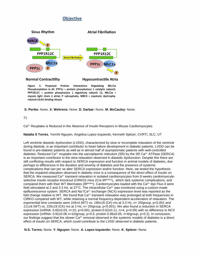
S. Perike: None. X. Wehrens: None. D. Darbar: None. M. McCauley: None.
71
Ca2+ Reuptake is Reduced in the Absence of Insulin Receptors in Mouse Cardiomyocytes
Natalia S Torres, Yennhi Nguyen, Angelica Lopez-Izquierdo, Kenneth Spitzer, CVRTI, SLC, UT
Left ventricle diastolic dysfunction (LVDD), characterized by slow or incomplete relaxation of the ventricle during diastole, is an important contributor to heart failure development in diabetic patients. LVDD can be found in pre-diabetic patients as well as in almost half of asymptomatic patients with well-controlled diabetes. Reduced Ca2+ reuptake into the sarcoplasmic reticulum (SR) by the SR Ca2+ ATPase (SERCA) is an important contributor to the slow relaxation observed in diastolic dysfunction. Despite this there are still conflicting results with respect to SERCA expression and function in animal models of diabetes, due perhaps to differences in the duration and severity of diabetes and the presence of systemic complications that can per se alter SERCA expression and/or function. Here, we tested the hypothesis that the impaired relaxation observed in diabetic mice is a consequence of the direct effect of insulin on SERCA. We measured Ca2+ transient relaxation in isolated cardiomyocytes from 8 weeks cardiomyocyte-selective insulin receptor knockout (CIRKO) mice (Cre-IRlox/lox),, which lack systemic complications, and compared them with their WT littermates (IRlox/lox). Cardiomyocytes loaded with the Ca2+ dye Fluo-4 were field stimulated at 2 and 3.3 Hz, at 37°C. The intracellular Ca2+ was monitored using a custom-made epifluorescence system. SERCA and Na+/Ca2+ exchanger (NCX) expression level was reported as the fold change relative to WT. We found that Ca2+ transient relaxation was prolonged at both frequencies in CIRKO compared with WT, while retaining a normal frequency-dependent acceleration of relaxation. The exponential time constants were 104±4 (WT) vs. 190±16 (CK) ms at 3.3 Hz, n= 20/group, p<0.001 and 111±6 (WT) vs. 226±19 (CK) ms at 2 Hz, n= 20/group, p<0.001). We also found a reduction in SERCA expression (mRNA: 0.65±0.03, n=10, p<0.001; protein:0.62±0.11, n=4, p<0.05) with no difference in NCX expression (mRNA: 0.92±0.06 n=10/group, p=0.5; protein 0.96±0.05, n=6/group, p=0.2). In conclusion, our findings suggest that the slower Ca2+ removal observed in the systemic models of diabetes is a direct effect of insulin on SERCA, which could contribute to the LVDD observed in diabetic patients.
N.S. Torres: None. Y. Nguyen: None. A. Lopez-Izquierdo: None. K. Spitzer: None.

72
Human Vascular Smooth Muscle Gene Expression Profiling Between Incracranial and Extracranial Vasculature
Hyung-Seok Kim, Jong Tae Park, Chonnam Natl Univ Medical Sch, Gwangju, Korea, Republic of; Joo Young Na, Chonnam Natl Univ Hosp, Gwangju, Korea, Republic of; Man Seok Park, Chonnam Natl Univ Medical Sch, Gwangju, Korea, Republic of
Background: Intracranial arteries (IA) have unique histological, pharmacological, and pathological characteristics when compared to extracranial arteris (EA). Despite its importance, little is known about the molecular characteristics of those vessels. In this study, we carried out a genome-wide characterization on expressions of mRNAs, and performed integrative analyses in conjunction with changes of the transcriptome between IA and EA vascular smooth muscle cells. Methods: mRNA expression screening was performed from same autopsied body (n=3) using Agilent Human mRNA arrays for IA (middle cerebral a.) and EA (left ant. descending a. of coronary a.) vascular smooth muscle (vSMC) layer. Total gene expression compared with intracranial vs. extracranial vSMC. Results: As compared to EA, we identified 304 mRNA that were differentially expressed in the IA vSMC (p<0.05 and fold change >2), including 224 upregulated and 80 downregulated. Through this comparison we have identified angiogenesis, cell migration, cell proliferation, neurogenesis, and inflammatory response genes whose expression is enriched in IA vSMC. This analysis has identified that RAR activation, and Caveolar-mediated endocytosis signaling cascade is specifically enriched at the IA vSMC, implicating this pathway in regulating this arterial system. Conclusions: This dataset provides a resource for understanding the different arterial regulation and disease susceptibility in IA and EA, especially atherosclerosis and diabetes.Funding: This study was supported by a grant from the Basic Science Research Program through the National Research Foundation of Korea (NRF) funded by the Ministry of Education, Science and Technology (Kim HS, NRF-2016R1A2B4008316).
H. Kim: None. J. Park: None. J. Na: None. M. Park: None.
73
Pre- and Post-Translational Regulation of Lysyl Oxidase/β1-Intergrin in Aortic Vascular Smooth Cell of Hypertension
Ben Ma, Ning Zhou, Shaunrick Stoll, Hongyu Qiu, Loma Linda Univ, Loma Linda, CA
Background An increase in aortic stiffness is a fundamental component of hypertension. We previous showed that aortic vascular smooth muscle cells (VSMCs) play an causative role in aortic stiffening not only due to the increased intrinsic stiffness in VSMC itself but also due to its interaction with extracellular matrix (ECM). However, the molecular mechanism involved is unclear. Here, we test the hypothesis that the aortic VSMCs mediates pre- and post-translational regulation of Lysyl Oxidase (Lox)/β1intergrain pathway which may be one of the mechanisms of VSMCs-ECM remodeling in hypertension. Methods and Results 4 months old male spontaneously hypertensive rats (SHR) and normotensive Wistar-Kyoto (WKY) rats were studied. Aortic pressure and stiffness were measured with an invasive catheter and Doppler imaging echocardiography, respectively. Compared to WKY, SHR showed a significant higher aortic pressure (mean arterial pressure (MAP) 138.7±11.8 vs 102.7±7 mmHg, (p<0.01) and a greater aorta stiffness reflected by 2.5-fold higher arterial stiffness index (ASI) in SHR vs WKY (p<0.01). VSMCs were isolated from thoracic aorta, then cultured and measured at passages 2 to 4. Real time PCR and western blot were used to detect the gene and protein expression. Compared to WKY, VSMCs from SHR showed a significant increase in mRNA Lox, a copper-dependent amine oxidase mediating the crosslinking of collagens and elastin (2.7 folds vs WKY, p<0.01). The abundance of LOX protein was significant increased not only at pro-enzyme lox level, but also at the regulatory pro-peptide lox and matured active enzyme Lox in SHR VSMCs by 3.5 folds and 1.4 folds respectively (p<0.01), indicating an increased post-translational regulation of LOX in SHR VSMCs. In addition, the glycosylated β1 integrin, a transmembrane receptor that facilitate cell-ECM adhesion, was found to be significant increase in SHR VSMCs vs WKY. Conclusion The regulation of pre- and post-translational regulation of Lox/β1intergrain

pathway in aortic VSMCs presents a novel mechanism of cell-ECM interaction, which may contribute to the increased aortic stiffness in SHR.
B. Ma: None. N. Zhou: None. S. Stoll: None. H. Qiu: None.
74
Whole Genome Sequencing as a Diagnostic Tool for Cardiomyopathy
Wenyu Pan, Ctr for Genetic Med, Northwestern Univ, Chicago, IL; Megan J Puckelwartz, Ctr for Genetic Med and Dept of Pharmacology, Northwestern Univ, Chicago, IL; Lisa Dellefave-Castillo, Ctr for Genetic Med and Dept of Med, Northwestern Univ, Chicago, IL; Lorenzo L Pesce, Computational Inst, Univ of Chicago, Chicago, IL; Elizabeth M McNally, Ctr for Genetic Med and Dept of Med, Northwestern Univ, Chicago, IL
Background: Congestive heart failure (CHF) is an increasing medical problem that is disabling and costly, and CHF currently affects more than 5 million Americans. A leading cause of CHF is cardiomyopathy, a disorder with a high heritable component. Genetic studies of familial cardiomyopathy have identified more than 50 responsible genes. Knowledge of the genetic underpinnings of disease provides prognostic information and can guide clinical decision- making. Clinical genetic testing employs gene panels for diagnosis, and typical panels include 20-100 genes. We assessed the viability of whole genome sequencing (WGS) to identify potentially pathogenic cardiomyopathy-associated genetic variants in established and novel genes. Methods: WGS was conducted on 91 families (99 individuals) with cardiomyopathy. In 46/91 (50%) families, there was a prior history of unrevealing panel-based clinical genetic testing. WGS was performed using Illumina next generation sequencing. Variants were called and annotated using MegaSeq and MegaSeq2. MegaSeq2 refines the number of candidate variants from ~4 million to <30 using variant effect prediction, conservation at variant site, frequency, and expression data. Results: MegaSeq2 was applied to 99 cardiomyopathy genomes and in 45/91 families (49%), a potentially pathogenic or likely pathogenic variant was identified. Here we describe identification and segregation analysis in 4 families. Two families have unique RBM20 variants that segregate with cardiomyopathy. One family was found to carry a premature stop in SCN5A and exhibits variable phenotypes including sudden death, arrhythmia and cardiomyopathy. The fourth family has cardiomyopathy that segregates with a BAG3 early termination variant. Conclusions: WGS was able to identify potentially pathogenic variants in nearly half (45/91). While some of the variants identified were in novel genes, the majority of pathogenic variants were in established cardiomyopathy genes despite previous panel-based testing. More comprehensive analysis, like WGS, allows for re-evaluation of variation as additional genetic information becomes available.
W. Pan: None. M.J. Puckelwartz: None. L. Dellefave-Castillo: None. L.L. Pesce: None. E.M. McNally: None.
77
The Role of Vinculin in Cardiomyocyte Adhesion and Mechanical Continuity
Chelsea D Merkel, Adam V Kwiatkowski, Univ of Pittsburgh Sch of Med, Pittsburgh, PA
Cardiomyocytes are joined end-to-end by a complex adhesive structure known as the intercalated disc (ICD). The ICD is composed of mechanical and electrical junctional complexes including the adherens junction (AJ). The core of the AJ is the cadherin/catenin complex, which links the actin cytoskeletons of neighboring cells to provide mechanical continuity. Myofibrils – the contractile machinery of the heart – are coupled to the ICD, presumably through the AJ; however, this mechanical linkage is poorly defined. Tension applied to the AJ is sensed in part through alpha-catenin, which connects the cadherin/catenin complex to actin. Force alters the conformation of alpha-catenin to reveal binding sites for actin-binding ligands, including vinculin and afadin, which are both found at the ICD. We investigated the role of alpha-catenin and its ligands in linking the AJ to actin in cardiomyocytes. Using a Förester resonance energy transfer tension-sensing module inserted into N-cadherin, we found that N-cadherin is under tension at the ICD. We then measured protein mobility of EGFP-tagged N-cadherin and found that N-cadherin

dynamics were reduced at sites of myofibril integration, suggesting that myofibrils stabilize N-cadherin. To examine the composition of AJs in cardiomyocytes, we used immunofluorescence and found that vinculin and afadin were preferentially recruited to myofibril integration sites at the ICD. To further examine the linkage between AJs and myofibrils, we built a series of EGFP-tagged N-cadherin:alpha-catenin fusion constructs to delineate the individual and combined roles of alpha-catenin, vinculin and afadin. A constitutively active alpha-catenin increased vinculin recruitment and decreased the mobility of the N-cadherin fusion, suggesting that vinculin recruitment stabilizes the AJ. In contrast, restricting the AJ-actin interface to afadin reduced stability, indicating afadin alone cannot replace vinculin in linking AJs to the myofibril network. Together, our data support a model in which alpha-catenin, vinculin and afadin cooperate to anchor myofibrils at the ICD to provide mechanical continuity between cells.
C.D. Merkel: None. A.V. Kwiatkowski: None.
This Research has received full or partial funding support from the American Heart Association.
78
Hippo Pathway and Dystrophin Glycoprotein Complex Regulate Cardiomyocyte Proliferation
Yuka Morikawa, Todd Heallen, Texas Heart Inst, Houston, TX; John Leach, Yang Xiao, James Martin, Baylor Coll of Med, Houston, TX
Regeneration of mammalian heart is limited due to the extremely low renewal rate of cardiomyocytes and their inability to reenter the cell cycle. The Hippo pathway controls heart size during development and represses postnatal heart regeneration by repressing cardiomyocyte proliferation. Our approach for activating adult heart regeneration is to uncover the mechanisms responsible for repression of cardiomyocyte proliferation. We have previously found that deletion of Salv, a modulator of the Hippo pathway, results in myocardial damage repair in postnatal and adult hearts. Deletion of Salv results in activation of the transcription factor, Yap, which positively regulates cytoskeleton and cell cycle genes. We also found that the components of dystrophin glycoprotein complex (DGC) are the target of Yap and DGC regulates heart regeneration. The dystrophin glycoprotein complex (DGC) is essential for muscle maintenance by anchoring the cytoskeleton and extracellular matrix. Disruption of the DGC results in muscular dystrophies, including Duchenne muscular dystrophy, resulting in both skeletal and cardiac myopathies. To explore the connection between DGC and the Hippo pathway, we conditionally deleted Salv in the mdx background, a mouse model of muscular dystrophy. We found that simultaneous disruption of the DGC and the Hippo pathway leads an increased cardiomyocyte proliferation after heart damage. This is associated with increased activity of Yap, suggesting DGC negatively regulate Yap to repress proliferation. We also found that one of the components DGC, dystroglycan directly binds Yap and anchors to the membrane. Our findings provide new insights into the mechanisms leading to heart repair through proliferation of endogenous cardiomyocytes.
Y. Morikawa: None. T. Heallen: None. J. Leach: None. Y. Xiao: None. J. Martin: None.
79
Sex-dependent Role of Macrophage-derived Insulin-like Growth Factor-1 in Body Growth, Cardiac Homeostasis and Responses to Cardiac Tissue Stress
Alexei Ilinykh, Monash Univ, Melbourne, Australia; Christopher Chan, Harvard Univ, Boston, MA; Micheal A McLellan, Galen T Squiers, Jane Branca, The Jackson Lab, Bar Harbor, ME; Grant R Drummond, La Trobe Univ, Melbourne, Australia; Nadia A Rosenthal, Alexander R Pinto, The Jackson Lab, Bar Harbor, ME
Biological sex is a key factor influencing the development and progression of cardiovascular disease. While sexual dimorphism has been identified in cardiac physiology and sex-based differences are observed in clinical contexts, the molecular and cellular bases for these differences are not understood. Recently, we found for the first time that female and male hearts are different in their cellular composition. Female hearts comprise significantly greater numbers of resident mesenchymal cells, including cardiac

fibroblasts, compared to male hearts, and female hearts consist of differing proportions of multiple leukocyte subsets. We found that these differences are regulated by gonadal hormones. Extending on our previous work, this study investigates sexual dimorphism in cell-specific IGF-1 production and its impact on cardiac inflammatory responses. IGF-1 is a growth factor that is essential for development and injury responses, and that has been previously shown to function in a sex-dependent manner. Using genetic, transcriptomic and high-dimensional flow cytometry approaches, we found that macrophages and fibroblasts are the principle IGF-1 producing cells in the heart, with macrophages from female mouse hearts producing twice as much IGF-1 as in males. This contributed to a ~20% greater level of IGF-1 found in the hearts of females compared to males. Furthermore, constitutive genetic ablation of macrophage-derived IGF-1 resulted in ~30% reduction in total IGF-1 in hearts of female mice, whereas no reduction was observed in male hearts. Surprisingly, we also found that ablation of macrophage-derived IGF-1 reduces body weight in female mice by ~10%. Finally, we found that macrophage-derived IGF-1 regulates cardiac inflammatory responses in a sex-dependent manner. To our knowledge, this is the first study to demonstrate that a macrophage-derived growth factor is essential for normal body growth and plays a sex-dependent role in cardiac inflammatory responses. Our observations provide a cellular and molecular basis for the sexual dimorphism observed in cardiac physiology and pathology, and underscore the importance of considering biological sex in experimental models of cardiac development and disease.
A. Ilinykh: None. C. Chan: None. M.A. McLellan: None. G.T. Squiers: None. J. Branca: None. G.R. Drummond: None. N.A. Rosenthal: None. A.R. Pinto: None.
This Research has received full or partial funding support from the American Heart Association.
80
Interplay Between Cell-cell and Cell-extracellular Matrix Forces Regulate Myocardial Proliferation
Caimei Zhang, Glenn Radice, Thomas Jefferson Univ, Philadelphia, PA
Changes in expression and distribution of integrin, fibronectin (FN), and N-cadherin in the postnatal heart accompany the switch from hyperplastic to hypertrophic growth. As FN decreases after birth, N-cadherin/catenin complex accumulates at the cell termini creating a specialized type of cell-cell contact called the intercalated disc (ID). Integrin-FN interactions promote cardiomyocyte and cardiac progenitor cell proliferation. However, little is known regarding the reciprocity between integrin and N-cadherin adhesions in the regulation of myocardial proliferation. Alpha-catenins function as mechanosensors and transduce the intercellular force from N-cadherin to the actin cytoskeleton. To investigate mechanotransduction in the heart, we generated cardiac-specific αE- and αT-catenins double knockout (DKO) mice. The relationship between N-cadherin, integrin, and FN was examined at postnatal day (P) 4, P7, P14, and P60. DKO hearts exhibited aberrant N-cadherin expression accompanied by increased expression of α5/β1 integrin, the primary receptor for FN. Normally found at the lateral membrane, α5 and FN accumulated at the ID along with N-cadherin in DKO hearts. FN-integrin binding leads to the formation of FN fibrils that are initially soluble in the detergent deoxycholate (DOC) but are gradually converted into a stable, DOC-insoluble form that comprises the mature matrix. Both α5 and FN were increased in the DOC-insoluble fraction consistent with enhanced matrix assembly in DKO hearts. Activation of focal adhesion kinase (FAK) and p130CAS were observed in the DKO hearts consistent with increased cell-extracellular matrix interactions. Complementary experiments performed with deformable substrata demonstrated that stiffness-mediated Yap nuclear accumulation was dependent on FAK activity. These data demonstrate that α-catenins regulate the balance between cell-cell and cell-matrix adhesions, which, in turn, controls Yap subcellular localization, thus providing a molecular explanation for loss of regenerative potential in the adult heart.
C. Zhang: None. G. Radice: None.
This Research has received full or partial funding support from the American Heart Association.

81
Hemogenic Endocardium Contributes to Cardiac Tissue Macrophages
Ayako Shigeta, Vincent Huang, Jonathan Zuo, Univ of California Los Angeles, Los Angeles, CA; Bin Shou, Einstein Medical Coll, Bronx, NY; Haruko Nakano, Atsushi Nakano, Univ of California Los Angeles, Los Angeles, CA
Transient phase of hematopoiesis in mammalian embryo occur in multiple anatomical sites including yolk-sac, placenta and the aorta-gonad-mesonephros region. A recent report demonstrates that endocardium also contribute to definitive hematopoiesis during embryogenesis. CD41+ hemogenic endocardial cells are enriched in the endocardial cushion in the outflow tract and atrial-ventricular canal at around embryonic day 9.5-11.5. Here we show the hemogenic endocardium as a novel source of cardiac tissue macrophages (cTMs). The fate of endocardial cells were traced using nuclear factor of activated T-cells 1 (Nfatc1)-cre mouse line, a Cre driver specific to the endocardium. Flow-cytometry study using NFATc1cre/+; R26YFP reporter/+ with CD45, CD11b and F4/80 demonstrated that endocardially-derived cardiac tissue macrophages (EcTMs) were found in the heart from embryonic day 10.5 to adult stages. The number of EcTMs gradually expanded from embryonic day 15.5 to postnatal 8 and were maintained until adult stage. Immunofluorescent staining with CD68, F4/80 and CD206 revealed that EcTMs were identified in the cardiac cushion at embryonic day 13.5 and 15.5, and persisted in the valve mesenchyme and atria after birth. To assess their functional potential, we characterized their M1/M2 polarity. Surface marker analyses indicate that EcTMs in embryo were classified exclusively as M2 type whereas non-endocardially-derived cTMs (non-EcTMs) contributed to both M1 and M2 type. EcTMs in postnatal hearts were also predominantly M2 type. Genome-wide transcriptome analysis showed that genes related to antigen presenting and phagocytosis were significantly more upregulated in EcTMs compared to non-EcTMs. These findings indicate that the hemogenic endocardium contributes to M2-type cTMs in endocardial cushion and valve mesenchyme throughout embryogenesis and postnatal heart. Cardiac tissue macrophages migrate to the heart in multiple waves including postnatal bone marrow, fetal liver during embryonic stages, and possibly yolk sac prior to the liver colonization. Our results suggest that the 4th population of macrophages originate directly from the local hemogenic endocardium.
A. Shigeta: None. V. Huang: None. J. Zuo: None. B. Shou: None. H. Nakano: None. A. Nakano: None.
82
Wnt11 Regulates Chamber Specific Neonatal Cardiomyocyte Proliferation During Perinatal Circulatory Transition
Marlin Touma, Xuedong Kang, Fuying Gao, Yan Zhao, Reshma Biniwale, Jau-Nian Chen, Giovanni Coppola, Brian Reemtsen, Yibin Wang, UCLA-David Geffen Sch Med, Los Angeles, CA
Background: Fetal to neonatal transition of heart involves major changes in cardiomyocytes (CMC) including proliferative capacity. However, the chamber specific CMC proliferation programs of remain poorly understood. Elucidating the mechanisms involved is critical to develop chamber specific therapies for newborn infants with single ventricle physiology and other congenital heart defects (CHDs). Methods: Transcriptomes of mouse left ventricle (LV) and right ventricle (RV) were analyzed by RNA-seq at postnatal days 0 (P0), P3 and P7. R package and Ingenuity suite were used for weighted gene co-expression network analysis (WGCNA) and gene ontology studies. Mechanistic analysis was conducted using gain and loss of function approaches. Results: Mouse neonatal cardiac transcriptome was mostly affected by developmental stage. WGCNA revealed 5 LV and 8 RV modules that were significantly correlated with maturation stage and highly preserved between both ventricles at P0 and P7. In contrast, P3 specific gene modules exhibited the largest chamber specific variations in cell signaling, involving proliferation in LV and Wnt signaling molecules, including Wnt11, in RV. Importantly, Wnt11 expression significantly decreased in cyanotic CHDs phenotypes and correlated with O2 saturation levels in hypoxemic infants with Tetralogy of Fallot (TOF). Notably, Perinatal hypoxia treatment in mice suppressed Wnt11 expression, induced CMC proliferation, downregulated Rb1 expression and enhanced Rb1 phosphorylation more robustly in RV vs. LV. Remarkably, Wnt11 inactivation was sufficient to induce myocyte proliferation in perinatal mouse heart and reduced Rb1 expression and phosphorylation in

primary neonatal CMC. Importantly, downregulated Wnt11 in hypoxemic TOF infantile heart was also associated with Rb1 suppression and inversely correlated with proliferation marker Plk1 in human. Conclusion: Using integrated systems genomic and functional biology analyses of perinatal cardiac transcriptome, we revealed a previously uncharacterized function for Wnt11 in chamber specific growth and cyanotic CHD. Reduction of Wnt11 expression by hypoxia plays a critical role in neonatal CMC proliferation via modulating Rb1 expression and activity.
M. Touma: None. X. Kang: None. F. Gao: None. Y. Zhao: None. R. Biniwale: None. J. Chen: None. G. Coppola: None. B. Reemtsen: None. Y. Wang: None.
85
The AAA-ATPase p97 is a Critical Regulator of Cardiac Homeostasis
Matthew J Brody, Michelle A Sargent, Jeffery D Molkentin, Cincinnati Childrens Hosp, Cincinnati, OH
p97 is a AAA-ATPase that plays critical roles in a myriad of cellular protein quality control processes, including the endoplasmic reticulum (ER)-associated degradation (ERAD) pathway that targets misfolded proteins in the ER for degradation in the cytosol by the ubiquitin proteasome system. Mutations in p97 cause a multisystem degenerative proteinopathy disorder called inclusion body myopathy with Paget disease of bone and frontotemporal dementia (IBMPFD) that includes pathologies of the nervous system, skeletal muscle, bone, and heart. Previous studies in the laboratory into the mechanisms whereby thrombospondin 4 has its cardioprotective effects and enhanced ERAD activity identified p97 as a direct interacting partner. This observation suggested that p97 itself could be an important cardioprotective effector by benefiting protein quality control in the heart. To address this hypothesis here we generated cardiac-specific transgenic mice overexpressing wildtype p97 or a p97K524A mutant with deficient ATPase activity, the latter of which functioned as a dominant negative. Mice overexpressing wildtype p97 exhibit normal cardiac structure and function while mutant p97 overexpressing mice develop cardiomyopathy, upregulate several ERAD complex components, and have elevated levels of ubiquitinated proteins. Proteomics and immunoprecipitation assays identified overwhelming interactions between endogenous p97 and a number of interesting protein complexes that suggest unique functions for this protein in regulating protein quality control in the heart. The results and novel regulatory relationships will be presented, which suggests entirely unique pathways whereby p97 functions in the heart.
M.J. Brody: None. M.A. Sargent: None. J.D. Molkentin: None.
86
Mapping and Inducing Ventricular Tachycardia in Cardiomyopathic Animal Models
Ike Chinyere, Kyle Weigand, Univ of Arizona, Tucson, AZ; Talal Moukabary, Carondelet Heart and Vascular Inst, Tucson, AZ; Russell Witte, Miensheng Chu, Stefanie Novak, Mathew Hutchinson, Jordan Lancaster, Carol Gregorio, Steven Goldman, Elizabeth Juneman, Univ of Arizona, Tucson, AZ
Introduction In the United States, one in three deaths is attributed to cardiovascular disease (CVD). With CVD, sudden cardiac death is a common cause of mortality, specifically by way of ventricular tachycardia (VT) and ventricular fibrillation. We propose the application of our custom software to evaluate the electrophysiologic (EP) properties of animal models of ischemic and non-ischemic dilated cardiomyopathies. Methods Adult male Sprague-Dawley rats with left coronary artery ligation and adult male and female transgenic Fragile X cardiomyopathic mice were sedated with Inactin and Isoflurane, respectively, and underwent hemodynamic measurements and/or EP testing. Using a PowerLab system and LabChart software, three-lead electrocardiograms were recorded. Using a pressure catheter, hemodynamic parameters were calculated. Using a concentric microelectrode (World Precision Inc.), a clinical EP catheter (Bard Inc.), and custom MATLAB software, local epicardial monophasic action potentials (MAP) and local epicardial voltages were recorded. Using custom MATLAB software for programmed electrical stimulation (PES), VT was induced epicardially with a clinically-accepted drivetrain. Animals underwent eight equidistant ‘S1’ stimulations followed by a premature ‘S2’ stimulation. The S2 stimulation was decreased by 5 milliseconds until loss of capture, indicating the effective refractory period. Sustained ventricular tachycardia (sVT) has been defined as more than fifteen consecutive
premature ventricular contractions. Results The chronic heart failure (CHF) rats had documented hemodynamic heart failure with elevated LV EDPs, and decreased EFs. Mapping and PES was performed on the two groups of rats, namely CHF and Sham-operated. In the CHF group, 71% (27/39) of the rats exhibited sVT, while 0% (0/10) of the Sham-operated rats exhibited sVT. Mapping was also performed on the two groups of mice, namely Wild-Type and Fragile X. Conclusions We have performed clinically-relevant EP studies in CHF rats and in Fragile X dilated cardiomyopathic mice. These EP studies demonstrate our ability to evaluate and validate the phenotypes of animal models of cardiomyopathies and demonstrate the potential to evaluate the effectiveness of new therapies.
I. Chinyere: None. K. Weigand: None. T. Moukabary: None. R. Witte: None. M. Chu: None. S. Novak: None. M. Hutchinson: None. J. Lancaster: None. C. Gregorio: None. S. Goldman: None. E. Juneman: None.
87
Alterations to Cardiac Mechanics Do Not Preserve Normal Cardiac Function in a Novel Ossabaw Swine Model of Heart Failure with Preserved Ejection Fraction
Jenna C. Edwards, Madeleine Dionne, T. Dylan Olver, Jan R. Ivey, Pamela K. Thorne, Jaume Padilla, Univ of Missouri, Columbia, MO; David A. Ford, Saint Louis Univ, St. Louis, MO; Christopher P. Baines, R. Scott Rector, Craig A. Emter, Univ of Missouri, Columbia, MO
Introduction: Heart failure with preserved ejection fraction (HFpEF) is clinically characterized by an increased incidence in females and many comorbidities including type 2 diabetes (T2D) and obesity. Animal models accurately representing clinical HFpEF are lacking; thus, the purpose of this study was to examine left ventricular (LV) mechanics in a novel Ossabaw swine model of chronic pressure-overload (aortic-banding; AB) and T2D (Western diet; WD) using two dimensional speckle tracking echocardiography (2D-STE). We hypothesized that global LV strain would be decreased primarily in the longitudinal direction in WD-AB animals. Methods: Female Ossabaws were randomly divided into 2 groups: CON (n=5) and WD-AB (n=5). LV function and strain were measured at 1 year of age after 6 mo. of AB and 9 mo. of WD via pressure-volume relations and 2D-STE. Significance was set at P < 0.05 using t-test vs. CON. Results: In the WD-AB group, ejection fraction (EF%) and end diastolic volume were normal (>50%), and observed in parallel with increased LV weight, lung weight, and LV diastolic wall thickness (i.e. concentric hypertrophy). WD-AB group had increased HOMA-IR and body surface area, two common features in T2D. In WD-AB animals, although global longitudinal systolic strain rate and end systolic displacement were increased, stroke volume index was decreased. Early diastolic rotation rate was decreased, while global longitudinal late diastolic strain rate was increased in the WD-AB group. These changes, considered in parallel with an increased end diastolic pressure-volume relationship in WD-AB animals, are consistent with diastolic dysfunction. In contrast, longitudinal, radial, and circumferential early diastolic strain rates increased in the WD-AB group. Conclusion: Contrary to our hypothesis, LV longitudinal strain was increased during both systole and diastole, and observed in parallel with decreased early diastolic untwisting in WD-AB animals. Our results suggest alterations to LV mechanics do not preserve normal systolic and diastolic cardiac function, despite normal resting EF%, in this novel translational model of pressure-overload HF with potential relevance to human HFpEF including associated clinical comorbidities (sex, obesity, and T2D).
J.C. Edwards: None. M. Dionne: None. T.D. Olver: None. J.R. Ivey: None. P.K. Thorne: None. J. Padilla: None. D.A. Ford: None. C.P. Baines: None. R.S. Rector: None. C.A. Emter: None.

88
A Crucial Role of BAG3 in Preventing Dilated Cardiomyopathy
Xi Fang, Julius Bogomolovas, Wei Zhang, Tongbin Wu, Canzhao Liu, Jennifer Lowe, Univ of California San Diego, San Diego, CA; Kunfu Ouyang, Peking Univ Shenzhen Graduate Sch, Shenzhen, China; Zhiyuan Zhang, Xiaolong Ma, Yongxin Mu, Matthew Stroud, Dieu-Hung Lao, Nancy Dalton, Yusu Gu, Celine Wang, Michael Wang, Yan Liang, Univ of California San Diego, San Diego, CA; Stephan Lange, San Diego, CA; Kirk Peterson, Sylvia Evans, Ju Chen, Univ of California San Diego, San Diego, CA
Defective protein quality control (PQC) systems are implicated in multiple diseases, with molecular chaperones/co-chaperones being critical to PQC. Cardiomyocytes are constantly challenged by mechanical and metabolic stress, placing great demand on the PQC system. Mutations and downregulation of the co-chaperone protein Bcl-2-associated athanogene 3 (BAG3) are associated with cardiac myopathy and heart failure, and a BAG3 E455K mutation leads to Dilated cardiomyopathy (DCM). However, the role of BAG3 in the heart and mechanisms by which the E455K mutation lead to DCM remained obscure. Here, we found that cardiac-specific BAG3 knockout (CKO) and cardiac-specific E455K BAG3 knockin mice developed DCM. Comparable phenotypes in the two mutants demonstrated that the E455K mutation resulted in loss-of-function, and experiments revealed that the E455K mutation disrupted interaction between BAG3 and HSP70. In both mutants, decreased levels of small heat shock proteins (sHSPs) were observed, and a specific subset of proteins required for metabolic and contractile function of cardiomyocytes was enriched in the insoluble fraction. Together, these observations suggested that interaction between BAG3 and HSP70 was essential for BAG3 to stabilize sHSPs and maintain cardiomyocyte protein homeostasis. Our results provide new insight into the pathogenesis of heart failure caused by defects in BAG3 pathways, suggesting that increasing protein levels of BAG3 may be of therapeutic benefit in heart failure.
X. Fang: None. J. Bogomolovas: None. W. Zhang: None. T. Wu: None. C. Liu: None. J. Lowe: None. K. Ouyang: None. Z. Zhang: None. X. Ma: None. Y. Mu: None. M. Stroud: None. D. Lao: None. N. Dalton: None. Y. Gu: None. C. Wang: None. M. Wang: None. Y. Liang: None. S. Lange: None. K. Peterson: None. S. Evans: None. J. Chen: None.
This Research has received full or partial funding support from the American Heart Association.

89
Differences in Orexin-related Gene Expression in the Brain in Early versus Late Stage Heart Failure in Male Rats
Jacqueline Watkins, Jennifer Vu, Linda Hayward, UF Coll of Vet Med, Gainesville, FL
Orexin is a neuropeptide produced exclusively by neurons in the lateral hypothalamus. Orexinergic neurons send projections throughout the brain and the system has been hypothesized to be critical for the coordination of “survival related-processes. Emerging evidence has identified that the orexin system is dysregulated in several forms of cardiovascular disease. In hypertension for example, upregulation of the orexin system contributes to elevated sympathetic drive. In heart failure, genomic changes in the orexin type 2 receptor have been linked to reduced cardiac function and pretreatment with an orexin type 2 receptor agonist prior to cardiac stress was identified to be cardio-protective. These data suggest changes in orexin system activation before or during cardiac stress may have therapeutic potential. The present study was undertaken to evaluate changes in the brain orexin system at 4 weeks following myocardial injury compared to the chronic changes previous documented at 16 weeks post infarction. Methods: Adult male rats underwent either sham operation (n=9) or ligation of the left main coronary artery (n=9) while under anesthesia and were allowed to recover either 4 weeks or 16 weeks. Results: The infarct size ranged from 21 to 39% of the left ventricle (28+/-6%, mean+/-STDEV). At 4 weeks of age hypothalamic orexin gene expression was upregulated 54% (P<0.07) when compared to sham operated animals. This was in contrast to a 78% reduction in gene expression at 16 weeks following myocardial infarction. No significant difference in orexin receptor expression (type 1 or type 2) within the hypothalamus was identified at either time point. These findings demonstrate that the orexin system undergoes dynamic changes following myocardial insult. Since orexin supplementation has been reported to be both neuroprotective and cardioprotective, it is possible that early orexin upregulation is an adaptive/protective response and sustained stimulation of the orexin system over a longer time period may improve health outcomes associated with heart failure.
J. Watkins: None. J. Vu: None. L. Hayward: None.
90
Inimitable Ouabain: The Endogenous Circulating Cardiotonic Steroid that Singularly Stimulates Sodium Potassium Pump Activity at Low Doses
Yeon Jae Kim, Elisha Hamilton, Kolling Inst, Royal North Shore Hosp, Sydney, Australia; William Hannam, Flinders Univ, Adelaide, Australia; Chia-Chi Liu, Rachel Teh, Helge H Rasmussen, Kolling Inst, Royal North Shore Hosp, Sydney, Australia; Alvaro Garcia, Univ of Sydney, Sydney, Australia
Rationale: Cardiotonic steroids (CTS), such as digoxin, have been used to treat heart failure (HF) for over 200 years. They inhibit the sodium-potassium pump (NKP), and increase cardiac contractility by inhibiting efflux of sodium through the pump (“digitalis hypothesis”). CTS possess three structural components: a saturated/unsaturated lactone ring, steroid core, and sugar moiety, each of which may be involved in NKP inhibition/stimulation. It is now known that inhibition of the NKP in patients with HF increases mortality, and all major beneficial treatments increase its activity. Endogenous circulating CTS such as ouabain are generally thought to inhibit the NKP, despite studies sporadically reporting ouabain-induced pump stimulation. This study aims to identify whether ouabain-induced pump stimulation occurs, and if so, which structural components are involved in causing pump stimulation. Methods & Results: Cardiac myocytes were isolated from male New Zealand White rabbits, placed in a Tyrode’s solution, and whole-cell patch clamped. They were exposed to 0-30nM ouabain, 0-50nM dihydroouabain (ouabain with a saturated lactone ring) or 0-500nM ouabagenin (ouabain lacking a sugar moiety) for 1 min, followed by a potassium-free solution, with the difference in current yielding the NKP current. Compared to the 0.47±0.05 pA/pF Tyrode’s solution control (n=11), 5nM ouabain significantly increased NKP current to 0.69±0.09 pA/pF (P<0.05, n=6). Exposure to dihydroouabain or ouabagenin did not significantly change NKP current in the studied concentration range. Cell viability assays carried out on the breast cancer cell line MCF7, which have an NKP structure extremely similar to that of cardiomyocytes, showed significantly elevated viability above control values (n=2) following 24h treatment with 0-9nM ouabain; maximum viability was 116±5% at 0.28nM (P˂0.05, n=4). A significant change in viability was not observed for

ouabagenin or digoxin in the same concentration range. Conclusion: Low-dose ouabain uniquely stimulates NKP activity. Low-dose dihydroouabain and ouabagenin do not, suggesting that a sugar moiety and unsaturated lactone ring are required for pump stimulation. Ouabain in its unaltered form may be a potential treatment for HF.
Y. Kim: None. E. Hamilton: None. W. Hannam: None. C. Liu: None. R. Teh: None. H.H. Rasmussen: None. A. Garcia: None.
91
Cardiac-directed Expression of Adenylyl Cyclase Catalytic Domain Reduces Left Ventricular Systolic and Diastolic Dysfunction in Pressure-overload
Zhen Tan, VA San Diego Healthcare System, San Diego, CA; Young Chul Kim, Ngai C Lai, Zhenxing Fu, Univ of California-San Diego, La Jolla, CA; Dimosthenis Giamouridis, Tracy Guo, Bing Xia, VA San Diego Healthcare System, San Diego, CA; Mei Hua Gao, H. Kirk Hammond, Univ of California-San Diego, La Jolla, CA
BACKGROUND AND OBJECTIVE: A fusion protein (C1C2) constructed by fusing the intracellular C1 and C2 segments of adenylyl cyclase type 6 (AC6), retains beneficial effects of AC6 expression, without increasing cAMP generation. For example, transgenic mice with cardiac-directed C1C2 expression have normal left ventricular (LV) function despite reduced cAMP generation. Furthermore, sustained isoproterenol (Iso) infusion reduces LV function in normal mice, but, in contrast, C1C2 mice show increased LV function with sustained Iso infusion. The effects of C1C2 expression in pressure-overload is unknown. METHODS: LV pressure overload was induced by transaortic constriction (TAC) in C1C2 mice and in transgene negative mice. Three weeks after TAC, LV systolic and diastolic function were measured, and Ca2+ handling was assessed in isolated cardiac myocytes. RESULTS: C1C2 expression reduced LV hypertrophy (p=0.017), increased LV peak pressure development (+dP/dt, p=0.018), and improved LV peak pressure decay (-dP/dt, p=0.038) in the pressure-overload (Table). Cytosolic peak Ca2+ concentration was increased (p=0.047), and time to peak Ca2+ transient and Tau were decreased (p=0.002 and p=0.003, respectively) in cardiac myocytes isolated from pressure-overloaded hearts (Table). CONCLUSIONS: Cardiac-directed C1C2 expression reduces LV hypertrophy, improves Ca2+ handling, and increases LV systolic and diastolic function in pressure-overload. These data provide a rationale for further exploration of C1C2 gene transfer as a potential treatment for heart failure.

Z. Tan: None. Y. Kim: None. N.C. Lai: None. Z. Fu: None. D. Giamouridis: None. T. Guo: None. B. Xia: None. M. Gao: None. H. Hammond: 8. Consultant/Advisory Board; Significant; Renova Therapeutics.
92
Cardiac Bridging Integrator 1 Gene Transfer Improves Left Ventricular Lusitropy in Mice With Continuous Infusion of Isoproterenol
Yan Liu, Kang Zhou, Sosse Agavanian, Bing Xu, Robin M. Shaw, TingTing Hong, Cedars-Sinai Medical Ctr, Los Angeles, CA
Background Cardiac bridging integrator 1(cBIN1) is a membrane scaffolding protein, which organizes t-tubule dyad microdomains critical for normal calcium transient development. BIN1 is transcriptionally reduced in heart failure, and reduced BIN1 diminishes calcium transients, limiting excitation-contraction coupling and impairing contractility. Furthermore, cBIN1-microdomains organize with acute β-adrenergic activation. It remains unclear whether over-expression of cBIN1 improves myocardial function in mice with chronic sympathetic overdrive. Methods Adult male C57BL/6 mice received retro-orbital injections of adeno-associate virus 9 (AAV9) transducing either control GFP, sBIN1 (small BIN1), BIN1+13, BIN1+17, or cBIN1 (BIN1+13+17). Three weeks after virus injection, mice were subjected to subcutaneous implantation of micro osmotic pumps continuously releasing isoproterenol at 30mg /kg/day for four weeks. Left ventricular (LV) geometry and function were evaluated by echocardiography before and weekly after isoproterenol infusion. Results In the control GFP group, isoproterenol infusion significantly increased LV mass and relative wall thickness (RWT), consistent with concentric hypertrophy. Over-expression of any of the four BIN1 isoforms attenuated left ventricular hypertrophy (n=5 per group). However, only cBIN1 over-expressing mice became more efficient. Their hearts had improved left ventricular relaxation, and were able to preserve ejection fraction yet with increased end systolic and diastolic volumes. As a result of improved relaxation, cBIN1 over-expression increased stroke volume and cardiac output without their hearts becoming hyperdynamic. Thus cBIN1 conveys a general BIN1 protection against isoproterenol induced hypertrophy together with a cBIN1 specific improvement in lusitropy. Conclusions In mice with chronic sympathetic overdrive, over-expression of cBIN1 increases LV compliance, preserves heart lusitropy, and improves cardiac function. This athletic heart like phenotype induced by cBin1 gene therapy indicates that exogenous cBIN1 can reverse the diastolic dysfunction associated with sympathetic overdrive and heart failure progression.
Y. Liu: None. K. Zhou: None. S. Agavanian: None. B. Xu: None. R.M. Shaw: None. T. Hong: None.
This Research has received full or partial funding support from the American Heart Association.
93
The Radiation Mitigator MMS350 Prevents Bradyarrhythmias in Irradiated Mice
Gina M Morgan, William J Kutschke, Daniel S Matasic, Univ of Iowa, Iowa City, IA; Michael W Epperly, Joel S Greenberger, Univ of Pittsburgh, Pittsburgh, PA; Amanda L Kalen, Timothy J Waldron, Joshua D Schoenfield, Michael L McCormick, Jin-Young Yoon, Univ of Iowa, Iowa City, IA; Peter Wipf, Univ of Pittsburgh, Pittsburgh, PA; Douglas R Spitz, Barry London, Univ of Iowa, Iowa City, IA
Introduction: Radiation exposure is a health hazard and the potential exposure from healthcare and other anthropogenic sources necessitates radioprotective therapy. MMS350, a water soluble oxetanyl sulfoxide, is a radiation mitigator which has protective effects on pulmonary and bone marrow cells following irradiation. We have previously shown that radiation exposure causes cardiac conduction damage including bradyarrhythmia and atrioventricular (AV) block in mice. This study investigates whether MMS350 protects against cardiac conduction damage in C57Bl/6 mice following radiation exposure. Methods: Mice were treated with MMS350 (400 µM) in drinking water ad libitum for 14 days prior to and for 30 days following irradiation, and were injected with MMS350 (20 mg/kg IV) 30 minutes prior to irradiation. Control (n=12) and MMS350-dosed (n=8) mice were total body irradiated (TBI) using a Pantak HF-320 Orthovoltage X-ray machine for a total dose of 6 Gy (1.38 Gy/min). Four-lead ECGs were

performed on anesthetized mice at baseline and at day 30; PR, QRS, and QTc (QT normalized to heart rate) intervals were analyzed. Results: TBI control mice had PR (51.2 ± 5.8 vs. 43.4 ± 2.6 ms, P=0.003) and QRS (13.7 ± 0.2 vs. 13.0 ± 0.6 ms, P=0.02) interval prolongation at 30 days compared to baseline, while QTc trended towards a significant lengthening (88.4 ± 6.8 vs. 80.1 ± 10.4 ms, P=0.06). TBI MMS350 mice had no significant PR (46.2 ± 7.8 vs. 42.9 ± 5.1 ms, P=0.41) or QRS (14.0 ± 2.4 vs. 13.7 ± 0.8 ms, P=0.71) interval prolongation at 30 days compared to baseline, while QTc trended towards a significant lengthening (86.1 ± 7.9 vs. 73.0 ± 15.2 ms, P=0.05). New prolonged pauses and/or AV block occurred by day 30 post-irradiation in a third of TBI control mice but not in TBI MMS350 mice. Additionally, compared to baseline, day 6 post- irradiation TBI-only mice lost weight (males: 25.0 ± 1.8 to 24.2 ± 1.6g, P=0.004; females: 21.2 ± 1.0 to 19.2 ± 1.3g, P=0.01) while MMS350-dosed mice did not (males: 25.4 ± 1.7 to 27.8 ± 0.5g, P=0.12); females: 19.3 ± 0.2 to 21.0 ± 1.2g, P=0.08). Conclusion: Our results suggest that MMS350 protects irradiated mice from bradyarrhythmias, cardiac conduction damage, and radiation-associated weight loss. These findings lend further support to MMS350 as a radiation mitigator.
G.M. Morgan: None. W.J. Kutschke: None. D.S. Matasic: None. M.W. Epperly: 7. Ownership Interest; Modest; co-inventor on a patent on MMS350 issued to University of Pittsburgh. J.S. Greenberger: 7. Ownership Interest; Modest; co-inventor on a patent on MMS350 issued to University of Pittsburgh. A.L. Kalen: None. T.J. Waldron: 7. Ownership Interest; Significant; Stock ownership in Varian Medical Systems which makes radiation treatment machines. J.D. Schoenfield: None. M.L. McCormick: None. J. Yoon: None. P. Wipf: 7. Ownership Interest; Modest; co-inventor on a patent on MMS350 issued to University of Pittsburgh. D.R. Spitz: 2. Research Grant; Modest; CUMC - Mitigation of Radiation. B. London: 2. Research Grant; Modest; a pilot grant from the Opportunity Funds Management Core of the Centers for Medical Countermeasures against Radiation, National Institute of Allergy and Infectious Diseases; grant number U19AI067773.. 3. Other Research Support; Modest; Receipt of MMS350.
95
Unfolded Protein Response Regulates Cardiac Performance in a Novel Preclinical Model of Heart Failure with Preserved Ejection Fraction (HFpEF)
Gabriele G Schiattarella, Francisco Altamirano, Dan Tong, Kristin French, Nan Jiang, Theodore M Hill, Zhao V Wang, Sergio Lavandero, Thomas G Gillette, Joseph A Hill, UT Southwestern Medical Ctr, Dallas, TX
Introduction. No reliably predictive, preclinical models of heart failure with preserved ejection fraction (HFpEF) exist. The spliced form of X-box binding protein 1 (Xbp1s), a downstream effector of the unfolded protein response (UPR), is a novel regulator of cardiomyocyte function in stress. However, its role in HFpEF pathophysiology is not known. Methods. C57BL/6 mice were fed (15w) with 1) high-fat diet (HFD); 2) N[w]-nitro-l-arginine methyl ester (L-NAME) in the drinking water; 3) a combination of both treatments (HFD/L-NAME) or 4) standard (CHOW) diet. Systolic and diastolic left ventricular (LV) function, glucose tolerance, blood pressure, exercise performance, cardiac morphometry and pulmonary congestion were assessed at study termination. Results. HFD caused obesity and glucose intolerance, and L-NAME elicited endothelial dysfunction-driven hypertension. Although all four groups manifested normal LV ejection fraction (EF%), only HFD/L-NAME mice showed LV diastolic dysfunction with increased filling pressures as evidenced by increased ratio between the mitral E wave (pulse Doppler) and E’ wave (mitral tissue Doppler). Moreover, only combination HFD/L-NAME mice exhibited exercise intolerance and pulmonary congestion. These data demonstrate that HFD/L-NAME mice manifest the systemic and cardiac alterations recognized in HFpEF patients. Interestingly, in contrast to observations in other experimental groups, HFD/L-NAME cardiomyocytes exhibited a significant reduction in mRNA/protein levels of XBP1s and other UPR effectors. Reduction in XBP1s was caused by chronic inflammation through increased inducible nitric oxide synthase (iNOS) activity, causing S-nitrosylation of a key UPR regulator, IRE1α, leading to a progressive decline in cardiomyocyte IRE1α-mediated XBP1 splicing activity. Finally, cardiomyocyte-specific overexpression of XBP1s partially ameliorated diastolic dysfunction, exercise intolerance and pulmonary congestion in HFpEF mice. Conclusions. We have developed a novel preclinical model of HFpEF that recapitulates most of the features of the human syndrome, unveiling inflammation-driven dysregulation of the UPR pathway as a crucial mechanism of cardiomyocyte dysfunction and pathophysiology.

G.G. Schiattarella: None. F. Altamirano: None. D. Tong: None. K. French: None. N. Jiang: None. T.M. Hill: None. Z.V. Wang: None. S. Lavandero: None. T.G. Gillette: None. J.A. Hill: None.
This Research has received full or partial funding support from the American Heart Association.
96
Inactivation of Neddylation Causes Left Ventricular Noncompaction Cardiomyopathy Through Suppressing YAP Signaling
Jianqiu Zou, Wenxia Ma, Jie Li, Rodney Littlejohn, Il-man Kim, Jiliang Zhou, Huabo Su, Augusta Univ, Augusta, GA
Rationale: Cardiac development is orchestrated by a number of growth factors, transcription factors and epigenetic regulators, perturbation of which can lead to congenital heart diseases and cardiomyopathies. However, the role of novel ubiquitin-like protein modifiers, such as NEDD8 (neural precursor cells expressed developmentally downregulated 8), in cardiac development is unknown. Objectives: The objective of this study was to determine the significance of NEDD8 modification (neddylation) during perinatal cardiac development. Methods and Results: Neddylated proteins and NEDD8 enzymes were highly abundant in fetal and neonatal hearts but downregulated in adult hearts. We employed an αMHCCre transgene to delete NAE1, a subunit of the NEDD8 E1 enzyme, in the perinatal mouse heart. Cardiac-specific deletion of NAE1 (NAE1CKO) significantly decreased neddylated proteins in the heart. The NAE1CKO mice displayed cardiac hypoplasia, ventricular non-compaction and heart failure during late gestation, which became more pronounced by postnatal day 1 and led to perinatal lethality. Mechanistically, genetic deletion or pharmacological inhibition of NAE1 resulted in accumulation of Hippo kinases Mst1 and LATS1/2, which in turn phosphorylated and inactivated YAP, a transcription cofactor necessary for cardiomyocyte proliferation, leading to dysregulation of a number of cell cycle-regulatory genes and blockade of cardiomyocyte proliferation in vivo and in vitro. Reactivation of YAP signaling by overexpression of a constitutively-active YAP mutant (YAP5SA), but not its wild-type counterpart, overcame the blockade of cardiomyocyte proliferation induced by inhibition of NAE1. Conclusions: Our findings establish the importance of neddylation in the heart, more specifically, in ventricular chamber maturation, and identify neddylation as a novel regulator of Hippo-YAP signaling to promote cardiomyocyte proliferation.
J. Zou: None. W. Ma: None. J. Li: None. R. Littlejohn: None. I. Kim: None. J. Zhou: None. H. Su: None.
97
Induction of Hexosamine Biosynthetic Pathway Promotes Cardiac Hypertrophy through Activation of O-GlcNAcylation
Diem H Tran, Jianping Li, Xiaoding Wang, Herman I May, Gabriele G Schiattarella, Thomas G Gillette, Zhao V Wang, Div of Cardiology, Dept of Internal Med, Univ of Texas Southwestern Medical Ctr, Dallas, TX
Background & significance: Heart failure affects approximately 6 million Americans, with 5-year survival of 50%, which is responsible for a huge burden on the US economy and healthcare system. The relevance and significance of the metabolic alteration to the pathogenesis of pressure overload-induced cardiac hypertrophy and heart failure are largely unknown. The hexosamine biosynthetic pathway (HBP) that is linked to metabolism of glucose, fatty acids and amino acids, has been implicated in the pathophysiology of heart diseases. Methods & results: Thoracic aortic constriction (TAC) was performed to induce heart failure by pressure overload in mice. At the in vitro levels, treatment of phenylephrine (PE, 50 μM) was used to induce cellular hypertrophy in neonatal rat ventricular myocytes (NRVM). Our data revealed that all the enzymes of the HBP were upregulated while induction of hypertrophy at both in vivo and in vitro levels. Consistently, the intermediate product of the HBP was elevated in heart by afterload stress, as measured by metabolomics analyses. In the transgenic mice model for Gfat1, the rate-limiting enzyme of the HBP, we found more profound cardiac hypertrophy and cardiac remodeling in response to pressure overload.

The increase of O-GlcNAc was also observed. In addition, the regulation of O-GlcNAcylation by specific targeting of two enzymes of the HBP (1 mM Alloxan, an inhibitor of OGT and 10 μM PUGNAc, an inhibitor of OGA) in NRVM suggested an involvement of the mTOR signaling in the activation of O-GlcNAc levels and the hypertrophy response. Targeting of the HBP by either specific siRNA or Gfat1 inhibitor (Azaserine, 5 μM) led to decrease in cellular hypertrophic response. Conclusions: Together, our data strongly suggest that the HBP participates in cardiac hypertrophic growth and pharmacologic targeting of the HBP may represent a novel approach to ameliorate pathological remodeling.
D.H. Tran: None. J. Li: None. X. Wang: None. H.I. May: None. G.G. Schiattarella: None. T.G. Gillette: None. Z.V. Wang: None.
98
Canonical TGF-β1 Signaling in Cardiomyocytes is Essential to Maintain Basal Cardiac Function
Prachi Umbarkar, Anand P Singh, Vipin K Verma, Manisha Gupte, Yuanjun Guo, Qinkun Zhang, Thomas L Force, Hind Lal, Vanderbilt Univ, Nashville, TN
The role of canonical transforming growth factor-β (TGF-β) pathway is well recognized in fibroblast biology and fibrosis in diseased hearts. However, its role in cardiomyocyte (CM) biology is not clear. SMAD4 is the central intracellular mediator of canonical TGF-β signaling. Herein, we investigate the role of SMAD4 in CM-biology and cardiac pathophysiology. SMAD4 homozygous floxed (SMAD4fl/fl) and heterozygous floxed (SMAD4fl/-) mice were crossed with α-myosin heavy chain (Mer-Cre-Mer) to create conditional CM-specific SMAD4-KO (SMAD4fl/flCre+/-) and SMAD4 haploinsufficiency (SMAD4fl/-Cre+/-), respectively. At 10 Wks of age, mice were subjected to well established tamoxifen diet protocol for 2 Wks. Echocardiographic analysis at 4 Wks post-tamoxifen treatment reveals that CM-specific loss of SMAD4 leads to dilatative ventricular remodeling as reflected by significantly increased LVIDs. This dilatative remodeling was associated with severe ventricular dysfunction as reflected by significantly reduced ejection fraction (EF) and fractional shortening (FS). Both HW/BW and LW/BW were significantly elevated in SMAD4 KO mice, suggesting pathological hypertrophy and heart failure in KOs. Analysis of Masson trichrome stained heart sections reveals a marked increase in fibrosis in the KO hearts. Q-PCR analysis showed the re-expression of fetal gene program (ANP, BNP), further confirming pathological remodeling in the KO hearts. As the heart functions of heterozygous mice and littermate controls were comparable at baseline, we stress them with TAC surgery. Consistent with KO findings, at 4 Wks post-TAC, heterozygous mice demonstrated characteristic heart failure phenotype. Taken together, these findings suggest that SMAD4 is required to maintain basal cardiac function and to prevent adverse ventricular remodeling in a pressure-overloaded heart.
P. Umbarkar: None. A.P. Singh: None. V.K. Verma: None. M. Gupte: None. Y. Guo: None. Q. Zhang: None. T.L. Force: None. H. Lal: None.
This Research has received full or partial funding support from the American Heart Association.
98a
A Novel Rodent Model of HFpEF: a Potential Platform for Understanding Disease Mechanisms and Evaluating New Therapies
Huawei Zhao, Merck, Kenilworth, NJ; Barry R Campbell, Merck, South San Francisco, CA; Xiaolan Shen, Merck, Kenilworth, NJ; Jeffrey B Madwed, Mitsubishi Tanable Phama Development, Jersey City, NJ; Maarten Hoek, Merck, South San Francisco, CA
Heart failure with preserved ejection fraction (HFpEF) occurs in over 50% of total HF population today and is growing with no approved therapies today. A major challenge to the field is that truly representative experimental models of HFpEF do not exist and therefore, the advancement of the science and treatment is minimal. We have set out to develop a reliable and reproducible HFpEF rodent model to provide a platform for preclinical investigation of potential therapies. Diagnostic criteria to validate the model

included (a) LVEDP > 15 mmHg, (b) left ventricular (LV) stiffness and impaired LV relaxation, (c) preserved ejection fraction (EF ≥ 55%), and (d) LV concentric hypertrophy. Adult SHR were given isoproterenol by subcutaneous mini pump infusion for 4-weeks. After first week of isoproterenol treatment initiated, L-NAME was added for 8-weeks to accelerate transition from concentric hypertrophy to HFpEF. Echocardiography and invasive hemodynamics were used to evaluate cardiac structure and function at baseline, immediately after stopping L-NAME and at 4 and 8 weeks. Treated SHR had significantly increased LVEDP, Tau and left ventricular hypertrophy with preserved EF. A follow-up study confirmed these initial data and demonstrated this HFpEF phenotype remained for at least 8-week after end of L-NAME/Iso treatment (see table). This rodent model mimics many characteristics of HFpEF patients, such as concentric cardiac hypertrophy, slow LV relaxation, and increased LV stiffness with a chronic hypertensive background and may provide a valuable platform for understanding the pathophysiology of HFpEF, as well as facilitating preclinical investigation of potential therapies.
H. Zhao: None. B.R. Campbell: None. X. Shen: None. J.B. Madwed: None. M. Hoek: None.
100
A MicroRNA Signature for Left Ventricular Reverse Remodeling in Chronic Systolic Heart Failure
Olivia Ziegler, Ravi Shah, Kirsty Danielson, Chun Yang Xiao, Dustin Rabideau, Mass General Hosp, boston, MA; Kristina Hanspers, Gladstone Inst, UCSF, San Francisco, CA; Venkatesh Murthy, Univ of Michigan at Ann Arbor, Ann Arbor, MI; Arianna Belcher, Mass General Hosp, boston, MA; Michael Tackett, Abcam, Cambridge, MA; Pablo Pinzon-Quintero, BIDMC, boston, MA; Avash Das, Mass General Hosp, boston, MA; Alexander Pico, UCSF, San Francisco, CA; James Januzzi, Mass General Hosp, boston, MA; Saumya Das, Mass General Hosp, Boston, MA
Introduction: Left ventricular reverse remodeling (LVRR) in heart failure (HF) is linked to improved patient outcomes. Current strategies to identify individuals who respond favorably to therapy in HF are limited. Though circulating microRNAs (miRs) show epigenetic regulation of LVRR in vivo, there is little clinical data linking plasma-circulating miRs to LVRR. Here we employ a point-of-care microRNA assay, Firefly, in silico analyses, and murine models of HF to characterize profiles of circulating miRs that prognosticate LVRR in patients with HF undergoing guideline directed therapy. Hypothesis: Profiles of miRs in circulating plasma will prognosticate LVRR in patients with HF better than the current clinical model alone and may play a functional role in LVRR. Methods: Plasma from 64 patients from the PROTECT study who had serial echocardiography and available plasma at pre-randomization study visit were run on Abcam’s Firefly platform to assay levels of 51 miRs, selected on prior sequencing efforts (for discovery) and published roles in CVD. miR levels were subject to PC analysis to predict LVRR. Candidate miRs were then validated in a murine model of transverse aortic constriction-induced heart failure (TAC-HF) and their function assessed in cardiomyocyte culture systems. Results: Principle component analysis revealed 4 PCs accounting for 62.4% of the observed variation without significant association with extant markers of HF. When PC2 miR levels were added to the clinical model prognostic ability for LVRR improved substantially (AIC 79.5, (OR=6.84, 95% CI 1.81-25.80, P= 0.005). In silico analysis was then applied to generate networks of mRNAs regulated by PC2 miRs. In the murine TAC-HF model, miRs 423, 212, 221, 193b were differentially regulated, as were the predicted targets of these miRs. These miRs, particularly in combination, appeared to be regulators of cardiac hypertrophy in vitro. Conclusions: Circulating miR profiles in HF improve prognosticative ability for patients who will undergo LVRR over the clinical model alone. Additionally these miRs and their targets are dynamically regulated in murine models of HF, suggesting they may have functional and potential therapeutic utility in addition to their prognostic power.

O. Ziegler: None. R. Shah: None. K. Danielson: None. C. Xiao: None. D. Rabideau: None. K. Hanspers: None. V. Murthy: None. A. Belcher: None. M. Tackett: 1. Employment; Significant; He is an employee of Abcam therapeutics, who owns Firefly, an assay employed in this abstract. P. Pinzon-Quintero: None. A. Das: None. A. Pico: None. J. Januzzi: None. S. Das: None.
101
FHL2-null Mice Showing Less Cardiac Remodeling
Pao Hsien Chu, Chang Gung Memorial Hosp, Taipei, Taiwan
The four and a half Lin11, Isl-1 and Mec-3 (LIM) domain protein 2 (FHL2) is a member of the four and a half LIM domain-only (FHL) gene family, and has been shown to play an important role in cardiac remodeling. Here, we tested the hypothesis that isoproterenol-induced myocardial fibrosis in mice lacking FHL2 is related to resistant to myocardial fibrosis. We compared the cardiac phenotypes of FHL2-null mice. Extra-cellular matrix remodeling and apoptosis are examined to determine the roles of FHL2 on cardiac fibrosis induced by isoproterenol. Compared to wild type littermates, FHL2-null mice exhibited lower mortality. Masson's Trichrome and silver stains showed significantly less severe ventricle fibrosis in FHL2-null mice. In addition, FHL2-null mice exhibited lower levels of TGF-β, collagen I/III, but higher membrane-type matrix metalloproteinase-1 (MT1-MMP/MMP-14) during cardiac remodeling. This study shows that alternations of FHL2 might be implicated in the pathogenesis of cardiac fibrosis and suggests FHL2 as novel targets for cardiac fibrosis therapy. Further studies are required to define the mechanism by which FHL2 modulates cardiac remodeling.
P. Chu: None.
103
Role of miRNA-7 in Cardiac Function
Manveen K Gupta, Wang Xi, Elizabeth Martelli, Sathyamangla V Naga Prasad, Cleveland Clinic, Cleveland, OH
Studies in cancer have established clearly the role of epidermal growth factors receptors as targets of microRNA-7 (miR-7) but there are no studies so far to elucidate the role and function of miR-7 in human heart. Our studies show that ERBB2 (a member of epidermal growth factors receptor family) is targeted by miR-7 in human heart failure as well as in mice hearts undergoing stress. In order to understand the role of miR-7 in the heart, we generated miR-7 transgenic (Tg) mice with cardiac myocyte-specific overexpression of miR-7. miR-7 Tg mice have age dependent deterioration in cardiac dysfunction associated with cardiac dilation as measured by echocardiography (3 months - 60% FS, 6 month -52% FS and 12 months - 24%FS) and yet, they survive well for more than a year. To investigate whether pathological stress would accelerate the deterioration in cardiac function, miR-7 Tg mice were subjected to transverse aortic constriction (TAC) for two weeks. In contrast to the wild type littermates which undergo hypertrophic response following TAC, miR-7 Tg mice have accelerated cardiac dysfunction and dilation within two weeks. Histological analysis shows increased fibrosis in miR-7 Tg mice hearts as shown by Picro Sirius red and Meson Trichome staining which is further accelerated after TAC stress in hearts of the miR-7 Tg mice as compared to their sham controls. We also find that there is a difference in the mitochondrial morphology and structure in miR-7 Tg mice as compared to Wildtype controls as seen by transmission Electron Microscopy (TEM) explaining the reason for deteriorated cardiac function in terms of energy generation. Our study will discuss the mechanism underlying the deteriorated cardiac function in the miR-7 Tg mice after pathological stress as compared to wildtype littermates.
M.K. Gupta: None. W. Xi: None. E. Martelli: None. S.V. Naga Prasad: None.
This Research has received full or partial funding support from the American Heart Association.

104
Activated Cardiac Fibroblast Can Deactivate to Become Resting Fibroblasts Upon Injury Resolution
Onur Kanisicak, Hadi Khalil, Jason Karch, Bryan Maliken, Xing Fu, Jeffery D. Molkentin, Cincinnati Childrens Hosp, Cincinnati, OH
Resident cardiac fibroblasts (CFs) are potential therapeutic targets in treating heart failure given the prominent role that fibrosis plays in this disorder. CFs directly convert to myofibroblasts (MFs) with injury where they mediate both adaptive wound healing after acute myocardial infarction as well as long-standing fibrosis during chronic disease states. However, the fate of activated MFs after injury resolution remains poorly understood, in part because the field has lacked a definitive strategy for identifying and tracing MFs and CFs in vivo. To address this issue we recently generated a novel mouse model that permits lineage tracing of all activated MFs in the heart after injury or stress stimulation. MFs were lineage traced with a tamoxifen inducible periostin allele knockin of the MerCreMer cDNA (PostnMCM) in combination with a Rosa26-eGFP Cre-dependent reporter. PostnMCM x R26-eGFP mice were transiently injured with the combined infusion of angiotensin II and phenylephrine (Ang/PE) for 2 weeks, during which time tamoxifen was also given to permanently label all newly formed MFs with eGFP. Ang/PE infusion was then removed and mice were then allowed to recover for 4 weeks as the fibrotic response regressed. The data show that during the Ang/PE infusion period nearly all the eGFP+ periostin lineage-traced myofibroblasts were αSMA positive with and activated myofibroblast gene expression profile. However, when the fibrotic response regressed weeks later, numerous periostin-lineage traced eGFP+ cells remained in the heart and these cells showed a phenotypic and molecular reversion back to resident CFs with a loss of myofibroblast marker genes. These results suggest that CFs are very unique cell types that can differentiate to MFs then de-differentiate back into resident CFs.
O. Kanisicak: None. H. Khalil: None. J. Karch: None. B. Maliken: None. X. Fu: None. J.D. Molkentin: None.
This Research has received full or partial funding support from the American Heart Association.
105
Cardiomyocyte Secretory Beta-2 Microglobulin Contributes to Myocardial Fibrosis During Pressure Overload
Gong Hui, Zhongshan Hosp, Shanghai, China; Yang Li, Inst of Biomedical Science, Fudan Univ, Shanghai, China; Xiaoyi zhang, Zhongshan Hosp, Shanghai, China
Plasma Beta-2 microglobulin (β2M) level is inversely associated with glomerular filtration rate (GFR) and ejection fraction (EF) in patients with chronic kidney disease. However, few studies have evaluated roles of β2M in cardiac remodeling induced by hypertension. The present data showed that serum β2M level was increased in patients with hypertension as compared to control group, and it was significantly higher in patients with chronic heart failure than in those with hypertension. Serum β2M level was related to diastolic dysfunction in patients with hypertension and heart failure. During 4 weeks after transverse aortic constriction (TAC) in mice, β2M level in serum or heart tissue increased progressively in time-dependent manner. Cardiomyocytes but not fibroblasts secretory β2M was rapidly increased after mechanical stretch (MS) in vitro. Exogenous β2M showed pro-fibrotic effects and enhanced migratory capability in cardiac fibroblasts. Conditional medium (CM) from mechanical stretched cardiomyocytes showed the similar effects, but these effects were partly abolished by CM from β2M-downregulated-cardiomyocytes subjected to MS. In vivo, sh-β2M-AAV9 injection greatly improved the cardiac fibrosis and dysfunction induced by TAC. Further analysis indicated that β2M promoted the phosphorylation of epidermal growth factor receptor (EGFR) in fibroblasts and inhibition of EGFR significantly attenuated β2M-induced-activation of cardiac fibroblasts. The present data demonstrated the importance of β2M as a paracrine pro-fibrotic mediator and the relevance of β2M in the cardiac fibrosis associated with hypertension or heart failure. It suggests that inhibition of β2M may have the therapeutic potential for the prevention of cardiac fibrosis or the progression of heart failure associated with hypertension.
G. Hui: None. Y. Li: None. X. zhang: None.

106
microRNA-33a Regulates Myocardin Expression and Attenuates Cardiac Remodeling in Renal Artery Ligation Model of Heart Failure
Madhu Khullar, Anupam Mittal, PGIMER, Chandigarh, India; Santanu Rana, Univ of Calcutta, Calcutta, India; Rajni Sharma, PGIMER, Chandigarh, India; Vikas Arige, Indian Inst of Technology, Chennai, India; Sanskriti Khanna, PGIMER, Chandigarh, India; Nitish Mahapatra, Indian Inst of Technology, Chennai, India; Sagartirtha Sarkar, Univ of Calcutta, Calcutta, India; Uma N Saikia, Ajay Bahl, PGIMER, Chandigarh, India
Myocardin (MYOCD), a cardiac specific transcriptional co-activator is up-regulated in heart failure (HF). Up-regulation of MYOCD expression has been proposed to be an important adaptive response in cardiac remodelling. However, molecular mechanisms contributing to increased cardiac MYOCD expression in HF are not known. The goal of this study was to identify microRNA(s) regulating cardiac MYOCD expression and to study the effect of cardiac modulation of MYOCD specific miRNA in an animal model of HF. miRNA(s) targeting MYOCD were identified using in silico approach and validated by 3’-UTR luciferase reporter assay. Cardiac expression of miRNA was measured in endomyocardial biopsies from idiopathic DCM (IDCM) patients, renal artery ligation rat model of HF (RAL) and in Ang II treated cardiomyocytes by real-time PCR. miRNA-33a, miRNA-33b, miRNA-139 and miRNA-542 were identified with MYOCD as putative target. Cardiac expression of miRNA-33a and miRNA-33b was significantly decreased, whereas expression of miRNA-139 and miRNA-542 was not altered in IDCM. miRNA-33a expression was also decreased in RAL and in Ang II treated cardiomyocytes. Luciferase assay confirmed MYOCD as target gene for miRNA-33a. miRNA-33a overexpression significantly decreased expression of MYOCD, ANP and fibrotic genes in Ang II treated cardiomyocytes. Cardiac specific delivery of miRNA-33a, using a homing peptide conjugated siRNA, attenuated cardiac hypertrophy and fibrosis, decreased expression of ANP, β-MHC and fibrotic genes and ameliorated the impaired diastolic dysfunction in RAL. Our results provide the first evidence that miRNA-33a regulates MYOCD expression and cardiac specific augmentation of miRNA-33a attenuated cardiac remodelling and partially restored left ventricular function. Our results suggest miR-33a as a potential therapeutic target in reversal of cardiac remodelling and improvement in heart function in HF.
M. Khullar: None. A. Mittal: None. S. Rana: None. R. Sharma: None. V. Arige: None. S. Khanna: None. N. Mahapatra: None. S. Sarkar: None. U.N. Saikia: None. A. Bahl: None.
107
RNA Seq Transcriptome Analysis Reveals Genes and Pathways Involved in the Cardiac Protection of VCP Against Pressure Overload
Christiana Leimena, Xin Chen, Ning Zhou, Shaunrick Stoll, Charles Wang, Hongyu Qiu, Loma Linda Univ, Loma Linda, CA
Aims: We previously showed that the valosin containing protein (VCP), a member of ATPases associated protein, protects the heart against pressure overload induced cardiac hypertrophy and dysfunction in transgenic (TG) mice. The mechanisms remain unknown. We hypothesize that VCP regulated alteration in transcriptome contributes to its cardio-protection by targeting cardiac cell growth and survival. Methods and results: Cardiac-specific VCPTG mice and their litter-matched wild type (WT) mice were subjected to transverse aortic constriction (TAC) for 2 and 5 weeks and compared to the sham mice. By using echocardiography and hemodynamic analysis, cardiac hypertrophy and dysfunction were confirmed in 2 weeks- and 5 weeks- TAC models respectively in WT but not in VCPTG mice. Total RNA was extracted from left ventricular tissues and gene expression was determined by RNA-Seq transcriptome analysis. Upon 2 weeks TAC, 690 differentially expressed genes were identified between VCPTG and WT (Fold Change ≥ 1.5, P value ≤ 0.05). Among these genes, VCPTG TAC mice, compared to WT TAC mice, showed significant activation of the genes linked to transcriptional factors, Protein Kinase CGMP-Dependent Type I (Prkg1) and Kruppel Like factor 15 (Klf15), the known repressors of cardiac hypertrophy under stress. On the contrary, there is significant increase in cardiac hypertrophy associated genes in WT TAC mice, such as myosin light chain 7 (Myl7), periostin (Postn) and tropomyosin beta chain (Tpm2), and in fetal gene natriuretic peptide A (Nppa), but these alterations were not observed in VCPTG

TAC mice, which may contribute to repression of cardiac hypertrophy in VCPTG mice. In addition, pro-apoptosis genes, such as platelet factor 4 (Pf4), Pleckstrin homology like domain A1 (Phlda1) and Radical S-Adenosyl Methionine Domain Containing 2 (Rsad2), are significantly downregulated continually in 2 weeks through 5 weeks TAC in VCPTG mice, but not in WT TAC, which may contribute to the protection of cell survival in VCPTG mice. Conclusion: Significant difference of gene regulation exists between VCPTG and WT in the heart under the pressure overload which may be the mechanism of VCP mediated cardiac protection.
C. Leimena: None. X. Chen: None. N. Zhou: None. S. Stoll: None. C. Wang: None. H. Qiu: None.
108
Exercise Promotes a Cardioprotective Gene Program in Resident Cardiac Fibroblasts
Janet K. Lighthouse, Ryan Burke, Lissette S. Velasquez, Ronald A. Dirkx, Univ of Rochester, Rochester, NY; Christine S. Moravec, Cleveland Clinic, Cleveland, OH; Jeffrey D. Alexis, Alex Rosenberg, Eric M. Small, Univ of Rochester, Rochester, NY
Exercise and disease both induce hypertrophic cardiac growth, but only disease results in fibrosis and compromised heart function. Transcriptional profiling of resident cardiac fibroblasts (CFs), the primary cellular source of fibrosis, was used to define the gene expression programs (GEP) underlying this divergent functional outcome. Bioinformatic analyses revealed distinct transcriptional responses to exercise and disease, including induction of Rho- and SRF-dependent remodeling genes in disease and NRF2-dependent antioxidant genes in exercise. The expression of a number of antioxidant genes, including metallothioneins (Mt1 and Mt2), are specifically maintained in CFs after exercise and lost in disease. Mice lacking Mt1/2 show signs of cardiac dysfunction after exercise, including cardiac fibrosis, vascular rarefaction, and reduced heart function. Importantly, Mt levels are also reduced in human heart failure (HF) patients, suggesting a potentially conserved cardioprotective role in humans. Non-canonical TGF-β1-mediated p38-MAPK signaling has previously been implicated in HF, therefore we tested the role of p38 signaling in Mt regulation. Pharmacological inhibition of p38 in human HF fibroblasts restores Mt1 and Mt2 expression and blunts the pathological fibroblast phenotype. Taken together, our study defines the transcriptional response of CFs to exercise and disease and reveals a cardioprotective mechanism that is lost in disease.
J.K. Lighthouse: None. R. Burke: None. L.S. Velasquez: None. R.A. Dirkx: None. C.S. Moravec: None. J.D. Alexis: None. A. Rosenberg: None. E.M. Small: None.
This Research has received full or partial funding support from the American Heart Association.
109
PI3Kγ Regulates Release of Myocyte-derived Factors Responsible for Myofibroblast Differentiation and Cardiac Fibrosis
Maradumane L Mohan, Lisa M Grove, Robert S Papay, Elizabeth E Martelli, Belinda B Willard, Mitchell A Olman, Sathyamangla V Naga Prasad, Cleveland Clinic Fndn, Cleveland, OH
Phosphoinositide 3 Kinase γ (PI3Kγ) is a lipid kinase that regulates downstream anti-apoptotic Akt signaling. Thus, pressure overload in PI3Kγ null (PI3Kγ-/-) mice leads to significant cardiac fibrosis, a key underlying cause of fatal heart failure. Classical hallmark of tissue fibrosis is differentiation of fibroblasts to myofibroblasts characterized by smooth muscle α-actin (αSMA) overexpression. However, less is known about the role of PI3Kγ in cardiac myofibroblast differentiation. Assessment of αSMA expression in cardiac lysates from WT and PI3Kγ-/- showed significant baseline upregulation in PI3Kγ-/- showing that loss of PI3Kγ predisposes the hearts towards fibrosis. To directly confirm that PI3Kγ-/- cardiac fibroblasts (CF) exhibit a myofibroblast phenotype, CF were isolated from hearts of WT and PI3Kγ-/- and assessed by immunostaining for αSMA in stress fibers. Greater number of CF from PI3Kγ-/- exhibited αSMA in stress fibers than CF from WT. Correspondingly, immunoblotting showed significantly higher expression of αSMA in PI3Kγ-/- CF compared to WT showing enhanced myofibroblast differentiation by PI3Kγ-/-

fibroblasts. Surprisingly, abundance of αSMA protein is significantly reduced in the hearts of mice with cardiomyocyte-specific expression of kinase-dead PI3Kγ (PI3Kγinact) in the PI3Kγ-/- (PI3Kγinact/PI3Kγ-/-) suggesting that myocytes derived factors responsible for myofibroblast differentiation are regulated by kinase-independent function of PI3Kγ. To directly evaluate the PI3Kγ-dependent cardiomyocyte derived factors responsible for myofibroblast differentiation; fibroblasts were treated with conditioned media derived from primary adult cardiomyocytes from WT, PI3Kγ-/- and PI3Kγinact/PI3Kγ-/- mice. Conditioned media derived from PI3Kγ-/- showed pro-fibrotic effects, while that from PI3Kγinact/PI3Kγ-/- showed fibrosis protective biological activity compared to WT. These findings reveal that kinase-independent function of PI3Kγ is a key regulator of the myocyte-initiated pathway that ultimately drives myofibroblast conversion. Proteomic analysis of conditioned media identified several pro-fibrotic factors that are regulated by PI3Kγ, the results of which will be discussed.
M.L. Mohan: None. L.M. Grove: None. R.S. Papay: None. E.E. Martelli: None. B.B. Willard: None. M.A. Olman: None. S.V. Naga Prasad: None.
110
Statin Therapy Alters the Transcriptome of Ventricular Fibroblasts From Human Failing Heart
Gracious R Ross, Stacie Edwards, Farhan Rizvi, Aurora Res Inst, Milwaukee, WI; Paul Werner, A Jamil Tajik, Aurora Cardiovascular Services, Aurora Sinai/Aurora St. Luke's Medical Ctrs, Univ of Wisconsin Sch of Med and Public Health, Milwaukee, WI; Arshad Jahangir, Ctr for Integrative Res on Cardiovascular Aging, Aurora Sinai/Aurora St. Luke's Medical Ctrs, Univ of Wisconsin Sch of Med and Public Health, Milwaukee, WI
Introduction: The mechanical and electrical dysfunction in heart failure (HF) is associated with excessive cardiac fibrosis (CF). Activation of human ventricular fibroblasts (hVF) and transdifferentiation to myofibroblasts underlies the increased CF. We recently reported that statin therapy reduced differentiation of hVF in HF patients. However, the underlying mechanism is not known. Therefore, we studied the effect of statin therapy on the transcriptome of hVF from HF patients. Hypothesis: We tested the hypothesis that statin therapy alters the expression of differentiation-associated transcription factors (TF) in hVFs from HF patients. Methods: Primary cultures of hVF obtained from HF patients undergoing left ventricular assist device implantation either under statin therapy for at least 1 year (n=3) or not (n=3). The extent of transcriptomic changes induced by statin therapy in hVFs was studied from total RNA using RT2 ProfilerTM PCR array - human transcription factors (Qiagen, Catalog No: PAHS-075Z ) run on Roche LightCycler 96-well block. Fold change was calculated by 2-ΔΔCt method. Data were analyzed by Student’s t test, and P value <0.05 was considered significant. Results: Out of the 84 related genes profiled, statin therapy upregulated significantly (P<0.05) at least two-fold the following genes: CREB1 (Cyclic AMP-responsive element-binding protein 1), SMAD1, TCF7L2 (transcription factor 7-like 2 ), MEF2A(myocyte enhancer factor-2), ATF1(activating transcription factor 1), and SP3. CREB1, SMAD1, TCFL2, and MEF2A are mainly involved in signaling pathways of G-protein coupled receptors, bone morphogenetic proteins, Wnt, and mitogen-activated protein kinases/extracellular signal-regulated kinases, respectively, while ATF1 and SP3 are involved in various signaling pathways. TFAP2A (transcription factor AP-2 alpha) tends to be downregulated by two-fold, however, did not reach statistical significance. Conclusion: Statin therapy mitigates differentiation of hVFs from human failing heart patients by associated changes in the transcriptome. Selective targeting of hVF transcription factor may be a potential therapeutic strategy to de-differentiate myofibroblasts and mitigate the progression of CF and HF.
G.R. Ross: None. S. Edwards: None. F. Rizvi: None. P. Werner: None. A. Tajik: None. A. Jahangir: None.

111
Reduced MK5 Expression Alters Migration, Proliferation, and Collagen Biosynthesis in Murine Ventricular Fibroblasts
Pramod Sahadevan, Sherin A Nawaito, Angelo Calderone, Montreal Heart Inst, Montreal, QC, Canada; Matthias Gaestel, Hannover Medical Sch, Hannover, Germany; Bruce G Allen, Montreal Heart Inst, Montreal, QC, Canada
MAP kinase-activated protein kinase-5 (MK5), a protein serine/threonine kinase expressed in the heart, has been identified as a substrate for p38α/β and ERK3/4 MAPKs. However, the interacting partners and physiological function of MK5 are just beginning to be understood. This study examined the role of MK5 in murine cardiac ventricular fibroblasts. Confocal immunocytofluorescence microscopy (CIFM) revealed that MK5 immunoreactivity localized primarily to the nucleus whereas ERK3 immunoreactivity was in the cytoplasm. Following serum stimulation, phospho-MK5 immunoreactivity was observed in the cytoplasm and appeared to be associated with the cytoskeleton and pseudopodia. ERK3 immunoreactivity redistributed to membrane ruffles and/or lamellipodia. ERK3 has been reported to be unstable in the absence of MK5: in cardiac fibroblasts, ERK3 immunoreactivity was unaffected by acute knockdown of MK5 with siRNA (MK5-kd), suggesting an as-yet unidentified binding partner may stabilize ERK3 in these cells. MK5 immunoprecipitates from fibroblast lysates contained ERK3 immunoreactivity and proximity ligation assays indicated the presence of ERK3-MK5 complexes in the cytoplasm: these complexes were less abundant in MK5-kd fibroblasts. Cell migration, in response to serum and/or Ang-II was reduced significantly in MK5-kd fibroblasts. In addition, cell proliferation was decreased in fibroblasts isolated from MK5-/- mice compared to fibroblasts from wild-type litter mate mice (MK5+/+). Surprisingly, both the abundance of type 1 collagen (COL1A1) mRNA and the secretion of soluble COL1A1 were increased in MK5-kd fibroblasts whereas the ability of Ang-II to increase collagen secretion was unaffected. CIFM revealed staining for COL1A1 was diffuse in MK5+/+ fibroblasts but condensed in the perinuclear region of MK5-/- cells. A similar pattern of subcellular distribution of COL1A1 immunoreactivity was observed in MK5-kd fibroblasts and was observed in passages 0 through 3. Taken together, these data suggest 1) one or more proteins in addition to MK5 serve to stabilize ERK3 in fibroblasts and 2) MK5 may be involved in cardiac fibroblast proliferation, migration, collagen biosynthesis and, possibly, myocardial remodelling.
P. Sahadevan: None. S.A. Nawaito: None. A. Calderone: None. M. Gaestel: None. B.G. Allen: None.
112
Cardiac-specific Constitutive Activation of Nrf2 Induces Reductive Stress and Pathological Cardiac Remodeling
Gobinath Shanmugam, Cardiac Aging & Redox Signaling Lab, Div of Molecular & Cellular Pathology, Dept of Pathology, Univ of Alabama at Birmingham, Birmingham, AL; Madhusudhanan Narasimhan, Dept of Pharmacology and Neuroscience, Texas Tech Univ Health Sciences Ctr, Lubbock, TX; Jolyn Fernandes, Div of Pulmonary, Allergy, Critical Care and Sleep Med, Emory Univ,, Atlanta, GA; Kevin Whitehead, Dept of Cardiovascular Med, Univ of Utah, Salt Lake City, UT; Silvio H Litovsky, Dept of Pathology, Univ of Alabama at Birmingham, Birmingham, AL; John R Hoidal, Dept of Med, Univ of Utah, Salt Lake City, UT; Sethu Palaniappan, Dept of Biomedical Engineering, Univ of Alabama at Birmingham, Birmingham, AL; Thomas W Kensler, Div of Endocrinology and Metabolism, Brigham and Women's Hosp and Harvard Medical Sch, Boston, MA; Dale E Abel, Dept of Internal Med, Univ of Iowa, Iowa, IA; Dean P Jones, Div of Pulmonary, Allergy, Critical Care and Sleep Med, Emory Univ, Atlanta, GA; Namakkal-Soorapppan Rajasekaran, Cardiac Aging & Redox Signaling Lab, Div of Molecular & Cellular Pathology, Dept of Pathology, Univ of Alabama at Birmingham, Birmingham, AL
Background: Heart failure is a growing cause of human morbidity and mortality. Supplementations of free radical scavenging antioxidants have largely failed to protect the myocardium from oxidative stress diseases. While endogenous transcriptional activation of antioxidants appears to be promising, their chronic effects are unknown. Here, we tested a hypothesis that chronic activation of antioxidant system will result in reductive stress (RS) and lead to pathological cardiac hypertrophy. Methods: Novel

transgenic (TG) mice expressing constitutively active Nrf2 in the heart (α-MHC-caNrf2-TG) and their littermates were used to study the effects on structure and function of the myocardium. Myocardial glutathione redox state (GSH/GSSG), transcript levels (qPCR), and protein (immunoblotting) for Nrf2-related antioxidants and structure and function analysis (echocardiography - Vevo2100 Imager) in Non-transgenic (NTg), TG-low and TG-high mice (n=6-12/gp.) were performed at 6-8 months of age. Further, changes in cardiomyocytes and rate of survival in TG mice were analyzed. Results: Kaplan-Meier survival plots demonstrated 10 and 40% mortality in TG-low and TG-high, respectively, compared to NTG by 60 weeks of age. The myocardial glutathione and its redox ratio (GSH/GSSG) were significantly increased (p<0.05) in the TG-low and TG-high compared with NTg mice indicates development of RS. A significant increase in Nrf2-ARE (promoter) binding with increased expression of antioxidant genes and proteins (p<0.05) were noted in TG vs. NTg mice. Increased heart-to-body weight and heart weight to tibia length ratios were prominent in TG-high relative to NTg or TG-low mice. Histological analyses (WGA, H&E staining) showed increased cardiomyocyte size, ventricular wall thickening and decreased chamber volume in TG mice. Echocardiography analyses revealed significant hypertrophic cardiomyopathy with abnormally increased ejection fraction (HCMiEF) due to chronic reductive stress. Conclusion: Thus, basal attenuation of the obligatory oxidative signaling with chronic activation of Nrf2-antioxidants could shift the redox equilibrium to “reductive” side and thereby causing pathological cardiac remodeling.
G. Shanmugam: None. M. Narasimhan: None. J. Fernandes: None. K. Whitehead: None. S.H. Litovsky: None. J.R. Hoidal: None. S. Palaniappan: None. T.W. Kensler: None. D. E Abel: None. D.P. Jones: None. N. Rajasekaran: None.
This Research has received full or partial funding support from the American Heart Association.
114
Myocardin-Related Transcription Factors Are Required for Compensatory Signaling in Maladaptive Cardiac Remodeling
Michael A Trembley, Deanne M Mickelson, Ronald Dirkx, Jeffrey D Alexis, Univ of Rochester SMD, Rochester, NY; Christine Moravec, Cleveland Clinic, Cleveland, OH; Eric M Small, Univ of Rochester SMD, Rochester, NY
Intercellular communication is essential for coordinating the contraction of cardiomyocytes. This synchrony is established by coupling electrical activity and force transmission at specialized junctions called intercalated discs (ICDs). We have recently found that myocardin-related transcription factor A (MRTF-A) and –B (MRTFs) accumulate at ICDs of healthy mouse and human hearts, but are redistributed in cardiac pathology. MRTFs are mechanosensitive transcriptional co-activators of serum response factor (SRF) that regulate the actin cytoskeleton; however, the role of MRTFs in cardiomyocyte homeostasis remains unclear. We conditionally deleted Mrtfb in cardiomyocytes of mice harboring a homozygous null allele of Mrtfa (called MRTFcmdKO). MRTFcmdKO mice exhibit no changes in cardiac function for up to a year; however, MRTF depletion results in conspicuous gap junction remodeling at ICDs and reduced cardiomyocyte communication. This phenotype stems from a significant reduction in genes important for trafficking gap junction proteins, including RP/EB family member 1 (Mapre1), which we show is a direct transcriptional target of SRF/MRTFs. The absence of a functional phenotype amid gap junction displacement prompted us to model heart failure and mechanical stress at ICDs by subjecting MRTFcmdKO mice to transaortic constriction (TAC) surgery. Unexpectedly, MRTFcmdKO mice display partially penetrant sudden cardiac death within 2 weeks post-TAC surgery. MRTFcmdKO survivors undergo accelerated heart failure with a reduced adaptive gene response compared to controls. Future investigations are underway to determine the mechanisms governing accentuated heart failure in MRTFcmdKO mice. Taken together, the subcellular localization of MRTFs to ICDs creates a paradigm in which potent transcription factors are anchored to regions of electromechanical stress, which may represent a signaling circuit for ICD maintenance and remodeling in disease.
M.A. Trembley: None. D.M. Mickelson: None. R. Dirkx: None. J.D. Alexis: None. C. Moravec: None. E.M. Small: None.
This Research has received full or partial funding support from the American Heart Association.

115
Myocardial Hypertrophy Upregulates the Calcium-activated Chloride Channel TMEM16A in the Heart
Yaofang Zhang, Lingyu L Ye, Dayue Darrel Duan, Univ Nevada Reno Sch Med, Reno, NV
Background: Transmembrane member 16A (TMEM16A), or Anoctamin-1, is a protein encoded by the Ano1 gene. TMEM16A forms a voltage-sensitive calcium-activated chloride (Cl-) channel in the cardiac atrial and ventricular myocytes. In this study we tested our hypothesis that activation of TMEM16A may be a novel adaptive mechanism for the pressure-overload induced structural and electrical remodeling of the heart. Methods: Wild type C57BL/6 (8-10 week old, male) mice were subjected to minimally-invasive transaortic banding (MTAB) surgery to create a pressure-over load myocardial hypertrophy model. Echocardiography at 2-D and M-mode were performed weekly after MTAB or sham-operation to monitor changes in left ventricular structure (wall thickness and chamber diameters) and cardiac function such as ejection fraction (EF) and fraction shortening (%FS). TMEM16A protein expression in left ventricles of control mice subjected to sham-operation or MTAB was estimated by capillary electrophoresis immunoblotting system (or Simple Western) using Wes platform. Whole-cell voltage- or current- clamp recordings were used to examine the functional expression of TMEM16A in left ventricular myocytes. Results: MTAB caused a time-dependent increase the left ventricular wall mass (LVWM) and wall thickness in the mice while no significant changes in the EF and SF, TMEM16A expression was increased significantly, which picked at week 4 and maintained at an expression level higher than normal control at week 10 after MTAB. Electrophysiological recordings followed the similar pattern of changes as the molecular expression of TMEM16A. Conclusions: Myocardial hypertrophy caused a significant upregulation of TMEM16A Cl- channels in cardiac myocytes and it may provide a novel adaptive mechanism for the structural and electrical remodeling induced by pressure overload.
Y. Zhang: None. L.L. Ye: None. D.D. Duan: None.
120
The CoExpressed 1a (Liver) Isoform of Carnitine Palmitoyl Transferase 1 is Critical to Cardiac Function and Transcriptional Activation of Metabolic Genes in Aging Hearts
Andrew Carley, Sanford Burnham Prebys Medical Discovery Inst, Orlando, FL; Ahlke Heydemann, Univ of Illinos Coll of Med at Chicago, Chicago, IL; E. Douglas Lewandowski Ph.D., Sanford Burnham Prebys Medical Discovery Inst, Orlando, FL
Carnitine palmitoyl transferase (CPT) 1 is the rate-limiting enzyme controlling long chain fatty acid (LCFA) oxidation in the heart. The muscle isoform, CPT1b, is predominantly expressed in heart along with the liver isoform, CPT1a, that is co-expressed at comparatively low levels. However, CPT1a content increases in response to pathological stress. Both neonatal and pathologically hypertrophied hearts display elevated CPT1a, despite low LCFA oxidation. To understand the role of cardiac CPT1a, cpt1afl/fl
(fl/fl) mice were crossed with mice heterozygous for cre recombinase under the control of the cardiac specific α-MHC promoter to generate cardiac specific cpt1a null mice (cpt1a null). Echocardiography revealed that CPT1a deletion induces age-related declines in systolic function and reduced left ventricular wall thickness. At 5 mos, cpt1a null showed no significant functional defects. At 10 mos, cpt1a null hearts displayed reduced ejection fraction (EF) and fractional shortening (FS) vs fl/fl mice (EF 63±1 fl/fl vs. 21±5 cpt1a null; FS 52±1 fl/fl vs. 18±2 cpt1a null, p<0.0001) with elevated systolic and diastolic volumes. Also at 10 mos, PPARα and PGC1α gene expression declined in CPT1a null hearts by 44% and 22% respectively, vs f/f mice (from arbitrary units referenced to f/f/ control signal, AU). The reduced activation of PPARα and PGC1α genes is the potential consequence of reduced ligand from ATGL-dependent triacylglycerol lipolysis, due to low ATGL gene expression (38% decrease vs. 5 mos f/f, p<0.01, AU). CPT1b gene expression was reduced by 26% (AU) at 10 mos vs f/f/, which is consistent with changes in PPARα, but was not sufficient to alter CPTb protein content. As with CPT1b content, the fractional contribution of exogenous LCFA to oxidative metabolism in hearts perfused with media containing 13C
palmitate, glucose, and lactate, was similar among groups, ranging from 0.70-0.73. Therefore, CPT1a does not impact the contribution of LCFA oxidation to mitochondrial energy metabolism in unstressed hearts. However, as hearts age, CPT1a plays a critical role in maintaining transcriptional activation of genes for LCFA metabolism enzymes, via PGC-1α and PPARα and in maintaining normal cardiac function and pathophysiology.
A. Carley: None. A. Heydemann: None. E. Lewandowski: None.
121
Development of Enzyme Replacement Therapy in Mammalian Models of Barth Syndrome
Ana Dinca, Wei-Ming Chien, Michael Chin, Univ of Washington, Seattle, WA
Barth Syndrome (BTHS) is caused by a single gene mutation in the mitochondrial transacylase, tafazzin (TAZ), which results in impaired lipid metabolism leading to dysfunction in highly energetic tissues such as the heart and skeletal muscle. TAZ remodels the signature mitochondrial phospholipid, cardiolipin (CL), which is responsible for providing support to the electron transport chain. BTHS patients suffer from growth deficiencies, cardiomyopathy, hypotonia and neutropenia. Currently, treatment for patients with BTHS is supportive, seeking to ameliorate rather than prevent heart problems, skeletal muscle problems and recurring infections. Protein therapy, on the other hand, might treat and even prevent cardiac, skeletal muscle as well as infection-related morbidities. We designed a recombinant TAZ protein containing a cell penetrating peptide in its C-terminus, which enables the recombinant TAZ to penetrate cells and then treated TAZ-deficient cells with it. We tested the permeability of the recombinant protein by direct delivery to H9C2 cardiomyoblasts and found that the protein is successfully taken up by the cells. We have generated a CRISPR-mediated TAZ knock out cardiomyoblast cell line and we found that TAZ knock out cells show a decrease in oxygen consumption as compared to the wild type cells; this is consistent with data from BTHS patient-derived cells. We are using this cell line to assess the enzymatic activity of the delivered protein by conducting mitochondrial respiration measurements. We have also acquired a mouse model of BTHS and are testing the recombinant TAZ in vivo. Preliminary data shows an augmentation in oxygen consumption following treatment with TAZ. These results indicate that the protein is able to reach the mitochondria, where it is enzymatically active and able to enhance respiration. As the protein is able to rescue respiration in cells in which tafazzin was absent, this suggests that our approach should not only be able to prevent onset of symptoms, but also rescue the phenotype in already affected tissues.
A. Dinca: None. W. Chien: None. M. Chin: 7. Ownership Interest; Significant; TransCellular Therapeutics.
122
Adenylate Kinase Isoform Network and AMP Metabolic Signaling in Heart Regeneration
Song Zhang, Petras Dzeja, Mayo Clinic, Rochester, MN
Metabolic signaling mechanisms of tissue regeneration is still an enigma. Energy state and metabolite signals regulate cell commitment to self-renewal, lineage specification, differentiation and regeneration. In a favorable metabolic environment, cells can grow and proliferate, however when energy is low - metabolic monitoring system sends signals to cell cycle checkpoint to halt cell division and preserve fuel resources. We demonstrate that loss of heart regenerative capacity after birth in mice is associated with marked changes in metabolome, AK-catalyzed phosphotransfer flux (β-ATP[18O]), ATP turnover (γ-ATP[18O]) and AMP-AMPK signaling along with changes in expression levels of p21, cyclins A and E, pGSK3β and thymidine kinase. Marked reduction of thymidine phosphorylation capacity prevents DNA

synthesis and cell proliferation. It emerges, that in adult heart augmented ATP turnover and AMP signal dynamics is misread by AMPK-sensor as "low energy" state inducing blockade of cell cycle metabolic checkpoint and cardiomyocyte proliferation and regeneration after injury. This occurs through augmented adenylate kinase (AK)-mediated AMP signaling which turns on AMPK consequently silencing p53/p21/cyclin cell cycle checkpoint. Changes in expression levels AK1, AK1β, AK2 and AK5 isoforms occur with arrest of heart regeneration. Protein knockdown using siRNA and CRISPR/Cas9 approach indicates that AK2 is critical for cardiomyocyte mitochondrial biogenesis and network formation. Furthermore, we have discovered that deficiency of the AK2 isoform, which is localized in mitochondria intermembrane-intra-cristae space, arrests developmental programming and is embryonically lethal in mice. The uncovered shift in metabolic signaling mechanisms opens new avenues for targeted regulation of heart regenerative potential critical for repair of injured hearts.
S. Zhang: None. P. Dzeja: None.
123
Increased Susceptibility of Mitochondria to Permeability Transition Pore Opening in Alcoholic Cardiomyopathy
Larisa Emelyanova, Ctr for Integrative Res on Cardiovascular Aging (CIRCA), Milwaukee, WI; Ekhson Holmuhamedov, Inst of Theoretical and Experimental Biophysics, Russian Acad of Sciences, Lab of Pharmacological Regulation of Cellular Resistance, Project #14.Z50.31.0028, Pushchino, Russian Federation; Sean Ryan, Kelsey Kraft, Stacie Edwards, Ulugbek Negmadjanov, Alisher Holmuhamedov, Farhan Rizvi, Gracious R. Ross, Yang Shi, Ctr for Integrative Res on Cardiovascular Aging (CIRCA), Milwaukee, WI; A. Jamil Tajik, Aurora Cardiovascular Services, Aurora Sinai/Aurora St. Luke's Medical Ctrs, Univ of Wisconsin Sch of Med and Public Health, Milwaukee, WI; Arshad Jahangir, Ctr for Integrative Res on Cardiovascular Aging (CIRCA), Milwaukee, WI
INTRODUCTION: Although generations of reactive oxygen species and mitochondrial dysfunction have been implicated in pathogenesis of alcoholic cardiomyopathy (ACM), molecular target(s) responsible for myocardial dysfunction in ACM are not well known. OBJECTIVE: To determine the impact of chronic alcohol exposure on mitochondrial oxidative phosphorylation system (OXPHOS), permeability transition pore (mPTP) opening, and oxidative stress using a rat model of ACM. METHODS: Sprague Dawley male rats (1 mo old) were exposed to alcohol (7.5% ethanol) for 3 months to develop ACM. Activity of OXPHOS was assessed enzymatically and by measuring mitochondrial oxygen consumption rate (OCR). The mPTP opening was determined by monitoring abrupt release of Ca2+ after exposure of mitochondria to Ca2+. Western blot and RT-PCR were used to assess the expression of OXPHOS and mPTP protein components and corresponding genes. Malondialdehyde (MDA) and 4-hydroxynonenal (4-HNE) were determined in heart tissue homogenates. RESULTS: There was no significant change in OCR or activity of OXPHOS complexes I-V, despite a decrease in complex V protein and Atp6v1e2 gene expression (-1.7-fold, p<0.01) in Alc rats. Mitochondria from Alc rats were more sensitive to Ca2+-induced mPTP opening. This was associated with increase in protein expression level of adenine nucleotide translocase 1/2 and voltage dependent anion channel 1. There was no difference in MDA or 4-HNE levels. CONCLUSION: Increased sensitivity of mitochondria to mPTP opening during long-term alcohol consumption may compromise cardiac energetic reserves and contractility, leading to ACM development and progression.

L. Emelyanova: None. E. Holmuhamedov: None. S. Ryan: None. K. Kraft: None. S. Edwards: None. U. Negmadjanov: None. A. Holmuhamedov: None. F. Rizvi: None. G. Ross: None. Y. Shi: None. A. Tajik: None. A. Jahangir: None.
124
Chronic Beta-Adrenergic Stimulation Prevents Protective Insulin-Induced Increases in Cardiomyocyte Respiratory Capacity
Jessica Pfleger, Jessica Ibetti, Walter J Koch, Temple Univ, Philadelphia, PA
Cardiac injury or stress increases circulating catecholamines (CA), stimulating cardiac β adrenergic receptors (βAR) and adaptive positive inotropy. Under such conditions of increased energy demand, cardiomyocyte metabolism and bioenergetics are altered to maintain cellular function and survival. During chronic CA stimulation, however, receptor desensitization occurs, contributing to loss of contractile reserve and heart failure (HF) development. This progression to HF is again marked by metabolic and bioenergetic changes that remain incompletely understood. Notably, chronic CA exposure induces insulin resistance (IR) in the heart, which decreases substrate uptake and prevents protective signaling. Our laboratory has shown that G protein-coupled receptor kinase 2 induces IR in the heart following myocardial infarction. However, neither the molecular mechanisms of CA-induced IR, nor its effects on cardiomyocyte bioenergetics are well characterized. We hypothesize that βAR stimulation induces IR by decreasing insulin-stimulated mitochondrial respiratory capacity, thus, compromising cardiomyocyte survival during stress. Bioenergetics were evaluated by measuring cellular respiration under coupled (basal) and uncoupled (maximal) conditions. Our results show that acute insulin treatment selectively increases (25%) maximal and reserve respiratory capacity in the presence of glucose, which positively correlates with cellular survival. This effect is completely inhibited with chronic βAR stimulation using isoproterenol or clenbuterol, as is glucose transporter (GLUT4) translocation to the membrane. A similar

effect is reproduced by high glucose and/or palmitate-induced IR. Notably, chronic βAR stimulation does not affect basal or maximal respiration, while acute stimulation increases basal respiration (25%). In short, the data demonstrate a unique insulin-induced increase in mitochondrial respiratory capacity in cardiomyocytes that is antagonized by chronic CA stimulation. We propose that this effect contributes to the detrimental effects of βAR stimulation during HF. Thus, understanding this phenomenon and its mechanisms is critical for inhibiting IR and improving cardiac metabolism and function during HF.
J. Pfleger: None. J. Ibetti: None. W.J. Koch: None.
125
Perturbation of Mitochondrial Calcium Uniporter Promotes Cardiac Oxidative Stress and Autophagy During Heart Failure
Sudarsan Rajan, Santhanam Shanmughapriya, Dhanendra Tomar, Zhiwei Dong, Joseph Y Cheung, Temple Univ LKSOM, Philadelphia, PA; Peter B Stathopulos, Western Univ, London, ON, Canada; Madesh Muniswamy, Temple Univ LKSOM, Philadelphia, PA
Mitochondrial calcium ([Ca2+]m) is essential for cardiomyocyte viability, and aberration of [Ca2+]m is known to elicit multiple cardiac stress conditions associated with ATP depletion, reactive oxygen species, and mitochondrial permeability transition pore opening, all of which can lead to metabolic stress and the loss of dysfunctional mitochondria by aberrant autophagy. Elucidating the regulatory role of mitochondrial calcium uniporter (MCU)-mediated [Ca2+]m in modulating cardiac mitochondrial bioenergetics and autophagy has high significance and clinical impact for many pathophysiological processes. [Ca2+]m is exquisitely controlled by the inner mitochondrial membrane uniporter, transporters, regulators and exchangers including MCU, MCUR1, EMRE, MICU1, MICU2 and LETM1. Our recently published findings revealed that Mitochondrial Ca2+ Uniporter Regulator 1 (MCUR1) serves as a scaffold factor for uniporter complex assembly. We found that deletion of MCUR1 impaired [Ca2+]m uptake, mitochondrial Ca2+ current (IMCU) and mitochondrial bioenergetics and is associated with increased autophagy. Our new findings indicate that the impairment of [Ca2+]m uptake exacerbated autophagy following ischemia-reperfusion (I/R) injury. In support of our mouse model, human failing hearts show that MCUR1 protein levels are markedly decreased and autophagy markers are increased, demonstrating a crucial link between [Ca2+]m uptake and autophagy during heart failure. Additionally, our results reveal that either oxidation or disruption of human MCU Cys-97 (in mouse Cys-96; gain-of-function MCUC96A mutant) produces a conformational change within the N terminal β-grasp fold of MCU which promotes higher-order MCU complex assembly and increased IMCU activity and mitochondrial ROS levels. The results of our studies using a novel cardiac-specific MCUR1-KO model and a constitutively active global MCUC96A KI mouse model (CRISPR-Cas9 genome edited) elucidate the regulatory role of [Ca2+]m in cardiac bioenergetics and autophagy during oxidative stress and myocardial infarction. Thus, targeting assembly and the activity of MCU complex will offer a new potential therapeutic target in the treatment of cardiomyopathy and heart failure.
S. Rajan: None. S. Shanmughapriya: None. D. Tomar: None. Z. Dong: None. J.Y. Cheung: None. P.B. Stathopulos: None. M. Muniswamy: None.
This Research has received full or partial funding support from the American Heart Association.
126
Peroxisomal Proliferator Activated Receptor Alpha Overexpression in Hypertrophied Cardiomyocytes Restores Mitochondrial Integrity and Function
Sagartirtha Sarkar, Santanu Rana, Univ of Calcutta, Kolkata, India
Cardiac tissue engineering is an interdisciplinary field that engineers modulation of viable molecular milieu to restore, maintain or improve heart function. Myocardial workload (energy demand) and energy substrate availability (supply) are in continual flux to maintain specialized cellular processes, yet the heart has a limited capacity for substrate storage and utilization during pathophysiological conditions. Damage to heart muscle, acute or chronic, leads to dysregulation of cardiac metabolic processes associated with
gradual but progressive decline in mitochondrial respiratory pathways resulting in diminished ATP production. The Peroxisome Proliferator Activated Receptor Alpha (PPARα) is known to regulate fatty acid to glucose metabolic balance as well as mitochondrial structural integrity. In this study, a non-canonical pathway of PPARα was analyzed by cardiomyocyte targeted PPARα overexpression during cardiac hypertrophy that showed significant downregulation in p53 acetylation as well as GSK3β activation levels. Targeted PPARα overexpression during hypertrophy resulted in restoration of mitochondrial structure and function along with significantly improved mitochondrial ROS generation and membrane potential. This is the first report of myocyte targeted PPARα overexpression in hypertrophied myocardium that results in an engineered heart with significantly improved function with increased muscle mitochondrial endurance and reduced mitochondrial apoptotic load, thus conferring a greater resistance to pathological stimuli within cardiac microenvironment.
S. Sarkar: None. S. Rana: None.
127
Vinculin Mediated Increase in Cardiac Function Confers Systemic Metabolic Efficiency and Increases in Healthspan and Lifespan
Ayla Sessions, Peter Min, Thekla Cordes, Christian Metallo, Adam Engler, Univ of California San Diego, La Jolla, CA
Despite limited regenerative capacity, compensatory mechanisms in cardiomyocytes may help maintain their function as we age, e.g. vinculin expression and localization to intercalated discs and costameres in aged organisms. This molecular mechanism, which is conserved from flies to non-human primates, helps sustain the sarcomere lattice structure and maintains cardiomyocyte function. In Drosophila melanogaster cardiac-restricted vinculin overexpression can extend lifespan up to 150%, but connections between cytoskeletal structure and metabolism are not apparent. We found that by prolonging heart function, the local and systemic energy requirements are dramatically altered; increased contractility resulted in sustained O2 consumption with age but without localized metabolic changes in cardiomyocytes. However, systemic changes resulting from vinculin-mediated increases in cardiac function improved physical activity levels in Drosophila and energy metabolism. This work is the first to identify systemic metabolic improvements from a cytoskeletal perturbation in cardiomyocytes that result in extended organismal lifespan.
A. Sessions: None. P. Min: None. T. Cordes: None. C. Metallo: None. A. Engler: None.
This Research has received full or partial funding support from the American Heart Association.
128
Mitochondrial CaMKII Plays Important Physiological and Pathological Roles
Qinchuan Wang, Elizabeth Luczak, Meera Viswanathan, Yuejin Wu, An-Chi Wei, Ashish Gupta, Johns Hopkins Univ, Balitmore, MD; Eleonora Corradini, Albert Heck, Utrecht Univ, Utrecht, Netherlands; Anthony Cammarato, Robert Weiss, Mark Anderson, Johns Hopkins Univ, Balitmore, MD
Calcium/calmodulin-dependent protein kinase II (CaMKII) is known for its roles in fight-or-flight responses, where it mobilizes ATP-consuming processes to maximize power output by regulating cellular Ca2+. We hypothesized that CaMKII may also influence ATP-generating processes in a feedforward mechanism to match ATP consumption with demand. Most cellular ATP is produced by oxidative phosphorylation (OXPHOS) in mitochondria; therefore, we generated transgenic animals whose mitochondrial CaMKII activity is boosted or inhibited by overexpressing mitochondrial targeted CaMKII (mtCaMKII), or a specific CaMKII inhibitor (mtCaMKIIN). We found that mitochondria from mtCaMKII mice have increased activity of pyruvate dehydrogenase and various TCA cycle enzymes. However, these mitochondria failed to provide sufficient ATP for cardiac function, likely due to detrimental remodeling of electron transport chain complex I. As a result, mtCaMKII mice develop a unique dilated cardiomyopathy soon after birth, which could be rescued by overexpressing the mitochondrial but not the myofibrillar form of creatine kinase.

mtCaMKIIN mice are protected against lipopolysaccharides (LPS)-induced mortality, ischemia/reperfusion injury, adverse cardiac remodeling after myocardial infarction and asthma. Furthermore, Drosophila melanogaster overexpressing mtCaMKIIN have longer lifespan under heat stress (29oC) and are resistant to paraquat, an ROS-inducing agent. However, in the mice, inhibiting mitochondrial CaMKII reduces metabolic fitness required to sustain the isoproterenol-induced fight-or-flight response, leading to elevated utilization of the cardiac glycogen store and increased lactate production. mtCaMKIIN mice have reduced spontaneous activity and a higher tendency to gain fat mass when fed a high-fat diet; mtCaMKIIN flies have a shorter lifespan in the absence of thermal stress (25oC). We are using phosphoproteomics, metabolomics and in silico modeling approaches to determine the mechanisms by which mtCaMKII regulates energy metabolism and stress responses. Our results have provided important insights into the physiological and pathological roles of mitochondrial CaMKII.
Q. Wang: None. E. Luczak: None. M. Viswanathan: None. Y. Wu: None. A. Wei: None. A. Gupta: None. E. Corradini: None. A. Heck: None. A. Cammarato: None. R. Weiss: None. M. Anderson: 7. Ownership Interest; Modest; Ownership interest in Allosteros Therapeutics.
129
Smyd1 is an Essential Regulator of Adaptive Response to Glucose Starvation in Cardiomyocytes
Junco S Warren, Amira Sabry, Keiko Cawley, Aman Makaju, Marta W Szulik, James Cox, David Nix, Alexey V Zaitsev, Sarah Franklin, Univ of Utah, Salt Lake Cty, UT
Smyd1, a muscle-specific histone methyltransferase, has been implicated in global metabolic remodeling in cardiac hypertrophy and failure. We previously showed that cardiac-specific ablation of Smyd1 in mice led to metabolic perturbations prior to overt cardiac dysfunction, suggesting that Smyd1 positively regulates cardiac metabolism. However, the role of Smyd1 in adaptive response to nutritional stress (NS) in cardiomyocytes is largely unknown. Here, we found that glucose deprivation-induced NS led to upregulation of Smyd1 in cultured rat neonatal ventricular myocytes (NRVMs) (FC=1.87, p<0.05), which was associated with the increased mRNA level of PGC-1α, a key regulator of mitochondrial energetics (FC=2.71, p<0.05). Strikingly, siRNA-mediated knockdown of Smyd1 (Smyd1-KD) in NRVM prior to glucose starvation led to extensive cell death not observed in control NRVMs (scrambled siRNA), suggesting that Smyd1 is required for cell survival in NS. To elucidate the mechanism how Smyd1 is involved in adaptive response to NS, we performed unbiased proteomic and metabolomic screening of Smyd1-KD NRVMs. Bioinformatic analysis of proteins and metabolites that were differentially expressed in Smyd1-KD NRVM revealed that oxidative phosphorylation was the most perturbed metabolic pathway in Smyd1-KD NRVMs, concomitant with a reduction in mitochondrial substrates (BCAAs; pyruvate; lactate, all p<0.05). Gene expression analyses using RT-PCR and RNA-seq in Smyd1-KD NRVMs further identified PGC-1α and Perm1 (the muscle-specific PGC-1α and ESRR induced regulator) as potential downstream targets of Smyd1 in regulation of cardiac energetics (FC=-1.92 and -1.66, respectively, both p<0.05). Consistent with downregulation of Perm1, the known Perm1-target genes (Tfb1m; Ctp1b; Glut4; Myl2) were all downregulated at the mRNA levels in Smyd1-KD NRVMs (p<0.05). Lastly, Smyd1-KD NRVMs exhibited accelerated loss of mitochondrial membrane potential during hypoxia, revealing an increased vulnerability to metabolic stress. Taken together, these results show that Smyd1 is an essential regulator of adaptive response and cell survival during metabolic insults, presumably through regulating PGC-1α/Perm1 gene expression.
J.S. Warren: None. A. Sabry: None. K. Cawley: None. A. Makaju: None. M.W. Szulik: None. J. Cox: None. D. Nix: None. A.V. Zaitsev: None. S. Franklin: None.
130
Developing Optogenetic Tools for Mitochondria and sarco/endoplasmic Reticulum Interactions
Masayuki Yazawa, Fan Shi, Columbia Univ, New York, NY
Interaction between mitochondria and sarco/endoplasmic reticulum (SR/ER) is essential for cardiac calcium handling, metabolism and autophagy. Here we present novel light-induced systems to control the interaction between mitochondria and SR/ER in mammalian cardiomyocytes. To develop this system, first

we have tested and optimized multiple light-induced hetero dimer domains in mammalian cell lines, HEK 293T and NIH 3T3 cells. Using electron and confocal microscopes with an optimized system, next we confirm that blue light (~450-470nm, shot illumination ~1sec) could induce coupling of SR/ER with mitochondria. In addition, we developed reversible and non-reversible types of light-induced coupling of mitochondria and SR/ER for further applications in human and mouse cardiomyocytes with viral transduction. The new optogenetic tools will help us study the molecular mechanisms underlying cardiac calcium signaling, metabolism and autophagy.
M. Yazawa: None. F. Shi: None.
131
Anti-inflammatory and Mitochondrial Effects of Butyrate in Shr Astrocytes in vitro
Tao Yang, Ty Redler, Carla G Bueno Silva, Rebeca Arocha, Jordan Schmidt, Christopher Martyniuk, Jasenka Zubcevic, Univ of Florida, Gainesville, FL
Emerging evidence demonstrates a significant link between gut dysbiosis and hypertension (HTN). Butyrate is one of the major fermented end-products of gut microbiota that reportedly produces beneficial effects on the immune system and metabolism. A contraction in butyrate-producing bacteria in the gut of spontaneously hypertensive rats (SHR) suggests that reduced butyrate may be associated with HTN. Considering its role in mitochondrial metabolism, we proposed that the positive anti-inflammatory effects of butyrate may be mediated via improvement in mitochondrial function in astrocytes. Methods: Sprague Dawley (SD) and SHR primary astrocytes from two-day old pups were cultured in DMEM, supplemented with 10% FBS and 1% pen/strep, for 14 days, prior to treatment with butyrate (0-1mM) for 4 hours. Cells were then subjected to the Seahorse XFe24 Extracellular Flux Analyzer to evaluate mitochondrial function following butyrate treatment. Additional samples were collected for total RNA isolation for real time PCR analysis of inflammatory factors and transcripts related to mitochondrial function and stress. Results: Butyrate significantly increased both basal and maximal mitochondrial respiration (by 3-4 fold, P<0.001) and elevated proton leak (by 4 fold, P<0.01) in astrocytes from SD rats but not SHR. Furthermore, we observed a trend for an increase in both ATP-linked and non-mitochondrial respiration in SD astrocytes compared to SHR (by 2-3 fold, P=0.07). This was associated with a significant reduction in relative expression levels in catalase (by 50%, P<0.05) and a trend in reduction in Sod1 and Sod2 (by 25%-50%, P=0.1) in astrocytes harvested from SD rats but not the SHR. Conversely, butyrate significantly lowered expression of pro-inflammatory Ccl2 (by 33%, P<0.05) and Tlr4 (by 48%, P <0.05) in astrocytes of SHR, but not SD rats. Conclusion: Butyrate modulated mitochondrial bioenergetics in SD but not the SHR, suggesting that the mitochondria of astrocytes may be less sensitive to the effects of butyrate in HTN. In addition, butyrate reduced inflammatory mediators in the SHR, but had no effect in the SD rat astrocytes. Thus, central anti-inflammatory effects of butyrate may be mediated via a mitochondria-independent mechanism.
T. Yang: None. T. Redler: None. C.G. Bueno Silva: None. R. Arocha: None. J. Schmidt: None. C. Martyniuk: None. J. Zubcevic: None.
This Research has received full or partial funding support from the American Heart Association.
136
Understanding Multi-allelic Heterozygous Variant Contributions to Dilated Cardiomyopathy
Cassandra Happe, Dekker Deacon, Rossin Erbe, Kevin Tenerelli, Vivien Tran, Eric Alder, Neil Chi, Adam Engler, Univ of California San Diego, La Jolla, CA
Dilated cardiomyopathy (DCM) is a multivariate disease with poorly understood mechanisms, but recently 30+ different mutations have been suggested to contribute to disease pathology. We have identified the first incidence where a family with high DCM prevalance is caused by the co-segregation of two heterozygous mutations in dissimilar cytoskeletal proteins, i.e. α-tropomyosin (TPM1; +/c.G97A) and
vinculin (VCL; +/c.659dupA). To better understand the disease mechanism, we generated human induced pluripotent stem cell-derived cardiomyocytes (hiPSC-CMs) from the family cohort carrying the variants and modeled these variants in human embryonic stem cell-derived CMs (hESC-CMs) via CRISPR to discern the functional consequences that could induce DCM. Affected CMs contracted with decreased energy compared to non-carrier control CMs. Decreased sodium and potassium channel expression accompanied slowed action potential kinetics in affected vs. non-carrier-derived CMs, which together suggests that VCL and/or TPM1 mutations may cause unique downstream transcriptome regulation that leads to the dysfunction we observed in vitro. To assess the combinatorial regulation by mutations in these dissimilar genes, hESC-CMs were engineered to mirror altered protein expression; these cells exhibited prolonged calcium transients and contractions with decreased energy and irregular timing, suggesting that reduced VCL creates CMs with dysfunctional mechanical properties, resulting in part from prolonged Ca2+ handling, which would likely adversely affect TPM1 mutant phenotypes leading to DCM when the variants co-segregated. Given the lack of disease in single variant carriers, these data provide a unique set of analyses that result in identification of how dissimilar but co-segregating variants can result in disease.
C. Happe: None. D. Deacon: None. R. Erbe: None. K. Tenerelli: None. V. Tran: None. E. Alder: None. N. Chi: None. A. Engler: None.
This Research has received full or partial funding support from the American Heart Association.
137
Ghrhr is a Cell-surface Marker of Human Pluripotent Stem Cell-derived Cardiomyogenic Precursors
Konstantinos E. Hatzistergos, Anastasia Vedenko, Krystalenia Valasaki, Wayne Balkan, Joshua Michael Hare, Univ of Miami ISCI, Miami, FL
Introduction: A major roadblock for generating human pluripotent stem cell (hPSCs) derivatives highly enriched in cardiomyogenic precursors (CPCs), has been the lack of CPC-specific cell surface markers. Hypothesis: Based on observations that adult CPCs are responsive to growth hormone-releasing hormone (GHRH) signaling, we hypothesized that the GHRH receptor (GHRHR) is a specific cell-surface marker for hPSC-derived CPCs. Methods: We performed temporal analysis of GHRHR expression in an in-vitro model of human cardiogenesis using induced hPSCs (hiPSCs) and SOX10::GFP embryonic hPSCs (hESCs); and mouse (in-vivo) cardiogenesis in wild-type (WT), MEF2c-AHF-Cre, Wnt1-Cre2 and cKit-CreERT2/+ reporter mice. Results. Gene expression and confocal immunofluorescence analyses during chemically-defined, stage-specific, cardiac lineage differentiation indicated that GHRHR is not expressed in undifferentiated hiPSCs or during specification into primitive streak-like Brachyury+ or Mesp1+ precardiac cells; but is induced in cardiogenic mesoderm-like cells, at the stage of commitment into NKX2.5+ and/or ISL1+ CPCs (p=0.001) and persists in Troponin-T+ cardiomyocytes. Similarly, experiments modeling cardiac neural crest (CNC) with SOX10::GFP hESCs indicated that GHRHR is not expressed by GFP+ CNCs but is induced following differentiation into NKX2.5+ and/or ISL1+ derivatives. Importantly, stimulation with 1μm recombinant GHRH during days 5-7 of hiPSCs differentiation increased NKX2.5 expression 2.5-fold, an effect that was abolished by exposure to 1μM Somatostatin, a GHRH antagonist (p=0.0009). Last, in vivo analyses in WT, MEF2c-AHF-Cre, Wnt1-Cre2 and cKit-CreERT2/+ reporter embryonic and postnatal hearts corroborated that GHRHR specifically marks NKX2.5+ mesoderm- and CNC-lineage descendants in vivo, whereas GHRHR is not expressed by Wnt1-Cre2 and cKit-CreERT2/+ CNCs descendants that are Nkx2.5─. Conclusions: Together these findings indicate that GHRHR is universally expressed by NKX2.5+/ISL1+ CPCs and cardiomyocytes of both mesoderm and CNC origin. Therefore, GHRHR appears to be a valuable cell-surface marker for the selection and enrichment of CPCs from hPSCs for biomedical and regenerative medicine applications.
K.E. Hatzistergos: 7. Ownership Interest; Modest; Vestion Inc. A. Vedenko: None. K. Valasaki: 7. Ownership Interest; Modest; Vestion Inc. W. Balkan: None. J.M. Hare: 7. Ownership Interest; Significant; Vestion Inc, Longeveron.

138
Identification of a Novel Small Molecule Activator of ATF6 that Confers Protection Against Ischemia/reperfusion Injury in the Heart
Erik A Blackwood, San Diego State Univ, San Diego, CA; Lars Plate, Ryan J Paxman, The Scripps Res Inst, La Jolla, CA; Kyle Malter, San Diego State Univ, San Diego, CA; Luke Wiseman, Jeff Kelly, The Scripps Res Inst, La Jolla, CA; Christopher C Glembotski, San Diego State Univ, San Diego, CA
Rationale: Reactive oxygen species generated during myocardial ischemia/reperfusion (I/R) potentiate myocyte death and cardiac dysfunction. Recently, our lab published a newly described role for the adaptive ER stress sensor and transcription factor, ATF6, as a novel inducer of an adaptive antioxidant gene family. These results highlight the need for the development of small molecule drug candidates that preferentially activate endogenous ATF6 to promote the adaptive effects of ER stress and ameliorate myocardial I/R damage. To this end we used of a cell-based high throughput-screen to identify a novel small molecular activator of endogenous ATF6, herein called compound 147, and tested its efficacy in cardiac myocytes and in the heart. Objective/Methods: The ability of compound 147 to activate endogenous ATF6, as measured by nuclear localization of ATF6 and ATF6-specific target gene induction was examined in cultured neonatal rat ventricular myocytes (NRVM). The effects of compound 147 on the viability of NRVM treated with H2O2 to generate ROS, or simulated I/R were assessed. Finally, the effects of compound 147 in the mouse heart were examined in vivo by administering the compound to mice and, 24h later, determining the effects of simulated I/R on cardiac myocytes isolated from the mice, or determining the effects of ex vivo I/R on hearts isolated from compound 147-treated mice. Results: Compared to a control compound, treatment of NRVM with compound 147 specifically and acutely activated ATF6 and primed cells to mount an adaptive response when treated with H2O2 or subjected to simulated I/R. Treatment of both neonatal and adult ventricular myocytes with compound 147 increased survival in cells subjected to simulated I/R. Compared to control, the cardiac myocytes and hearts from mice treated with compound 147 exhibited increased viability and functional recovery in response to I/R, respectively. Conclusions: Compound 147 specifically activates ATF6 in cardiac myocytes and confers cardioprotection during I/R, in vitro and in vivo. Thus, compound 147 represents a potential first- in-class small molecule drug candidate that enhances myocardial recovery from I/R damage, specifically by activating the endogenous adaptive ATF6 gene program in the heart.
E.A. Blackwood: None. L. Plate: None. R.J. Paxman: None. K. Malter: None. L. Wiseman: None. J. Kelly: None. C.C. Glembotski: None.
139
Deep Sequencing Identifies MicroRNA Let-7 Regulating Cardiomyocyte Apoptosis in a Porcine Model of Early Myocardial Infarction
Chen-Yun Chen, Patrick C.H. Hsieh, Inst of Biomedical Sciences, Taipei, Taiwan
Myocardial infarction (MI) is a leading cause of mortality and heart failure worldwide. Several animal models have been utilized to mimic human MI. Porcine is considered one of the most representative models. Here, we take advantage of a porcine MI model and next-generation sequencing techniques to discover differential microRNA expression occurring in early MI. We found that microRNAs let-7a and let-7f were enriched in heart tissues and showed significant downregulation within 24 hours post-MI. Inhibition of let-7 by Tough Decoy (TuD) RNA in vivo enhanced cardiomyocyte apoptosis. Overexpression of let-7 downstream target GeneA activated p38 MAPK and led to cardiomyocyte apoptosis. Inhibition of p38 MAPK rescued GeneA induced activation of Caspase 3. We therefore hypothesized that downregulation of let-7 promotes cardiomyocyte death through GeneA and p38 MAPK signaling pathway. In support of this, overexpression of let-7 or inhibition of GeneA reduced p38 MAPK activity and rescued cell death induced by serum starvation in vitro. Finally, we confirmed that in pigs with acute MI, the plasma level of let-7 was significantly reduced and the plasma level of GeneA was significantly increased.
This study identifies differential expression of microRNAs in early MI, uncovers the roles of let-7 and GeneA in cardiovascular diseases, and also provides potential biomarkers and therapeutic targets for further investigation.
C. Chen: None. P.C. Hsieh: None.
140
Role of miR-181c in Mitochondrial Matrix Calcium Accumulation During Ischemia/Reperfusion Injury in the Heart
Soroosh Solhjoo, Johns Hopkins Sch of Med, Baltimore, MD; Sangeetha M Kannan, Johns Hopkins Univ, Baltimore, MD; Deepthi Ashok, Brian O'Rourke, Johns Hopkins Sch of Med, Baltimore, MD; Charles Steenbergen, Samarjit Das, Johns Hopkins Univ, Baltimore, MD
Background. We have identified a microRNA, miR-181c, which can translocate into the mitochondria of cardiomyocytes, and regulate the mitochondrial gene mt-COX1. Recently, we have also demonstrated that miR-181c/d-/- mice are protected against ischemia/reperfusion (I/R) injury by attenuating oxidative stress in the heart. Previous data also suggest that overexpression of miR-181c in the heart can activate Ca2+ entry into mitochondria. Here, we investigate the mechanism by which miR-181c regulates Ca2+ influx into the mitochondrial matrix. Methods and Results. We found both Mitochondrial Calcium Uptake 1 (MICU1) and mitochondrial respiratory complex IV (COX IV) expression are markedly higher in the miR-181c/d-/- mouse heart. Immunoprecipitated with MICU1, and then immunoblot for different sub-units of COX IV confirmed a protein-protein interaction between MICU1 and COX IV. We have also found significantly less Pyruvate Dehydrogenase (PDH) activity in neonatal mouse ventricular myocytes (NMVMs) isolated from miR-181c/d-/- mouse compared to C57BL6 (WT), suggesting significantly lower mitochondrial Ca2+-concentrations in the miR-181c/d-/- group. Utilizing a coverslip induced I/R-model, we observe that siRNAs against MICU1 (si-MICU1) during the ischemic phase significantly increase Ca2+-entry into the mitochondria of the NMVMs. Lowering MICU1 also significantly increases Ca2+-entry into the mitochondria after 30 min of ischemia in miR-181c/d-/- NMVMs. Furthermore, 30 min ischemia followed by 30 min reperfusion in NMVM monolayers led to significantly less oscillatory instability in mitochondrial inner membrane potential (ΔΨm) in miR-181c/d-/- NMVMs compared with WT NMVMs. However, using si-MICU1 in the miR-181c/d-/- NMVM group attenuated mitochondrial protection against I/R-injury. Conclusions. MICU1 is directly associated with complex IV. Thus, miR-181c can regulate mitochondrial Ca2+-entry by targeting mt-COX1 during I/R injury.
S. Solhjoo: None. S.M. Kannan: None. D. Ashok: None. B. O'Rourke: None. C. Steenbergen: None. S. Das: None.
This Research has received full or partial funding support from the American Heart Association.
141
Class I Histone Deacetylases Localize to Cardiac Myocyte Mitochondria and Contribute to Ischemia Reperfusion Injury
Daniel J Herr, Mauhamad Baarine, Sverre Aune, Craig C Beeson, Lauren E Ball, Donald R Menick, Medical Univ of South Carolin, Charleston, SC
Approximately half of the damage done to the heart by a myocardial infarction occurs during reperfusion of the ischemic region while the patient is in the care of the treatment team. While many different adjuvant treatments have been explored in an attempt to attenuate this ischemia-reperfusion (I/R) injury, little progress has been made in translating novel therapies to the clinic. Recently, it was discovered that epigenetic enzymes contribute to reperfusion-induced damage, but little is known about the exact mechanism by which they exacerbate I/R injury. Previously, we have shown that class I histone deacetylase (HDACs) activity acutely exacerbates I/R injury, and that inhibition of class I HDACs with MS-

275 (entinostat) preserves left-ventricular (LV) function and substantially reduces the area of infarcted tissue in isolated rat hearts subjected to ischemia-reperfusion (IR) injury. Notably, this protective effect occurs whether MS-275 is given as a pretreatment or during the reperfusion phase alone. Given the acute nature of this protective effect, we hypothesized that class I HDACs mediate reperfusion injury by modulating the acetylation state of non-histone proteins in signaling cascades that are essential to cell survival. To examine this, hearts from male Sprague-Dawley rats were subjected to ex vivo I/R injury +/- class I HDAC inhibition during reperfusion. We then performed mass spectrometry to analyze the changes in the acetylome between sham and I/R groups with and without class I HDAC inhibition. Unexpectedly, mass spectrometry analysis revealed significant changes in the acetylation state of multiple mitochondrial enzymes. Further biochemical studies show that class I HDACs localize to cardiac mitochondria and may directly modulate mitochondrial acetylation. Interestingly, these effects are correlated with a reduction in the mitochondrial respiratory capacity and mitochondrial oxidative stress during reperfusion. This study is the first to identify a class I HDAC that localizes to the mitochondria and emphasizes the importance of exploring class I HDAC inhibitors for protection against ischemia-reperfusion injury.
D.J. Herr: None. M. Baarine: None. S. Aune: None. C.C. Beeson: None. L.E. Ball: None. D.R. Menick: None.
142
IL-10 Regulates Inflammation to Improve LV Physiology After Myocardial Infarction by Stimulating M2 Macrophage Polarization and Fibroblast Activation
Mira Jung, Yonggang Ma, Rugmani Padmanabhan Iyer, Mississippi Ctr for Heart Res, Dept of Physiology and Biophysics, Univ of Mississippi Medical Ctr, Jackson, MS; Andriy Yabluchiansky, Reynolds Oklahoma Ctr on Aging, Donald W. Reynolds Dept of Geriatric Med, Univ of Oklahoma Health Sciences Ctr, Oklahoma, OK; Michael R Garrett, Dept of Pharmacology and Toxicology, UMMC, Jackson, MS; Merry L Lindsey, Mississippi Ctr for Heart Res, Dept of Physiology and Biophysics, Univ of Mississippi Medical Ctr, Jackson, MS
Inflammation resolution is important for scar formation following myocardial infarction (MI) and requires the coordinated actions of macrophages and fibroblasts. In this study, we hypothesized that exogenous interleukin-10 (IL-10), an anti-inflammatory cytokine, promotes post-MI repair through actions on these cardiac cell types. To test this hypothesis, C57BL/6J mice (male, 3-6 months old, n=24/group) were treated with saline or IL-10 (50 μg/kg/day) by osmotic mini-pump infusion starting at day 1 post-MI and sacrificed at day 7 post-MI. IL-10 infusion doubled plasma IL-10 concentrations by day 7 post-MI. Despite similar infarct areas and mortality rates, IL-10 treatment significantly decreased LV dilation (1.6-fold for end-systolic volume and 1.4-fold for end-diastolic volume) and improved ejection fraction 1.8-fold (both p<0.05). IL-10 treatment attenuated inflammation at day 7 post-MI, evidenced by decreased numbers of Mac-3+ macrophages in the infarct (p<0.05). LV macrophages isolated from day 7 post-MI mice treated with IL-10 showed significantly elevated gene expression of M2 markers (Arg1, Ym1 and TGF-β1; all p<0.05). We further performed RNA-seq analysis on post-MI cardiac macrophages and identified 410 significantly different genes (155 increased, 225 decreased by IL-10 treatment). By functional network analysis grouping, the majority of genes (133 out of 410) were part of the cellular assembly and repair functional group. Of these, Hyaluronidase 3 (Hyal 3) showed the largest fold change between groups. IL-10 treatment decreased Hyal3 by 28%, which reduced hyaluronan degradation and limited collagen deposition (all p<0.05). In addition, ex vivo IL-10 treatment increased fibroblast activation (proliferation, migration, and collagen production), an effect that was both directly and indirectly influenced by macrophage M2 polarization. Combined, our results indicate that in vivo infusion of IL-10 post-MI improves the LV microenvironment to dampen inflammation and facilitate cardiac wound healing.
M. Jung: None. Y. Ma: None. R. Padmanabhan Iyer: None. A. Yabluchiansky: None. M.R. Garrett: None. M.L. Lindsey: None.

143
The Inner Mitochondrial Membrane Protein Mpv17 is Crucial for Myocardial Recovery From Ischemia Reperfusion Injury
Ngonidzashe B Madungwe, Yansheng Feng, Mihaela Lie, Jean C Bopassa, UT Health Science Ctr, San Antonio, TX
Introduction: The MPV17 mutation in humans is an autosomal recessive disorder that results in mitochondrial DNA depletion syndromes (MDS) leading to hepatocerebral defects. Previously attributed to unknown functions, the MPV17 protein has recently been shown in the liver and kidney to be a non-selective channel in the inner membrane of mitochondria whose function is tied to membrane potential (ΔΨm), pH and reactive oxygen species (ROS) generation. These mitochondrial functions are critical in ischemia/reperfusion (I/R) injury caused by myocardial infarction, but despite this, however, Mpv17’s role in the myocardium is still unknown. We hypothesized that Mpv17 plays a critical role in cardiac responses to I/R injury. Methods: To investigate the role of cardiac Mpv17 protein in response to I/R injury, we measured cardiac functional recovery and myocardial infarct size in isolated hearts from in Mpv17-/- and wildtype mice subjected to I/R. Additionally, we assessed mitochondrial ROS production, calcium retention capacity (CRC) required to induce mitochondrial permeability transition pore (mPTP) opening, ΔΨm and cristae morphology in isolated mitochondria from the same mice. Results: We found that normal Mpv17-/- mice exhibit similar cardiac and mitochondrial characteristics to wildtype, but after I/R, knockout mice show less cardiac functional recovery and increased myocardial infarct size. Mitochondria in these mice had more damaged cristae and reduced CRC, but surprisingly less ROS production compared to wildtype mice. Conclusion: These results shed more light onto the function of the Mpv17 protein and suggest it is critical in cardioprotection against I/R injury, and the cardiac mitochondrial response to I/R stress.
N.B. Madungwe: None. Y. Feng: None. M. Lie: None. J.C. Bopassa: None.
This Research has received full or partial funding support from the American Heart Association.
144
Cardiac Expression of Mitochondrial Acetyltransferase Gcn5l1 Contributes to Ischemia-Reperfusion Injury and Alters Mitochondrial Energetics After Injury
Janet R Manning, Dharendra Thapa, Manling Zhang, Michael W. Stoner, Iain Scott, Univ of Pittsburgh, Pittsburgh, PA
Introduction: The increasing global burden of ischemic heart disease demands a closer examination of the mechanisms by which myocardial reperfusion produces injury to initiate long-term heart failure. Reactive oxygen species (ROS) generated after ischemia reperfusion (IR), in conjunction with the dysfunction of mitochondrial metabolic enzymes, have been identified as a primary mediator of cardiac reperfusion injury. The acetylation of mitochondrial proteins, regulated by opposing actions of NAD+-dependent sirtuin deacetylases and the recently identified mitochondrial acetyltransferase GCN5L1, has emerged as a key point of intersection between nutrient status and mitochondrial protein function in cardiomyoctyes. This makes the association between acetylation and ROS production an important topic of investigation. Intriguingly, global protein acetylation was recently reported to be upregulated in the hearts of human patients with ischemic heart failure. Despite this, it remains unknown whether GCN5L1 acetyltransferase activity plays a role in the regulation of metabolic proteins during IR injury. Hypothesis: Cardiac deletion of the acetyltransferase GCN5L1 reduces the acetylation of mitochondrial proteins during IR, reducing aberrant activity and preventing ROS production. Methods: Isolated work-performing hearts from cardiac-specific inducible GCN5L1 knockout mice were subjected to global ischemia and reperfusion. Contractility (+/- dP/dT) of the left ventricle was measured throughout as an index of post IR functional recovery. Tissue damage was assessed by measuring the release of lactate dehydrogenase and post-reperfusion staining of viable tissue with triphenyltetrazolium chloride. Acetylation levels of mitochondrial proteins were measured during IR using immunoblotting of homogenized hearts, which were also used to evaluate ROS production. Results and Conclusions: Mitochondrial acetylation was

decreased in GCN5L1 hearts compared to WT, coinciding with improved post-IR recovery. We therefore conclude that acetylation of mitochondrial proteins by the acetyltransferase GCN5L1 is an important regulatory mechanism of IR-induced, ROS-mediated damage.
J.R. Manning: None. D. Thapa: None. M. Zhang: None. M.W. Stoner: None. I. Scott: None.
145
Rational Design of a Novel Peptide That Selectively Inhibits δPkc-mediated Pyruvate Dehydrogenase Kinase Phosphorylation Induce Protection From Ischemia and Reperfusion Injury
Nir Qvit, Daria Mochly-Rosen, Stanford, Palo Alto, CA
Rationale: Protein kinase C delta (δPKC), whose activation induces cardiac injury following heart attack [ischemia andreperfusion (I/R) injury], phosphorylates a number of protein substrates. To determine which of these phosphorylation events is critical to induce cardiac injury, we used a rational approach and identified a novel separation-of-function inhibitor peptide that selectively inhibits specific protein-protein interaction. We showed that following I/R, δPKC-mediated phosphorylation of pyruvate dehydrogenase kinase (PDK) increased PDK-inducedpyruvate dehydrogease phosphorylation, which results in inhibition of ATP regeneration. We therefore determined if selective inhibition of δPKC-mediated PDK phosphorylation alone is sufficient to induce cardiac protection.Methods: We designed a short peptide inhibitor, called ψPDK, to selectively inhibit δPKC binding and phosphorylation of PDK without affecting δPKC-mediated functions. The peptide was used in an ex vivo isolated rat hearts model and in vivo rat model of heart attack [by inducing 30 min ischemia followed by reperfusion (I/R)]. I/R-induced injury was determined by infarct size, cell lysis and activation of stress-induced response.Results: Our approach identified a selective inhibitor of PDK dockingto δPKC with an in vitro Kd of ∼50 nM and reducing cardiac injury IC50 of ∼5 nM. This inhibitor, which did not affect thephosphorylation of other δPKC substrates even at 1 μM, demonstrated that PDK phosphorylation alone is critical for δPKC mediated injury by heart attack.
N. Qvit: None. D. Mochly-Rosen: 2. Research Grant; Significant; HL52141.
146
Adenosine Production by Biomaterial-supported Mesenchymal Stromal Cells Reduces the Innate Inflammatory Response in Myocardial Ischemia/Reperfusion Injury
Eric Y Shin, Lanfang Wang, Emory Univ, Atlanta, GA; Marina Zemskova, Univ of Arizona, Tucson, AZ; Juline Deppen, Kai Xu, Emory Univ, Atlanta, GA; Andrés J Garcia, Georgia Inst of Technology, Atlanta, GA; Rabindra Tirouvanziam, Rebecca D Levit, Emory Univ, Atlanta, GA
Rationale: During myocardial ischemia/reperfusion (MI/R) injury there is excessive release of immunogenic metabolites that activate cells of the innate immune system. These metabolites include adenine nucleotides such as adenosine triphosphate (ATP) and adenosine monophosphate (AMP) which up-regulate chemotaxis, migration, and effector function of early infiltrating inflammatory cells. These cells subsequently drive further tissue devitalization. Mesenchymal stromal cells (MSCs) are a potential treatment modality for MI/R due to their powerful anti-inflammatory capabilities; however, the manner in which they regulate the acute inflammatory milieu requires further elucidation. Surface membrane ecto-5’-nucleotidase CD73 may play a critical role in inflammatory regulation by converting pro-inflammatory AMP to anti-inflammatory adenosine (ADO). We hypothesized that the MSC-mediated conversion of AMP into ADO reduces inflammation in early MI/R, favoring a micro-environment that attenuates excessive innate immune cell activation and facilitates earlier cardiac recovery. Objective: To determine the contribution of MSC-mediated production of ADO in regulating the innate inflammatory response following MI/R. Methods and Results: Adult rats were subjected to 30 minutes of MI/R injury. MSCs were encapsulated

within a hydrogel vehicle and implanted onto the myocardium. A subset of MSCs were treated with the CD73 inhibitor, α,β-methylene adenosine diphosphate, prior to implantation. Using liquid chromatography/mass spectrometry, we found MSCs increase myocardial ADO availability following injury via CD73 activity. We also demonstrated that MSCs reduce innate immune cell infiltration as measured by flow cytometry and hydrogen peroxide formation as measured by Amplex Red assay. These effects were also dependent on MSC-mediated CD73 activity. Furthermore, through echocardiography we found CD73 activity on MSCs was critical to optimal protection of cardiac function following MI/R injury. Conclusion: MSC-mediated conversion of AMP to ADO by CD73 ecto-5’-nucleotidase activity exerts a powerful anti-inflammatory effect following MI/R injury.
E.Y. Shin: None. L. Wang: None. M. Zemskova: None. J. Deppen: None. K. Xu: None. A.J. Garcia: None. R. Tirouvanziam: None. R.D. Levit: None.
This Research has received full or partial funding support from the American Heart Association.
147
The Valosin-Containing Protein Regulates Mitochondrial Respiration and Membrane Permeability via Inducible Nitric Oxide Synthase in the Mouse Heart
Shaunrick A Stoll, Ning Zhou, Hongyu Qiu, Loma Linda Univ, loma linda, CA
Introduction: Mitochondrial dysfunction plays a critical role in mediating cell death during ischemia/reperfusion (IR). We previously found that overexpressing valosin-containing protein (VCP), an ATPase associate-binding protein, in a transgenic (TG) mouse heart increases the expression of inducible nitric oxide synthase (iNOS) and reduces infarct size during IR. We tested the hypothesis that VCP mediates cardiac protection by increasing mitochondrial respiration and inhibiting the opening of the mitochondrial permeability transition pore (mPTP) via iNOS. Methods and Results: Cardiac mitochondria were isolated from the heart tissues of adult VCP TG and their litter-matched WT mice (N=6/group). VCP TG mice increased the expression of VCP by 3.1 folds and iNOS by 4.5 folds in the mitochondrial fraction vs WT (p<0.01). Mitochondrial respiration was determined by oxygen consumption rate (OCR) in an airtight chamber with a Clark-type electrode. Compared to WT, mitochondria from VCP TG showed a significant increase in OCR during ADP-dependent State 3 under Complex I stimulation while no significant change was seen in oligomycin induced-State 4, increasing the efficiency of mitochondrial respiration, as measured by the respiratory control ratio (State 3/State 4). VCP TG mice also displayed a significant increase in their maximum respiration capacity versus WT, measured after the addition of the uncoupler carbonyl cyanide 4-(trifluoromethoxy)phenylhydrazone. However, there was no difference between VCP TG and WT on Complex II-dependent respiration rate, suggesting VCP’s predominance on Complex I-stimulated respiration. mPTP opening was induced by Ca2+ overload in isolated mitochondria and was measured by a swelling assay. Compared to WT mice, VCP TG showed significantly less mitochondrial swelling under the Ca2+ overload (p<0.05). To test whether protection by VCP is mediated by iNOS, a bigenic VCP TG /iNOS knock out mouse was generated. Deletion of iNOS from the VCP TG abolishes the effects of VCP on mitochondrial respiration and mPTP opening. Conclusion: VCP prevents the opening of the mPTP and increases mitochondrial respiratory efficiency in an iNOS dependent manner, which may represent a novel mechanism of cardiac protection.
S.A. Stoll: None. N. Zhou: None. H. Qiu: None.
148
Gut Microbial Metabolites Modulate Immune Response for Cardiac Repair
Tony W.H. Tang, Patrick C.H. Hsieh, IBMS, Taipei, Taiwan

Antibiotic abuse has been a serious issue on human health and homeostasis. Appreciation of the role of gut microbiota in regulating vertebrate tissue repair has exploded recently. However, the effects of gut microbiota on cardiac repair after myocardial infarction (MI) remain to be elucidated. Here, we report that although administration of antibiotics doesn’t influence cardiac function before injury, antibiotic-mediated depletion of the gut microbiota caused high mortality in mice after MI. Since the effect happens only after the mice with MI have received antibiotics, it suggests that the gut microbiota is important for effective cardiac repair, especially in early stage after MI. Because the gut microbiota is an important immune modulator, we speculate that immune response, which plays an important role in cardiac repair, is significantly impaired in mice treated with antibiotics. We have determined that short-chain fatty acids (SCFAs), which are a gut microbiota-derived fermented product, contribute to efficacious cardiac repair after MI. Furthermore, the profile of gut microbiota dynamic changes clues a decisive role of Lactobacillus species in more powerful cardiac repair after MI. Mice with MI that were treated with Lactobacillus showed a significant improvement in left ventricular ejection fraction and smaller infarct size. Finally, the beneficial effects of Lactobacillus come from the alternation of SCFA metabolism which modulates the immune response in the heart. Our study demonstrates that the gut microbiota contributes to cardiac repair after MI via immune response modulation driven by gut microbiota-derived SCFAs. Manipulation of gut microbiota composition or its metabolites may provide opportunities to modulate pathological outcome in the heart after MI.
T.W. Tang: None. P.C. Hsieh: None.
149
Targeting Protein Tyrosine Phosphatase PTP-PEST for Therapeutic Interventions in Acute Myocardial Infarction
Chiu-Fen Yang, Hualien Tzu Chi General Hosp, Hualien, Taiwan; Yi-Yun Chen, Inst of Biological Chemistry, Academia Sinica, Taipei, Taiwan; Ching-Feng Cheng, Tzu Chi General Hosp and Univ, Hualien, Taiwan; Tzu-Ching Meng, Inst of Biological Chemistry, Academia Sinica, Taipei, Taiwan
Introduction: To date reperfusion remains the only established clinical management to salvage ischemic myocardium. However, reperfusion also results in additional damage. New interventions that prevent myocardial injury caused by ischemia/reperfusion (I/R) are the unmet clinical need. A hallmark of I/R-induced cardiac signaling perturbation is dysregulation of protein tyrosine phosphorylation (pTyr), which is dynamically controlled by protein tyrosine phosphatases (PTPs) and protein tyrosine kinases (PTKs). Recent works showed that forced activation of myocardial PTKs reduces I/R injury to the heart, suggesting a detrimental effect of protein tyrosine dephosphorylation by PTPs on the survival of myocardium. However, the role of PTPs in this disease context has never been explored. Hypothesis: PTPs may act to promote I/R-induced injury to the heart. Methods: Mice received the surgery for myocardial ischemia (1 h) followed by reperfusion (4 h). We measured the myocardial PTP activity, the pTyr level of cardiac proteins, and the degree of heart injury. To suppress endogenous PTP activity, mice were treated with phenyl vinyl sulfone (PVS), a pan-PTP inhibitor. Results: We found that the overall myocardial PTP activity was significantly elevated after ischemia and I/R, in line with a decrease of pTyr in cardiac proteins. Interestingly, PTP-PEST, which is abundantly expressed in myocardium, underwent protease-processed activation in the heart exposed to I/R. Consistently, the pTyr of paxillin and p130CAS, both which are the substrates of PTP-PEST, was significantly diminished. Treatment of the mice with PVS, an inhibitor that inactivates PTP-PEST, protected the heart from I/R injury. Conclusion: Our finding suggests that PTP-PEST may be a new target against myocardial I/R injury. The current focus is to validate the role of PTP-PEST in cardiac I/R damage and to test the therapeutic effect of drugable PTP-PEST inhibitors. New pharmacological interventions may be designed for improving the outcome of myocardial infarction.
C. Yang: None. Y. Chen: None. C. Cheng: None. T. Meng: None.

150
Irisin Enhances Cardiac Progenitor Cell-induced Cardiac Repair in the Infarcted Heart
Yu T Zhao, Hao Wang, Roger Williams Medical Ctr, Boston Univ Sch of Med, Providence, RI; Patrycja Dubielecka-Szczerba, Rhode Island Hosp, Brown Univ, Providence, RI; Ting C Zhao, Roger Williams Medical Ctr, Boston Univ Sch of Med, Providence, RI
BACKGROUND: Our recent evidence has demonstrated that irisin, a newly identified cardiokine, is important for the development of cardiac protection. We and others have shown that engraftment of cardiac progenitor cells (CPCs) into infarcted hearts led to myocardium regeneration and neovascularization. The purpose of this study is aimed at investigating the functional role of irisin, a cardiokine, in facilitating engrafted Nkx2.5/CPCs to promote cardiac repair and preserve cardiac performance in infarcted hearts. METHODS: Nkx2.5 CSCs were isolated from mouse embryonic stem cells and re-introduced into the infarcted myocardium in which the mouse MI model was created by permanent ligation of the left anterior descending artery. Nkx.2.5 CPCs were treated with or without irisin (5 ng/ml) for 24 hours to precondition CPCs. CPCs were transplanted into the infarcted heart through the pegylated fibrin delivery approach. Myocardial functions were evaluated by serial echocardiographic measurements. Histological analysis was employed to assess newly formed cardiogenesis and cardiac remodeling. RESULTS: Eight weeks after engraftment, the retention of CPCs in the infarcted heart was increased significantly following pegylated fibrin delivery of CPCs as compared to the direct injection of cells. Engrafted CPCs demonstrated newly formed and proliferative cardiomyocyte structures, which was enhanced by treatment with irisin. Echocardiography showed improvements in ventricular function following the engraftment of CPCs, which was promoted by irisin in association with the attenuation of remodeling. CONCLUSION: Our results indicate that irisin-promoted, CPC-derived cardiac regeneration improves the restoration of cardiac function in infarcted hearts, suggesting that irisin holds promise in developing a potentially new therapeutic strategy in myocardial repair and suppressing remodeling.
Y.T. Zhao: None. H. Wang: None. P. Dubielecka-Szczerba: None. T.C. Zhao: None.
151
The Cardioprotective Effect of HIF-1α/miR-21/PDCD4 Pathway During Transplanted Heart Cold Storage
Liang Zhu, Jingzhou Mu, Chang Qu, Yunhong Wu, Shuzhuang Li, Tonghui Ma, Dalian Medical Univ, Dalian, China
Hypoxia-inducible factor 1 (HIF-1) is a transcription factor that plays a major role under hypoxia condition. Cold storage during heart transplantation causes the donor heart long-term hypoxia. There are some evidence indicating a conceivable HIF-1α/miR-21/PDCD4 pathway. We assessed the hypothesis that Hypoxia-inducible factor -1α (HIF-1α) has a cardioprotective effect during donor heart cold storage, by making the miR-21 upregulate reduce the expression of PDCD4. We establish the rats heart cold storage model and stratified into 6-hour groups from 0 to 24 hours. Western blot and RT-qPCR were performed to detected the expression of HIF-1α, miR-21, PDCD4 and PTEN. After cold storage the expression of HIF-1α increased from 0 to 6 hours, then gradually decreased but the expression level was relatively higher compared to control group. The miR-21 was upregulated from 0 to 12 hous then downregulated. The mRNA expression of PDCD4 was upregulated gradually, but the protein expression was significantly downregulated in the 12 hour then continued to upregulate. Interestingly, the expression level of miR-21 was highest in the 12 hour, which indicated miR-21 could inhibite the PDCD4. And we subsequently detected the mRNA of PTEN which can inhibit the HIF-1α and be inhibited by miR-21.The expression of PTEN was also significantly downregulated in the 12 hour. In conclusion, there is possible interaction between HIF-1α and miR-21, and the conceivable HIF-1α/miR-21/PDCD4 pathway play a protective role in cold storage of heart.
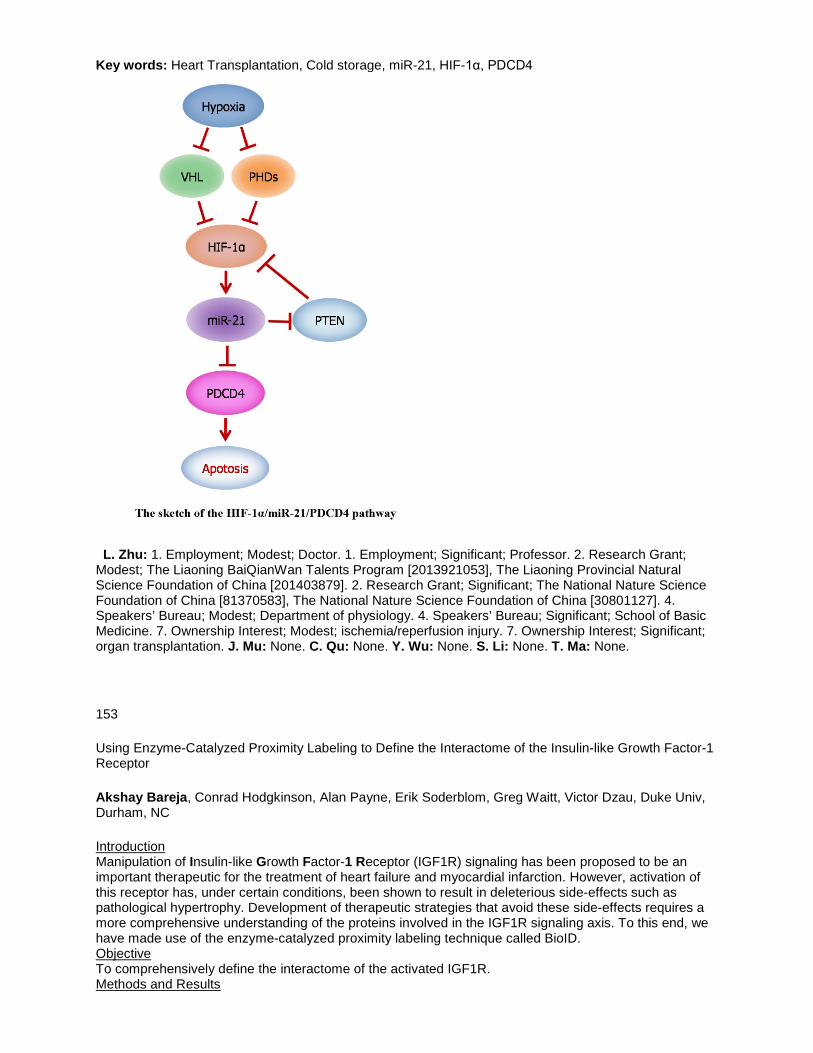
Key words: Heart Transplantation, Cold storage, miR-21, HIF-1α, PDCD4
L. Zhu: 1. Employment; Modest; Doctor. 1. Employment; Significant; Professor. 2. Research Grant; Modest; The Liaoning BaiQianWan Talents Program [2013921053], The Liaoning Provincial Natural Science Foundation of China [201403879]. 2. Research Grant; Significant; The National Nature Science Foundation of China [81370583], The National Nature Science Foundation of China [30801127]. 4. Speakers’ Bureau; Modest; Department of physiology. 4. Speakers’ Bureau; Significant; School of Basic Medicine. 7. Ownership Interest; Modest; ischemia/reperfusion injury. 7. Ownership Interest; Significant; organ transplantation. J. Mu: None. C. Qu: None. Y. Wu: None. S. Li: None. T. Ma: None.
153
Using Enzyme-Catalyzed Proximity Labeling to Define the Interactome of the Insulin-like Growth Factor-1 Receptor
Akshay Bareja, Conrad Hodgkinson, Alan Payne, Erik Soderblom, Greg Waitt, Victor Dzau, Duke Univ, Durham, NC
Introduction Manipulation of Insulin-like Growth Factor-1 Receptor (IGF1R) signaling has been proposed to be an important therapeutic for the treatment of heart failure and myocardial infarction. However, activation of this receptor has, under certain conditions, been shown to result in deleterious side-effects such as pathological hypertrophy. Development of therapeutic strategies that avoid these side-effects requires a more comprehensive understanding of the proteins involved in the IGF1R signaling axis. To this end, we have made use of the enzyme-catalyzed proximity labeling technique called BioID. Objective To comprehensively define the interactome of the activated IGF1R. Methods and Results

Briefly, BioID makes use of a biotin protein ligase called BirA* that promiscuously biotinylates neighboring proteins. The protein of interest is fused with BirA* and is then expressed in a particular cell line. Following administration of an excess amount of biotin, BirA* will biotinylate those proteins that are adjacent to or interact with the protein of interest. Interacting proteins are identified by tandem mass spectrometry (MS/MS). We have made a HEK 293T cell line that stably expresses the BirA* biotin ligase fused to the C-terminus of IGF1R. We performed BioID using this cell line (N=3) to identify a set of proteins that bind to the IGF1R following stimulation by IGF1. This set included many known interactors of the IGF1R, such as IRS2 and SHC1, thereby validating this technique. Of the many interesting novel interactors, we chose to pursue validation of sorting nexin 6 (SNX6) as some SNX proteins have been shown to be involved in receptor recycling to the plasma membrane following stimulation. We have found that SNX6 knockdown results in a dramatic diminution of IGF1-mediated ERK phosphorylation (2-fold; N=4; p<0.05). However, there was no effect on IGF1-mediated Akt phosphorylation (N=4). We are currently assessing the role of SNX6 in IGF1R-mediated function and recycling of this receptor. Conclusion We have demonstrated the ability of the BioID technique to identify a set of proteins that can interact with the activated IGF1R. Moreover, BioID has identified a number of novel interactors that further define IGF1R signaling and function.
A. Bareja: None. C. Hodgkinson: None. A. Payne: None. E. Soderblom: None. G. Waitt: None. V. Dzau: None.
154
CCDC80 Functions as a Protein Kinase GI Substrate and is Secreted by Cardiac Myocytes
Robert M Blanton, Craig Cooper, Anja Hergruetter, Mark Aronovitz, Timothy D. Calamaras, Tufts Medical Sch, Boston, MA
Background: Protein kinase G I alpha (PKGIa) inhibits cardiac hypertrophy, remodeling, and dysfunction. Downstream PKGI substrates remain incompletely understood and represent potential novel therapeutic targets for myocardial disease. We previously identified through a molecular screen that PKGIa binds and phosphorylates the protein coiled-coiled domain containing 80 (Ccdc80; also termed SSG1 and URB) in vascular smooth muscle cells. Previous work also identified that Ccdc80 is secreted from adipocytes. However, the expression and secretion of Ccdc80 from the cardiac myocyte has not been investigated. The current study tested the hypothesis that Ccdc80 is expressed in and secreted from the cardiac myocyte. Results: In cultured rat cardiac myocytes (CM), we detected Ccdc80 by western blot. Western blot for Ccdc80 also detected a band of the predicted Ccdc80 molecular weight present in media from these cells, but not in uncultured media. Ccdc80 could be detected in the human left ventricle (LV), though expression did not differ between hearts of normal controls and patients with hypertrophic cardiomyopathy. In the setting of LV pressure overload induced by transaortic constriction (TAC), we observed an increase in Ccdc80 expression in 1 week TAC LVs, compared with sham LVs (5.0 +/- 0.3 arbitrary densitometric units in sham versus 9.6 +/- 0.9 in TAC; n=4 per group). Conclusion: Taken together, our findings identify that the PKGIa substrate Ccdc80 expresses in cardiac myocytes, becomes secreted from CMs, resides in the human heart, and increases in expression in the mouse LV in response to pressure overload. Given the anti-remodeling role of PKGIa, these findings support future studies to understand the in vivo role of Ccdc80 in the cardiovascular system. Future studies will also explore the significance of Ccdc80 secretion from the CM and its potential regulation by PKG.
R.M. Blanton: 2. Research Grant; Modest; Novartis Pharmaceuticals. 8. Consultant/Advisory Board; Modest; Ironwood Pharmaceuticals. C. Cooper: None. A. Hergruetter: None. M. Aronovitz: None. T.D. Calamaras: None.
This Research has received full or partial funding support from the American Heart Association.

155
ATF6 is a Mediator of Cardiac Hypertrophy During Pressure Overload in the Mouse Heart
Robert N. Correll, Jeffrey M. Lynch, Michelle A. Sargent, Allen J. York, Jeffery D. Molkentin, Cincinnati Children's Hosp, Cincinnati, OH
The endoplasmic reticulum (ER) stress response is one of many signaling events activated in the heart in response to injury and/or cardiac hypertrophy, although the role that this response pathway plays in such events remains under investigation. Recently, our laboratory demonstrated that overexpression of thrombospondin-4 (Thbs4) resulted in a protective ER stress response via activation of activating transcription factor 6 (ATF6), and that mice gene-deleted for Thbs4 demonstrated compromised ER stress signaling and decreased survival after transverse aortic constriction (TAC) or myocardial infarction (MI) surgery. Here we confirm that Thbs4-mediated expansion of the ER compartment requires ATF6 and examine whether the protective ER stress response mediated by transgenic overexpression of Thbs4 is eliminated when crossed with gene-deleted mice lacking ATF6. We also examined cardiac-specific transgenic mice overexpressing the transcriptionally-active ATF6 N-terminus, as well as mice gene-deleted for ATF6, in combination with disease stimuli including TAC and MI surgery. We find that ATF6 proteins are required for compensatory hypertrophy in the mouse heart and that loss of these proteins results in accelerated decompensation and failure, likely due to reductions in ER folding capacity that is required for the increased protein production necessary for cardiac hypertrophy. These results firmly position ATF6 as an essential regulator of compensatory cardiac hypertrophy during disease.
R.N. Correll: None. J.M. Lynch: None. M.A. Sargent: None. A.J. York: None. J.D. Molkentin: None.
157
Alterations in Signaling Pathways During Regression of Pathological Cardiac Hypertrophy
Deanna Langager, Leslie Leinwand, Univ of Colorado Boulder, Boulder, CO
Introduction Cardiac hypertrophy is initially, a compensatory mechanism to maintain cardiac output when there is an increased load on the heart. However, if cardiac hypertrophy persists for an extended time, there can be maladaptive changes to the myocardium. Even when the underlying cause of hypertrophy is treated, regression is often minimal or absent. Clinical cases of cardiac regression do exist, including patients receiving bariatric surgery or a left ventricular assist device. While many of the mechanisms leading to cardiac hypertrophy are well understood, little is known about the mechanisms of reversal of hypertrophy and why it is sometimes irreversible. We hypothesized that a reversal of isoproterenol (Iso) induced cardiac hypertrophy in the mouse will be observed within 7 days following the removal of the stimulus and we will be able to identify alterations in signaling pathways. Methods We induced pathological cardiac hypertrophy with Iso for 7 days, at which peak hypertrophy is achieved. To identify if/when regression occurs, the Iso treatment was stopped and the mice were monitored for 7 days. Heart weights were measured at peak hypertrophy, post-drug days 1, 2, 3 & 7, along with vehicle treated mice (8/group). We used left ventricle tissue for protein analysis and protein degradation activity assays. Results Regression from cardiac hypertrophy occurs by post-drug day 7 (p=0.016) in the Iso mouse model. p-Akt is increased with Iso treatment and returns to vehicle control levels by post-drug day 7. There is a decrease in p-mTOR and an increase in LC3-II levels at post-drug day 7, indicating a possible role of autophagy in cardiac regression. In addition, there was a decrease in cell size when neonatal rat ventricular myocytes were treated with the Akt inhibitor, Wortmannin, following phenylephrine induced hypertrophy. Conclusion Regression of Iso-induced cardiac hypertrophy occurs in the mouse after 7 days following the removal of the stimulus. The Akt pathway is activated with Iso treatment and when this pathway is inactivated during regression, autophagy is activated, which may be an important mechanism to degrade proteins and lead to a decrease in cardiac hypertrophy. Finally, when the Akt pathway is inhibited in vitro, hypertrophic cells regress.
D. Langager: None. L. Leinwand: None.

158
A Tether Containing a UBX Domain (TUG) Mediates Glucose Transporter Translocation in the Ischemic Heart
Ji Li, Univ of Mississippi Med Ctr, Jackson, MS; Yina Ma, Jonathan Bogan, Yale Univ Sch of Med, New Haven, CT
Introduction: The adaptive metabolic regulation of glucose and fatty acid in the heart plays a critical role in limiting cardiac damage caused by ischemia and reperfusion (I/R). TUG (tether containing a UBX domain, for GLUT4) can be cleaved to mobilize glucose transporter GLUT4 from intracellular vesicles to the cell surface in skeletal muscle and adipose in response to insulin stimulation. The energy sensor AMP-activated protein kinase (AMPK) plays an important cardioprotective role in response to ischemic insults by modulating GLUT4 translocation. Hypothesis: TUG is one of the downstream targets of AMPK in the heart. TUG could be phosphorylated by ischemic AMPK and cleaved to dissociate with GLUT4 and increase GLUT4 translocation in the ischemic heart. Methods: In vivo regional ischemia by ligation of left anterior coronary artery and ex vivo isolated mouse heart perfusion Langendorff system were used to test the hypothesis. Results: Antithrombin (AT) is an endogenous AMPK agonist in the heart and used to define the role of TUG in regulating GLUT4 trafficking during ischemia and reperfusion in the heart. AT showed its cardioprotective function through recovering cardiac pumping function and activating AMPK. The results showed that AMPK activation by AT treatment was through LKB1 and Sesn2 complex. Furthermore, the ex vivo heart perfusion data demonstrated that AT administration significantly increase GLUT4 translocation, glucose uptake, glycolysis and glucose oxidation during ischemia and reperfusion (p<0.05 vs. vehicle). Moreover, AT treatment increased abundance of a TUG cleavage product (42 KD) in response to I/R. The TUG protein was clearly phosphorylated by activated AMPK in HL-1 cardiomyocytes. The in vivo myocardial ischemia results demonstrated that ischemic AMPK activation triggers TUG cleavage and significantly increases GLUT4 translocation to the cell surface. Moreover, an augmented interaction between AMPK and TUG was observed during ischemia. Conclusions: Cardiac AMPK activation stimulates TUG cleavage and causes the dissociation between TUG and GLUT4 in the intracellular vesicles. TUG is a critical mediator that modulates cardiac GLUT4 translocation to cell surface and enhances glucose uptake by AMPK signaling pathway.
J. Li: None. Y. Ma: None. J. Bogan: None.
This Research has received full or partial funding support from the American Heart Association.
159
Eicosapentaenoic Acid and Docosahexaenoic Acid Have Distinct Membrane Locations and Lipid Interactions as Determined by X-ray Diffraction
R. Preston Mason, Brigham and Women's Hosp, Harvard Medical Sch, Boston, MA; Samuel C.R. Sherratt, Elucida Res LLC, Beverly, MA
Eicosapentaenoic acid (EPA) and docosahexaenoic acid (DHA) differentially influence lipid oxidation, signal transduction, fluidity, and cholesterol domain formation, potentially due to distinct membrane interactions. We used small angle x-ray diffraction to test EPA and DHA effects on model membrane structure. Vesicles were prepared to model human peripheral cell membranes using 1-palmitoyl-2-oleoyl-sn-glycero-3-phosphocholine (POPC) and cholesterol (C) (0.3 C:POPC mole ratio), and treated with vehicle, EPA, or DHA (1:10 mole ratio to POPC). Membrane width (d-space) was 59.5 Å at 10°C, decreasing to 54.7 Å at 30°C due to increased acyl chain dynamics. EPA or DHA had no effect on membrane d-space (<1 Å change). Electron density profiles from diffraction data were superimposed on corresponding vehicle profiles (Fig. 1). EPA increased membrane hydrocarbon core electron density over a broad area, up to ± 20 Å from the membrane center, indicating an energetically favorable extended membrane orientation for EPA, stabilized by van der Waals interactions. By contrast, DHA increased electron density in the phospholipid head group region starting at ± 12 Å from the membrane center, due to DHA-surface interactions, resulting in a pronounced electron density reduction and increased disorder

in the membrane hydrocarbon core centered ± 7-9 Å from the membrane center. DHA disordering effects were less apparent at higher temperatures, likely due to greater rotational dynamics, while EPA effects were stable. The contrasting EPA and DHA effects on membrane structure signify distinct molecular locations/orientations and may contribute to observed differences in biological activity.
R.P. Mason: 3. Other Research Support; Significant; Amarin Pharma, Inc.. S.C. Sherratt: None.
160
Cardiomyocyte CaMKII Mediates Expression of Pro-inflammatory Chemokines and Cytokines, Macrophage Infiltration and Cardiac Remodeling in Response to Pressure Overload
Takeshi Suetomi, Andrew Willeford, Shigeki Miyamoto, Joan Heller Brown, Dept of Pharmacology, Univ of California, San Diego, Sch of Med, La Jolla, CA
Background: There is evidence that inflammation is associated with pressure overload induced cardiac remodeling and heart failure, as well as evidence for a role of CaMKII in remodeling and heart failure development. Whether CaMKII mediates inflammatory responses that contribute to its role in adverse remodeling following TAC has not been established. Methods and Results: CaMKIIδ knockout (CKO) mice in which CaMKIIδ was selectively deleted from cardiomyocytes were subjected to pressure overload by transverse aortic constriction (TAC). By 3 days, the earliest time examined, there were marked increases in cardiac mRNA levels for pro-inflammatory chemokines CCL2 (MCP-1, ~15 fold), CCL3 (MIP1α, ~20 fold) and cytokines (IL-6, ~50 fold). These responses were markedly attenuated (by 56%, 43% and 42 % respectively) in the CKO mice. NFkB signaling was also increased in control heart at 3 days of TAC but not in CKO. Immunohistochemical analysis showed increases in CD68+ macrophage by 7 days TAC which further increased by 14days of TAC. Macrophages accumulation was also significantly attenuated in the CKO mice (50% decrease at 7day, 40% decrease at 14 days). Fibrosis was assessed by Masson trichrome staining and increases in collagen gene expression (col1a1 and col3a1 mRNA). Both were clearly elevated after 14 and 28 days TAC and significantly attenuated in the CKO mice. Conclusion: Our results indicate that activation of CaMKII localized in cardiomyocytes initiates an inflammatory transcriptional program within cardiomyocyte. We suggest that this drives immune cell recruitment and is in turn associated with development of fibrosis. Early inflammatory responses and their sequelae may thus be responsible for involvement of CaMKII in the progression from hypertrophy to heart failure.
T. Suetomi: None. A. Willeford: None. S. Miyamoto: None. J. Heller Brown: None.

161
Β-Arrestin-Biased Agonism of Β-Adrenergic Receptor Regulates Dicer-mediated MicroRNA Maturation to Promote Cardioprotective Signaling
Jian-Peng Teoh, Il-man Kim, Augusta Univ, Augusta, GA
MicroRNAs (miRs) are small, non-coding RNAs that function to post-transcriptionally regulate gene expression. First transcribed as primary miR transcripts (pri-miRs), they are enzymatically processed by Drosha into premature miRs (pre-miRs) and further processed by Dicer into mature miRs. Initially discovered to desensitize β-adrenergic receptor (βAR) signaling, β-arrestins are now well-appreciated to modulate multiple effector pathways independent of G protein-mediated signaling, a concept known as biased signaling. Using the β-arrestin-biased βAR ligand carvedilol (Carv), we previously showed that β-arrestin1-biased β1AR cardioprotective signaling stimulates Drosha-mediated processing of a subset of miRs. Here, we investigate whether Carv could regulate Dicer-mediated miR maturation, thereby providing a novel mechanism for its cardioprotective effects. In mouse hearts, Carv indeed upregulates 3 mature miRs, but not their pre-miRs and pri-miRs, in a β-arrestin1/2-dependent manner. Interestingly, Carv-mediated activation of miR-466g and miR-532 is dependent on β2AR, while the activation of miR-674 by Carv is β1AR-dependent. Mechanistically, β-arrestins regulate maturation of 3 newly identified βAR/β-arrestin-responsive miRs (β-miRs) by associating with the Dicer complex as well as hnRNPK and dyskerin on 3 pre-miRs (Fig. A). Cardiac cell approaches uncover that β-miRs act as gatekeepers of cardiac cell function by repressing detrimental target genes (Fig. B). In conclusion, our findings indicate a novel role for βAR-mediated β-arrestin signaling activated by Carv in miR maturation, which may be linked, in part, to its protective mechanism.
J. Teoh: None. I. Kim: None.
This Research has received full or partial funding support from the American Heart Association.

162
MicroRNAs Mediate the Hemodynamic Regulation of Endothelium-to-Smooth Muscle Signaling
Jing Zhou, Juanjuan Zhu, Yuefeng Liu, Peking Univ Health Science Ctr, Beijing, China; Shu Chien, Univ of California, San Diego, La Jolla, CA
Vascular endothelial cells (ECs) at arterial branches and curvatures experience disturbed blood flow and induce a quiescent-to-activated phenotypic transition of the adjacent smooth muscle cells (SMCs) and a subsequent smooth muscle hyperplasia. However, the mechanism underlying the flow pattern-specific initiation of EC-to-SMC signaling remains elusive. Our previous study has demonstrated that endothelial microRNA-126-3p (miR-126-3p) acts as a key intercellular molecule to increase turnover of the recipient SMCs, and that its release is reduced by atheroprotective laminar shear (LS) to ECs. In the current study we found that atherogenic oscillatory shear (OS), but not atheroprotective pulsatile shear (PS), promotes the regulated exocytosis, particularly the secretion of non-membrane-bound miR-126-3p and other microRNAs (miRNAs) via the activation of soluble N-ethyl-maleimide-sensitive fusion protein attachment protein receptors (SNAREs), VAMP3 and SNAP23. Quantitative PCR arrays identify 15 EC-secreted miRNAs whose levels in the vesicle-poor supernatant of flow perfusate are differentially regulated (P<0.05) by OS versus PS. The fold changes of 11 miRNAs are either greater than 2.0 or less than 0.5. Knockdown of VAMP3 and SNAP23 reduces the secretion of miR-126-3p and miR-200a-3p, as well as the proliferation, migration, and suppression of contractile markers in SMCs caused by EC-coculture. Pharmacological intervention of mTORC1 in ECs blocks endothelial secretion and EC-to-SMC transfer of miR-126-3p through transcriptional inhibition of VAMP3 and SNAP23. Systemic inhibition of VAMP3 and SNAP23 by rapamycin or periadventitial application of the endocytosis inhibitor dynasore ameliorates the disturbed flow-induced neointimal formation, whereas intraluminal overexpression of SNAP23 aggravates it. Our findings demonstrate the flow-pattern-specificity of SNAREs activation and its contribution to the miRNA-mediated EC-SMC communication, uncovering potential targets for future diagnosis and therapeutic interventions for proliferative vascular diseases.
J. Zhou: None. J. Zhu: None. Y. Liu: None. S. Chien: None.
165
Role of the Gut Microbiome in Obesity-Related Vascular Dysfunction
Micah Battson, Dustin Lee, Shuofei Hou, Dillon Jarrell, Kayl Ecton, Tiffany Weir, Christopher Gentile, Colorado State Univ, Fort Collins, CO
Vascular dysfunction, characterized by arterial stiffness and endothelial dysfunction, represents an important link between obesity and cardiovascular disease. Deleterious changes to the gut microbiome, termed dysbiosis, are associated with obesity and may contribute to the development of vascular dysfunction by promoting systemic and arterial inflammation. We tested the hypothesis that antibiotic treatment in diet-induced obese mice would improve vascular function by suppressing potentially harmful gut-derived bacterial products to reduce dysbiosis-related inflammation. Male C57BL/6 mice were fed either a standard diet (SD) or Western diet (WD) for 5 months. Obese WD-fed mice were then randomized to receive 8 weeks of antibiotic treatment via drinking water. Arterial stiffness was measured via in vivo aortic pulse wave velocity and endothelial function assessed via ex vivo pressure myography in isolated mesenteric arteries. Plasma and arterial tissue samples were collected for measures of endotoxemia and inflammation and fecal samples were collected for microbial sequencing. Our results showed that mice fed a WD for 5 months had significantly higher body weight and arterial stiffness compared to SD. Antibiotic treatment significantly reduced arterial stiffness in WD-fed mice without affecting body weight. Endothelial function was impaired in WD-fed mice, whereas WD-Abx mice had similar endothelial function to SD-fed mice. Plasma levels of the pro-inflammatory cytokine IL-6 and endotoxemia marker LPS binding protein (LBP) were significantly increased in WD vs SD, but reduced in WD-Abx vs WD. Gut microbial suppression with antibiotic treatment was confirmed using quantitative PCR and ongoing analyses are being conducted to determine specific diet-induced changes to the composition of the gut microbiome. Overall, our results support the hypothesis that gut dysbiosis contributes the development of vascular dysfunction in obesity and provide evidence that a gut-targeted therapy can improve endothelial function and arterial stiffness in diet-induced obese mice. Future studies

will further examine the mechanisms involved in, and help identify potential therapeutic targets for, obesity-related vascular dysfunction.
M. Battson: None. D. Lee: None. S. Hou: None. D. Jarrell: None. K. Ecton: None. T. Weir: None. C. Gentile: None.
166
Anti-cachectic Role of Neuregulin-1β in Heart Failure
Cristi L Galindo, Manisha Gupte, Vanderbilt Univ Med Ctr, Nashville, TN; Oleg Y Tikhomirov, Maine Medical Res Ctr, Scarborough, ME; Pranay Manda, John H. Cleator, Abigail Murphy, Ehab Kasasbeh, Vanderbilt Univ Med Ctr, Nashville, TN; Douglas B. Sawyer, Maine Medical Res Inst, Scarborough, ME
Neuregulin-1β (NRG-1β) is a growth and differentiation factor with pleiotropic systemic effects. Because NRG-1β has therapeutic potential for heart failure and known growth effects in skeletal muscle, we hypothesized that it might affect heart failure-associated cachexia, a severe co-morbidity characterized by a loss of muscle mass. We therefore assessed NRG-1β’s effect on skeletal muscle gene expression in a swine model of heart failure using recombinant Glial Growth Factor 2 (USAN - cimaglermin alfa), a version of NRG-1β currently being tested in humans with systolic heart failure. Animals received one of two intravenous doses (0.67 or 2 mg/kg) of NRG-1β bi-weekly for 4 weeks, beginning one week after infarct. Skeletal muscle was collected at the time of euthanasia from the intercostal space and paired-end RNA sequencing performed. NRG-1β treatment altered expression of 583 transcripts including 242 transcripts altered at both doses. These included genes required for myofiber growth, maintenance and survival such as MYH3, MYHC, MYL6B, KY and HES1. Importantly, NRG-1β altered the directionality of at least 85 genes associated with cachexia, including myostatin, which negatively regulates myoblast differentiation by down-regulating MyoD expression. Consistent with this, MyoD was increased in NRG-1β treated animals. In vitro experiments with myoblast cell lines confirmed that NRG-1β induces skeletal muscle differentiation with an absolute requirement for ERBB signaling on myoblast differentiation. These findings suggest a NRG-1β-mediated anti-atrophic, anti-cachexia effect that may provide additional benefits to this potential therapy in heart failure.
C.L. Galindo: None. M. Gupte: None. O.Y. Tikhomirov: None. P. Manda: None. J.H. Cleator: None. A. Murphy: None. E. Kasasbeh: None. D.B. Sawyer: None.
167
Inhibition of Endoplasmic Reticulum Stress Improves Vascular Function in Type II Diabetic Mice
Dustin M Lee, Micah L Battson, Dillon K Jarrell, Shuofei Hou, Kayl Ecton, Christopher L Gentile, Colorado State Univ, Fort Collins, CO
Cardiovascular disease is the leading cause of death in the United States with type 2 diabetes (T2D) representing a major risk factor in its development. Vascular dysfunction, characterized by arterial stiffness and endothelial dysfunction occur prior to overt cardiovascular disease and predict future

cardiovascular events and mortality in diabetic individuals. Dysfunction of the endoplasmic reticulum (ER), or ER stress, is associated with the development and progression of chronic metabolic diseases such as obesity and T2D. However, the role of ER stress in the development of vascular dysfunction observed in T2D is unclear. We hypothesized that inhibiting ER stress would improve both measures of vascular dysfunction observed in diabetic mice. Male C57BL/6J Leprdb (DB) male mice lacking the leptin receptor were used as a model of T2D. Starting at 4 months of age DB mice were given intraperitoneal injections of ER stress inhibitor, tauroursodeoxycholic acid (TUDCA) at 250mg/kg/day for 4 weeks. C57BL/6J mice were used as controls (n=8 for all groups). Pulse wave velocity (PWV) was measured at baseline (prior to treatment) and after TUDCA treatment. Secondary order mesenteric resistance arteries (MRA) were used to determine endothelial dependent dilation (EDD). DB mice not treated with TUDCA were used for acute studies of EDD by incubating the MRA for 1hr with 0.5mM TUDCA. At baseline, DB mice displayed increased arterial stiffness compared to C57BL/6J controls as measured by PWV (457±25 vs 348±26 cm/s, p<0.05). TUDCA treatment significantly decreased PWV (402±12 cm/s, p<0.05). Both acute and chronic TUDCA treatment improved DB EDD compared to untreated controls (58.4±13.8 and 49.1±6.1%, vs 19.8±9.6%, p<0.05). Expression of ER stress-associated genes CHOP and ATF4 were significantly elevated in DB perivascular adipose tissue and treatment with TUDCA significantly decreased ATF4 compared to DB. In the aorta, TUDCA significantly decreased GRP78 and XBP1 compared to DB. These data support the hypothesis that ER stress contributes to the vascular dysfunction observed in T2D and suggest that ER stress may be a potential target in the treatment of T2D related vascular stiffening and dysfunction.
D.M. Lee: None. M.L. Battson: None. D.K. Jarrell: None. S. Hou: None. K. Ecton: None. C.L. Gentile: None.
168
Stress Dependent MicroRNA Mediate Crosstalk Between DNA Repair ′ Senescence & Angiogenic Signaling
Cristina Espinosa-Diez, RaeAnna Wilson, Namita Chaterjee, Clay Hudson, Rebbeca Ruhl, Erin Helms, Sudarshan Anand, Oregon Health & Science Univ, Portland, OR
The endothelium is highly exposed to a myriad of stressors that can lead to endothelial dysfunction if persistent. One of the hallmarks of endothelial stress is increased DNA damage. MRN complex plays a vital role in DNA double strand break repair and replication. Nevertheless, its role in cardiovascular impair has not been studied in depth. Our results show that a cohort of microRNAs, upregulated under genotoxic and oxidative stress conditions, target MRN complex inducing senescent phenotype in endothelial cells. These microRNAs are rapidly transcribed in response to different types of stress, however the transcription factors that induce the expression of these miRs in response to DNA damage in endothelial cells is not known. We have profiled a subset of active transcription factors as c-Myc, Nrf2 and Nrf1 that robustly induce these miRs. Furthermore, our gain and loss-of-function studies indicate that these miRs, miR-99b and miR-494, affect telomerase activity, enable the activation of p21 and Rb pathways and regulate angiogenic sprouting. Ectopic expression of the miR mimics inhibits angiogenesis in vivo suggesting that targeting the MRN complex could be a viable anti-angiogenic strategy. Finally, our studies suggest a crosstalk between VEGF signaling and DNA Damage. Genetic and pharmacological disruption of VEGFR-2 signaling and the MRN complex reveal a surprising co-dependency of these pathways in regulating EC senescence and proliferation. Using a pathway focused gene expression array we find that disruption of the MRN complex leads to a robust transcription of CD44, a known driver of senescence as well as a negative regulator of VEGF signaling. Our work identifies a putative miR-facilitated mechanism by which endothelial cells can be insulated against VEGF signaling to facilitate the onset of senescence. We propose that stress response drives specific microRNA expression perturbing canonical VEGF signaling, proliferation and senescence by thresholding DNA damage repair in the vasculature.
C. Espinosa-Diez: None. R. Wilson: None. N. Chaterjee: None. C. Hudson: None. R. Ruhl: None. E. Helms: None. S. Anand: None.
This Research has received full or partial funding support from the American Heart Association.

169
miR-21-5p Regulation of Lipid Content and Peroxidation in H9C2 Cells
Sandra Chuppa, Alison J Kriegel, Medical Coll of Wisconsin, Milwaukee, WI
Cardiovascular pathologies are the leading single cause of death in chronic kidney disease (CKD) patients. We have found that the 5/6 nephrectomy model of CKD leads to an upregulation of miR-21-5p in the left ventricle 7 weeks after surgery, targeting peroxisome proliferator-activated receptor alpha (PPARα). PPARα is a regulator of fatty acid uptake and metabolism. In our model we find that suppression of miR-21-5p alters the expression of numerous genes involved with fatty acid oxidation and glycolysis, presumably through its regulatory action on PPARα and/or additional targets. We also find that 5/6Nx rats exhibit dyslipidemia and increased left ventricular lipid content at this time. In this study we evaluated the potential for knockdown or overexpression of miR-21-5p to regulate lipid content and peroxidation in H9C2 cells. Cells were transfected with anti-miR-21-5p (40nM), pre-miR-21-5p (20nM) or appropriate scrambled oligonucleotide controls. After 24 hours medium was changed and half of the cells from each transfection group were treated with lipid (0.66 mM oleic acid and 0.33 mM palmitic acid) for 48 hours (n=6/treatment group for each set of experiments). Lipid content, measured by AdipoRed assay (Lonza) was significantly increased with lipid treatment (nearly two-fold). Overexpression of miR-21-5p significantly attenuated this increase (228.0 ± 9.7 vs. 198.2 ± 8.9% of untreated control), while suppression of miR-21-5p augmented lipid content (235.8 ± 11.2 vs. 328.1 ± 12.3% of untreated control). These results were supported by imaging of Oil Red O stained cells. We found that the abundance of malondialdehyde (MDA), a product of lipid peroxidation, was significantly increased in response to lipid treatment. Overexpression of miR-21-5p reduced MDA content in untreated and lipid treated cells, suggesting that miR-21-5p reduces oxidative stress. Suppression of miR-21-5p had no effect on MDA levels. These results indicate that overexpression of miR-21-5p attenuates both lipid content and lipid peroxidation in H9C2 cells. Ongoing studies aimed at evaluation of alterations in fatty acid oxidation and oxidative stress will further aid in determining the functional impact of miR-21-5p on associated pathways in cardiac tissue.
S. Chuppa: None. A.J. Kriegel: None.
This Research has received full or partial funding support from the American Heart Association.
170
Differential Regulation of miRNA and mRNA Expression in the Myocardium of Nrf2 Knockout Mice
Justin M Quiles, Cardiac Aging & Redox Signaling Lab, Div of Molecular & Cellular Pathology, Dept of Pathology, Univ of Alabama at Birmingham, Birmingham, AL; Madhusudhanan Narasimhan, Dept of Pharmacology and Neuroscience, Texas Tech Univ Health Sciences Ctr, Lubbock, TX; Gobinath Shanmugam, Cardiac Aging & Redox Signaling Lab, Div of Molecular & Cellular Pathology, Dept of Pathology, Univ of Alabama at Birmingham, Birmingham, AL; Brett Milash, Huntsman Cancer Inst, Dept of Med, Univ of Utah Sch of Med, Salt Lake City, UT; John R Hoidal, Div of Pediatric Pulmonology, Dept of Internal Med, Univ of Utah Sch of Med, Salt Lake City, UT; Namakkal Soorappan Rajasekaran, Cardiac Aging & Redox Signaling Lab, Div of Molecular & Cellular Pathology, Dept of Pathology, Ctr for Free Radical Biology, Univ of Alabama at Birmingham, Birmingham, AL
Background: Preclinical studies indicate that disruption of the transcription factor nuclear factor, erythroid 2 like 2 (Nrf2) leads to pathological oxidative stress in several tissues. However, the transcriptional role of Nrf2 in the heart, a highly aerobic organ sensitive to redox disturbances and aberrant microRNA (miRNA) activity, remains elusive. Here, we tested the hypothesis that Nrf2 ablation disrupts cardiac miRNA abundance leading to impaired transcriptional regulation of the myocardial defense system.Methods: Age-matched wild-type (WT) and Nrf2 knockout (Nrf2-/-) mice (n=3-6/group) were used in this study. Independent next-generation sequencing experiments were conducted to evaluate differential expression of mRNA and miRNA in the heart. Real-time qPCR was used to validate RNA sequencing (RNAseq) data. Results: RNAseq for mRNA uncovered 152 differentially expressed genes (DEGs) in Nrf2-/- hearts, of which 129 were downregulated relative to WT. Novel DEGs previously unreported as Nrf2 targets were detected and Gene Ontology analysis revealed the enrichment (p<0.05) of several biological processes
distinct from canonical Nrf2 function including; wound healing, protein folding, cytokine response, cell migration and adhesion. In line with previous reports, altered regulation of apoptosis, stress response and oxidative metabolism was also implicated in Nrf2-/- DEG signatures. Next, small RNAseq detected a total of 27 miRNAs (11 up and 16 downregulated) altered in Nrf2-/- mice. Validation by qPCR revealed a significant (p<0.05) decrease in miR-10b-5p, miR-674-3p, miR-3535, and miR-378c while miR-30b-5p, miR-208a-5p, miR-350-3p, miR-582-5p, and miR-1249-3p were increased. In silico integration of RNAseq data discovered complementarity between 39 repressed mRNAs and 4 upregulated miRNAs; miR-30b-5p, miR-208a-5p, miR-350-3p, and miR-582-5p. These miRNAs may cooperatively regulate expression in Nrf2-/- hearts as 22 DEGs matched with 2 or more miRNAs. Conclusion: Our results highlight novel mRNA targets and indicate that Nrf2 regulates a subset of cardiac miRNAs. Biological changes distinct from canonical Nrf2 function suggest that regulatory cross-talk with miRNAs contribute to unique roles for Nrf2 in the myocardium.
J.M. Quiles: None. M. Narasimhan: None. G. Shanmugam: None. B. Milash: None. J.R. Hoidal: None. N. Rajasekaran: None.
171
Promoter Negative Elongation Factor a (NelfA) Occupancy Required for Gene Transcription in Heart During Hypertrophy
Elena Severinova, Zhi Yang, Maha Abdellatif, Danish H Sayed, Rutgers- NJMS, Newark, NJ
Adaptive change in gene expression is one of the earliest responses of heart to hypertrophy. Gene transcription is tightly regulated by systematic recruitment of factors including positive and negative elongation complexes of RNA Pol II. Recently we embarked on the task of deconstructing the transcriptional machinery in the heart, by examining Pol II and TFIIB dynamics across the genome in sham and hypertrophied hearts. In this study, we continue examining the active transcriptional code by measuring occupancy of two interdependent factors that belong to positive (PTEFb) and negative (NELF) elongation complexes, Cdk9 and NelfA, respectively. Studies in non-cardiac cells have implicated these two factors in the regulation of promoter (prom) clearance of pol II, where Cdk9 mediated phosphorylation of NelfA results in its release from paused pol II and initiation of productive elongation. As expected, we observed increase Cdk9 (>1.5) association with proms in 59.7% of expressed genes (8257 of 13816), while 6% show decrease in prom Cdk9 (<0.75) in TAC vs. sham hearts. In contrast to previous reports, we did not see any correlation between Cdk9 and NelfA occupancy, where 93.2% and 68% of genes did not show change in NelfA with increased or decreased Cdk9 levels in TAC hearts, respectively. Interestingly, highest enrichment of NelfA (av. density 53.5) was observed on genes regulated by prom clearance of pol II (av. prom den 9.5 vs. 7.4; av. Ingene den 3.0 vs. 3.6, sham vs. TAC; housekeeping genes) with no change in NelfA (89.8% of genes). On other hand, 3.3% of genes showed increase in NelfA (av. den 4.6 vs. 8.4, sham vs. TAC), along with increase in pol II recruitment (av. prom den 3.3 vs. 4.04; av. ingene den 1.8 vs. 2.4, sham vs. TAC; cytoskeletal, cell cycle genes). We confirmed increase in mRNA abundance of these genes, (eg. Acta 1) in TAC vs. sham hearts. Intriguingly, genes that were associated with reduced NelfA (av. den 21.7 vs. 14.8, sham vs. TAC) were mostly cardiac specific genes (e.g. cardiac muscle contraction), which show increase in pol II, but no change in mRNA levels with TAC (eg. Actc). While we are investigating the role of NelfA in heart, ChIP- Seq data suggest that NelfA might be required for productive elongation and increase in global gene expression during hypertrophy
E. Severinova: None. Z. Yang: None. M. Abdellatif: None. D.H. Sayed: None.
This Research has received full or partial funding support from the American Heart Association.
172
Vascular Smooth Muscle Cell Activation is Regulated by miR-25 Induced miR-9 Suppression of Nox4 and Myocardin
Brandon M Schickling, Maysam Takapoo, Eric J Devor, Univ of Iowa, Iowa City, IA; Francis J Miller, Duke Univ, Durham, NC

Vascular smooth muscle cell (SMC) de-differentiation with subsequent migration and proliferation into the subendothelial space is central to the progression of cardiovascular diseases. The Nox4 NADPH oxidase (Nox4) is implicated in maintaining the differentiated phenotype of SMC in part through myocardin, a master regulator of SMC gene expression. However, this process is poorly understood. We hypothesized that microRNAs (miR)-mediate changes in Nox4 expression and regulate SMC differentiation. Treatment of human SMCs with a miR-9 or miR-25 mimic silenced Nox4 mRNA through binding to the Nox4 3’UTR. However, only miR-25 was sufficient to downregulate Nox4 protein levels. We found that miR-25 induced the expression of miR-9 through a novel mechanism involving demethylation of the miR-9 promoter by Tet methylcytosine dioxygenase 2 (TET2). Inhibition of miR-9 induction by miR-25 with a miR-9 inhibitor restored Nox4 protein expression to basal levels. Furthermore, the miR-25-mediated decrease in Nox4 protein was ameliorated by inhibiting the proteasome with MG132. These data suggest a novel mechanism wherein miR-9 and miR-25 regulate Nox4 through both translational suppression and proteosomal degradation. Overexpression of miR-9 or miR-25 in human SMCs (1) suppressed myocardin mRNA and protein expression; (2) decreased expression of multiple SMC differentiation genes; and (3) was sufficient to induce cell migration. Thrombin and tumor necrosis factor increased the expression of miR-9 and miR-25 in human SMCs and inhibition of miR-9 prevented thrombin-mediated decrease in myocardin and SMC migration. Mir-9 and miR-25 levels were increased in SMCs derived from balloon injured rat aorta as compared to medial SMCs and in murine carotid artery ten days post carotid injury. A miR-9 inhibitor decreased neointimal formation by more than 50% in following partial carotid ligation in mice. These findings identify miR-9/Nox4 as a novel regulatory pathway of SMC differentiation and a potential therapeutic target in vascular disease.
B.M. Schickling: None. M. Takapoo: None. E.J. Devor: None. F.J. Miller: None.
This Research has received full or partial funding support from the American Heart Association.
174
Regulation of Kv11.1 C-terminal Isoform Expression by Polypyrimidine Tract Binding Protein
Qiuming Gong, Anastasiya Goldys, Zhengfeng Zhou, Oregon Health Science Univ, Portland, OR
The KCNH2 gene encodes the Kv11.1 potassium channel that conducts the rapidly activating delayed rectifier current in the heart. KCNH2 pre-mRNA undergoes alternative processing. Intron 9 splicing leads to the formation of a functional, full-length Kv11.1a isoform, and polyadenylation within intron 9 generates a non-functional, C-terminally truncated Kv11.1a-USO isoform. In this study we investigated the developmental regulation of Kv11.1 isoform expression. We showed that Kv11.1a expression was lower than that of Kv11.1a-USO in the adult heart, but the levels of Kv11.1a and Kv11.1a-USO were similar in the fetal heart. We studied the effect of polypyrimidine tract binding protein (PTB) on the alternative processing of KCNH2 pre-mRNA. PTB is an RNA-binding protein well known for its role in the regulation of alternative splicing. Recently, PTB has been shown to regulate polyadenylation. We showed that PTB increased Kv11.1a isoform expression and decreased Kv11.1a-USO isoform expression by the RNase protection assay and immunoblot analysis. In patch-clamp experiments, we found that PTB significantly increased Kv11.1 current. Our findings suggest that the relative expression of Kv11.1 C-terminal isoforms can be regulated by PTB. It has been reported that PTB protein abundance is progressively reduced during postnatal heart development. Thus, PTB may play an important role in developmental regulation of Kv11.1 isoform expression in the heart.
Q. Gong: None. A. Goldys: None. Z. Zhou: 2. Research Grant; Significant; NIH and AHA Grants.
This Research has received full or partial funding support from the American Heart Association.

185
Genome Editing of Isogenic Human Induced Pluripotent Stem Cells Allows for Functional and Transcriptomic Insights Into Hereditary Dilated Cardiomyopathy Caused by Phospholamban Mutations
Delaine K Ceholski, Irene C Turnbull, Simon Koplev, Icahn Sch of Med at Mount Sinai, New York, NY; Chi-Wing Kong, Hong Kong Univ, Hong Kong, Hong Kong; Francesca Stillitano, Mathieu Nonnenmacher, Icahn Sch of Med at Mount Sinai, New York, NY; Kobra Haghighi, Univ of Cincinnati, Cincinnati, OH; Przemyslaw A Gorski, Ninette Cohen, Icahn Sch of Med at Mount Sinai, New York, NY; Fotis Kolokathis, Dimitrios Th. Kremastinos, Onassis Cardiac Surgery Ctr, Athens, Greece; Arvind Babu, Icahn Sch of Med at Mount Sinai, New York, NY; Ronald A Li, Karolinska Instt, Stockholm, Sweden; Chiara Giannarelli, Icahn Sch of Med at Mount Sinai, New York, NY; Ioannis Karakikes, Stanford Univ, Stanford, CA; Evangelia G Kranias, Univ of Cincinnati, Cincinnati, OH; Kevin D Costa, Roger J Hajjar, Icahn Sch of Med at Mount Sinai, New York, NY
Dilated cardiomyopathy (DCM) can be caused by genetic mutations in numerous cardiac proteins, including phospholamban (PLN). PLN mutations are quite rare and obtaining patient samples for mechanistic insights can be challenging. We used genome editing with CRISPR/Cas9 to successfully insert R14del and R9C PLN mutations into a human induced pluripotent stem cell (hiPSC) line from an individual with no cardiovascular disease. HiPSC-cardiomyocytes (hiPSC-CMs) with the inserted R14del PLN mutation recapitulate the phenotype observed in patient-derived R14del hiPSC-CMs, characterized by abnormal intracellular calcium cycling and arrhythmogenicity. Insertion of R9C PLN results in hiPSC-CMs displaying an abnormal response to β-agonists, defective calcium handling, and a hypertrophic phenotype. In human engineered cardiac tissues (hECTs) created from hiPSC-CMs in a 3D matrix, R14del results in a progressive worsening of developed force and R9C PLN demonstrates an abnormal lusitropic response following β-adrenergic stimulation. Further, transcriptional profiling using RNAseq suggests a role for lipid metabolism in R14del. DNA methylation studies showed that differentially expressed genes were enriched for lipoprotein metabolism and chylomicron-mediated lipid transport pathways in R14del PLN. This was also confirmed with the observation of lipid deposition in R14del hECTs and human myocardial tissues from explanted hearts of affected patients. Furthermore, small RNAseq identified 2 miRNAs that were differentially regulated in R14del hiPSC-CMs (miR-449c-5p and miR-483-3p). For R9C PLN, RNAseq suggests that the mutation results in profibrotic signaling, activation of autophagy, and an altered metabolic state. Our findings demonstrate that gene editing of hiPSCs can be used to successfully create models and delineate molecular mechanisms of human PLN mutations associated with DCM.
D.K. Ceholski: None. I.C. Turnbull: None. S. Koplev: None. C. Kong: None. F. Stillitano: None. M. Nonnenmacher: None. K. Haghighi: None. P.A. Gorski: None. N. Cohen: None. F. Kolokathis: None. D.T. Kremastinos: None. A. Babu: None. R.A. Li: None. C. Giannarelli: None. I. Karakikes: None. E.G. Kranias: None. K.D. Costa: 7. Ownership Interest; Modest; NovoHeart, Ltd. R.J. Hajjar: None.
This Research has received full or partial funding support from the American Heart Association.
186
Heart Disease Associated Lamin A/C Mutations and Resultant Defects in Cell and Nuclei Structure
Jason Q Core, Zachery R Robinson, Mehrsa Mehrabi, Linda A McCarthy, Michael V Zaragoza, Anna Grosberg, Unversity of California, Irvine, CA
Changes in cell organelle structure or shape, which can be associated with a variety of diseases, can be caused by inherited genetic mutations. For example, the progeria disease is associated with a mutation to the LMNA gene, and it leads to a devastating early aging and death. However, other LMNA gene mutations do not cause early aging, but instead have a more subtle effect with patients presenting only with heart disease symptoms. One way to study the mechanisms that lead to these differences is in vitro with patient specific cell-lines. Here we will present a study of 10 cell-lines negative for mutations or with four different types of LMNA mutations: 1) Hutchinson-Gilford progeria syndrome LMNA gene mutation, 2) LMNA splice-site mutation (c.357-2A>G , p.N120Lfs*5), 3) LMNA nonsense mutation (c.736 C>T,

p.Q246X), and 4) LMNA missense mutation (c.1003C>T, p.R335W) in exon 6. Fibroblasts from each of these cells lines were cultured on either isotropic fibronectin or fibronectin patterned into anisotropic lines. The cells were then fixed and stained to visualize the nuclei, actin fibrils, and the underline remodeled fibronectin. To analyze this data, we applied existing algorithms to quantify the re-organization of actin in response to extracellular matrix cues by calculating both the actin and fibronectin orientational order parameter. Additionally, we have created a new algorithm to automatically identify nuclei defects, which is a known property of the progeria LMNA mutation. Using the new algorithm, it is possible to separately characterize the shape of normal and defective nuclei for each cell-line. Through this algorithm, we show the varied effects of the different mutations on the number of fibroblasts that present with nuclear defects and their properties. The current data is consistent with the known patient symptoms providing further avenues of study into the mechanisms triggered by these mutations. Furthermore, we probe whether the unique mechanical conditions present in the myocardium are responsible for the heart disease symptoms of some of the tested mutations.
J.Q. Core: None. Z.R. Robinson: None. M. Mehrabi: None. L.A. McCarthy: None. M.V. Zaragoza: None. A. Grosberg: None.
187
c-Kit Biology Revealed by Two Transgenic Reporter Models.
Natalie A Gude, Fareheh Firouzi, Kristine Nguyen, Christina Payne, Veronica Sacchi, Roberto Alvarez, Mark A Sussman, SDSU Res Fndn, San Diego, CA
Background: The biological significance of c-Kit as a marker of cardiac stem cells, and role(s) of c-Kit+ cells in myocardial development or in response to pathologic injury remain unresolved due to varied findings among investigators and experimental model systems. Alternative experimental models and approaches are needed to achieve a broader perspective of cardiac c-Kit biology that contextualizes discrepant published observations. Objectives: Tracking c-Kit expression using transgenesis overcomes limitations inherent to knock-in reporter models. Two novel, inducible transgenic c-Kit reporter models are presented in this study to further elaborate on myocardial c-Kit biology. Methods: A previously characterized mouse c-Kit promoter segment was engineered to generate a transgenic mouse in which rtTA transactivator is expressed in c-Kit+ cells (c-KitrtTA). c-KitrtTA crossed to Tet-Responsive-Element(TRE)-Histone2B-EGFP or TRE-Cre lines produces the CKH2B and CKCre double transgenic lines, which express doxycycline-inducible H2BEGFP or Cre proteins in c-Kit+ cells. The CKmTmG triple transgenic mouse, arising from CKCre crossed to the ROSAmTmG reporter line, utilizes doxycycline induced recombination to tag c-Kit+ cells irreversibly with membrane bound EGFP. Endogenous c-Kit and transgenic reporter expression was assessed in adult cardiac myocyte and nonmyocyte cells from these mice under resting and cellular stress conditions using immunohistochemistry and flow cytometry. Results: Coincidence of c-Kit and EGFP is observed in approximately 75% of freshly isolated nonmyocyte cells as detected by flow cytometry. A subpopulation of cardiomyocytes express H2BEGFP or mEGFP in the uninjured, doxycycline treated adult heart. H2BEGFP and c-Kit expression increase in myocytes in response to isoproterenol-induced pathologic stress in vivo and in vitro. Conclusion: These c-Kit transgenic reporter models provide sensitive, specific, inducible and persistent tracking of c-Kit promoter activation. Results presented here reveal an unexpected role for c-Kit expression in adult cardiomyocytes. Future studies will use both models to investigate c-Kit expression in all cell types during cardiac formation and repair.
N.A. Gude: None. F. Firouzi: None. K. Nguyen: None. C. Payne: None. V. Sacchi: None. R. Alvarez: None. M.A. Sussman: None.
This Research has received full or partial funding support from the American Heart Association.

188
Manipulation of Excitation-contraction Coupling in Cardiomyocytes Using Conductive Polyaniline Scaffolds
Eleanor J Humphrey, Michaella Kapnisi, Damia Mawad, Nicholas S Peters, Molly M Stevens, Cesare M Terracciano, Imperial Coll London, London, United Kingdom
The application of tissue engineered patches made of conductive polymer scaffolds combined with cardiomyocytes (CMs) could provide a dual method of improving the damaged myocardium after an infarction: firstly by introducing functional CMs to the area; secondly the conductive polymer could modulate electrical transmission across the scar tissue. Polyaniline (PANI) scaffolds are one such example, however, the consequences of growing CMs on conductive PANI scaffolds with regards to CM electrophysiology are unknown. In this study we assess the hypothesis that conductive PANI scaffolds affect CM calcium transients and action potential morphology in culture. Neonatal rat ventricular myocytes (NRVMs) and neonatal rat fibroblasts were co-cultured on conductive and non-conductive (sodium hydroxide treated) PANI scaffolds and remained viable after four days of culture, covering the surface of the construct. Compared to those cultured on non-conductive PANI scaffolds, NRVM cultured on conductive PANI scaffolds show faster calcium transients, measured using Fluo-4AM and field stimulated at 1 Hz, with a decrease in the time to peak (tp non-conductive=105±6 ms, tp conductive= 85±5 ms, p<0.05, n=6) and time to 50% (t50 non-conductive=212±12 ms, t50 conductive= 116±7 ms, p<0.001, n=6) and 90% decay (t90 non-conductive=404±24 ms, t90 conductive= 266±15 ms, p<0.001, n=6). Action potential morphology, assessed using FluoVolt membrane potential dye and stimulated at 1 Hz, remain unchanged for conductive and non-conductive PANI scaffolds. The PANI scaffolds are compatible with NRVMs and the cells have good viability after four days in culture. The conductive PANI scaffolds have a significant effect on myocyte calcium cycling but this is not caused by a change in action potential morphology. Further work is required to understand the mechanism behind the change in calcium handling in the CMs on the conductive PANI scaffolds.
E.J. Humphrey: None. M. Kapnisi: None. D. Mawad: None. N.S. Peters: None. M.M. Stevens: None. C.M. Terracciano: None.
189
HuR, RNA Stabilizing Protein, Regulation of Pluripotency and Differentiation in iPSCs
Sahana Suresh Babu, Houston Methodist Res Inst, Houston, TX; Johnson Rajasingh, Dept of Internal Med, Cardiovascular Res Inst, University of Kansas Medical Center, KS; Wing Tak Wong, Sch of Life Sciences, The Chinese Univ of Hong Kong, Shatin, NT, Hong Kong SAR, China; Prasanna Krishnamurthy, Univ of Alabama at Birmingham, Birmingham, AL
Background: The Hu family of RNA-binding proteins, HuR (also known as ELAVL1 or human embryonic lethal abnormal vision-like protein), binds to the 3'-untranslated region of mRNAs and regulates transcript stability and translation. Global deletion of HuR is embryonically lethal in mice and plays a critical role in progenitor cell survival and biology. Induced-pluripotent stem cells (iPSC) have distinct transcriptional machinery for the maintenance of pluripotency and achievement of differentiation. However, the exact role of HuR in pluripotency or differentiation of iPSC to cardiomyocytes (iCM) remains unclear. Methods: HuR knockdown in human dermal fibroblast-derived iPSCs was achieved by CRISPR/Cas9 or lentiviral shRNA transduction and subsequently differentiated into cardiomyocytes (iCM). Then, the expression of HuR, pluripotency and cardiomyocyte markers were evaluated on days 0, 1, 3, 6, 8 and 17 following the initiation of differentiation. Results: At basal level, HuR expression was higher in the iPSCs compared to dermal fibroblasts. Upon differentiation of iPSCs into iCM, HuR mRNA expression gradually reduced with significantly lower levels on day 17. As expected, pluripotency markers gradually reduced upon differentiation with significantly lower levels from day 6 onwards. We observed a corresponding increase in ISL1, MESP1 (mesoderm/cardiac progenitor markers) from day 3 through day 8 with a steep fall from day 8 to day 17. This was associated with Myosin light chain-2V and GATA4 expression increases from day 8 through day 17. Interestingly, knockdown of HuR resulted in clumps of colonies with differentiated cells and a corresponding increase in cardiac-troponin positive cells. However, as a general observation, HuR knockdown reduced beating intensity compared to wild type cells. Conclusions: Based on these

data, we could speculate that HuR might be necessary for maintenance of pluripotency and loss of which renders cells to differentiate in culture. HuR knockdown yields higher number of c-troponin positive cells but its effect on functional maturity of iCM needs to be further evaluated.
S. Suresh Babu: None. J. Rajasingh: None. W. Wong: None. P. Krishnamurthy: None.
This Research has received full or partial funding support from the American Heart Association.
190
Mesenchymal Stromal Cells (MSCs) Regulate Neutrophil Extracellular Traps (NETs) Through Adenosine
Rebecca Levit, Eric Shin, LanFang Wang, Kai Xu, Emory Univ, Atlanta, GA
Background: NETs released by neutrophils may be an important component of inflammation in MI/R. NETs consist of extruded DNA, histone, and other chromatin components and are pro-thrombotic and pro-inflammatory. MSCs as a cell therapy for MI/R may act by modulating the innate immune response including NETs. MSCs convert pro-inflammatory ATP into anti-inflammatory adenosine (ADO) via CD 73, a 5’ ectonucleotidase. Objective: To investigate the role of MSCs in regulating NETs through ADO production. Methods: Neutrophils were freshly isolated from peripheral blood of healthy donors. Human bone marrow derived MSCs were grown under standard conditions. Neutrophils were stimulated to produce NETs by PMA (1 μg/ml) and quantified by Sytox green fluorescence. Some neutrophils were also treated with MSCs or ADO. Results: Neutrophils treated with PMA had a 1.9±0.19, (p<0.05) fold increase in NET formation as quantified by Sytox green florescence. Treatment with MSCs in 1:10 ratio prevented increased NET production in response to PMA (1.02±0.12 fold increase). Pretreatment of MSCs with a CD 73 inhibitor APCP reduced their ability to prevent NET formation in some donors (1.62±0.10). ADO decreased NET formation in a dose dependent manner. Conclusion: MSCs may inhibit NET formation through ADO signaling and could be an important mechanism of MSC anti-inflammatory effects in MI/R. Future studies will investigate the ability of MSCs to inhibit NET formation in MI/R.
R. Levit: None. E. Shin: None. L. Wang: None. K. Xu: None.
This Research has received full or partial funding support from the American Heart Association.
191
Paracrine Impact of Cardiac Progenitor Cells on Macrophage Phenotypes and Human Ipsc-derived Cardiomyocyte Survival
Michela Noseda, Chrystalla Constantinou, Sara Samari, Patricia Chaves, Kevin Cheng, Chiara Beretta, Imperial Coll London, London, United Kingdom; Tiago Rito, Max Delbrück Ctr for Molecular Med, Berlin, Germany; Marta Abreu-Paiva, Imperial Coll London, London, United Kingdom; Ana Pombo, Max Delbrück Ctr for Molecular Med, Berlin, Germany; Michael D Schneider, Imperial Coll London, London, United Kingdom
The clinical progression of myocardial infarction, the foremost cause of death globally, to heart failure is proportionally higher with increasing infarct size. Thus, preventing cardiomyocyte loss at time of injury and stimulating self-repair are fruitful approaches to counteract the severity of cardiac damage. We previously found that intramyocardial injection of cardiac progenitor cells (CPCs) at time of ischemia improves cardiac function 12 weeks later. Given poor long-term engraftment in our studies and many others’, the consensus is that early paracrine effects are the probable cause of the benefits observed, including direct effects on cardiomyocytes but also macrophages as key players in the response to injury. Here, using transwell and conditioned medium experiments, we show that CPCs suppress the death of cardiomyocytes after oxidative stress. In this highly tractable model system, CPCs protect not only mouse cardiomyocytes, but also human iPSC-derived cardiomyocytes. Moreover, we show that CPC-conditioned medium affects macrophage differentiation and polarization. Cardioprotective CPCs disrupt the pro-inflammatory (“M1”) programme induced by GM-CSF/LPS/IFNγ, and promote instead the formation of anti-inflammatory (“M2”) macrophages as shown by single cell qRT-PCR. To identify the effectors of the cardioprotective paracrine cocktail, we performed RNA-Seq on unfractionated Sca1+ cardiac stromal cells, identified wound healing and immune response enriched GeneOntologies, then mapped noteworthy hits by single-cell qPCR to the PDGFRα+ CPC subpopulation. By a combination of single-cell gene expression profiling in protective versus non-protective cells, plus highly multiplexed immunoassays (LEGENDplex, O-Link), we identified 6-8 candidates as potential mediators of cardiomyocyte protection and M2 macrophage induction. In summary, our data: (1) Define a consistent single-cell paracrine gene signature in adult CPCs; (2) Resolve specific benefits conferred by CPCs in the M1 versus M2 macrophage phenotypes; (3) Demonstrate the utility of iPSC-derived cardiomyocytes as a human platform for paracrine protection studies.
M. Noseda: None. C. Constantinou: None. S. Samari: None. P. Chaves: None. K. Cheng: None. C. Beretta: None. T. Rito: None. M. Abreu-Paiva: None. A. Pombo: None. M.D. Schneider: None.
192
Persistence of First Generation Adenovirus in the Myocardium: Refuting Old Dogma
Jacqueline K Olive, Megumi Mathison, Jaya Pratap Pinnamaneni, Vivek P Singh, Deepthi Sanagasetti, Jianchang Yang, Todd K Rosengart, Baylor Coll of Med, Houston, TX
INTRODUCTION Adenovirus (Ad) vectors do not integrate into host genomes and thus offer clinical benefits as non-mutagenic, acute-term gene delivery vehicles. One limitation to Ad vector use is the transient duration of transgene expression, supposedly resulting from immune-mediated cytolysis of Ad-infected cells. Our recent observation of Ad-induced transdifferentiation of infected cardiac fibroblasts into induced cardiomyocytes over 28 days in vivo called this premise into question. Therefore, we hypothesized that Ad remains in the myocardium for over 28 days and, to our knowledge, are the first to suggest alternate mechanisms for this persistence. METHODS AND RESULTS Ad encoding green fluorescent protein (AdGFP) or mock was administered via intramyocardial injection into Sprague-Dawley rats, and Ad genome and GFP expression were analyzed 3 or 28 days later (n=6 per group, 3 euthanized at each time point). Polymerase chain reaction typing and amplicon sequencing confirmed the presence of Ad DNA in all 6 treated animals. Quantitative real time polymerase chain reaction assays further demonstrated more than a three-fold increase in Ad DNA levels at both days 3 and 28 compared to untreated control animals (3.41±1.40 and 3.37±1.23, respectively). Thus, at 28 days

Ad DNA persisted at a high level comparable to that which was present at 3 days. Next, AdGFP expression and host immune response were characterized by fluorescence microscopy and histology. We found that GFP-expressing cells decreased five-fold by 28 days (49±9 v. 3±0 cells). In addition, hematoxylin and eosin staining showed minimal infiltrating mononuclear inflammatory cells in AdGFP and mock-treated animals at both time points. CONCLUSIONS Ad DNA and infected host cell persistence suggests that host-mediated cytolysis may not be responsible for the downregulation of transgene expression, indicating that Ad vectors may consequently be useful for therapies like cellular reprogramming wherein host cell persistence is required.
J.K. Olive: None. M. Mathison: None. J. Pinnamaneni: None. V.P. Singh: None. D. Sanagasetti: None. J. Yang: None. T.K. Rosengart: None.
193
CRISPR-mediated Introduction of the Sodium-iodide Symporter to Enable Non-invasive Monitoring of Macaque Induced-pluripotent Stem Cell-derived Cardiomyocytes
John Ostrominski, Ravi Chandra Yada, Noriko Sato, Maryknoll Palisoc, Stefania Pittaluga, Yongshun Lin, Jizhong Zou, Natl Insts of Health, Bethesda, MD; Kah-Whye Peng, Imanis Life Sciences, Rochester, MN; So Gun Hong, Cynthia Dunbar, Natl Insts of Health, Bethesda, MD
Due to the limited regenerative capacity of mature cardiomyocytes, cardiac cell therapies constitute an exciting strategy for myocardial repair. However, there is limited understanding of the spatio-temporal distribution and survival of transplanted cells. Hence, there is demand for technologies enabling long-term non-invasive tracking of transplanted cellular therapeutics. Sodium-iodide symporter (NIS)-based in vivo imaging has many potential advantages, including predicted safety and immunotolerance due to reliance on an endogenous species-specific gene and on widely available imaging technologies. We believe that non-human primates represent ideal models for investigating the biology of allogenic or autologous cellular grafts, because of close physiologic similarity to humans. We report the development of NIS-based in vivo imaging to detect and track rhesus induced pluripotent stem cell (RhiPSC)-derived teratomas as a proof-of-concept model tested in mice, and characterization of NIS-positive RhiPSC (NIS-RhiPSC)-derived cardiomyocytes (CM). NIS-RhiPSCs were generated by CRISPR/Cas9-mediated integration of the rhesus NIS cDNA within the AAVS1 safe harbor locus. NIS was stably expressed and radiotracer uptake by NIS-RhiPSCs was demonstrated in vitro. To evaluate viability of NIS-mediated imaging in RhiPSCs, undifferentiated NIS-RhiPSCs were introduced intramuscularly into immunodeficient mice, and NIS imaging was performed via PET/CT at 2, 4, and 6-weeks post-injection. NIS-positive teratomas were readily detectable as early as 2 weeks post-injection, prior to development of any palpable mass. Using our previously established differentiation protocol, NIS-RhiPS-CMs were derived with high purity, exhibited spontaneous beating in culture, and were similar in all aspects to parental RhiPS-CMs. NIS-RhiPS-CMs maintained stable NIS expression that was comparable to undifferentiated NIS-RhiPSCs, suggesting that in vivo imaging of transplanted NIS-RhiPS-CMs should be feasible. Further functional characterization of NIS-RhiPS-CMs, including in vitro radiotracer uptake, post-transplantation imaging in a mouse myocardial infarction model, and electrophysiologic analysis is ongoing and data will be presented.
J. Ostrominski: None. R. Yada: None. N. Sato: None. M. Palisoc: None. S. Pittaluga: None. Y. Lin: None. J. Zou: None. K. Peng: 1. Employment; Significant; Imanis Life Sciences. S. Hong: None. C. Dunbar: None.
194
Frequency of Mononuclear Diploid Cardiomyocytes Underlies Natural Variation in Heart Regeneration
Michaela Patterson, Henry Sucov, Univ of Southern California, Los Angeles, CA
Strong evidence from the literature supports the model that mononuclear, diploid cardiomyocytes (MNDCMs), a relatively rare population in the adult mammalian heart, are a regeneration-competent population, but this has not previously been subjected to experimental analysis. This project explores the

hypothesis that the frequency of MNDCMs in the adult mammalian heart and regenerative potential are two interlinked, variable traits determined by multiple genetic parameters. If so, different individuals will have varying capacity to undergo heart regeneration following injury based on their unique genetic backgrounds. By surveying 120 inbred mouse strains, we found that the percentage of mononuclear cardiomyocytes in the adult mouse heart is a polygenic trait that is surprisingly variable (2.3%-17.0%). We confirmed experimentally that the degree of functional recovery and cardiomyocyte proliferation after permanent coronary artery ligation correlates with MNDCM content. Using genome-wide association, we identify Tnni3k as one gene with natural alleles that influence variation in this composition, and show in an isogenic strain background that Tnni3k knockout results in elevated MNDCM content and elevated cardiomyocyte proliferation after injury. Reciprocal to mammals, zebrafish are known to retain an almost pure MNDCM population through adulthood and are able to efficiently regenerate after adult heart injury. Here, we show that overexpression of Tnni3k in zebrafish promotes cardiomyocyte polyploidization and compromises heart regeneration, indicating a commonality in the regenerative process between the two species. Our results provide support for the requirement of the resident mononuclear diploid subpopulation of cardiomyocytes in heart regeneration. Moreover, our results imply that intrinsic heart regeneration is not uniform and limited in all individuals, but rather is a variable trait subject to the influence of multiple genes.
M. Patterson: None. H. Sucov: None.
195
Engineering Shear-thinning Injectable Biomimetic Hydrogel to Repair Damaged Cardiac Tissue
Arghya Paul, Renae Waters, Settimio Pacelli, Aparna Chakravarti, Univ of Kansas, Lawrence, KS
Rationale: In acute myocardial infarction (AMI), prognosis and mortality rate are closely related to the infarct size and the progression of post-infarction cardiac failure. Delivery of stem cell derived secretome (miR, growth factor, exosome) has presented a new approach for the treatment of AMI. However, protection of bioactivity of delivered secretome and its retention at target site remains a challenge that can lead to sub-optimal results. Methods and Results: Here, we present a shear-thinning nanocomposite tissue adhesive hydrogels composed of synthetic 2% silicate nanoplatelets and 5% gelatin as injectable platform for effectively deliver adipose stem cell-derived secretome. This nanogel forms a highly crosslinked gel which could sustain high strain and recover its original stiffness once the strain is removed. This property is essential to prevent the material from flowing out of the therapeutic site once injected and thus enabling longer retention of the secretome. We used a novel microfluidic technology to harness high concentration secretome from stem cell spheroids before loading it to the hydrogel. The hydrogel-secretome (HS) demonstrated significantly enhanced in vitro angiogenic activity and cardioprotection. We hypothesized that the HS might promote angiogenesis cooperatively with the delivered secretome. To evaluate our hypothesis, we employed a rat AMI model and injectable HS gel to the heart. A significant increase in capillary density and reduction in infarct sizes were noted in the infarcted hearts with HS treatment compared with ctrl infarcted hearts treated with saline, hydrogel or secretome only groups. Furthermore, the HS showed significantly higher cardiac performance in echocardiography (62.8±3 of ejection fraction vs 45.2±5 ctrl, P < 0.05, n=6) 3 weeks post MI. In combination with bioprinting and microscale technologies, we have also sucessfully fabricated an elastomeric scaffold with this HS gel that can be used as a cardiac patch. Conclusion: The combination of injectability, rapid mechanical recovery, cargo retention ability, bioprintability and ability to attenuate progression of cardiac dysfunction makes this HS gel a versatile platform for cardiac biotherapeutics and tissue engineering applications.
A. Paul: None. R. Waters: None. S. Pacelli: None. A. Chakravarti: None.

196
Genetically Modified Human Mesenchymal Stromal Cells (MSCs) Help Improve Glucose Homeostasis by Reducing Inflammation and Promotes Browning of Visceral Fat
Sabyasachi Sen, Cleyton Domingues, Nabanita Kundu, Neeki Ahmadi, George Washington Univ, Washington, DC
Background: MSCs are multipotent cells that can home-in to a site of inflammation. Upregulation of specific antioxidants in MSCs reduces intracellular inflammation and ROS formation in a hyperglycemic condition. Hypothesis: Antioxidant over-expressed MSCs will reach fat depots, reduce local & systemic inflammation and improve glucose homeostasis in diet-induced obese (DIO, 60% and 45% fat diet) hyperglycemic mouse models. Methods: We used GFP-containing adenoviral constructs to upregulate intracellular (SOD1, SOD2, Catalase) and extracellular (SOD3) antioxidants in human adipose-derived MSCs. The modified MSCs were delivered in DIO mice. Results: In-vitro, SOD2 (mitochondrial anti-oxidant) upregulation showed reduced inflammatory markers IL6 and TNFa mRNA while PCG1A mRNA (a gene upstream of UCP1), upregulated. SOD2 upregulated MSC delivery in both DIO models demonstrated improved glucose tolerance test (GTT) at week 4 compared to SOD1-MSC and Null-MSC (control). Catalase-MSC delivery not only improved GTT but also improved insulin tolerance test (ITT) in 60% DIO mice. Interestingly, RT-PCR of pericardial fat showed significant increases in mRNA expression of both UCP1 (25-100,000-fold) and PRDM-16 (2-10-fold) in both DIO mice models that received antioxidants upregulated MSCs, compared to mice receiving Null MSCs. For omental fat, an increase in mRNA expression of UCP1 was observed in 60% fat DIO mice (1,000-6000 fold) for SODs 1-3 and catalase, while for 45% DIO mice only those receiving SOD1 & SOD2 upregulated MSCs presented UCP1 mRNA upregulation (1,000 to 11,000-fold). Omental Fat histology showed less hyperplastic fat with SOD2 and Catalase-MSCs. UCP1 staining of omental fat was also positive with SOD2-MSC. Inflammatory molecules such as IL-6 and TNF alpha levels by ELISA, were reduced with SOD2-MSC in 60% DIO mice model. Conclusion: We conclude that delivery of antioxidant upregulated MSCs to the inflamed adipocyte depots in diabetic DIO models appear to upregulate UCP1 and PRDM-16 in visceral fat while reducing systemic inflammatory markers, which may explain improvements noted in GTT and ITT. Delivery of modified MSC is a novel & robust therapeutic tool that improves glucose homeostasis in diet induced diabetes.
S. Sen: None. C. Domingues: None. N. Kundu: None. N. Ahmadi: None.
197
Heat Shock Factor 1 Enhances Therapeutic Potential of c-kit+ Cardiac Progenitor Cells Derived Exosomes
Sudhish Sharma, Grace E Bigham, Rachana Mishra, Univ of Maryland, Baltimore, MD; Laxminarayana Korutla, Prashanth Vallabhajosyula, Univ of Pennsylvania, Philadelphia, PA; Sotirios Karanthasis, Medimmune, Inc, Gaithersburg, MD; Sunjay Kaushal, Univ of Maryland, Baltimore, MD
Background: Cardiac Progenitor cells (CPCs, identified by ckit+/CD45-/lin-) are being studied in 21 clinical trials (recruiting and completed) to restore heart function and promote tissue regeneration in response to injury. We have recently identified that CPCs derived exosomes (EXO) are critical for their functional activity. Hypothesis: We hypothesized that heat shock factor 1 (HSF1) modulates EXO biogenesis from CPCs and their enrichment with therapeutic miRs. Methods and Results: EXOs were isolated from cultured CPCs, generated from the biopsies of right atrial appendage (RAA) at the time of cardiac surgery from neonatal (nCPCs, < 1 month) and adult (aCPCs, > 40 years) patients with normal functioning myocardium using size exclusion chromatography (CL-2B). EXOs were analyzed by transmission electron microscopy, Nanosight (LM10) and the flow cytometry for EXO markers as CD63 and ALIX. Our results showed that chronological ageing affects EXOs biogenesis and their functional potential since nCPCs generate significantly more EXOs than aCPCs. HSF1 overexpression in aCPCs significantly enhanced their EXO biogenesis. nCPCs derived

EXOs (nEXOs) are significantly more effective (nEXOs 62.14±2.9% vs aEXO 56 ± 3.6%, p<0.05) to improve cardiac function and tissue repair in a myocardial infarction (MI) model in rats as compared to aCPCs derived EXOs. In a rat MI model, using florescent labelled HLA-A antibodies, nEXOs were retained for longer duration in rat serum as compared to aEXOs, which correlated with myocardial functional recovery. miRNA sequencing of EXOs identified significantly higher expression of cardio protective miRs like miR199b, 146a, 454, 590, 21 in nEXOs as compared to aEXOs. We also identified that HSF1 overexpression in aCPCs significantly enriched them with miR21 and miR590, suggesting that HSF1 can affect EXO biogenesis and its miR Cargo. Conclusion: We showed that nEXOs are functionally more active and modulation of HSF1 can affect EXOs biogenesis and its cargo from CPCs. These results also suggest that nEXOs has the potential to be utilized as an off the shelf cell-free therapy.
S. Sharma: None. G.E. Bigham: None. R. Mishra: None. L. Korutla: None. P. Vallabhajosyula: None. S. Karanthasis: None. S. Kaushal: None.
198
Multipotent Placenta-derived Cdx2 Cells Possess in vitro and in vivo Cardiomyogenic Potential
Sangeetha Vadakke Madathil, Amaresh Ranjan, Jesse Yoon, Joseph Tripodi, Icahn Sch of Med at Mount Sinai, New York, NY; Koen Raedschelders, Sarah Parker, Cedars-Sinai Medical Ctr, Los Angeles, CA; Vesna Najfeld, Icahn Sch of Med at Mount Sinai, New York, NY; Jennifer Van Eyk, Cedars-Sinai Medical Ctr, Los Angeles, CA; Hina Chaudhry, Icahn Sch of Med at Mount Sinai, New York, NY
Stem cell-based therapies for cardiac regeneration are of crucial importance and an ideal cell-type is yet to be established. We previously reported that fetal cells from placenta “home” to injured maternal heart and approximately 40% (40/100) of the migrating cells expressed homeodomain protein Cdx2. This interesting observation led us to hypothesize that placental Cdx2 could be a novel cell target for cardiac differentiation. To understand this phenomenon, we employed a cre-lox strategy that labeled Cdx2 cells in placenta with e-GFP and induced myocardial infarction (MI) in pregnant mice at mid-gestation. The maternal heart was analyzed 4 weeks post-MI for the presence of Cdx2-eGFP-derived cardiomyocytes. Additionally, Cdx2 cells were isolated from late-gestation placenta and assayed for cardiac differentiation in vitro followed by live cell imaging. Phenotypic and whole-cell proteomic analysis, clonal and vascular lineage differentiation and immune profiling were carried out subsequently. We observed that Cdx2 cells migrated to injured maternal hearts and differentiated into cardiomyocytes highlighting the functional significance of fetal-maternal stem cell transfer. Additionally, isolated Cdx2 cells from the late placenta differentiated into spontaneously beating cardiomyocytes and expressed structural proteins cardiac troponin T(cTnT), α-sarcomeric actinin and gap junction protein Cx43. These cells underwent clonal expansion and differentiated into endothelial and smooth muscle lineages in culture indicative of their multipotent nature. Low expression of MHC molecules and other components of the immune-response, infer that these cells possess the ability to evade host immune surveillance. Proteomic analysis demonstrated that 145 proteins were uniquely identified in the Cdx2 cells compared to embryonic stem cells. These protein networks reflected an increased activation of functions involving migration, fertility, homing, and chemotaxis. Our study is the first to demonstrate that Cdx2 may play a role in cardiac differentiation and delineate multipotent cells in placenta with an inherent “homing” ability. These findings point to a potential role for Cdx2 cells in cardiac regenerative therapies using allogeneic cells.
S. Vadakke Madathil: None. A. Ranjan: None. J. Yoon: None. J. Tripodi: None. K. Raedschelders: None. S. Parker: None. V. Najfeld: None. J. Van Eyk: None. H. Chaudhry: 7. Ownership Interest; Significant; Ventrinova Inc.

199
A 3-D Prevascularized Cardiac Muscle Construct for Analyzing hMSCs Engraftment and Differentiation Potential in vitro
Mani T Valarmathi, Univ of Illinois at Urbana-Champaign, Urbana, IL
Introduction: The most fundamental problem facing cardiac therapy, unlike vascular grafts and heart valves, is to repair and/or regenerate the damaged myocardium. Restricted myocardial regeneration after tissue damage and shortage of donor organs for cardiac transplantation are the major constraints of conventional therapies. The most daunting task in the field of cardiovascular tissue engineering is the creation and/or regeneration of an in vitro engineered cardiac muscle; tissue engineering is associated with two common underlying concerns for clinical applicability, viz., contractility and thickness. However, both the thickness and the contractility of the derived cardiac tissue are dependent on the vascularity of the construct. Hypothesis: Whether functioning vascularized cardiac tissue can be generated by the simultaneous interaction of cardiac myocytes, endothelial cells, and somatic stem cells, as would expect to occur during myocardial reparative/regenerative processes; by utilizing, viz., the embryo-derived embryonic cardiac myocytes (eCMs) and the human adipose-derived multipotent mesenchymal stem cells (hMSCs) on a three-dimensional (3-D) prevascularized collagen cell carrier (CCC) scaffold. Methods and Results: First, to generate the prevascularized scaffold, human cardiac microvascular endothelial cells (hCMVECs) and hMSCs were co-cultured onto a 3-D CCC for 7 days under vasculogenic culture conditions, hCMVECs/hMSCs underwent maturation, differentiation, and morphogenesis characteristic of microvessels, and formed dense vascular networks. Next, the eCMs and hMSCs were co-cultured onto this generated prevascularized CCCs for further 7 or 14 days in myogenic culture conditions. Lastly, expression and functional analyses of the differentiated progenies revealed neo-cardiomyogenesis and neo-vasculogenesis. In this milieu, not only were hMSCs able to couple electromechanically with developing eCMs, but also able to contribute to the developing vasculature as mural cells, respectively. Conclusions: Hence, our unique 3-D co-culture system provides us a reproducible and quintessential in vitro 3-D model of cardiomyogenesis, and a functional cardiac graft that can be utilized for personalized medicine.
M.T. Valarmathi: None.
This Research has received full or partial funding support from the American Heart Association.
200
Nanofibrous Microsphere Mediated High Engraftment of hESC Derived Cardiomyocytes Improves Rat Heart Function After Infarction
Zhong Wang, Michigan Univ Medical Sch, Ann Arbor, MI
Cell-based therapy is a promising strategy to regenerate the injured heart after myocardial infarction. However, the current strategies only show modest therapeutic benefits largely due to the low engraftment and poor regeneration of functional cardiac muscle tissue. To address these critical issues in heart regeneration, we applied poly (L-lactic acid) nanofibrous hollow microspheres (NF-HMS) as cell carriers to transplant human embryonic stem cell (hESC) derived cardiomyocytes (CMs) into infarcted rat hearts. Our studies showed that, 28 days after cell transplantation, graft size of the CM+NF-HMS group (1.85 ± 0.72 mm2) was significantly higher than that of the CM only group (0.49 ± 0.34 mm2) (P<0.01), resulting in a 3.78 fold increase of CM engraftment by application of NF-HMS. Consequently, the reduction of infarct size in CM+NF-HMS group was significantly greater than that of the CM only group (42.58% versus 12.07% reduction). Furthermore, the grafted cells could couple with host cells as indicated by connexin 43 connections between grafted and host cells. Functionally, the left ventricular ejection fraction and fractional shortening in CM+NF-HMS group was significantly improved compared with CM only and PBS control groups. The increased engraftment of CMs in CM+NF-HMS group also promoted a higher density of vasculature in the infarction border zone. Thus, the highly engrafted transplantation of CMs using NF-

HMS as a cell carrier resulted in significant improvement of CM engraftment, revascularization, and functional performance, providing an exciting strategy for heart regeneration.
Z. Wang: None.
201
High Fat Diet Selectively Decreases the Bone Marrow Lin-/c-kit+ Cell Population in Aging Mice Through Increased ROS Production
Yichao Xiao, Xuanyou Liu, Qingyi Zhu, Meng Jiang, Hong Hao, Govindasamy Ilangovan, Hua Zhu, Qiming Liu, Shenghua Zhou, Zhenguo LIU, Ohio State Univ Medical Ctr, Dublin, OH
Bone marrow-derived c-kit-positive (c-kit+) cells are a potential source for cell-based therapy and tissue repair and regeneration. The ability of tissue repair and regeneration is significantly decreased in the aging population for various reasons. Western diet usually has increased level of fat. The present study was designed to evaluate the effect of long-term high fat diet on bone marrow c-Kit positive cells and the role of ROS in aging mice. Aging male wild-type (WT) C57BL/6 mice (40 weeks) were fed with high fat diet (HFD) for 3 months with regular diet as the control. To evaluate the role of reactive oxygen species (ROS) in mediating the effect of HFD on bone marrow cell population, a separate group of mice were treated with N-acetylcysteine (NAC) to reduce ROS production. Bone marrow (BM) and blood cells were harvested and prepared in the mice after 3 months of HFD treatment for flowcytometry analysis for Sca-1+, or c-Kit+, or CD133+ cells. The lineage negative (Lin-) and c-Kit positive (Lin-/c-Kit+) cell population was significantly decreased in the BM, not in blood, in the mice with HFD, while no significant change was observed in Sca-1+ or CD133+ cell populations in the BM or blood. The BM Lin-/c-Kit+ cells also exhibited increased intracellular ROS level, increased level of apoptosis, and deceased level of proliferation in HFD-treated mice. NAC treatment significantly decreased intracellular ROS level and normalized the BM Lin-/c-Kit+ cell population in HFD mice. In conclusion, the present study demonstrated that long-term HFD selectively decreased BM Lin-/c-Kit+ cell population in the aging mice through increased ROS production.
Y. Xiao: None. X. Liu: None. Q. Zhu: None. M. Jiang: None. H. Hao: None. G. Ilangovan: None. H. Zhu: None. Q. Liu: None. S. Zhou: None. Z. Liu: None.
202
Engineering Artificial Sinus Node by Reprogrammed Cardiomyocytes
Hu Yufeng, Shih-Ann Chen, Taipei Veterans General Hosp, Taipei, Taiwan
The lack of clinically relevant sinoatrial node (SAN) disease model makes the pathophysiological investigation and therapeutic development stagnant. We hypothesize that engineering SAN by TBX18 somatic-reprogrammed cardiomyocytes on the three-dimension (3D) scaffold could create an in vitro SAN model, sharing similar features with a native SAN. Methods In addition to neonatal rat ventricular cardiomyocytes (NRVMs) alone, we chose cardiosphere-derived cells (CDCs), or fibroblasts as supportive cells with different mixing ratios to construct engineered SAN. Hydrogel scaffolds including matrigels or platelet gels were used and compared. The engineered tissue was reprogrammed by TBX18 over-expression. Results The over-expression of TBX18 increased HCN4 and CX45 transcriptions in cardiomyocytes. A stable spontaneous beating rate could be created in TBX18-reprogrammed engineered tissue, made of NRVMs and fibroblasts with matrigel scaffold (beating rate, TBX18 vs. control: 105.0 ± 10.7 bpm vs. 35.5±7.1 bpm, n=12, P<0.001). Although spontaneous beating could be observed in reprogrammed engineered tissues by NRVM alone, NRVM with CDCs, or NRVMs with CDCs and fibroblasts, the beating rates were not stable and slower. The beating rate in engineered tissue did not differ between scaffolds of matrigel and platelet gel. However, inter-experimental variation is higher in
platelet gels, compared to matrigels. By immunofluorescent staining, an unique spatial distribution of NRVMs and fibroblasts was identified. NRVMs formed the central core of engineered tissues, encapsulated by fibroblasts, which was similar to a native SAN. The application of a sympathomimetic drug (epinephrine) doubled the beating rate of reprogrammed engineered tissue (P=0.02, n=6-8). Conclusions A pilot model of engineered SAN was established by TBX18-reprogrammed cardiomyocytes. The supportive cells such as fibroblasts played an important role in tissue engineering of SAN.
H. Yufeng: None. S. Chen: None.
203
Glucose Stimulated Endothelial Microparticles Increase Endothelial Cell Apoptotic Susceptibility
Tyler Bammert, Jamie Hijmans, Whitney Reiakvam, Ma'ayan Levy, Kelly Stockelman, Lillian Bruster, Zoë Goldthwaite, Jared Greiner, Christopher DeSouza, Univ of Colorado, Boulder, CO
Clinical interest in endothelial cell-derived microparticles (EMPs) has increased due to their role in the pathogenesis of vascular disease. Although released by the endothelium, EMPs have autocrine properties that can significantly impact endovascular health. Hyperglycemic conditions, such as diabetes, are known to stimulate EMP release; however, the effects of these glucose-related microparticles on endothelial cell function are not well understood. High glucose concentrations induce endothelial cell apoptosis through a caspase-3-dependent mechanism. The aim of this study was to determine the effect of EMPs derived from a hyperglycemic condition on endothelial cell susceptibility to apoptosis. Human umbilical vein endothelial cells (HUVECs) were cultured (3rd passage) and plated in 6-well plates at a density of 5.0 x 10⁵ cell/condition. Cells were incubated with RPMI 1640 media containing 25mM D-glucose (concentration representing a diabetic glycemic state) or 5mM D-glucose (control, normoglycemic, condition) for 48 h to generate EMPs. EMPs derived from both conditions were pelleted by centrifugation and resuspended in culture media. EMP identification (CD144+ expression) and number was determined by flow cytometry. HUVECs (2 x10⁶ cells/condition) were treated with EMPs (2:1 ratio) generated from either the hyperglycemic or normoglycemic conditions for 24 h. Thereafter, cells were treated with staurosporine (1µmol/L) for 3 h at 37°C and biotin-ZVKD-fmk inhibitor for 1 h at 37°C. Intracellular concentration of active caspase-3 was determined by enzyme immune assay. Cellular expression of miR-Let7a, an anti-apoptotic microRNA, was determined by RT-PCR using the ΔΔCT normalized to RNU6. Hyperglycemic EMPs resulted in significant increase in basal (1.5+0.1 vs 1.0+0.1 ng/mL) and staurosporine-stimulated (2.2+0.2 vs 1.4+0.1 ng/mL) caspase-3 activity compared with normoglycemic EMPs. Additional, the expression of miR-Let7a was markedly reduced (~140%) in response to hyperglycemic EMPs (0.43+0.17 fold vs control). These results demonstrate that hyperglycemic-induced EMPs increase endothelial cell apoptotic susceptibility. This apoptotic effect may be mediated, at least in part, by a reduction in miR-Let7a expression.
T. Bammert: None. J. Hijmans: None. W. Reiakvam: None. M. Levy: None. K. Stockelman: None. L. Bruster: None. Z. Goldthwaite: None. J. Greiner: None. C. DeSouza: None.
204
Protective Roles of Eukaryotic Elongation Factor 2 Kinase on Rat Cardiomyoblast Death Under Glucose-deprived Condition
Satoshi Kameshima, Muneyoshi Okada, Hideyuki Yamawaki, Kitasato Univ, Towada, Japan
Eukaryotic elongation factor 2 (eEF2) kinase (eEF2K; also known as calmodulin-dependent protein kinase III) has both active and inactive phosphorylation sites. An intracellular energy sensor, AMP-activated protein kinase (AMPK) was reported to activate eEF2K via increasing dephosphorylation at

Ser366 (inactive site). Activated eEF2K phosphorylates and inactivates a specific substrate, eEF2, which results in the inhibition of protein translation consuming high energy. Glucose depletion (GD) is one of the primary causes for cardiomyocyte death in the developed cardiac hypertrophy. We have recently found that the expression and dephosphorylation of eEF2K (Ser366) and eEF2 phosphorylation were significantly increased in left ventricle of several cardiac hypertrophy models. However, it is almost unknown whether eEF2K/eEF2 signals affect GD-induced cardiomyocyte death. The aim of this study was to explore it. GD was induced by incubating H9c2 cells in a glucose-free medium. H9c2 cell viability, apoptotic-like nuclear condensation or protein expression was examined using a cell counting assay, DAPI staining or Western blotting, respectively. GD induced H9c2 cell death (p<0.01, n=6) and caspase-3 fragmentation (p<0.05, n=10-12). In addition, GD significantly increased phosphorylation of AMPK (p<0.05, n=6-8) and eEF2 (p<0.01, n=4-8) as well as eEF2K dephosphorylation at Ser366 (p<0.01, n=4-8). eEF2K gene knockdown (eEF2K KD) by siRNA transfection significantly increased GD-induced H9c2 cell death (p<0.05, n=7) and caspase-3 fragmentation (p<0.01, n=9). Moreover, eEF2K KD significantly facilitated GD-induced increase of nuclear condensation (44.0±3.3%, eEF2K siRNA vs. 30.9±2.4%, control siRNA p<0.01, n=5). AMPK KD did not affect GD-induced H9c2 cell death and eEF2K dephosphorylation. In conclusion, we for the first time revealed in H9c2 cells that activated eEF2K might play protective roles in GD-induced apoptosis via the inhibition of caspase-3 fragmentation, whereas AMPK activation is not directly related to the regulation of eEF2K/eEF2 signals in GD condition. The present results suggest eEF2K as a novel pharmacotherapeutic target for cardiac dysfunction.
S. Kameshima: None. M. Okada: None. H. Yamawaki: None.
205
Overexpression of the Mitochondrial FoF1 ATP Synthase regulators ATP12 and ATP5J are Protective against Ischemia-Reperfusion Injury in the Heart
Jason Karch, Demetria M Michael, Matthew J Brody, Onur Kanisicak, Bryan D Maliken, Michelle A Sargent, Jeffery D Molkentin, CCHMC, Cincinnati, OH
Loss of cardiomyocytes by necrotic cell death is a substantial underlying problem in progressive heart failure. One crucial step that is required for regulated necrosis to ensue is the opening of the mitochondrial permeability transition pore (MPTP), which is triggered by elevations in mitochondrial matrix calcium and reactive oxygen species levels. Opening of the MPTP causes permeabilization of the inner mitochondrial membrane and collapse of the electrochemical proton gradient required for ATP production. The latest model of the MPTP consists of the FoF1 ATP synthase as the pore forming component within the inner mitochondrial membrane, although the mechanistic aspects of how the ATP synthase generates the pore is unknown. Two regulators of the ATP synthase, ATP12 and ATP5J, were identified in an unbiased calcium overload necrotic cell death screen using a lenti-viral gain-of-function library containing over 13,000 human cDNAs. ATP12 is an assembly factor of the F1 component of the ATP synthase where it directly binds to the α-subunit and prevents homo-oligomerization. ATP5J is a component of the peripheral stalk that links the Fo and F1 portions of the ATP synthase together. To test if overexpression of ATP12 or ATP5J is indeed protective as we identified in the screen, here we generated cardiac-specific transgenic mice expressing each of these proteins. When subjected to ischemia-reperfusion injury, both ATP5J and ATP12 overexpressing mice were significantly protected. Additionally, mitochondria isolated from the overexpressing hearts demonstrated increased mitochondrial calcium uptake, a hallmark of MPTP desensitization. These data provide evidence that ATP12 and ATP5J are novel inhibitors of MPTP opening through their association with the FoF1 ATP synthase and thus protect against necrotic cell death.
J. Karch: None. D.M. Michael: None. M.J. Brody: None. O. Kanisicak: None. B.D. Maliken: None. M.A. Sargent: None. J.D. Molkentin: None.

206
Mitofilin Knockdown Induces Cell Death by Apoptosis via an AIF-PARP-dependent Mechanism and Cell Cycle Arrest
Jean C Bopassa, Ngonidzashe B Madungwe, Yansheng Feng, Mihaela Lie, UTHSCSA, San Antonio, TX
Introduction: Mitochondrial activity plays an essential role in the efficient function of cardiomyocytes. Mitofilin is an inner membrane protein that has been defined as a mitochondria-shaping protein in controlling and maintaining mitochondrial cristae structure and remodeling. In this study, we determined the role of mitofilin in H9c2 and C2C12 myoblasts survival by investigating the mechanism underlying mitofilin knockdown-induced cell death by apoptosis. Methods: H9c2 and C2C12 cells were cultured and treated with mitofilin siRNA or scramble siRNA for 24 hours. Cell death (apoptosis), caspase 3 activity, and cell cycle phases were assessed by flow-cytometry, cytochrome C release and intracellular ATP production were measured by ELISA. Mitofilin, AIF and PARP expression was measured by Western blot analysis and calpain activity assessed using calpain kit. Mitochondria images were taken using electron microscopy. Results: We found that mitofilin knockdown increases cell death by apoptosis mainly via activation of AIF pathway leading to nuclear fragmentation and subsequent PARP1 activation in the nucleus that is correlated with S phase arrest of the cell cycle. Knockdown of mitofilin in H9c2 as well as C2C12 myoblasts with siRNA led to mitochondrial swelling and damage of mitochondrial cristae that is associated with the increase in ROS production, decrease in intracellular ATP production and a marked decrease in mitochondrial membrane potential. Moreover, cells treated with mitofilin siRNA displayed an increase in calpain activity versus scramble siRNA. Conclusion: Together, these results indicate that mitofilin knockdown by siRNA increases calpain activity that presumably leads to mitochondrial structural degradation resulting in a critical reduction of mitochondrial function that is responsible for the increase in cell death by apoptosis via AIF-PARP mechanism which is associated with nuclear fragmentation and S phase arrest of cell cycle.
J.C. Bopassa: None. N.B. Madungwe: None. Y. Feng: None. M. Lie: None.
This Research has received full or partial funding support from the American Heart Association.
207
Cytotoxic Effect of Palmitate is Caused by a Change in Membrane Fatty Acid Composition and ER Stress
Tsunehisa Yamamoto, Jin Endo, Sch of Med Keio Univ, Tokyo, Japan; Ken Shinmura, Hyogo Coll of Med, Hyogo, Japan; Motoaki Sano, Keiichi Fukuda, Sch of Med Keio Univ, Tokyo, Japan
[Backgrounds] Saturated fat in diet is known to be detrimental to heart health, but its impact on the cardiac muscle remains unknown. We investigated the impact of saturated fatty acid overload on membrane fatty acid composition and survival in neonatal rat cardiomyocytes. [Methods and Results] (1) In neonatal rat cardiomyocytes, palmitate, a saturated fatty acid (16:0), induced cell death. (2) Palmitate-induced cell death was rescued by either co-administration of oleate (monounsaturated fatty acid; 18:1), EPA (polyunsaturated fatty acids; 20:5), or DHA (polyunsaturated fatty acids; 22:6). Among them, oleate was most effective. (3) Palmitate-overload did not alter the total amount of fatty acids in membrane phospholipid but changed the composition of fatty acids; it increased palmitate and decreased oleate. (4) Palmitate-induced cell death was associated with activation of ER stress sensors, referred as PERK, IRE1. Co-administration of oleate abolished ER stress. (5) Palmitate-induced cell death was significantly inhibited by APY29, an inhibitor of IRE1 phosphorylation pathway, but not by GSK260414 (PERK inhibitor) and by 4µ8c (IRE1-XBP1 inhibitor). [Conclusions] Palmitate-induced cell death in cardiomyocytes is associated with a change in membrane fatty acid
composition and activation of ER stress. Lipotoxic effect of palmitate is attenuated by co-administration of oleate and an inhibitor of IRE1 phosphorylation pathway.
T. Yamamoto: None. J. Endo: None. K. Shinmura: None. M. Sano: None. K. Fukuda: None.
208
Excessive Autophagy Contributes to Microvascular Endothelial Dysfunction in Prehypertensive Spontaneously Hypertensive Rats
Qianqian Dong, Wenjuan Xing, Feifei Su, Feng Gao, Siwang Wang, Haifeng Zhang, Fourth Military Medical Univ, Xian, China
Autophagy exists in vascular endothelial cells, however, the exact role of autophagy in hypertensive-associated vascular endothelial dysfunction remains largely elusive. We assessed the hypothesis that autophagy is one of the mechanisms of endothelial dysfunction in normotensive spontaneously hypertensive rats (SHRs). Age-matched male SHRs and Wistar Kyoto rats (WKY) aged 4 weeks (young) and 12 weeks (adult) were randomized into 2 groups and treated daily for 2 weeks by a) gavage with vehicle alone (normal saline) or b) intraperitoneally injection of rapamycin (1 mg/kg/day), and the vascular function of their isolated aorta and mesenteric artery was assessed in vitro. Furthermore, human umbilical vein endothelial cells (HUVECs) were incubated serum-starved for 6 h to induce excessive cell autophagy, and then incubated with DMEM containing 10% FBS and a) treated with 10 nmol/L insulin-like growth factor 1 (IGF-1) for 12 h, b) treated with 100 nmol/L Rapamycin for 1 h for 37°C. Compared with WKY, young and adult SHRs showed endothelial dysfunction of the aorta and mesenteric artery, along with decreased phosphorylated (p)-Akt, p-mTOR, and autophagic marker protein p62 and increased LC3 II/I in microvascular but not aortic tissues. Treatment of young SHRs with the mTOR inhibitor rapamycin aggravated the endothelial autophagy and subsequent impaired endothelial function. Moreover, IGF-1 significantly activated Akt/mTOR signaling in HUVECs and reduced autophagic levels, while rapamycin inhibited p-mTOR and increased autophagic levels. In conclusion, mesenteric endothelial dysfunction in prehypertensive SHRs is at least partly attributable to excessive autophagy in vascular tissues, and is likely mediated by the underactive Akt/mTOR signaling pathway; however, autophagy appears not to be responsible for the macrovascular endothelial dysfunction.
Q. Dong: None. W. Xing: None. F. Su: 2. Research Grant; Modest; National Natural Science Foundation of China (No. 81300077). F. Gao: None. S. Wang: None. H. Zhang: 2. Research Grant; Significant; National Natural Science Foundation of China (No. 81670449).
209
Association Between Cannabis Use and TakoTsubo Cardiomyopathy (TTC): Analysis from the NIS 2012 - 2014
Samson Alliu, Maimonides Medical Ctr, Brooklyn, NY; Adeyinka Adejumo, Univ of Massachusetts Medical Sch, Worcester, MA; Oluwole Adegbala, Englewood Hosp and Medical Ctr, Englewood, NJ; Vinod Namana, Pendkar Chetana, Maimonides Medical Ctr, Brooklyn, NY; Borovinskaya Yevgeniya, New York Inst of Technology, Old Westbury, NY; Melvyn Hecht, Lawrence Wolf, Stephan Kamholz, Gerald Hollander, Jacob Shani, Maimonides Medical Ctr, Brooklyn, NY; Modupeolowa Durojaiye, Univ of Alabama at Birmingham, Birmingham, AL
Background and Objective: Marijuana use causes catecholamine surge with consequent tachycardia and elevation of both systolic and diastolic blood pressure. It is unclear if the catecholamine surge associated is sufficient to cause left ventricular wall apical ballooning (TakoTsubo Cardiomyopathy (TTC)). Given the similarity in the pathophysiology of TTC and mechanism of action of cannabis, we sought to investigate if there is any association.

Methods: We obtained data from the HCUP-NIS of all patients older than 45 years hospitalized between 2012 - 2014. Our main outcome was diagnosis of TTC, and main exposure variables was cannabis use both identified using the ICD-9 codes. Using the SURVEYLOGISTICS procedure, we performed logistic regressions to estimate the odds of TTC diagnosis and in-hospital mortality among cannabis users adjusting for demographics, comorbidities, and other recreational drugs. Results: Of the 7,805,400 hospitalized patients who were > 45 years, 10,160 (0.1%) had a diagnosis of TTC, 54,311 (0.7%) were nondependent cannabis user and 5,045 (0.1%) were dependent cannabis users. We observed a significant association between TTC and nondependent cannabis use (OR 1.35, 95% CI: 1.10-1.65), but the association was nonsignificant for dependent cannabis use. After adjusting for potential confounders such as age, race, gender, comorbidities, cocaine, amphetamine and alcohol, nondependent cannabis use was associated with a 2-fold increased odds of TTC (AOR 2.00, 95% CI: 1.61-2.40). However, the association remained nonsignificant for dependent cannabis users (AOR 0.70, 95% CI: 0.25-1.92). Also, among patients diagnosed with TTC, there was no significant difference in the odds of in-hospital mortality among cannabis users (dependent and nondependent) when compared to nonusers (AOR 1.04, 95% CI: 0.39 - 2.70). Conclusion: In our study population, nondependent cannabis use was associated with significantly increased odds of TTC. However, among patients with TTC, in-hospital mortality rate was the same irrespective of cannabis exposure.
S. Alliu: None. A. Adejumo: None. O. Adegbala: None. V. Namana: None. P. Chetana: None. B. Yevgeniya: None. M. Hecht: None. L. Wolf: None. S. Kamholz: None. G. Hollander: None. J. Shani: None. M. Durojaiye: None.
210
Soluble Tumor Necrosis Factor Receptors, Marker of Arterial Stiffness in Patients With Coronary Atherosclerosis
Sang-Hyun Kim, Hack-Lyoung Kim, Jae-Bin Seo, Joo-Hee Zo, Myung-A Kim, Seoul Boramae Hosp, Seoul, Korea, Republic of
Background Soluble forms of tumor necrosis factor receptors (sTNFRs) are emerging target molecules of inflammatory disease. However, their role in vascular biology is not well known. This study was performed to investigate the association between serum concentrations of sTNFRs and arterial stiffness. Methods and Results A total of 117 consecutive patients with suspected coronary artery disease (CAD) (63.6 ± 11.0 years; men, 65%) who were referred for invasive coronary angiography (ICA) were prospectively enrolled. Arterial blood sTNFR1 and sTNFR2 were measured using commercially available ELISA kits. Brachial-ankle pulse wave velocity (baPWV) measurements were made within 24 hours of blood sampling for sTNFRs measurement. Most of the patients (86.3%) had significant CAD (stenosis ≥ 50%) in ICA. In simple linear regression analyses, there were significant positive correlations of baPWV with sTNFR1 (β = 0.483, P < 0.001) and sTNFR2 (β = 0.366, P < 0.001). The baPWV values increased proportionally with increasing sTNFR1 (P = 0.002) and sTNFR2 (P = 0.076) tertiles. In multiple linear regression analyses, sTNFR1 (β = 0.300, P < 0.001) and sTNFR2 (β = 0.206, P = 0.013) had independent association with baPWV even after controlling for potential confounders including age, gender, systolic blood pressure, diabetes mellitus and hypertension. Conclusions Taken together, sTNFR1 and sTNFR2 were independently associated with baPWV in patients undergoing ICA. This study suggests that sTNFR1 and sTNFR2 can be considered therapeutic targets as well as new makers of arterial stiffness.
S. Kim: None. H. Kim: None. J. Seo: None. J. Zo: None. M. Kim: None.

211
Myocardial Hypertrophy and Circulating RNAs
Ravi V Shah, Ravi Shah, Newburyport, MA; Olivia Ziegler, Massachusetts General Hosp, Boston, MA; Kahraman Tanriverdi, Univ of Mass-Worcester, Worcester, MA; Jian Rong, Martin Larson, Framingham Heart Study, Framingham, MA; Alexander Pico, UCSF, San Francisco, CA; Daniel Levy, Framingham Heart Study, Framingham, MA; Ramachandran S Vasan, Boston Univ, Boston, MA; Saumya V Das, Massachusetts General Hosp, Boston, MA; Jane Freedman, Univ of Mass-Worcester, Worcester, MA
While increased left ventricular mass (LVM) is strongly associated with incident heart failure (HF), events during transition from increased LVM to HF remain unclear. Extracellular non-coding RNAs (ex-RNAs) have been implicated in cardiac hypertrophy, though whether these ex-RNAs reflect important pathways in HF in humans is underexplored. In >2,000 individuals with concomitant M-mode echocardiography and ex-RNA measurements in the Framingham Heart Study, we found that lower circulating concentrations of three ex-RNAs—miR-20a-5p, miR-106b-5p, miR-17-5p—were associated with (1) greater LVM (+ one other pre-clinical phenotype, e.g., left atrial dimension or LVEDV) and (2) greater incident HF risk over a median follow-up 7.7 years (Fig. A). These 3 miRNAs were members of a tight cluster, regulating 883 mRNAs in common, associated with “hypertension” (OMIM) and biological process relevant to HF, including TGF-β signaling. We observed an increase in myocardial expression of these miRNAs during different phases of hypertrophy/HF development (Fig. C, D). Using gain and loss of function in vitro, our preliminary results suggest up-regulation of cardiomyocyte miR-106b expression abrogates expression of pathologic hypertrophy markers (ANP and BNP) during phenylephrine treatment, consistent with in silico results suggesting broad connections between miR-106b targets and natriuretic peptide signaling (Fig. B, E-F). These results provide translational evidence that circulating miRNAs associated with hypertrophy in patients may be protective in the transition from hypertrophy to HF at the molecular level.
R.V. Shah: None. O. Ziegler: None. K. Tanriverdi: None. J. Rong: None. M. Larson: None. A. Pico: None. D. Levy: None. R.S. Vasan: None. S.V. Das: None. J. Freedman: None.
This Research has received full or partial funding support from the American Heart Association.

212
Card9-mediated Signaling is Critical to Cytokine Production in Mice With PM Exposure
Xuanyou Liu, Yuqi Cui, Yichao Xiao, Qingyi Zhu, Meng Jiang, Yunyan Duan, Hong Hao, Joseph Abraham, Bimei Jiang, Rulong Shen, Qinghua Sun, Zhenguo LIU, Ohio State Univ Medical Ctr, Dublin, OH
Oxidative stress and inflammation are considered an important mechanism for the development of cardiovascular diseases. Cytokines including interleukin (IL)-6 and tumor necrosis factor-α (TNF-α) play an important role in oxidative stress and inflammation. It is known that ambient fine particulate matter (PM) exposure is closely associated with cardiovascular diseases and oxidative stress. Caspase-recruitment domain 9 (Card9) signaling is critically involvement in the function of macrophages, neutrophils and monocytes that are important for oxidative stress and inflammation. The present study was designed to evaluate the role of CARD9-mediated signaling in cytokines production in mice with PM exposure. Both male wild-type (WT) C57BL/6 mice (8-10 weeks) and age-matched CARD9 knockout (KO) mice (with C57BL/6 background) were exposed to PM2.5 for 6 weeks via intranasal approach with PBS as the control. Serum concentrations of the cytokines including IL-6, IL-1β, and TNF-α were measured with ELISA in the mice before and after PM exposure. There was no difference in the serum levels of IL-6, IL-1β, or TNF-α between WT mice and CARD9 KO mice exposed to PBS. As expected, PM exposure substantially increased the serum levels of IL-6, IL-1β, and TNF-α in the WT mice (by up to 6 times). However, no significant increase in the serum concentrations for IL-6, IL-1β, and TNF-α was observed in CARD9 KO mice exposed to PM. Increased inflammatory infiltrations in the lungs were observed in the WT mice as compared to the CARD9 KO mice with PM exposure. In conclusion, the present study demonstrated that increased cytokines were produced in WT mice, but not in CARD9 KO mice with PM exposure. The data suggested that CARD9 signaling played a critical role in the production of inflammatory cytokines in the mice in response to PM exposure, and might contribute to the development of cardiovascular diseases related to PM exposure.
X. Liu: None. Y. Cui: None. Y. Xiao: None. Q. Zhu: None. M. Jiang: None. Y. Duan: None. H. Hao: None. J. Abraham: None. B. Jiang: None. R. Shen: None. Q. Sun: None. Z. Liu: None.
213
Interleukins 17 a and 22: Are There Differences in Blood Concentrations of Patients With or Without Atherosclerosis?
Dinaldo Cavalcanti Oliveira, Federal Univ of Pernambuco, Recife, Brazil; Augusto Brasileiro, Faculdade de Medicina Nova Esperança (FAMENE), Recife, Brazil; Danielle Oliveira, Elayne Heide, Myrtson Gurgel, Orlando Medeiros, Federal Univ of Pernambuco, Recife, Brazil; Jessica Amorim, Hosp Ilha do Leite. HAPVIDA, Recife, Brazil
Introduction: The role of the immune and inflammatory pathways in patients with atherosclerosis is important but not complete understood. The objective of this study was to evaluate if patients with coronary atherosclerosis have higher concentrations of interleukins 17 A and 22 when compared to patients without carotid atherosclerosis. Hypothesis: There are higher concentrations of interleukins 17 A and 22 in patients with coronary artery disease than in patients without carotid disease Methods: This is a cross-sectional, prospective, analytical study, conducted from August to December 2015, that enrolled 60 patients. We included 30 patients with stable CAD with coronary stenosis ≥ 50% according to current coronary angiography and 30 patients with chronic infectious parasitic disease without carotid atherosclerosis according to intimal medial thickness. Interleukins (IL) were evaluated in serum of patients. IL concentrations were expressed in pg / ml and the detectable minimum values of interleukins were: 17A = 15.62 pg/ml and 22 = 7.81 pg/ml. Descriptive and analytical statistical analyzes were performed. The Shapiro-Wilk normality test was applied to verify the normality of the data. Statistical tests were used to compare the variables and p-value < 0.05 was significant Results: There were 18 men and 12 women in the group of patients with coronary disease and 14 men and 16 patients without carotid

disease. The main CAD risk factors (in the group of patients with coronary atherosclerosis) were: Hypertension 63%, Diabetes Mellitus 40%, dyslipidemia 33%, smoking 23%. The serum concentrations of interleukins 17A showed: patients with coronary atherosclerosis = 15.62 pg/ml vs patients without carotid atherosclerosis = 15.62 pg/ml. Serum concentrations of interleukins 22 showed: patients with coronary atherosclerosis = 7.81 pg/ml vs patients without carotid atherosclerosis = 7.81 pg/ml. Conclusions: Interleukin 17 and 22 concentrations were low in both groups of patients and there were no differences between patients with coronary atherosclerosis and no carotid atherosclerosis. Therefore, it is possible that these interleukins measured may not identify who has coronary atherosclerosis and who does not have carotid atherosclerosis.
D.C. Oliveira: None. A. Brasileiro: None. D. Oliveira: None. E. Heide: None. M. Gurgel: None. O. Medeiros: None. J. Amorim: None.
214
Tracking of CD16 Monocytes Following Delivery Into the Ischemic Limb - a Bench to Bedside Study
Ashish S Patel, Francesca E Ludwinski, Alexander Kerr, Jun Cho, King's Coll London, London, United Kingdom; Christopher Fisher, Andrew Hope, Guy's & St Thomas' NHS Fndn Trust, London, United Kingdom; Lefteris Livieratos, Prakash Saha, Oliver T Lyons, Alberto Smith, Bijan Modarai, King's Coll London, London, United Kingdom
Introduction The poor retention of cells delivered into ischemic tissue is a hurdle that needs to be addressed for cell therapy to be effective. We have previously shown that CD16+ monocytes expressing upregulated cell adhesion markers are mobilized in critical limb ischemia (CLI) and salvage limbs in pre-clinical studies of hindlimb ischemia (HLI). We hypothesized that these cells are well retained following delivery into ischemic muscle. Methods and Results CD16+ and CD16- monocytes were isolated from CLI patients, fluorescently labeled with QuantumDots and injected into the adductor muscle of nude athymic mice following HLI (n=15/group). Flow cytometric analysis of cell suspensions from injected muscle showed higher 3 and 7 day retention of CD16+ compared with CD16- monocytes (P<0.05). Five patients with CLI underwent leukapheresis and isolation of CD16+ cells (CliniMACS) under GMP conditions. Autologous cells (30x107) were radiolabeled with Indium111 and injected into ischemic legs. Cells were tracked at 30mins, 2, 24 and 72 hours using whole-body γ-camera imaging: radiodecay-corrected retention percentages were 60.0, 37.4, 29.9 and 22.0% respectively with evidence of “washout” into the liver. Analysis of muscle biopsies showed >85% viability of the cells at 10 days and areas of increased arteriogenesis compared with remote, non-injected muscle. Conclusion CD16+ Mo are preferentially retained within ischemic muscle in experimental HLI. This first in man study shows these cells remain viable in ischemic tissue. Isolation of sufficient numbers of these cells for delivery directly into poorly perfused muscle may provide a more effective cell therapy for CLI patients.

A.S. Patel: None. F.E. Ludwinski: None. A. Kerr: None. J. Cho: None. C. Fisher: None. A. Hope: None. L. Livieratos: None. P. Saha: None. O.T. Lyons: None. A. Smith: None. B. Modarai: 2. Research Grant; Significant; British Heart Foundation.
215
Allogeneic MSCs Improve Endothelial Function in Patients with Dilated Cardiomyopathy via an SDF-1α-mediated Mechanism and the Suppression of Pathologic Cytokines
Courtney Premer, Ivonne H Schulman, Wayne Balkan, Valeria Porras, Michael A Bellio, Joshua M Hare, Univ of Miami, Miami, FL
Endothelial dysfunction is central to the pathophysiology of heart failure, including dilated cardiomyopathy (DCM). Current drug therapies are unable to halt the progression of DCM, compelling the emergence of novel stem cell therapy approaches. Mesenchymal stem cells (MSCs) are pro-angiogenic, immunomodulatory, antifibrotic, and stimulate endogenous endothelial progenitor (EPC) proliferation and function, thus having the potential to ameliorate endothelial dysfunction. We demonstrated that patients with DCM who received allogeneic MSCs had a significant improvement in endothelial function 3-months post treatment, whereas patients who received autologous MSCs had no improvement. Therefore, we hypothesized that allogeneic MSCs preferentially improve endothelial function via a mechanism involving the suppression of pathologic levels of vascular endothelial growth factor (VEGF), stromal derived factor-1 alpha (SDF-1α), and tumor necrosis factor alpha (TNFα). Accordingly, patient serum VEGF and TNFα were measured at baseline and 3 months post MSC treatment. In vitro, MSC secretion of SDF-1α and TNFα was also measured. Our results show that patients with DCM had elevated levels of VEGF (n=21, 581.2±812.2 pg/mL) and TNFα (n=15, 22±9.4 pg/mL) at baseline, and that only allogeneic MSCs were able to restore these levels toward normal (VEGF: n=10, Δ-267.1±252.1, P=0.01; TNFα: n=8, Δ-7.1±3.1 pg/mL, P=0.0005). While there was no difference in TNFα secretion by autologous or allogeneic MSCs (0.01±0.14 vs. 0.4±0.6 pg/mL), autologous MSCs secreted significantly higher levels of SDF-1α compared to allogeneic MSCs (n=12, 79.3±16.7 vs. 14.2±9.4 pg/mL, P=0.0001). In vitro secreted SDF-1α and serum VEGF and TNFα levels correlated with EPC bioactivity (ΔSDF-1α to ΔEPC-CFUs, R=-0.9, P<0.0001; ΔVEGF to ΔEPC-CFUs, R=-0.7, P=0.001; ΔTNFα to ΔEPC-CFUs, R=-0.6, P=0.01). These findings reveal a novel mechanism by which allogeneic MSCs secrete physiologic levels of SDF-1α resulting in physiologic levels of VEGF signaling, reduced TNFα, increased EPC bioactivity, and improved endothelial function. These findings have important clinical and biological implications for the use of MSCs in patients with DCM.
C. Premer: None. I.H. Schulman: None. W. Balkan: None. V. Porras: None. M.A. Bellio: None. J.M. Hare: 8. Consultant/Advisory Board; Modest; Vestion Inc. and Longevron LLC. 9. Other; Modest; Patent for cardiac cell-based therapy.
This Research has received full or partial funding support from the American Heart Association.
216
Causal Role of Oxidized Lipids in Pulmonary Hypertension Development
Gregoire Ruffenach, Soban Umar, Mylene Vaillancourt, Victor Grijalva, Ellen I. O’Connor, Shayan Moazeni, Christine Cunningham, Abbas Ardehali, Aman Mahajan, Srininivasa T. Reddy, Mansoureh Eghbali, Univ of California, Los Angeles, CA
Pulmonary arterial hypertension (PAH) is a deadly disease characterized by increased pulmonary arterial pressure and pulmonary vascular occlusion. Recently, we and others demonstrated a robust increase in oxidized lipids, including 15-hydroxyeicosatetraenoic acids (15-HETE), in the lungs and plasma of PAH patients and animal models of pulmonary hypertension (PH). We hypothesized that diets rich in 15-HETE are sufficient to cause PH in wild type mice. We also examined whether 15-HETE or its metabolites are required to cause PH by comparing the effect of 15-HETE with 15-HETE methyl ester, which is a stable form of 15HETE that is not easily metabolized. C57BL/6 male mice were fed for 3 weeks with 15-HETE diet (5μg/day), 15-HETE methyl ester (15-HETE-ME, 5μg/day), or regular chow diet (n=8-21 mice/group). PH development was followed via weekly serial echocardiography. Right ventricular systolic pressure (RVSP) was measured via direct heart catheterization. RV hypertrophy index (RV/[IVS+LV]) was measured. Lung morphology and lipid accumulation were assessed using H&E and Oil red O staining. Echocardiography revealed the first sign of PH in mice on 15HETE diet as early as one week and a significant decrease in the pulmonary arterial acceleration time after 2 weeks of treatment (16.6±1.9 vs. 21.2±1.4 msec, p<0.05). Mice on 15HETE diet also had significantly higher RVSP (31.3±1.1 vs. 38.4±2.3 mmHg, p<0.05). Increase in RVSP was concomitant with significantly higher RV hypertrophy index (0.26 ± 0.02 vs. 0.33 ±0.02, p<0.05). Pulmonary arteriolar thickness was also significantly increased in mice on 15-HETE diet compared to regular diet (35.1±0.8 vs 53.4±1, p<0.05). Our new model of PH is not a model of atherosclerosis as there was no detectable plaque in aorta of the mice on 15-HETE diet. Finally, mice on 15-HETE-ME diet also developed PH as RVSP was significantly higher compared to control (31.3±1.1 vs. 39±3 mmHg, p<0.05). The severity of PH was similar in 15HETE-ME and 15HETE, confirming 15HETE itself and not its metabolites is sufficient to cause PH in wild type mice. We have developed a new and physiologically relevant animal model to study PH as a consequence of oxidized lipids overload as it occurs in humans with PAH.
G. Ruffenach: None. S. Umar: None. M. Vaillancourt: None. V. Grijalva: None. E.I. O’Connor: None. S. Moazeni: None. C. Cunningham: None. A. Ardehali: None. A. Mahajan: None. S.T. Reddy: None. M. Eghbali: None.
This Research has received full or partial funding support from the American Heart Association.
217
Study II - Antinuclear Antibody Levels Study in Postural Orthostatic Tachycardia Syndrome
Swathi Solipuram, Fabian Wayar, Apeksha Bhattarai, Chandralekha Ashangari, Amer Suleman, The Heartbeat Clinic, Frisco, TX
Background: Postural Orthostatic Tachycardia Syndrome (POTS) is a constellation of signs and symptoms that occurs when the patient stands upright. It affects primarily young women. The antinuclear antibody (ANA) test measures the amount and pattern of antibodies in blood that work against own body (autoimmune reaction). If there are more antibodies in the blood than normal, the test is positive. Previously, we have conducted a research on increased ANA levels and POTS and found that 41% of

them had positive results. Taking that one step ahead, the aim of this study is to demonstrate further pathologic conditions in patients found to have positive ANA values. Methods: 27 patients who were having positive ANA levels were selected and followed up on with phone calls about their contact with the rheumatologists out of whom 1 patient (4%) declined to participate in the research.Results: Out of 27 patients, 26 patients were Females (age 33.84±12.37) and 1 patient was male (age 24). 11 out of 27(40%) patients reported that they never visited rheumatologists, 15 out of 27 (55%) patients reported rheumatologist visit, 1 out of 27 (3.70%) patient reported endocrinologist visit, 2 out of 27(7.40%) patients reported that their ANA level was monitored by Primary Care Physician. 2(7.40%) patients were diagnosed with Systemic Sclerosis, 2(7.40%) patients were diagnosed with Rheumatoid Arthritis, 3(11.11%) patients were diagnosed with Systemic Lupus Erythematosus. 7 out of 15 patients had other diagnosis; 3(11.11%) patients had Ehlers Danlos Syndrome, 2(7.40%) patients had Osteoarthritis, 1(3.70%) patient had Larsen syndrome, 1(3.70%) patient had Hashimoto’s thyroiditis, 1(3.70%) patient had Fragile X Syndrome, 1(3.70%) patient had Fibromyalgia.Conclusion: 1) 41% of POTS patients had elevated ANA levels. 2) Elevated ANA levels represents a measure of autoimmune mechanism in a subset of POTS patients. 3) Elevated ANA levels incidentally found need to be further studied.
S. Solipuram: None. F. Wayar: None. A. Bhattarai: None. C. Ashangari: None. A. Suleman: None.
218
Nuclear Remodeling Following Mechanical Circulatory Support
Jun Luo, Stephen Farris, Deri Helterline, April Stempien-Otero, Univ of Washington Sch of Med, Seattle, WA
Rationale: Cardiomyocytes increase DNA content in normal growth and in response to stress in humans by both increases in nuclear number and ploidy. This observation complicates the analysis of human cardiomyocyte proliferation as DNA content can increase in the absence of cytokinesis. Proliferation has been reported in cardiomyocytes following LVAD unloading which may represent a reversal of this process. However, cardiac recovery from LVAD is rare. Thus, we sought to analyze changes in cardiomyocyte nuclear characteristics for clues to this paradox. Objective: We used a novel technique-imaging flow cytometry-to determine changes in nuclear content to test the hypothesis that adult cardiomyocytes can complete cell cycle progression by mitosis after long-term hemodynamic unloading of the failing heart. Methods and Results: Cardiomyocytes were isolated from 8 subjects undergoing primary heart transplantation and 15 subjects following unloading with left ventricular assist device (LVAD, mean unloading time 13.7 ± 9.1 months). Myocyte size, nuclear number and size, DNA content (per cell and per nucleus) and the frequency of cell cycling markers were evaluated by imaging flow cytometry. Myocyte size and nuclear morphology was not significantly different between the groups. However, DNA content per nucleus was significantly decreased (P < 0.01) and the correlation between nuclear size and DNA content lost. The frequency of the cell cycle markers, Ki67 and phospho-histone3 (H3P) were not increased after hemodynamic unloading. Conclusions: Our data demonstrate that unloading of failing hearts with mechanical ventricular assist devices does not alter nucleation state of cardiomyocytes. However, unloading is associated with decreased DNA content of nuclei independent of nucleation state within the cell. As these changes were associated with a trend to decreased cell size but not increased cell cycle markers, they may represent a regression of hypertrophic nuclear remodeling.
J. Luo: None. S. Farris: None. D. Helterline: None. A. Stempien-Otero: None.

219
Impact of Combination Therapy on Cardiovascular Outcomes in Patients with Symptomatic Subclavian Artery Stenosis
Fan Ye, North Florida Regional Medical Cent, Gainesville, FL; Narendranath Epperla, Medical Coll of Wisconsin, Milwaukee, WI; Hong Liang, North Florida Regional Medical Cent, Gainesville, FL; Po-Huang Chyou, Richard Dart, Joseph Mazza, Marshfield Clinic Res Fndn, Marshfield, WI; Amr Idris, Adeeb Sakkalaek, Steven Yale, North Florida Regional Medical Cent, Gainesville, FL
Background: Subclavian artery stenosis (SAS) is narrowing of the subclavian artery most commonly caused by atherosclerosis. It serves as a marker for cerebrovascular and myocardial ischemic events. Methods: A retrospective cohort study was conducted to determine the association of treatment via combination therapy (antiplatelet drug plus either by-pass surgery or percutaneous transluminal angioplasty (PTA) with or without stent implantation) versus antiplatelet drug therapy alone on cardiovascular events and all-cause mortality in Marshfield Clinic patients diagnosed with symptomatic SAS from January 1, 1995 to December 31, 2009. Results: Of the total 2153 cases, 100 patients were identified as eligible to be included in the study. Of these 100 patients that meet inclusion criteria, 30 underwent combination therapy while 70 were managed only with pharmacological drug treatment. Adverse cardiovascular events occurred in 5/30 (17%) of combination therapy patients compared to 28/70 (40%) of antiplatelet drug therapy only patients (p = 0.0355). Accordingly, all-cause mortality was higher (47%) in the antiplatelet drug therapy only group than the combination therapy group (13%) [hazard ratio = 3.45, p = 0.0218]. Conclusions: Preliminary findings in this pilot data set suggest that combination therapy (medications plus either surgical or interventional repair) of subclavian artery stenosis is associated with less cardiovascular adverse events and higher survival rates, benefits that extend beyond the target vessel. Prospective randomized studies with larger number of patients are needed to validate these findings.
F. Ye: None. N. Epperla: None. H. Liang: None. P. Chyou: None. R. Dart: None. J. Mazza: None. A. Idris: None. A. Sakkalaek: None. S. Yale: None.
220
Plasma Extracellular RNAs In Lv Remodeling Post MI
Ashish S Yeri, Kirsty Danielson, Xiaojun Liu, Ravi Shah, Fernando Camacho, Massachusetts General Hosp, Boston, MA; Kahraman Tanriverdi, Univ of Massachusetts Medical Sch, Worcester, MA; Chunyang Xiao, Massachusetts General Hosp, Boston, MA; Cory Tschabrunn, Elad Anter, Beth Israel Deaconess Medical Ctr, Boston, MA; Jane Freedman, Univ of Massachusetts Medical Sch, Worcester, MA; Raymond Kwong, Brigham and Women's Hosp, Boston, MA; Anthony Rosenzweig, Massachusetts General Hosp, Boston, MA; Yaoyu Wang, Dana Farber Cancer Inst, Boston, MA; Saumya Das, Massachusetts General Hosp, Boston, MA
Despite significant declines in early mortality after acute myocardial infarction (MI), heart failure (HF) remains a significant chronic complication following MI. Small non-coding RNAs are short (<200 nt) RNA transcripts that regulate networks of gene expression in cardiovascular disease (CVD), including fibrosis, atherosclerosis and arrhythmia. We hypothesized that plasma circulating extracellular RNAs (ex-RNAs) would be associated with cardiac remodeling by cardiac magnetic resonance in humans post-MI and dysregulated in animal models of ischemia. First, we found that plasma circulating ex-RNA profiles by RNA-seq are distinct in individuals with prior MI relative to healthy individuals without cardiovascular disease with several novel ex-RNAs detected (e.g., y-RNAs; Figure 1). Principal components of candidate RNAs selected from differential expression n RNA-seq were associated with LV phenotypes post-MI, specifically LV mass and fibrosis at 4-6 weeks post-MI and change in LV mass at 6 months. In animal and cellular models of ischemia, we found that (1) candidate miRNAs found in humans were expressed in a temporally and cell-specific fashion in the myocardium and (2) candidate miRNA expression increased in cardiomyocyte culture after hypoxia/reoxygenation. Collectively, these data add

to a burgeoning literature implicating plasma circulating ex-RNAs as functional markers of acute cardiovascular disease involved in cardiac remodeling en route to HF.
A.S. Yeri: None. K. Danielson: None. X. Liu: None. R. Shah: None. F. Camacho: None. K. Tanriverdi: None. C. Xiao: None. C. Tschabrunn: None. E. Anter: None. J. Freedman: None. R. Kwong: None. A. Rosenzweig: None. Y. Wang: None. S. Das: None.
221
A Non-invasive Technique for Fast Assessment of Optimal LVAD Outflow Graft Implant Sites
Huidan Whitney YU, Anurag Deb, Xi Chen, Rou Chen, Indiana Univ-Purdue Univ, Indianapolis, Indianapolis, IN; Dongfang Wang, Univ of Kentucky, Lexington, KY; I-Wen Wang, Indiana Univ Sch of Med, Indianapolis, IN
We develop a novel noninvasive technique, named InVascular, for fast hemodynamic assessment of optimal LVAD (left ventricle assist device) outflow-graft (OG) implant suites. LVADs has been a proven and effective therapy for patients with severe end-stage heart failure. Due to the improved pump technology and better patient management, minimally invasive LVAD implantation has emerged as a paradigm shift for clinical surgical techniques. Appropriate OG insertion to either ascending aorta (a), descending thoracic aorta (b), or axillary artery (c) (Fig. 1) is critical in an LVAD surgery. We have recently invented a novel technique that integrates advanced computational modeling based on patient’s CT and ECHO images (Fig. 1d), with cutting-edge GPU (Graphic Processing Units) parallel computing technology to noninvasively quantify 4-D (time+space) flow (Fig. 1e) and pressure in the segment. Comparisons of flow and pressure among 3 OG locations (Fig. 1f) can guide an appropriate OG location for an LVAD implantation. Our results from one study case show that the OG location has important effects on flow in the ascending and descending aorta: OG locations (a) and (b) generate similar flow in the ascending aorta whereas in descending aorta OG location (a) results in larger flow than (b), which is consistent with results in public data. Meanwhile, we found that location (c) produces the smallest flow among the 3 locations in both ascending and descending aorta. Invascular can complete such assessment for an optimal OG site within 30 minutes after a patient’s imaging data are available, which is promising for clinical use in future.

H.W. Yu: None. A. Deb: None. X. Chen: None. R. Chen: None. D. Wang: None. I. Wang: None.
222
Inhibition of Phosphodiesterase 1 Confers Striking Therapeutic Benefit to HFpEF in Mice
Hanming Zhang, Xuejun Wang, The Univ of South Dakota, Vermillion, SD
Heart Failure with preserved Ejection Fraction (HFpEF) is the most prevalent HF but with virtually no effective treatment proven. Targeted removal of misfolded proteins by the ubiquitin-proteasome system (UPS) is pivotal to protein quality control. UPS function insufficiency contributes to HF genesis, making UPS priming an attractive strategy to treat HF. Previously we reported that protein kinase G (PKG) positively regulates cardiac UPS functioning while others showed PKA stimulates the proteasome. Thus, we propose that simultaneous activation of PKG and PKA might be a powerful strategy to improve cardiac UPS function and thereby benefit HF treatment. Here we provide the first evidence that strongly supports this proposition. We found that myocardial protein and mRNA levels of phosphodiesterase 1 (PDE1), a major cardiac PDE that degrades both PKG and PKA, were markedly elevated in CryABR120G transgenic (tg) mice which are a classic model of cardiac proteinopathy with HFpEF as the major manifestation of its cardiac dysfunction. To test whether PDE1 inhibition (PDE1I) primes cardiac UPS and thereby ameliorates cardiac proteotoxicity, we performed cell cultures and mouse in vivo experiments taking advantage of a proven surrogate UPS substrate (GFPdgn) and a bona fide misfolded protein (CryABR120), which demonstrates that pharmacological PDE1I significantly improves cardiac UPS performance and enhances degradation of the misfolded protein. We then treated CryABR120G tg mice with PDE1 inhibitor LSN2790158 (3mg/kg/day x 28 days) or vehicle control via osmotic mini-pumps, starting at 4 months of age when HFpEF has clearly developed in the tg mice as reflected by significant decreases in left ventricular (LV) end-diastolic volume, stroke volume, and cardiac output and increases in end-diastolic LV posterior wall thickness along with unchanged EF, compared to littermate non-tg mice. Echocardiography revealed that that 4 weeks of PDE1 inhibitor treatment resulted in drastic improvement of HFpEF and Kaplan-Meier survival analysis shows striking delay of premature death in the PDE1I group vs. vehicle-treated group. Taken together, our data provide compelling evidence for the first time that PDE1I shall be explored as a new therapeutic strategy for HFpEF.
H. Zhang: None. X. Wang: None.
This Research has received full or partial funding support from the American Heart Association.

223
Troponin I Tyrosine Phosphorylation Modulation of Cardiac Function
Elizabeth A Brundage, Brendan Agatisa-Boyle, Vikram Shettigar, Jae-Hoon Chung, Ziqing Qian, Hussam E Salhi, Paul M l Janssen, Dehua Pei, Jonathan P Davis, Mark T Ziolo, Brandon J Biesiadecki, The Ohio State Univ, Columbus, OH
Heart failure results in depressed contraction and slowed relaxation, both of which limit heart function and contribute to the progression of heart disease. Currently there is no chronic therapy to accelerate relaxation and reverse the diastolic dysfunction present in heart failure. Myocardial relaxation is regulated by serine/threonine phosphorylation of key regulatory proteins. Tyrosine (Tyr) specific kinases are expressed in the heart but the Tyr phosphorylation of regulatory proteins to modulate heart function has not been demonstrated. To investigate the effects of Tyr kinase phosphorylation on cardiac contraction we employed a novel cell penetrating peptide to deliver a direct Tyr kinase activator into isolated adult myocytes. Results demonstrate Tyr kinases activation increases Tyr phosphorylation of the regulatory protein troponin I (TnI) at Tyr26. We have demonstrated that TnI Tyr26 phosphorylation is beneficial to cardiac health by decreasing calcium sensitivity and accelerating myofilament deactivation (key determinants in accelerating myocardial relaxation) and that TnI Tyr26 phosphorylation undergoes functional integration with TnI Ser23/24 resulting in further accelerated calcium dissociation (accelerated relaxation) without further decreased calcium sensitivity (no further depression of contraction). We now demonstrate TnI Tyr26 also undergoes novel signaling integration with TnI Ser23/24 phosphorylation increasing the rate of Tyr kinase mediated Tyr26 phosphorylation. For the first time we demonstrate tyrosine kinase phosphorylation of TnI at Tyr26 modulates cardiac function resulting in accelerated relaxation. Increasing TnI Tyr26 phosphorylation may therefore serve as a novel targeted mechanism for future therapeutic development to accelerate depressed myocardial relaxation and improve diastolic dysfunction in heart failure.
E.A. Brundage: None. B. Agatisa-Boyle: None. V. Shettigar: None. J. Chung: 2. Research Grant; Significant; AHA. Z. Qian: None. H.E. Salhi: None. P.M. Janssen: 2. Research Grant; Significant; NIH. D. Pei: None. J.P. Davis: 2. Research Grant; Significant; NIH. M.T. Ziolo: 2. Research Grant; Significant; AHA. B.J. Biesiadecki: 2. Research Grant; Significant; NIH.
This Research has received full or partial funding support from the American Heart Association.
225
Ischemic Cardiomyopathy Perturbs GSK-3β Myofilament Localization and Reduces Function
Marisa J. Stachowski, Maria Papadaki, Jody L Martin, Loyola Univ Chicago, Maywood, IL; Christine S. Moravec, Cleveland Clinic, Cleveland, OH; Jonathan A Kirk, Loyola Univ Chicago, Maywood, IL
In ischemic cardiomyopathy (ICM), regions with ischemic damage are weaker than surrounding tissue, leading to contractile heterogeneity (ischemia-induced dyssynchrony, IID) that worsens function and mortality. IID is distinct from conduction abnormality-induced dyssynchrony that is treated with Cardiac Resynchronization Therapy (CRT). Our previous work found CRT reactivates glycogen synthase kinase 3β (GSK-3β) and restores myofilament function. Pacing an infarct cannot strengthen it, so CRT is ineffective in IID. However, we hypothesized GSK-3β may modulate myofilament function in ICM and could be leveraged to treat IID. We measured GSK-3β in whole tissue and myofilament-enriched samples from human rejected donor (Control), ICM, and dilated cardiomyopathy (no IID, DCM) LV. GSK-3β was detected in all the myofilament samples, but there was a 71±12% reduction in ICM. In whole tissue, there was very little phospho-Y216 GSK-3β, however it was highly enriched in the myofilament. Immunofluorescence on adult human myocytes showed weak co-localization of total GSK-3β and α-actinin at the z-disc compared to a strong correlation with p-Y216 GSK-3β. Furthermore, co-IP of GSK-3β and the myofilament show total GSK-3β had low-affinity to myofilament proteins, while p-Y216 GSK-3β binds with a high affinity. This led us to hypothesize Y216 phosphorylation modulates GSK-3β binding to the myofilament. To remove the

confounding effect of antibodies, we created adenoviral constructs of myc-tagged wild-type, Y216F (unphosporylatable) and Y216E (constitutively phosphorylated) GSK-3β and transfected them into rat neonatal ventricular myocytes (NRVMs). The Y216E construct alone associated with the myofilament in co-IP experiments. We then performed skinned myocyte functional studies on human Control and ICM LV. The ICM myocytes were desensitized to calcium compared to Control and this was restored with exogenous GSK-3β treatment, but had no effect on Control myocytes. While GSK-3β is a promiscuous kinase in the myocyte, we have identified a specific regulatory mechanism involving Y216 phosphorylation, a site with a largely unknown role, that could allow precise therapeutic intervention to improve contractile function in the ICM heart.
M.J. Stachowski: None. M. Papadaki: None. J.L. Martin: None. C.S. Moravec: None. J.A. Kirk: None.
This Research has received full or partial funding support from the American Heart Association.
226
Elucidating the Mechanism of Reduced Length-dependent Activation Due to a Dilated Cardiomyopathy-associated Mutation in Tropomyosin
Joseph D. Powers, Farid Moussavi-Harami, Maria Razumova, Univ of Washington, Seattle, WA; Jil Tardiff, Univ of Arizona, Tucson, AZ; Michael Regnier, Univ of Washington, Seattle, WA
At the subcellular level, the Frank-Starling law of the heart is described by an increase in calcium sensitivity and force with increased sarcomere length (SL). We examine how this relationship is affected by a dilated cardiomyopathy-associated mutation in tropomyosin (D230N, denoted TmD230N) by measuring contractility of intact and permeabilized cardiac muscle preparations at short (2.0 μm) and long (2.3 μm) SL. Transgenic mouse hearts containing the TmD230N mutation have significantly dilated hearts and reduced cardiac output by ~6 months of age. Intact trabeculae were electrically stimulated and paced at 1 Hz with oxygenated solution (30°C) circulating through the experimental chamber, and permeabilized preparations were bathed in solutions (15°C) of progressively increased [Ca2+] for measures of steady-state force. For intact muscle we found that the TmD230N mutation results in significantly reduced twitch forces at SL 2.0 and 2.3 μm relative to wild-type (WT). Also, WT trabeculae displayed a significant increase in twitch force upon increase in SL (as expected) but TmD230N trabeculae did not, demonstrating a loss of SL dependence of contraction. In permeabilized preparations, maximal activation (pCa 4.5) of both WT and TmD230N preparations exhibited significant SL-dependent increases in force. However, at submaximal Ca2+ (pCa 5.8), where the heart operates, WT preparations had significant increases in force with increasing length (comparing SL 2.0 to 2.3 μm), while this length-dependence of force augmentation in TmD230N was absent. The increase in pCa50 (pCa that produces half-maximal force) going from SL 2.0 to 2.3 μm was significantly less for TmD230N preparations compared to WT, owing to a significantly smaller increase in pCa50 at SL 2.3 μm (the pCa50 at SL 2.0 μm was not significantly different between WT and TmD230N). These results suggest that the TmD230N mutation limits an increase in the Ca2+ sensitivity of contraction as the muscle lengthens by damping thin filament activation. To further examine length-dependent effects of the TmD230N mutation, future experiments will test conditions that augment cross-bridge binding/inhibition, and other models of dilated cardiomyopathy that inhibit thin filament activation. Funding: HL111197
J.D. Powers: None. F. Moussavi-Harami: None. M. Razumova: None. J. Tardiff: None. M. Regnier: None.

227
Skinned Myocardial Strips From Non-failing and Failing Human Hearts Produce More Force at Physiological Temperature
Bertrand Cw Tanner, Peter O Awinda, Washington State Univ, Pullman, WA; Cheavar A Blair, Maya E Guglin, Kenneth S Campbell, Univ of Kentucky, Lexington, KY
Introduction: Heart failure contributes to 1 in 9 deaths in the United States. Contractile deficits at the myofilament level may contribute to the heart inadequately pumping blood throughout the body. Few studies have investigated force production and myosin cross-bridge kinetics at physiological temperature in human myocardium from non-failing and failing hearts. Hypothesis: Myosin cross-bridge kinetics speed up with temperature, thereby altering contractility in non-failing and failing human myocardium. Methods: Skinned myocardial strips from the left ventricle free wall were Ca2+-activated at 2.3 µm sarcomere length. Isometric force production and cross-bridge attachment and detachment rates were measured at 17 and 37°C (4 non-failing hearts, 8 failing hearts; 4-6 strips tested at each condition). Results: Maximal Ca2+-activated, force was ~35% greater in non-failing vs. failing tissue at both 17 and 37°C (p<0.001 for condition effect). Moreover, increasing temperature from 17 to 37°C increased maximal force by roughly 30% and 40% in non-failing and failing myocardial strips, respectively (p<0.001 for temperature effect). Ca2+-sensitivity of the force-pCa relationship was reduced for non-failing vs. failing myocardial strips (p=0.03 for condition effect), with differences in pCa50 being greatest at 17°C between non-failing and failing samples. These contractile differences between non-failing and failing myocardium, were not driven via differences in myosin cross-bridge kinetics, which were similar among non-failing and failing samples at each temperature. However, faster cross-bridge cycling rates accompanied greater force production at physiological temperature in both non-failing and failing samples. Conclusions: These findings suggest that heart failure compromises force production without significantly altering cross-bridge kinetics, both at sub-physiological and physiological temperatures in human myocardium.
B.C. Tanner: None. P.O. Awinda: None. C.A. Blair: None. M.E. Guglin: None. K.S. Campbell: None.
This Research has received full or partial funding support from the American Heart Association.
228
FADS2 Regulates Cardiometabolic Risk Phenotypes in Mice
Adam J Chicco, Christopher M Mulligan, Catherine H Le, Melissa A Routh, Dina Nemr, Lance C Li Puma, Peter E Linde, Gerrit J Bouma, Daniel P Regan, Colorado State Univ, Fort Collins, CO; Manabu T Nakamura, Univ of Illinois Urbana-Champaign, Urbana, IL; Karen S Moulton, Univ of Colorado Anschutz Medical Campus, Aurora, CO
Single nucleotide polymorphisms of the FADS2 gene associate with cardiometabolic risk in humans. Additionally, serum fatty acid profiles reflecting hepatic hyperactivity of the FADS2 gene product, delta-6 desaturase (D6D), correspond to cardiometabolic syndrome (CMS) phenotypes in humans and animal models. D6D catalyzes rate-limiting steps in essential polyunsaturated fatty acid (PUFA) metabolism, but its role in the pathogenesis of CMS has not been defined. In the present study, we employed pharmacological and genetic gain- and loss-of-function approaches to investigate the links between D6D activity and CMS phenotypes in mice. Transgenic overexpression (TG) of FADS2 in normal (FVB) mice modestly increases hepatic D6D protein expression and serum PUFA product/precursor ratios reflecting greater enzyme activity in vivo. FADS2 TG mice develop a mild, but progressive obesity and insulin resistance with age compared to WT mice, as well as elevated serum triglycerides and LDL/HDL and hepatic macrophage infiltration, but not hepatic steatosis. Global FADS2 ablation prevents obesity/insulin resistance and hyperlipidemia induced by high-fat feeding in C57Bl/6J mice, but promotes severe hepatic steatosis. Pharmacological D6D inhibition in vivo with SC-26196 (100 mpk 4-8 weeks) ameliorates hepatic inflammation and glucose intolerance in FADS2 TG mice and leptin-deficient (ob) mice, and prevents severe hyperlipidemia and atherosclerosis in ldlr-/- mice fed an atherogenic diet; despite augmenting hepatic steatosis in all cases. Tissue phospholipid analyses across these models revealed

consistent positive relationships between D6D activity, pro-inflammatory eicosanoid accumulation, and a higher phosphatdiylcholine/phosphatidylethanolamine (PC/PE) ratio previously linked to increased hepatic VLDL synthesis and release. These studies establish an important role of D6D activity in the development of CMS and inflammation, and reveal novels links with tissue phospholipid class distribution and metabolism relevant to the development an atherogenic serum lipid profile, hepatic lipid homeostasis, and perhaps other aspects of cardiovascular risk currently under investigation in our laboratory.
A.J. Chicco: None. C.M. Mulligan: None. C.H. Le: None. M.A. Routh: None. D. Nemr: None. L.C. Li Puma: None. P.E. Linde: None. G.J. Bouma: None. D.P. Regan: None. M.T. Nakamura: None. K.S. Moulton: None.
This Research has received full or partial funding support from the American Heart Association.
229
Searching Genetic Modifiers For bag3-based Cardiomyopathy Using Adult Zebrafish Models
Yonghe Ding, Alexey Dvornikov, Xiao Ma, Hong Zhang, Xiaolei Xu, Mayo Clinic, Rochester, MN
We recently developed a forward mutagenesis screening strategy in adult zebrafish to screen gene-breaking transposon (GBT) mutants, and identified four genetic modifiers for doxorubicin-induced cardiomyopathy. However, it remains unclear whether these genetic modifiers identified from an acquired cardiomyopathy model exert similar modifying effects on inherited cardiomyopathy models. To address this question, we generated BCL2-associated athanogene 3 (bag3) gene knockout in adult zebrafish, using the transcription activator-like effector nucleases (TALEN) genome editing technology. In the bag3-/- fish, progressive cardiac phenotypes reminiscent of human cardiomyopathy such as fetal gene activation, myofibril loss and cardiac dysfunction were detected. At the single myofibril level, reduced active contractility was observed, supporting the dilated cardiomyopathy (DCM)-like phenotype. Based on the ejection fraction index quantified using a newly developed ex vivo assay, different pathogenesis stages including pre-DCM, early-DCM and late DCM were defined. Next, we assessed the potential modifying effects of the four DIC-modifying mutants on bag3-based cardiomyopathy model. Different from the other three GBT mutants, GBT0411+/-, which tags the long isoform of dnajb6b gene, dramatically accelerated the cardiac dysfunction and fish mortality in the bag3-/- fish, suggesting dnajb6b as a sensitive genetic modifier for bag3-based cardiomyopathy. Mechanistically, we showed that Bag3 physically interacts with Dnajb6, and we hypothesize that impaired autophagy and/or endoplasmic reticulum stress convey the synergistic cardiac dysfunction and fish mortality phenotypes in the GBT0411+/-;bag3-/- double mutants. In summary, this study demonstrates that an inherited cardiomyopathy model can be established in an adult zebrafish, which can be utilized to search genetic modifiers. Future studies employing this simple vertebrate model amenable to forward mutagenesis screening promise systematic identification of genetic modifiers for different types of cardiomyopathies, a foundation for individualized medicine.
Y. Ding: None. A. Dvornikov: None. X. Ma: None. H. Zhang: None. X. Xu: None.
This Research has received full or partial funding support from the American Heart Association.
231
A Hand1 Left Ventricular Enhancer that is Associated With Prolonged QRS Interval
Joshua W Vincentz, Wenjun Zhang, Weinian Shou, Indiana Univ Sch of Med, Indianapolis, IN; Dan Arking, Ctr for Complex Disease Genomics, McKusick-Nathans Inst of Genetic Med, Johns Hopkins Univ Sch of Med, Baltimore, MD; Nona Sotoodehnia, Dept of Epidemiology, Div of Cardiology, Univ of Washington, Seattle, WA; Michael Rubart-von der Lohe, Anthony B Firulli, Indiana Univ Sch of Med, Indianapolis, IN
Throughout the process of cardiogenesis, dynamic changes in cardiomyocyte morphology are defined by gene regulatory networks that drive atrial, ventricular, septal, compact zone or trabecular cardiomyocyte

cellular identity. The bHLH transcription factors Hand1 and Hand2 are both critical for heart development. We have isolated a 750 bp evolutionarily conserved non-coding sequence 5’ to the Hand1 transcription start site that is necessary and sufficient to drive left ventricle (LV)-specific Hand1 expression. Regulatory cis-elements include Gata and T-box binding sights. We show this enhancer as necessary and sufficient for LV expression using CrispR/Cas9 deletion. Mice homozygous for this enhancer deletion (Hand1αLV/αLV) are viable and fertile. Adult Hand1αLV/αLV mice exhibit a prolonged QRS interval. Interestingly, human HAND1 SNPs associated with prolonged QRS are located near the LV enhancer, but these SNP sequences are not conserved in mice. More refined examination of the human sequences revealed additional prolonged QRS-associated human nucleotide changes occurring directly within the conserved enhancer that alter cis-element DNA binding. Ventricular conduction system gene expression is altered in Hand1 enhancer deletion mice, validating in vivo a human GWAS association study that links Hand1 function to the development and perhaps maintenance of the LV purkinje fiber network. Additionally, using the Hand1 LV-enhancer, we have generated a novel Cre transgenic mouse line (Hand1LV-Cre), in which LV-specific recombination is observed. We use this Cre to delete both cardiomyocytes and Hand genes from the developing LV, discovering that specification of subpopulations of ventricular cardiomyocytes regulates myocardial growth and, thereby, cardiac function.
J.W. Vincentz: None. W. Zhang: None. W. Shou: None. D. Arking: None. N. Sotoodehnia: None. M. Rubart-von der Lohe: None. A.B. Firulli: None.
This Research has received full or partial funding support from the American Heart Association.
235
GJA1-11k Localizes to the Nucleus and Inhibits Cell Cycle Progression
Irina Epifantseva, Shaohua Xiao, Nan Deng, TingTing Hong, Robin Shaw, Cedars-Sinai Medical Ctr, Los Angeles, CA
Gap Junction (GJ) channels, including the most common Connexin 43 (Cx43), are involved in the exchange of ions and small molecules to maintain homeostasis and also have fundamental roles in excitable tissues by facilitating rapid transmission of action potentials between adjacent cells. For instance, synchronization during each heartbeat is regulated by these ion channels at the cardiomyocyte cell-cell border. Numerous studies have found that Cx43 (predominantly the C-domain) is associated with control of cell proliferation, yet it is unclear how Cx43-based membrane channels effect these processes. We recently identified the existence of six endogenous Cx43 isoforms which are produced from the same full-length mRNA molecule in human cells by means of alternative translation and many of the known non-canonical roles of Cx43 can be attributed to these recently identified isoforms. Here we report that the smaller, non-channel related, alternatively translated isoform of Cx43 can directly affect cell growth. We found that 11KDa isoform (GJA1-11k) is localized in the nucleus of HEK293FT cells. GJA1-11k is a more potent growth suppressor than the wild type and full length isoform GJA1-43k. GJA1-11k-mediated growth suppression was achieved by limiting cell cycle progression from G0/G1 to S phase, in part, through regulation of internal translation of GJA1-11k. Nuclear localization experiments indicate that the cell growth-suppressive properties of GJA1-11k is linked to nuclear activity. RNA-seq based genome-wide mRNA transcriptome analysis revealed that the heat shock protein HSPA6, which is normally upregulated in certain cancer cells, was significantly downregulated in GJA1-11k overexpressing cells compared to cells overexpressing the full length isoform, suggesting a potential mechanism and/or new binding protein for the Cx43 C-terminal domain. In general, further understanding of the role of small Cx43 isoforms will provide a way not only to understand the mechanism controlling myocyte proliferation to improve the repair of the myocardium, but also to understand the pathobiology of cell growth associated with tumor suppressive properties of Cx43. It may be possible to develop new therapeutics by regulating alternative translation of Cx43.
I. Epifantseva: None. S. Xiao: None. N. Deng: None. T. Hong: None. R. Shaw: None.
This Research has received full or partial funding support from the American Heart Association.
236
Shaping Waves of BMP Inhibition During Vasculogenesis
Pierre Guihard, Jiyai Yao, Yina Guo, Ana Blazquez-Medela, Medet Jumabay, Yucheng Yao, Alan Garfinkel, Kristina Boström, UCLA, Los Angeles, CA
During early embryogenesis, gradients of bone morphogenetic proteins (BMPs) have been shown to be critical in controlling patterning and growth of developing tissues. Two members of the BMP family are well known as strong modulators of vascular development, BMP4 and BMP9. Hereditary hemorrhagic telangiectasia 2 (HHT2) is caused by mutations in a BMP receptor, the activin receptor-like kinase 1 (Alk1) gene, and characterized by abnormal vascular networks and arteriovenous malformations (AVMs). ALK1 expression and signaling are tightly regulated by BMP4 and 9 and their respective inhibitors, matrix Gla protein (MGP) and Crossveinless-2 (CV2), through negative feedback regulation. Aberrant ALK1 signaling disrupting endothelial cell (EC) differentiation is closely related to vascular morphogenesis and AVMs, and depends on normal action of MGP and CV2. We have previously shown that deletion of the Mgp gene leads to AVMs in multiple organs (100% penetrance), characterized by large-caliber shunts and abnormal endothelium with increased cellularity and EC marker expression. Using in vitro, in silico and in vivo approaches, we found that the vascular growth, patterning and EC maturation are orchestrated by “shaping waves” resembling those in somatogenesis. In cultures ECs, the waves are seen as temporal oscillations of MGP and CV2 expression in response to BMP9, whereas in tissue such as the retina, the waves are observed as stripes perpendicular to the direction of growth. Thus, lack of MGP or disruptions in BMP9/ALK1/Endoglin signaling, as in HHT, will cause alterations in the expression of the BMP inhibitors and the patterning during vascular development.
P. Guihard: None. J. Yao: None. Y. Guo: None. A. Blazquez-Medela: None. M. Jumabay: None. Y. Yao: None. A. Garfinkel: None. K. Boström: None.
237
PRMT1-p53-Slug Axis Regulates Epicardial EMT and Ventricular Development
Olan Jackson-Weaver, Jian Wu, Yongchao Gou, Yibu Chen, Meng Li, Henry Sucov, Jian Xu, Univ of Southern California, Los Angeles, CA
Rationale: Epicardial epithelial-to-mesenchymal trasition (EMT) is a vital process in embryonic heart development. During EMT, epicardial cells acquire migratory and invasive properties, and differentiate into new cell types, including cardiac fibroblasts and coronary smooth muscle cells. Non-histone protein methylation is an emerging modulator of cell signaling. We have recently established a role for protein arginine methyltransferase-1 (PRMT1) in TGF-β-induced EMT in cultured cells. Objective: To determine the role of PRMT1 in epicardial EMT. Methods and Results: We investigated the role of PRMT1 in epicardial EMT in mouse epicardial cells. Embryonic day 9.5 (E9.5) tamoxifen administration of WT1-CreERT;PRMT1fl/fl;ROSA-YFPfl/fl mouse embryos was used to delete PRMT1 in the epicardium. Epicardial PRMT1 deletion led to reduced epicardial migration into the myocardium, a thinner compact myocardial layer, and dilated coronary blood vessels at E15.5. Using the epicardial cell line MEC1, we found that PRMT1 siRNA prevented the increase in mesenchymal proteins Slug and Fibronectin and the decrease in epithelial protein E-Cadherin during TGF-β treatment-induced EMT. PRMT1 siRNA also reduced the migration and invasion of MEC1 cells. We further identified that PRMT1 siRNA also increased the expression of p53, a key regulator of the Slug degradation pathway. PRMT1 siRNA increases p53 expression by decreasing p53 degradation, and shifted p53 localization to the cytoplasm. In vitro methylation assays further demonstrated that PRMT1 methylates p53. Knockdown of p53 increased Slug levels and enhanced EMT, establishing p53 as a regulator of epicardial EMT through controlling Slug expression. Furthermore, RNAseq experiments in MEC1 cells demonstrated that 40% (545/1,351) of TGF-β-induced transcriptional changes were prevented by PRMT1 siRNA. Furthermore, when p53 and PRMT1 were simultaneously knocked down, TGF-β induced transcriptional control of 37% (201/545) of these PRMT1-dependent genes was restored. Conclusions: The PRMT1-p53-Slug pathway is necessary for epicardial EMT in cultured MEC1 cells as well as in the epicardium in vivo. Epicardial PRMT1 is required for the development of compact myocardium and coronary blood vessels.

O. Jackson-Weaver: None. J. Wu: None. Y. Gou: None. Y. Chen: None. M. Li: None. H. Sucov: None. J. Xu: None.
This Research has received full or partial funding support from the American Heart Association.
238
S-nitrosoglutathione Reductase (GSNOR) Plays a Critical Role in Placental Vascularization Working Through the VEGF-NO Pathway
Shathiyah Kulandavelu, Michael A Bellio, Julia Fritsch, Wayne Balkan, Joshua M Hare, Univ Miami Miller Sch Med, Miami, FL
Introduction: Preeclampsia (PE), a leading cause of maternal and fetal mortality and morbidity, is characterized by increased levels of reactive oxygen species (ROS) and S-nitrosylated protein, and decreased levels of the antioxidant, ascorbate (Asc), which is required for the release of nitric oxide (NO) from nitrosylated proteins. Mice lacking S-nitrosoglutathione reductase (GSNOR−⁄−), a denitrosylase that regulates protein S-nitrosylation, exhibit a PE-like phenotype including maternal hypertension, cardiac concentric hypertrophy and impaired placental vascularization. We hypothesized that impaired placental vascularization, one of the primary causes of preeclampsia is mediated by alteration in S-nitrosylation of the VEGF-NO pathway, and ascorbate treatment rescues this pathologic phenotype. Methods: Pregnant GSNOR−⁄− and control (WT) mice (n=5-7) were studied at late pregnancy (day 17.5). Ascorbate was provided in drinking water beginning at day 0.5. Fetoplacental capillary density was determined from isolectin staining and reactive nitrosative stress determined from nitrotyrosine staining in placental sections. S-nitrosylation of VEGF was determined using SNO-Rac and eNOS levels by Western blot analysis. Results: Fetoplacental capillary density was reduced 19% in GSNOR−⁄− fetuses compared to WT (P<0.001). GSNOR−⁄− placentas exhibited higher nitrotyrosine staining than WT placentas, indicating the presence of nitrosative stress. These increases were associated with reduced level of eNOS protein (P<0.05) and decreased S-nitrosylation of VEGF (P<0.05) in the GSNOR−⁄− placentas as compared to WT. Ascorbate treatment decreased nitrotyrosine staining, and increased fetoplacental capillary density ~10% (P<0.001), eNOS protein levels (P<0.05) and S-nitrosylation of VEGF (P<0.05) in the GSNOR−⁄− placentas as compared to WT. Conclusion: These findings suggest that GSNOR plays an essential role in promoting placental vascularization in part working through the VEGF-NO pathway. Ascorbate treatment rescued the nitrosative stress and improved placental vascularization, suggesting that it can be used therapeutically to treat or prevent preeclampsia.
S. Kulandavelu: None. M.A. Bellio: None. J. Fritsch: None. W. Balkan: None. J.M. Hare: 8. Consultant/Advisory Board; Significant; Longeveron LLC; Vestion Inc..
239
Transcriptomic Profiling Maps Anatomically Patterned Subpopulations Among Single Embryonic Cardiac Cells
Guang Li, Stanford Univ, Stanford, CA; Adele Xu; Sean Wu
Embryonic gene expression intricately reflects anatomical context, developmental stage, and cell type. To address whether the precise spatial origins of cardiac cells can be deduced solely from their transcriptional profiles, we established a genomewide expression database from 118, 949, and 1,166 single murine heart cells at embryonic day 8.5 (e8.5), e9.5, and e10.5, respectively. We segregated these cells by type using unsupervised bioinformatics analysis and identified chamber-specific genes. Using a random forest algorithm, we reconstructed the spatial origin of single e9.5 and e10.5 cardiomyocytes with 92.0% ± 3.2% and 91.2% ± 2.8% accuracy, respectively (99.4% ± 1.0% and 99.1% ± 1.1% if a ±1 zone margin is permitted) and predicted the second heart field distribution of Isl-1-lineage descendants. When applied to Nkx2-5/ cardiomyocytes from murine e9.5 hearts, we showed their transcriptional alteration and lack of ventricular phenotype. Our database and zone classification algorithm will enable the discovery of novel mechanisms in early cardiac development and disease.

G. Li: 2. Research Grant; Significant; AHA POSTDOC FELLOWSHIP.
This Research has received full or partial funding support from the American Heart Association.
240
Asb2-dependent Proteolysis of the Cytoskeleton Directs Cardiac Development and Disease
Abir Yamak, Dongjian Hu, Ibrahim J. Domian, Massachusetts General Hosp, Boston, MA
Congenital heart diseases (CHDs) account for 25% of birth defects and are major risk factors for adult cardiovascular problems. Partial disease penetrance is seen even in autosomal dominant disorders and genotype/phenotype correlations remain a clinical challenge; thus, the need to understand different regulators of cardiac formation. The Ubiquitin-Proteasome System (UPS) is important in controlling protein turnover during organ development but its role in the mammalian heart remains ambiguous. We have identified a specificity subunit of ubiquitin-mediated proteolysis (Asb2) as being specific for the cardiac myogenic lineage. Asb2 was previously shown to regulate hematopoietic and skeletal muscle cell differentiation through targeting filamin proteins (FlnA, B and C), actin-binding proteins important for cytoskeleton stabilization. In our present study, we show that Asb2 is markedly enriched in myocardial progenitor cells and cardiomyocytes. To investigate the role of Asb2 and UPS dependent proteolysis in heart development, we generated two cardiac-specific murine knockouts (KOs): NkxCre.Asb2-/- and Mef2cCre.Asb2-/- (deleting Asb2 in early cardiomyocyte progenitors and anterior heart field progenitors, respectively). Both KOs are embryonic lethal with pericardial edema. We used tissue clarifying and confocal microscopy to define the morphological defects of the Asb2 null heart. Moreover, we found that FlnA is overexpressed in the hearts of these mice and its deletion therein partially rescues their lethality. In addition, using transcriptomic analysis on Asb2-null e9.5 hearts, we identified novel potential Asb2 targets in the heart. Finally, to understand the role of Asb2 in the differentiation and function of human cardiomyocytes, we used CRISPR/Cas9 genome editing technique to generate Asb2-null human induced pluripotent stem cells. Collectively, our study provides a novel mechanistic understanding of the role of the UPS proteasome in cardiac development, myocardial function, and disease pathogenesis. Given recent interests in both the UPS and the cytoskeleton as therapeutic targets, our study provides an innovative platform for the development of pharmacotherapy for cardiac disease.
A. Yamak: None. D. Hu: None. I.J. Domian: None.
245
Heart Failure With Preserved Ejection Fraction- a New Animal Model and a Novel Mechanistic Perspective
Claire L Curl, Univ Melbourne, Melbourne, Australia; Vennetia R Danes, Univ Sam Ratulangi, Monado, Indonesia; James R Bell, Antonia Ja Raaijmakers, Wendy Tk Ip, Chanchal Chandramouli, Tristan W Harding, Univ Melbourne, Melbourne, Australia; Kimberley M Mellor, Univ Auckland, Auckland, New Zealand; Jonathan Kalman, Stephen B Harrap, Lea M Delbridge, Univ Melbourne, Melbourne, Australia
Diagnosis of heart failure with preserved ejection fraction (HFpEF) is becoming the prevalent form of disease type. Established treatments for heart failure with reduced ejection fraction (HFrEF) have proven minimally effective for HFpEF. This may reflect differences in underlying cardiomyocyte pathophysiology. In HFrEF cardiomyocyte phenotype is characterized by impaired contractile response and diminished systolic Ca2+ levels. Studies of intact HFpEF-derived cardiomyocytes are lacking. Progress in understanding the etiology of HFpEF has been impeded by limited availability of appropriate pre-clinical models. Our goal was to validate and characterize a new rodent model of HFpEF, the ‘Hypertrophic Heart Rat’ (HHR), undertaking longitudinal investigations to delineate the associated cardiac and cardiomyocyte pathophysiology. The selectively inbred HHR strain exhibits adult cardiac enlargement (without hypertension) and premature mortality (40% at age 50 weeks) compared to the control ‘Normal Heart Rat’ (NHR). Echo analyses established that cardiac hypertrophy was characterized in vivo by maintained systolic

parameters (i.e. ejection fraction at 85-90% control) with marked diastolic dysfunction (i.e. increased E/E’). Diastolic dysfunction was detectable in young adult HHR, as an early disease marker. Histological examination identified regions of focal reparative fibrosis in HHR hearts, most prominent in the transverse midwall area of the left ventricle adjacent to the interventricular septum. Evaluation of cardiomyocyte function using left ventricular myocytes isolated from hearts of 30 week (prefailing ) HHR revealed a hypercontractile phenotype with high Ca2+ operational levels and arrhythmogenic vulnerability. HHR cardiomyocytes exhibited dramatically increased L-type Ca2+ channel current density (almost 2-fold), and molecular analyses identified hyperphosphorylation of key sarcoplasmic reticulum Ca2+ regulatory proteins, without change in total phospho-titin. These findings strongly support the contention that HFpEF and HFrEF can have different underlying cardiomyocyte phenotypes. New directions for HFpEF therapies are indicated, and the HHR provides a new model for preclinical HFpEF investigations.
C.L. Curl: None. V.R. Danes: None. J.R. Bell: None. A.J. Raaijmakers: None. W.T. Ip: None. C. Chandramouli: None. T.W. Harding: None. K.M. Mellor: None. J. Kalman: None. S.B. Harrap: None. L.M. Delbridge: None.
246
Rescuing an Acquired Cardiac Retinoic Acid Deficiency Prevents Hypertrophy and Sudden Cardiac Death in a Pressure Overload/Chronic Catecholamine Model of Hypertrophy and Heart Failure
Ni Yang, Ting Liu, Robert O'Meally, Kai Kammers, Djahida Bedja, C. Conover Talbot Jr., Robert N Cole, David A Kass, Johns Hopkins Sch of Med, Baltimore, MD; Maureen Kane, Univ of Maryland Sch of Pharmacy, Baltimore, MD; Brian O'Rourke, D Brian Foster, Johns Hopkins Sch of Med, Baltimore, MD
Introduction: Recently, we have investigated a pressure-overload/chronic catecholamine guinea pig model (ACi) of cardiac hypertrophy (HYP) and HF with the unique features of acquired long QT syndrome and sudden cardiac death (SCD) by quantitative global-scale proteomics using isobaric tags for relative and absolute quantitation. Hypothesis: By compiling proteins altered significantly both compensated HYP and HF, it may be possible to identify novel early contributors to pressure overload- induced HF pathogenesis. Results: Pathway mapping of proteins differentially regulated in HYP that were also changed in HF (p<0.05 in both HYP and HF) revealed altered “retinoate biosynthesis” (p<0.01) and “RAR Activation” (p<0.05), suggesting that guinea pig HYP &HF might be related to impaired Vitamin A metabolism, specifically, to a deficit in the bioactive metabolite, all-trans retinoic acid. Causal Regulator Analysis indicated that coordinate regulation of ATRA-responsive proteins was unlikely to have occurred by chance (p=1.88x10⁻¹¹) and that the ATRA program was downregulated (z-score<-1.2). ATRA deficit would attenuate transcriptional programs that control fatty acid oxidation, excitation/contraction-coupling, contraction, and antioxidant defense - all of which were decreased in heart failure. Metabolomic profiling by mass spectrometry showed that ATRA, alone among the resident cardiac retinoids, was downregulated in HYP and HF (Down 32% & 33% respectively p<0.05). Secondly, the specific agonist of RXR signaling, 9-cis RA, is not present in adult guinea pig hearts. Proteome and transcriptome data indicate that the ATRA deficit stems from downregulation of retinaldehyde dehydrogenase 1 (RALDH1) by 23% in HYP and 39% in HF (p<0.01 each). Treatment with ATRA (2mg/kg/day) mitigated heart weight/tibia length (ACi: 0.71 , ACi + ATRA: 0.57; p<0.01), improved fractional shortening (27.4% vs 37.5%, p<0.01) and ejection fraction (50.4% vs. 64.8%, p<0.01). Preliminary studies indicate that ATRA treatment also reduces the incidence of sudden cardiac death (55% vs 15% (p<0.01)) in the guinea pig ACi model. Conclusion: The data support a causal role for ATRA deficiency in the pathogenesis of pressure overload-induced HF and susceptibility to SCD.
N. Yang: None. T. Liu: None. R. O'Meally: None. K. Kammers: None. D. Bedja: None. C. Talbot Jr.: None. R.N. Cole: None. D.A. Kass: None. M. Kane: None. B. O'Rourke: None. D. Foster: None.
This Research has received full or partial funding support from the American Heart Association.

247
The Endothelial Transcription Factor GATA2 Prevents the Development of Heart Failure During Pressure Overload Through Suppression of the Two Secreted Long Non-coding RNAs GADLOR1 and 2
Natali Froese, Malgorzata Szaroszyk, Badder Kattih, Mortimer Korf-Klingebiel, Hannover Medical Sch, Hannover, Germany; Sally A. Camper, Univ of Michigan Medical Sch, Ann Arbor, MI; Jeffery D. Molkentin, Cincinnati Children's Hosp Medical Ctr, Cincinnati, OH; Kai C. Wollert, Johann Bauersachs, Jörg Heineke, Hannover Medical Sch, Hannover, Germany
Capillary endothelial cells influence myocardial growth and function during pathological stress by releasing paracrine factors. We found that the transcription factor GATA2 is suppressed in cardiac endothelial cells by mechanical stimuli, and that GATA2 is downregulated in human failing hearts. To investigate the functional consequence of reduced endothelial GATA2 expression, we exposed endothelial cell specific, inducible GATA2 knock-out (G2-EC-KO) or wild-type (WT) mice to pressure overload through transverse aortic constriction (TAC). G2-EC-KO mice developed aggravated heart failure after TAC, but not enhanced fibrosis or capillary rarefaction. Investigation of stress signaling pathways revealed a prominent activation of p38 MAP kinases and Akt in cardiomyocytes after TAC only in WT mice, but not in G2-EC-KO mice, which in addition exerted increased calcineurin/NFAT activation. Transcriptional profiling revealed a strong upregulation of two distinct previously unknown long non-coding (lnc) RNAs in cardiac endothelial cells from G2-EC-KO mice, which we termed GADLOR1 and GADLOR2 and which were also induced in human failing hearts. Both RNAs were also induced in cultured cardiac endothelial cells in vitro after ablation of GATA2 and were found to be secreted within extracellular vesicles. Isolated cardiomyocytes incubated with extracellular vesicles from GATA2 depleted endothelial cells efficiently incorporated GADLOR1 and 2. Uptake of GADLORs by cardiomyocytes led to a profound reduction of p38 MAPK and Akt activation. Proteomic screening revealed that GADLOR1/2 bind the Ras-like protein TC21 in cardiomyocytes and block downstream signaling by preventing TC21 binding to its target PI3K. Application of both lncRNAs to the mouse myocardium by exosomal gene-transfer triggered aggravated cardiac dysfunction and disturbed stress signaling, while in turn inhibition of GADLOR1/2 by specific GapmeRs rescued stress signaling and cardiac dysfunction in G2-EC-KO mice during TAC. In summary, GATA2 protects the heart during pressure overload by suppressing the endothelial release of two long non-coding RNAs, which interfere with stress signaling in cardiomyocytes and ultimately induce heart failure.
N. Froese: None. M. Szaroszyk: None. B. Kattih: None. M. Korf-Klingebiel: None. S. Camper: None. J.D. Molkentin: None. K.C. Wollert: None. J. Bauersachs: None. J. Heineke: None.
248
Elamipretide Reverses Dysregulation of mRNA Expression Levels of FoxO Transcription Factors 1, 3a and 4 in Left Ventricular Myocardium of Dogs with Advanced Heart Failure
Ramesh C. Gupta, Vinita Singh-Gupta, Kefei Zhang, Jiang Xu, Hani N Sabbah, Henry Ford Hosp, Detroit, MI
Background: FoxO (Forkhead box) family of transcription factors play important roles in regulating the expression of genes involved in physiologic cellular functions that include 1) cell proliferation and growth, 2) oxidative stress and DNA repair, 3) apoptosis and autophagy, 4) energy metabolism and 5) immune system function. Many components of all 5 cellular functions are abnormal in the failing heart. FoxO1, FoxO3, and FoxO4 are members of the FoxO family and are expressed in adult cardiomyocytes. We previously showed that elamipretide (ELAM), a novel mitochondria-targeting peptide, reverses many of the abnormalities of the above captioned cellular function in dogs with advanced heart failure (HF). Abnormalities of expression and/or phosphorylation of FoxO1, FoxO3a and FoxO4 have been described in HF. Objective: This study examined the effects of chronic therapy with ELAM on levels of mRNA expression of FoxO1, FoxO3a and FoxO4 in LV myocardium of dogs with coronary microembolization-induced HF (LV ejection fraction ~30%). Methods: LV tissue from 14 HF dogs randomized to 3 months monotherapy with subcutaneous injections of ELAM (0.5 mg/kg once daily, n=7) or no therapy at all (control, CON, n=7) and tissue from 6 normal

(NL) dogs was used in the study. Using specific primers, mRNA levels of FoxO1, FoxO3a, and FoxO4 normalized to β-actin, an internal control, were measured using real-time PCR in isolated RNA from LV tissue. Results: There were no differences in the levels of β-actin among the 3 study groups. mRNA expression levels of FoxO1 and 3a were significantly decreased and FoxO4 increased in HF-CON dogs compared to NL dogs (0.16, 0.19, and 2.48 fold change from NL respectively, p<0.05). Treatment of HF dogs with ELAM restored the expression of all 3 FoxO transcription factors to near NL (0.53, 0.65, and 1.21 fold change from NL respectively, P<0.05 vs. CON). Conclusions: mRNA levels of FoxO1 and FoxO3a are reduced and that of FoxO4 is increased in LV myocardium of HF dogs. Chronic therapy with ELAM normalizes expression of all 3 FoxO transcription factors. This improvement in FoxO expression is consistent with the observed reduction of oxidative stress, apoptosis and cytokines and improved energy metabolism in HF dogs following chronic therapy with ELAM.
R.C. Gupta: None. V. Singh-Gupta: None. K. Zhang: None. J. Xu: None. H.N. Sabbah: 2. Research Grant; Significant; Stealth Biotherapeutics, Inc. 8. Consultant/Advisory Board; Modest; Stealth Biotherapeutics, Inc.
249
Chronic Neuregulin-1β Treatment Mitigates the Progression of Post-myocardial Infarction Heart Failure in the Setting of Type 1 Diabetes Mellitus
Manisha Gupte, Hind Lal, Firdos Ahmad, Lin Zhong, Douglas B Sawyer, Michael F Hill, Vanderbilt Univ Medical Cente, Nashville, TN
Aim: Neuregulin-1β (NRG-1β), a growth factor critical for cardiac development as well as maintenance of heart function after injury has been shown to significantly improve heart function in preclinical rodent models. Importantly, number of studies are ongoing to test the efficacy of NRG-1β as a treatment for patients with chronic heart failure. However, the efficacy of recombinant NRG-1β in a typ1 diabetic model of heart failure due to myocardial infarction (MI) has not been investigated. The aim of the present study was to determine the efficacy of exogenous NRG-1β to improve residual cardiac function after MI in type1 diabetic rats. Methods and Results: Sprague Dawley rats were induced type 1 diabetes by a single injection of streptozotocin (STZ) (65 mg/kg). Two weeks after induction of type 1 diabetes, rats underwent left coronary artery ligation to induce MI. STZ-diabetic rats were treated with saline or NRG-1β (100 ug/kg) twice a week for 7 weeks, starting two weeks prior to experimental MI. Residual left ventricular (LV) function was significantly greater in the NRG-1β-treated STZ-diabetic MI group compared to the vehicle-treated STZ-diabetic MI group 5 weeks after MI as assessed by high-resolution echocardiography. Furthermore, NRG-1β treatment in STZ-diabetic MI rats reduced myocardial fibrosis and apoptosis as well as decreased gene expression of key oxidant-producing enzymes. Conclusion: This study demonstrates that augmentation of NRG-1β signaling in STZ-diabetic post-MI rats via therapy with exogenous recombinant NRG-1β will alleviate subsequent HF through improvements in residual LV function via protection against adverse remodeling and apoptosis.
M. Gupte: None. H. Lal: None. F. Ahmad: None. L. Zhong: None. D.B. Sawyer: None. M.F. Hill: None.
250
Canonical Transient Receptor Potential Channel 6 Ameliorates Increased Cardiac S-nitrosylation in Duchenne Muscular Dystrophy
Grace E Kim, Div of Cardiology, Johns Hopkins Sch of Med, Baltimore, MD; Heaseung S Chung, Ronald J Holewinski, Med and Heart Inst, Cedars Sinai Medical Ctr, Los Angeles, CA; Guangshuo Zhu, Div of Cardiology, Johns Hopkins Sch of Med, Baltimore, MD; Vidya Venkatraman, Med and Heart Inst, Cedars Sinai Medical Ctr, Los Angeles, CA; Djahida Bedja, Div of Cardiology, Johns Hopkins Sch of Med, Baltimore, MD; Jennifer E Van Eyk, Med and Heart Inst, Cedars Sinai Medical Ctr, Los Angeles, CA; David A Kass, Div of Cardiology, Johns Hopkins Sch of Med, Baltimore, MD

Duchenne muscular dystrophy (DMD) is an X-linked disorder that markedly weakens skeletal and cardiac muscle to cause early death. Its elimination of dystrophin disrupts nitric oxide (NO) signaling and amplifies intracellular Ca2+ responses to mechanical load. We have shown the latter is linked to hyperstimulated transient receptor potential canonical 6 (TRPC6) cation channels. As Ca2+ also activates NO synthase, we hypothesized TRPC6 couples to redox-dependent nitrosative stress to broadly impact protein S-nitrosylation (SNO). Using an unbiased, dual-labeling proteomic strategy we identified 1276 SNO sites on 491 proteins in DMD hearts (dystrophin/utrophin+/-), of which 102 sites among 69 proteins were unique to DMD. Many of the targeted proteins were mitochondrial or metabolic regulators and sarcomere proteins - including titin, myosin binding protein-C, α-myosin heavy-chain, and tropomyosin α1 - that were hyper-nitrosylated. A key redox regulator peroxiredoxin1 was also hyper-nitrosylated at Cys173, a site previously shown to be a requisite regulator of its dimerization and enzymatic activity. DMD mice were then crossed into a Trpc6-/- background, and proteomic analysis now found 70% of SNO targeted residues in DMD were reversed towards normal (p<0.01, Χ2). Trpc6 deletion improved left ventricular dilation (13.7±1.2mm, 22.4±3.9mm, 15.3±2.3mm; p<0.01), fractional shortening (58.5±0.5%, 50.3±1.0%, 59.6±1.2%, p<0.001), and fibrosis (2.3±0.9%, 6.2±0.9%, 3.7±0.6%; p<0.0001) in WT, DMD and DMD-TRPC6-/- respectively (1-way ANOVA), and reversed pro-fibrotic gene activation (connective tissue growth factor, fibronectin1 and osteopontin). These results provide the first broad-based SNO analysis of the DMD heart, and support linkage between abnormal calcium via TRPC6, nitrosative stress and cardiac disease.
G.E. Kim: None. H.S. Chung: None. R.J. Holewinski: None. G. Zhu: None. V. Venkatraman: None. D. Bedja: None. J.E. Van Eyk: None. D.A. Kass: None.
This Research has received full or partial funding support from the American Heart Association.
251
Lna- Mediated Mir-92a Inhibition Induces Therapeutic Neovascularization in a Pig Model of Chronic Myocardial Ischemia
Katharina Klett, Rabea Hinkel, Medische Klinik und Poliklinik, Klinikum rechts der Isar, Muenchen, Germany; Ariane Fischer, Inst for Cardiovascular Regeneration, Goethe Univ, Frankfurt, Germany; A. Bonauer, Inst for Cardiovascular Regeneration, Frankfurt, Germany; A. Zeiher, Internal Med III, Cardiology, Goethe Univ, Frankfurt, Germany; Stefanie Dimmeler, Inst for Cardivascular Regeneration, Goethe Univ, Frankfurt, Germany; Christian Kupatt, Medische Klinik und Poliklinik, Klinikum rechts der Isar, Muenchen, Germany
miRNA are post-transcriptional gene regulators which modulate RNA silencing and gene expression. In chronic myocardial ischemia, miRNA are altered and dysregulated. MiR92a inhibition in a mouse model of LAD occlusion led to improved neovascularisation and reduced infarct sizes. We aimed to investigate this therapeutic effect of miR92a- inhibition in a pig model of chronic myocardial ischemia. Methods: In pigs (n=5/group) reduction stent graft implantation in the circumflex artery led to a gradual occlusion of the vessel within 28 days (d28) after intervention. Using selective pressure-regulated retroinfusion the nucleic acid-modified antisense miR92a (LNA-92a; 5 mg/KG heart weight) was injected 28 days post implantation. The global myocardial function (Ejection Fraction (EF)) and the left end-diastolic pressure (LVEDP) were obtained at day 28 and 56. At day 56 after stent implantation regional myocardial function (subendocardial segmentshortning) as well as post mortem angiographies for collateral growth were analyzed. For the histological quantification the ischemic tissue was stained for PECAM-1 positive cells (capillaries) and for NG2- positive cells (vessel maturation, pericyte coverage). Results: The regionally applied locked nucleic acid-modified anti-sense miR-92a significantly reduced miR-92a expression in the heart and enhanced capillary coverage in the ischemia. Improved collateral growth (3±1 control vs. 6±1 in LNA-92a treated hearts) and distal perfusion of the occluded coronary artery (Rentrop score: 1.3±0.2 control vs. 2.4±0.2 in LNA-92a treated hearts) was seen after miR-92a inhibition. Moreover the increased LVEDP at day 28 (control: 16±1 mmHg vs. LNA-92a 15±1mmHg) was reduced after regional miRNA-92a inhibition (control: 16±1 mmHg vs. LNA-92a 15±1mmHg) and EF clearly increased after LNA-92a application (39±4% vs. 25±2 % in control). Regional myocardial function, obtained in the ischemic area under increased heart rate, was significantly improved after miR-92a inhibition (SES at hear rate 150bpm: 7±3 % of non-ischemic in control vs. 70±11 %% of non-ischemic in LNA-92a treated animals). Conclusion: Even single application of LNA-92a was able to reduce ischemia dependent impaired myocardial function

K. Klett: None. R. Hinkel: None. A. Fischer: None. A. Bonauer: None. A. Zeiher: None. S. Dimmeler: None. C. Kupatt: None.
252
The Capsaicin Sensitive Afferent Neuron Innervating Skeletal Muscle is Abnormal in the Mdx Mouse
Qinglu Li, Univ of Minnesota, Shoreview, MN; Mary Garry, Univ of Minnesota, Minneapolis, MN
Duchenne muscular dystrophy (DMD) is a severe type of muscular dystrophy caused by a mutation of the dystrophin gene at locus Xp21, located on the short arm of the X chromosome. Muscle wasting and weakness are common in DMD and in the murine mdx model. We previously demonstrated Group III and IV afferent neurons, which innervate skeletal muscle and control blood pressure and heart rate in response to exercise, are abnormal in settings of ischemia and atrophy; such as cardiomyopathy. We hypothesized that these afferent neurons would also display abnormalities in the mdx mouse. To test this hypothesis, we developed a decerebrate mouse model using 10 wk and 6 mo old male BL10 WT and MDX mice to test mean arterial pressure (MAP) responses to intra-arterial capsaicin (IA-Cap; a specific stimulant of group IV afferent neurons). Mice were anesthetized and MAP was continuously recorded with a pressure transducer in the left carotid artery after which the animal was rendered decerebrate. Following decerebration, anesthesia was discontinued and IA-Cap (0,003-1ug/100ul) was delivered via the left common iliac artery. In rats, we have demonstrated this to be a valid model for evaluating MAP responses to activation of metabolically active afferent neurons. We observed that MAP increased in a dose-related fashion in both 10wk and 6 mo old WT and MDX, while 10 wk old MDX mouse had a normal response, the 6 months MDX mouse response was significantly blunted when compared to WT. To test whether these abnormalities are related to the onset of cardiomyopathy, Echocardiography was performed using 6 months old BL10 WT and MDX mice, no abnormality was found in terms of LV dimensions and function in MDX mice comparing with WT mice. Further studies will be performed to determine whether these abnormalities are inherent to changes in the skeletal muscle of the mdx mouse. We conclude that this murine model displays pressor responses to IA-Cap, similar to the rat and that MDX mice have a blunted blood pressure response to IA-Cap. These results indicate that abnormalities exist within the skeletal muscle afferent neurons in the mdx model.
Q. Li: None. M. Garry: None.
253
HSPB7 is Required for Maintaining the Intercalated Disc Structure to Prevent Cardiac Conduction System Failure in Mouse
Wern-Chir Liao, Liang-Yi Juo, Yen-Hui Chen, Yu-Ting Yan, Academia Sinica, Taipei, Taiwan
HSPB7 is belonged to small heat-shock protein (HSPB) family and considered to function as a co-chaperone, which prevents protein aggregation and maintains protein structure. Single-nucleotide polymorphisms of HSPB7 associated with sporadic cardiomyopathy and heart failure have been identified in human patients. Additionally, HSPB7 is constitutively expressed in heart and rapidly increased in blood plasma after myocardial infarction, suggesting a functional role in the heart. In this study, we found that HSPB7 is highly colocalized with N-cadherin during the assembly and maturation of intercalated disc, suggesting that HSPB7 may involve in organizing and maintaining the cardiac cytoarchitecture. To elucidate the physiological function of HSPB7 in the adult heart, we generated a cardiac-specific inducible HSPB7 knockout mouse. Ablation of HSPB7 in the cardiomyocyte rapidly leads to heart failure, abnormal conduction properties and sudden arrhythmias death. Loss of HSPB7 did not cause significant changes in the organization of contractile proteins in sarcomeres, whereas severe abnormality in the intercalated disc was detected. The expression of connexin 43, a gap-junction protein located at the intercalated disc, was downregulated in HSPB7 knockout cardiomyocytes. Mislocalizations of desmoplakin (desmosomal proteins), and N-cadherin (adherens junction proteins) were also observed in the HSPB7 CKO hearts. Furthermore, filamin C, the interaction protein of HSPB7, was mislocalized and aggregated in HSPB7 mutant cardiomyocytes. The expressivity of the phenotype in the HSPB7 CKO mice is similar to human arrhythmogenic cardiomyopathy patients. Conclusively, we provide the first study characterizing HSPB7 as an intercalated disc protein. Our findings demonstrate that HSPB7 plays an essential role to maintain

the structure and function of gap-junction complexes and intercalated disc and has vital implications for human heart disease.
W. Liao: None. L. Juo: None. Y. Chen: None. Y. Yan: None.
254
MicroRNA Regulation of G Protein-Coupled Receptor Kinase 2 After Cardiac Injury
Melissa Lieu, Kurt Chuprun, Erhe Gao, Walter J Koch, Temple Univ Lewis Katz Sch of Med, Philadelphia, PA
The development and progression of heart failure (HF) involves many processes that occur after cardiac stress and/or injury. Compensatory neuro-hormonal activation and chronic stimulation of the sympathetic nervous system (SNS) is accompanied by the pathological increase in G protein-coupled receptor kinase (GRK) 2 expression. Data indicates that elevated GRK2 activity participates in the progression of HF after cardiac injury such as a myocardial infarction (MI) and this includes chronic β1-adrenergic receptor (β1AR) desensitization. The molecular transcriptional or translational mechanism of GRK2 upregulation in the heart is largely unknown. MicroRNAs (miRNAs) have been discovered as critical regulatory molecules in the heart as their expression profiles become dysregulated after cardiac injury. Further, miRNAs can play key roles in orchestrating many phenotypic changes observed in HF. Previous studies have shown that components of the βAR pathway, including the β1AR, are targeted by miRNAs whose levels are aberrant during cardiac injury. This study was started to determine if GRK2 may also be targeted by a miRNA that goes down after injury, participating in the up-regulation of this pathological kinase. MiRNA profiles of 2 week post-MI C57BL/6 mice were examined through miRNA microarray analysis. MiRNAs that were down-regulated after MI and had seed sequence complementarity to GRK2’s 3’UTR were selected as candidates for further testing. MiR-378a-5p (miR-378a) was chosen as a suitable candidate and transfection of a miR-378a mimic into neonatal rat ventricular myocytes was able to significantly reduce GRK2 protein levels compared to a negative control, while mRNA levels seem to be unaffected. This suggests miR-378a may work primarily through translational inhibition rather than transcriptional inhibition. Using a 3’UTR reporter system, we found that miR-378a was able to directly bind to the 3’UTR of GRK2 and inhibit reporter activity. These data support the hypothesis that GRK2 is regulated by a miRNA in the heart and provides a possible mechanism for GRK2 post-transcriptional regulation. In vivo studies are currently in progress to determine if miR-378a may be a novel therapeutic strategy to decrease GRK2 levels in the failing heart.
M. Lieu: None. K. Chuprun: None. E. Gao: None. W.J. Koch: None.
This Research has received full or partial funding support from the American Heart Association.
255
Sex Differences in Survival, Ventricular Remodeling, and Cardiomyocyte Contractility After Isoproterenol
Angela K. Peter, Christa L. Trexler, Kimberly R. Lugo, Amy R. Perry, Leslie A. Leinwand, CU Boulder, Boulder, CO
Introduction: Significant sexual dimorphisms have been demonstrated to be important modifiers of cardiovascular disease (CVD). However, women have traditionally been omitted from clinical trials and female animals have been excluded from most preclinical research studies, ultimately leading to therapeutics that are not as effective, or with different side effects, in women relative to men. In light of the effects that sex has on CVD, the National Institutes of Health has called for the inclusion of both male and female animals in preclinical CVD research. Methods and Results: Through recent RNAseq experiments, we found both the α- and β-adrenergic receptors were significantly increased in isolated female adult rat ventricular myocytes (ARVMs) when compared to male ARVMs. In order to test whether this difference in gene expression translated to functional differences, the β-adrenergic receptor agonist, isoproterenol (ISO), was utilized to induce pathological cardiac hypertrophy in male and female rats. Survival of both sexes following a 7 day ISO
treatment was significantly lower compared to vehicle. In addition, male ISO treated rats had significantly lower survival compared to female ISO treated rats (44% survival versus 77% survival, respectively). Both sexes developed significant cardiac hypertrophy compared to vehicle controls, but male ISO treated rats developed greater cardiac hypertrophy compared to ISO treated females. ARVM contractility experiments revealed no significant increases in peak shortening in response to ISO treatment in either sex. However, in both sexes, ARVMs isolated from ISO treated animals took less time to reach peak shortening and displayed increased departure velocity. Conversely, only male ISO treated ARVMs took less time to relax, exhibiting an increase in return velocity. Conclusions: Chronic in vivo β-adrenergic receptor stimulation revealed dramatic sex differences both in the intact heart and at the cellular level including increased mortality, increased cardiac hypertrophy, and altered myocyte contractility in males compared to females. These findings suggest that 7 day ISO treatment in rats may serve as a reliable, reproducible animal model for testing current and future CVD therapeutics in both sexes.
A.K. Peter: None. C.L. Trexler: None. K.R. Lugo: None. A.R. Perry: None. L.A. Leinwand: None.
256
Xpln a New Target Protein in the Pathogenesis of Cardiac Dysfunction
Mirjam Seiz, H.a. Katus, M. Voelkers, Univ of Heidelberg, Heidelberg, Germany
Backround: mTOR (mammalian target of rapamycin) is an essential multiprotein complex in cells. It consists in two different complexes, mTORC1 and mTORC2, each with distinct cellular functions. mTORC1 is important for protein synthesis, whereas mTORC2 influences cell survival. Both are important for maintaining cell homeostasis. A negative feedback mechanism between the mTORC1/mTORC2 is indispensable for controlled activity of the protein complex. Adrenergic overstimulation of mTORC1 contributes to severity of myocardial damage, including cardiac hypertrophy and heart failure. Thus the regulatory influence of mTORC2 seems to be important for physiological mTORC1 activation. Hypothesis: The activation of mTORC2 affects the pathogenesis of cardiac dysfunction. Therefore XPLN, a new found mTORC2 Inhibitor, could prove as a pivotal regulator protein for the mTOR complex and cardiac function. Methods: Combined pharmacological and molecular biological methods were used for disruption of the mTORC1/mTORC2 equilibrium. The experiments were performed in cultured cardiac myocytes as well as in human samples. Gain and loss of function of XPLN was used for demonstrating it´s relevance on mTORC2 regulation and the functionally consequence of mTORC1/mTORC2 interconnection. The importance of the mTORC equilibrium was demonstrated by application of pathological growth factors (Phenylephrine). mTORC2 inhibition after XPLN overexpression resulted in mTORC1 activation, hypertrophic cell formation and cardiac stress. Conversely, knockdown of XPLN results in shifting toward mTORC2 activation with significant reduction in BNP expression.Conclusion: The regulatory influence of mTORC2 is essential for physiological mTORC1/mTORC2 activation. Disruption of this equilibrium results in myocardial stress. We demonstrate XPLN as a potent mTORC2 inhibitor with significant influence on the mTORC2 regulation. Furthermore we could show it´s relevant consequences in cell survival and the extensive effects on mTORC1 activation. Therefore XPLN is an essential regulator protein in the pathogenesis of cardiac dysfunction and could prove as a new target protein for medical treatment.
M. Seiz: None. H. Katus: None. M. Voelkers: None.

257
Endogenous Activating Transcription Factor 6 Preserves Heart Structure and Function in a Mouse Model of Myocardial Infarction-induced Heart Failure
Winston T Stauffer, Khalid Azizi, Erik A. Blackwood, San Diego State Univ, San Diego, CA; Randal J Kaufman, Sanford Burnham Prebys Medical Discovery Inst, La Jolla, CA; Christopher C Glembotski, San Diego State Univ, San Diego, CA
Rationale: The ER stress response is activated by the accumulation of misfolded, toxic proteins in the endoplasmic reticulum (ER), and upregulates proteins that restore ER protein-folding capacity. The ER-transmembrane protein, activating transcription factor 6 (ATF6) senses ER stress and responds by transcriptionally inducing many of these genes and is thus a key component of the adaptive ER stress response. We previously showed that in the heart, ischemia activates ATF6. Furthermore, transgenic mouse hearts expressing a conditionally activated form of ATF6, and subjected to ex vivo ischemia/reperfusion, exhibited preserved heart function and smaller infarcts. Our lab also showed that by serving as a novel inducer of a global anti-oxidant gene program, endogenous ATF6 limits cardiac damage caused by reactive oxygen species during reperfusion. However, the effect of endogenous ATF6 in the failing heart is not known. Given that acute ischemia caused by occlusion of the coronary arteries is the cause of myocardial infarction (MI), we hypothesized that endogenous ATF6 limits infarct size and preserves heart function during MI. Additionally, since deleterious cardiac remodeling and heart failure can be long-term consequences of MI, we hypothesized that ATF6 can mitigate these effects. Objective/Methods: To examine the role of endogenous ATF6 in heart failure, in vivo, we used a mouse model of MI-induced heart failure in mice with a global deletion of the ATF6 gene (ATF6 KO). Infarct size was measured by TTC staining and heart function was observed via longitudinal echocardiogram. Results: We found that following infarction, ATF6 KO mouse hearts had larger infarcts compared to control. Thus, ischemic cardiac tissue in the peri-infarct region requires ATF6 to limit cardiac myocyte death. Interestingly, ejection fraction following MI decreased more over 13 weeks in ATF6 KO mice relative to control. While control and ATF6 KO mouse hearts hypertrophied to a similar degree, KO mice showed greater cardiac dilation. Conclusions: Together these findings show for the first time that endogenous ATF6 acts to preserve heart structure and function in an MI model of heart failure, suggesting that ATF6 may be a viable therapeutic target for treatment of this disease.
W.T. Stauffer: None. K. Azizi: None. E.A. Blackwood: None. R.J. Kaufman: None. C.C. Glembotski: None.
258
Pulmonary Overexpression of SERCA2A Improves Electrical Dysfunction and Suppresses Arrhythmias in Monocrotaline-induced Right Ventricular Failure
Benjamin Strauss, Yassine Sassi, Irene C Turnbull, Carlos Bueno-Beti, Erik Kohlbrenner, Roger J Hajjar, Lahouaria Hadri, Fadi G Akar, Icahn Sch of Med at Mount Sinai, New York, NY
Background: Pulmonary Arterial Hypertension (PAH) is a major cause of mortality due, in large part to right ventricular (RV) failure. Its electrophysiological (EP) effects however are poorly defined. Recently, a gene therapy approach targeting SERCA2a (S2A) to the lungs improved RV mechanical function in a rodent PAH model. Whether this approach improves myocardial EP properties remains unknown. Methods: Male Sprague Dawley rats received a subcutaneous injection of monocrotaline (MCT 60mg/kg) leading to PAH induced RV failure. Three wks later, rats underwent intratracheal delivery of aerosolized AAV1.S2A (1E11 gc, N=6) or no treatment (MCT, N=7). Age matched rats served as controls (CTRL, N=5). The EP substrate and risk of VT were assessed using high resolution optical action potential (AP) mapping in ex vivo perfused hearts. Results: MCT (6/7) but not CTRL (0/5) hearts were prone to pacing-induced VT (P<0.01). S2A gene therapy markedly suppressed the incidence of VT to <15% (P<.05 vs MCT). Investigation of the EP substrate revealed complete reversal of slow myocardial conduction in S2A treated compared to untreated MCT rats (Fig A, B). AP duration (APD) and heterogeneity were increased

in MCT and partially reversed by AAV1.S2A. Underlying the rise in heterogeneity was selective APD prolongation on the RV side causing >90 degree clockwise shift in the orientation of the transepicardial gradient, an effect which was not reversed by AAV1.S2A (Fig C arrows). Conclusion: S2A gene therapy to the lungs ameliorates PAH induced EP remodeling and arrhythmia propensity. Our findings highlight for the first time the utility of a non-cardiac gene therapy approach for arrhythmia suppression.
B. Strauss: None. Y. Sassi: None. I.C. Turnbull: None. C. Bueno-Beti: None. E. Kohlbrenner: None. R.J. Hajjar: 8. Consultant/Advisory Board; Modest; Celladon Corporation. L. Hadri: None. F.G. Akar: None.
259
Cardiomyocytes Release cBIN1 Microparticles
Bing Xu, Cedars-Sinai Medical Ctr, W Hollywood, CA; Ying Fu, Yan Liu, Agvanian, Sosse, TingTing Hong
Abstract Microparticles are cell-cell communication vesicles derived from cell surface plasma membrane, although they are not known to originate from cardiac ventricular muscle. In ventricular cardiomyocytes, the membrane deformation protein cardiac bridging integrator 1 (cBIN1 or BIN1+13+17) shapes t-tubule membrane to form microfolds which facilitate ion channel trafficking, modulate local ionic concentrations, and help organize components of the calcium signaling apparatus. cBIN1 membrane microdomains at t-tubules are formed continuously and in response isoproterenol stimulation. cBIN1 is also blood available. We explored mechanisms by which cBIN1 can be released from cardiomyocytes. Using electron microscopy imaging with immunogold labeling, we found in mouse plasma that cBIN1 exists in membrane vesicles ~200 nm in size, corresponding to microparticles. In mice with cardiac specific heterozygous Bin1 deletion, flow cytometry identified less cBIN1-microparticles in plasma, confirming cardiac origin. Cardiac release was also evidenced by flow cytometry detection of cBIN1-microparticles in medium bathing a pure population of isolated adult mouse cardiomyocytes. Exploring putative microparticle release mechanisms, we found that the membrane fission complex ESCRT-III subunit CHMP4B, colocalizes with cBIN1 at t-tubule membrane, and co-immunoprecipitates with cBIN1’s N-BAR domain, an interaction enhanced by actin stabilization. Using plated HeLa cells, knockdown of CHMP4B reduced release of cBIN1 containing microparticles. This study links the BAR domain-containing protein superfamily to the ESCRT pathway for microparticle biogenesis in mammalian cardiac ventricular cells, identifying elements of a pathway by which cytoplasmic cBIN1 is released into blood.
B. Xu: None.
This Research has received full or partial funding support from the American Heart Association.

262
Myofibroblast-Specific Transforming Growth Factor β Suppression Reduces Fibrosis in a Proteotoxic CryabR120g Mouse Model of Heart Failure
Bidur Bhandary, Qinghang Meng, James Gulick, Hanna Osinska, Kritton Shay-Winkler, Jeffrey Robbins, Heart Inst, Cincinnati, OH
Introduction: Transforming Growth Factor beta (TGFβ) is an important cytokine in mediating cardiac fibrosis. Cardiomyocyte-specific expression of a mutant αB-crystallin (CryABR120G) that is responsible for human Desmin Related Myopathy results in significant cardiac fibrosis and cardiac remodeling leading to heart failure. Onset of fibrosis is initiated by the activation of a quiescent fibroblast population to an active, “myofibroblast” state and TGFβ binding is thought to mediate an essential signaling pathway underlying this process. Our central hypothesis is that myofibroblast-based TGFβ signaling can result in significant cardiac fibrosis. Here, we have partially ablated TGFβ signaling in cardiac myofibroblasts to observe if cardiac fibrosis is altered. Objective: To understand the contributions of myofibroblast-based TGFβ signaling to the development of cardiac fibrosis. Methods and Results: To test the hypothesis we partially ablated myofibroblast specific TGFβ signaling by crossing CryABR120G mice with mice containing a floxed allele of TGFβ’s receptor 1 (TGFβr1). The double transgenic animals were further crossed with activated myofibroblast specific Cre mice in which Cre expression was driven off the periostin promoter so that TGFβr1 would be ablated subsequent to myofibroblast conversion as the periostin promoter became active. Echocardiography, Masson’s Trichome staining, the hydroxyproline assay, PCR arrays, immunohistochemistry and western blots were used to characterize fibrosis and cardiac function in mice lacking TGFβr1 in the myofibroblasts were used to characterize the resultant animals. Conclusion: Myofibroblast-targeted knockdown of Tgfβr1 signaling resulted in reduced fibrosis and improved cardiac function.
B. Bhandary: None. Q. Meng: None. J. Gulick: None. H. Osinska: None. K. Shay-Winkler: None. J. Robbins: None.
This Research has received full or partial funding support from the American Heart Association.
263
Protein Tyrosine Phosphatase 1B-Regulation of Gene Silencing and Cardiac Hypertrophy
Benoit Boivin, SUNY Polytechnic Inst / CNSE, Albany, NY
Abundant evidence indicates that transcriptional control of protein synthesis during cardiac hypertrophy is complex and that protein expression levels do not reflect the rate of transcription of the corresponding genes: while the overall quantitative change in mRNA transcription occurs as a direct consequence of chronic stress on the heart, qualitative gene reprogramming is also tightly controlled by the RNA-Induced Silencing Complex-mediated gene silencing. We have previously shown that the inactivating phosphorylation of Argonaute 2 (Ago2) on tyr-393 was an important switch regulating the loading of miRNAs onto Ago2 and shaping miRNA-mediated post-transcriptional regulation. Since we know that PTP1B regulates Ago2 tyr-393 phosphorylation, we explored whether PTP1B activity was altered in hypertrophic hearts. We found that PTP1B was reversibly oxidized and Ago2 phosphorylated on tyr-393 in hearts subjected to pressure-overload (PO). To better understand the role of PTP1B inhibition in cardiac hypertrophy, we generated cardiomyocyte-specific PTP1B knockout (PTP1B CKO) mice. Subjecting PTP1B CKO mice to PO caused severe systolic and diastolic dysfunction when compared to control mice subjected to PO for the same period. Interestingly, characterization of several markers of hypertrophy revealed that the increased expression of β-MHC observed in control mice subjected to PO was compromised in PTP1B CKO-PO mice. Since PTP1B inactivation leads to the inactivation of Ago2 and compromises miRNA-mediated mRNA repression, we investigated whether certain abundant miRNAs utilized by Ago2 in PTP1B Flox/Flox mice undergoing hypertrophy were differently used in PTP1B CKO mice. Using a substrate trapping approach, we confirmed that Ago2 was a substrate of PTP1B in myocytes and in hearts undergoing hypertrophy. We also found that miR-208 and MED13

mRNA were not bound to Ago2 in PTP1B CKO-PO hearts. Using propylthiouracil to induce hypothyroidism, we could prevent PTP1B CKO-induced hypertrophy and confirmed that the Ago2-MED13-TRβ1 (Thyroid Receptor β1) pathway regulating β-MHC expression is the molecular mechanism downstream of PTP1B that contributes to cardiac hypertrophy and heart failure.
B. Boivin: None.
264
p38 Regulates Programmed Myofibroblast Differentiation and Fibrosis
Darrian Bugg 98109, Univ of Washington, Seattle, WA; Natasha Ghearing, Cincinnati Children's Hosp Medical Ctr, Cincinnati, OH; Lisa E Dorn, The Ohio State Univ Coll of Med, Columbus, OH; Michelle A Sargent, Cincinnati Children's Hosp Medical Ctr, Cincinnati, OH; Ambika Gunajea, Peter Kim, Univ of Washington, Seattle, WA; Kinya Otsu, King's Coll London, London, United Kingdom; Jeffery D Molkentin, Cincinnati Children's Hosp Medical Ctr, Cincinnati, OH; Jennifer Davis, Univ of Washington, Seattle, WA
Fibrosis is the excess collagen deposition that occurs with nearly every form of heart disease. A myriad of different stimuli including mechanical strain, cytokines and neuroendocrine effectors like TGFβ and Angiotensin II induce fibrotic matrix deposition. Current paradigms suggest that the differentiation of resident fibroblasts into myofibroblasts underlies the heart’s fibrotic response through yet unknown molecular mechanisms. Recently the p38 mitogen activated protein kinase (MAPK) signaling axis was implicated as a nodal pathway regulating myofibroblast differentiation in vitro, but never examined in vivo. To tactically address the role of p38 in the heart’s fibrotic response tamoxifen-inducible Cre recombinase knock-in mice (Tcf21MCM) were used to excise p38α from cardiac fibroblasts of conditional p38α knockout mice (p38F/F) subjected to ischemic injury. Deletion of p38 in fibroblasts reduced scar area by more than 50%, which we ascribe to the significantly reduced number of myofibroblasts in these hearts. Moreover, p38F/F- Tcf21MCM mice had markedly improved diastolic function relative to control littermates with intact p38 activity. Conversely, mice with fibroblast-specific expression of a constitutively active MAPK kinase 6 transgene (MKK6 Tg), which directly initiates p38α activity, developed interstitial and perivascular fibrosis at baseline. These mice had severe diastolic dysfunction in comparison to non-transgenic littermates with myofibroblasts present throughout heart. To investigate whether p38 MAPK regulates the fibrotic response in other tissues, p38α was deleted from activated dermal fibroblasts in mice using a tamoxifen-inducible periostin Cre recombinase (PostnMCM) that were subjected to subcutaneous circular wounds. Mice lacking p38 signaling in activated fibroblasts had significantly delayed wound closure relative to p38 replete littermates, whereas mice expressing the activated MKK6 transgene in activated fibroblasts had accelerated wound closure and scarring suggesting that p38α activity in fibroblasts is universally vital to the fibrotic phase of wound healing. Collectively, these data suggest that p38 inhibition is a viable candidate for anti-fibrotic therapies.
D. Bugg: None. N. Ghearing: None. L.E. Dorn: None. M.A. Sargent: None. A. Gunajea: None. P. Kim: None. K. Otsu: None. J.D. Molkentin: None. J. Davis: None.
265
Deficiency of Mir-1954 Promotes Cardiac Remodeling
Sudhiranjan Gupta, Ana Paula Takano, Rakeshwar Guleria, Texas A&M Univ, Temple, TX
Cardiac remodeling due to hemodynamic overload is associated with significant morbidity and mortality. In response to stress, cardiomyocyte (CM) become hypertrophied whereas cardiac fibroblasts convert into myofibroblasts. The phenomenon leads to the development of cardiac hypertrophy, fibrosis and impair cardiac function. Previously, we have shown the pivotal role of miRNA (a new class of post-transcriptional regulators) in cardiac remodeling, but, loss of miRNA contributing to the onset of cardiac remodeling remains elusive. Using next generation miRNA sequencing, we discovered a panel of novel dysregulated miRNAs from read-data, secondary structure and miRPara classification score analysis in wild-type mice (WT) infused with Angiotensin II (Ang II). Among them, one was identified as miR-1954, a novel miRNA which was significantly reduced in Ang II-infusion and transverse aortic constriction (TAC). Following an unbiased approach, we confirmed that Sp1-Gata4-Col I-Tsp1-axis is the bona-fide targets.
Our hypothesis is that deficiency of miR-1954 exacerbates cardiac remodeling leading to hypertrophy and fibrosis through paracrine mechanism; and overexpression of miR-1954 mitigates the cardiac damage and abrogates remodeling by modulating Sp1-Gata4-Col I-Tsp1-axis. Our data demonstrated that depletion of miR-1954 in CM triggers hypertrophic response by modulating Sp1 and Gata4; releases soluble factors (Tgfβ1) that triggers cardiac fibroblasts proliferation; upregulation of thrombospondin 1 (Tsp1) and collagen I (Col I). Overexpression of miR-1954 in CM reverses these processes implicated a cellular cross-talk. Cardiac-specific overexpression of pre-miR-1954 transgenic mice (miR-1954 Tg) showed reduced cardiac mass and improved function compared to WT littermate after Ang II treatment. Inhibition of miR-1954 by locked nucleic acid of anti-miR-1954 exacerbates cardiac hypertrophy and fibrosis. Our findings provide evidence that loss of miR-1954 promotes cardiac remodeling by targeting Sp1-Gata4-Col I-Tsp1-axis and, overexpression of miR-1954 reverses the process. We conclude that miR-1954 could be a triggering factor in cardiac remodeling and providing new mechanistic information for therapeutic benefit.
S. Gupta: None. A.P. Takano: None. R. Guleria: None.
This Research has received full or partial funding support from the American Heart Association.
266
Phosphodiesterase 9A Deficiency Does Not Attenuate Chronic-hypoxic Pulmonary Hypertension
Todd M Kolb, Laura Johnston, Haarika Srinath, Mahendra Damarla, David A Kass, Paul M Hassoun, Johns Hopkins Univ, Baltimore, MD
Background: Phosphodiesterase (PDE) inhibition is a cornerstone of therapy for pulmonary arterial hypertension. A novel role for PDE9A in natriuretic peptide-mediated left ventricular cyclic guanosine monophosphate (cGMP) signaling and remodeling has recently been described. Since cGMP-signaling can promote vaso-relaxation and prevent vascular smooth muscle cell proliferation and cardiac myocyte hypertrophy, we hypothesized that PDE9A deficiency would attenuate right ventricular (RV) and pulmonary vascular remodeling during chronic RV pressure overload. Methods: Chronic-hypoxic pulmonary hypertension (CH-PH) was induced in mice lacking PDE9A expression (Pde9a-/-) and wild-type littermates (Pde9a+/+) by exposure to normobaric hypoxia (FiO2 = 10%) for 3 weeks. RV systolic pressures were measured to quantify PH, and RV hypertrophy was quantified as the ratio of RV free wall mass to LV/septal mass (RV/LV+S). Heart, lung, and serum were flash frozen for biochemical analyses. Results: In wild-type mice, exposure to chronic hypoxia resulted in significant increases in RV pressure (30 ± 2.4 vs. 20 ± 3.2 mm Hg; P = 0.0006) and RV hypertrophy (0.34 ± 0.04 vs. 0.25 ± 0.02; P < 0.0001). Serum ANP levels were increased 5-fold with CH-PH (P = <0.0001), associated with a 72% reduction in lung Npr3 receptor expression (responsible for ANP clearance; P < 0.0001) but no increase in RV Nppa expression. After 3 weeks of CH-PH, tissue levels of cGMP were not increased in lung (993 ± 502 pg/mg vs. 667 ± 210 pg/mg; P = 0.27) or RV (47 ± 18 pg/mg vs. 49 ± 13 pg/mg; P = 0.88) homogenates. Accordingly, serine-239 phosphorylation of vasodilatory-stimulated phosphoprotein (VASP; a surrogate for protein kinase G activation) was not increased by CH-PH in lung or RV. Expression of Pde9a RNA was not increased by CH-PH in lung or RV. Pde9a-/- mice did not have attenuated CH-PH induced increases in RVH (P = 0.27, 2-way ANOVA) or RVSP (P = 0.23, 2-way ANOVA) when compared to Pde9a+/+ mice. In both Pde9a-/- and Pde9a+/+ mice, CH-PH was associated with significant increases in lung PDE5A expression. Conclusions: In a murine model of chronic RV pressure overload, PDE9A deficiency does not attenuate RV remodeling or PH, despite robust increases in circulating natriuretic peptide levels.
T.M. Kolb: None. L. Johnston: None. H. Srinath: None. M. Damarla: None. D.A. Kass: None. P.M. Hassoun: None.
267
Activation of Non-canonical Estrogen-dependent Pathways to Mitigate Pathological Cardiac Remodeling
Marissa A Lopez-Pier, Matthew Koppinger, Preston Royal Harris, Joshua Strom, Rachel M Mayfield, Carol C Gregorio, John P Konhilas, Univ Arizona, Tucson, AZ

Prior to menopause, women are protected against cardiovascular disease (CVD) compared to age-matched men; this protection is gradually lost after menopause. Mechanisms responsible for loss of CVD protection are unknown. We previously demonstrated that menopause and CVD suppress the AMP-activated protein kinase (AMPK) signaling pathway in mice. We also validated the cellular mechanism by which estrogen (E2) potentiates AMPK activity through a direct interaction of estrogen receptors (ER) with members of the AMPK kinase complex. Because AMPK signaling is down in CVD and menopause, we hypothesized that activation of AMPK will prevent pathological cardiac remodeling in menopausal female mice. First, we demonstrated that E2 potentiates AMPK activity in neonatal rat cardiomyocytes (NRCMs) subjected to energy stress. NRCMs, cultured in estrogen-free media, were treated (10-30 minutes at 100nm) with the electron transport chain uncoupler carbonyl cyanide 4-(trifluoromethoxy)phenyphydrazone (FCCP). As expected, AMPK activity determined by phosphorylation of threonine 172 (p-AMPK172) was increased over controls. Adding 1-100nm of E2 potentiated p-AMPK172 over control-treated NRCMs by 5-fold. Next, we used our novel model of menopause with 4-vinylcyclohexene diepoxide (VCD), which induces gradual ovarian failure, preserving the perimenopause transitional period and androgen secreting capacity of residual ovarian tissue. Starting at 2 months, females received daily (i.p.) injections of VCD (160mg/kg, 20 consecutive days) or sesame oil as vehicle. Peri/menopause were confirmed by vaginal cytology. Menopausal females receiving angiotensin II (Ang II, 800 ng/kg/min via alzet s.c. mini-pump, 14 days) demonstrated exacerbation of hypertension and pathological cardiac remodeling compared to pre- and peri-menopausal mice. Female mice treated with Ang II following surgical removal of ovaries (OVX) experienced a similar exacerbation of cardiac remodeling. Daily adminstration of the AMPK activator (A-769662, s.c. 30mg/kg) prevented pathological remodeling in menopausal and OVX female mice subjected to Ang II. We conclude that AMPK represents a non-canonical target for the mitigation of menopausal susceptibly to CVD.
M.A. Lopez-Pier: None. M. Koppinger: None. P. Royal Harris: None. J. Strom: None. R.M. Mayfield: None. C.C. Gregorio: None. J.P. Konhilas: None.
This Research has received full or partial funding support from the American Heart Association.
268
Strain-specific Cardiac Fibroblast Response to Isoproterenol-induced Cardiac Fibrosis
Shuin Park, Sara Ranjbarvaziri, Fides Lay, Peng Zhao, Aldons J. Lusis, Reza Ardehali, Univ of California, Los Angeles, Los Angeles, CA
Fibroblasts are a heterogeneous population of cells that function within the injury response mechanisms across various tissues. Despite their importance in pathophysiology, the effects of different genetic backgrounds on fibroblast contribution to the development of disease has yet to be addressed. It has previously been shown that mice in the Hybrid Mouse Diversity Panel, which consists of 110 inbred mouse strains, display a spectrum in severity of cardiac fibrosis in response to chronic treatment of isoproterenol (ISO). Here, we characterized cardiac fibroblasts (CFbs) from three different mouse strains (C57BL/6J, C3H/HeJ, and KK/HIJ) which exhibited varying degrees of fibrosis after ISO treatment. The select strains of mice underwent sham or ISO treatment via intraperitoneally-implanted osmotic pumps for 21 days. Masson’s Trichrome staining showed significant differences in fibrosis in response to ISO, with KK/HIJ mice demonstrating the highest levels, C3H/HeJ exhibiting milder levels, and C57BL/6J demonstrating little to no fibrosis. When CFbs were isolated and cultured from each strain, the cells demonstrated similar traits at the basal level but responded to ISO stimuli in a strain-specific manner. Likewise, CFbs demonstrated differential behavior and gene expression in vivo in response to ISO. ISO treatment caused CFbs to proliferate similarly across all strains, however, immunofluorescence staining showed differential levels of CFb activation. Additionally, RNA-sequencing analysis revealed unique gene expression profiles of all three strains upon ISO treatment. Our study depicts the phenotypic heterogeneity of CFbs across different strains of mice and our results suggest that ISO-induced cardiac fibrosis is a complex process that is independent of fibroblast proliferation and is mainly driven by the activation/inhibition of genes involved in pro-fibrotic pathways.
S. Park: None. S. Ranjbarvaziri: None. F. Lay: None. P. Zhao: None. A.J. Lusis: None. R. Ardehali: None.

269
Epicardial Deletion of Myocardin-related Transcription Factors Prevent Pathological Cardiac Remodeling After Ischemic Injury
Pearl J Quijada, Michael A Trembley, Lissette Velasquez, Ronald Dirkx, Eric M Small, Univ of Rochester, Rochester, NY
Epicardium-derived progenitor cell (EPDC) activation after injury is a cardio-restorative mechanism in the zebrafish heart, but this response is limited in adult mammals. Identification of mechanisms that regulate adult EPDC function is critical to enhance epicardium-mediated cardiac repair. We previously found that myocardin-related transcription factors (MRTFs) regulate EPDC mobilization and coronary vessel maturation during development. Adult mice lacking both MRTF-A and -B in the epicardium (DKO) were evaluated at baseline and after myocardial infarction (MI) in this study. In non-injured DKO hearts, MRTF expression is reduced in epicardium-derived lineages such as fibroblasts and pericytes, but is unchanged in cardiomyocytes and endothelial cells. DKO mice display increased cellular retention in the epicardium as compared to wildtype mice (WT), consistent with observations during embryonic development. While WT mice demonstrate a decline in cardiac function after MI, ejection fraction is preserved in DKO mice, which also display reduced fibrotic scarring and cardiomyocyte hypertrophy. In order to determine the expression levels of pro-fibrotic genes in epicardial-derived lineages, we isolated CD31-NG2+PDGFRβ+ (pericytes), CD31-NG2-PDGFRβ- (fibroblasts) and CD31+ (endothelial cells) populations from sham and 7-day post-MI hearts. Postn (11-fold), Col1a1 (4-fold), Col3a1 (4.8-fold) and Acta2 (5-fold) are similarly activated in pericyte and fibroblast populations after MI. Of note, pericytes express abundant levels of fibronectin (38-fold vs. 16-fold in fibroblasts) and pro-inflammatory interleukin-1β (41-fold vs 12-fold in fibroblasts) suggesting that pericytes may also contribute to inflammation and fibrosis after cardiac injury. Indeed, NG2+ pericytes are dramatically reduced in remote (5-fold) and damaged regions (3.3-fold) of DKO hearts compared to WT. From this data, we hypothesize that MRTF deletion alters pro-migratory and fibrotic gene programs and reduces inflammatory signals in EPDCs decreasing the presence of scar and improving cardiac function after ischemic injury.
P.J. Quijada: None. M.A. Trembley: None. L. Velasquez: None. R. Dirkx: None. E.M. Small: None.
270
Peroxisome Proliferator Activated Receptor- α is a Connecting Link Between Cardiac Fibrosis and Oxidative Stress in Atrial Fibrillation
Farhan Rizvi, Larisa Emelyanova, Stacie Edwards, Breanna Aldred, Peter Homer, David Kress, Gracious Ross, Ctr for Integrative Res on Cardiovascular Aging (CIRCA), Aurora UW Medical Group, Milwaukee, WI; Jasbir Sra, A. Jamil Tajik, Aurora Cardiovascular Services, Aurora Sinai/Aurora St. Luke's Medical Ctrs; Univ of Wisconsin Sch of Med and Public Health, Milwaukee, WI; Arshad Jahangir, Ctr for Integrative Res on Cardiovascular Aging (CIRCA), Aurora UW Medical Group, Milwaukee, WI
Background: AF, a common sustained arrhythmia that predisposes heart failure and stroke, is associated with cardiac fibrosis and oxidative stress. Peroxisome proliferator-activated receptor (PPAR)-α, important for ROS and lipid degradation, is reported to negatively correlate with kidney, liver, and lung fibrosis, but its role in cardiac fibrosis is less clear. We hypothesize that reduced expression of PPARα in AF contributes to oxidative stress and interferes with TGFβ signaling, a cytokine implicated in cardiac fibrosis. Methods: Myocardial fibrosis was detected (Masson trichrome) in right atrial appendages of AF and non-AF patients. Levels of 4-hydroxynonenal (4-HNE) (ELISA) and superoxide production (MitoSOX) were assayed in patients’ atrial tissue homogenates and cardiac myofibers respectively. Using atrial fibroblasts (hAF) from AF and non-AF patients, α-SMA, COL1A1 and c-Jun/c-Fos expressions were determined (RT-PCR and/or Western Blot) in the absence and presence of TGF-β (5 ng) with or without ciprofibrate (200 µM), a PPARα agonist. Results: Increased atrial fibrosis, levels of 4-HNE (101.7 vs 7.4 ng/mg protein; p=0.02(n=6)) and oxidative stress (8.7 fold; p=0.04) correlated with reduced PPAR-α mRNA (2.1 fold; p<0.01) in AF patients. Activation of PPARα by ciprofibrate reduced TGFβ1-induced increased expression of α-SMA and COL1A1 by repressing AP-1 signaling in hAF.

Conclusion: Reduced expression of PPARα is associated with impaired cardiac mitochondrial metabolism and promotes TGFβ-induced profibrotic pathway. Preliminary data suggest PPARα agonist might confer therapeutic benefits for patients with cardiac fibrosis and oxidative stress in AF.
L. Emelyanova: None. S. Edwards: None. B. Aldred: None. P. Homer: None. D. Kress: None. G. Ross: None. J. Sra: None. A. Tajik: None. A. Jahangir: None.
271
Deletion of Delta-like 1 Homolog Accelerates Fibroblast-myofibroblast Differentiation and Induces Myocardial Fibrosis
Luca Troncone, Patricia Rodriguez, Yassine Sassi, Ludovic Benard, Kiyotake Ishikawa, Ronald E Gordon, Icahn Sch of Med, New York, NY; Santiago Lamas, Ctr de Biologia Molecular "Severo Ochoa", Madrid, Spain; Jorge Laborda, Univ of Castilla-La Mancha, Albacete, Spain; Roger J Hajjar, Djamel Lebeche, Icahn Sch of Med, New York, NY
Myocardial fibrosis is associated with profound changes in ventricular architecture and geometry, resulting in diminished cardiac function. Here we uncover that Delta-like homologue 1 (Dlk1), a paternally imprinted gene encoding a transmembrane protein belonging to the Epidermal Growth Factor (EGF)-like family, orchestrates the process of cardiac fibroblast to myofibroblast differentiation and controls myocardial fibrosis. We first show that cardiomyocytes and cardiac fibroblasts express different Dlk1 mRNA spliced variants and its absence accelerates fibroblast differentiation into myofibroblasts in vitro. Overexpression of Dlk1 in cardiac fibroblasts resulted in inhibition of fibroblast proliferation and differentiation into myofibroblasts. This process appears to be regulated by TGFβ-1 signaling, since fibroblasts lacking Dlk1 exhibited a higher activation of the TGFβ-1/Smad-3 pathway at baseline, leading to an earlier acquisition of the myofibroblast phenotype. Dlk1-null mice myocardium displayed increased TGFβ-1/Smad3 profibrotic activity, resulting in infiltration/accumulation of myofibroblasts, and induction and deposition of the extracellular matrix fibronectin extra domain A isoform and collagen, supporting a role for Dlk1 in cardiac fibrosis. Furthermore, these profibrotic events were associated with reduced myofibril integrity, myocyte hypertrophy and cardiac dysfunction. Interestingly, Dlk1 expression was downregulated in ischemic heart tissue from human patients and in the border and scar-zones of infarcted pigs’ hearts. This phenotype was paralleled by increased expression of the profibrotic markers, collagen I, lysyl oxidase and α-smooth muscle actin. Mechanistically, the inhibitory action of Dlk1 on cardiac fibroblast-myofibroblast differentiation is mediated by miR-370 direct targeting of TGFβ-R2/Smad-3 signaling in the myocardium. Given the deleterious effects of continuous activation of this pathway, we propose Dlk1 as a new potential candidate for therapy in cases where aberrant TGFβ signaling leads to chronic fibrosis.
L. Troncone: None. P. Rodriguez: None. Y. Sassi: None. L. Benard: None. K. Ishikawa: None. R.E. Gordon: None. S. Lamas: None. J. Laborda: None. R.J. Hajjar: None. D. Lebeche: None.

272
The Orphan Receptor GPRC5B Increases Pro-Inflammatory Signaling in Neonatal Rat Cardiac Fibroblasts and Cardiac MMP-9-expression in vivo
Friedrich A von Samson-Himmelstjerna, Greta Freundt, Norbert Frey, Hans-Joerg Hippe, UKSH Kiel, Kiel, Germany
Background: Inflammatory processes driven by cardiac fibroblasts (CF) play a major role in cardiac fibrosis and eventually diastolic dysfunction leading to heart failure. GPRC5B, a novel G protein-coupled orphan receptor, is involved in inflammatory pathways in adipocytes and endogenously expressed in CF. Hypothesis: We assessed the impact of GPRC5B on inflammatory and fibrotic pathways in isolated neonatal rat CF and mouse heart. Methods & Results: We show that 48h-stimulation of CF with Tumor Necrosis Factor α (TNFα, 50ng/ml) or Lipopolysaccharides (LPS, 100ng/ml) leads to a significant up-regulation of GPRC5B protein levels compared to unstimulated cells (TNFα +74%, p<0.01; LPS +111%, p<0.001, n=8). This is confirmed by evaluation of mRNA levels after 12h-stimulation (TNFα +69%, p<0.01; LPS +61%, p<0.01, n=9). Similarly, a mechanical stretch of 18% of CF length for 24h increases GPRC5B-mRNA by 83% (p<0.01, n=6). Adenoviral overexpression of GPRC5B results in an increased TNFα- (+84%, p<0.01), Interleukin 1ß- (IL1ß, +56%, p<0.05), Interleukin 6- (IL6, +32%, p<0.05) and Matrix-Metalloproteinase 9- (MMP9, +133%, p<0.01) mRNA production in CF (AdLacZ vs. AdGPRC5B, n=9). After an additional 24h-stimulation with LPS (10ng/ml), IL1ß-, IL6- and MMP9-mRNA increases as compared to AdLacZ-infected control cells (AdLacZ+LPS vs. AdGPRC5B+LPS: IL1ß +114%, p<0.01; IL6 +113%, p<0.05; MMP9 +195%, p<0.01; n=8). Conversely, siRNA mediated knockdown to 25% of endogenous protein levels followed by 24h-stimulation with LPS (10ng/ml) lowers significantly the expression of TNFα-, IL1ß- and IL6-mRNA when compared to control-transfected cells (siNeg+LPS vs. siGPRC5B+LPS: TNFα -28%, p<0.05; IL1ß -30%, p<0.05; MMP9 -34%, p<0.05; n=8). These findings are supported by Western Blot analysis of heart tissue from GPRC5B-transgenic mice revealing an increased protein expression of MMP9 in contrast to wild type mice (WT vs. GPRC5B-TG: MMP9 +60%, p<0.001; n=10). Conclusion: GPRC5B is up-regulated in cardiac models of inflammation and mechanical stress. GPRC5B modulates cellular inflammatory response by increasing the levels of key cytokines in CF and of MMP9 expression in mouse heart pointing to a role in myocardial inflammation and cardiac remodeling.
F.A. von Samson-Himmelstjerna: None. G. Freundt: None. N. Frey: None. H. Hippe: None.
273
Pressure Overload Induces Cardiac Insulin Resistance in an Angiotensin II-Independent Manner
Ning Zhou, Section of Cardiology, Dept of Internal Med, Tongji Hosp, Tongji Medical Coll, Huazhong Univ of Science and Technology, Wuhan, China; Yong Ye, cShanghai Inst of Cardiovascular Diseases, Zhongshan Hosp, Fudan Univ, Shanghai, China; Hongyang Shu, Jingjing Fan, Section of Cardiology, Dept of Internal Med, Tongji Hosp, Tongji Medical Coll, Huazhong Univ of Science and Technology, Wuhan, China; Yunzeng Zou, Shanghai Inst of Cardiovascular Diseases, Zhongshan Hosp, Fudan Univ, Shanghai, China; Dao Wen Wang, Section of Cardiology, Dept of Internal Med, Tongji Hosp, Tongji Medical Coll, Huazhong Univ of Science and Technology, Wuhan, China
Aims: Pressure overload is able to provoke the cardiac insulin resistance via angiotensin II (AngII). However, the direct effect of pressure overload on the myocardial insulin sensitivity was poorly understood. This study was aimed to explore whether and how pressure overload impairs myocardial insulin sensitivity independent of AngII. Methods and Results: To mimic cardiac pressure overload, C57BL/6 mice were subjected to transverse aortic constriction (TAC) for 2 weeks in vivo. Cultured neonatal rat cardiomyocytes (NRCMs) were imposed with mechanical stretch for 24 hours in vitro. Although pressure overload dramatically restrained the glucose uptake and expression of glucose transporter 4 (GLUT4) in the myocardial membrane, it didn’t increase the local expression of AngII in TAC mouse hearts and mechanically-stretched NRCMs. C57BL/6 mice were treated with Enalapril and then
subjected to TAC for 2 weeks. The glucose uptake and expression of GLUT4 in the myocardial membrane were dramatically restrained in the left ventricles (LVs) of 2 week TAC mice, which were preserved in mice treated with Enalapril. Angiotensinogen knock out (ATG-/-) mice were subjected to TAC for 2 weeks. Angiotensin converting enzyme (ACE)-deficient NRCMs were mechanically stretched. Insulin sensitivity was significantly impaired by TAC in ATG-/- mouse hearts and mechanical stretch in NRCMs evidenced by a notably decreased glucose uptake and expression of GLUT4 in myocardial membrane. Pressure overload impaired the myocardial insulin signals including decreased phosphorylation of insulin receptor, insulin receptor substrate 1 and Akt. Candesartan, an AngII receptor 1 (AT1R) blocker, partly attenuated the myocardial insulin resistance induced by mechanical stress in vivo and in vitro. Conclusion: In conclusion, we unraveled that pressure overload is able to provoke myocardial insulin resistance independent of AngII which was partly mediated by AT1R.
N. Zhou: None. Y. Ye: None. H. Shu: None. J. Fan: None. Y. Zou: None. D. Wang: None.
274
Defective Branched Chain Amino Acid Catabolism Impairs Exercise Endurance
Lauren Abell, Rufeng Shi, Dan Shao, Stephen Kolwicz Jr., Rong Tian, Univ of Washington, Seattle, WA
The branched chain amino acids (BCAA), leucine, isoleucine and valine, are essential for mammalians, and they play a positive role in exercise capacity, muscle development, and a lean body phenotype. BCAA supplementation is commonly paired with exercise in order to promote muscle growth, increase resistance to fatigue and reduce muscle soreness. On the other hand, elevated serum BCAA is strongly and positively correlated with the development of insulin resistance, coronary heart disease, and type II diabetes, and is predictive of patient response to therapeutics and intervention outcomes. We have previously shown that defective BCAA catabolism in mice impaired glucose metabolism in the heart and increased susceptibility to stress-induced cardiac damage. In this study we sought to determine the effects of elevated BCAA levels on skeletal muscle performance and response to exercise training using mouse models with systemically elevated BCAA levels. Supplementation of BCAA (1.5mg/g bodyweight/day, ratio of Leu:Ile:Val = 1.5:0.8:1) was administered to mice with impairment of BCAA catabolism due to the deletion of mitochondrial-localized protein phosphatase 2C (KO), a key enzyme in activating BCAA catabolism, and their littermate controls (CON). Mice were subjected to one week of daily exercise training via forced treadmill running and an exercise capacity test was performed at the beginning and end of training. Baseline maximum running time was decreased in the KO compared to CON (mean 73.4 and 82.5 min, respectively). One week of training resulted in increased exercise capacity in CON with an attenuated increase in KO mice (mean 136 and 112 min, respectively). BCAA supplementation did not further improve exercise capacity in CON (mean 131 min) and abrogated the response to training in KO (mean 70.1 min). Reduced exercise capacity positively correlated with elevated serum BCAA levels. Additionally, KO supplemented with BCAAs demonstrated elevated serum succinate, alanine and glutamate levels, which are metabolic markers of physiological stress. We conclude that short term supplementation of BCAA has no benefit for exercise capacity and accumulation of BCAAs has a negative effect on endurance exercise capacity.
L. Abell: None. R. Shi: None. D. Shao: None. S. Kolwicz Jr.: None. R. Tian: None.
275
Activating Transcription Factor 3 Protects Against Obesity and Metabolic Dysfunction Through Promoting Lipolysis and Adipocytes Browning
Ching-Feng Cheng, Hui-Chen Ku, Pei-Fang Lai, Tzu Chi General Hosp, Hualien, Taiwan; Heng Lin, Taipei Medical Univ, Taipei, Taiwan
Recent studies implying that promoting lipolysis or enhancing brown remodeling of white adipose tissue is a potential strategy to treat obesity and metabolic syndrome. Activating transcription factor 3 (ATF3) is a member of the ATF/cAMP-response element binding protein family of transcriptional factors. It is upregulated under stress condition in a variety of tissues, including the adipocytes. However, the mechanism of ATF3 in adipocytes and obesity regulation are not clear. We gave high-fat-diet (HFD) to

both ATF3-/- (KO) and their wild type littermates (WT) for 16 weeks. KO showed obvious obesity as compared to WT. The obese KO mice then received AAV8-mediated gene transfer of ATF3 (AAV8-ATF3) at 16 weeks, in which decrease body weights were observed as compared to KO receiving AAV8-GFP. Histology demonstrated increased adipocyte cell diameter in KO, in which such enlargement of adipocytes can be reversed by AAV8-ATF3 therapy. We then used ATF3-overexpressing 3T3-L1 preadipocytes for in vitro studies. These cells demonstrated decreased lipid accumulation with diminished adipogenic markers (C/EBPα, PPARγ2, FABP4, and perilipin 1) and lipogenic markers (ACC1, ACC2, FAS, ChREBP, and SREBP1) as compared to the control cells. To our surprise, ATF3 overexpressed 3T3-L1 cells also demonstrated increased lipolysis (ATGL, HSL, and MGL), upregulated mitochondrial protein UCP1 and PCG1α expression, and enhanced brown/beige genes expression (Prdm16, Dio2, CIDEA, Tbx1, and Elovl3) as compared to control cells. Mechanistically, our ChIP assay found that ATF3 can directly repress ChREBP activity and ATF3 enhanced adipocyte browning via inhibition of SCD1. Lastly, we established a screening platform for ATF3 inducer, and found compound 5 could induce ATF3 expression. Our results demonstrated that compound 5 could induce UCP1 and promote lipolysis and white adipocyte browning in vitro and inhibit HFD induced obesity and increase insulin sensitivity in vivo. In summary, our findings confirmed ATF3 and its inducer, can promote lipolysis, increase UCP1 expression, and enhance adipocytes white-to-brown trans-differentiation through ChREBP and SCD1 pathways, thus represent an attractive drug candidate to protect against obesity and metabolic dysfunction.
C. Cheng: None. H. Ku: None. P. Lai: None. H. Lin: None.
276
The Role of Diacylglycerol Acetyltransferase 1 and 2 in Cardiac Metabolism and Function
Nathan D Roe, Michal K Handzlik, Tao Li, Rong Tian, Univ of Washington, Seattle, WA
Cardiac triglyceride (TG) plays an important role in myocardial metabolism. TG synthesis is catalyzed by diacylglycerol:acetyltransferase (DGAT). Enhancing cardiac TG synthesis and turnover, by means of overexpression DGAT1, has protected hearts against stresses while blocking the TG turnover causes cardiomyopathy. In the meantime DGAT inhibitors are being developed for lipid lowering therapy, raising concerns whether DGAT inhibition affects cardiac function. Here we determined the role of the two cardiac DGAT isoforms in TG synthesis and turnover in the heart and their contribution to cardiac fatty acid metabolism. Using an inducible cardiac specific DGAT1 deletion mouse (iKO) together with DGAT2-specific inhibitor, we were able to achieve graded inhibition of TG synthesis and turnover as determined by 13C-NMR spectroscopy of isolated perfused mouse heart. The iKO heart has normal TG level (CON 5.7±1.2 vs. 6.9±0.7 µg/mg wwt) and perfusing hearts with glucose (5.5mM), fatty acids (0.4mM) and lactate (1.2mM) for 1hr did not change the TG content in control (CON, pre-perfusion 3.9 ± 0.6 vs. post-perfusion 4.5 ± 0.7 µg/mg wwt), iKO (3.8±0.6 vs. 4.2±0.5 µg/mg wwt) or iKO+DGAT2 inhibitor (3.8±0.6 vs. 5.0±0.8 µg/mg wwt). Relative to CON, the rate of 13C labeled fatty acids incorporation into the TG pool decreased by 32% in iKO (AUC 5.170 of 7.547, p<0.05) which was accompanied by an increase the oxidation of exogenous fatty acids (relative FAO: 48.5±5.3 for CON vs. 67.0±4.1% for iKO, p<0.05). Cardiac function, assessed by echocardiography (FS: CON 47.5±1.3 vs. iKO 45.8±3.0%, p>0.05) or by rate pressure product of the isolated perfused heart (CON 39149±1047 vs. iKO 40836±3424 bpm*mmHg, p>0.05) is normal in iKO hearts and remained unchanged after treatment with the DGAT2 inhibitor (37083±8507 bpm*mmHg). Coinhibition of DGAT1 and 2 abrogated 13C labeled fatty acids incorporation into the TG pool by 58% (AUC 5.042 of 11.82, p<0.05) and suppressed expression of PPARα target genes relative to non-treated control hearts (p<0.05). Taken together, our data show that both DGAT1 and 2 contribute TG synthesis in the heart. Inhibition of both isoforms abrogated TG synthesis and reduced PPARα activity in the heart but did not affect cardiac function in isolated perfused heart.
N.D. Roe: None. M.K. Handzlik: None. T. Li: None. R. Tian: None.

278
PGC1α Activation Corrects Cardiac Dysfunction Caused by Anti-diabetic Dual-PPARα/γ Therapy
Charikleia Kalliora, Melissa J. Lieu, Yujia Yue, Christine Pol, Mesele Christina Valenti, Ying Tian, Temple Univ, Philadelphia, PA; Diego Scerbo, NYU-Langone Sch of Med, Div of Endocrinology, Diabetes & Metabolism, New York, NY; Christian Schulze, Dept of Internal Med I, Div of Cardiology, Angiology, Intensive Medical Care and Pneumology, Univ Hosp Jena, Erlanger Allee, Jena, Germany; Muniswamy Madesh, Temple Univ, Philadelphia, PA; Mete Civelek, Dept of Biomedical Engineering, Ctr for Public Health Genomics, Univ of Virginia, Charlottesville, VA; Brian N. Finck, Div of Geriatrics and Nutritional Sciences, Ctr for Human Nutrition, Dept of Med, Washington Univ Sch of Med, St. Louis, MO; Ira J. Goldberg, NYU-Langone Sch of Med, Div of Endocrinology, Diabetes & Metabolism, New York, NY; Konstantinos Drosatos, Temple Univ, Philadelphia, PA
Peroxisome proliferator-activated receptor (PPAR) agonists are used in metabolic diseases. PPARα agonists target hyperlipidemia and PPARγ agonists correct hyperglycemia. Dual PPARα/γ agonists were developed to combine the benefits in type 2 diabetes patients. Despite their beneficial effect, PPARα/γ agonists caused cardiac dysfunction. We studied the mechanisms that underlie this toxic effect aiming to improve a failed therapy. Wild type mice were fed with chow and high fat diets containing Tesaglitazar (TESA) a dual PPARα/γ agonist for 6 weeks. Although, TESA lowered plasma triglyceride and glucose levels compared to controls, 2D-echo revealed severe cardiac dysfunction. Assessment of the expression of cardiac fatty acid metabolism genes that are regulated by PPARs showed that mRNA and protein levels of PPARγ-coactivator (PGC1α), a regulator of mitochondrial biogenesis, had the most profound reduction. Furthermore, we observed increased acetylation of PGC1α levels, which indicates lower activation and lower mitochondrial respiration. We then examined the mechanisms that underlie lower expression of PGC1α upon activation of PPARα and PPARγ. Thus, we administered C57BL/6 mice with both single PPARα and PPARγ agonists (WY14643 and rosiglitazone). This treatment reproduced the reduction in PGC1α expression that we observed with TESA. Moreover, mitochondria abundance was lower. Luciferase promoter analysis in a human cardiomyocyte cell line showed that PPARα and PPARγ compete for binding on PPAR elements of the human Pgc1a promoter, as well as that PPARα binding compromises PPARγ-mediated activation of the Pgc1α promoter. Aiming to decipher the mechanism via which dual PPARα/γ activation increases acetylation and thus inhibits PGC1α, we measured cardiac protein levels of the deacetylase sirtuin 1 (SIRT1), which were lower in primary cardiomyocytes from TESA-treated mice. We then treated mice with diet containing TESA and Resveratrol (RSV), which stimulates SIRT1. RSV attenuated TESA-driven cardiac dysfunction and corrected mitochondrial respiratory rates in primary cardiomyocytes. We propose that SIRT1-mediated activation of PGC1α blunts the cardiotoxic effect of dual PPARα/γ agonists and improves their therapeutic potential.
C. Kalliora: None. M.J. Lieu: None. Y. Yue: None. C. Pol: None. M.C. Valenti: None. Y. Tian: None. D. Scerbo: None. C. Schulze: None. M. Madesh: None. M. Civelek: None. B.N. Finck: None. I.J. Goldberg: None. K. Drosatos: None.
279
Mitochondrial Bioenergetic Signaling Drives Myofibroblast Transdifferentiation
Alyssa A Lombardi, Ehtesham Arif, Timothy S. Luongo, John W. Elrod, Lewis Katz Sch of Med at TU, Philadelphia, PA
When the heart is injured, quiescent fibroblasts differentiate into contractile, synthetic myofibroblasts. Initially fibrosis is reparative, but when chronic it becomes maladaptive and contributes to HF. Intracellular Ca2+ (iCa2+) signaling is reported to be necessary for myofibroblast transdifferentiation yet the role of mitochondrial Ca2+ (mCa2+) exchange has not been explored. The Mcu gene encodes the channel-forming subunit of the mCa2+ uniporter channel (MCUc) and is required for acute mCa2+ uptake. To examine the contribution of mCa2+ in cardiac fibrosis, we generated conditional, fibroblast-specific knockout mice by

crossbreeding Mcufl/fl mice with Col1a2-CreERT mice (Col1a2-Mcu-/-), permitting tamoxifen-inducible gene deletion in adult mice. Col1a2-Mcu-/- mice and controls were subjected to ligation of the left coronary artery and cardiac function was examined by echocardiography. Loss of fibroblast Mcu worsened LV function and increased fibrosis, as evaluated by Mason’s trichrome staining and qPCR analysis of fibrotic gene expression. To examine the cellular mechanisms responsible for the increased fibrosis we isolated mouse embryonic fibroblasts (MEFs) from Mcufl/fl mice and deleted Mcu with Cre-adenovirus. When challenged with pro-fibrotic ligands (TGF-β and AngII), Mcu-/- MEFs exhibited decreased mCa2+ uptake and enhanced iCa2+ transient amplitude. Loss of Mcu promoted myofibroblast transdifferentiation: increased α-SMA expression and contractile function (gel retraction) and decreased migration and proliferation. Mcu-/- MEFs were more glycolytic with increased phosphorylation (inactivation) of pyruvate dehydrogenase. Genetic activation of glycolysis with a Pfk2 mutant in WT MEFs promoted myofibroblast differentiation. Conversely, genetic inhibition of glycolytic flux ablated the increased transdifferentiation observed in Mcu-/- MEFs. Further, TGF-β and AngII altered the expression of regulatory MCUc components in WT MEFs. Our results suggest that alterations in mCa2+ uptake and bioenergetic pathways are necessary for myofibroblast transdifferentiation. Thus, energetic signaling represents a novel therapeutic target to impede HF progression and other progressive fibrotic diseases.
A.A. Lombardi: None. E. Arif: None. T.S. Luongo: None. J.W. Elrod: None.
This Research has received full or partial funding support from the American Heart Association.
280
Cardiac Deletion of the Mitochondrial Pyruvate Carrier Results in Dilated Cardiomyopathy with Preservation of Fatty Acid Catabolism
Kyle S McCommis, Carrie M Gierasch, Attila Kovacs, Carla J Weinheimer, Washington Univ, Saint Louis, MO; Timothy R Koves, Deborah M Muoio, Duke Univ Sch of Med, Durham, NC; Brian N Finck, Washington Univ, Saint Louis, MO
Pyruvate is an important metabolic substrate for the heart that is formed in the cytosol by glycolysis or conversion of lactate, and then must be transported into the mitochondrial matrix for further metabolism. The mitochondrial pyruvate carrier (MPC) is composed of MPC1 and MPC2 proteins that are each required for complex stability and transport activity. Indeed, mice with cardiac-specific knockout of MPC2 (CS-MPC2-/- mice) exhibited concomitant MPC1 degradation and marked reduction in pyruvate-stimulated mitochondrial respiration. While cardiac function and heart size was normal in 6 week old CS-MPC2-/- mice, serial echocardiograms demonstrated drastic increases in heart size, chamber dilation, and loss of contractile function at 10 and 16 weeks of age. Gene markers of heart failure, hypoxia, and fibrosis were markedly increased in CS-MPC2-/- hearts. Mitochondria isolated from 16 week old failing CS-MPC2-/- hearts exhibited normal respiration on glutamate/malate, succinate, palmitoylcarnitine, and 3-hydroxybutyrate/malate, indicating preservation of mitochondrial energetics with anaplerotic malate, or substrates to produce acetyl-CoA independent of pyruvate. Expression of genes encoding fat and ketone oxidation enzymes was not down-regulated in failing CS-MPC2-/- hearts as is typically observed in heart failure, suggesting these hearts may rely on fat or ketone body oxidation for ATP production. However, targeted metabolomics of hearts from 6 week old CS-MPC2-/- chow-fed mice suggested TCA cycle dysfunction due to decreased acetyl-CoA levels that are insufficient to condense with oxaloacetate, causing an accumulation of oxaloacetate/aspartate, malate, and fumarate. To determine whether increasing the availability of usable substrates (fatty acids and ketones) would rescue the cardiac dysfunction, CS-MPC2-/- mice were fed a high fat, low carbohydrate (ketogenic) diet. Ketogenic diet strikingly decreased hypertrophy and improved functional parameters in 10 week old mice. In conclusion, loss of mitochondrial pyruvate utilization leads to altered cardiac substrate metabolism and inability to maintain TCA cycle flux, resulting in dilated cardiomyopathy that can be corrected by administration of a ketogenic diet.
K.S. McCommis: None. C.M. Gierasch: None. A. Kovacs: None. C.J. Weinheimer: None. T.R. Koves: None. D.M. Muoio: None. B.N. Finck: None.
281
Hepatic Liver Receptor Homolog-1, a Key Regulator of Lipid Storage and Phospholipid Diversity
Diego A Miranda, William C Krause, UCSF, San Francisco, CA; Amaury Cazenave, Singapore Lipidomics Incubator, Singapore, Singapore; Hazel A Escusa, UCSF, San Francisco, CA; Diyala S Shihadih, Andreas Stahl, Univ of California Berkeley, Berkeley, CA; Markus R Wenk, Singapore Lipidomics Incubator, Singapore, Singapore; David L Silver, Duke-Natl Univ of Singapore, Singapore, Singapore; Holly A Ingraham, UCSF, San Francisco, CA
Cardiovascular disease and malignancy are the most common cause of death in Non-alcoholic Steatohepatitis (NASH) patients. Aside from lifestyle modification, there is currently no treatment for NASH. Activation of Liver Receptor Homolog-1 (Lrh-1), known to bind phospholipid ligands, has been shown to effectively reduce liver triglyceride (TG) in DIO mice, raising Lrh-1 as a possible target for treating NASH. Despite this finding, hepatic TGs are equivalent in controls and liver-specific Lrh-1 knockout (LKO or Lrh1AlbCre) mice, regardless of diet. Given this discrepancy, we sought to characterize Lrh-1’s role in hepatic lipid metabolism by acutely deleting Lrh-1 in the adult liver, thus eliminating potential compensatory developmental effects associated with LKO. To acutely eliminate Lrh-1 in hepatocytes, 6-week old Lrh-1fl/fl male mice were infected with AVV8-TBG-eGFP (Control) or AAV8-TBG-Cre (LKOAAVCre) via retro-orbital injection and fed chow or high fat diet. LKOAAVCre mice developed hepatic steatosis after six weeks on standard chow or high fat diet. Furthermore, LKOAAVCre hepatocytes exhibited large lipid droplets, which were visible as early as 2 wks post-infection, thus suggesting that lipid handling is significantly altered in LKOAAVCre hepatocytes, independent of fatty acid transport or oxidation. LKOAAVCre exhibited lower Pcsk9 expression, which correlated with decreased fasting plasma LDL-C. Consistent with other studies showing that perturbations in phospholipid pools affect lipid storage, lipidomic analyses revealed a significant reduction in phospholipid species containing arachidonic acid (AA), thus reducing the overall diversity of key membrane phospholipids. RNA-Seq analyses from LKOAAVCre livers confirmed that factors promoting lipid droplet size (Cidec, Plin4) were greatly increased while key enzymes in biosynthesis of unsaturated fatty acids were reduced (Fads1, Fads2 and Elovl5). In addition, expression of human LRH-1 in LKOAAVCre decreased hepatic TG and improved glucose tolerance in DIO mice, in a ligand dependent manner. Collectively our data establish a novel role for Lrh-1 as a key regulator of lipid storage, thereby providing the first in vivo evidence as to why phospholipid serve as Lrh-1 ligands.
D.A. Miranda: None. W.C. Krause: None. A. Cazenave: None. H.A. Escusa: None. D.S. Shihadih: None. A. Stahl: None. M.R. Wenk: None. D.L. Silver: None. H.A. Ingraham: None.
This Research has received full or partial funding support from the American Heart Association.
282
A Drosophila Model of Cardiac Lipotoxicity
Bryon F Tuthill, Binghamton Univ, Binghamton, NY; Louis A Searcy, Richard A Yost, Univ of Florida, Gainesville, FL; Laura P Musselman, Binghamton Univ, Binghamton, NY
Obesity is associated with cardiovascular disease in humans and a number of model organisms, including the fruit fly Drosophila melanogaster. We use a fly model of high-sugar-diet-induced metabolic disease that includes hyperglycemia, insulin resistance, obesity, cardiovascular disease, and reduced longevity. High-sugar-fed flies accumulate lipids in a variety of organs, including the heart. We are using tissue-specific metabolomics and loss-of-function genetics to probe the roles of individual organs and lipids in cardiovascular disease. Our goal is to focus on conserved pathways to better understand how overnutrition with high-calorie diets leads to adverse outcomes in patients with complications of type 2 diabetes.
B.F. Tuthill: None. L.A. Searcy: None. R.A. Yost: None. L.P. Musselman: None.
This Research has received full or partial funding support from the American Heart Association.

284
Elevation of Cardiac NAD+ Levels Improves Mitochondrial Function in Mice Lacking Sirtuin 3
Matthew A Walker, Rong Tian, Univ of Washington, Seattle, WA
The mitochondria rely heavily on the ratio between NAD+ and its reduced form NADH to maintain proper function and generate 95% of cardiac cellular energy. Sirtuin 3 (SIRT3) is the major mitochondrial deacetylase and its activity level has been linked to the NAD+/NADH ratio. Numerous studies have shown that deletion of SIRT3 results in hyperacetylation and impairment of mitochondrial enzymes involved in fatty acid metabolism and in the mitochondrial respiratory chain. Because impairments in these processes have shown to be associated with decreases in the NAD+/NADH ratio and contribute to deficits in energy production, we hypothesized that the SIRT3 knockout mice (SIRT3-/-) would have a decreased NAD+/NADH ratio and that normalizing the ratio would improve mitochondrial respiratory function and inner membrane potential. In the present study, we observed increases in both NAD+ (1.1-fold, n=3, p<0.05) and NADH (1.5-fold) in cardiac tissue from 14-week old SIRT3-/- mice compared to wild-type (WT) controls, resulting in a 26±2.2% reduction in the NAD+/NADH ratio. These changes correlated with decreased Complex I ADP stimulated respiration (173±16 vs 118±14 nmolO2/min/mg for WT and SIRT3-/-, respectively, n=3, p<0.05) and suppressed mitochondrial membrane potential. Intraperitoneal injection (I.P.) of Nicotinamide Riboside (NR) increased NAD+ levels in cardiac tissue lysates (WT 1.5-fold and SIRT3-/- 1.4-fold, n=5, p<0.05) and in mitochondria isolated (WT 1.9-fold and SIRT3-/- 1.7-fold) from the mice with no significant changes in NADH levels. Therefore, the NR I.P. injections normalized the NAD+/NADH ratio, partially restored the Complex I supported mitochondrial respiration (123±11 vs 157±8 nmolO2/min/mg for vehicle and NR treated SIRT3-/- mice, respectively, n=3, p<0.05), and improved mitochondrial membrane potential in the SIRT3-/- mice. These results suggest that increasing cardiac NAD+ levels can rescue mitochondrial dysfunction independent of SIRT3 protein deacetylation and warrants further investigation. The next step will be to test whether increasing cardiac NAD+ levels can improve mitochondrial function and reduce injury in SIRT3-/- mice subjected to chronic heart stress induced by transverse aortic constriction surgery.
M.A. Walker: None. R. Tian: None.
285
Osteopontin Deficiency Ameliorates Heart Failure with Preserved Ejection Fraction <HFpEF> Pathology by Upregulating Mitochondrial 2-Oxoglutarate Dehydrogenase Like <OGDHL> Enzyme
Keyvan Yousefi, Wen Ding, Lina A Shehadeh, Universiy of Miami-Miller Sch of Med, Miami, FL
HFpEF is an increasingly prevalent syndrome associated with impaired myocardial energetics, for which no etiologic therapy is available. Osteopontin (OPN) is a matricellular protein that is upregulated in the circulation of HFpEF patients, and reported to induce mitochondrial stress in rodent cardiomyocytes. Here we evaluate the role of circulating OPN in regulating myocardial function in the nephrotic Col4a3-/- mouse model of HFpEF. We performed extensive cardiac, biochemical and mitochondrial analyses of the Col4a3-/- mouse and found a striking HFpEF phenotype. We showed OPN levels were elevated in Col4a3-
/- mice (FC=2.1, n=6; p<.01). Col4a3-/- mice were hypertensive, had diastolic dysfunction, myocyte hypertrophy and interstitial fibrosis - all of which were ameliorated in Col4a3-/-OPN-/- mice (n=5-20; p<.05). Col4a3-/- hearts had dysmorphic mitochondria (EM), lowered antioxidant capacity as a 50% reduction in GSH/GSSG ratio (n=6; p<.05) and lower protein levels of mitochondrial respiratory complexes I, II and IV (p<.05). Flux assay in adult cardiomyocytes showed that maximal respiration was reduced in Col4a3-/- hearts (575.84±37.6 vs 322.34±25.48 pmol/min in WT, n=9; p<.0001). Microarray data (validated by mitochondrial blot) implicated OGDHL as decreased in Col4a3-/- hearts but increased in double knockout Col4a3-/-OPN-/- hearts compared to WT (n=3; p<.05). OGDH activity was also lower in Col4a3-/- hearts (17.1±7.3 vs 2.5±1.1 mU/mg in WT; n=6; p<.05). In Col4a3-/- mice, heart-specific AAV9-mediated overexpression of OGDHL, similar to global OPN KO, improved survival by ~50-100% (p<.0001). Isovolumetric relaxation time, a marker of diastolic dysfunction, which is prolonged in Col4a3-/- mice (26.17 vs 15.30±1 ms, n=26; p<.001) was decreased in Col4a3-/-OPN-/- mice (18.1±1 ms, n=37; p<.01) as

well as in AAV9-cTnT-OGDHL-treated Col4a3-/- mice (16.7±2.5 ms, n=8; p<.05). In conclusion, we present a new mouse model for HFpEF in which diastolic function and lifespan can be improved by genetic deletion of OPN or cardiac OGDHL gene therapy. Our results elucidate for the first time the pivotal roles of circulating OPN and cardiac OGDHL in HFpEF pathophysiology and present two related potential therapeutic targets for HFpEF.
K. Yousefi: None. W. Ding: None. L.A. Shehadeh: None.
286
ATPase Inhibitory Factor 1 Regulates Glycolysis in Cardiomyocytes
Bo Zhou, Mitochondria and Metabolism Ctr, Depts of Anesthesiology and Pain Med, Univ of Washington, Seattle, WA; Yanhua Qiu, Anesthesia Dept, West China Hosp, Sichuan Univ, Sichuan, China; Stephen C. Kolwicz, Rong Tian, Mitochondria and Metabolism Ctr, Depts of Anesthesiology and Pain Med, Univ of Washington, Seattle, WA
It is known that pathological cardiac hypertrophy is associated with decreased fatty acid oxidation (FAO) and increased reliance on glycolysis. This metabolic remodeling is generally recognized and considered ultimately maladaptive for sustaining myocardial energetics and function. The mechanisms responsible for the switch are poorly understood but appear to be coupled with impaired mitochondrial function. We found that mitochondrial ATPase inhibitor factor 1 (ATPIF1) was up-regulated (by 4-fold, p=0.01) in the failing heart induced by TAC surgery as well as in hypertrophied adult rat cardiomyocytes (CMs) induced by 10 uM phenylephrine (PE, by 3-fold, p=0.01), but not in the physiological hypertrophied heart (p=0.9). ATPIF1 is an inhibitor of ATPase in the mitochondrial ATP synthase, or Complex V. It was shown that upregulation of ATPIF1 increased glycolysis in non-cardiomyocytes. Here we wish to test the hypothesis that ATPIF1 stimulates glycolysis in the heart undergoing pathological hypertrophy. Overexpress ATPIF1 (OE) by adenovirus vector in CMs, which mimics the up-regulation of ATPIF1 induced by PE, increased glycolysis in CMs (control: 8.54±0.61 mpH/min, OE: 11.46±0.48 mpH/min; p=0.006), while ATPIF1 knockdown (KD) by shRNA in PE treated CMs abolished the upregulation of glycolytic capacity (control: 29.27±1.31 mpH/min, KD: 28.61±1.81 mpH/min, control-PE: 40.12±1.33 mpH/min, KD-PE: 32.51±2.09 mpH/min; p=0.003). ATPIF1OE or pathological hypertrophy induced by PE increased the expression of glycolytic enzymes, e.g. GAPDH, GLUT1, LDHA and PKM2, which were abolished by ATPIF1KD. We also found that elevation of ATPIF1 decreased mitochondrial oxygen consumption rate (OCR) and promoted mitochondrial reactive oxygen species (mtROS) production, while the generation of mtROS caused by PE was abrogated in the ATPIF1 deficient CMs. Blockade of mtROS production by expressing mitochondria localized catalase suppressed the increased glycolysis in ATPIF1-OE CMs. In conclusion, our results identify ATPIF1 as a regulator of glycolysis in pathological cardiac hypertrophy, which may provide a mechanism for the regulation of glycolysis through mitochondrial bioenergetics.
B. Zhou: None. Y. Qiu: None. S.C. Kolwicz: None. R. Tian: None.
287
Metabolic Control of Ischemic Cardiac Injury in Human Cardiomyocytes
Dongjian Hu, Nikita Shah, Ibrahim J Domian, Massachusetts General Hosp, Cambridge, MA
Acute myocardial infarction is a leading cause of cardiac dysfunction and heart failure. As a result, developing in vitro models of myocardial ischemia has been important for the elucidation of the mechanisms of hypoxic injury and the development of novel cardioprotective strategies. During acute ischemia, cardiac injury occurs in minutes to hours in vivo but in hours to days in vitro. This difference in response to ischemia is compounded by differences in cardiomyocytes’ (CMs) metabolism and substrate utilization under standard in vitro growth conditions. An important challenge in modeling ischemia has

been to faithfully recapitulate the in vivo cellular response to hypoxic stress. To address this limitation, we have performed comprehensive phenotypic characterization of myocardial metabolism and contractile functions in vitro during normoxia and hypoxia under different metabolic conditions. We show that, unlike CMs in the adult heart, adult murine CMs and hPSC-CMs cultured in glucose as the primary energy source utilize aerobic glycolysis and not oxidative phosphorylation (OXPHOS) for ATP generation. In contrast, CMs cultured with fatty acids as the primary energy source are dependent on OXPHOS for ATP generation and contractile function. Consistent with these findings, only CMs cultured in fatty acid have an acute drop in ATP production and contractility in response to hypoxia. We then show that by modulating the transcriptional activity of hypoxia-inducible factor 1-alpha, its upstream regulator sirtuin, and its downstream target lactate dehydrogenase A, CMs cultured in glucose shift their metabolism from glycolysis to the more metabolically appropriate OXPHOS. Collectively, our results provide novel mechanistic insights into the key regulatory pathways that control hPSC-CMs’ energy metabolism and highlight the significance of comprehensive characterization of CMs’ metabolic and functional responses to ischemic stress in vitro. By creating in vitro models of ischemic injury that can faithfully recapitulate cellular responses of myocardial infarction, it is then possible to identify relevant therapeutic targets for the treatment the disease.
D. Hu: None. N. Shah: None. I.J. Domian: None.
288
Use of hiPSC-Derived Cardiomyocytes to Explore Functional Cardiotoxicities of Direct Acting Antiviral Nucleoside Based Structures
John P Imredy, Bharathi Balasubramanian, Armando Lagrutta, David B Olsen, Merck & Co., West Point, PA; Stephane Bogen, Merck & Co., Kenilworth, NJ; Paul G Bulger, Merck & Co., Rahway, NJ; Heather Vu, Qiuwei Xu, Merck & Co., West Point, PA
Nucleoside-derived structures constitute important Direct-Acting Antivirals (DAAs) for Hepatitis C Virus (HCV) treatment. BMS-986094 (BMS-094: a 2’-C-modified guanosine (G) derivative prodrug) was discontinued in phase II due to heart/kidney failure and cardiomyopathy, whereas sofosbuvir (a 2’-C-modified uridine prodrug) is an effective HCV DAA. Given the importance of purine nucleosides in cardiac signaling, we investigated BMS-094 in the human induced pluripotent stem cell derived cardiomyocyte (hiPSC-CM) model (iCells, CDI Fujifilm). Beating myocyte monolayer impedance signals were monituored for up to 7 days in the RTCA Cardio platform (ACEA-Biosystems). BMS-094 was acutely myotoxic at high concentrations (≥ 17 µM), as evidenced by cessation of myocyte beating and loss of baseline impedance or ‘Cell Index’ (C.I.), a measure of cell adherence, within hours. On prolonged exposure to BMS-094 at concentrations not associated with loss of C.I. (< 1 µM), we observed progressive, dose-dependent decreases in impedance amplitude (myocyte contractility) which were accompanied by increases in the spontaneous myocyte beating rate. The same beating phenotype could be elicited over a multi-day exposure duration by extracellular application of the BMS-094 core nucleoside (2’-C-methyl-guanosine) alone, but not by unmodified G. Loss of contractility with the G derivatives was accompanied by a decrease of the Ca2+ transient amplitude in a Ca2+ influx fluorescence assay (FDSS μCell, Hamamatsu). We investigated the impact of purine or pyrimidine base substitutions in the 2’-CMe-modified nucleoside: 2’-CMe-adenosine, -cytidine, -G, and -uridine. The uridine analog had the most limited effects on hiPSC-CMs beating parameters. The effect of 2’-CMe-cytidine on beating rate and impedance amplitude was similar to that of the G derivative, including a progressive multi-day onset. In contrast, 2’-CMe-adenosine slowed beating acutely, and led to loss of C.I. at higher concentrations. These results demonstrate the varied effects on CM function of base substitutions in antiviral nucleoside chemistry and support the utility of hiPSC-CMs as a model for screening in-vitro the potential cardiotoxicities of nucleoside derivative drugs.
J.P. Imredy: 1. Employment; Significant; Merck & Co., Inc. B. Balasubramanian: 1. Employment; Significant; Merck & Co., Inc. A. Lagrutta: 1. Employment; Significant; Merck & Co., Inc. D.B. Olsen: 1. Employment; Significant; Merck & Co., Inc. S. Bogen: 1. Employment; Significant; Merck & Co., Inc. P.G. Bulger: 1. Employment; Significant; Merck & Co., Inc. H. Vu: 1. Employment; Significant; Merck & Co., Inc. Q. Xu: 1. Employment; Significant; Merck & Co., Inc..

289
Improved Disease Modeling Reveals 9p21 Risk Allele Regulates Connexins to Induce Arrhythmic Phenotypes
Aditya Kumar, Stephanie Thomas, Kirsten Wong, Univ of California San Diego, La Jolla, CA; Valentina Lo Sardo, The Scripps Res Inst, La Jolla, CA; Daniel Cheah, Kevin Tenerelli, Univ of California San Diego, La Jolla, CA; William Ferguson, Eric Topol, Kristin Baldwin, The Scripps Res Inst, La Jolla, CA; Adam Engler, Univ of California San Diego, La Jolla, CA
Genome-wide association studies have identified single nucleotide polymorphisms (SNPs) in the non-coding 9p21 gene locus associated with increased risk of coronary artery disease (CAD) and myocardial infarction (MI). In addition, SNP correlations with sudden and arrhythmic death, even after accounting for patient and family history for CAD and MI, suggest an altered cardiac remodeling response. However, little is known about a possible cardiac phenotype as studies have largely focused on its effect on CAD and have trouble describing regulation with non-coding loci. Using induced pluripotent stem cell-derived CMs from patients that are homozygous risk/risk (R/R) and non-risk/non-risk (N/N) for 9p21 SNPs, we assessed cardiomyocyte (CM) function when cultured on hydrogels capable of mimicking the fibrotic stiffening associated with disease post-heart attack, i.e. stiffening from 10 kiloPascals (kPa) to 50 kPa. While all CMs independent of genotype beat synchronously on soft matrices, R/R CMs cultured on dynamically stiffened hydrogels exhibited asynchronous contractions versus N/N CMs in the same conditions. Dynamic stiffening reduced connexin 43 expression and gap junction assembly in R/R CMs but not N/N CMs. To eliminate patient-to-patient variability, we created an isogenic line by deleting the 9p21 locus from a R/R patient, i.e. R/R KO. R/R KO CMs maintained synchronous contractions and organized connexin 43 junctions after stiffening. The 9p21 locus suppresses the activity of the cell cycle regulator CDKN2A. p16, a protein produced by CDKN2A, prevents JNK phosphorylation (p-JNK), which in turn reduces gap junction expression in CMs and contributes to the development of arrhythmias in rabbit myocardium in response to stress. We observed that treatment with the p-JNK antagonist SP600125 after stiffening restored synchronous contractions and organized gap junction assembly to R/R CM. As a non-coding locus, 9p21 appears to repress connexin transcription, but only when the niche is stiffened as in disease. These data are the first to demonstrate that disease-specific niche remodeling can differentially affect CM function depending on SNPs within a non-coding locus.
A. Kumar: None. S. Thomas: None. K. Wong: None. V. Lo Sardo: None. D. Cheah: None. K. Tenerelli: None. W. Ferguson: None. E. Topol: None. K. Baldwin: None. A. Engler: None.
290
Concurrent Gout is Associated with Reduced In-hospital Mortality Among Postmenopausal Women with Acute Myocardial Infarction: Insights from a Nationwide Sample
Akintunde Mike Akinjero, Icahn Sch of Med at Mount Sinai, Englewood, NJ; Oluwole Adegbala, Seton Hall Univ Sch of Health and Medical Sciences at Englewood Hosp and Medical Ctr, South Orange, NJ; Eseosa Edo-Osagie, Seton Hall Univ Sch of Health and Medical Sciences at Englewood Hosp and Medical Ctr, Englewood, NJ; Nike E Akinjero, Spring Arbor Univ, Spring Arbor, MI; Tomi Akinyemiju, Univ of Alabama at Birmingham, Birmingham, AL
Background: Gout has been shown to increase the risk of cardiovascular diseases among postmenopausal women. The mechanisms remain unclear. We sought to evaluate in-hospital outcomes among postmenopausal women hospitalized with coexisting gout and acute myocardial infarction (AMI). Methods: Data was retrieved from the Nationwide Inpatient Sample (NIS), using ICD-9-CM codes. We excluded women less than 55yrs old. Patients with acute myocardial infarction (AMI) and co-occurring gout were compared with those without gout. We then tested for associations with stroke, in-hospital mortality and length of stay using multivariate analysis to adjust for confounders. All analyses were performed using SAS.

Results: There was a total of 335,403 hospital admissions for AMI. Out of these, 484 had co-occurring gout. Women with both co-occurring conditions were more likely to be white (64.1%). In adjusted models, co-occurring gout and AMI was significantly associated with a reduced likelihood of in-hospital mortality (aOR=0.50, 95% CI: 0.35-0.72, Table 1). However, co-occurring gout and AMI was not significantly associated with stroke rates (aOR=0.95, 95% CI: 0.59-1.55) or length of stay (aOR=1.46, 95% CI: 0.77-2.16). Conclusion: Results from this nationally representative sample suggests that co-occurring gout is associated with reduced in-hospital mortality among postmenopausal women admitted for AMI, while no significant associations were found for length of stay and stroke rates. Further prospective studies are needed to explore these associations.
A.M. Akinjero: None. O. Adegbala: None. E. Edo-Osagie: None. N.E. Akinjero: None. T. Akinyemiju: None.
291
Platelet Function Monitoring After Percutaneous Coronary Intervention: Updated Meta-analysis of Randomized Trials.
Akintunde Mike Akinjero, Icahn Sch of Med at Mount Sinai, Englewood, NJ; Oluwole Adegbala, Esosa Edo-Osagie, Seton Hall Univ, Englewood, NJ; Nike Akinjero, Spring Arbor Univ, Spring Arbor, MI; Tomi Akinyemiju, Univ of Alabama at Birmingham, Birmingham, AL
Background: The need to balance bleeding and clotting risks after percutaneous coronary intervention (PCI) has led to interest in platelet function monitoring as a strategy to improve post-PCI outcomes. The prognostic value of platelet function testing in monitoring response to antiplatelet therapy after PCI remains unclear. Prior studies have been inconclusive. We sought to conduct an updated meta-analysis to address this gap in knowledge. Methods: We conducted a systematic search of EMBASE, PUBMED and the Cochrane libraries for studies since inception to December 2016 on platelet function monitoring. Our search yielded 203 studies, out of which 83 were extracted for full-text review. Only 3 studies met inclusion criteria. We pooled odds ratios using random-effects statistics, Mantel-Haenszel method. I2 and Chi- squared statistic was used to evaluate for heterogeneity. Publication bias was assessed using the funnel plot. Primary outcome was major adverse cardiovascular events (MACE). This was defined in the studies as a composite of cardiovascular death, myocardial infarction, stroke, stent thrombosis, and bleeding complications. Results: The 3 randomized controlled trials that were analyzed involved 3701 patients. There were 550 MACE (29.76%) in the platelet function monitored group compared with 514 (27.74%) in the control. MACE (Figure 1) was not significantly higher for the platelet function monitored group during follow-up compared with control (pooled Odds Ratio:1.11 [95% CI: 0.96-1.28], p = 0.15). Tests for heterogeneity were not significant, with I2 of 0%, Chi2 = 1.52 (p = 0.47); and small study bias was absent on visual inspection of the funnel plot. Conclusions: Platelet function monitoring continues to be used in practice. Results from this meta-analysis show no benefit of platelet function monitoring compared with conventional strategy with regards to MACE after PCI. Future research is needed to further evaluate this finding.

A.M. Akinjero: None. O. Adegbala: None. E. Edo-Osagie: None. N. Akinjero: None. T. Akinyemiju: None.
292
Novel Synthesis of PFC Nanobubble-magnetic Nanoparticles Conjugate for Acute Myocardial Infarction
Mark C Arokiaraj, Pondicherry Inst Medical Science, Pondicherry, India
Aims: The potential aim of this study was to prepare this conjugate and analyze its basic characteristics. This could help in myocardial salvage or reduction in infarct size which is potentially very useful. Methods and Results: Perfluorocarbon nanobubbles were prepared using standard techniques. PFC nanobubbles-magnetic nanoparticle conjugation was performed and a final solution prepared had a pH of 8.0. The conjugation was performed to form covalent bonding and PBS buffer was used. The final preparation is an emulsion with a PFC content of 25% and 40% concentration in each emulsion of 300 microl. The final volume achieved in the synthesis was 300microl of each concentration, and the final compound was stable. The final sample when evaluated under microscope gives an image shown in figure 1. The polydispersity index was <0.25. The magnetic nanoparticle concentration was 15mg/ml and the particle concentration in 300microl was the particle content was 4.5mg. A drop of emulsion subjected to the magnetic field by a small magnet tends to polarize to one side due to the effect of the magnetic field. These particles are easy and respond quickly to magnetic field. On a glass slide when allowed to dry the conjugate forms a thick film like a mountain, and when a small amount of water is added, and when a small magnet is applied nearby, the particles start moving towards the magnet. The size of the particles is about 100nm by dynamic light scattering. Conclusion: Synthesis of magnetic nanoparticle conjugation with PFC nano bubbles is feasible and stable.

M.C. Arokiaraj: None.
293
MicroRNA-532 Protects the Heart in Acute Myocardial Infarction by Repressing a Positive Regulator of Endothelial-to-mesenchymal Transition, Prss23
Ahmed S Bayoumi, Jian-Peng Teoh, Il-man Kim, Augusta Univ, Augusta, GA
Aims: Acute myocardial infarction (MI) leads to cardiac remodeling and development of heart failure. Insufficient myocardial capillary density after MI is considered a critical determinant of this process. MicroRNAs (miRs), negative regulators of gene expression, have emerged as important players in MI. We previously showed that miR-532-5p (miR-532) is upregulated by the β-arrestin-biased β-adrenergic receptor antagonist (β-blocker) carvedilol, which activates protective pathways in the heart independent of G protein-mediated second messenger signaling (Figure A-C). Here, we hypothesize that β2-adrenergic receptor/β-arrestin-responsive miR-532 confers cardioprotection against MI. Methods and Results: Using cultured cardiac endothelial cell (CEC) and in vivo approaches, we show that

CECs lacking miR-532 exhibit increased transition to a fibroblast-like phenotype via endothelial-to-mesenchymal transition (EndMT). We also demonstrate that knockdown of miR-532 in mice causes abnormalities in cardiac structure and function as well as reduces CEC proliferation and cardiac vascularization after MI. Mechanistically, cardioprotection elicited by miR-532 is in part attributed to repression of a positive regulator of maladaptive EndMT, prss23 (a vascular protease serine 23) in CECs (Figure D). Lastly, cardiac expression of miR-532 and prss23 is inversely correlated during MI. Conclusions: In conclusion, these findings reveal a pivotal role for miR-532-prss23 axis in regulating CEC function after MI, and this novel axis could be suitable for therapeutic intervention in ischemic heart disease.
A.S. Bayoumi: None. J. Teoh: None. I. Kim: None.
This Research has received full or partial funding support from the American Heart Association.
294
Mitochondrial Inner Membrane Protein Plays a Critical Role in the Ischemic Myocardial Dysfunction and Determines the Outcome of Cardiac Recovery
Jean C Bopassa, UTHSCSA, San Antonio, TX
Disorders characterized by ischemia/reperfusion (I/R), such as myocardial infarction and stroke, is the leading cause of death in the US. Indeed, reoxygenation of cardiomyocytes after an ischemic insult leads to mitochondrial Ca2+ overload and increase in ROS generation that triggers the opening of mitochondrial permeability transition pore (mPTP), making this event crucial in the mechanism of cell death after I/R. Several proteins have been proposed contributing to formation and function of mPTP, but its exact molecular identity and mechanism still need to be elucidated. Cyclophylin D (CypD) deleted mice displayed a marked desensitization of the mPTP to Ca2+ overload and oxidative stress establishing it as a key component of mPTP. However, CypD null mitochondria still exhibit mPTP activity. Using 2D-DIGE and mass spectrometry, we identified mitofilin, which plays an important role of maintaining mitochondrial cristae morphology, as a protein that the expression is reduced after I/R versus sham. We thus investigated the impact of mitofilin regulation in the mPTP formation. We found that: i) mitofilin-/- mice subjected to I/R exhibited an increase in myocardial infarct size, a reduced cardiac functional recovery, a reduced mitochondrial Ca2+ retention capacity required to induce the mPTP opening and an increase in mitophagy compared to WT; ii) Knockdown of mitofilin in H9c2 myoblasts with siRNA led to: increase in apoptosis via activation of AIF-PARP1 pathway that is associated with S phase arrest of the cell cycle

and nuclear fragmentation, increase in mitochondrial cristae disorganization, in ROS production and Calpain activity, as well as decrease in intracellular ATP production and mitochondrial membrane potential versus scramble siRNA; iii) mitofilin structurally bound to CypD and this interaction is abridged after the mPTP opening triggered by Ca2+ overload after I/R. These results indicate that mitofilin plays an essential role in the mechanism of mPTP formation and cardiomyocyte death.
J.C. Bopassa: None.
This Research has received full or partial funding support from the American Heart Association.
296
Fibroblast Proliferation Dynamics in the Mouse Heart with Myocardial Infarction Injury in vivo
Xing Fu, Onur Kanisicak, Hadi Khalil, Jeffery Molkentin, Cincinnati Children's Hosp, Cincinnati, OH
After MI, dead cardiomyocytes are permanently replaced by scar tissue mainly composed of fibroblasts and extracellular matrix (ECM) proteins due to the lack of cardiomyocyte regeneration. Previous attempts to characterize fibroblasts after MI were greatly hindered by the lack of specific cardiac fibroblast markers. By employing newly established mouse lines with the Tcf21 locus-driven MerCreMer cDNA or Postn locus-driven MerCreMer cDNA, which when used with a loxP-dependent genetic reporter line will lineage trace either all resident cardiac fibroblasts and all activated cardiac fibroblasts, respectively. Here we show that upon MI injury, cardiac fibroblasts within the area of injury were quickly activated and showed dramatic increases in cellular proliferation followed by an overlapping differentiation phase, resulting in a large number of smooth muscle α-actin (αSMA) positive myofibroblasts within the developing scar region. However, αSMA+ cardiac fibroblasts were no longer observed by 2 weeks post MI, although collagen deposition increased significantly. Lineage tracing indicates that loss of αSMA+ fibroblasts within the stabilized scar was due to the loss of αSMA expression in these lineage traced and differentiated fibroblasts, rather than due to their apoptosis. Moreover, additional stimulation of these activated fibroblasts within the stable scar with Angiotensin II - phenylephrine osmotic pumps, which robustly induces new fibroblasts to differentiate and express αSMA in previously uninjured hearts, was not able to re-activate or induce proliferation of the fibroblasts within the scarred region 4 weeks post MI, which suggests that these cardiac fibroblasts were not fully dedifferentiated. We proposed that αSMA+ myofibroblasts are unique to the early post MI stage in providing structural support before the collagen matrix and additional ECM is fully matured. We will present the results of RNA-sequencing of cardiac fibroblasts isolated from the scar region at different post MI stages to show their more stable intermediate stages.
X. Fu: None. O. Kanisicak: None. H. Khalil: None. J. Molkentin: None.
297
GJA1-20k Protects the Heart From Ischemic Injury by Inducing Mitochondrial Metabolic Quiescence
Ying Fu, Wassim Basheer, Baiming Sun, Sosse Agvanian, Shaohua Xiao, TingTing Hong, Robin M Shaw, Cedars-Sinai Medical Ctr, Los Angeles, CA
The mRNA of the GJA1 gene that encodes gap junction protein connexin 43 (Cx43) undergoes alternative translation, producing N-terminal truncated smaller protein isoforms. Unlike the well-characterized full-length Cx43 (GJA1-43k), little is known about the function and regulation of the smaller isoforms. Here, we report that global ischemia/reperfusion injury in Langendorff-perfused mouse hearts upregulates endogenous GJA1-20k, the most abundant isoform of alternative translation. Biochemical fractionation indicates that the induced GJA1-20k is preferentially enriched in cardiomyocyte mitochondria. When introduced in vitro through adenovirus-medicated gene expression, exogenous

GJA1-20k, but not full length protein GJA1-43k, localizes to mitochondria and improves the survival and viability of adult cardiomyocytes when subjected to oxidative stress. In vivo gene transfer of GJA1-20k through retro-orbital injection of AAV9 virus results in lower mitochondria-dependent basal oxygen consumption as well as maximal respiratory capacity in cardiomyocytes, unlike GFP or GJA1-43k controls. Thus GJA1-20k, but not GJA1-43k, protects the heart against ischemic injury induced by permanent LAD ligation. As compared to GFP control group, myocardial infarct size in GJA1-20k treated hearts is reduced by 30% at 72 hours post LAD ligation. These results indicate that endogenous GJA1-20k is induced upon stress and has strong tropism to mitochondria. Increased GJA1-20k induces mitochondrial metabolic quiescence and affords cardioprotection in vitro and in vivo. Alternatively translated GJA1-20k acts as a novel mitochondrial stress protein and demonstrates therapeutic potential against ischemic injury.
Y. Fu: None. W. Basheer: None. B. Sun: None. S. Agvanian: None. S. Xiao: None. T. Hong: None. R.M. Shaw: None.
This Research has received full or partial funding support from the American Heart Association.
298
Circular Rna Mmu_circ_008396 Attenuates Cardiac Remodeling After Myocardial Infarction in Mice
Venkata N Garikipati, Suresh K Verma, Zhongjian Cheng, May M Trungcao, Temple Univ, Philadelphia, PA; Dongming Liang, Univ of Pennsylvania, Philadelphia, PA; Mohsin Khan, Cindy Benedict, Maria Cimini, Jessica Ibetti, Laurel Grisanti, Sarah M Schumacher, Erhe Gao, Joseph E Rabinowitz, Temple Univ, Philadelphia, PA; Jeremy E Wilusz, Univ of Pennsylvania, Philadelphia, PA; David Goukassian, Steven Houser, Walter J Koch, Raj Kishore, Temple Univ, Philadelphia, PA
Recent studies highlighted that circular RNAs (circRNA) can play an important role in cardiac hypertrophy. However, the circRNAs in cardiac diseases is still limited. Using global circRNA expression profiling, we identified several circRNA transcripts that were differentially regulated post-MI in mice, including mmu_circ_008396 that is significantly down regulated. Cell fractionation experiments indicated that mmu_circ_008396 is highly enriched in endothelial cells in post-MI mice. Interestingly, we found a mmu_circ_008396 circRNA ortholog in humans, which was also significantly down regulated in ischemic cardiomyopathy patients. Further, overexpression of mmu_circ_008396 significantly enhanced tube formation and reduced apoptosis of human umbilical vein endothelial cells. For cardiac overexpression of mmu_circ_008396 circRNA, we created AAV9 viral particles and found that in vivo over expression attenuated LV dysfunction post-MI and enhanced neovascularization. Mechanistically, mmu_circ_008396 binds to its potential target miRNAs (mmu-miR-93-3p, mmu-miR-412-3p and mmu-miR-298-5p) and regulate hemeoxygenase-1/ VEGF signaling, thereby enhancing neovascularization and cardiac repair post-MI. These results indicate that mmu_circ_008396 circRNA might be a novel potential target to prevent cardiac remodeling and also highlight the significance of circRNAs in cardiovascular diseases.
V.N. Garikipati: None. S.K. Verma: None. Z. Cheng: None. M.M. Trungcao: None. D. Liang: None. M. Khan: None. C. Benedict: None. M. Cimini: None. J. Ibetti: None. L. Grisanti: None. S.M. Schumacher: None. E. Gao: None. J.E. Rabinowitz: None. J.E. Wilusz: None. D. Goukassian: None. S. Houser: None. W.J. Koch: None. R. Kishore: None.
This Research has received full or partial funding support from the American Heart Association.

299
Cardioprotection from Ischemia/Reperfusion Injury by β-Arrestin-Biased β2-Adrenergic Receptor Activation
Laurel A Grisanti, Claudio de Lucia 19140, Erhe Gao, Walter J Koch, Temple Univ, Philadelphia, PA; Jeffrey L Benovic 19107, Thomas Jefferson Univ, Philadelphia, PA; Douglas G Tilley 19140, Temple Univ, Philadelphia, PA
β-Adrenergic receptors (βAR) are important regulators of cardiac function in the normal and failing heart. Activation of the β1AR subtype increases contractility and cardiomyocyte death whereas the β2AR can promote survival. In response to cardiac injury, increased catecholamines activate and downregulate β1AR thus, β-adrenergic receptor antagonists or β-blockers are commonly prescribed in to restore β1AR expression and preserve contractility. However, promoting the pro-survival effects of β2AR, which are known to occur in part through β-arrestin (ARR)-dependent signaling, may be a beneficial therapeutic strategy. Pepducins have been developed based on the intracellular loop (ICL) domains of β2AR to activate either Gαs- or βARR-dependent β2AR signaling pathways. We hypothesized that pepducin-mediated engagement of βARR-dependent β2AR signaling in the heart would be therapeutically advantageous following ischemia/reperfusion (I/R) injury. To test this, wild-type (WT) C57BL/6 mice received three intracardiac injections of either a βARR-biased pepducin (ICL1-9) or a scrambled control pepducin at the time of ischemia (30 min) followed by reperfusion. Assessment of cardiomyocyte death 24h post-I/R using TUNEL staining showed a decrease in cell death in animals treated with ICL1-9 when compared to scrambled pepducin, correlating with decreased infarct size and improved cardiac function as measured by echocardiography. Assessment of cardiac function at later time points showed that the cardioprotective effects of ICL1-9 were preserved over time and resulted in decreased cardiac remodeling. Although βARR1KO mice displayed similar cell death, infarct size, and contractility changes following I/R as their WT counterparts, they did not manifest a cardioprotective response from ICL1-9 treatment, indicating that βARR signaling is essential in relaying ICL1-9-dependent cardioprotection. These results demonstrate that pharmacologically-mediated activation of βARR-biased β2AR signaling provides a therapeutic benefit in the context of I/R-induced injury and may provide an improved strategy for the treatment of acute cardiac injury.
L.A. Grisanti: None. C. de Lucia: None. E. Gao: None. W.J. Koch: None. J.L. Benovic: None. D.G. Tilley: None.
300
Topical Application of IcyHot Reduces Myocardial Infarct Size in Rodents by a TRPA1-dependent Mechanism
Yun Wu, Yao Lu, Eric R Gross, Stanford Univ, Stanford, CA
Toxic reactive aldehydes are formed during ischemia-reperfusion. The ion channel transient receptor potential ankryin 1 (TRPA1) is irreversibly modified by reactive aldehydes which can cause calcium influx and cell death. Here we tested whether topically applied creams containing a reversible TRPA1 agonist could reduce myocardial infarct size. Male Sprague-Dawley rats 8-10 weeks age were subjected to an in vivo myocardial ischemia-reperfusion model of 30 minutes of left anterior descending (LAD) coronary artery ischemia followed by 2 hours reperfusion. Prior to ischemia, rats were untreated or had 1g of cream applied to the abdomen. The creams tested were IcyHot, Bengay, Tiger Balm, or preparation H (Fig. 1A). Hearts were negatively stained for the area at risk and the infarct size was determined by using TTC staining (Fig. 1B). A subset of rodents prior to receiving IcyHot also received an intravenous bolus of the TRPA1 antagonist TCS-5861528 (1mg/kg) or AP-18 (1mg/kg). Interestingly, both IcyHot and Bengay reduced myocardial infarct size compared to untreated rodents (Fig. 1C and 1D IcyHot: 41±3%*, Bengay: 50±2%* versus control 62±1%, n=6/group, *P<0.001). Both preparation H and Tiger Balm failed to reduce myocardial infarct size (Tiger Balm: 63±2%, preparation H 59±2%). Giving a TRPA1 antagonist prior to IcyHot also blocked the reduction in infarct size. Our

additional data also indicates the methyl salicylate (mint) in IcyHot and Bengay is the agent that limits myocardial infarct size. Since IcyHot and Bengay are safely used by humans, targeting TRPA1 by using products such as these could be quickly translatable and widely used to reduce ischemia-reperfusion injury.
Y. Wu: None. Y. Lu: None. E.R. Gross: None.
301
CITED4 Gene Therapy Alters Maladaptive Cardiac Remodeling After Ischemia-reperfusion Injury
Carolin Lerchenmüller, Charles P Rabolli, Dongjian Hu, Massachusetts General Hosp, Boston, MA; Vassilios J Bezzerides, Boston Children's Hosp, Boston, MA; Colin Platt, Laura Liu, Massachusetts General Hosp, Boston, MA; Patrick Most, Univ Hosp Heidelberg, Heidelberg, Germany; Anthony Rosenzweig, Massachusetts General Hosp, Boston, MA
Introduction: Cardiac hypertrophy is an adaptive response to increased physiologic or pathologic hemodynamic stress. Previous work from our laboratory suggested that the CEBPβ/ CITED4 pathway plays an important role in exercise-induced cardiac hypertrophy. Consistent with this model, our laboratory recently found that inducible cardiac expression of CITED4 in adult mice increases in heart weight and cardiomyocyte size with normal systolic function and a gene expression profile consistent with physiologic growth. After ischemia-reperfusion injury (IRI), induced CITED4 mice show significant functional recovery and evidence for decreased adverse remodeling. Hypothesis: Here, we assessed the hypothesis that CITED4 gene therapy delivered in a clinically relevant time frame after IRI in a mouse model, will also lead to improved systolic function and favorable cardiac remodeling. Methods and Results: Cardiomyocyte-specific CITED4 gene delivery via intravenous AAV9 (CITED4 and GFP control) injections in young wild type (WT) mice led to a steady 4-fold increase in cardiac CITED4 expression. After four weeks, CITED4 treated animals developed physiologic cardiac hypertrophy with increased heart weights (heart weight to tibia length controls 6.54±0.17g/mm vs. CITED4 7.31±0.12g/mm), as well as increased left ventricular mass index and wall thickness with unchanged systolic function evaluated by echocardiography. CITED4 gene therapy in the setting of IRI, delivered 20min. after reperfusion, promoted decreased maladaptive remodeling with improved systolic function (%FS controls 37.5±3.6 vs. C4KO 47.9±1.6), a smaller scar size (% fibrotic area controls 9.1±1.9 vs. C4KO 2.4±0.5) and a favorable gene expression profile eight weeks after IRI. After injury, CITED4 gene therapy led to a 6-fold overexpression already after one week post-IRI, responsible for less

apoptosis, fibrosis and inflammation when compared to control mice. Conclusion: Taken together, our data identify CITED4 as a regulator of physiologic cardiac growth that protects against adverse remodeling after ischemic injury in a clinically relevant therapeutic intervention after IRI. CITED4 may represent a novel therapeutic target to mitigate adverse ventricular remodeling.
C. Lerchenmüller: None. C.P. Rabolli: None. D. Hu: None. V.J. Bezzerides: None. C. Platt: None. L. Liu: None. P. Most: None. A. Rosenzweig: None.
302
Chronic Neuropathic Pain Increases Susceptibility to Myocardial Ischemia/Reperfusion Injury by Sirt1 Carbonylative Inactivation
Chen Li, Dept of Pathophysiology, The Fourth Military Medical Univ, Xi’an, China; Jingrun Yang, Dept of Physiology, The Fourth Military Medical Univ, Xi’an, China; Lu Yu, Dept of Pathology, Xijing Hosp, The Fourth Military Medical Univ, Xi’an, China; Chunhu Gu, Dept of Cardiovascular Surgery, Xijing Hosp, Fourth Military Medical Univ, Xi’an, China; Heng Ma, Dept of Pathophysiology & Physiology, Fourth Military Medical Univ, Xi’an, China
Pain is not a symptom that exists alone, but whether chronic pain enhances susceptibility to myocardial ischemia/reperfusion (MI/R) injury and the underlying mechanisms remain unknown. Reactive aldehydes contribute to pain pathologies and cardiac injury, suggesting that aldehyde dehydrogenase (ALDH2), which detoxifies aldehydes, may regulate chronic pain related MI/R injury. In this study, chronic neuropathic pain was induced by chronic compression of the dorsal root ganglion (CCD). CCD for 2 weeks, ALDH2 KO or wild-type (WT) littermates were subjected to in vivo MI/R. CCD-WT mice exhibited heightened nociception and correlated with circulating aldehyde (4-HNE) accumulation and cardiac protein carbonylation. CCD induced 4-HNE overload provoked cardiac SIRT1 carbonylative inactivation and impairment the cardioprotection of LKB1-mediated AMPK activation, which resulting in enhanced MI/R injury and higher mortality compare with pain free WT mice. Chronic neuropathic pain enhanced susceptibility to MI/R injury was further exacerbated by ALDH2 deficiency in which associated with more impaired SIRT1-LKB1-AMPK signaling. However, treatment of CCD-WT mice with ALDH2-selective activator (Alda-1) or cardiac specific ALDH2 upregulation by AAV9-cTNT-mediated gene delivery significantly reduced chronic neuropathic pain-induced SIRT1 carbonylative inactivation and decreased MI/R injury (minor infarct size, less apoptosis, and elevated cardiac function). These results strongly suggest that elevated reactive aldehyde concentration, like that observed in the presence of chronic pain, may render cardiomyocytes more susceptible to MI/R injury by SIRT1 carbonylative inactivation and impairment the cardioprotection of LKB1-mediated AMPK activation. ALDH2 activation blocked reactive aldehyde overproduction induced carbonyl stress and attenuated myocardial ischemic vulnerability in chronic pain individual. Keywords: chronic pain; myocardial ischemia/reperfusion; ALDH2; 4-HNE; carbonylation
C. Li: None. J. Yang: None. L. Yu: 2. Research Grant; Modest; Natural Science Foundation of China No. 31571413, Natural Science Foundation of China No. 31201037. C. Gu: None. H. Ma: 2. Research Grant; Modest; Natural Science Foundation of China No. 31671424, Natural Science Foundation of China No. 81322004.
303
C/ebpβ Plays a Crucial Role in Protecting the Heart From Ischemic Injury Through its Phosphorylation at Thr299
Jihoon Nah, Rutgers Univ NJMS, Newark, NJ; Yasuhiro Maejima, Tokyo Medical and Dental Univ, Tokyo, Japan; Peiyong Zhai, Narayani Nagarajan, Junichi Sadoshima, Rutgers Univ NJMS, Newark, NJ
CCAAT-enhancer-binding protein (C/EBP)β is a basic-leucine zipper transcription factor family member that regulates various cellular mechanisms, including cell differentiation, proliferation, cell death and survival, and inflammation. Post-translational modifications of C/EBPβ, such as phosphorylation, acetylation, and SUMOylation, play an important role in regulating the function of C/EBPβ under stress conditions. However, the function of C/EBPβ in cardiomyocytes is not fully understood. Here, we

investigated the role of C/EBPβ in the heart under stress. C/EBPβ is phosphorylated at Thr299 in response to oxidative stress in cardiomyocytes in vitro and Mst1, a serine/threonine kinase, mediates this phosphorylation. C/EBPβ is also phosphorylated at Thr299 in the heart during three-hour myocardial ischemia. Overexpression of C/EBPβ and its phosphomimetic mutant (T299E) increases expression of antioxidant-genes, including catalase and MnSOD, and decreases pro-apoptotic genes, such as NOXA, in cardiomyocytes. To elucidate the role of phosphorylation of C/EBPβ at Thr299 in mediating cardiac function during cardiac stress, we generated C/EBPβ-T250E knock-in mice (Thr250 in mice corresponds to Thr299 in humans). C/EBPβ-T250E knock-in mice are born with a normal Mendelian ratio and no apparent basal phenotype. However, similar to the findings in cardiomyocytes, heterozygous C/EBPβ-T250E knock-in mice showed increased expression of anti-oxidant genes, including catalase (1.93±1.0-fold vs. wild-type mice) and MnSOD (5.93±3.24-fold vs. wild-type mice), in the heart at baseline. Moreover, heterozygous C/EBPβ-T250E knock-in mice showed protective effects against three-hour ischemia (% infarct size per AAR: 39.88±3.25 vs. 32.90±2.79). These results indicate that phosphorylation of C/EBPβ at Thr299 plays a crucial role in protecting the heart during ischemic injury by regulating both antioxidant and pro-apoptotic gene expression.
J. Nah: None. Y. Maejima: None. P. Zhai: None. N. Nagarajan: None. J. Sadoshima: None.
304
Molecular Mechanisms of Electrically-induced Cardioprotection
Anne E Roessler, Loyola Univ Chicago, Maywood, IL; Kristin Luther, Cedars-Sinai, Los Angeles, CA; Xiaoping Ren, Harbin Medical Univ, Harbin, China; Yang Wang, Zilong Shen, Qing Miao, W. Keith Jones, Loyola Univ Chicago, Maywood, IL
Ischemic preconditioning (IPC) is a well-characterized cardioprotective phenomenon involving multiple brief, non-lethal ischemic episodes to the heart. Remote ischemic preconditioning (RIPC) elicits cardioprotection via repetitive ischemia and reperfusion to a distant vascular bed, organ, or limb. A non-ischemic, nociceptive stimulus in distant tissues can result in the most powerful cardioprotection reported - an 80% decrease in infarct size - called remote nociceptor-induced cardioprotection (NIC). Electrically induced cardioprotection via electrical stimulation with electroacupuncture needles (EA) or skin patches (ES) on the abdomen may be a clinically feasible way to induce NIC. A 5 volt, 15 minute electrical stimulus applied as either preconditioning or postconditioning reduces infarct size in a mouse surgical model of myocardial infarction. This study’s objective is to determine the molecular mechanism of electrically-induced NIC. Our previous work shows that IPC requires upregulation and post-transcriptional regulation of heat shock proteins (HSPs), which act in unity to assist in protein folding, repair, and degradation following myocardial injury. However, Western blotting and PCR revealed non-significant changes in HSP mRNA and protein relative to sham, which were inconsistent with the large, significant increases in HSPs observed after IPC. Studies using mice with a genetic deletion of HSP70.1 revealed that this heat shock protein is not required for protection. The transcription factor NF-κB regulates many cardioprotective genes in the heart after IPC, including, but not limited to, heat shock proteins. Based on our work in IPC, we hypothesize that electrical stimulation of the skin is cardioprotective via a unique induction of NF-κB-dependent genes. Pharmacological and genetic inhibition of NF-κB were utilized to evaluate the role of this transcription factor in electrically-induced cardioprotection. Deep sequencing studies are in progress to delineate the presence and quantity of additional transcripts that may be involved in electrically-induced cardioprotection. Future investigations will address the neurobiology of electrically-induced cardioprotection via skin stimulation.
A.E. Roessler: None. K. Luther: None. X. Ren: None. Y. Wang: None. Z. Shen: None. Q. Miao: None. W. Jones: 7. Ownership Interest; Modest; IP in area of remote preconditioning, founding member of Cardioception, LLC..

305
Cxcr6 Aggravates Myocardial Ischemia/reperfusion Injury Through Inducing Il-17a Producing Nkt Cells
Shi-Jun Wang, Jian Wu, Gang Zhao, Jia-yuan Huang, Guoliang Jiang, Yunzeng Zou, Junbo Ge, Fudan Univ, Shanghai, China
Objective: Immune inflammation is highly associated with myocardial ischemia/reperfusion (I/R) injury. Invariant natural killer T (iNKT) cells are important participants in early-stage innate immune responses. However their roles in I/R-induced cardiac injury are not well defined. We previously showed that chemokine receptor, CXCR6 deficiency prevented heart from I/R injury through reducing cardiac infiltration of inflammatory cells. In this study, we further analyze the role of CXCR6 in activating iNKT cells and iNKT cells-mediated cardiac dysfunction in response to I/R injury. Approach and Results: After creation of cardiac I/R injury, the mobilization of iNKT cells in thymus gland, spleen and heart were measured by flow cytometry. Loss of CXCR6 significantly attenuated the cardiac infiltration of iNKT cells, but not affected the recruitment of iNKT cells in thymus gland and spleen. This phenomenon did not change even in the case of intravenous injection with α-galactosylceramide (α-GC), the activator of iNKT cells. Also, Index of Infarct size(IFS)/left ventricular (LV) and IFS/area at risk (AAR), measured by image analyzer after TTC staining, were both lower in CXCR6-/- mice than in wild-type(WT) mice. M-mode echocardiography showed that left ventricular ejection fraction (LVEF) was significantly increased in CXCR6-/- mice when compared with that in WT control mice. In addition, autophagy-related proteins, Beclin-1 and ATG5 were upregulated, and p62 was downregulated in the early stage of reperfusion in CXCR6-/- mice. In vitro, IL-17a secretion was significantly increased in spleen-derived iNKT cells after inducing by α-GC, but α-GC stimulation failed to enhance IL-17a production in CXCR6-/- iNKT cells. Further experiment revealed that I/R-induced protein levels of Beclin-1, ATG5 and p62 were all reversed in CXCR6-/- mice with the pretreatment of recombinant mouse IL-17a, whereas the autophagic flux was improved in WT mice after using anti-IL-17a blocking antibody.Conclusions: Our findings highlight CXCR6 as a key chemokine receptor that regulates iNKT cells maturation and cardiac infiltration, which promotes I/R-induced myocardial injury and dysfunction via increasing IL-17a secretion and IL-17a-dependent autophagy inhibition.
S. Wang: None. J. Wu: None. G. Zhao: None. J. Huang: None. G. Jiang: None. Y. Zou: None. J. Ge: None.
306
Endothelial Arnt Mediates Cardiac Remodeling After Ischemia Reperfusion Injury Through an MMP Pathway
Mei Zheng, Maura Knapp, Nikola Sladojevic, Anna Chen, Jorge Andrade, Univeristy of Chicago, Chicago, IL; Qiong Zhao, Inova Heart and Vascular Inst, Inova Fairfax Hosp, Falls Church, IL; James K Liao, Rongxue Wu, Univeristy of Chicago, Chicago, IL
Background: Prevention of myocardial injury following heart attack is a challenge due to lack of effective treatments and the potential to develop chronic heart failure (HF). Impaired endothelial function is thought to contribute to the progression of HF after cardiac ischemia-reperfusion injury (IR). However, the underlying mechanisms are unknown. Aryl Hydrocarbon Receptor Nuclear Translocator (ARNT), also known as hypoxia-inducible factor 1-beta (HIF1-β), plays a major role in vascular endothelial function and is implicated in ischemic diseases. We hypothesize that endothelial ARNT mediates cardiac remodeling following IR. Methods and Results: We generated inducible endothelial specific ARNT knockout mice (ecARNT-/-) by crossing mice with loxP sequences flanking exon 6 of ARNT with Cre ERT2 mice under the VE-cadherin promoter. A 90% deletion of ecARNT was achieved following two weeks of oral tamoxifen administration. ecARNT-/- mice exhibit severe cardiac blood vessel leakage and impaired cardiac function after IR. Following 1 month of IR, ecARNT-/- mice had a 50% mortality rate, while no mortality was observed in the control group (ARNTf/f mice treated with tamoxifen). To determine the underlying mechanisms by which ARNT regulates endothelial function, we performed DNA sequencing on cardiac microvascular endothelial cells (CMECs) isolated from control and ecARNT-/- hearts. Data suggest a significant increase in the matrix metalloproteinases (MMP) family of proteins following ARNT knockdown. An increase in MMP activity in the serum of ecARNT-/- CMECs after IR was also detected.

Moreover, use of an MMP inhibitor in vivo rescues heart failure induced by IR in ecARNT-/- mice. Taken together, these results suggest that ecARNT may mediate heart failure during IR through an MMP pathway. Conclusion: Endothelial ARNT is a critical regulator of endothelial function and could potentially serve as a therapeutic target for HF in response to IR injury.
M. Zheng: None. M. Knapp: None. N. Sladojevic: None. A. Chen: None. J. Andrade: None. Q. Zhao: None. J.K. Liao: None. R. Wu: None.
This Research has received full or partial funding support from the American Heart Association.
307
GJA1-20k Regulates Actin Cytoskeleton and Promotes Delivery of Connexin 43 to Cardiac Intercalated Discs
Wassim A Basheer, Shaohua Xiao, Shan-Shan Zhang, Irina Epifantseva, Ying Fu, André G Kléber, TingTing Hong, Robin M Shaw, Cedars Sinai Medical Ctr, Los Angeles, CA
Rationale: Delivery of connexin 43 (Cx43) to the intercalated disc is a continuous and rapid process critical for intercellular coupling. By a pathway of targeted delivery involving microtubules and actin cytoskeleton rest stops, Cx43 hemichannels are efficiently trafficked to adherens junctions at intercalated discs. It has recently been shown that an internally translated isoform of Cx43, GJA1-20k, facilitates full-length Cx43 trafficking in cell lines, although the mechanism remains unknown. Objective: We explored the mechanism by which GJA1-20k regulates the trafficking of full-length Cx43 to intercalated discs. Methods and Results: In vivo overexpression of exogenous GJA1-20k, administered via AAV9-mediated gene delivery, increases the delivery of full length Cx43 to intercalated discs in mouse hearts. Using electron microscopy and fluorescence microscopy, together with biochemical co-immunoprecipitation, we found in micro-patterned HeLa cells and cardiomyocytes that GJA1-20k not only substantially increases the number and length of actin fibers, but can also rescue the effect of actin disruption. GJA1-20k complexes with actin and tubulin, improving microtubule targeting to cell-cell borders in the setting of actin disruption. Actin is also disrupted in acute ischemia-reperfusion (IR) injury. The ex vivo rescue potential of GJA1-20k was further tested in mouse hearts subjected to myocardial IR injury. As compared to control GFP and full length GAJ1-43k, only GJA1-20k gene transfer significantly improves the targeting of Cx43 to intercalated discs following IR injury. Conclusions: These results indicate that GJA1-20k positively modulates actin cytoskeleton to facilitate microtubule-based Cx43 trafficking machinery, promoting the delivery of full-length Cx43 to cardiac cell-cell junctions under normal and ischemic conditions. Therefore, up regulation of GJA1-20k is a potential therapeutic option to reverse the loss of Cx43 in IR injuries.
W.A. Basheer: None. S. Xiao: None. S. Zhang: None. I. Epifantseva: None. Y. Fu: None. A.G. Kléber: None. T. Hong: None. R.M. Shaw: None.
This Research has received full or partial funding support from the American Heart Association.
308
CTRP9 Regulates the Fate of Implanted Mesenchymal Stem Cells and Mobilizes Their Protective Effects Against Ischemic Heart Injury via Multiple Novel Signaling Pathways
Wenjun Yan, Wayne Lau, Theodore Christopher, Bernard Lopez, Thomas Jefferson Univ, Philadelphia, PA; Erhe Gao, Walter Koch, Temple Univ, Philadelphia, PA; Yajing Wang, Xinliang (Xin) Ma, Thomas Jefferson Univ, Philadelphia, PA
Cell therapy remains the most promising approach against ischemic heart failure. However, the poor survival of engrafted stem cells in the ischemic environment limits their therapeutic efficacy for cardiac repair post-MI. CTRP9 is a novel pro-survival cardiokine with significantly downregulated expression after MI. Here, we tested a hypothesis that CTRP9 might be a cardiokine required for a healthy microenvironment promoting stem cell survival and cardioprotection. Mice were subjected to MI and treated with adipose-derived mesenchymal stem cells (ADSCs, intramyocardial transplantation), CTRP9,

or their combination. Administration of ADSCs alone failed to exert significant cardioprotection. However, administration of ADSCs in addition to CTRP9 further enhanced the cardioprotective effect of CTRP9 (P<0.05 vs. CTRP9 alone), suggesting a synergistic effect. CTRP9 significantly increased ADSCs survival and migration after implantation. Conversely, the number of engrafted ADSCs was significantly reduced in the CTRP9KO heart. CTRP9 promoted ADSCs proliferation and migration in vitro, and protected ADSCs against hydrogen peroxide-induced cellular death. Discovery-drive approaches followed by cause-effect analysis identified that CTRP9 enhances ADSCs proliferation/migration by ERK1/2-MMP-9 signaling. CTRP9 promotes anti-apoptotic/cell survival via ERK-Nrf2/anti-oxidative protein expression. Mass spectrometry, immunocytochemistry, and immunoprecipitation identified N-cadherin as the novel CTRP9 binding partner on ADSC. N-cadherin knockdown completely abolished the above noted CTRP9 biological effects. Finally, CTRP9 promotes Sod-3 expression and secretion from ADSCs, protecting cardiomyocytes against oxidative stress-induced cell death. We provide the first evidence that CTRP9 promotes ADSCs proliferation/survival, stimulates ADSCs migration, and attenuates cardiomyocyte cell death by previously unrecognized signaling mechanisms (N-cadherin-ERK/MMP-9 and N-cadherin-ERK/Nrf2-SOD). These results suggest that CTRP9 is a cardiokine critical in maintaining a healthy microenvironment facilitating stem cell engraftment in infarcted myocardial tissue, thereby enhancing stem cell therapeutic efficacy.
W. Yan: None. W. Lau: None. T. Christopher: None. B. Lopez: None. E. Gao: None. W. Koch: None. Y. Wang: None. X. Ma: None.
312
Visualization of Cardiomyocyte cGMP Dynamics in Hypoxia/Reoxygenation
Nadja I Bork, Viacheslav O Nikolaev, Univ Medical Ctr Hamburg Eppendorf, Hamburg, Germany
The second messenger cyclic guanosine 3´5´-monophosphate (cGMP) plays an important role in the regulation of multiple physiologic processes in the cardiovascular system for example it is known to inhibit hypertrophy and to protect against ischemia-reperfusion injury. As a consequence of hypoxia in the heart, maladaptive signaling cascades are activated that can result in cardiac damage and finally lead to heart failure. However, cellular responses to hypoxia/reoxygenation (H/R) are still incompletely understood and almost nothing is known about cGMP dynamics in the context of H/R. The aim of this project was to investigate the effects of H/R on cGMP dynamics in mammalian cardiomyocytes. Therefore transgenic mice with cardiomyocyte-specific expression of the cytosolic Förster resonance energy transfer (FRET)-based cGMP sensor red cGES-DE5 were used to study cGMP dynamics in single adult cardiomyocytes exposed to H/R. In addition, a Langendorff system was used for FRET measurements in whole-heart. Basal cGMP levels in single adult cardiomyocytes exposed to H/R were increased. This increase was generated already during the hypoxic period and was maintained during reoxygenation. PDE3 protein expression and activity were significantly downregulated after H/R, however RNA expression level of PDE3 was not significantly changed. At the same time, protein levels of the soluble guanylyl cyclase β-subunit showed a tendency towards downregulation during H/R. In whole heart measurements, we could show that cGMP levels increase during anoxia and decrease during reoxygenation. This indicates that there is also an influence of other cell-types on cGMP dynamics or on cGMP cell-cell transfer. In conclusion, we developed FRET-based cGMP measurements in single cardiomyocytes and whole hearts in context of H/R and found an increase of intracellular cGMP in hypoxia. This offers great opportunities to dissect the molecular mechanism of cGMP signaling regulation during ischemic injury and should help to distinguish between direct protective effects on cardiomyocytes and indirect mechanisms such as cell-cell interactions in cGMP signaling during and after H/R.
N.I. Bork: None. V.O. Nikolaev: None.

313
Sphingosine-1-Phosphate and RhoA Signaling Regulate Dynamin-Related Protein 1 and Cardiac Mitochondrial Fission
Cameron S Brand, Valerie P Tan, Joan Heller Brown, Shigeki Miyamoto, Univ of California San Diego, La Jolla, CA
In cardiomyocytes, signaling through RhoA is protective against ischemia/reperfusion injury. Cardioprotective receptor agonists such as sphingosine-1-phosphate (S1P) provide protection through RhoA activation. Mitochondrial fission has been suggested to play a role in cardioprotection, allowing for selective degradation of smaller damaged mitochondria. Our previous studies showed that RhoA signaling can protect cardiomyocytes against ischemia/reperfusion injury by blocking mitochondrial death pathways under oxidative stress, however it is not known if RhoA activation also regulates mitochondrial quality control. We tested the possibility that activated RhoA regulates cardiac mitochondrial fission. Adenoviral expression of constitutively active RhoA in cardiomyocytes caused an increase in small, fragmented mitochondria observed by both fluorescent confocal microscopy and electron microscopy. This mitochondrial fission phenotype was attenuated by inhibition of the downstream RhoA target Rho-associated Protein Kinase (ROCK), or by siRNA knockdown of the primary fission protein Dynamin-related Protein 1 (Drp1). Activated Drp1 causes mitochondrial fission when it translocates from the cytosol to the mitochondria, a process regulated by Drp1 phosphorylation. We determined that expression of active RhoA in cardiomyocytes stimulated phosphorylation of Drp1 at serine-616, and increased mitochondrial levels of Drp1. Both phosphorylation and mitochondrial translocation of Drp1 by active RhoA could be blocked by ROCK inhibition. Endogenous RhoA activation by S1P also increased Drp1 phosphorylation and mitochondrial translocation in a RhoA-dependent and ROCK-dependent mechanism. In conclusion, RhoA activation in cardiomyocytes can increase Drp1 phosphorylation and Drp1 mitochondrial localization, and induce mitochondrial fission. We propose that mitochondrial fission, elicited in response to increased availability of S1P and activation of RhoA in response to ischemia/reperfusion injury, represents a previously unknown pathway for cardioprotection.
C.S. Brand: None. V.P. Tan: None. J.H. Brown: None. S. Miyamoto: None.
314
BRaf Plays a Major Role in Gene Expression in Cardiomyocytes in Mouse Hearts in vivo
Michelle A Hardyman, Stephen J Fuller, Daniel N Meijles, Kerry A Rostron, Sam J Leonard, Peter H Sugden, Angela Clerk, UNIVERSITY OF READING, Reading, United Kingdom
Introduction. Raf kinases lie upstream of ERK1/2 with BRaf being the most highly expressed and having the highest basal activity. V600E BRaf mutations constitutively activate ERK1/2 and are common in cancer. The role of BRaf in the adult heart is yet to be established. ERK1/2 regulate cardiomyocyte gene expression, promoting cardiac hypertrophy and cardioprotection, but effects of ERK1/2 may depend on signal strength. Hypothesis. Our hypotheses are that BRaf is critical in regulating ERK1/2 signaling in cardiomyocytes and, whilst moderate ERK1/2 activity is beneficial, excessive ERK1/2 activity is detrimental to the heart. Methods. We generated heterozygote mice for tamoxifen- (Tam-) inducible cardiomyocyte-specific knockin of V600E in the endogenous BRaf gene. Mice (12 wks) received 2 injections of Tam or vehicle on consecutive days (n=4-10 per group). Kinase activities and mRNA expression were assessed by immunoblotting and qPCR. Echocardiography was performed (Vevo2100). M-mode images (short axis view) were analyzed; data for each mouse were normalized to the mean of 2 baseline controls. Results. V600E knockin did not affect overall BRaf or cRaf levels in mouse hearts, but significantly increased ERK1/2 activities within 48 h (1.51±0.05 fold). Concurrently, mRNAs for hypertrophic gene markers including BNP and immediate early genes (IEGs) increased signficantly. At 72 h, expression of BNP, Fosl1, Myc, Ereg and CTGF increased further, other IEGs (Jun, Fos, Egr1, Atf3) declined, and ANF was upregulated. In contrast, expression of α and β myosin heavy chain mRNAs was substantially downregulated (0.46/0.41±0.05 relative to controls). Within 72 h, left ventricular (LV) mass and diastolic LV wall thickness had increased (1.23±0.05 relative to controls), but cardiac function was severely compromised with significant decreases in ejection fraction and cardiac output (0.53/0.68±0.09 relative to controls) associated with increased LV internal diameters and cardiac volumes. Conclusions.

Endogenous cardiomyocyte BRaf is sufficient to activate ERK1/2 in mouse hearts and induce cardiac hypertrophy associated with dynamic temporal changes in gene expression. However, excessive activation of ERK1/2 in isolation is detrimental to cardiac function.
M.A. Hardyman: None. S.J. Fuller: None. D.N. Meijles: None. K.A. Rostron: None. S.J. Leonard: None. P.H. Sugden: None. A. Clerk: None.
315
Mechanical Stretch Activates Biased AT1R Signaling and a Distinct β-arrestin Conformation
Clarice Gareri, Jialu Wang, Duke Univ, Durham, NC; Kenji Hanada, Hirosaki Univ, Aomori, Japan; Alem W Kashai, Xinyu Xiong, Sudarshan Rajagopal, Howard A Rockman, Duke Univ, Durham, NC
Introduction: Angiotensin II Type 1 receptor (AT1R) is a member of the G protein-coupled receptors (GPCRs) family, playing an important role in several cardiovascular diseases. Depending on the stimulus, AT1R activates a cascade of signaling pathways including those mediated by G proteins and the multifunctional proteins β-arrestins. AT1R is also mechanosensor and respond to membrane stretch by activating ligand-independent β-arrestin-biased signaling. Objective: The aim of this study is to investigate the precise molecular mechanism for mechanoactivation of the AT1R. Methods and Results: Our previous work demonstrated that mechanical stretch induced by hypotonicity (osmotic-stretch, OSM) allosterically activates the AT1R to mediate β-arrestin signaling. Using Proximity Ligation Assays (PLA) and co-IP we now demonstrate that OSM uniquely promotes Gαi recruitment to AT1R to initiate β-arrestin signaling. In sharp contrast to the β-arrestin-biased ligand TRV120023, the Gαi inhibitor Pertussis Toxin (PTX) blocked the OSM-induced β-arrestin recruitment, EGFR transactivation and ERK signaling. To determine whether the two biased stimuli (OSM and TRV120023) can activate different β-arrestin conformations, we used β-arrestin FLAsH constructs and monitored BRET following AT1R activation by either ligand or stretch stimulation. A FlAsH constructs located in the N-domain domain and two located in the C-domain domain showed a distinct BRET pattern indicating that in response to OSM, β-arrestin assumes a distinct conformation from either the balanced ligand Angiontensin II and the TRV120023 (N=7; p<0.05). Current studies are underway using site-specific stable-isotope labeling strategies coupled with mass spectrometry-based quantitative analysis (CDSiL-MS) to obtain biophysical information regarding the precise AT1R conformations induced by these balanced and β-arrestin-biased stimuli. Conclusion: The principal driver for differential signaling is thought to be different receptor conformations stabilized by each ligand. Our data suggest that in response to two β-arrestin-biased stimuli, AT1R likely adopt distinct conformations indicating remarkable conformational heterogeneity in activating intracellular responses.
C. Gareri: None. J. Wang: None. K. Hanada: None. A.W. Kashai: None. X. Xiong: None. S. Rajagopal: None. H.A. Rockman: 7. Ownership Interest; Modest; ownership stock in Trevena Inc..
316
Adap1 Prevents Cardiomyocyte Hypertrophy by Interfering with Integrin Signaling
Hugo Giguere, Audrey-Ann Dumont, Jonathan Berthiaume, Gino Laberge, Mannix Auger-Messier, Univ de Sherbrooke, Sherbrooke, QC, Canada
The Arf GTPase activating protein with dual PH domains 1 (Adap1), a putative regulator of the small G protein ADP-ribosylation factor 6 (Arf6), might be expressed to some extent in the heart. Although Arf6 roles in cardiomyocytes are not well defined, this small G protein was shown to alter surface expression of integrins in other cell types. Considering the importance of integrins on cellular mechanisms of adhesion and spreading, we hypothesized that Adap1 might impact cardiomyocyte hypertrophy by regulating integrin signaling. In order to determine the role of Adap1 in cardiomyocytes, we analyzed its cardiac expression in Sprague Dawley rats. Comparatively to its high mRNA expression in the brain, we showed that Adap1 transcript is expressed at relatively low levels in the heart of adult and neonatal rats. However, Western blot detection of Adap1 showed that it is well expressed in the whole heart of adult and

neonatal rats at levels reaching 13% and 17% relative to the brain, respectively. Moreover, Adap1 was 3-fold more abundant in isolated rat neonatal ventricular cardiomyocyte (RNVC) comparatively to partially enriched non-cardiomyocyte cells. Adenoviral-mediated overexpression of ADAP1 partially prevented the increase in cell surface area of cultured RNVCs in response to increasing serum concentrations. Interestingly, ADAP1 overexpression completely blocked constitutively active mutant Mek1- and phenylephrine-induced hypertrophy of RNVCs. In these conditions, ADAP1 did not impair the Mek1ca-induced fetal gene program in RNVCs, suggesting that its anti-hypertrophic effect was not mediated by transcriptional regulation. In fact, cell surface biotin labeling indicated that ADAP1 significantly reduced the localization of integrin β1 at the membrane of RNVCs without changing its total expression. Strikingly, integrin β1 was almost completely absent from the surface of RNVCs overexpressing both ADAP1 and Mek1ca. These findings demonstrate for the first time that Adap1 expressed in cardiomyocytes interferes with the surface expression of integrin β1, thereby slowing the development of hypertrophy in vitro. Further in vivo studies are needed to characterize the anti-hypertrophic function of Adap1 in response to pathological stresses.
H. Giguere: None. A. Dumont: None. J. Berthiaume: None. G. Laberge: None. M. Auger-Messier: None.
317
Synthetic HDL Inhibits Endotoxin-induced Inflammatory Cytokine Production in Macrophages Through Neutralization of Endotoxins and Suppression of TLR Signaling
Ling Guo, Univ of Kentucky SAHA CRVC, Lexington, KY; Emily Morin, Univ of Michigan, Ann Arbor, MI; Yaxia Yuan, Chang-guo Zhan, Univ of Kentucky SAHA CRVC, Lexington, KY; Anna Schwendeman, Univ of Michigan, Ann Arbor, MI; Xiang-an Li, Univ of Kentucky SAHA CRVC, Lexington, KY
HDL plays critical roles in the regulation of inflammatory signaling in immune effect cells and its plasma levels are negatively correlated with the occurrence of cardiovascular diseases. Numerous efforts have been made to increase circulating HDL levels or improve HDL functions. In this study, we assessed the effect of synthetic HDL (sHDL) ETC642, a second generation of sHDL, on endotoxin-induced inflammatory cytokine production in macrophages and the underlying mechanism. We first calculated the binding of ETC642 to endotoxins via computer simulation which shows that ETC642 is capable of binding to both LPS and LTA. We next investigated the effect of ETC642 on LPS/LTA-induced NF-ĸB activation using HEK-Blue cells stably expressing either human TLR-4 or TLR-2. We observed that ETC642 effectively suppressed LPS/LTA-induced NF-ĸB activation in a dose-dependent manner. We further observed that pre-incubation of the HEK-Blue cells with ETC642 partly suppressed LPS/LTA-induced NF-ĸB activation, indicating that ETC642 not only neutralized LPS/LTA but also suppressed TLR4- or TLR2-NF-kB signaling. We demonstrated that ETC642 regulates the TLR signaling through modulation of TLR4- or TLR2 to lipid rafts. We finally confirmed the effect of ETC642 in RAW cells and in bone marrow-derived macrophages.
L. Guo: None. E. Morin: None. Y. Yuan: None. C. Zhan: None. A. Schwendeman: None. X. Li: None.
318
Jnk3 Regulates Angiogenesis via a Sirtuin1, Creb1 Axis in Peripheral Tissue
Shashi Kant, Siobhan M Craige, Univ of Massachusetts Medical Sch, Worcester, MA; Kai Chen, Univ of Connecticut Health Ctr, Farmington, CT; Michaella M Reif, Univ of Maryland Sch of Med, Baltimore, MD; Kasmir Ramo, Roger J Davis, John F Keaney, Jr, Univ of Massachusetts Medical Sch, Worcester, MA
Peripheral artery disease (PAD) affects nearly 10 million people in the United States alone, yet patients with clinical manifestations of PAD (e.g. claudication and limb ischemia) have limited and ineffective treatment options. In ischemic tissues, stress kinases, c-Jun N-terminal kinase (JNK), are activated. Here we show that inhibition of the JNK3 isoform, which is most highly expressed in the peripheral nerves, strikingly potentiates angiogenesis and blood flow recovery from mouse hind limb ischemia. JNK3 deficiency leads to increased pro-angiogenetic growth factors such as Vegfa, Pdgfb, Pgf, Hbegf and Tgfb3 in ischemic muscle and cells by repression of the transcription factor Creb1. JNK3 acts through

sirtuin 1 (SirT1) to suppress the activity of Creb1. With Sirt1 suppression, as in JNK3 -deficient mice, Creb1 is more active and upregulates pro-angiogenic factors. Together these data suggest that the JNK3/Sirtuin1/Creb1 axis coordinate the vascular remodeling response in peripheral ischemia.
S. Kant: None. S.M. Craige: None. K. Chen: None. M.M. Reif: None. K. Ramo: None. R.J. Davis: None. J.F. Keaney, Jr: None.
This Research has received full or partial funding support from the American Heart Association.
319
Role of Phosphodiesterases 4B and 4D in cAMP Signal Compartmentation Around Calcium Handling Proteins
Axel E Kraft, Viacheslav O Nikolaev, Univ Medical Ctr Hamburg-Eppendorf, Hamburg, Germany; Marco Conti, UCSF Sch of Med, San Francisco, CA
Phosphodiesterase subfamilies 4B and 4D are critically involved in the regulation of cAMP signaling in mammalian cardiomyocytes. Alterations in activity of these enzymes in human hearts have been shown to result in arrhythmia and heart failure. The aim of this project was to systematically investigate specific roles of PDE4B and PDE4D in regulating cAMP dynamics in three distinct subcellular microdomains formed around Ca2+ handling proteins, such as L-type calcium channels (LTCCs), sarco/endoplasmic reticulum Ca2+-ATPase (SERCA) and ryanodine receptors (RyRs), to understand their impact on heart function and disease. Transgenic mice expressing three different Förster resonance energy transfer (FRET) based cAMP biosensors targeted to caveolin rich plasma membrane, SERCA and RyR microdomains, were crossed with PDE4B and PDE4D knockout mice. Using FRET imaging in ventricular cardiomyocytes freshly isolated from wildtype and knockout mice, direct analysis of the specific effect of both PDE subfamilies in these microdomains could be performed by measuring the kinetics of local cAMP degradation. Our results indicate that the cAMP kinetics around the LTCC microdomain is critically regulated by PDE4B and PDE4D. So far was it has been known that the isoform that is associated with the RyR microdomain belongs to the PDE4D family, however we found PDE4B to be involved in regulating the cAMP signaling in this microdomain. PDE4D deletion also revealed the critical role of this subfamily for the control of cAMP dynamics in the SERCA microdomain of adult mouse cardiomyocytes. Basal levels of cAMP were elevated when PDE4B was absent from any of the PDE4B-regulated microdomain, whereas no such alterations were detected for PDE4D knockout cells. These data demonstrate that all three microdomains are differentially regulated by PDEs. Even within one organelle such as sarcoplasmic reticulum, we could show the existence of at least two distinct cAMP microdomains, i.e. around RyR and SERCA which are preferentially controlled by PDE4B and PDE4D, respectively. In the future, we aim to systematically analyze biochemical composition of the three microdomains, their distinct roles in cardiac function and disease as well as ways of their pharmacological modulation.
A.E. Kraft: None. V.O. Nikolaev: None. M. Conti: None.
320
Apoa1 Binding Protein Regulates Lymphangiogenesis by Control of Caveolin-1
Renfang Mao, Cardiovascular Science, Houston, TX
ApoA-1 binding protein (AIBP) plays a vital role in zebrafish angiogenesis by targeting cholesterol effux, which reduces caveolae abundance, inhibits VEGFR2 activation. Although reduced caveolae abundance often attenuates receptor signaling, it has not bee studied whether AIBP affects VEGFR3 signaling and then plays a role in lymphangiogenesis. Thus, we studied the experesion of AIBP in lymphedema patients skin tissue. Interestingly, we found increased AIBP experession in human lymphedematous tissue and it is expressed in the lymphatic vessels as perilymphatic distribution, which indicated that AIBP plays a role in lymphangiogenesis. We then generated Aibp-/- mice, which are viable and fertile and compared dermal

lymphatic development in Aibp-/- mice and wild-type control mice by whole mount immunostaining. Our data show that AIBP deficiency in mice results in significantly decreased lymphangiogenesis. In vitro lymphangiogenesis also show that AIBP/apoA1 significantly promote human lymphatic endothelia cell (HLEC) tube formation. These data suggeste that caveolae abundance controls lymphangiogenesis. Because caveolin1, a core protein in caveolae, inhibits VEGFR3 signaling, we thus determined the effect of cav1 deficiency, which results in loss of caveolae, on HLEC tube formation and showed cav1 knockdown promotes tube formation. We also found that VEGFR3 located in caveolae and binds to cav1 in LEC. What’s more, MβCD, which disrupts caveolae by sequestering cholesterol, enhances VEGFR3 activation. Therefore, our study demonstrated that AIBP promotes lymphangiogenesis. Mechanically, AIBP enhances cholesterol efflux from HLEC to apoA1, which might disrupt caveolae and then abolish cav1 inhibition of VEGFR3 signaling in lymphangiogenesis.
R. Mao: None.
This Research has received full or partial funding support from the American Heart Association.
321
Protein Tyrosine Phosphatase 1B: a Novel Regulator of Proliferation and Apoptosis in the Development of Pulmonary Arterial Hypertension
Shayan Moazeni, Gregoire Ruffenach, Shervin Sarji, Christine Cunningham, Mylene Vaillancourt, Soban Umar, Mansoureh Eghbali, UCLA, Los Angeles, CA
Background: Pulmonary arterial hypertension (PAH) is characterized by pulmonary vascular remodeling that leads to an increase in pulmonary arterial pressure resulting in right ventricle failure and death. PAH is driven by pulmonary artery smooth muscle cell (PASMC) proliferation and resistance to apoptosis. Protein Tyrosine Phosphatase 1B (PTP1B), a negative regulator for platelet-derived growth factor (PDGF) and BCL-2, has recently been implicated in PAH in humans. While PDGF and BCL-2 are increased in PAH patients, the pathway for regulating BCL-2 and PDGF is poorly understood. We aim to investigate if PTP1B has a role in proliferation and resistance to apoptosis in PAH in human PACMCs and in the Sugen/Hypoxia/Normoxia (Su/Hx/Nx) PH rat model. Method: Adult male Sprague-Dawley rats were treated with single intraperitoneal dose of SU5416 (20 mg/kg) and kept in Hx for 3 weeks followed by Nx for 2 weeks. Saline treated rats kept in Nx for 5 weeks served as control (n=4/group). RV catheterization was performed terminally for recording RV systolic pressure (RVSP). RV, LV, and interventricular septum (IVS) were isolated for Fulton index (FI, RV/IVS+LV). We analyzed gene expression in lungs via qPCR. Healthy hPASMCs were incubated with a PTP1B inhibitor (Ethyl-3,4-dephostatin) at IC50=0.58ug/ml for 24hrs under Nx conditions and cells were stained with Ki67 to assess proliferation. Results: Su/Hx/Nx rats had severe PH evidenced by a significantly elevated RVSP compared to control (88.97+/- 13.67 vs 28.47+/- 2.22 mmHg, p<0.05). PH rats also showed severely reduced RV function and increased RV hypertrophy (FI= 0.7+/- 0.063 vs 0.274 +/-0.01, p<0.05). PH lungs exhibited severe pulmonary vascular remodeling with excessive growth of the PASMCs. PTP1B was significantly decreased in PH lungs compared to controls (0.158+/-0.0647 vs 1+/-0.06, P<0.05). BCL-2 expression was significantly increased in PAH compared to control (2.01+/-0.162 vs 1 +/-0.1, P<0.01). Inhibition of PTP1B in cultured hPASMCs increased proliferation by ~2 fold as assessed by Ki67 positive cells (n=3). Conclusion: Severe angioproliferative PH in rats is associated with a downregulation of PTP1B and increased expression of BCL-2 and PASMC proliferation.
S. Moazeni: None. G. Ruffenach: None. S. Sarji: None. C. Cunningham: None. M. Vaillancourt: None. S. Umar: None. M. Eghbali: None.
This Research has received full or partial funding support from the American Heart Association.
323
Metabolic Transformation of Fatty Acids Defines Resolving Versus Non-Resolving Inflammation in Obesity-Associated Heart Failure
Ganesh V Halade, Vasundhara Kain, The Univ of Alabama at Birmingham, Birmingham, AL; Xavier de la Rosa, Paul C Norris, Brigham and Women's Hosp, Boston, MA; Kevin A Ingle, Sumanth D Prabhu, The Univ of Alabama at Birmingham, Birmingham, AL; Charles N Serhan, Brigham and Women's Hosp, Boston, MA
Whether and how fat intake contributes to persistent inflammation in obesity is unclear. Here, we report the differential metabolic transformation of fatty acids in heart failure (HF) pathology in the setting of obesity. We used permanent coronary ligation to induce myocardial infarction (MI) leading to acute (d1), healing phase (d5) and subsequent chronic HF phase (d28) in male C57BL/6J mice, while maintaining naïve controls at day (d)0. Prior to MI, 100 two month-old mice were subjected to n-6 fatty acids (safflower oil; SO; 22Kcal) for 3 months, with subsequent randomized allocation of 50 mice to docosahexaenoic acid (DHA; 22Kcal) enriched n-3 fatty acids for the next 2 months, while maintaining SO control in the remaining 50 mice. Both groups gained significant weight and fat mass independent of lean mass confirmed by magnetic resonance imaging. Post-MI survival and left ventricular (LV) function was decreased in SO-fed mice with downregulation of multiple inflammation resolution mechanisms in the spleen, infarcted LV, and kidney. DHA increased Ly6Clow macrophages, MRC, Arg-1, YM1 and FFAR4 (GPR120) during the acute and resolving phase from d1 to d5 post-MI as compared with SO-fed mice. DHA-improved post-MI survival at d28, together with increased levels of D-series resolvins (RvD1, RvD2, RvD4, RvD5), maresin 1, and RvE3, and lower levels of prostaglandins (PGD2, PGE2, PGF2α), leukotriene B4 (LTB4), 20-OH-LTB4, thromboxane B2 and lipoxin A4 in the resolving phase. During chronic HF at d28 post-MI, DHA increased CD4+/CD127- with increased expression of FOXp3 in the LV. DHA reduced FFAR1-3, neprilysin activity and simultaneously increased FFAR4 in the kidney as compared with SO post-MI. In contrast, SO-induced overactivation of pro-inflammatory FFAR1 (GPR40) and reduction of anti-inflammatory FFAR4 tracked with increased serum creatinine and IL-1β, and decreased ALX/FPR2 in kidney at d28 post-MI. Overall, DHA intake modulates the splenocardiac resolving phase with generation of proresolving mediators, augmentation of Ly6Clow macrophages, and suppression of prostaglandins post-MI. Thus, the metabolic transformation of n-3 and -6 fatty acids can regulate resolution versus non-resolution of inflammation in HF post-MI in the setting of obesity.
G.V. Halade: 2. Research Grant; Significant; AT006704. V. Kain: None. X. Rosa: None. P.C. Norris: None. K.A. Ingle: None. S.D. Prabhu: None. C.N. Serhan: 7. Ownership Interest; Significant; C.N.S. is an inventor on patents [resolvins] assigned to BWH and licensed for clinical development; His interests are reviewed and managed by the Brigham and Women's Hospital and Partners Health Care.
324
Identification of Regulators of Cardiac DNA Methylation Using a Systems Genetics Approach in Mice
Christoph D Rau, Milagros Romay, UCLA, Los Angeles, CA; Eleanor Wong, Wilson Tan, Natl Inst of Singapore, Singapore, Singapore; Aldons J Lusis, UCLA, Los Angeles, CA; Roger Foo, Natl Inst of Singapore, Singapore, Singapore; Yibin Wang, UCLA, Los Angeles, CA
Systems biology approaches to studying heart failure in humans are hampered by numerous environmental factors and inciting incidents which combine to complicate the application of standard population-based tools and algorithms to the syndrome. We recently reported a study of heart failure in a large cohort of inbred mouse strains (the Hybrid Mouse Diversity Panel; HMDP) treated with the beta-adrenergic agonist isoproterenol. We demonstrated that it was possible to combine systems-level tools with the benefits of an animal model to capture and identify numerous genetic variations which drive heart failure. Recent research on the epigenome has identified changes to DNA methylation which affect the progression of cardiovascular diseases and suggests that DNA methylation may serve as an important additional marker or even driver of genes involved in heart failure-associated phenotypes. We have supplemented our prior study by completing reduced representational bisulfite sequencing on 88 strains from our panel both with and without catecholamine challenge.

We have identified 75,000 CpGs which vary among the strains of the HMDP across the genome. Each CpG was mapped to the genome using GWAS to identify methylation quantitative trait loci (mQTLs). These mQTLs were then grouped together to identify mQTL hotspots, regions on the genome which regulate a sizable fractions of all observed CpGs. We then overlapped these hotspots with hotspots which regulate the expression of gene transcripts. Three genomic regions were identified as multi-omic master regulators which affect both DNA methylation and gene expression for over 5% of the expressed transcripts and varying CpGs. Closer examination of these loci identified the serine peptidase inhibitor Serpina3n as a likely candidate gene for the regulation of DNA methylation, gene expression and cardiac phenotypes. Subsequent in vitro and in vivo work demonstrate that Serpina3n knockdown results in reduced cellular hypertrophy and changes to hypertrophy-related gene expression. Further analysis of the other master regulatory regions will likely reveal additional genes which will improve our understanding of how DNA methylation acts to regulate pathways underlying heart failure.
C.D. Rau: None. M. Romay: None. E. Wong: None. W. Tan: None. A.J. Lusis: None. R. Foo: None. Y. Wang: None.
This Research has received full or partial funding support from the American Heart Association.
325
Probenecid Downregulates Kidney Pendrin and AQP-2 and Potentiates Hydrochlorothiazide Diuresis
Manoocher Soleimani, Sharon Barone, Kamyar Zahedi, Jie Xu, Univ, Cincinnati, OH
Background: Concurrent inactivation of the kidney Na-Cl co-transporter NCC and the Cl-/HCO3- exchanger pendrin leads to significant salt wasting, whereas single deletion of NCC does not cause any significant salt wasting, indicating that pendrin mitigates the salt excretion caused by NCC inactivation. Probenecid is a uricosuric agent that, in addition, exhibits positive ionotropic effect in the heart and inhibits pendrin in the kidney. Hypothesis: Pretreatment with probenecid will inactivate/downregulate pendrin; therefore, leaving NCC as the main salt absorbing transporter in the distal nephron, and hence enhancing the hydrochlorothiazide (HCTZ) diuresis. Results: Male Sprague Dawley rats were treated with probenecid intraperitoneally (IP) at 250 or 100 mg/kg for 6 days and then received HCTZ while being maintained on probenecid for 4 more days. Urine output increased from 9.8 at baseline to 15.9 ml/24 hrs after 10 days of Probenecid at 250 mg/kg (p<0.01, n=5). Treatment with HCTZ alone for 4 days caused a mild diuresis, with urine output increasing to 13.8 ml/24 hrs (p>0.05, vs. baseline, n=5). However, rats pretreated with Probenecid for 6 days exhibited a profound diuresis when HCTZ was added for 4 additional days, with urine output increasing to 42.9 ml/day, a more than 300% increase vs. rats treated with either Probenecid or HCTZ (p<0.003 vs. both groups, n = 5). In the absence of pretreatment with Probenecid, the diuresis caused by concurrent Probencid plus HCTZ treatment was not different vs. HCTZ alone (p>0.05). Immunofluorescent, Northern and/or Western hybridization studies demonstrated a significant reduction in the expression of pendrin and AQP2 in kidneys of probenecid treated rats. At 100 mg/kg, Probenecid alone had no significant effect on urine output but caused a robust diuresis when HCTZ was added, with the urine output increasing from 9.93 baseline to 24.23 (p<0.001, n=7). Conclusion: Probenecid pretreatment downregulates pendrin and AQP2 and robustly enhances diuresis by HCTZ-mediated NCC inhibition in the kidney distal nephron. We propose that the combination of Probenecid and a thiazide derivative offers a powerful diuretic regimen for the treatment of fluid overloaded states such as CHF.
M. Soleimani: None. S. Barone: None. K. Zahedi: None. J. Xu: None.

330
MicroRNA Dysregulation in Patients with Hereditary Hemorrhagic Telangiectasia
Anthony Cannavicci, Univ of Toronto, Toronto, ON, Canada
Hereditary hemorrhagic telangiectasia (HHT) is a rare, understudied, autosomal dominant disorder that is characterized by angiogenic abnormalities. Gene mutations in HHT involve key angiogenic factors. Clinical manifestations of HHT include benign telangiectasias and potentially life threatening arteriovenous malformations (AVMs) that can occur in the pulmonary and cerebral circulation. Currently, there is no effective treatment for HHT. PBMCs have been implicated in the pathogenesis of HHT and the goal of this study was to profile and characterize PBMC microRNAs, to determine whether they had a role in the observed angiogenic abnormalities. A total of 28 HHT patients with confirmed mutations in either ENG, ACVRL1 or SMAD4, and 25 controls were recruited. PBMCs were isolated from 16 ml of peripheral blood and total RNA was obtained. PBMC expression profiling was conducted with a human miR array analysis. Dysregulated miRs identified from the analysis were then validated with RT-qPCR. Significant differences were determined using a two-tailed t- test. Of the 800 miRs screened, 167 dysregulated miRs were identified in PBMCs. Selected PBMC miRs (MiRs-28-5p, -30b-5p, -361-3p and -374a-5p) were validated with RT-qPCR. PBMC miR-361-3p (p=0.025) known to target IGF1, was found to be significantly decreased compared to controls. IGF1 messenger RNA (mRNA) levels in PBMCS of HHT patients was significantly elevated (p=0.005) compared to controls. Our results show dysregulation of select miRs in PBMCs from HHT patients. We also show an increase in IGF1 mRNA, an important angiogenic factor, in PBMCs that may be related to the downregulation of miR-361-3p in HHT patients. This may represent an important pathogenic mechanism that is involved in the angiogenic dysfunction observed in HHT and may provide for a unique miR therapeutic target.
A. Cannavicci: None.
331
Genome-wide Analysis of Cardiac and Cardiac Fibroblasts Regulatory Elements Reveals Combinatorial Control of Gene Expression
Tal Golan Lagziel, Lilac Caspi, Yair Lewis, Izhak Kehat, Technion-Israel Inst of Technology, Haifa, Israel
The mammalian body contains several hundred cell types that share the same genome, but can express distinct gene signatures. This specification of gene expression is achieved through the activity of cis-regulatory genomic elements (CRE), such as enhancers, promoters, and silencers. The Assay for Transposase-Accessible Chromatin followed by sequencing (ATAC-seq) can identify nucleosome evicted open chromatin, an established marker of regulatory regions. Using a differential ATAC-seq approach, coupled with RNA-seq, H3K27ac ChiP-seq, and computational transcription factor (TFs) binding analysis we comprehensively mapped cell-type and condition specific cis regulatory elements for cardiac fibroblasts and cardiomyocytes, and outlined the TFs that control them. We show that in cardiomyocytes six main transcription factor groups, that control their own and each other’s expression, cooperatively bind discrete distal enhancers that are located at a variable distance from the transcription start site of their target genes. None of these factors is entirely tissue specific in expression, yet various combination of binding sites for these factors, densely clustered within a nucleosome length of genomic stretch make these CREs tissue specific. Multiple tissue specific CREs in turn, are clustered around highly tissue specific genes, and multiple factors, acting from the same and from different CREs can converge on these genes to control their tissue specific expression. Together our data puts forward a mechanistic multi-level combinatorial model for cardiac specific genes expression
T. Golan Lagziel: None. L. Caspi: None. Y. Lewis: None. I. Kehat: None.

332
A Data Mining Strategy Identifies microRNA-15b-5p as a Potencial Bio-Marker in Non-Ischemic Heart Failure
Georthan Mancilla, Ingrid Oyarzun, Rocio Artigas, Clara Quiroga, Ignacio Wichmann, Hugo Verdejo, Pablo Castro, Catholic Univ of Chile, Santiago, Chile
Advances in bioinformatics has provided us with large databases that characterize the complex genetic and epigenetic changes associated with human diseases. The use of data mining strategies on public access databases to identify previously unknown disease markers is an innovative approach to identify potential biomarkers or even new therapeutic targets in complex diseases such as heart failure (HF). We assessed the hypothesis that a bioinformatic strategy of meta-analysis using existing public databases may predict novel differentially expressed microRNA (miR) in non-ischemic HF patients.We systematically reviewed the literature for miR profiling and HF (2006-2014). Four studies fulfilled quality criteria for analysis. Raw data from the largest study (discovery dataset, n=35) was normalized and analyzed by unsupervised hierarchical clustering. Significance analysis selected miRs with a fold change of 2 and a false discovery rate of less that 1%. The miRs reported as significant in the other three papers were incorporated in a meta-analysis using a robust rank aggregator approach and then contrasted with the results of the unsupervised analysis of raw data. To validate the in-silico analysis, we evaluated the miRs with higher predicted fold change in plasma samples from 23 patients with HF and five healthy controls using RT-qPCR. Internal controls included miR-39 for standardization of the extraction procedures and miR23a, miR451 as hemolysis markers.Results: The in-silico strategy identified 54 miR that fulfilled our criteria. The meta-analysis confirmed nine differentially expressed miRs, six of them previously reported (let-7b, miR-100, miR-103, miR-199a, miR-23a) and three with no known relation with HF (miR-125b, miR-140, miR-15b). RT-qPCR of HF plasma samples revealed that miR-15b-5p was significantly reduced in HF subjects (p=0.004). Conversely, the previously described miR-23a (p = 0.0021) and let-7b (p = 0.0195) were largely increased in HF subjects.In conclusion, bioinformatics analysis allows the identification of previously unreported miR associated with HF. This novel approach using publicly available data for the identification of new potential biomarkers may accelerated the pre-analytic phase of biomarker research.
G. Mancilla: None. I. Oyarzun: None. R. Artigas: None. C. Quiroga: None. I. Wichmann: None. H. Verdejo: None. P. Castro: None.
333
MED13-dependent Regulation of Cardiac Thyroid Hormone Signaling
Rachel Minerath, Ines Martins, Chad Grueter, Univ of Iowa, Iowa City, IA
Thyroid hormone (TH) is a key regulator of cardiac metabolism. While hypothyroidism is known to result in adverse cardiac effects, the molecular mechanisms that modulate TH signaling are not completely understood. Mediator is a multiprotein complex that coordinates signal dependent transcription factors with basal transcriptional machinery. Mediator complex protein, MED13, was previously demonstrated to repress numerous thyroid receptor (TR) response genes in the heart. Furthermore, we have previously demonstrated that mice overexpressing cardiac MED13 (MED13cTg) treated with propylthiouracil (PTU), an inhibitor of T3 biosynthesis, were resistant to PTU-dependent decreases in cardiac contractility. Here we demonstrate that MED13 expression is induced in the hearts of mice in response to a PTU diet. To elucidate the role of MED13 in transcriptional regulation of cardiac TH signaling, cardiac-specific Med13 knockout mice (MED13cKO) and control mice were placed on a PTU diet or normal chow diet for 4 weeks. An additional group of mice on PTU diet were treated acutely with thyroid hormone (T3). While heart weight to body weight ratios did not differ between genotypes, RNA sequencing was performed from hearts of these mice to understand the role of MED13 in TR-dependent transcription. Echocardiography was performed to assess cardiac function in these mice. In addition, histology was performed to evaluate cardiac structure and fibrosis. These studies demonstrate that MED13 is induced in response to hypothyroidism and further deciphers molecular mechanisms of MED13 regulation of TR-dependent transcription.

R. Minerath: None. I. Martins: None. C. Grueter: None.
335
Inhibition of Histone Deacetylases Post Myocardial Infarction, Upregulates Wisp-1, a Novel Regulator of Cardiac Angiogenesis
Lillianne Wright, Daniel Herr, Symone Brown, Harinath Kasiganesan, Donald Menick, Medical Univ of South Carolina, Charleston, SC
Introduction: Re-establishing vasculature after a myocardial infarction (MI) may spare cardiomyocytes from death due to lack of sufficient oxygen. WISP-1 -a secreted matricellular protein that regulates angiogenesis in certain cancers- promotes cell survival in isolated cardiomyocytes in vitro. However, the potential role WISP-1 plays post-MI, has not been evaluated. Histone deacetylase inhibition (HDACi) attenuate adverse effects of an MI in small animal models but it is unclear which genes and or targets contribute to this benefit. Our preliminary data shows that Wisp-1 is upregulated 15-fold in response to MI injury compared to sham-operated mice, but is upregulated 45-fold in mice that are subjected to an MI injury and treated with the HDAC inhibitor, Vorinostat. Therefore, we hypothesized that HDACi mediated upregulation of Wisp-1 contributes to beneficial angiogenesis, post-MI. Methods: To test this, we subjected 10-12 week old male mice to ligation of the L.A.D. coronary artery or a sham operation. Mice were injected daily with either DMSO/vehicle, or Vorinostat. Seven days post-MI, mice were euthanized and their heart tissue was assessed for the expression of Wisp-1 and microvasculature. We also assessed the impact of recombinant WISP-1, and respective lentiviral mediated upregulation and shRNA mediated suppression of WISP-1 expression on human coronary artery endothelial cells (HCAECs). Results: In vivo, HDACi mediated upregulation and expression of Wisp-1/Wisp-1 is observed at the border zone of infarction and its expression is proximal to microvasculature. Recombinant WISP-1 protein promotes expression of pro-angiogenic genes and phenotypic characteristics in HCAECs. Lastly, lentiviral activation and shRNA-targeted suppression of endogenous WISP-1 respectively enhances and reduces endothelial cell network branching in vitro. Conclusion: Therapeutic interventions after a heart attack impact the extent of infarct injury, cell survival and overall prognosis. Our studies shown here identify a novel pro-angiogenic target, Wisp-1, that may be useful in post-MI treatment modalities.
L. Wright: None. D. Herr: None. S. Brown: None. H. Kasiganesan: None. D. Menick: None.
336
Reactive Ion Surface Modification of Vascular Graft Materials Enhances Endothelialization Without Promoting Thrombosis
Deirdre E. J. Anderson, Patrick L. Jurney, Oregon Health & Science Univ, Portland, OR; Marie F. A. Cutiongco, Natl Univ of Singapore, Signapore, Singapore; Grace Pohan, Univ of Waterloo, Waterloo, ON, Canada; Pascale Chevallier, Diego Mantovani, Laval Univ, Québec City, QC, Canada; Evelyn K. F. Yim, Univ of Waterloo, Waterloo, ON, Canada; Monica T. Hinds, Oregon Health & Science Univ, Portland, OR
Previous work demonstrated that poly(vinyl alcohol) (PVA) holds great potential as a cardiovascular biomaterial. PVA prevents thrombosis at least as well as expanded polytetrafluoroethylene (ePTFE) clinical vascular grafts in whole blood studies. However, long term in vivo success of this material will likely depend on its ability to support an endothelial cell (EC) monolayer. To promote EC attachment and growth we are treating the PVA surface with high energy reactive ions. We hypothesize that increasing the concentration of reactive groups on the surface of the PVA will increase endothelial outgrowth cell (EOC, a type of endothelial progenitor cell isolated from whole blood) attachment without increasing thrombosis. To test this hypothesis we exposed PVA films to reactive ion modifications using O2, N2, and Ar gases at a variety of powers and durations. Treated samples were then characterized with X-ray photoelectron spectroscopy to quantify surface chemistries. Samples were seeded with EOCs to quantify attachment and proliferation using immunohistochemistry. Platelet and fibrin accumulation was measured dynamically in PVA tubes using a baboon arteriovenous shunt with radiolabeled platelets for 1hr. Flow rate was controlled and no anticoagulants were given. O2 and N2 treatments encouraged cell adhesion on the PVA films in an energy dependent manner, which correlated to an increase in surface nitrogen. Initial shunt data (n=3) indicate that treated samples did not have increased platelet attachment compared to

untreated PVA or ePTFE. In conclusion, reactive ion modified-PVA demonstrates a promising biomaterial by supporting EC growth without increasing thrombosis.
D.E. Anderson: None. P.L. Jurney: None. M.F. Cutiongco: None. G. Pohan: None. P. Chevallier: None. D. Mantovani: None. E.K. Yim: None. M.T. Hinds: 2. Research Grant; Significant; NIH.
337
Cytoglobin Promotes Cardiac Stem Cell Survival Against Oxidative Stress via the Upregulation of the NFkB/iNOS Signal Pathway and Nitric Oxide Production
Shuning Zhang, Xiuchun Li, Frances L. Jourd’heuil, Shunlin Qu, Neil Devijian, Edward Bennett, David Jourd’heuil, Chuanxi Cai, Albany Medical Coll, Albany, NY
Cardiac stem cells may serve in regenerative medicine to repair the infarcted heart. However, this approach is severely limited by the poor survival of donor cells. We previously showed that preconditioning human cardiac stem cells (hCSCs) with a nitric oxide (NO) donor enhanced cell survival through activation of survival signaling pathways. Recent studies suggest that the mammalian globin cytoglobin (CYGB) regulates NO metabolism and may regulate cell death. Therefore, the goal of the present study was to test the hypothesis that CYGB may serve important pro-survival functions in hCSCs. We found that CYGB is expressed in hCSCs, in the absence of any significant expression of myoglobin (MB), the primary hemoglobin expressed in adult cardiomyocytes. Through molecular approaches aimed at increasing or decreasing CYGB expression in hCSCs, we found that CYGB functions as a pro-survival factor in response to oxidative stress. This was associated with the upregulation of primary antioxidant systems such as peroxiredoxin 1 (PRDX1) and heme oxygenase-1 and anti-apoptotic factors, including BCL2, BCL-XL, and MCL1. Most significantly, we established that CYGB increased the expression of NFкB-dependent genes including iNOS (NOS2) and that iNOS-dependent NO production was required for a feedforward loop that maintains CYGB expression. Our study delineates for the first time a role for a globin in regulating hCSC survival and establishes novel mechanistic insights in the functional role of CYGB. It provides a rationale for the exploration of the CYGB pathway as a novel molecular target that can be used to enhance the effectiveness of stem cell therapy for ischemic heart disease.
S. Zhang: None. X. Li: None. F.L. Jourd’heuil: None. S. Qu: None. N. Devijian: None. E. Bennett: None. D. Jourd’heuil: None. C. Cai: None.
338
Cardioprotective Action of the Exosome Secreted From TGFbRI Inhibitor-primed Nkx2.5+ Cardiomyoblasts
Wen-Pin Chen, Wei-Feng Hsiao, Natl Taiwan Univ, Taipei, Taiwan
Using transgenic cardiac Nkx2.5 enhancer-eGFP mice to label Nkx2.5+ cardiomyoblasts, an embryonic Nkx2.5+ cell population representing the common precursor for cardiovascular lineages was identified in the developing heart and a residual small population was also found in the postnatal heart. Our previous study found that A83-01, an inhibitor of TGFb receptor type I (TGFbRI), could improve cardiac function in post-injured adult heart through inducing the proliferation of Nkx2.5+ cardiomyoblasts and enhancing paracrine benefit in the enhancement of myocardial survival. The aim of the present study is to examine whether the exosome secreted from A83-01-primed Nkx2.5+ cell (exo(Nkx-A83)) could increase cardiomyocyte survival and to investigate the cardioprotective mechanism of the exosomal molecules. Adult mice cardiomyocytes were isolated and cultured in IMDM medium in vitro. A83-01 alone did not directly affect myocyte viability. The myocyte viability was 33.3±2.0 % on day 5, and could be prominently increased to be 63.8 ±0.9% when co-cultured with Nkx2.5+ cell in the presence of A83-01. The effect was abolished by GW4869 to block exosome secretion. Moreover, treating cardiomyocyte with the purified exo(Nkx-A83) could markedly increase cell viability in vitro. The miRNA microarray and qPCR validation were performed to identify the different miRNAs in the exosome secreted from Nkx2.5+ cell treated with or without A83-01. It was found the marked increase of miR-30c, miR-489, miR-92b, and miR-98 in exo(Nkx-A83). Functional study demonstrated miRNA mimics of miR-30c and miR-92b could significantly enhance myocyte survival in vitro. Furthermore, administration of the purified exo(Nkx-A83) via intravenous injection in isoprenaline-induced heart injured mice could significantly improve cardiac function monitored by echocardiography. In conclusion, the findings revealed miR-30c and miR-92b in exo(Nkx-A83) could contribute to its cardioprotective action for the enhancement of myocardial survival.
W. Chen: None. W. Hsiao: None.
339
Hematopoietic-derived Progenitor Cells Identified in Mouse Heart Valves are also Present in Human Mitral Valves
Zoltan Hajdu, Edward Via Coll of Osteopathic Med, Spartanburg, SC; Agnes Nagy Mehesz, Clemson Univ, Greenville, SC
Tissue engineered heart valve constructs (TEHVCs) are thought to be the solution for the increasing needs of heart valve replacements. Using decellularized xenograft valve matrices reseeded with autologous cells seems to fulfill the demands for durability, lack of coagulation and immune reactions, in the same time allowing growth and adaptability. The presently used cell sources are not viable as the TEHVCs frequently exhibit durability issues due to inefficient seeding and subsequent calcification. The natural valve interstitial cells are very heterogeneous, their role is to maintain the extracellular matrix which is responsible for bearing the mechanical and hemodynamic load. We have previously identified, in murine heart valves, a hematopoietic bone marrow-derived progenitor cell population. These progenitor cells are continuously migrating into the heart valves and appear to participate in the normal homeostasis. The heart valve progenitor cells identified in mice seems a good candidate for TEHVCs, but first, their existence must be validated in human heart valves. We used commercially available, paraffin-embedded, human mitral valve sections from 4 different individuals to immunohistochemically identify the hematopoietic-derived progenitor cells. Combination of anti-CD45 (pan hematopoietic marker) and anti-CD133 (progenitor marker) antibodies revealed a double-positive cell population in the proximal region (toward the annulus), atrial side of the mitral leaflets - exactly where we have found the similar cells in the murine system. By further phenotypic experiments, we have excluded the presence of B- and T-lymphocytes, granulocytes and macrophages. In contrast to the mouse findings, the human CD45+/CD133+ cells are also expressing CD34 which further emphasizes their hematopoietic origin (in mouse the CD34 is not a hematopoietic marker). By co-labeling with CD31 (Pecam-1) markers, we found that the CD45+/CD133+ cells are never part of the endocardium, although they can be observed closely underneath. In conclusion, we have identified the CD45+/CD133+ cells, previously identified in mice as

progenitor cells, in human mitral valves. Functional characterization of these cells and their use in TEHVCs is the focus of our future work.
Z. Hajdu: None. A. Nagy Mehesz: None.
340
Short Telomeres Induce Autophagy and Modulate Cardiac Progenitor Cell Fate
Nirmala Hariharan, Collin Matsumoto, Univ of California at Davis, Davis, CA; Jacqueline Emathinger, San Diego State Univ, San Diego, CA; Saba Daneshpooy, Minyoung Shin, Aimee B Levinson, Univ of California at Davis, Davis, CA; Pearl Quijada, Nathalie Nguyen, Mark Sussman, San Diego State Univ, San Diego, CA
Aging severely limits myocardial regeneration. Delineating the impact of age-associated factors such as short telomeres is critical to enhance the regenerative potential of cardiac progenitor cells (CPCs). We hypothesize that short telomeres induce autophagy and elicit the age-associated change in cardiac progenitor cell fate. We compared mouse strains with different telomere lengths (TL) for phenotypic characteristics of aging and also isolated CPCs from them. Naturally occurring wild mouse strain Mus musculus castaneus (CAST) possessing short telomeres (TL:18Kb) exhibits early cardiac aging with diastolic dysfunction, hypertrophy, fibrosis and increase in senescence markers p53 and p16, as compared to common lab strains FVB (TL:75Kb) and C57 (TL:50Kb). CAST CPCs with short TLs have altered cell fate as characterized by slower proliferation (p<0.01); increased senescence identified by beta-galactosidase activity (p<0.05); increased basal commitment as determined by expression of lineage markers smooth muscle actin, Tie2, and sarcomeric actinin (16.6, 1.7 and 1.75, p<0.05); as well as loss of quiescence marker expression. Consistent findings of altered cell fate are also evident in old CPCs isolated from aged mice with significantly shorter TLs. Cell fate changes occurring downstream from short TL are at least partially p53 dependent, as p53 inhibition rescues the irreversible cell cycle arrest observed in CAST CPCs. Mechanistically, short TLs induce autophagy, a catabolic protein degradation process. Autophagy flux is increased in CAST CPCs as evidenced by increased LC3 (p<0.05), reduced p62 expression (-52%, p<0.05) and increased accumulation of autophagic puncta. Pharmacological inhibition of autophagosome formation, but not autolysosome formation reverses the cell fate to a more youthful phenotype. Overall the data suggests that short TLs activate autophagy to accommodate cell fate changes that tip the equilibrium away from quiescence and proliferation into differentiation and senescence, leading to age-associated exhaustion of CPCs. The study provides the mechanistic basis underlying age-associated cell fate changes that will enable identification of molecular strategies to enhance the therapeutic effects of aged CPCs.
N. Hariharan: 2. Research Grant; Significant; American Heart Association Scientist Development Grant. C. Matsumoto: None. J. Emathinger: None. S. Daneshpooy: None. M. Shin: None. A.B. Levinson: None. P. Quijada: None. N. Nguyen: None. M. Sussman: None.
This Research has received full or partial funding support from the American Heart Association.
341
Disturbance of p53-indcued Long Noncoding RNA Meg3 - FUS Complex by AAV9 System Preserves Heart Function in Myocardial Infarction
Hongchun Wu, Kaili Hao, You Yu, Xinglong Han, Jingjing Li, Zhen-Ao Zhao, Soochow Univ, Suzhou, China; Junwei Liu, Huazhong Univ of Science and Technology, Wuhan, China; Joseph C Wu, Stanford Univ, Stanford, CA; Shijun Hu, Soochow Univ, Suzhou, China
Introduction: The injured heart undergoes a major process of cellular apoptosis in the initial stage of MI, therefore, the most fundamental method to prevent post-MI remodeling is to suppress cardiomyocyte apoptosis. In this study, we have illustrated the key role of long noncoding RNA, Maternally expressed gene 3 (Meg3), on cardiomyocyte apoptosis and the underlying mechanisms in heart. Methods: Neonatal murine cardiomyocytes and human ESC-derived cardiomyocytes were subjected to hypoxia, and cellular

apoptosis was evaluated with Annexin V assay. The Meg3 regulation by p53 was measured by luciferase reporter assay. The complex of Meg3 and RNA-binding protein FUS (Fused in sarcoma) was determined by EMSA and RIP. Adeno-Associated Virus serotype 9 (AAV9) system was employed to knock down Meg3 in cardiomyocytes in vivo, and the cardiac function was evaluated by echocardiography and ex-vivo assays. Results: We first found that Meg3 was progressively upregulated in the murine injured heart after MI, and it showed the pro-apoptotic functions in primary cardiomyocytes. Meg3 could be directly upregulated by p53 during hypoxia condition, and was involved in apoptotic regulation via its direct binding with FUS. The Meg3 knockdown in cardiomyocytes by AAV9 system could preserve heart function in murine myocardial infarction. Moreover, its pro-apoptotic function was conserved in human cardiomyocytes. Conclusion: Together, these results indicate that p53-induced Meg3 - FUS complex plays an important role on cardiomyocyte apoptosis post-MI, and its specific knockdown in cardiomyocytes with AAV9 system represents a promising method to treat myocardial infarction.
H. Wu: None. K. Hao: None. Y. Yu: None. X. Han: None. J. Li: None. Z. Zhao: None. J. Liu: None. J.C. Wu: None. S. Hu: None.
This Research has received full or partial funding support from the American Heart Association.
342
Serum Albumin Hydrogels Alter Excitation-Contraction Coupling in Neonatal Rat Myocytes and Human Induced Pluripotent Stem Cell Derived Cardiomyocytes
Eleanor J Humphrey, Manuel M Mazo, Nadav Amdursky, Nicholas S Peters, Molly M Stevens, Cesare M Terracciano, Imperial Coll London, London, United Kingdom
Tissue engineering provides a promising method of introducing functional cardiomyocytes (CMs) to damaged myocardium after myocardial infarction; however, finding a biocompatible construct with the chemical and mechanical properties capable of supporting CM function is challenging. Serum albumin hydrogels are novel autogenic scaffolds with elastic properties that can be tailored to mimic the stiffness of native adult myocardium. We assessed the hypothesis that culturing immature CMs on these serum albumin hydrogels would affect CM gene expression and calcium handling. Neonatal cardiomyocyte (NRVM) viability was maintained for at least 14 days on the hydrogels, with clear sarcomeric striations. Cardiac gene expression was quantified using RT-qPCR and demonstrated an up regulation in many genes of cells cultured on hydrogels compared to glass (e.g. relative expression (log 2-ΔΔCt) of ryanodine receptor 2: glass= -2.3±0.5, hydrogel= -0.3±0.1,p<0.01; connexin 43:glass= -1.7±0.5, hydrogel= 0.3±0.1,p<0.01,n=4-6). Compared to glass, NRVMs on hydrogels have an increased time to peak of the calcium transients measured using Fluo-4AM and field stimulated at 1 Hz (tp glass=38±3 ms, tp hydrogel= 54±2 ms, p<0.01,n=4-6). Compared to glass the hydrogels also have a reduced time 50% decay (t50 glass=108±13 ms, t50 hydrogel=78±6 ms, p<0.05,n=4-6) and 80% decay (t80 glass=217±19 ms, t80 hydrogel= 152±10 ms,p<0.05,n=4-6). Human induced pluripotent stem cell derived cardiomyocytes (iPSC-CMs) were cultured on the hydrogels for up to 28 days. Calcium handling was faster in the iPSC-CMs cultured on the hydrogels in comparison to glass with a reduced time to peak (tp glass=281±43 ms, tp hydrogel= 186±8 ms, p<0.05, n=4) and time to 50% decay (t50 glass=269±15 ms, t50 hydrogel=204±10 ms,p<0.01,n=4) and 90% decay (t90 glass=535±33 ms, t90 hydrogel=397±19 ms, p<0.01,n=4). The serum albumin hydrogels are compatible with NRVM and iPSC-CM culture for at least 28 days. We demonstrate that the serum albumin hydrogels have significant effects on CM calcium cycling and have the potential for use in myocardial repair. Further study is required to determine the mechanisms involved in calcium handling alterations and then assess this engineered patch in vivo for cardiac repair.
E.J. Humphrey: None. M.M. Mazo: None. N. Amdursky: None. N.S. Peters: None. M.M. Stevens: None. C.M. Terracciano: None.
343
Embryonic Extracellular Matrix and Cardiomyocyte Maturation
Stefan M Kren, Daniel J Garry, Mary G Garry, Univ of Minnesota, Minneapolis, MN
Background- The progression of cell maturation is governed by intrinsic factors, but also by interaction with the milieu in which they reside. The extracellular matrix (ECM) from rapidly developing tissue should form a rich signaling environment for cellular development. Hypothesis- Murine embryonic ECM can be prepared by detergent decellularization that is morphologically preserved, biocompatible for cell culture, and at E13.5, substantial enough to permit vascular catheterization and recellularization by perfusion. When used as a scaffold for cell culture, it can be shown to have a salutatory effect on the morphologic and physiologic maturation of post-natal cardiomyocytes (CM). Methods and Results- To test the contribution of embryonic ECM to the performance of cardiomyocytes in culture, we undertook the isolation of ECM from developing murine embryos. Using a detergent decellularization protocol, we established an acellular scaffold that was shown to be free of native cells and returned to a biocompatible state, to serve as a scaffold for cell culture. Primary P7 mCherry expressing cardiomyocytes were isolated and introduced by perfusion into the embryonic ECM heart. The same cells were also plated, and the performance of the two culture modalities was compared at 3 and 28 days. Histologic comparison demonstrated the maintenance of the rod cellular morphology. Analysis of cellular contractile performance by video microscopy demonstrated improved contractile performance of CMs when cultured on ECM and paced at 1 Hz (3 Day plated contraction velocity/relaxation velocity (μm/sec) 1.18±0.3/0.91±0.2 and ECM contraction velocity/relaxation velocity (μm/sec) 6.65±1.0/4.52±0.7). At 28

days plated contraction velocity/relaxation velocity was (μm/sec) 2.71±0.2/1.59±0.1 and ECM contraction velocity/relaxation velocity was (μm/sec) 8.83±4.2/6.20±2.6). Conclusion- Biocompatible, acellular morphologically preserved embryonic ECM can be extracted from E13.5 murine embryos. By E13.5 the structural integrity of the acellular matrix can sustain vascular perfusion for delivery of P7 cardiomyocytes to internal organoid structures. These ECM preparations support the maturation and physiologic performance of cardiomyocytes.
S.M. Kren: None. D.J. Garry: None. M.G. Garry: None.
344
Cardiac Grafts Engineered From Human Induced Pluripotent Stem Cell Ventricular Pure or Heterogeneous Cardiomyocytes Display Synchronous and Spontaneous Contraction and Reduce Ventricular Tachycardia in Rats with Chronic Heart Failure
Jordan J Lancaster, Jennifer Koevary, Ikeotunye R Chinyere, Saffie-Alrahman E Mohran, Elizabeth B Juneman, Steven Goldman, Univ of Arizona, Tucson, AZ
Background: Twenty six million people worldwide are diagnosed with chronic heart failure (CHF). With only 5000 heart transplants performed annually, the prognosis for the remaining individuals is poor and new therapeutic options are needed. Previously we have described a cardiac engineered graft comprised of human induced pluripotent stem cell (iPSC) derived cardiomyocytes co-cultured with fibroblasts in a bio-absorbable mesh. Few studies to date have examined comparatively the therapeutic benefits of different cardiac populations. In the present study we engineered grafts generated with either hiPSC derived heterogeneous cardiac myocytes (hetCMs) or ventricular pure myocyte (VMs) populations to compare functional outcomes in a rat model of CHF. Methods: Cardiac grafts were generated with hetCMs or VMs (Axiogenesis) by co-culture into a 3D bioabsorbable mesh with human dermal fibroblasts. The hetCMs contain 60% ventricular, 40% atrial and nodal-like cardiomyocytes. The VMs contain 90% ventricular and 10% atrial cardiomyocytes. Grafts were evaluated in culture out to 6 days. Adult male Sprague-Dawley rats underwent permanent left coronary artery ligation and were randomized to Sham, CHF or graft treatment. Hemodynamic pressure measurements were performed at 6 weeks post-ligation with Millar solid state micromanometer pressure catheters. Ventricular tachycardia (VT) induction studies were performed at study endpoint (3 weeks after implant) to evaluate VT susceptibility using methods developed in the laboratory. Results: Both HetCM and VM generated cardiac grafts displayed synchronous and spontaneous contractions after 48hrs in culture and maintained an average contraction rate of 67±6 (hetCM) and 71±9 (VM) beats per minute (n=10 per group). Implantation of HetCM and VM grafts lowered (p<0.05) EDP 45% and 14% (n=10) and resulted in decreased susceptibility of VT induction 54% and 65%, respectively (n=4). Conclusion: Cardiac grafts engineered with hetCMs or VMs and implanted in a rat CHF model can lower LV EDP in-vivo while decreasing susceptibility of induced VT. This approach raises the possibility that iPSC derived engineered grafts may be used as a treatment for VT in patients with CHF.
J.J. Lancaster: 7. Ownership Interest; Significant; Avery Therapeutics, Inc. J. Koevary: 7. Ownership Interest; Significant; Avery Therapeutics, Inc. I.R. Chinyere: None. S.E. Mohran: None. E.B. Juneman: None. S. Goldman: 7. Ownership Interest; Significant; Avery Therapeutics, Inc.
345
Maturation of Cardiomyocytes via Rbfox1
Josh Z Lee, Chen Gao, UCLA David Geffen Sch of Me, Los Angeles, CA; Ivan Pushkarsky, Dept of Bioengineering, Univ of California, Los Angeles, Los Angeles, CA; Chaowei Hu, Haodong Xu, Dept of Pathology and Lab Med, Los Angeles, CA; Dino DiCarlo, Dept of Mechanical Engineering, Univ of California, Los Angeles, Los Angeles, CA; Yibin Wang, Molecular Biology Inst, UCLA, Los Angeles, CA
Background: Cardiovascular disease is the leading cause of death in the world with a dearth of effective therapies. Heart undergoes a complex differentiation and maturation process throughout embryonic and post-natal stages. Intense efforts have been made in the study of cardiomyocyte differentiation, maturation and pathological remodeling. With the aid of stem cells, investigators are able to recapitulate

events in early cardiac development and gain valuable insight in transcriptional regulatory networks directing early cardiomyocyte differentiation. However, the molecular network that determine myocyte maturation in postnatal development, which are especially important for understanding diseases and developing cell based clinical applications, are far less well characterized. Human induced pluripotent stem cell derived cardiomyocytes (hIPSC-CMs) are lineage committed but remain immature and fetal-like in molecular, morphological and functional characteristics. Their application in cell based therapy or serving as a disease model for heart failure is limited, in part due to the lack of sufficient insight to promote their maturation into adult myocytes. Results: We find from previous studies that postnatal cardiomyocyte development is marked by global alternative splicing (AS) programming. In mouse heart, we find RNA splicing regulator RBFox1 is markedly induced in heart only in post-natal period and functions as a key regulator to post-natal global RNA alternative splicing reprogramming during cardiomyocyte maturation. Transcriptome analysis revealed that exogenous expression of RBFox1 promotes gene expression in neonatal ventricular myocytes reminiscent of mature adult heart, and results in cellular maturation based on sarcomere organization and calcium cycling characteristics. Furthermore, ectopic expression of rodent RBFox1 in human iPSC derived CMs resulted in similar maturation effects, implicating a conserved mechanism in human and rodent myocytes. Conclusion: RBFox1 mediated RNA splicing is an important contributor to post-natal myocyte maturation and it can be manipulated to promote cardiomyocyte maturation for cell-based therapy or disease modeling.
J.Z. Lee: None. C. Gao: None. I. Pushkarsky: None. C. Hu: None. H. Xu: None. D. DiCarlo: None. Y. Wang: None.
346
Glucose Inhibits Cardiomyocyte Maturation Through Nucleotide Biosynthesis
Haruko Nakano, UCLA, Los Angeles, CA; Itsunari Minami, 2. Inst for Integrated Cell-Material Sciences (WPI-iCeMS), Kyoto Univ,, Kyoto, Japan; Daniel Braas, Herman Pappoe, Xiuju Wu, Addelynn Sagadevan, Laurent Vergnes, Kai Fu, Marco Morselli, Xueqin Ding, Adam Z Stieg, James K Gimzewski, Matteo Pellegrini, Peter M Clark, Karen Reue, Aldons J Lusis, Bernard Ribalet, Siavash K Kurdistani, Heather Cristofk, UCLA, Los Angeles, CA; Norio Nakatsuji, Norio Nakatsuji, Inst for Integrated Cell-Material Sciences (WPI-iCeMS), Kyoto Univ, Kyoto, Japan; Atsushi Nakano, UCLA, Los Angeles, CA
The heart switches its energy substrate from glucose to fatty acids at birth, and maternal hyperglycemia is known to be associated with congenital heart disease. However, little is known about the mechanism how blood glucose impacts heart formation. Using a chemically-defined human pluripotent stem cell-derived cardiomyocyte differentiation system, we found that high glucose inhibits the maturation of cardiomyocytes at genetic, structural, metabolic and electrophysiological levels via nucleotide biosynthesis through the pentose phosphate pathway. Even though blood glucose level stays stable in utero during normal pregnancy, glucose uptake by fetal cardiac tissue was found drastically reduced at late gestational stages in the mouse. Interestingly, perturbation of glucose dynamics during gestation in a murine model of diabetic pregnancy promoted mitosis and inhibited maturation of fetal cardiomyocytes. Therefore, this observation suggests that the metabolic switch is not only to meet the energy demand but also to induce a genetic program to facilitate cardiac maturation, providing a possible mechanistic basis for the congenital heart disease in diabetic pregnancy.
H. Nakano: None. I. Minami: None. D. Braas: None. H. Pappoe: None. X. Wu: None. A. Sagadevan: None. L. Vergnes: None. K. Fu: None. M. Morselli: None. X. Ding: None. A.Z. Stieg: None. J.K. Gimzewski: None. M. Pellegrini: None. P.M. Clark: None. K. Reue: None. A.J. Lusis: None. B. Ribalet: None. S.K. Kurdistani: None. H. Cristofk: None. N. Nakatsuji: None. N. Nakatsuji: None. A. Nakano: None.

347
The Success of Humanizing Miniature Hearts is the Right Cannulation
Duong T Nguyen, Matthew O'Hara, Cecilia Graneli, Bo Peng, Navina Gunther, David Hammer, Li-Ming Gan, Magnus Althage, Sepideh Hagvall, AstraZeneca, Molndal, Sweden
The development of humanized miniature organs through decellularization of rodent organs and recellularization with human cells will provide higher quality translational evidence for in vitro models from animal to human within basic research and pharmacological applications. Here, we have focused on the preservation of the circulation network within rat hearts by cannulating the superior vena cava (SVC), ascending aorta (A), pulmonary vein (PV), and pulmonary artery (PA) (deemed 4-Flow). Compared to the conventional Langendorff, the 4-Flow cannulation is superior by allowing control of both atria and ventricles for non-invasive mechanical stimulation and most advantageous is access to both the coronary arteries and cardiac veins. Hence, the objectives are to thoroughly recellularize the heart and to conduct ventricular mechanical stimulation. Controlled perfusion was conducted with inflow via the SVC and A, and outflow via PV and PA. The resin cast of a decellularized heart in Figure 1A demonstrates that the retrograde flow in addition to antegrade flow through both coronary vascular networks permit efficient whole heart perfusion of solutions, cells, and factors. Multiphoton microscopy (1B) reveals that cells are perfused to capillaries and tissues but more so with enzymatic treatment (1C-D). All of the valves’ functionality are maintained, hence, mechanical stimulation of the left ventricle is possible via perfusion through the PV to expand the left chamber until the aortic valve opens to relieve the pressure (1E-F). Consequently, a 4-Flow humanized miniature heart is the complex in vitro model that will revolutionize the translation into patient studies.
D.T. Nguyen: None. M. O'Hara: None. C. Graneli: None. B. Peng: None. N. Gunther: None. D. Hammer: None. L. Gan: None. M. Althage: None. S. Hagvall: None.
348
Interleukin 13 is Required for Neonatal Heart Regeneration
Caitlin C O'Meara, Dana Murphy, Angela Lemke, Michael J Flister, Medical Coll of Wisconsin, Wauwatosa, WI
Shortly after birth neonatal mice can fully regenerate their hearts, but this potential is lost in the first week of life. Cell cycle entry of existing cardiomyocytes is thought to be an essential mechanism enabling neonatal mouse heart regeneration. In previous studies we found that the cytokine interleukin 13 (IL13) was a an upstream regulator of differentially expressed gene networks during neonatal heart regeneration and stimulated cell cycle activity of cultured rat cardiomyocytes, suggesting that this factor might be important in neonatal heart regeneration in vivo. In the present study, we subjected wildtype and IL13

knockout mice to ventricular apical resection at one day of age and assessed heart regeneration 21 days post resection (dpr). Compared to wildtype controls, IL13 knockout mice failed to regenerate their hearts as determined by extensive scar formation at the ventricular apex. To gain insight into the mechanism of impaired regeneration, we quantified cardiomyocyte proliferation and expression of macrophage markers at 7 dpr. We found no difference in gene expression of macrophage markers in IL13 knockout mice compared to wildtype. Interestingly, IL13 knockout mice demonstrate a significant increase cardiomyocyte cell cycle activity as determined by phosphorylated Histone H3 (pH3) staining. This seemingly contradictory result appears to be due to an underlying developmental defect in IL13 knockout hearts. Cardiomyocytes in IL13 knockout mice appeared large and disorganized. Cardiomyocytes from IL13 knockout unoperated mice showed decreased pH3 staining and had increased expression marker of hypertrophic growth such as Nppb and Nppa. Histologically, hearts from IL13 knockout mice appeared to have a dilated cardiomyopathy phenotype. Collectively our data suggests that during heart development IL13 influences proliferative versus hypertrophic growth. We surmise that following neonatal apical resection in IL13 knockout mice the significant increase in cardiomyocyte proliferation is a compensatory attempt to repair the injury, but the underlying cardiomyocyte phenotype inhibits complete regeneration. These data are the first to report a role for IL13 in normal heart development and neonatal heart regeneration.
C.C. O'Meara: 2. Research Grant; Significant; American Heart Association SDG. D. Murphy: None. A. Lemke: None. M.J. Flister: None.
This Research has received full or partial funding support from the American Heart Association.
349
Tracking Human Pluripotent Stem Cell-Derived Cardiomyocytes in Post-Myocardial Infarction Heart with Photoacoustic Imaging
Xulei Qin, Haodong Chen, Kai Li, Huaxiao Yang, Haodi Wu, Xin Zhao, Heike Daldrup-Link, Joseph C Wu, Stanford Univ, Stanford, CA
Background Molecular imaging has helped unravel potential benefits and barriers of stem cell therapy, yet in vivo tracking of cell injection and engraftment with high 3D spatial resolution remains a challenge. This study aims to testify the feasibility of photoacoustic imaging (PAI) as an efficient method to monitor transplanted therapeutic cells in post-myocardial infarction (MI) hearts. Methods Cell penetrating peptide-conjugated photoacoustic nanoparticles (PANPs) were synthesized and applied directly to cultured human pluripotent stem cell-derived cardiomyocytes (hPSC-CMs) at the concentration of 4nM for 6h. Post exogenous labeling, cells were injected into mouse MI hearts (n=5) guided by PAI and then monitored by PAI in vivo. These engraftments were further confirmed by immunostaining. Results Flow cytometry and confocal imaging indicated that PANP labeling significantly increased the labeling efficiency of hPSC-CMs from 38% to 90% compared to endocytosis labeling. Moreover, calcium imaging and contractile analysis indicated that PANP labeling had no cardiotoxicity to both electrical and mechanical functions of hPSC-CMs. In addition, quantitative polymerase chain reaction (qPCR) showed no adverse effects of PANP labeling on the genes related to cardiomyocyte structures and functions. Finally, animal experiments demonstrated the feasibility of PAI for both injection guidance and followed tracking of hPSC-CMs in vivo. The real-time acquired PAI images showed a high spatial resolution (~100 µm) for mice hearts and a high intensity for the labeled cells. Conclusion We utilized PAI for the first time to monitor cardiac regenerative therapy in a preclinical study. PAI demonstrated its superior potential to track cell therapy in vivo due to its high spatial resolution, low cost, and non-radioactivity. It will be a useful tool for the investigation of cardiac regenerative therapy.
X. Qin: None. H. Chen: None. K. Li: None. H. Yang: None. H. Wu: None. X. Zhao: None. H. Daldrup-Link: None. J.C. Wu: None.
This Research has received full or partial funding support from the American Heart Association.

350
The Developmental Origin Of Cardiac Fibroblasts Influences the Efficiency of Direct Reprogramming to Cardiomyocytes
Sara Ranjbarvaziri, Reza Ardehali, UCLA, Los Angeles, CA
Direct conversion of fibroblasts to cardiomyocytes (CM) is advancing the field of cardiac regeneration. Despite advantages of direct reprogramming, the presence of residual epigenetic memory of the original cells may hinder clinical transition. Thus, choosing a starting cell with similar ontogeny to the desired reprogrammed cell may overcome some of the limitations. Expression of key cardiogenic genes shared between cardiac fibroblasts (CFb) and CM in addition to the abundance of these cells, suggest that CFb may be the optimal autologous cell source for therapeutic manipulation in treating heart disease. We hypothesized that progenitors, transiently expressing Mesp1 generate a sub-population of CFb which are more prone to direct reprogramming and adopt a cardiomyocyte gene profile due to their maintained epigenetic memory. We generated a Mesp1CremTmG mouse to label all cells expressing Mesp1 and their progeny and we observed that the majority of the cells in the heart including CFb are derived from Mesp1 cells. We showed that ~80% of resident CFb are derived from Mesp1 while a minor non-Mesp1 subset also exists. We compared the reprogramming efficiency of CFb of Mesp1 origin to CFb of other origin to induced CM (iCM) by overexpression of specific cardiac transcription factors. Results from immunostaining and gene analysis showed higher expression of cardiac muscle markers in CM induced from Mesp1 CFb. To further delineate potential differences between two subsets, we performed RNAseq and our results showed that the non Mesp1 CFb were enriched in neural crest related genes. We generated Pax3CremTmG mice to lineage trace neural crest-derived cells. Our results confirmed a minor contribution of Pax3 cells to CFb. We developed a modified CLARITY technique to transform the heart into an optically-transparent organ for light-sheet fluorescence imaging. We observed that Pax3 CFb were mainly located in the wall of aorta while Mesp1 CFb were distributed throughout the heart. Additionally, we are studying whether each CFb subset has the tendency to generate a specific subtype of iCM (ventricular, atrial, and nodal CM). These results can identify distinct sub-populations of CFb, which can generate functional cardiomyocytes for cardiac-regenerative therapies.
S. Ranjbarvaziri: None. R. Ardehali: None.
This Research has received full or partial funding support from the American Heart Association.
351
Cardiac Progenitor Cells Recover Injured Myocardium More Effectively Than Cardiosphere Derived Cells
Progyaparamita Saha, Rachana Mishra, Sudhish Sharma, Sunjay Kaushal, Univ of Maryland Baltimore, Baltimore, MD
Background: Transplantation of autologous cardiac progenitor cells (c-kit+/CD45- CPC; SCIPIO) recovered myocardial function better than transplantation of autologous cardiosphere derived cells (CDC; CADUCEUS trial). In order to eliminate the confounding variables of patient characteristics in terms of remodeling capacity of hearts we isolated CPCs and CDCs from same myocardial biopsy specimen. Hypothesis: We hypothesized that CPCs have higher functional recovery and tissue regenerative capabilities in response to myocardial ischemia (MI) in a rat model as compared to CDCs isolated from same patient. Method and results: We obtained myocardial specimens from the right atrial appendage of adult patient (55-65 years) undergoing coronary artery bypass grafting surgery. CPCs and CDCs were isolated from the same patient biopsy using our established protocol. A comparative study was done at passage 3 between CPCs and CDCs regarding their growth properties and functional activity. In vitro studies were performed in order to compare their functional abilities like proliferation, migration and their senescence property. In vivo studies were done to compare the myocardial remodeling abilities between transplanted CPCs and CDCs in our established MI rat model by injecting 1 million cells in rat myocardium. Interestingly, CDCs attained senescence earlier (passage 6) than CPCs as identified by various senescence marker (p16 and B-gal activity). The CPCs secreted higher levels of VEGF-A, IGF, HGF, ANG, SDF-1α and PDGF compared to CDCs. These results showed that CPCs have superior growth and

migratory potentials compared to CDCs. Echocardiography data reveled that CPCs treated animals have significantly improved left ventricular ejection fraction as compared to CDCs. Additionally the Masson’s Trichrome staining showed both CPCs and CDCs significantly reduce the fibrosis compared to IMDM. Conclusion: From our results we concluded that CPCs have more robust regenerative ability than CDCs both in vivo and in vitro.
P. Saha: None. R. Mishra: None. S. Sharma: None. S. Kaushal: None.
352
Using Extracellular Matrix Biomolecules to Modify Poly(vinyl alcohol) Vascular Graft Surface Properties
Katie P. Truong, Deirdre E.j. Anderson, Oregon Health & Science Univ, Portland, OR; Evelyn K.F. Yim, Univ of Waterloo, Waterloo, ON, Canada; Monica T. Hinds, Oregon Health & Science Univ, Portland, OR
The demand for biosynthetic vascular grafts is increasing due to the growing prevalence of occlusive arterial disease in the population. Autologous vessels are often used for treatment but are limited due to a patient’s pre-existing condition or previous surgeries. There is a clinical need for engineered small diameter blood vessel grafts with mechanical and physiologic properties that mimic native arteries. Poly(vinyl alcohol) hydrogel (PVA) has been previously shown to be a suitable biomaterial for grafting. However, its hydrophilic and bioinert properties prevent in vivo endothelialization. Previously, our work has shown that modification of the PVA surface with different biomolecules can alter endothelial growth and function. Coating with gelatin exhibited a pro-endothelial environment for tissue growth but displayed pro-thrombotic properties. When cyclicRGD (cRGD) peptide was incorporated in the PVA graft, we observed increased endothelial cell viability without an increase in platelet adhesion. We hypothesize that short peptide sequences of the basement membrane extracellular matrix will produce peak endothelial cell growth without compromising the hemocompatibility of the graft. Planar PVA underwent surface modification with different basement membrane proteins and peptides including collagen, laminin, cRGD, and YIGSR. To characterize the hydrophobicity of the material, we measured the static water contact angle on the surface of the PVA. Cell attachment and proliferation of primary endothelial cells were quantified with immunohistochemistry by staining for nuclei, actin, and VE-cadherin. Initial results showed increased hydrophobicity and cell attachment with the surface modifications. Currently, we are assessing thrombotic potential by obtaining plasma clotting time, and platelet attachment and activation during incubation with platelet rich plasma. In the future, we will measure cell migration and retention under flow and characterize the cell phenotype under pro-thrombotic and anti-thrombotic flow conditions using real time PCR. This work is a significant step toward advancing the fabrication of biosynthetic vascular grafts for the treatment of cardiovascular disease
K.P. Truong: None. D.E. Anderson: None. E.K. Yim: None. M.T. Hinds: 2. Research Grant; Significant; NIH.
353
Bone Marrow Fibroblast Progenitor Cell-derived Exosomes Activate Resident Fibroblast and Augment Pressure Overload Induced Cardiac Fibrosis in IL10KO Mice
Suresh K Verma, Venkata Ns Girikipathi, Maria Cimini, Zhongjian Cheng, Moshin Khan, May Truongcao, Cindy Benedict, David Goukassian, Raj Kishore, Temple Univ, Philadelphia, PA
Background: Activated fibroblasts (myoFBs) play critical role in cardiac fibrosis, however, their origin in diseased heart remains uncertain. Previous studies suggest the contribution of bone marrow fibroblasts progenitor cells (FPC) in pressure overload (PO)-induced cardiac fibrosis and inflammation acts as catalyst in this process. Recently others and we have shown that paracrine mediators packaged in exosomes play important role in cardiac pathophysiology. Thus, we hypothesized that exosome-derived from IL10KO-FPC augments PO-induced resident cardiac fibroblast activation and therefore, aggravate cardiac fibrosis. Methods and Results: Cardiac fibrosis was induced in Wild-type (WT) and IL10-knockout (IL10KO) mice by transverse aortic constriction (TAC). TAC-induced left ventricular (LV) dysfunction and fibrosis were further exaggerated in IL10KO mice. PO-enhanced FPC (Prominin1+ cells) mobilization and homing in IL10KO mice compared to WT mice. To establish the IL10KO-FPC paracrine
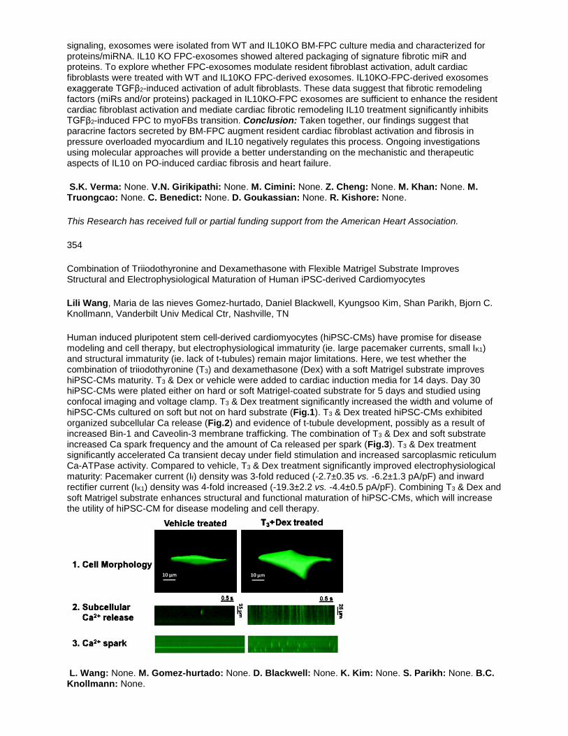
signaling, exosomes were isolated from WT and IL10KO BM-FPC culture media and characterized for proteins/miRNA. IL10 KO FPC-exosomes showed altered packaging of signature fibrotic miR and proteins. To explore whether FPC-exosomes modulate resident fibroblast activation, adult cardiac fibroblasts were treated with WT and IL10KO FPC-derived exosomes. IL10KO-FPC-derived exosomes exaggerate TGFβ2-induced activation of adult fibroblasts. These data suggest that fibrotic remodeling factors (miRs and/or proteins) packaged in IL10KO-FPC exosomes are sufficient to enhance the resident cardiac fibroblast activation and mediate cardiac fibrotic remodeling IL10 treatment significantly inhibits TGFβ2-induced FPC to myoFBs transition. Conclusion: Taken together, our findings suggest that paracrine factors secreted by BM-FPC augment resident cardiac fibroblast activation and fibrosis in pressure overloaded myocardium and IL10 negatively regulates this process. Ongoing investigations using molecular approaches will provide a better understanding on the mechanistic and therapeutic aspects of IL10 on PO-induced cardiac fibrosis and heart failure.
S.K. Verma: None. V.N. Girikipathi: None. M. Cimini: None. Z. Cheng: None. M. Khan: None. M. Truongcao: None. C. Benedict: None. D. Goukassian: None. R. Kishore: None.
This Research has received full or partial funding support from the American Heart Association.
354
Combination of Triiodothyronine and Dexamethasone with Flexible Matrigel Substrate Improves Structural and Electrophysiological Maturation of Human iPSC-derived Cardiomyocytes
Lili Wang, Maria de las nieves Gomez-hurtado, Daniel Blackwell, Kyungsoo Kim, Shan Parikh, Bjorn C. Knollmann, Vanderbilt Univ Medical Ctr, Nashville, TN
Human induced pluripotent stem cell-derived cardiomyocytes (hiPSC-CMs) have promise for disease modeling and cell therapy, but electrophysiological immaturity (ie. large pacemaker currents, small IK1) and structural immaturity (ie. lack of t-tubules) remain major limitations. Here, we test whether the combination of triiodothyronine (T3) and dexamethasone (Dex) with a soft Matrigel substrate improves hiPSC-CMs maturity. T3 & Dex or vehicle were added to cardiac induction media for 14 days. Day 30 hiPSC-CMs were plated either on hard or soft Matrigel-coated substrate for 5 days and studied using confocal imaging and voltage clamp. T3 & Dex treatment significantly increased the width and volume of hiPSC-CMs cultured on soft but not on hard substrate (Fig.1). T3 & Dex treated hiPSC-CMs exhibited organized subcellular Ca release (Fig.2) and evidence of t-tubule development, possibly as a result of increased Bin-1 and Caveolin-3 membrane trafficking. The combination of T3 & Dex and soft substrate increased Ca spark frequency and the amount of Ca released per spark (Fig.3). T3 & Dex treatment significantly accelerated Ca transient decay under field stimulation and increased sarcoplasmic reticulum Ca-ATPase activity. Compared to vehicle, T3 & Dex treatment significantly improved electrophysiological maturity: Pacemaker current (If) density was 3-fold reduced (-2.7±0.35 vs. -6.2±1.3 pA/pF) and inward rectifier current (IK1) density was 4-fold increased (-19.3±2.2 vs. -4.4±0.5 pA/pF). Combining T3 & Dex and soft Matrigel substrate enhances structural and functional maturation of hiPSC-CMs, which will increase the utility of hiPSC-CM for disease modeling and cell therapy.
L. Wang: None. M. Gomez-hurtado: None. D. Blackwell: None. K. Kim: None. S. Parikh: None. B.C. Knollmann: None.

355
CardioClusters: Harnessing the Power of Multi-lineage Cardiac Stem Cells
Megan M Monsanto, Kevin White, Kristina Fisher, Jessica Wang, Mark A Sussman, SAN DIEGO STATE UNIVERSITY, San Diego, CA
Existing approaches to modify stem cells for myocardial regeneration desperately need innovative solutions that enhance cell engraftment and persistence. Although this deficiency has been attacked through combinatorial stem cell delivery, there is no evidence that these stem cell injections provide for direct cellular crosstalk to promote stem cell survival and proliferation. Therefore, we created a CardioCluster, a 3D microenvironment consisting of three defined cell populations from the human heart: c-kit+ cardiac progenitor cells (CPCs), CD90+/CD105+ mesenchymal stem cells (MSCs) and CD133+
endothelial progenitor cells (EPCs). The size of the CardioCluster can be controlled by the quantity of cells used to create the cluster, allowing them to be infused into the heart without being reduced to single cell suspensions as is the case for cardiosphere-derived cells where the structural and cell-cell contact information is lost when delivered. Unlike cardiospheres, these cardiac cells are combined into a rationally designed cluster with MSCs and CPCs in the central core and EPCs forming the outer layer. EPCs play a vital role in forming neovasculature that will connect the CardioClusters to living heart tissue not damaged by ischemia and allow for revascularization of the damaged myocardium. In vitro we have shown that EPCs are better able to form tubular networks on matrigel-coated plates compared to either CPCs or MSCs. MSCs reinforce the 3D structure by releasing growth factors that attract and maintain cells within the cluster, as well as release immunomodulatory signaling factors. Upon induction of an oxidative stress by hydrogen peroxide CardioClusters show improved cell survival with a lower percentage of apoptotic and/or necrotic cell populations compared to the three populations individually. Upon myocardial injection CardioClusters have been shown to maintain their 3D structural integrity. Future directions will be to assess long-term engraftment and regenerative potential in a mouse model of myocardial infarction.
M.M. Monsanto: None. K. White: None. K. Fisher: None. J. Wang: None. M.A. Sussman: None.
This Research has received full or partial funding support from the American Heart Association.
357
Activation of Autophagic Flux Blunts Cardiac Ischemia/Reperfusion Injury
Geoffrey W Cho, UCLA, Los Angeles, CA; Min Xie, Univ of Alabama at Birmingham, Birmingham, AL; Yongli Kong, Dan L Li, Xiang L Luo, Francisco Altamirano, Cyndi Morales, Herman May, Gabriele Schiattarella, Jessica Medina, Nan Jiang, John Shelton, Anwarul Ferdous, Thomas G Gillette, Joseph A Hill, UT Southwestern Medical Ctr, Dallas, TX
Background: Reperfusion injury accounts for a significant portion of myocardial damage in acute coronary syndromes. Autophagy, a process of cell catabolism, plays a vital role in the heart’s response to stress. We have reported that re-induction of ischemia/reperfusion (I/R)-suppressed cardiomyocyte autophagy with histone deacetylase (HDAC) inhibitors affords significant cardioprotection. However, as HDACs govern many processes and may have off-target effects, we set out to modulate autophagy in a manner independent of HDAC activity. Here, we hypothesized that induction of autophagy with a novel agent, Tat-Beclin, at the time of reperfusion, will reduce I/R injury and rescue cardiac function. Methods: Wild type and ATG7 (protein required for autophagic flux) knockout mice were randomized among 3 treatment groups prior to surgical I/R injury [45 min LAD artery ligation; 24h reperfusion]: vehicle control (VC), Tat-Scrambled (TS), or Tat-Beclin (TB). Each agent was delivered at coronary reperfusion. To define molecular mechanisms, cultured adult and neonatal rat ventricular cardiomyocytes (ARVMs/NRVMs) were subjected to simulated I/R. Results: Induction of cardiomyocyte autophagy at reperfusion reduced infarct size 20.1% (±6.3%, n=23, p<0.02 vs VC). This treatment was associated with improved systolic function (declines in fractional shortening:

19.8±3.7% VC; 18.7±2.1% TS; 8.5±1.7% TB, n=11, p<0.01 vs VC). In NRVMs subjected to I/R injury, cell death was reduced 41% (±6%, n=12, p<0.001 vs VC). Improvements correlated with increased autophagic flux measured by the marker LC3-II, particularly at the infarct border zone. Additional data suggested that autophagy rescues I/R injury through reduction of oxidative stress. ATG7 KO mice or NRVM depleted of ATG7 (RNAi) manifested significantly less cardioprotection. Conclusion: Direct induction of cardiomyocyte autophagy reduces infarct size and declines in contractile function. Autophagy rescues I/R injury in part through reduction of oxidative stress. Critically, this cardioprotection was observed when intervention occurred at the time of reperfusion, the clinically relevant context.
G.W. Cho: None. M. Xie: None. Y. Kong: None. D.L. Li: None. X.L. Luo: None. F. Altamirano: None. C. Morales: None. H. May: None. G. Schiattarella: None. J. Medina: None. N. Jiang: None. J. Shelton: None. A. Ferdous: None. T.G. Gillette: None. J.A. Hill: None.
This Research has received full or partial funding support from the American Heart Association.
358
Factor Xa Deteriorates Atherosclerosis by Facilitating Inflammasome Formation via PAR-2-mediated Autophagy Suppression
Yasuhiro Maejima, Yusuke Ito, Natsuko Tamura, Masanori Konishi, Kenzo Hirao, Mitsuaki Isobe, Tokyo Medical and Dental Univ, Tokyo, Japan
Background: It is known that an endogenous blood coagulation factor Xa (FXa) plays a critical role in facilitating atherosclerosis by activating protease-activated receptor-2 (PAR-2). However, the precise mechanism how FXa-mediated PAR-2 activation promotes atherogenesis remains to be elucidated. Purpose: The aim of this study is to explore how FXa promotes atherosclerosis through PAR-2-associated signaling pathway. Methods & Results: Administration of direct FXa inhibitor rivaroxaban (Riv; 120 mg/kg/day) to the mice significantly suppressed the plasma FXa activity compared with untreated mice. Administration of Riv to ApoE knockout mice fed with high fat diet (ApoE-KO-HFD) significantly reduced atherosclerotic area in the aorta compared with those in the untreated ApoE-KO-HFD. The plaque size of ApoE-KO mice crossed with PAR-2 knockout mice fed with HFD was similar to those of Riv-treated ApoE-KO-HFD. Ultrastructural examinations of atherosclerotic lesions revealed that the number of autophagosomes in the plaque-resident macrophages of Riv-treated ApoE-KO-HFD was significantly smaller than those of the untreated ApoE-KO-HFD. Immunostaining of NLRP3 revealed that Riv attenuated the inflammasome formation in the atherosclerotic lesion in ApoE-KO-HFD. In vitro experiments demonstrated that treatment of 7-ketocholesterol (7KC) markedly enhanced autophagy activity in the murine macrophages. The addition of FXa significantly promoted mTOR (Ser2448) phosphorylation and blocked autophagy activity induced by 7KC, which was reversed in the presence of Riv (1 µM). Furthermore, immunoblot analyses demonstrated that FXa administration significantly accelerated inflammasome formation induced by 7KC, which was blocked in the presence of Riv. On the other hand, treatment with FXa failed to inhibit 7KC-induced autophagy and inflammasome activation in PAR-2-KO mice-derived macrophages. Conclusion: These results suggest that FXa worsens atherogenesis through PAR-2-mediated pathway by inhibiting macrophage autophagy which, in turn, promoting inflammasome activation.
Y. Maejima: None. Y. Ito: None. N. Tamura: None. M. Konishi: None. K. Hirao: None. M. Isobe: None.

359
Autosis is Triggered by Ischemia/Reperfusion in the Heart
Jihoon Nah, Peiyong Zhai, Rutgers Univ NJMS, Newark, NJ; Beth Levine, Univ of Texas Southwestern Medical Ctr, Dallas, TX; Junichi Sadoshima, Rutgers Univ NJMS, Newark, NJ
Although autophagy generally acts as a protective function in cardiomyocytes (CMs), uncontrolled or excessive activation of autophagy can be detrimental to CMs under some conditions. However, whether autophagy mediates cell death for CMs remains largely unknown. Recently, it has been shown in HeLa cells that excessive activation of autophagy induces cell death with characteristic morphological and biochemical features, and this form of cell death is termed autosis. Here we investigated whether death of CMs is induced by autosis in the presence of TAT-Beclin1, an autophagy-inducing peptide, and ischemia/reperfusion. TAT-Beclin1 treatment dose-dependently increased the level of LC3-II and decreased that of p62. TAT-Beclin1-treated CMs showed increases in trypan-blue-positive cells suggesting that autophagy-related cell death is induced by TAT-Beclin1. TAT-Beclin1-treated CMs showed the typical morphological features of autosis, including increased autophagic vacuoles and empty vacuoles, perinuclear spaces with cytoplasmic materials, and ballooning under electron microscopic observation. TAT-Beclin1-induced autosis in CMs was not inhibited by inhibitors of apoptosis, necrosis, or lysosomal degradation. Although autosis is known to be inhibited by cardiac glycosides, since cardiomyocyte Na+-K+-ATPase in rodents is relatively resistant to cardiac glycosides, we generated adenovirus harboring shRNA-α1 subunit of Na+-K+-ATPase. We found that downregulation of the α1 subunit of Na+-K+-ATPase inhibits TAT-Beclin1-induced autosis in CMs. We also found that ischemia/reperfusion induces autosis in the mouse heart, as evidenced by the presence of characteristic morphological features of autosis, including the presence of autophagosomes, empty vacuoles, and perinuclear space. Lastly, in order to elucidate the molecular mechanism mediating autosis in the heart, we conducted unbiased screenings of 150 kinase inhibitors and identified several classes of kinase inhibitors suppressing TAT-Beclin1-induced autosis in CMs. These results indicate that autosis occurs in the heart during ischemia/reperfusion injury. Our findings may provide novel therapeutic targets for treatment of cardiac cell death during ischemia/reperfusion.
J. Nah: None. P. Zhai: None. B. Levine: None. J. Sadoshima: None.
360
Peptidyl-prolyl Isomerase Domain on SPG7 Is the Critical Mediator for Activation of Permeability Transition Pore Opening
Shanmughapriya Santhanam, Zhiwei Dong, Dhanendra Tomar, Neeharika Nemani, Edmund Carvalho, Aparna Tripathi, Sudarsan Rajan, Madesh Muniswamy, Temple Univ, Philadelphia, PA
Introduction: In Cardiomyocytes, physiological MCU-mediated [Ca2+]m uptake is required for the activation of energy pathways that support increased contractility, but reciprocally [Ca2+]m overload is the key driver for the mitochondrial permeability transition pore (PTP) opening. Though [Ca2+]m mediated PTP has been implicated in several necrosis models, multiple proteins have been proposed and these identifications remain controversial. Using a RNAi-based screen to identify genes that modulate Ca2+ and ROS-induced PTP, we identified a necessary and conserved role for Spastic Paraplegia 7 (SPG7) in CypD-dependent PTP opening. Absence of this protein caused a remarkable increase in [Ca2+]m retention capacity with sustained mitochondrial membrane potential (ΔΨm). Silencing of SPG7 prevented Ca2+ and ROS-induced mitochondrial dysfunction and cell death. Hypothesis: We hypothesis that CypD isomerase (PPIase) activity is required for SPG7 oligomerization and PTP activation. Results: The PPIase role is to interconvert the cis and trans isomers of peptide bonds. Proline followed by phenylalanine (XXPF) constitutes the putative signature motif for PPIase activity. We performed PPIase domain prediction to identify whether SPG7 has PPIase recognition sites. Two putative PPIase sites were identified: proximity to regions corresponding to ATP binding Walker Motif (aa 368-371) and CypD/SPG7 interaction domain (aa 690-691). We have shown that CypD sequesters SPG7 through SPG7690-691 region as substrate, facilitates the assembly of higher-order oligomer on the inner mitochondrial membrane and forms the active PTP. Our biochemical studies indicate that knocking in (KI) a point mutation (SPG7Δ690-691) in MEFs resulted in the disruption of CypD dependent SPG7 oligomerization, and an incompetent PTP thus

significantly alleviating necrotic cell death. We have also adopted CRISPR/Cas9-mediated KI strategy to generate a mouse model (SPG7Δ690-691 KI) to decipher the region of SPG7 that emerged as a critical domain for PTP assembly and opening. Conclusion: In Conclusion, various cellular and biochemical evidences envisioned SPG7 and CypD as integral components of the PTP complex that are Ca2+ and ROS sensitive.
S. Santhanam: None. Z. Dong: None. D. Tomar: None. N. Nemani: None. E. Carvalho: None. A. Tripathi: None. S. Rajan: None. M. Muniswamy: None.
This Research has received full or partial funding support from the American Heart Association.
361
Regulation of Activity of Apoptosis Inducing Factor by SUMO Conjugation
Eun Y KIim, Yi Zhang, Abha Grover, Texas Heart Inst, Houston, TX; Wei Yu, Univ of Houston, Houston, TX; Jun Wang, Texas Heart Inst, Houston, TX
Apoptosis inducing factor (AIF), a mitochondrial oxidoreductase, is primarily involved in maintaining mitochondrial function. However, in response to apoptotic insults, AIF is released from mitochondria via cleavage and relocates to the nucleus, where it binds chromatin, subsequently causing its condensation and cell death. Thus, AIF plays an important role in both cell survival and death. The regulatory pathways/signaling events that govern the key step of AIF translocation and activity in cell death are not completely deciphered. SUMO conjugation, a highly conserved posttranslational modification, plays an important role in cardiac homeostasis. Herein, we report AIF as a novel SUMO substrate, and SENP5, a SUMO deconjugation enzyme, increased cardiomyocyte death once overexpressed in the mouse hearts, which coincided with an increase level of truncated AIF in cytosol. In line with the above findings, adenoviral mediated transduction of SENP5 wild type (wt), but not the C713L mutant, the latter of which lacks de-sumoylation activity, triggered cell death and AIF nuclear occupancy. Also, the SENP5-wt, but not the catalytically inactive mutant, de-sumoylated AIF. We further identified lysine residue 89 (K89) on AIF as one of the major SUMO attachment sites, and mutation of K89 to arginine (K89R) promoted AIF nuclear translocation in AC16, a cardiac cell line, and necrosis in L929 cells induced by TNFα/Z-vad-fmk. Our findings provide a novel mechanism by which SUMO conjugation is involved in mediating the function of AIF, which is important for cardiomyocyte survival and death.
E.Y. KIim: None. Y. Zhang: None. A. Grover: None. W. Yu: None. J. Wang: None.
This Research has received full or partial funding support from the American Heart Association.
362
Restoring Diabetes-induced Autophagic Flux Arrestment in Ischemic/Reperfused Heart by Adiponectin Receptor Activation Involves Both AMPK-Dependent and AMPK-Independent Signaling
Yajing Wang, Wayne Lau, Bernard Lopez, Theodore Christopher, Thomas Jefferson Univ, Philadelphia, PA; Erhe Gao, Walter Koch, Temple Univ, Philadelphia, PA; Xinliang (Xin) Ma, Thomas Jefferson Univ, Philadelphia, PA
Autophagy is increasingly recognized as an important regulator of myocardial ischemia-reperfusion (MI-R) injury. However, whether and how diabetes may alter autophagy in response to MI-R remains unknown. Deficiency of adiponectin (AdipoQ), a cardioprotective molecule, markedly increases MI-R injury. However, the role of diabetic hypoadiponectinemia in cardiac autophagy alteration after MI-R is unclear. Utilizing normal control (NC), high-fat-diet-induced diabetes, and Adipoq knockout (adipoq-/-) mice, we demonstrated that autophagosome formation was modestly inhibited and autophagosome clearance was markedly impaired in the diabetic heart subjected to MI-R. adipoq-/- largely reproduced the phenotypic alterations observed in the ischemic-reperfused diabetic heart. Treatment of diabetic and adipoq-/- mice with AdipoRon, a novel adiponectin receptor agonist, stimulated autophagosome formation, markedly increased autophagosome clearance, reduced infarct size, and improved cardiac function (P<0.01 vs

vehicle). Mechanistically, AdipoRon caused significant phosphorylation of AMPK-BECN1 (Ser93/Thr119)-PtdIns3K (Ser164) and enhanced lysosome protein LAMP2 expression both in vivo and in isolated adult cardiomyocytes. Pharmacologic AMPK inhibition or genetic AMPK alpha2 mutation abolished AdipoRon-induced BECN1 (Ser93/Thr119)-PtdIns3K (Ser164) phosphorylation and AdipoRon-stimulated autophagosome formation. However, AdipoRon-induced LAMP2 expression, AdipoRon-stimulated autophagosome clearance, and AdipoRon-suppressed superoxide generation were not affected by AMPK inhibition. Treatment with MnTMPyP (a superoxide scavenger) increased LAMP2 expression and stimulated autophagosome clearance in simulated ischemic-reperfused cardiomyocytes. However, no additive effect between AdipoRon and MnTMPyP was observed. Collectively, these results demonstrate that hypoadiponectinemia impairs autophagic flux, contributing to enhanced MI-R injury in the diabetic state. Adiponectin receptor activation restores AMPK-mediated autophagosome formation and antioxidant-mediated autophagosome clearance, representing a novel intervention effective against MI-R injury in diabetic conditions.
Y. Wang: None. W. Lau: None. B. Lopez: None. T. Christopher: None. E. Gao: None. W. Koch: None. X. Ma: None.
365
Change in Saturation of Oxygen and Patent Foramen Ovale in Postural Orthostatic Tachycardia Syndrome
Mu'taz F AbuAlSha'r, Ra'ed K Abu-Jajeh, Mo'yed N Ahmad, Chandralekha Ashangari, Stacy E Hamid, Amer Suleman, The Heartbeat Clinic, Dallas, TX
Background : Postural Orthostatic Tachycardia Syndrome (POTS) is a constellation of signs and symptoms that occurs when the patient stands upright. It affects primarily young women. On the other hand, PFO is a hidden condition that doesn’t cause signs and symptoms in most cases. Method : 331 POTS patients were randomly selected during the period of June 2014 to April 2016 from our clinic and they underwent PFO test. In addition to that, patients underwent tilt test with SPO2 recordings every 2 minutes until 10 minutes to calculate the change in SpO2 of that period of time. Results : Out of 331 patients, 292 patients were females (88%; n=292 age 32.48 ± 10.78), 39 patients were males (12%; n=39 age 28.87 ± 11.69). 136 patients (41%) had no drop in their SPO2 and 17 of 136 patients had positive(+) PFO test (Grade 1 PFO in 4 patients, Grade 2 PFO in 2 patients, Grade 3 PFO in 3 patients and Grade 4 PFO in 8 patients). 77 patients (23%) had a drop of 1% in their SPO2 and 11 of 77 patients had + PFO test (Grade 1 PFO in 3 patients, Grade 2 PFO in 1 patient, Grade 3 PFO in 2 patients and Grade 4 PFO in 5 patients). 46 patients (14%) had a drop of 2% in their SPO2 and 10 of 46 patients had + PFO test (Grade 1 PFO in 2 patients, Grade 3 PFO in 2 patients and Grade 4 PFO in 6 patients). 16 patients (5%) had a drop of 3% in their SPO2 and 3 of 16 patients had + PFO test (Grade 1 PFO in 1 patient, Grade 2 PFO in 1 patient, Grade 3 PFO in 1 patient). 9 patients (3%) had a drop of 4% in their SPO2 and 1 of 9 patients had + PFO test (Grade 2). 5 patients (1.5%) had a drop of 5% in their SPO2 and 2 out of 5 patients had + PFO test (Grade 1 PFO in 1 patient and Grade 4 PFO in 1 patient). 2 patients (0.6%) had a drop of 6% in their SPO2 and both of them had + PFO test (Grade 2 PFO in 1 patient, Grade 4 PFO in 1 patient). 40 patients (12%) had a rise in their SPO2 and 4 of 40 patients had + PFO test (Grade 2 PFO in 2 patients, Grade 3 PFO in 1 patient and Grade 4 PFO 1 patient). Conclusion : Our research results demonstrated that 15% of patients with POTS have PFO. Though orthodeoxia is relatively uncommon, this study demonstrated orthodeoxia in 59% of patients. Only 2% patients showed orthodeoxia of >5%, but further studies should be done to define the clinical significance of the study.
M.F. AbuAlSha'r: None. R.K. Abu-Jajeh: None. M.N. Ahmad: None. C. Ashangari: None. S.E. Hamid: None. A. Suleman: None.

367
Plasma Based cBIN1 Correlates with Myocardial Health in Heart Failure with Reduced Ejection Fraction
Rachel Baum, Bing Xu, Yu Xie, Ronit Zadikany, Tara Hitzeman, Sosse Agvanian, Dael Geft, David Chang, Jaime Moriguchi, Antoine Hage, Babak Azarbal, Lawrence Czer, Michelle Kittleson, Jignesh Patel, Jon Kobashigawa, Michele Hamilton, TingTing Hong, Robin M Shaw, Cedars-Sinai Medical Ctr, Los Angeles, CA
BACKGROUND: Acquired heart failure (HF) originates with failing heart muscle, yet it is currently not possible to assay the biochemical health of cardiomyocytes. A key pathophysiology of HF is weakened calcium transient under the regulation of a membrane deformation protein bridging integrator 1 (BIN1). BIN1 decreases with HF and is blood available. We hypothesized that a recently cloned cardiac BIN1 isoform (cBIN1, BIN1+13+17) can be selectively measured to diagnose heart muscle health. METHODS: Expression of cBIN1 in human heart and plasma was determined by immunoprecipitation, western blotting, and mass spectrometry analysis. A cBIN1 specific ELISA was developed using the combination of anti-BIN1 exon 17 (clone 99D from Sigma) and 13 (gift from Sarcotein Diagnostics) antibodies. Plasma cBIN1 concentration was then measured in a large clinical cohort of HFrEF (N=180) patients and compared to a sex and age matched cohort of healthy volunteers. Plasma cBIN1 concentration was also compared to NT-proBNP for its ability to detect patients with HFrEF. RESULTS: Biochemistry and mass spectrometry confirms that cBIN1 is expressed in human myocardium. An ELISA assay selects for the cBIN1 isoform which significantly reduced in HFrEF (3.0±0.3, n=286, mean±SEM, ng/ml, p<0.001) versus matched healthy volunteers (3.0±0.3, n=340). Low plasma cBIN1 diagnoses HFrEF with a ROC area under the curve of 0.92. cBIN1 ROC characteristics are additive to those of NT-proBNP. In addition, low plasma cBIN1 predicts future cardiovascular hospitalization and death over a 12 month follow-up period. CONCLUSIONS: A cBIN1 specific ELISA can quantify cBIN1 in human plasma. Low plasma cBIN1 diagnoses diseased heart muscle in HFrEF patients and predicts future hospitalization and death.
R. Baum: None. B. Xu: None. Y. Xie: None. R. Zadikany: None. T. Hitzeman: None. S. Agvanian: None. D. Geft: None. D. Chang: None. J. Moriguchi: None. A. Hage: None. B. Azarbal: None. L. Czer: None. M. Kittleson: None. J. Patel: None. J. Kobashigawa: None. M. Hamilton: None. T. Hong: None. R.M. Shaw: None.
This Research has received full or partial funding support from the American Heart Association.
368
The Effect of Long-term Administration of PGE1 on the Morphological Change of Ductus Arteriosus
Ryuma Iwaki, Hironori Matsuhisa, Yoshihiro Oshima, Dept of Cardiovascular Surgery, Kobe Children's Hosp, Kobe, Japan; Masato Hoshino, Japan Synchrotron Radiation Res Inst (SPring-8), Sayo, Japan; Susumu Minamisawa, Toru Akaike, Dept of Cell Physiology, Jikei Univ Sch of Med, Tokyo, Japan; Kiyozo Morita, Gen Shinohara, Dept of Cardiovascular Surgery, Jikei Univ Sch of Med, Tokyo, Japan; Yukihiro Kaneko, Hiroyuki Morishita, Div of Cardiovascular Surgery, Natl Medical Ctr for Children and Mothers, Tokyo, Japan; Masashi Takahashi, Div of Thoracic and Cardiovascular Surgery, Niigata Univ Graduate Sch of Medical and Dental Sciences, Nigata, Japan; Naoto Yagi, Japan Synchrotron Radiation Res Inst (SPring-8), Sayo, Japan; Takuro Tsukube, Div of Cardiovascular Surgery, Japanese Red Cross Kobe Hosp, Kobe, Japan; Yutaka Okita, Div of Cardiovascular Surgery, Graduate Sch of Med, Kobe Univ, Kobe, Japan
Introduction Plostagrandin E1 (PGE1) is essential for maintaining the ductus arteriosus (DA) in ductal-dependent congenital heart disease. Recently, relatively long-term administration of PGE1 became common, especially in patient with hypoplastic left heart syndrome (HLHS) . Staged Norwood approach which consists of bilateral pulmonary artery banding, PGE1 infusion, and Norwood procedure beyond newborn period provided excellent survival benefit. However, microstructural changes of DA after long-term PGE1 administration remain unknown. Therefore, we sought to investigate their features of DA after long-term PGE1 administration using synchrotron based X-ray phase-contrast tomography (XPCT), a novel modality to inspect biological soft tissue, and pathological scrutiny including DA specific immunostaining. Methods Seventeen DA tissues of HLHS were obtained during Norwood procedure.

The median duration of PGE1 was 48 days (Range 3-123). Radiographic microstructural analysis of DA was performed using X-ray phase contrast tomography at SPring-8 (Hyogo, Japan). Histological changes focused on elastic fiber and smooth muscle formation were evaluated by Elastica van Gieson staining, and expression of prostaglandin E2 receptor EP4 was evaluated by immunohistochemical analysis. Results XPCT study showed a sclerotic changes of ductal media. There was a significant correlation between the duration of PGE1 infusion and the mass density of ductal media (R: 0.723, p=0.001). Histological study showed that the duration of PGE1 infusion was positively correlated with the elastic fiber formation (R: 0.795, P=0.002), and negatively correlated with smooth muscle formation (R: -0.83, P<0.001). The EP4 expression was disappeared in all DA. Conclusion Long-term administration of PGE1 induced elastogenesis and density growth of the ductal media. In addition, downregulation of EP4 was observed. These results suggested that dosage of PGE1 could be decreased after definite period of administration.
R. Iwaki: 2. Research Grant; Modest; JSPS KAKENHI Grant Number 15K15505 and 26293249. H. Matsuhisa: 2. Research Grant; Modest; JSPS KAKENHI Grant Number 15K15505 and 26293249. Y. Oshima: 2. Research Grant; Modest; JSPS KAKENHI Grant Number 15K15505 and 26293249. M. Hoshino: None. S. Minamisawa: 2. Research Grant; Modest; JSPS KAKENHI Grant Number 15K15505 and 26293249. T. Akaike: 2. Research Grant; Modest; JSPS KAKENHI Grant Number 15K15505 and 26293249. K. Morita: None. G. Shinohara: None. Y. Kaneko: None. H. Morishita: None. M. Takahashi: None. N. Yagi: None. T. Tsukube: None. Y. Okita: None.
369
Engineering Rrm2 to Elevate 2-deoxy-ATP and Improve Cardiomyocyte Function
Jason D Murray, Xuan Guan, Farid Moussavi-Harami, Sigurast S Olafsson, Charles E Murry MD, PhD, Michael Regnier, Univ of Washington, Seattle, WA
Overexpression of ribonucleotide reductase (RNR) in cardiomyocytes increases the amount of cytosolic 2-deoxy-ATP (dATP), which can be used by myosin and significantly increases contraction of cardiac muscle at all levels of calcium activation. Our group is working to develop enhanced dATP as a therapeutic option for heart failure. We have demonstrated that virally-mediated overexpression of RNR elevates dATP and increases the rate and magnitude of contraction and increases left ventricular contraction in normal hearts as well as rodent models of myocardial infarction and dilated cardiomyopathy. RNR is a heterotetramer containing two subunits, Rrm1 and Rrm2. While cardiomyocyte-specific overexpression of Rrm1 is stable, we have observed high variability in expression levels of the Rrm2 subunit in multiple disease models. We hypothesized that this variability was largely due to protein degradation via the ubiquitin-proteasome complex (UPC). We found that pharmacological inhibition of proteasome activity leads to increased expression of Rrm2 in virally-transduced cardiomyocytes in vitro. To confirm the hypothesis that the overexpressed Rrm2 is degraded via UPC-mediated degradation, we engineered mutations in specific ubiquitin-binding degrons of the Rrm2 gene. Transfecting human induced pluripotent stem cell-derived cardiomyocytes resulted in higher levels of Rrm2 than those overexpressing wild-type protein and resulted in higher levels of cytosolic dATP as measured by Liquid chromatography-mass spectrometry. Ongoing and planned experiments will compare the effects of this engineered mutation on Rrm2 overexpression, dATP production, and contractility in cultured cardiomyocytes. Our goal is to develop an improved RNR vector that will be resistant to degradation through the ubiquitin-proteasome pathway and therefore enable more stable and consistent RNR enzyme activity and deoxynucleotide levels of cardiomyocytes transduced in vivo.
J.D. Murray: None. X. Guan: None. F. Moussavi-Harami: None. S.S. Olafsson: None. C.E. Murry: 7. Ownership Interest; Modest; BEAT Biotherapeutics, Inc. M. Regnier: 7. Ownership Interest; Modest; BEAT Biotherapeutics, Inc.

370
Wt1 Influences Regenerative Angiogenic Function of the Vascular Endothelium
Robert Ogley, Richard McGregor, Nick Hastie, Patrick Hadoke, Univ of Edinburgh, Edinburgh, United Kingdom
Ischaemia causes irreversible tissue damage in cardiovascular disease. Since regenerative angiogenesis (new blood vessel formation from existing vasculature) fails to consistently induce sufficient reperfusion to facilitate repair, a targeted approach to manipulating angiogenesis is clinically desirable. The Wilms’ tumour suppressor (Wt1) is a transcription factor which regulates numerous genes and cellular processes, including many intrinsic to angiogenesis. We hypothesise that Wt1 influences the angiogenic function of endothelial cells. Wt1 was identified in endothelial and non-endothelial cells comprising vessel outgrowths generated by cultured aortic rings from Wt1-GFP reporter mice. Inducible deletion of Wt-1 from the endothelium (VE-Wt1 KO) significantly delayed angiogenesis in this assay (p<0.05 relative to Cre and vehicle controls, n=5-8). In vivo, Wt-1 expression was evident in vascular endothelial cells and perivascular cells of the hindlimb as early as 3 days following femoral artery ligation to induce ischaemia. Less expression was detected by day 28. VE-Wt1 KO had no effect on hindlimb reperfusion (laser Doppler; days 0-28) or on vessel density (day 28). Similarly, VE-Wt1 KO had no effect on vessel density (histology, immunohistochemistry) or expression of angiogenic factors (qRT-PCR) in sponges inserted beneath the skin of mice (20 days). To further understand the role of Wt1 in angiogenesis, transcriptomic analysis was performed for RNA expression in Wt1+ and Wt1- cells isolated (FACs) from sponges 7 and 21 days after implantation in Wt1-GFP mice. Wt1+ cells exhibited higher expression of genes involved in a number of processes relevant to tissue repair, including vasculature development (D7: p=10-7.43, D21: p=10-3.47, n=3) and angiogenesis (D7: p=10-3.90, D21: p=10-4.74, n=3). These results show that deletion of endothelial Wt-1 delays, rather than permanently inhibiting, angiogenesis: suggesting a more complex mechanism of action than reported previously. Transcriptomic data suggest that expression in endothelial cells (and other cells in the endothelial lineage) contributes to tissue repair in ischaemia. This work has improved our understanding of the regulatory role of Wt1 in angiogenesis.
R. Ogley: None. R. McGregor: None. N. Hastie: None. P. Hadoke: None.
371
Assessment of Catheter Position Above or Below the Aortic Valve by Evaluation of Characteristics of the Local Electro Gram: an Acute Canine Study
Deepak Pa, Chance Witt, Alan M Sugrue, Scott Suddendorf, Joanne Powers, Dejae Ladewig, Budimir Drakulic, Thomas Foxall, Samuel Asirvatham, Mayo Clinic, Rochester, MN
Background: Ablation in the sinuses of Valsalva (SoV) within the aorta is fraught with risk, owing to the proximity of the coronary arteries. Assessment of catheter tip position is not very easy at this location owing to complex local anatomy and poor images from available modalities. Objectives: We propose the use of novel local unipolar electrogram characteristics using a new signal recording system to guide the assessment of catheter tip position in this region. Methods: We recorded unipolar signals from above the SoV, within the coronaries and below the SoV using the PURE-EP™ (BioSig Technologies, MN) signal processing system in 4 anesthetized canines in the setting of an acute study. We used fluoroscopy, intracardiac echocardiography and angiography for confirmation of the position of the catheter tip. We recorded changes in the signals when transitioning between (1) the coronary arteries and the SoV and (2) the SoV and below the SoV. Results: The unipolar signal showed a negative current of injury (COI) when the catheter tip was present in the SoV, whereas within the coronary artery or below the SoV, the COI was positive. We propose that the difference in the distribution of ventricular muscle below and above the SoV is responsible for this phenomenon. These changes in the COI were noted immediately when transitioning from one location to another and were present reproducibly in all the dogs. (Figure 1) Conclusions: Analysis of the changes on the local electrogram can provide information about the location of the catheter tip which could prove critical in saving lives.

D. Pa: None. C. Witt: None. A.M. Sugrue: None. S. Suddendorf: None. J. Powers: None. D. Ladewig: None. B. Drakulic: None. T. Foxall: None. S. Asirvatham: None.
372
CD16 Monocytes Stimulate Arteriogenesis to Salvage the Ischemic Limb
Ashish S Patel, Francesca E Ludwinski, Puja Kapoor, Paul R Jones, Alexander Kerr, Tamer El-Sayed, Prakash Saha, Oliver T Lyons, Alberto Smith, Bijan Modarai, King's Coll London, London, United Kingdom
Introduction Cell therapy using unselected mononuclear cell populations has had modest benefits in patients with critical limb ischemia (CLI). We hypothesized that tissue-remodeling monocytes, identified by their expression of CD16 (CD16+ Mo), may be a novel cellular therapy for CLI. Methods and Results Flow cytometry showed that the proportion of circulating CD16+ Mo was greater in CLI patients (n=25) compared with matched controls (n=15, P<0.0001). Removal of ischemia following revascularization or amputation resulted in a fall in CD16+ Mo to control levels (P<0.05). CLI CD16+ Mo expressed higher levels of the adhesive proteins VLA, ICAM-1 and CD11c compared with controls (P<0.05). Conditioned media from these cells contained higher levels of HB-EGF, PlGF, endoglin, VEGF-C and VEGF-D (P<0.05) and induced greater endothelial cell tubule formation (P<0.05) compared with CD16- Mo from the same patients (n=9). CD16+ Mo preferentially migrated towards ischemic muscle supernatants isolated from CLI patients (n=7, P<0.02). CD16+ and CD16- Mo were isolated from 12 CLI patients and 1x10⁶ cells injected into the adductor muscles of nude athymic mice following femoral artery excision. More ischemic hindlimbs were salvaged when treated with CD16+ compared with CD16- Mo (83% [10/12] vs 17% [2/12] limbs, P<0.05) and this was associated with enhanced arteriogenesis (αSMA-stained vessels, P<0.05). Conclusion Circulating CD16+ Mo from CLI patients have increased expression of adhesion markers, are preferentially retained within ischemic muscle and promote robust arteriogenesis and limb salvage in experimental HLI. This monocyte subset may be an effective cellular therapy for CLI.

A.S. Patel: None. F.E. Ludwinski: None. P. Kapoor: None. P.R. Jones: None. A. Kerr: None. T. El-Sayed: None. P. Saha: None. O.T. Lyons: None. A. Smith: None. B. Modarai: 2. Research Grant; Significant; British Heart Foundation.
373
Effects of Coronary Wall Mechanics on Smooth Muscle Cell Phenotypic Switch and CD44+ Mesenchymal Cell Repopulation in Saphenous Vein Grafts
Matthijs S Ruiter, Ctr Cardiologico Monzino, Tissue Engineering Unit, Milan, Italy; Marco Piola, Politecnico di Milano, Dept di Elettronica, Informazione e Bioingegneria, Milan, Italy; Gloria Garoffolo, Ctr Cardiologico Monzino, Tissue Engineering Unit, Milan, Italy; Erica Gianazza, Ctr Cardiologico Monzino, Unit of Proteomics, Milan, Italy; Marco Agrifoglio, Ctr Cardiologico Monzino, Dept of Cardiac Surgery, Milan, Italy; Claudio Saccu, Stefano Zoli, Ctr Cardiologico Monzino, Dept of Cardiovascular Surgery, Milan, Italy; Marco Zanobini, Ctr Cardiologico Monzino, Dept of Cardiac Surgery, Milan, Italy; Cristina Banfi, Ctr Cardiologico Monzino, Unit of Proteomics, Milan, Italy; Monica Soncini, Gianfranco B Fiore, Politecnico di Milano, Dept di Elettronica, Informazione e Bioingegneria, Milan, Italy; Maurizio Pesce, Ctr Cardiologico Monzino, Tissue Engineering Unit, Milan, Italy
Background. Despite the preferred application of arterial conduits, the greater saphenous vein (SV) remains indispensable for bypass grafting, especially in multi-vessel coronary artery disease. Early remodeling induced by altered wall mechanics has been recognized to play a key role in SV graft disease. The mechanism remains, however, unknown. Aim. To investigate mechanical factors involved in early graft remodeling, we characterized SV-derived smooth muscle cells (SMCs) after both ex vivo coronary-like mechanical stimulation of SV segments and in vitro unidirectional strain. Methods. SV segments from patients receiving coronary artery bypass grafts were stimulated in a custom-made coronary pulse-duplicator bioreactor. After 7 (n=6) or 14 (n=5) days, stretched and control SVs were fixed and stained for immunofluorescence. Additionally, SMCs isolated from SVs of 7 patients undergoing saphenectomy were subjected to uniaxial cyclic strain (10% elongation, 1 Hz) for 24 or 72 hours using a Flexcell platform. SMCs analysis was performed by western blotting and mass spectrometry-based secretome analysis. Results. Coronary stimulation elevated apoptosis of SV medial cells after 7 days, and consistently reduced the percentage of cells positive for contractile markers α-SMA and calponin. Conversely, synthetic phenotype marker tropomyosin-4 (TM4) and early contractile marker SM22α were elevated at T14. Mesenchymal marker CD44 was markedly upregulated in cells populating the media after 14 days of stimulation. In accordance, strained SMCs displayed decreased α-SMA and SM22α, and increased TM4 protein expression after 72h. Analysis of the supernatant showed a significant increase of plasminogen activator inhibitor-1 and thrombospondin 1.

Conclusions. Mechanical stimulation of SVs leads to apoptosis of medial cells and a decrease of contractile SMC markers, followed by repopulation with cells expressing the mesenchymal marker CD44. In addition, unidirectional strain induces a switch of SMC phenotype and secretion of proteins related to vascular remodeling. We are currently investigating whether CD44+ cells derive from SMCs undergoing phenotypic switch, or from progenitor cells localized in the adventitia.
M.S. Ruiter: None. M. Piola: None. G. Garoffolo: None. E. Gianazza: None. M. Agrifoglio: None. C. Saccu: None. S. Zoli: None. M. Zanobini: None. C. Banfi: None. M. Soncini: None. G.B. Fiore: None. M. Pesce: None.
374
Evidence for Hyper-reductive and Hyper-oxidative Conditions in Heart Failure Patients
Thiagarajan Sairam, PSG Inst of Medical Sciences and Res, Coimbatore, India; Gobinath Shanmugam, Univ of Alabama at Birmingham, Birmingham, AL; Madhusudhanan Narasimhan, Texas Tech Univ Health Sciences Ctr, Lubbock, TX; Meenu Subramanian, PSG Inst of Medical Sciences and Res, Coimbatore, India; Amit N Patel, Univ of Miami, Miami, FL; Rajendran Gopalan, Ramalingam Sankaran, PSG Inst of Medical Sciences and Res, Coimbatore, India; Rajasekaran N Soorappan, Univ of Alabama at Birmingham, Birmingham, AL
Background: Oxidative stress has been linked to heart failure (HF) in humans. Antioxidant-based treatments are not always effective. Hence, we tested a hypothesis that some patients with heart failure may have a hyper-reductive state. Identifying this condition may lead to personalized optimization and better outcomes. Methods: Blood samples were collected from age and sex matched healthy control (n=50) and HF patients (n=55). Serum was separated and stored immediately in -80°C until analysis. Reduced glutathione (GSH) and its redox ratio (GSH/GSSG) using sigma GSH kit (38185) and malondialdehyde (MDA) levels by HPLC in the serum of HF patients were quantified. Further, the activities of key antioxidant enzymes including catalase, SOD, glutathione peroxidase and glutathione reductase were analyzed using a kinetic spectrophotometer. Results: While majority of the HF patients had significantly decreased the glutathione redox status (GSH/GSSG) and increased MDA levels (lipid peroxidation index) indicating a hyper-oxidative state, a subset of HF patients (n=8) displayed a significantly increased GSH/GSSG ratio along with decreased MDA levels in the serum, suggesting a strong association for a hyper-reductive state in the development of HF. Moreover, closer analyses of echocardiography revealed a lower ejection fraction (EF) with substantial diastolic dysfunction (MV E/A) in the HF patients with hyper-reductive state. Conclusion: These results suggest that hyper-reductive condition may indicate a worsening clinical course for HF patients. Thus a thorough diagnosis of redox state and personalized approach for antioxidant treatment is warranted.
T. Sairam: None. G. Shanmugam: None. M. Narasimhan: None. M. Subramanian: None. A.N. Patel: None. R. Gopalan: None. R. Sankaran: None. R.N. Soorappan: None.

376
Acute Modulation of Sodium Channel Auxiliary Subunit β1-mediated Adhesion: a Novel Antiarrhythmic Strategy
Rengasayee Veeraraghavan, Gregory S Hoeker, Virginia Polytechnic Univ, Roanoke, VA; Anita Alvarez Laviada, Imperial Coll London, London, United Kingdom; Xiaoping Wan, Isabelle Deschenes, Case Western Reserve Univ, Cleveland, OH; James Smyth, Virginia Polytechnic Univ, Roanoke, VA; Julia Gorelik, Imperial Coll London, London, United Kingdom; Steven Poelzing, Robert G Gourdie, Virginia Polytechnic Univ, Roanoke, VA
Cardiac impulse propagation is thought to occur by direct cell to cell flow of current via connexin43 (Cx43) gap junctions (GJs). We recently demonstrated that cardiac sodium channels (Nav1.5) localized within the perinexus, an intercalated disk (ID) nanodomain adjacent to Cx43 GJ, may enable ephaptic coupling between cardiac myocytes. We hypothesized that β1-mediated adhesion may closely approximate membranes within ID nanodomains, facilitating ephaptic coupling. Super-resolution STochastic Optical Reconstruction Microscopy-based Relative Localization Analysis (STORM-RLA) and immuno-electron microscopy identified two NaV1.5 populations within the ID in guinea pig ventricles (GPVs): A perinexal population, accounting for 47% of ID-localized NaV1.5 and a plicate population, co-distributing with N-Cadherin, accounting for 29%. β1 was preferentially localized to the perinexus (48% of ID-localized β1) over N-Cadherin-rich plicate regions (8%). βadp1, a novel peptide inhibitor of β1 adhesion, selectively and dose-dependently inhibited barrier function in β1-overexpressing 1610 cells in electric cell-substrate impedance spectroscopy studies. Neither βadp1 nor a scrambled control peptide (βadp1-scr) affected INa or action potentials in isolated GPV myocytes. However, βadp1 reduced peak current recorded from NaV1.5 clusters adjacent Cx43-EGFP at cell-cell contacts using scanning ion conductance microscopy-guided patch clamp. In GPVs, βadp1 (100 µM) compromised the diffusion-resistance of the ID as assessed by perfusion of fixable fluorescent dyes. βadp1 (48±4 µm) but not βadp1-scr (22±1 µm) increased perinexal intermembrane spacing compared to control GPVs (17±1µm). Optical mapping revealed that βadp1 but not βadp1-scr slowed conduction in GPVs and iPSC-derived cardiomyocyte monolayers. Importantly, in GPVs, βadp1 increased conduction anisotropy and precipitated spontaneous tachyarrhythmias in a dose-dependent manner. Thus, β1-mediated adhesion generates close apposition between NaV1.5-rich perinexal membranes, facilitating ephaptic conduction in the heart. Importantly, β1-mediated adhesion may be a target for novel, mechanistically-driven antiarrhythmic therapy.
R. Veeraraghavan: None. G.S. Hoeker: None. A.A. Laviada: None. X. Wan: None. I. Deschenes: None. J. Smyth: None. J. Gorelik: None. S. Poelzing: None. R.G. Gourdie: None.
This Research has received full or partial funding support from the American Heart Association.
378
A Systematic Review of Mobility/Immobility in Thromboembolism Risk Assessment Models for Hospitalized Patients
Fan Ye, North Florida Regional Medical Cent, Gainesville, FL; Carolyn Stalvey, Univ of Florida, Gainesville, FL; Matheen Khuddus, North Florida Regional Medical Ctr, Gainesville, FL; David Winchester, Univ of Florida, Gainesville, FL; Hale Toklu, North Florida Regional Medical Cent, Gainesville, FL; Joseph Mazza, Marshfield Clinic Res Fndn, Marshfield, WI; Steven Yale, North Florida Regional Medical Cent, Gainesville, FL
Introduction: Venous thromboembolism (VTE) is a potentially fatal disorder. Prophylaxis is often suboptimal in medical inpatients, attributed to the difficulty in identifying at-risk patients. Simple and validated risk-assessment models (RAMs) are available to assist clinicians in identifying and stratifying patients who have a higher likelihood for developing VTE. Despite the well-documented association of immobility with increased risk of thrombosis, immobility is not consistently defined in clinical studies. Methods: We conducted a systematic review of published RAMs, based on objective criteria, to determine how the term immobility is defined in RAMs. Results: We identified seventeen RAMs with six being externally validated. The concept of immobility is vaguely described in different RAMs, impacting the validity of these models in clinical practice. The

widespread variability in defining mobility in RAMs precluded its accurate clinical application further limiting generalization of published RAMs. Conclusion: Externally validated RAMs with clearly defined qualitative or quantitative terms of immobility are needed to assess VTE risk in real-time at the point-of-care.
F. Ye: None. C. Stalvey: None. M. Khuddus: None. D. Winchester: None. H. Toklu: None. J. Mazza: None. S. Yale: None.
380
Mapping the in vitro Interactome of the Cardiac Sodium-Calcium Exchanger 1
Cathrine R Carlson, Tandekile L Hafver, Pimthanya Wanichawan, Marianne Lunde, Marita Martinsen, Gustavo A de Souza, Ole M Sejersted, Oslo Univ Hosp, Oslo, Norway
The sodium (Na+)-calcium (Ca2+) exchanger 1 (NCX1) is an antiporter membrane protein encoded by the SLC8A1 gene. In the heart, it maintains cytosolic Ca2+ homeostasis, serving as the primary mechanism for Ca2+ extrusion during relaxation. Dysregulation of NCX1 is observed in end-stage human heart failure. In this study we used affinity purification coupled with mass spectrometry in rat left ventricle lysates to identify novel NCX1 interacting proteins in the heart. Two screens were conducted using: 1) anti-NCX1 against endogenous NCX1 and 2) anti-His with His-TF-NCX1cyt recombinant protein as bait. The respective methods identified 112 and 350 protein partners, of which several were known NCX1 partners from the literature and 29 occurred in both screens. Selected protein partners were validated for binding to NCX1 expressed in HEK293 cells. A cardiac NCX1 protein-protein interaction map was constructed. The map was highly connected, containing distinct clusters of proteins with different biological functions, where cell communication and signal transduction formed the largest clusters. The NCX1 interactome was also significantly enriched with proteins/genes involved in cardiovascular disease. Exploring the molecular mechanisms of these protein partners in future studies may aid in elucidation of NCX1 regulation and facilitate selective therapeutic targeting of NCX1.
C.R. Carlson: None. T.L. Hafver: None. P. Wanichawan: None. M. Lunde: None. M. Martinsen: None. G.A. de Souza: None. O.M. Sejersted: None.
381
Ex vivo Heart Function Analysis in Adult Zebrafish
Alexey V Dvornikov, Hong Zhang, Xiaolei Xu, Mayo Clinic Rochester, Rochester, MN
Zebrafish (Danio rerio) is an efficient vertebrate model of human cardiomyopathy which is amenable to the medium throughput screening approaches opening opportunities to search new genetic modifiers via mutagenesis screening and assessing compound-based therapies at larger scale. The advent of genome editing technology enables the generation of a panel of genetic models of cardiomyopathy with mutations in leading cardiomyopathy genes. However, one of the major bottlenecks for adult zebrafish as a cardiomyopathy model is the lack of appropriate cardiac functional assays. Due to small heart size, in vivo methods such as those based on echocardiography, are limited by their insufficient resolution. Here, we report the development of an ex vivo approach aimed to facilitate phenotyping in adult zebrafish. We show that our method is able to quantify parameters of pump function of the heart, including end-diastolic/systolic length/volumes, ejection and shortening fractions, and velocities of contraction/relaxation. We defined the basic parameters of these indices using different wild-type strains, age, and sex, and then demonstrated that our method can be useful in definition of progression of pathogenesis of both acquired (doxorubicin-injected) and inherited cardiomyopathy models. We conclude that our novel approach shall facilitate cardiac phenotyping in adult zebrafish models of heart diseases.
A.V. Dvornikov: None. H. Zhang: None. X. Xu: None.
382
p300 Mediated Acetylation of the Cardiac Calcium Pump Reduces Its Activity and Contributes to Cardiac Dysfunction
Przemyslaw A. Gorski, Ahyoung Lee, Jaegyun Oh, Roger J. Hajjar, Changwon Kho, Icahn Sch of Med at Mount Sinai, New York, NY
Impaired calcium uptake resulting from reduced expression and activity of the cardiac calcium ATPase (SERCA2a) is a hallmark of heart failure (HF). The calcium sequestering activity of SERCA2a is tightly regulated by the endogenous protein phospholamban that lowers its apparent affinity for calcium. However, other mechanisms of SERCA2a regulation, including post-translational modifications (PTMs), have recently emerged. Our lab has identified SUMOylation as a PTM that enhances the stability and activity of SERCA2a. Our latest analysis of SERCA2a PTMs has identified lysine acetylation as another PTM which might play a significant role in regulating SERCA2a activity. Indeed, we have demonstrated that SERCA2a is acetylated, and that this acetylation is more prominent in failing human hearts. In line with these observations, we determined the expression levels of key acetyltransferases to also be elevated in human HF tissues. Using a HEK293 cell-based co-expression screen, we found that p300 directly interacts with and acetylates SERCA2a. This interaction was confirmed in human cardiac tissues and using in vitro acetylation of purified SERCA2a by p300. Moreover, we have shown that p300 mediated acetylation profoundly reduced SERCA2a ATPase activity. Analysis of in vitro acetylated SERCA2a by mass spectrometry revealed several lysine residues in SERCA2a susceptible to acetylation by p300. We functionally characterized acetylated (Lys-to-Gln) and deacetylated (Lys-to-Arg) mimicking mutants of SERCA2a and found the identified acetylation sites on SERCA2a, specifically Lys492 located in the nucleotide binding pocket, to be essential for SERCA2a function. This was also corroborated by our structural models of acetylated forms of SERCA2a. Finally, we have demonstrated that adenovirus-mediated overexpression of acetyl-mimicking mutants of SERCA2a in cardiomyocytes isolated from SERCA2 conditional knockout mice does not restore normal cardiomyocyte function. Taken together, our data demonstrate that p300 mediated acetylation of SERCA2a is a critical post-translational mechanism that regulates calcium pump function and provides invaluable information necessary to develop new therapies to improve cardiac function in HF.
P.A. Gorski: None. A. Lee: None. J. Oh: None. R.J. Hajjar: None. C. Kho: None.
383
Modulating Calcium Dysregulation in Troponin T Linked Hypertrophic Cardiomyopathy
Sarah Lehman, Univ of Arizona, Tucson, AZ; Lauren Grinspan, Albert Einstein Coll of Med, Bronx, NY; Mark E. Anderson, Johns Hopkins, Baltimore, MD; Jil C. Tardiff, Univ of Arizona, Tucson, AZ
Mutations in cardiac troponin T (cTnT) account for approximately 5-10% of all Hypertrophic Cardiomyopathy (HCM). Residue Arg92 of cTnT represents a HCM-mutational hotspot, including Arg92Leu (R92L) and Arg92Trp (R92W). These mutations, although differing by only a single amino acid, present with varying degrees of cardiac remodeling and disease severity in the human population. Studying the differential effects of the R92 mutations on Ca2+ homeostasis revealed an age-dependent disruption in down-stream calcium handling in R92W mice, including differential diastolic calcium levels and SERCA2a uptake activity. To determine the effects of treating this differential calcium handling disruption, R92L and R92W mice were treated with diltiazem hydrochloride, an L-type Ca2+ channel blocker. While R92L mice showed a progressive worsening in diastolic function and remodeling throughout treatment, R92W mice showed improvement in diastolic function but no change in cardiac remodeling, suggesting an improvement but not amelioration of disease with diltiazem treatment. Of note, previous studies also suggested an increase in phospholamban (PLB) phosphorylation at Thr17 (CaMK-II-mediated) over time in R92W mice only. Thus, we hypothesized a mutation-specific role of CaMK-II in cTnT-linked HCM progression. We tested this via genetic partial inhibition of CaMK-II (AC3-I peptide) in both R92W and R92L transgenic mice. Both R92W and AC3-IxRW hearts exhibited a normalization of SERCA2a activity to non-transgenic levels over time. This improved Ca2+ re-uptake was coupled with a decrease in atrial mass, improved ventricular relaxation, and normalized diastolic function only in AC3-IxRW animals, consistent with blunting of disease progression in vivo. In contrast, AC3-IxRL mice showed

depressed SERCA2a activity, increased atrial mass and ventricular thickening, and diastolic dysfunction as compared to R92L alone mice, indicating a CaMK-II independent progression to HCM. These results suggest that closely related primary mutations can result in specific ventricular remodeling and targeting these discrete molecular secondary effects represent a novel targeted treatment approach in HCM.
S. Lehman: None. L. Grinspan: None. M.E. Anderson: None. J.C. Tardiff: None.
384
A Luminal Kinase Regulates Sarcoplasmic Reticulum Calcium Cycling
Adam Pollak, Sandra Wiley, Shigeki Minamoto, Joan Heller Brown, Jack Dixon, UCSD/Pharmacology, La Jolla, CA
The mechanisms and machinery regulating calcium cycling through the sarcoplasmic reticulum (SR), a secretory pathway calcium storage organelle, remain incompletely understood, and further insight is critical for discerning normal and diseased heart function. An overlooked component is luminal SR protein phosphorylation. Our lab recently identified an atypical protein kinase in the secretory pathway lumen, Fam20C, and have demonstrated its role in calcium homeostasis for proper development of bone and teeth. This novel kinase phosphorylates Ser residues within highly conserved Ser-x-Glu/pSer (SxE) motifs, and is interestingly responsible for the overwhelming majority of secretory pathway protein phosphorylation. Therefore, we hypothesize that Fam20C phosphorylates proteins in the cardiac SR lumen, and that this phosphorylation will play an important role in SR calcium cycling. Indeed, we demonstrate that Fam20C phosphorylates SR proteins that have SxE sites, including Calsequestrin 2, Triadin, Sarcalumenin, Calreticulin, and Calumenin, all of which play important roles in SR calcium handling. Furthermore, we developed a cardiac specific Fam20C knockout mouse model. Following transverse aortic constriction (TAC) induced pressure overload, we find that Fam20C ablation reduces cardiac function and shows significantly increased signs of heart failure. Also, Fam20C ablation following TAC causes reduction in key regulators of SR calcium cycling, including SR Calcium-ATPase type 2a (SERCA2a) and phospholamban. Our results establish Fam20C as a novel cardioprotective signaling molecule, and open new avenues for potential therapeutic approaches to cardiovascular disease.
A. Pollak: None. S. Wiley: None. S. Minamoto: None. J. Heller Brown: None. J. Dixon: None.
385
Cardiac Myosin Binding Protein-C Mutants Disrupt Ubiquitin Proteasome System and Hsc70 Functions
Amelia A Glazier, Adam Helms, Neha Hafeez, Univ of Michigan, Ann Arbor, MI; Srisha Kotlo, Johns Hopkins Univ, Baltimore, MD; Jaime Yob, Vi Tang, Sharlene Day, Univ of Michigan, Ann Arbor, MI
The most commonly mutated gene in hypertrophic cardiomyopathy (HCM) is cardiac myosin binding protein C (MYBPC3). Over 90% of MYBPC3 mutations are nonsense, but whether these mutations manifest in loss- or gain-of-function is unresolved. Evidence suggests MYBPC3 mutants impact protein quality control mechanisms. The objective of this study was to evaluate interactions of MYBPC3 with proteostatic systems and test the hypothesis that these interactions affect protein homeostasis in cardiomyocytes. WT and mutant MYBPC3 constructs were expressed in neonatal rat ventricular myocytes (NRVMs) via adenovirus. Mutant MYBPC3 induced ubiquitin proteasome system reporter GFPu accumulation (fold increase in GFPu-positive cells vs control: WT 138±14.0%, mutant 198±27.2%, mean±SEM, p<0.05 vs control and WT), indicating proteasome dysfunction. Affinity purification/mass spectrometry identified molecular chaperones Hsp70 and Hsc70 as prominent interactors with MYBPC3. We observed MYBPC3 degradation by cycloheximide chase in response to Hsc70 siRNA knockdown or pharmacological treatment with Hsp70 activator YM-1. Hsc70 knockdown slowed degradation of WT and mutant MYBPC3 (WT control t½=5.47±0.70 hr, WT Hsc70 knockdown t½=13.5±1.62 hr; mutant control t½=3.42±0.61 hr, mutant Hsc70 knockdown t½=9.87±0.95 hr), while YM-1 treatment accelerated degradation (WT DMSO t½=10.2±3.28 hr, WT YM-1 t½=3.16±0.61 hr; mutant DMSO t½=11.7±2.67 hr, mutant YM-1 t½=1.37±0.16 hr). We then evaluated whether transferrin uptake via clathrin mediated endocytosis, a critical Hsc70-dependent activity, was affected by mutant MYBPC3. Transferrin uptake was significantly decreased in
NRVMs expressing mutant MYBPC3 compared to WT and untreated controls (transferrin-positive cells: control 22.93±3.34%, WT 17.47±0.70%, mutant 9.30±1.63%, mean±SEM, p<0.05 vs control and WT). In conclusion, we have demonstrated that Hsp70 chaperones interact with MYBPC3 in cardiomyocytes and affect MYBPC3 degradation, suggesting MYBPC3 is a client of Hsp70 and Hsc70. Additionally, expression of mutant MYBPC3 causes ubiquitin proteasome impairment and interferes with normal Hsc70 function. These results support our hypothesis that mutant MYBPC3 affects protein homeostasis in HCM.
A.A. Glazier: 2. Research Grant; Significant; NIH 1 F31 HL131327-01 NRSA Research Grant, AHA Award 15PRE25090023 Predoctoral Research Grant. A. Helms: None. N. Hafeez: None. S. Kotlo: None. J. Yob: None. V. Tang: None. S. Day: 2. Research Grant; Significant; AHA Grant in Aid. 3. Other Research Support; Significant; Internal Funding.
This Research has received full or partial funding support from the American Heart Association.
386
Identification of Genetic Regulatory Networks for Insulin Resistance in Multiple Populations of Diverse Ethnicities
Le Shu, Yuqi Zhao, UCLA, Los Angeles, CA; The GENESIS Consortium; Aldons J Lusis, UCLA, Los Angeles, CA; Ke Hao, Icahn Inst for Genomics and Multiscale Biology, New York, NY; Thomas Quertermous, Joshua W Knowles, Stanford Univ, Stanford, CA; Xia Yang, UCLA, Los Angeles, CA
Insulin resistance (IR) is a critical pathogenic factor for highly prevalent modern cardiometabolic diseases, including coronary artery disease (CAD) and type 2 diabetes (T2D). However, the molecular circuitries underlying IR remain to be elucidated. The GENEticS of Insulin Sensitivity Consortium (GENESIS) conducted genome-wide association studies (GWAS) for direct measures of IR using euglycemic clamp or insulin suppression test. We sought to identify gene networks and their key intervening drivers for IR by performing a comprehensive integrative analysis leveraging GWAS data from seven GENESIS cohorts representing three ethnic groups - Europeans, Asians and Hispanics, along with expression quantitative trait loci, ENCODE, and tissue-specific gene network models (both co-expression and graphical models) from IR relevant tissues. Integration of the multi-ethnic GWAS with diverse functional genomics information captured shared IR pathways and networks across ethnicities that are independent of body mass index, including GLUT4 translocation regulation, insulin signaling, MAPK signaling, interleukin signaling, extracellular matrix, branched-chain amino acids metabolisms, cell cycle, and oxidative phosphorylation. Further integration of these GWAS-informed IR processes with graphical gene networks uncovered potential key regulators including HADH, COX5A, VCAN and TOP2A, whose network neighbors are consistently enriched for the genetic association signals of IR across ethnicities, and show significant correlation with IR, fasting glucose and insulin levels in the transcriptomic-wide association data from a Hybrid Mouse Diversity Panel comprised of >100 strains fed with high-fat diet. Findings from this in-depth assessment of genetic and functional data from multiple human cohorts provide new understanding of the pathways, gene networks and potential regulators contributing to IR. These results will also facilitate future functional investigations to unveil how DNA variations translate into IR.
L. Shu: None. Y. Zhao: None. A.J. Lusis: None. K. Hao: None. T. Quertermous: None. J.W. Knowles: None. X. Yang: None.

387
Weighted Gene Co-Expression Network Analysis Identification and Experimental Validation of Genetic Determinants of Right Ventricular Function
Christoph Rau, David Geffen Sch of Med at Univ of California Los Angeles, Los Angeles, CA; Katie Lee, Columbia Univ Coll of Physicians & Surgeons, New York, NY; Jeffrey Chukwuneke, Columbia Univ Coll of Physicians & Surgeons, New York, NY; Yanan Liu, Qing Li, Columbia Univ Coll of Physicians & Surgeons, New York, NY; Yibin Wang, David Geffen Sch of Med at Univ of California Los Angeles, Los Angeles, CA; Emily J Tsai, Columbia Univ Coll of Physicians & Surgeons, New York, NY
Right ventricular dysfunction (RVD) portends worse outcomes in chronic heart failure (HF), irrespective of left ventricular function. Despite the significance and prevalence of RVD, its molecular pathobiology is poorly understood. Gene networks can identify and explore complex relationships between genes, gene pathways, and phenotypes. We used weighted gene co-expression network analysis (WGNA) of human heart samples to identify genetic determinants of RV function and validated candidate genes in animal models of RV versus LV dysfunction. We isolated and sequenced total RNA from explanted left and right ventricles of ischemic cardiomyopathy (ICM) patients (n=10) who underwent cardiac transplantation. Non-failing donor hearts that were declined for transplant were used as controls (n=5). Half the ICM patients had severe RV failure (RVF) based upon hemodynamic indices (mean±SEM) with mean RA 25.8±1.7 mmHg, RA:PCWP ratio 1.05±0.11, and PA pulsatility index 1.6±0.2. Using direct gene membership comparison and z-score analysis, we found that more than half the modules in RV-specific networks are shared by combined RV-LV gene networks. We used principle component analysis to examine modules that correlated with RVF-related phenotypes. Two modules unique to the RV network identified selenium-binding protein gene Selenbp1 as a potential driver of RVF. We also identified a strong association between anti-apoptotic gene Mif and vesicle fusion gene Snap47 with RA:PCWP and PA pulsatility indices, suggesting potential important roles of these genes in RVF. Finally, peroxiredoxin 5 PRDX5 and E3 ubiquitin-protein ligase SHPRH were associated with protection against RVF. We sought to validate these candidate gene drivers of RV function in mouse models of RV versus LV-specific dysfunction induced by pulmonary artery banding (PAB) and transverse aortic constriction (TAC), respectively. PRDX5 expression was markedly reduced in RV of PAB versus Sham but unchanged in LV of TAC versus Sham. Selenbp1 was diminished in both RV of PAB and LV of TAC mice, relative to Sham. Expression of other candidate genes did not vary significantly with PAB or TAC. Our preliminary findings suggest that cardiac PRDX5 may play an important role in regulating RV function.
C. Rau: None. K. Lee: None. J. Chukwuneke: None. Y. Liu: None. Q. Li: None. Y. Wang: None. E.J. Tsai: 2. Research Grant; Significant; NHLBI R03.
390
Actin Arcs are Essential Templates for Sarcomere Assembly in Cardiomyocytes
Dylan T Burnette, Vanderbilt Sch of Med, Brentwood, TN; Aidan Fenix, Nilay Taneja, Annabelle Williams, David Bader, Vanderbilt Sch of Med, Nashville, TN; Jennifer Gutzman, Univ of Wisconsin-Milwaukee, Milwaukee, WI; Matthew Tyska, Vanderbilt Sch of Med, Nashville, TN
The sarcomere is the basic contractile unit within cardiomyocytes. The proper assembly of sarcomeres during development and their maintenance during homeostasis are critical for the contraction of the heart. How molecular components of sarcomeres assemble remains a major unanswered question. Here we use newly plated human induced pluripotent stem cell-derived cardiomyocytes (hiCM) combined with high-resolution microscopy to elucidate the steps of de novo sarcomere assembly. We found that sarcomere formation was preceded by bundles of actin filaments resembling the so-called “actin arcs” prevalent in migrating non-muscle cells. Live-cell imaging revealed that sarcomeres appeared along the length of actin arcs; suggesting they are acting as a template for sarcomere assembly. Actin arc formation in non-muscle cells is dependent on the actin filament nucleator, formin, and the molecular motor, non-muscle myosin II (NMII). Inhibiting formin with the small molecule SMIFH2 in hiCM stopped the formation of actin arcs and subsequent sarcomere assembly, but had no effect on pre-assembled sarcomeres. We

found that two isoforms of NMII, NMIIA and NMIIB, localized to the actin arcs in hiCM. Knockdown of NMIIB, but not NMIIA, in hiCM resulted in a loss of sarcomere assembly, but, much like formin inhibition, did not affect pre-assembled sarcomeres. To test if loss of NMIIB resulted in less sarcomere assembly in vivo, we knocked down NMIIB in zebrafish embryos, and found a significant loss of sarcomeres within both the atrium and ventricle. Finally, we use super-resolution microscopy to show that NMII and the muscle isoform, β myosin II (βMII), are found in the same filaments in hiCM and in vivo in neonatal mice and in human patients with hypertrophic cardiomyopathy, suggesting individual molecular components within actin arcs (e.g., NMII) could be acting as seeds for their muscle counterparts (e.g., βMII). Taken together, our data supports a model in which contractile systems in cardiomyocytes evolved from non-muscle contractile systems. Furthermore, cardiomyocytes still use some non-muscle contractile components for sarcomere assembly during development, and potentially during aberrant sarcomere formation during hypertrophic cardiomyopathy.
D.T. Burnette: None. A. Fenix: None. N. Taneja: None. A. Williams: None. D. Bader: None. J. Gutzman: None. M. Tyska: None.
This Research has received full or partial funding support from the American Heart Association.
391
Lumican-null Mice Showed Characteristics of Aortic Aneurysm
Pao Hsien Chu, Chang Gung Memorial Hosp, Taipei, Taiwan
A replacement gene targeting vector was designed such 3-kb EcoRI-BamHI segment containing all of exon 2, the largest of the three exons was replace with a 1.33-kbPGKneo expression cassette for positive selection of ES cell. Correctly targeted clones were identified by Southern hybridization as previous report. The homozygous mutant mice were borne alive, in the expected Mendelian ratio. The lumican-null mice are viable but with lower blood pressure comparing to the wild-type mice in the both normal and stress condition. The aortic walls showed disarrayed medial layer but flatten endothelial layer, disarrayed medial layers, the elastic fibers in the media were decreased and disarrayed in the lumican-null mice. The multiple layers of elastic fibers in the media were much decreased, and disarrayed in the lumican-null mice. The elastic fiber in the adventitia also decreased also. The argyrophilic (silver staining) fibrous structures are present in basement membrane mostly. The looser reticular fibers were seen in the cross-sections in the media and adventitia layers of aorta in lumicnan-null mice. Moreover, the lumican-null mice showed increased and improved fibrocytes after losartan treatment. The results showed more fibrocytes in lumican-null mice at the baseline and treatment comparing to the wild type mice. The possible mechanism may be related to the imbalance between vasodilating and vasoconstricting substances produced by (or acting on) the vascular smooth muscles and endothelium. The vascular dysfunction may be associated with reduced anticoagulant properties as well as increased adhesion molecule expression, chemokine and other cytokine release, and reactive oxygen species production from the endothelium.
P. Chu: None.
392
Impacto of Nifedipine on Polymorphonuclear Cells in Wounds
Augusto Brasileiro, Faculdade de Medicina Nova Esperança (FAMENE), João Pessoa, Brazil; Dinaldo Cavalcanti Oliveira, Danielle Oliveira, Federal Univ of Pernambuco, Recife, Brazil; Jessica Amorim, Marina Rocha, Bruno Medeiros, Hosp Ilha do Leite. HAPVIDA, Recife, Brazil; Lais Dantas, Faculdade de Medicina Nova Esperança (FAMENE), Recife, Brazil; Maria Isabel Gadelha, Vitor Nunes, Hosp Ilha do Leite. HAPVIDA, Recife, Brazil
Introduction: Skin wounds are a problem and many occur due to vascular diseases. There is a need for drugs (low cost) that can heal wounds. We aimed to evaluate if the use of nifedipine in wounds is

associated with recruitment of polymorphonuclear cells higher than placebo. Hypothesis: Nifedipine stimulates the recruitment of polymorphonuclear in wounds. Methods: We performed 32 wounds in pigs and locally applied placebo or nifedipine (1%, 10% or 20%). Wounds were evaluated macro and microscopically at 6 different moments. A logistic longitudinal model of mixed effects was applied. For this purpose, the response to moderate or marked polymorphonuclear cells was considered as a dependend variable, with the comparison groups (placebo, nifedipine 1%, 10% and 20%) as the explanatory variable. Based on the placebo group, the OR was estimated with its respective confidence interval for the nifedipine groups at the different doses. Value of p <0.05 was considered significant. This study was approved by the animal research ethics committee. Results: The table below shows the proportion of polymorphonuclear cells among the comparison groups over the 6 analyzed moments. Conclusions: Wounds treated with nifedipine had higher recruitment of polymorphonuclear, regardless of the concentration used. This drug has potential for recruitment of this type of cell and may have a beneficial effect on healing.
A. Brasileiro: None. D.C. Oliveira: None. D. Oliveira: None. J. Amorim: None. M. Rocha: None. B. Medeiros: None. L. Dantas: None. M. Gadelha: None. V. Nunes: None.
393
Cronos Titin in Human Heart Development
Rebecca J. Zaunbrecher, Xiulan Yang, Hans Reinecke, Lil Pabon, Michael Regnier, Charles E. Murry, Univ of Washington, Seattle, WA
Titin is a giant myofibrillar protein that provides passive tension and facilitates sarcomere formation in striated muscle, where it spans from the Z-disk to the M-line. Recently, an alternative start site has been identified in the titin gene that is predicted to allow for expression of a novel isoform, Cronos, that is ~2/3 the size of full-length titin and lacks the Z-disk and most of the I-band domains. Because the start site for this isoform is spliced out of full-length titin transcripts, we are able to determine the expression levels of Cronos compared to full-length titin. Reverse-transcription followed by quantitative PCR using primers specific to this region indicate Cronos transcript levels are higher in human fetal ventricle and atrial tissue (days 90-105) than in adult samples, while full-length titin transcripts are higher in adult than fetal cardiac tissue. Additionally, human ventricular tissue stained with a custom antibody raised to recognize this specific region of Cronos titin indicated high levels of protein expression and integration into myofibrils in early fetal samples (days 54 and 81), while this novel isoform was nearly undetectable in later fetal (day 130) and adult tissue. The Cronos titin transcript and protein were also detected in high quantities in human induced pluripotent stem cell-derived cardiomyocytes (hiPSC-CMs), which are believed to have a fetal level of maturity. Based on these data, we conclude that Cronos is predominantly a fetal isoform of titin. Future experiments will investigate the effects of a Cronos titin knockdown in hiPSC-CMs during and after differentiation to establish its functional role in human heart development.
R.J. Zaunbrecher: None. X. Yang: None. H. Reinecke: None. L. Pabon: None. M. Regnier: None. C.E. Murry: None.

394
Medium Chain Acyl-Coenzyme a Dehydrogenase Gene Therapy Induces Physiological Cardiac Hypertrophy and Protects Against Pathological Remodeling
Bianca C Bernardo, Kate L Weeks, Thawin Pongsukwechkul, Xiaoming Gao, Helen Kiriazis, Yow Keat Tham, Xiao-Jun Du, Paul Gregorevic, Julie R McMullen, Baker Heart and Diabetes Inst, Melbourne, Australia
Introduction: Gene-based therapies represent a realistic and feasible avenue for the treatment of cardiac disease. We previously demonstrated that activation of phosphoinositide 3-kinase [P13K(p110α)] was beneficial in heart failure mouse models and identified PI3K(p110α)-regulated mRNAs. We found that gene expression of medium chain acyl-coenzyme A dehydrogenase (MCAD) to be i) elevated in hearts with increased PI3K activity, ii) reduced in hearts with decreased PI3K activity, iii) reduced in settings of cardiac injury, and iv) positively correlated with cardiac function. Aim: The aim of this study was to determine whether gene delivery of MCAD could attenuate pressure overload-induced pathological remodeling. Methods: Recombinant adeno-associated viral vectors (rAAV) encoding MCAD or a control (2x10¹¹ vector genomes) were delivered to normal adult mice (n=9) and mice with pre-existing pathological hypertrophy and cardiac dysfunction due to pressure overload, induced by transverse aortic constriction (TAC, n=16). Cardiac function was assessed by echocardiography. Molecular/histological analyses were performed on heart tissue. Results: rAAV6:MCAD delivery promoted physiological growth of the heart in normal mice. Treated mice had larger hearts (Heart weight/tibia length [HW/TL] of rAAV6:control 8.3±0.5 mg/mm vs. HW/TL of rAAV6:MCAD 10.1±0.5 mg/mm, n=4-5/group, P<0.05), thicker left ventricular walls (rAAV6:control 0.82±0.03mm vs. rAAV6:MCAD 1.00±0.05mm, n=3-4/group, P<0.05), preserved systolic function, and no evidence of cardiac fibrosis, lung congestion or re-expression of the fetal genes atrial- and B- type natriuretic peptides. In a setting of pressure overload, mice that received rAAV:MCAD had less left ventricular fibrosis (3.4-fold lower vs. control, n=6-10/group, P<0.05), higher levels of SERCA2a gene expression, and a more favorable metabolic gene expression profile (increased gene expression of Pgc1α, Cpt1b and Glut4, n=6-10/group, P<0.05) than control mice at the end of the treatment period. Conclusion: In summary, we show that MCAD can promote physiological cardiac hypertrophy in normal mice, and can attenuate fibrosis in a mouse model of cardiac injury.
B.C. Bernardo: None. K.L. Weeks: None. T. Pongsukwechkul: None. X. Gao: None. H. Kiriazis: None. Y. Tham: None. X. Du: None. P. Gregorevic: None. J.R. McMullen: None.
395
A Synonymous Coding SNP Alters SCN5A Regulation by miR-24 and Associates With Non-Arrhythmic Death in Heart Failure
Xiaoming Zhang, Jin-Young Yoon, Univ of Iowa, Iowa City, IA; Michael Morley, Univ of Pennsylvania, Philadelphia, PA; Patrick Breheny, Univ of Iowa, Iowa City, IA; Heather Bloom, Emory Univ, Atlanta, GA; Samuel Dudley, Brown Univ, Providence, RI; Patrick Ellinor, Massachusetts General Hosp, Boston, MA; Kenneth Margulies, Univ of Pennsylvania, Philadelphia, PA; Barry London, Ryan L Boudreau, Univ of Iowa, Iowa City, IA
Mutations disrupting SCN5A coding sequence cause inherited arrhythmias and cardiomyopathy, and SNPs linked to SCN5A splicing, localization and function associate with heart failure-related sudden cardiac death. However, the clinical relevance of SNPs that modulate SCN5A expression levels remains understudied. Recently, we generated a transcriptome-wide map of microRNA (miR) binding sites in human heart and evaluated their interface with polymorphisms. Among >500 common SNPs residing within miR target regions, we identified a synonymous SNP (rs1805126) adjacent to a miR-24 site within SCN5A coding sequence. This SNP is known to reproducibly associate with heart rhythm measurements, but is not considered to be “causal”. Here, we show that miR-24 potently suppresses SCN5A and that rs1805126 modulates this regulation. In further exploring the clinical significance of this, we found that rs1805126 minor allele homozygosity associates with decreased cardiac SCN5A expression and increased mortality in heart failure patients. Unexpectedly, this risk was not linked with arrhythmic sudden cardiac death, but rather, with clinical signs of worsening heart failure (e.g. reduced ejection fraction) and

myocardial gene expression changes related to bioenergetics, inflammation and extracellular remodeling. Together, these data attribute a molecular mechanism to this firmly-established GWAS SNP and highlight a novel and surprising link between common variations in SCN5A expression and non-arrhythmic death in heart failure.
X. Zhang: None. J. Yoon: None. M. Morley: None. P. Breheny: None. H. Bloom: None. S. Dudley: None. P. Ellinor: None. K. Margulies: None. B. London: None. R.L. Boudreau: None.
This Research has received full or partial funding support from the American Heart Association.
396
GRK5-mediated Exacerbation of Ischemic Heart Failure Involves Cardiac Immune-Inflammatory Responses
Claudio de Lucia, Laurel A. Grisanti, Jessica Ibetti, Anna Maria Lucchese, Erhe Gao, Douglas G Tilley, CTM-Lewis Katz Sch of Med, Philadelpia, PA; Dario Leosco, Nicola Ferrara, Giuseppe Rengo, Federico II Univ of Naples, Naples, Italy; Walter J Koch, CTM-Lewis Katz Sch of Med, Philadelpia, PA
RATIONALE: Coronary artery disease and subsequent myocardial ischemia (MI) are the most common cause of heart failure (HF) in the US. G protein-coupled receptor (GPCR) kinase 5 (GRK5) has been shown to be upregulated in failing human myocardium. While the canonical role of GRKs is to desensitize receptors via phosphorylation, it has been shown that GRK5 can also locate to the nucleus of cardiomyocytes where it can exert GPCR-independent effects that promote maladaptive cardiac hypertrophy after hypertrophic stress. Despite numerous data indicating the importance of GRK5 in hypertrophy, it is still unknown if GRK5 has a role in ischemic heart disease. OBJECTIVE: In this study, we investigated the critical role that GRK5 plays after myocardial ischemic injury with a novel aspect being the regulation of immune and inflammatory responses including recruitment of immune cells to the injured heart. METHODS AND RESULTS: Cardiac-specific GRK5 transgenic mice (Tg-GRK5) and non-transgenic littermate (NLC) control mice were subjected to MI. Tg-GRK5-HF mice showed decreased cardiac function (both global and segmental contractility) as well as augmented left ventricular diameters and volumes compared to NLC-HF mice. Heart weight (HW) to body weight or tibia length ratios as well as mRNA expression of all major adverse remodeling-associated biomarkers (ANF, BNP, b-MHC) were increased in the TgGRK5-HF compared to NLC-HF. Cardiac fibrosis in the border zone (BZ) area as well as mRNA levels of Collagen-1 (Col-1), Col-3, MMP2 and CTGF were higher in TgGRK5-HF compared to NLC-HF, strongly suggesting increased adverse remodeling. Inflammatory Cytokines (IL-6 and IL-1beta) were augmented in TgGRK5-HF compared to NLC-HF and contribute at least in part to increased immune cell recruitment in the heart. In fact, we found both Neutrophils and T-lymphocytes (T- cells) augmented in the BZ and infarct zone. CONCLUSIONS: Our study shows that cardiac GRK5 has a detrimental effect during ischemic HF. GRK5 overexpression causes reduced cardiac function and increased immune cell recruitment/inflammation. Further, these results suggest GRK5 as a potential therapeutic target to limit HF development after ischemic injury.
C. de Lucia: None. L.A. Grisanti: None. J. Ibetti: None. A.M. Lucchese: None. E. Gao: None. D.G. Tilley: None. D. Leosco: None. N. Ferrara: None. G. Rengo: None. W.J. Koch: None.
397
Chronic Infusion of Growth Hormone Releasing Hormone Prevents Heart Failure with Preserved Ejection Fraction Phenotype Development in Murine Cardiomyocytes by Reducing Myofilament Sensitivity to Calcium
Raul A Dulce, Rosemeire M Kanashiro-Takeuchi, Lauro M Takeuchi, Alessandro G Salerno, Wayne Balkan, Interdisciplinary Stem Cell Inst, Miami, FL; Renzhi Cai, Andrew V Schally, Endocrine, Polypeptide and Cancer Inst, Veteran Affairs Medical Ctr, Miami, FL; Joshua M Hare, Interdisciplinary Stem Cell Inst, Miami, FL
Introduction: Heart failure with preserved ejection fraction (HFpEF) represents ~50% of heart failure cases and is characterized by impaired relaxation, ventricular stiffening and fibrosis. Growth hormone releasing hormone agonists (GHRH-A) reduce fibrosis in rat and swine models of ischemic myocardial injury. However, their effect on failing cardiomyocytes (CMs) is unknown. We hypothesized that activation of GHRH receptor signaling targets proteins associated with excitation-contraction coupling, reduces affinity of myofilaments for Ca2+ and prevents the development of HFpEF. Methods: CD1 mice, implanted with mini-osmotic pump (Alzet) to deliver angiotensin-II (Ang-II) for 4 weeks, received daily injections of GHRH-A (MR-356; n=8) or vehicle (n=8). CMs were isolated and Ca2+ and sarcomere length recorded. Expression and phosphorylation of Ca2+ handling and sarcomeric proteins were assessed. Unmanipulated CD1 mice (n=7) acted as normal controls. Results: Ang-II-treated CMs exhibited reduced sarcomere length consistent with shorter cell length, indicating an inability to completely relax. These CMs also exhibited impaired contractility that correlated with reduced myosin binding protein C (cMyBPC) expression with no changes in phosphorylation. Response of [Ca2+] transient amplitude to increasing pacing rate was depressed and Ca2+ decay was delayed and associated with lower expression of SERCA2 and NCX1, increased SR Ca2+ leak but no change in phospholamban phosphorylation (p-PLB) at Ser16. Slower sarcomere re-lengthening and reduced phospho-cTnI (p-cTnI) at Ser 23/24 were observed in HFpEF CMs. MR-356 treatment maintained resting sarcomere length as well as sarcomere shortening at control values, and completely abrogated Ang-II-induced delay in Ca2+ decay and sarcomere relaxation. SR Ca2+ leak was reduced. p-PLB was further enhanced by MR-356, and cMyBPC and p-cTnI were maintained at control levels. Conclusion: Our findings demonstrate that chronic administration of Ang-II mediates functional changes in CMs consistent with HFpEF and suggest that activation of the GHRH receptor signaling pathways desensitizes myofilaments and prevents HFpEF-associated alterations in Ca2+ handling and dysfunctional CM relaxation.
R.A. Dulce: None. R.M. Kanashiro-Takeuchi: None. L.M. Takeuchi: None. A.G. Salerno: None. W. Balkan: None. R. Cai: None. A.V. Schally: None. J.M. Hare: 8. Consultant/Advisory Board; Significant; Vestion Inc., Longeveron LLC.
398
Growth Hormone Releasing Hormone Agonist (GHRH-A) Restores Cardiac Function in a Rodent Model of Heart FailureWith Preserved Ejection Fraction (HFpEF)
Rosemeire M Kanashiro-Takeuchi, Lauro M Takeuchi, Chen Chin Yuan, Diana Katz, Darrell Hardin, Marilia Zuttion, Wayne Balkan, Renzhi Cai, Andrew V Schally, Joshua M Hare, Univ of Miami, Miami, FL
Background: Roughly half of patients with heart failure (HF) have preserved EF (HFpEF) and this rate is increasing. The pathophysiology of HFpEF is unclear and treatment of HFpEF remains a critical unmet need. Hypothesis: Growth hormone releasing hormone agonist (GHRH-A) restores cardiac function in a rodent model of HFpEF. Methods: C57BL/6N mice (n=4-5) received angiotensin-II (Ang-II: 0.8 mg/kg/day) via mini-osmotic pump for 4 weeks with concurrent daily administration of GHRH-A (MR-356: 200 µg/kg) or vehicle (DMSO+propylene-glycol). Echocardiography was assessed at baseline and 4 weeks after Alzet pump placement. Hemodynamic studies were performed and the titin N2BA/N2B ratio measured. Results: Ang-II administration increased end-diastolic pressure (EDP, p=0.0186) with no changes in EF (p=ns) or end-systolic pressure (ESP, p=ns) in comparison to control mice. Isovolumetric relaxation time (IVRT, p<0.05) and end-diastolic pressure-volume relationship (EDPVR, p=0.0229) were significantly increased in the Ang-II/vehicle group, consistent with increased ventricular stiffness and impaired relaxation. Importantly, GHRH-A treatment reset these parameters to normal conditions (table).

HFpEF mice exhibited higher HW/BW ratios and lung weight. The titin N2BA/N2B ratio, which was increased (p<0.05) in the Ang-II group, was restored by GHRH-A treatment. Conclusion: Chronic administration of Ang-II mediates structural and functional changes that mimic HFpEF. GHRH-A treatment improves diastolic dysfunction and impaired relaxation. Therefore, GHRH-A therapy may be beneficial in the treatment of HFpEF.
R.M. Kanashiro-Takeuchi: None. L.M. Takeuchi: None. C.C. Yuan: None. D. Katz: None. D. Hardin: None. M. Zuttion: None. W. Balkan: None. R. Cai: None. A.V. Schally: None. J.M. Hare: 7. Ownership Interest; Significant; Vestion Inc.. 8. Consultant/Advisory Board; Significant; Vestion Inc., Longeveron LLC.
399
The Roles of NAD+-sensitive Protein Acetylation in Diastolic Dysfunction
Chi Fung Lee, Juan Chavez, Ying Ann Chiao, James Bruce, Michael Regnier, Rong Tian, Univ of Washington, Seattle, WA
Half of the heart failure (HF) patients in US have preserved ejection fraction but diastolic dysfunction. Without a specific medication, better understanding on the mechanisms of diastolic dysfunction is urgently needed. NAD+-sensitive lysine acetylation emerges as regulators of HF pathogenesis. We generated mouse models to manipulate NAD+ redox states in the heart. We used cardiac Ndufs4-KO mice (cKO) as a model of reduced NAD+/NADH ratio and cardiac NAMPT over expression (cNAMPT), as a model of elevated NAD+ levels. Using labelled-free quantitative proteomics, we identified NAD+-sensitive changes of acetylation landscapes in the heart. Muscle contraction is one of the top categories of acetylated proteins identified by gene ontology analysis. Analysis of non-mitochondrial acetylome showed that many proteins regulating SR Ca2+ homeostasis and myofilament movement were hyperacetylated in cKO hearts. Therefore, we hypothesized that increased acetylation of components of SR Ca2+ homeostasis and myofilament movement impairs diastolic function. Using Doppler echocardiography, we observed a decline in E’/A’ ratio and increased myocardial performance index in cKO hearts. Systolic function and cardiac geometry were normal in cKO hearts. In addition, elevation NAD+ levels by cNAMPT expression (cKO:cNAMPT) normalized protein hyperacetylation and reversed diastolic dysfunction in cKO. These data support that increased acetylation contributes to the decline in diastolic function in cKO hearts. Future experiments using isolated myofibril and cardiomyocytes will determine the roles of protein acetylation to regulate myofilament, SR Ca2+ homeostasis and thus diastolic dysfunction.
C. Lee: None. J. Chavez: None. Y. Chiao: None. J. Bruce: None. M. Regnier: None. R. Tian: None.
This Research has received full or partial funding support from the American Heart Association.

401
Nuclear Remodeling After Mechanical Circulatory Support
Jun Luo, Stephen Farris, Deri Helterline, April Stempien-Otero, UW Med Cardiology, Seattle, WA
Rationale: Cardiomyocytes increase DNA content in normal growth and in response to stress in humans by both increases in nuclear number and ploidy. This observation complicates analysis of human cardiomyocyte proliferation as DNA content can increase in the absence of cytokinesis. Proliferation has been reported in cardiomyocytes following LVAD unloading which may represent a reversal of this process. However, cardiac recovery from LVAD is rare. Thus, we sought to analyze changes in cardiomyocyte nuclear characteristics for clues to this paradox.Objective: We used a novel technique to determine changes in nuclear content to test the hypothesis that adult cardiomyocytes can complete cell cycle progression by mitosis after long-term hemodynamic unloading of the failing heart. Methods and Results: The makeup of myocyte nuclear number, ploidy (per cell and per nucleus) and the frequency of cell cycling markers were evaluated by imaging flow cytometry. Hypertrophic hearts from 15 subjects with left ventricular assist device (LVAD) were compared with 8 non-LVAD unloaded hearts. After hemodynamic unloading for 13.7 ± 9.1 months, myocyte nuclear makeup, specifically the average sizes of both cell and individual nuclei, did not significantly change. DNA content per nucleus was significantly decreased (P < 0.01). The frequency of cell cycle markers, i.e. Ki67 and phospho-histon3 (H3P) were not increased after hemodynamic unloading.Conclusions: Our data demonstrate that unloading of failing hearts with mechanical ventricular assist devices does not alter nucleation state of cardiomyocytes. However, unloading is associated with decreased DNA content of nuclei independent of nucleation state within the cell. As these changes were associated with a trend to decreased cell size but not increased cell cycle markers, they may represent a regression of hypertrophic nuclear remodeling.
J. Luo: None. S. Farris: None. D. Helterline: None. A. Stempien-Otero: None.
402
Vitamin C Deficiency Impairs Cardiac Function and Post-infarction Survival in the Mouse
Adolfo G Mauro, Donatas Kraskauskas, Bassem M Mohammed, Bernard J Fisher, Eleonora Mezzaroma, Alpha A Fowler III, Ramesh Natarajan, Stefano Toldo, Virginia Commonwealth Univ, Richmond, VA
Introduction. L-gulonolactone oxidase (Gulo) is the rate limiting enzyme for Vitamin C (VitC) biosynthesis. Humans rely on dietary VitC for collagen synthesis, extracellular matrix formation, and tissue regeneration. VitC deficiency is an unrecognized condition and its role in cardiac homeostasis and post-acute myocardial infarction (AMI) remodeling is unknown. Hypothesis. Low levels of VitC impair cardiac function and tissue repair following AMI. Methods. Adult male Gulo-/- knockout mice (C57BL6 background, N=8) and control C57BL (N=8), which are able to synthesize VitC were used. VitC deficiency was maintained supplying low levels of VitC (30mg/l) to Gulo-/- mice in drinking water. Mice underwent M-mode and Doppler echocardiography to measure left ventricular (LV) diameters and wall thicknesses, fractional shortening (FS), E and A waves, E/A ratio, isovolumetric relaxation time (IRT) and myocardial performance index (MPI). Experimental AMI was induced by coronary artery ligation for 7 days. An additional group of Gulo-/- were mice supplemented with physiological levels of VitC (330 mg/l) and underwent AMI. Results. VitC deficient Gulo-/- mice exhibited significantly reduced LV wall thicknesses, reduced FS, and impaired diastolic function, measured as significantly reduced E/A ratio and longer IRT (Panel A, B & C). Following AMI, 100% (8/8) of deficient Gulo-/- mice died within 5 days. Supplementation with physiological levels of VitC significantly improved survival after AMI (Panel D). Conclusion. VitC deficiency impairs systolic and diastolic function. Moreover, VitC is critical for the post-AMI survival.

A.G. Mauro: None. D. Kraskauskas: None. B.M. Mohammed: None. B.J. Fisher: None. E. Mezzaroma: None. A.A. Fowler III: None. R. Natarajan: None. S. Toldo: None.
403
TNFα-receptor Associated Factor 2 (TRAF2) Interaction With GRK2 Mediates βAR Desensitization to TNFα
Sromona Mukherjee, Mohan L Maradumane, Sathyamangla V Naga Prasad, Cleveland Clinic Fndn, Cleveland, OH
β- Adrenergic receptor (βAR), a prototypical G-protein coupled receptor (GPCR) is a key regulator of cardiac function. Loss of surface receptors (downregulation) and impaired G-protein coupling (desensitization) of βARs are hallmarks of heart failure. Desensitization occurs by phosphorylation of βAR by GRK2 in response to sympathetic overdrive leading to β-arrestin binding and its endocytosis into endosomes. Our studies indicate that TNFα induces βAR phosphorylation/desensitization in HEK 293, neonatal and adult cardiomyocyte cells via GRK2 recruitment. The TNFα induced GRK2 increase is independent of agonist or Gβγ pathway, suggesting a non-canonical mechanism of GRK2 recruitment to the plasma membrane. In conditions of obesity and type 2 diabetes which are clear risk factors for heart failure are consistently associated with significant increase in TNFα which is a known cardio-depressant. Correspondingly, β-blockers are known to be contra-indicative in obesity which we believe is due to βAR desensitization through non-canonical GRK2 recruitment to the βAR in response to elevated TNFα. However the underlying mechanisms for βAR desensitization are not known. We hypothesized that TRAF2 (TNFα-receptor associated factor 2) recruits GRK2 to the plasma membrane causing receptor phosphorylation. Using co-immunoprecipitation studies, we identified that scaffolding protein TRAF2 (responsible for TNFR signaling) interacts with GRK2 in untreated whole cell lysate. Furthermore, co-immunoprecipitation and immunoblotting show that TRAF2-GRK2 interaction is increased in the plasma membrane after 30 minutes of TNFα treatment. Immunoblotting also shows that TNF receptor 2 (TNFR2) engages more robustly with GRK2-TRAF2 complex than TNFR1, suggesting GRK2-TRAF2 dependent phosphorylation of βARs with TNFα is TNFR2 driven. Furthermore, TNFα induced βAR phosphorylation/desensitization is GRK2 dependent as significant loss in βAR phosphorylation is observed with GRK2 inhibitor paroxetine despite presence of TNFα. These observations suggest that cardiac dysfunction mediated by TNFα in conditions of obesity could occur through a novel GRK2-TRAF2-TNFR2 axis that could mediate agonist-independent βAR phosphorylation.
S. Mukherjee: None. M.L. Maradumane: None. S.V. Naga Prasad: None.
404
NLR Family, Pyrin Domain-containing 3 Knockout Rescues Cardiac Dysfunction Induced by High-fat Diet Feeding
Matthew R. Peterson, Samantha Haller, Tracy Ta, Luiza Bosch, Aspen Smith, Alyssa Sanders, Jelard Aquino, Guanglong He, Univ of Wyoming, Laramie, WY
NLR family, pyrin domain-containing 3 (NLRP3) is a pattern recognition receptor responsible for perpetuating an inflammatory response through production of pro-inflammatory cytokines IL-1β and IL-18. It has been implicated in the sustained inflammatory response in obesity and multiple cardiovascular disease conditions. In order to investigate NLRP3 as a potential therapeutic target in metabolic syndrome, C57BL/6 wild-type (WT) and NLRP3 knockout (NLRP3-\-) mice were fed a normal diet (ND; 12% fat chow) or a high fat diet (HFD; 45% fat chow) for 5 months. At 5 months, echocardiography and glucose tolerance tests (GTTs) were performed. Cardiac function assessed by fractional shortening (FS) was significantly impaired by HFD feeding in the WT group (0.335 HFD vs. 0.456 ND; p<0.05) but not in the NLRP3-\- (0.449 HFD vs. 0.492 ND; p>0.05). FS was higher in NLRP3-\-HFD than in WT-HFD (p<0.05). Two-dimensional analysis shows the FS difference between NLRP3-\-HFD and WT-HFD was primarily explained by the difference in left ventricular end-systolic dimension (0.2716 cm WT vs. 0.1883 cm NLRP3-\-; p<0.05). Glucose tolerance measured by area under the curve (AUC) was significantly impaired by HFD feeding for both WT (23183 ND vs. 57298 HFD; p<0.001) and NLRP3-\- (23197 ND vs. 44626 HFD; p<0.001), but significantly better in the NLRP3-\-HFD than in WT-HFD (p<0.01). HFD feeding increased fasting blood glucose (FBG) for both WT (97.7 mg.dl-1 ND vs. 164.7 mg.dl-1 HFD; p<0.01) and NLRP3-\- (80.50 mg.dl-1 ND vs. 108.8 mg.dl-1 HFD; p<0.05), but significantly less in NLRP3-\- mice (NLRP3-\- vs. WT; p<0.05). For GTTs, body weight was significantly higher in the WT than NLRP3-\- fed HFD (47.93 g vs. 36.5 g; p<0.001). Body weight explained 92% of variation in glucose tolerance (p<0.0001) and 69% of variation in fasting blood glucose (p<0.0001). WT-HFD averaged 1.31X heavier than NLRP3-\-HFD, while the AUC for the IGTT was 1.28X larger for the WT-HFD than NLRP3-\-HFD. Body weights were not significantly different between genotypes at the time of echo. The results suggest that knockout of NLRP3 may be protective against HFD induced cardiovascular dysfunction. A protective effect on glucose tolerance is not strongly supported.
M.R. Peterson: None. S. Haller: None. T. Ta: None. L. Bosch: None. A. Smith: None. A. Sanders: None. J. Aquino: None. G. He: None.
405
Bcl-xL Serine 14 Phosphorylation is Critical for Pressure Overload-induced Cardiac Hypertrophy
Michinari Nakamura, Nadezhda Fefelova, Lai-Hua Xie, Peiyong Zhai, Shohei Ikeda, Junichi Sadoshima, Rutgers New Jersey Medical Sch, Newark, NJ
Cardiac hypertrophy is an adaptive response to various stresses, wherein Ca2+ serves as a messenger to induce hypertrophy. We found that phosphorylation of Bcl-xL at Serine (Ser) 14 is significantly increased in the heart within one hour after transverse aortic constriction (TAC)-induced pressure overload. However, the functional significance of the increased Ser14-phosphorylation in hypertrophy is unknown. To address this question, wild-type (WT) and phosphorylation-resistant knock-in (KI) mice with an Alanine mutation at Ser14 in the Bcl-x gene were subjected to TAC. The KI mice had significantly higher mortality than WT mice (p=0.001), accompanied by heart failure (LVEDP, 34.6 vs 16.5 mmHg, p<0.05) with contractile dysfunction (EF, 38.2% vs 67.5%, p<0.001) and increased fibrosis (1.6-fold, p<0.001). Although the level of apoptosis was similar between the KI and WT mice one week after TAC, as assessed by TUNEL staining and cleaved caspase 3 and 9 expressions, cardiomyocyte and cardiac hypertrophy were inhibited in the KI mice (cardiomyocyte size, 0.32-fold; heart weight/tibia length, 0.61-fold, both P<0.001). Consistent with this finding, ANP and BNP gene expressions were significantly suppressed in the KI mice despite worse cardiac function (0.17-fold (p<0.001) and 0.69-fold (p<0.05), respectively). Adult cardiomyocytes isolated from the KI mice two days after TAC showed significantly reduced contractility compared to those isolated from WT mice (0.32-fold, p<0.001). Mechanistically,

gene set enrichment analysis using the RNA-seq data obtained from the mouse heart one day after TAC revealed significantly altered channel activity in the KI mice. Although angiotensin II (AngII) increases Ser14-phosphorylation and cytosolic Ca2+ level in WT-MEFs in vitro, MEFs isolated from the KI mice showed a significantly lower elevation of cytosolic Ca2+ against AngII stimulation. Taken together, these data suggests that Bcl-xL-Ser14 phosphorylation promotes Ca2+ release from the sarcoplasmic reticulum in response to pressure overload or AngII stimulation, leading to compensatory hypertrophy. Our findings indicate that targeting the Bcl-xL-Ser14 phosphorylation could be a potential therapeutic strategy for acute heart failure.
M. Nakamura: None. N. Fefelova: None. L. Xie: None. P. Zhai: None. S. Ikeda: None. J. Sadoshima: None.
406
Dantrolene, a Ryanodine Receptor Stabilizer, Can Significantly Attenuate Atrial Fibrillation Inducibility in a Rat Myocardial Infarction-Heart Failure Model Under Sympathetic Stimulation
Colleen Nofi, Ada Wong, Ying Li, Youhua Zhang, NYIT Coll of Osteopathic Med, Old Westbury, NY
Background: Compared to sympathetic stimulation (SS), normal hearts are more sensitive to vagal stimulation induced atrial fibrillation (AF). In contrast, we have recently demonstrated that failing hearts are less sensitive to vagal stimulation induced AF and more sensitive to SS induced AF. The exact mechanism underlying this SS enhanced AF in heart failure (HF) remains to be investigated. We now hypothesize that SS can worsen ryanodine receptor dysfunction already present in HF, leading to enhanced AF vulnerability. Consequently dantrolene, by stabilizing ryanodine receptors, can decrease SS enhanced AF inducibility in HF. Methods and Results: A rat myocardial infarction (MI)-HF model was used. MI was produced in rats by coronary artery ligation. HF was confirmed in 17 rats 2 months after the surgery. These animals were randomized into control (vehicle treated, n=9) and dantrolene (10mg/kg, IP, n=8) groups. In vivo atrial electrophysiology and AF inducibility tests under SS were studied using a catheter approach in all animals 30 minutes after the respective treatments. SS was mimicked by intravenous isoproterenol (0.1ug/min) infusion. Compared with the control group, dantrolene treatment significantly reduced AF inducibility and AF duration under SS (figure). Conclusion: The ryanodine receptor stabilizer dantrolene can significantly attenuate SS enhanced AF inducibility in a rat MI-HF model, suggesting that ryanodine receptor dysfunction may play a critical role in enhancing AF inducibility under SS in HF. The ryanodine receptor stabilizer dantrolene may be a new treatment option in reducing AF in HF.
C. Nofi: None. A. Wong: None. Y. Li: None. Y. Zhang: None.

407
Divergent Functions of Thrombospondin Genes in Mammals
Tobias G Schips, Davy Vanhoutte, Matthew Brody, Nathan Correll, Andoria Tjondrokoesoemo, Michelle Sargent, Marjorie Maillet, Jeffery D Molkentin, Cincinnati Childrens Hosp, Cincinnati, OH
Thrombospondin (Thbs) proteins are multidomain, matricellular proteins comprised of 5 genes that each share similar domains and have been largely ascribed the same functional characteristics. The Thbs protein family is divided in 2 subgroups based on multimerization as trimers, (group A: Thbs1 and 2), or as pentamers (group B: Thbs3, 4 and 5). Thbs proteins modulate various aspects of cell- matrix interactions. Thbs1 and 2 can also alter angiogenesis and modulate MMP- as well as TGF beta activity. More recently, we have shown that they can also serve an intracellular chaperone function along the secretory pathway in response to ER stress (Lynch et. al. Cell 2012). The overall aim of this study is to functionally compare the two Thbs subgroups and elucidate their involvement in cardiac homeostasis and disease. Hence we analyzed gain and loss of function mouse models for Thbs1 as a representative of group A and Thbs3 for group B. Our overall conclusion was that Thbs4 serves a protective function in the heart while Thbs1 and Thbs3 are either overtly maladaptive or they predispose to worsening heart disease with injury stimulus. Taken together, this is the first study comparing Thbs subfamilies, unraveling previously unrecognized functions of Thbs1 and 3 in the mammalian heart, which appears to oppose the protective function of Thbs4.
T.G. Schips: None. D. Vanhoutte: None. M. Brody: None. N. Correll: None. A. Tjondrokoesoemo: None. M. Sargent: None. M. Maillet: None. J.D. Molkentin: None.
408
Spliced X-box Binding Protein 1 Promotes Cardiac Hypertrophy through Activation of mTORC1
Xiaoding Wang, Dept of Cardiology, Renmin Hosp of Wuhan Univ, Wuhan, China; Jianping Li, Diem Hong Tran, Div of Cardiology, Dept of Internal Med, Univ of Texas Southwestern Medical Ctr, Dallas, TX; Lin Xu, Dept of Cardiology, Renmin Hosp of Wuhan Univ, Wuhan, China; Thomas G. Gillette, Div of Cardiology, Dept of Internal Med, Univ of Texas Southwestern Medical Ctr, Dallas, TX; Xuejun Jiang, Dept of Cardiology, Renmin Hosp of Wuhan Univ, Wuhan, China; Zhao V. Wang, Div of Cardiology, Dept of Internal Med, Univ of Texas Southwestern Medical Ctr, Dallas, TX
Background: Spliced X-box binding protein 1 (Xbp1s) is a key signal transducer and the most conserved branch of the unfolded protein response (UPR). Its role in cardiac hypertrophy and heart failure however remains poorly understood. Aims: To explore the role of Xbp1s in the development of pathological cardiac hypertrophy and remodeling.Methods and Results: Here, we showed that the expression of Xbp1s was highly and acutely induced by pressure overload in mouse hearts. Transgenic overexpression of Xbp1s in vivo led to cardiac hypertrophy, and exacerbated the hypertrophic response in response to transverse aortic constriction. At the mechanistic levels, we found that FK506 binding protein 11 (Fkbp11) is a bona fide transcriptional target of Xbp1s. Overexpression of Xbp1s stimulated Fkbp11 at both in vitro and in vivo levels. In so doing, Xbp1s directly augmented the mTORC1 signaling, the master regulator of cell growth. Indeed, siRNA-mediated knockdown of Fkbp11 significantly diminished Xbp1s-induced mTORC1 activation and hypertrophic growth in cardiac myocytes.Conclusions: Our results uncovered a novel link between protein-folding and cell growth with Xbp1s, Fkbp11 and mTORC1 as the key mediators, which may provide insights about our understanding of pathological cardiac remodeling in pressure overload.
X. Wang: None. J. Li: None. D. Tran: None. L. Xu: None. T.G. Gillette: None. X. Jiang: None. Z.V. Wang: None.
This Research has received full or partial funding support from the American Heart Association.

409
LRRC10 Associates With and Regulates Cardiac CaV1.2 L-type Ca2+ Channels, and I195T LRRC10 Variant is Linked to Dilated Cardiomyopathy
Marites T Woon, Univ of Wisconsin-Madison, Madison, WI; Pamela A Long, Mayo Clinic, Rochester, MN; Louise Reilly, Univ of Wisconsin-Madison, Madison, WI; Jared M Evans, Mayo Clinic, Rochester, MN; Alexis M Keefe, Martin R Lea, Carl J Beglinger, Ravi C Balijepalli, Youngsook Lee, Univ of Wisconsin-Madison, Madison, WI; Timothy M Olson, Mayo Clinic, Rochester, MN; Timothy J Kamp, Univ of Wisconsin-Madison, Madison, WI
Leucine-rich repeat containing (LRR) proteins facilitate protein-protein interactions critical in a number of cellular functions including ion channel regulation. Leucine-rich repeat containing protein 10 (LRRC10), a cardiac-specific protein expressed in zebrafish, mice and humans, is essential for normal cardiac physiology with loss of Lrrc10 in Lrrc10-/- mice resulting in dilated cardiomyopathy (DCM). However, the mechanism by which LRRC10 contributes to DCM is unknown. Further, the functional role of LRRC10 in the regulation of ion channels in the heart has not been explored. Here we recorded L-type Ca2+ channel currents (ICa,L) from isolated ventricular myocytes of WT and Lrrc10-/- mice, demonstrating a significant reduction in ICa,L in Lrrc10-/- myocytes (-4.9 ± 0.19 pA/pF, n=10) compared to WT (-6.5 ± 0.24 pA/pF, n=8). Co-immunoprecipitation (co-IP) experiments showed an association between LRRC10 and Cav1.2 in WT, but not Lrrc10-/- mouse lysates, with immunocytochemistry studies further demonstrating colocalization of LRRC10 and Cav1.2 in isolated ventricular myocytes from wild-type (WT) mice. Additionally, whole exome sequencing revealed a novel homozygous recessive missense variant in LRRC10, I195T, found in a pediatric DCM patient. To determine whether the I195T variant impacts Cav1.2 L-type Ca2+ channels, whole-cell patch clamp experiments were performed using HEK293 cells transiently expressing the L-type Ca2+ channel complex (LTCC) alone or with WT or I195T LRRC10. These electrophysiology experiments demonstrated a significant increase in ICa,L with WT LRRC10 coexpression (-81 ± 5.3 pA/pF, n=12), but a decrease in ICa,L when I195T (-18.2 ± 3.3 pA/pF, n=9) was coexpressed compared to the LTCC alone (-34.1 ± 2.2 pA/pF, n=17). Parallel surface biotinylation experiments demonstrated that WT and I195T LRRC10 did not alter L-type Ca2+ channel expression on the plasma membrane, while co-IP experiments using lysates prepared from the transiently transfected HEK293 cells showed the association between Cav1.2 and I195T LRRC10 variant. Overall, these findings newly identify LRRC10 as a cardiac-specific component of the Cav1.2 macromolecular complex and demonstrate dysregulation of ICa,L by the DCM associated I195T LRRC10 variant.
M.T. Woon: None. P.A. Long: None. L. Reilly: None. J.M. Evans: None. A.M. Keefe: None. M.R. Lea: None. C.J. Beglinger: None. R.C. Balijepalli: None. Y. Lee: None. T.M. Olson: None. T.J. Kamp: None.
This Research has received full or partial funding support from the American Heart Association.
410
Rhein Administration Prevents Cardiac Fibrosis by Interfering with the TGF-β Pathway
David Barbosa, German Diabetes Ctr, Düsseldorf, Germany; Melanie Wehmöller, Heinrich-Heine Univ Duesseldorf, Düsseldorf, Germany; Maximilian R. Spinner, German Diabetes Ctr, Düsseldorf, Germany; Ulrich Rüther, Heinrich-Heine Univ Duesseldorf, Düsseldorf, Germany; Margriet Ouwens, German Diabetes Ctr, Düsseldorf, Germany
Fibrosis, which occurs in various heart diseases like acute myocardial ischemia and pressure overload, is triggered by the differentiation of fibroblasts into myofibroblasts. Dysregulation of this reparative mechanism results in excessive collagen accumulation leading to cardiac stiffness and impaired heart function. The aim of this study was to determine whether the rhubarb anthraquinone Rhein, a drug already used as treatment for chondroarthritis, prevents the transdifferentiation of cardiac fibroblasts. We observed that Rhein pre-treatment ameliorates the cardiac function and reduces adverse remodeling after acute myocardial infarction in mice, in vivo. In primary human cardiac fibroblasts, Rhein incubation dose-dependently inhibited the TGF-β-mediated upregulation of α-SMA, the master marker for myofibrolasts, and prevented the contraction of fibroblast-populated collagen gel lattices upon TGF-β stimulation.

Further, Rhein reduced TGFβ-R1 expression in primary human cardiac fibroblast, resulting in decreased SMAD2 phosphorylation and blunting of the fibrogenic response. Furthermore, Rhein stabilized protein levels of SMAD7, a key inhibitor of TGF-β signaling. Collectively, these data show for the first time that Rhein administration prevents cardiac fibrosis in vivo and in vitro by blunting the TGF-β signaling pathway, and identify Rhein as potential therapeutic treatment to prevent excessive fibrosis and adverse remodeling in cardiac pathologies.
D. Barbosa: None. M. Wehmöller: None. M.R. Spinner: None. U. Rüther: None. M. Ouwens: None.
411
The Activated Fibroblast Secretome Drives Cardiomyocyte Dedifferentiation in Pulmonary Hypertension-induced Right Ventricular Dysfunction: a Role for Interleukin-1β?
Danielle R Bruns, Philip D Tatman, Kurt R Stenmark, Peter M Buttrick, Lori A Walker, Univ of Colorado Denver, Aurora, CO
Chronic exposure to hypobaric hypoxia (HH) causes pulmonary hypertension (PH) and right ventricular (RV) dysfunction, a strong predictor of survival in a variety of clinical contexts. Inflammatory-fibrotic remodeling underlies PH-induced RV dysfunction, a process regulated in part by cardiac fibroblasts (Cfib). In response to insult, Cfib undergo phenotypic changes including hypersecretion of bioactive molecules which may contribute to ventricular dysfunction. We hypothesized that persistent changes in the activated Cfib phenotype would be mediated through DNA methylation and that secretion of signals from the activated Cfib would contribute to cardiomyocyte dysfunction. We tested this hypothesis using RV Cfib derived from the neonatal calf exposed to HH- a novel large animal model with significant resonance with human disease. HH exposure resulted in an epigenetically modified pro-inflammatory Cfib characterized by global DNA hypomethylation, elevated IL-1β signaling, and activation of a myofibroblast phenotype. IL-1β treatment of control Cfib also elicited DNA hypomethylation and phenotype conversion, albeit less robustly than Cfib derived from the HH-exposed RV. Treatment of adult rat ventricular myocytes (ARVM) with conditioned media from HH-derived Cfib caused dedifferentiation of ARVM to a neonatal-like phenotype characterized by activation of the fetal gene program. Immunodepletion of IL-1β from HH conditioned media attenuated expression of fetal genes and myocyte dedifferentiation. However, IL-1β treatment alone was not sufficient to cause dedifferentiation and fetal gene expression, suggesting other secreted (inflammatory) factors contribute to myocyte disarray and dysfunction. Together, these data suggest a key role of the epigenetically modified activated Cfib in the pathogenesis of PH-induced RV dysfunction, mediated in part by IL-1β signaling. Ongoing investigations will identify other pathways of Cfib-mediated ventricular dysfunction to facilitate manipulation of the fibroblast for therapeutic benefit in right heart disease.
D.R. Bruns: None. P.D. Tatman: None. K.R. Stenmark: None. P.M. Buttrick: None. L.A. Walker: None.
This Research has received full or partial funding support from the American Heart Association.
412
Galectin-3 Promotes Pro-Hypertrophic Communication Between Fibroblasts and Cardiomyocytes
Mario Bustamante, Ingrid Oyarzun, Georthan Mancilla, Clara Quiroga, Hugo E Verdejo, Pablo Castro, P. Univ Católica de Chile, Santiago, Chile
Galectin-3 (gal-3) is a β-galactoside-binding protein used as a prognostic biomarker in chronic heart failure (CHF) patients. Genetic and pharmacologic studies show that gal-3 is required for cardiac remodeling in animal models of CHF, suggesting an active role for gal-3 in the progression of the disease, but the mechanism involved is largely unknown. We assessed the hypothesis that gal-3 induces cardiac remodeling, causing both cardiomyocyte hypertrophy and fibroblasts activation through a mechanism involving intercellular communication. As a model, we used neonatal rat ventricular cardiomyocytes (NRVM), and fibroblasts (NRVF) stimulated with recombinant gal-3. We assessed cell death and
proliferation by flow cytometry and MTT assays. To elucidate the signaling pathways involved, we quantified relative changes in protein levels by Western blot. To determine cell size and sarcomerization, we used confocal microscopy. Changes in mRNA and microRNA expression were analyzed by qPCR. Our results show that gal-3 does not induce hypertrophy or sarcomerization on isolated NRVM culture. On the other hand, NRVF treated with gal-3 exhibit an increase in ERK1/2 (n=3, p<0.05) and AKT (n=3, p<0.05) phosphorylation, as well as a marked increase in cell proliferation (n=4, p<0.05), without changes in αSMA and collagen. Interestingly, gal-3 increased tgfb1 expression in cardiac fibroblasts (n=5, p<0.05) and induced an increase of miR-21 and miR-23a levels in the culture media (15- and 6-folds over basal, respectively, n=4, p<0.05). To test a possible paracrine communication between cardiomyocytes and fibroblasts, we stimulated NRVM with fibroblast-derived, gal-3-stimmulated conditioned medium. Our results show that in this condition there is an increase in both cardiac remodeling markers (Nppa and Myh7), as well as cardiomyocyte area. In conclusion, gal-3 activates pro-survival and proliferation signaling pathways in cardiac fibroblasts without a direct effect on cardiomyocytes. However, gal-3 can induce cardiomyocyte hypertrophy by a mechanism involving paracrine communication between fibroblasts and myocytes, which likely includes TGFβ1 and the hypertrophy-related miR-21 and miR-23a.
M. Bustamante: None. I. Oyarzun: None. G. Mancilla: None. C. Quiroga: None. H.E. Verdejo: None. P. Castro: None.
413
Podoplanin Neutralization Improves Cardiac Remodeling and Function After Acute Myocardial Infarction
Maria Cimini, Venkata N Garikipati, Suresh K Verma, Cindy Benedict, Zhongjian Cheng, Claudio de Lucia, Chunlin Wang, Anna Maria Lucchese, David Goukassian, Raj Kishore, Temple Univ, Philadelphia, PA
Variety of cardioprotective and reparative therapeutic approaches have emerged for the treatment of cardiac remodeling after myocardial infarction (MI). Here we propose a novel mechanism using a neutralizing antibody that target Podoplanin (PDPN), a platelet aggregation-inducing type I transmembrane glycoprotein, expressed on a cohort of myocardial cells that migrate to the infarcted area after MI and contribute significantly to scar formation. The PDPN+ cells were isolated from infarcted hearts two days after MI, using magnetic beads sorting. We tested in vitro the effect of PDPN neutralizing antibody (5μg/ml) in a transwell migration assay and the activation of monocytes co-cultured with PDPN+ cells. The neutralizing antibody decreased significantly PDPN+ cells migration. Monocytes co-cultured with PDPN+ cells produced high levels of IL1α and IL12, whereas treatment of co-cultures with podoplanin neutralizing antibody inhibited IL1α and IL12 production and increased IL9 and IL10 production, suggesting a switch form pro-inflammatory to anti-inlammatory phenotype. To tests the effect of podoplanin neutralizing antibody in vivo, C57BL/6 wild type mice were subjected to experimental MI and anti-PDPN antibody (25μg/ml) was injected i.p. on days 1, 2, 7 and 15 after MI and mice were scarified two months after. At 7 days after MI echocardiography revealed comparable ~30% of ejection fraction (EF) in control and antibody-injected mice. After one month EF% remained unchanged in control group and increased up to 45% in antibody-treated group, suggesting improvement in cardiac function. Histologically, in the control group the ischemic area was composed by fibrotic tissue highly positive for fibronectin and αSMA, whereas in the antibody-treated group revealed large number of survived, as well as proliferating myocytes expressing αSARC-actin and Phospho-H3. Further, there was a significant increase in CD31 positive cells in the infarct border-zone of antibody-treated vs. control hearts, suggesting increased angiogenesis. Our findings suggest that inhibition of PDPN during first two weeks after MI intensely enhances cardiac regeneration and angiogenesis. This may represent a new therapeutic support for the tissue renewal after MI.
M. Cimini: None. V.N. Garikipati: None. S.K. Verma: None. C. Benedict: None. Z. Cheng: None. C. de Lucia: None. C. Wang: None. A. Lucchese: None. D. Goukassian: None. R. Kishore: None.
414
Role of Red Blood Cell Derived Extracellular Vesicles in Cardiac Remodeling After Myocardial Infarction in a Transgenic Murine Model
Avash Das, Olivia Ziegler, Massachusetts General Hosp, Boston, MA; Kirsty Danielson, Univ of Otago, Dunedin, MA; Bridget Simonson, Massachusetts General Hosp, Boston, MA; Shulin Lu, Vasilis Toxavidis, Tigges John, Ionita Ghiran, Saumya Das, Beth Israel Deaconess Medical Ctr, Boston, MA
Introduction: Extracellular vesicles (EVs) function as novel mediators of intercellular communication. Here, we describe a novel, fluorescence switch-based, experimental model to study EV-mediated communication between RBCs and the heart that permits characterization of cross-talk between RBCs and cardiomyocytes at homeostasis and after myocardial infarction. Methods: Mice with RBC-specific expression of cre (Erythropoietin Receptor (EpoR) Cre) were crossed with reporter mTmG Rosa26 mice to yield EpoRCre/mTmG off-springs with membrane GFP expression in RBCs and RBC-derived EVs. Cultured dermal fibroblasts from mTmG mice and a mT/floxed/mGFP HEK 293 reporter cell line were used to assess transfer of functional cre in RBC-derived EVs. To determine targets of RBC-EVs, organs from i)EpoRCre/mTmG (n=3), ii) mTmG (n=3) or iii) mTmG mice transfused with RBC-EVs from EpoR-cre mice and targets of RBC-EVs (determined by mGFP expression due to cre-recombination) were assessed by confocal microscopy. Finally, ischemia-reperfusion-infarction (30 min. LAD ligation) was done in EpoRCre/mGmT mice (n=3) and their blood and organs harvested after a span of 4 weeks to analyze changes in quality and quantity of RBC-EV targets following MI. Results: 1. RBC-EVs (mGFP positive) in plasma accounted for about 9% of total fluorescent EVs as detected by nano-flow cytometry and microscopy. 2. RBC-EVs contained cre protein by EM, and in vitro dermal fibroblasts from mTmG mice or mT/floxed/mGFP HEK 293 reporter cells showed mGFP expression with EpoRCre RBC-EVs, suggesting EV-mediated transfer of functional cre. 3. Cre-mediated recombination was noted in diverse organs in EpoRCre/mTmG mice and mTmTG mice transfused with EpoRCre- EVs with the bone marrow, heart, lungs, kidney and spleen showing the largest degree of recombined cells. 4. Target profile of RBC-EVs demonstrates a distinct pattern of EV-mediated communication among the organs at baseline that may be altered in different disease models. Conclusion: We show proof-of-concept for a novel model to study origin and targets of EV-mediated intercellular communication with significant EV-mediated communication between RBCs and cardiomyocytes under homeostatic conditions.
A. Das: None. O. Ziegler: None. K. Danielson: None. B. Simonson: None. S. Lu: None. V. Toxavidis: None. T. John: None. I. Ghiran: 2. Research Grant; Significant; U01HL126497. S. Das: 2. Research Grant; Significant; UH3TR000901 MPI, U01 HL126497, RO1 HL122547, AHA-SFRN.
This Research has received full or partial funding support from the American Heart Association.
415
Transgenic Expression of Dimethylarginine Dimethylaminohydrolase Attenuates Exercise-induced Fatigue in Duchenne Muscular Dystrophy Carrier Mice
Joanne F. Garbincius, Lauren E. Merz, Ashley J. Cuttita, Kaitlynn V. Bayne, Sara Schrade, Emily A. Armstead, Kimber L. Converso-Baran, Steven E. Whitesall, Louis G. D'Alecy, Daniel E. Michele, Univ of Michigan, Ann Arbor, MI
Duchenne muscular dystrophy (DMD) is an X-linked disease caused by mutations in dystrophin and characterized by muscle degeneration, cardiomyopathy, and impaired muscle nitric oxide (NO) production that disrupts muscle blood flow regulation and leads to excessive post-exercise fatigue. Interestingly, circulating levels of the endogenous NO synthase inhibitor asymmetric dimethylarginine (ADMA) are

elevated in dystrophin-deficient subjects. Therefore, we hypothesized that excessive circulating ADMA impairs muscle NO production and thus negatively impacts exercise tolerance in DMD. The objectives of this study were to determine whether increased circulating ADMA is itself sufficient to affect exercise performance, and whether transgenic modulation of ADMA metabolism could improve exercise-induced fatigue in the mdx mouse model of DMD. Although infusion of exogenous ADMA did impair exercise performance in healthy, wild-type mice, transgenic expression of dimethylarginine dimethylaminohydrolase 1 (DDAH), the enzyme that degrades ADMA, did not affect exercise-induced fatigue in dystrophin-deficient male mdx mice. Surprisingly, DDAH transgene expression did attenuate exercise-induced fatigue in dystrophin-heterozygous female mdx carrier mice. This improvement in exercise tolerance was associated with reduced heart weight, improved cardiac contractile function, and improved chronotropic responsiveness to beta-adrenergic stimulation in DDAH-transgenic female mdx carriers. In contrast, DDAH transgene expression did not significantly affect skeletal muscle pathology in female mdx carriers. We conclude that DDAH transgene expression improves exercise tolerance in dystrophin-heterozygous females, possibly by limiting pathological cardiac remodeling and helping to maintain heart performance. These findings emphasize the importance of methylated arginines to the development of cardiomyopathy in female DMD carriers, and have interesting implications for current genetic therapies that may only partially restore dystrophin expression in the hearts of male DMD patients.
J.F. Garbincius: None. L.E. Merz: None. A.J. Cuttita: None. K.V. Bayne: None. S. Schrade: None. E.A. Armstead: None. K.L. Converso-Baran: None. S.E. Whitesall: None. L.G. D'Alecy: None. D.E. Michele: None.
416
NF-kB-Mir-23a-27a: a Critical Modulator in Post-myocardial Infarction Remodeling
Sudhiranjan Gupta, Li Li, Texas A&M Univ, Temple, TX
Myocardial infarction (MI) is one of the leading causes of death around the world. Myocardial ischemia contributes to the development of MI and initiates cardiomyocyte (CM) apoptosis, myofibroblasts differentiation, fibrosis and left ventricle (LV) remodeling leading to cardiac dysfunction. Previously, we have demonstrated the direct involvement of miRNAs (a new class of post-translational regulator) and NF-κB in LV remodeling. However, NF-κB-mediated miRNA modulation and miRNA/gene networks in post-MI remodeling remain elusive. Furthermore, with an emerging interest to combine cell transplantation with gene therapy, mesenchymal stem cells (MSC) are considered most beneficial of the stem cells in regenerative medicine. We have a paucity of data in genetically modified MSC on their mobilization within infarcted myocardium. We identified a panel of novel dysregulated miRNAs in the wild type (WT) mice subjected to MI using conventional miRNA array. These miRNAs were restored in cardiac-specific IκBα triple-mutant mice (3M, resistant to NF-κB activation) mice indicated NF-κB-dependent regulation. Among them, miR-23a and miR-27a were significantly upregulated in MI. Our study has identified PPARγ-GSK3β-Bcl2-axis, a bona-fide target for miR-23a/-27a after unbiased in vitro screening. Our hypothesis is that NF-κB-dependent miR-23a/-27a is a pathogenic niche, exacerbates post-MI remodeling through several target pathways; and inhibition of NF-κB, miR-23a/-27a and transplantation of 3M-MSC mitigates the cardiac damage and abrogates the cardiac damage by restoring PPARγ-GSK3β-Bcl2-axis. Inhibition of NF-κB normalized miR-23a/-27a expression and reduced CM death in hypoxia/ reoxygenation (H/R) model. Our data also indicated that miR-23a is a key regulator in simulated H/R-induced injury. Inhibition of miR-23a showed reduction of cell death and improved mitochondrial function in H/R model. Interestingly, 3M-MSC showed significant reduction in apoptosis, inflammation and oxidative stress in H/R-induced CM injury, compared to WT-MSC. Our findings conclude that miR-23a/-27a could be a triggering factor and 3M-MSC provides a better protection in post-MI remodelling indicate new mechanistic information for therapeutic benefit.
S. Gupta: None. L. Li: None.
This Research has received full or partial funding support from the American Heart Association.

417
Syndecan-4 Regulates the Effect of Osteopontin on Cardiac Fibroblast Function and Phenotype
Kate M Herum, Andreas Romaine, Arne Olav Melleby, Mari E Strand, Inst for Experimental Medical Res, Oslo Univ Hosp and Univ of Oslo, Oslo, Norway; Bjørn Braathen, Dept of Cardiothoracic Surgery, Oslo Univ Hosp Ullevål, Oslo, Norway; Andrew D McCulloch, Dept of Bioengineering, Univ of California San Diego, La Jolla, CA; Cathrine R Carlson, Geir Christensen, Inst for Experimental Medical Res, Oslo Univ Hosp and Univ of Oslo, Oslo, Norway
Pressure overload of the heart induces cardiac fibrosis due to excessive production of extracellular matrix by activated cardiac fibroblasts. This compromises cardiac function and may lead to heart failure. The transmembrane proteoglycan syndecan-4 is upregulated in response to pressure overload and can bind extracellular signaling proteins via its heparan sulfate glycosaminoglycan chains. The matricellular protein osteopontin is associated with fibrosis and contains a heparan binding domain that overlaps with its thrombin cleavage site. We here examine the role of the extracellular part of syndecan-4 in regulating proteolytic cleavage and pro-fibrotic activity of the matricellular protein osteopontin. Syndecan-4 and osteopontin mRNA displayed similar regulation patterns in response to left ventricular pressure overload induced by aortic banding in mice. Syndecan-4 and osteopontin bound to each other in left ventricles and fibroblasts as shown by native western blots, immunoprecipitation and immunofluorescent staining. Osteopontin was protected from cleavage by thrombin when pre-incubated with the extracellular domain of syndecan-4. Thrombin-cleaved osteopontin induced a pro-fibrotic phenotype when given to fibroblasts, whereas full-length osteopontin favored a more quiescent phenotype. Thus, our data suggest that the extracellular domain of syndecan-4 protects against the pro-fibrotic effects of osteopontin. Following the initial stage of extracellular remodeling, the extracellular part of syndecan-4 is shed from the cell surface. Accordingly, increased cleaved osteopontin was detected in hypertrophic pressure-overloaded left ventricles three days after aortic banding. Cleaved osteopontin was also detected in plasma from patients with aortic stenosis and displayed a higher cleaved/full-length ratio in the coronary sinus than the radial artery, suggestion that osteopontin is in fact cleaved locally in the heart. Our results indicate an anti-fibrotic effect of the extracellular part of syndecan-4 when it is still attached to the cell surface. Shedding of syndecan-4 from the cell surface at later stages of remodeling exposes osteopontin to cleavage by thrombin, enabling pro-fibrotic effects on cardiac fibroblasts.
K.M. Herum: None. A. Romaine: None. A. Melleby: None. M.E. Strand: None. B. Braathen: None. A.D. McCulloch: None. C.R. Carlson: None. G. Christensen: None.
418
Genetic Rescue of Connexin 43 Remodeling in Muscular Dystrophy Cardiomyopathy
Eric Himelman, Jorge E Contreras, Natalia Shirokova, Diego Fraidenraich, Rutgers - NJMS, Newark, NJ
Dilated cardiomyopathy has emerged as a major cause of lethality in Duchenne Muscular Dystrophy (DMD). Despite the growing influence on mortality, studies of dystrophic cardiomyopathy remain limited. We have found key evidence suggesting that aberrant expression of Connexin 43 (Cx43), a critical component of cardiomyocyte gap junctions, plays a detrimental role in DMD-cardiomyopathy. We showed Cx43 protein upregulation and lateralization away from the intercalated discs (IDs) to the lateral sarcolemma borders in both mouse (mdx) and human DMD cardiac tissues. Selective inhibition of the function of lateralized Cx43 protected Isoproterenol (Iso) challenged mdx mice from severe arrhythmias. We determined that Cx43 in mdx hearts is hypo-phosphorylated in a specific triplet of serine residues (S325/S328/S330). Reduction of phosphorylation in this same triplet promotes Cx43 lateralization and has been directly linked to cardiac ischemia, hypertrophy and arrhythmias. To investigate the role of the serine-triplet toward Cx43 remodeling and DMD cardiac pathology, we generated knock-in mutant mdx mice where the serine-triplet was modified to either non-phosphorylatable alanine (mdx:S3A) or phospho-mimetic glutamic acid residues (mdx:S3E). The changes in Cx43 phosphorylation status were confirmed by immunoblotting wherein mdx:S3E cardiac extracts prominently display slowly-migrating phosphorylated Cx43 bands while mdx:S3A extracts display a faster migrating isoform. We observed that 4 month-old mdx:S3E mice are protected from arrhythmias upon Iso challenge. Unlike mdx and mdx:S3A, mdx:S3E cardiomyocytes displayed strong Cx43 co-localization with N-Cadherin at the IDs,

demonstrating improved Cx43 gap junction targeting. Together, these results suggest that the Cx43-serine triplet contributes toward Cx43 localization and cardiac dysfunction in mdx mice. This project will help identify possible molecular targets for pharmaceutical treatments to protect against cardiac failure and extend the lives of DMD patients.
E. Himelman: None. J.E. Contreras: None. N. Shirokova: None. D. Fraidenraich: None.
This Research has received full or partial funding support from the American Heart Association.
419
OLA1 Regulates GSK3β/eIF2-alpha Signaling Axis in Angiotensin II-treated Human Ventricular Cardiomyocytes
Prince Jeyebal, Zheng-Zheng Shi, Houston Methodist Res Inst, Houston, TX; Prasanna Krishnamurthy, Univ of Alabama at Birmingham, Birmingham, AL
Pathological cardiac hypertrophy in response to stress or disease such as hypertension, myocardial infarction, valvular disease or neurohomonal dysregulation often result in heart failure. Cardiac hypertrophy is associated with changes in key molecular features including protein synthesis, sarcomeric organization, fibrogenesis, cell death and energy metabolism. OBG like ATPase 1 (OLA1) belongs to the translation factor-related (TRAFAC) class, Obg family, and YchF subfamily of P-loop GTPases that play a crucial role in the regulation of diverse cellular processes including protein translation, intracellular transport, signal transduction and growth. Recent study has shown that OLA1 functions as an intrinsic regulator in cellular stress responses, such as oxidative stress and heat shock. However, its role in cardiac hypertrophy is unknown so far. Here, we investigated the effect of OLA1 deficiency on angiotensin II (ANG II)-induced hypertrophic signaling response in immortalized human ventricular cardiomyocytes (hCM) and in heart tissue from OLA1 knockout mice. We observed that ANG II-induced increase in cardiomyocyte cell size was lower in OLA1 knockdown hCM cells as compared to control transfected cells. Interestingly, OLA1 knockdown inhibits the above effect and increases phosphorylation of GSK3β and eIF2α (inactive state). Furthermore, OLA1 homozygous knockout mice showed increase in phosphorylation of GSK3β. Taken together, these findings show that OLA1 deficiency decreases ANG II-induced hypertrophy signaling response in ventricular cardiomyocytes. This study provides the first evidence for the role of OLA1 in ANG II-induced hypertrophic response of human ventricular cardiomyocytes.
P. Jeyebal: None. Z. Shi: None. P. Krishnamurthy: None.
This Research has received full or partial funding support from the American Heart Association.
420
MMI-0100 Inhibits Cardiac Fibrosis in a Model of Cardiac Myosin Binding Protein C Hypertrophic Cardiomyopathy
Qinghang Meng, Bidur Bhandary, Na Xu, Hanna Osinska, Kritton Shay-Winkler, Cincinnati Children's Hosp Medical Ctr, Cincinnati, OH; Monte Willis, McAllister Heart Inst, Univ of North Carolina, Chapel Hill, NC; Jeffrey Robbins, Cincinnati Children's Hosp Medical Ctr, Cincinnati, OH
Background Hypertrophic cardiomyopathy is considered one of the most common genetic heart disorders, occurring with a frequency of about 1 in 200, with approximately 30% of those affected showing mutations within the cardiac myosin-binding protein C (cMyBP-C) gene (MYBPC3). Cardiac stress, as well as cMyBP-C mutations, can trigger production of a 40kDa truncated fragment derived from the amino terminus of cMyBP-C. Genetic expression of this 40kDa fragment in mouse cardiomyocytes (MyBP-C40k) leads to hypertrophic cardiomyopathy), fibrosis and heart failure. Fibrosis can occur in many cardiovascular diseases and the p38-MK2 signaling pathway has been implicated in a variety of fibrotic processes. Recent studies demonstrated that MK2 inhibition using the cell-permeant peptide inhibitor MMI-0100 is protective in the setting of acute myocardial infarction. We hypothesized that MMI-0100

might also be protective in a chronic model of fibrosis, produced as a result of cMyBP-C40k cardiomyocyte expression. Methods and Results-Non-transgenic and MyBP-C40k transgenic (Dtg) mice were given MMI-0100 or PBS daily for 30 weeks. In control groups, long term MMI-0100 was benign, with no measurable effects on cardiac anatomy, function, cell viability, hypertrophy or probability of survival. In the Dtg group, MMI-0100 treatment reduced cardiac fibrosis, decreased cardiac hypertrophy and prolonged survival. Conclusions-Pharmaceutical inhibition of p38-MK2 signaling via MMI-0100 treatment is beneficial in the context of fibrotic MyBPC40k disease.
Q. Meng: None. B. Bhandary: None. N. Xu: None. H. Osinska: None. K. Shay-Winkler: None. M. Willis: None. J. Robbins: None.
421
Cardiac Hypertrophy Induced by Swimming Exercise in Mice and the Cardiac Renin-Angiotensin System: The More, the Better?
Santiago Alonso L Tobar, Douglas S Soares, Graziela H Pinto, Daniel S l Caetano, Amanda Lopes, Michael E Andrades, Nadine O Clausell, Luis Eduardo P Rohde, Andréia Biolo, Hosp De Clinicas De Porto Alegr, Porto Alegre, Brazil
Cardiac hypertrophy is an adaptive process which is triggered by different mechanism in order to improve blood flow to organism and it may progress as physiological or pathological. Physical exercise offers a wide range hemodynamic stimulus; consequently it may modulate several molecular mechanisms associated to cardiac hypertrophy, for instance the local cardiac renin-angiotensin system (RAS). Thus, the aim of this study was to analyze the classical (ANGII/AT1) and alternative (ANG1-7/MAS) axis of the RAS in the cardiac muscle of mice submitted to exercise with different volumes/intensity training for the development of cardiac hypertrophy. Therefore, male Balb/c mice were divided in three groups: (i) Sedentary (SED), (ii) swimming training twice a day (T2), and (iii) swimming training three times a day with 2% of body weight overload (T3), for six weeks of training. The cardiac hypertrophy was assessed by the left ventricle weight and tibial length (LV/mm) ratio and cardiomyocytes cross-sectional area. Angiotensin peptides were analyzed by HPLC and angiotensin receptor measured by western blotting. We have also analyzed fibrosis by masson’s tricrome and the fetal genes reactivation was assessed by qRT-PCR. Both swimming training induced cardiac hypertrophy, the CHI for groups was T2 (6.34±0.44 mg/mm) and T3 (6.74 ± 0.70 mg/mm) compared to SED (5.55±0.5 mg/mm, p = 0.002). There was no observed change in the levels of angiotensin peptides ANG-I, ANG-II, and ANG1-7 between training groups and sedentary, however when we analyze angiotensin receptors, group T3 showed higher levels of AT1 when compared to SED (p=0.004), while MASR levels was higher in T2 compared to SED (0.017). Further, there was moderate reactivation of fetal genes as evidenced by increased in MHC-β expression observed in T3, but without fibrosis in either group. Our results suggest that increasing volumes/intensity of exercise beyond moderate does not influence the magnitude or the structural phenotype of physiological cardiac hypertrophy. However, it might promote the activation of molecular mechanisms involved in pathological cardiac hypertrophy.
S.L. Tobar: None. D.S. Soares: None. G.H. Pinto: None. D.S. Caetano: None. A. Lopes: None. M.E. Andrades: None. N.O. Clausell: None. L.P. Rohde: None. A. Biolo: None.

422
Small Molecule and Activated Fibroblast Targeting of the Gβγ-GRK2 Interface After Myocardial Ischemia Attenuates Heart Failure Progression
Joshua G Travers, Fadia A Kamal, Inigo Valiente-Alandi, Cincinnati Children's Hosp Medical Ctr, Cincinnati, OH; Michelle L Nieman, Univ of Cincinnati Coll of Med, Cincinnati, OH; Michelle A Sargent, Cincinnati Children's Hosp Medical Ctr, Cincinnati, OH; John N Lorenz, Univ of Cincinnati Coll of Med, Cincinnati, OH; Jeffery D Molkentin, Burns C Blaxall, Cincinnati Children's Hosp Medical Ctr, Cincinnati, OH
Cardiac fibroblasts are a critical cell population responsible for myocardial extracellular matrix homeostasis. Upon injury or pathologic stimulation, these cells transform to an activated myofibroblast state and play a fundamental role in myocardial fibrosis and remodeling. Chronic sympathetic overstimulation, a hallmark of heart failure, induces pathologic signaling through G protein βγ subunits and their interaction with G protein-coupled receptor kinase 2 (GRK2). We hypothesized that Gβγ-GRK2 inhibition/ablation after myocardial injury would attenuate pathologic myofibroblast activation and cardiac remodeling. The therapeutic potential of small molecule Gβγ-GRK2 inhibition alone or in combination with activated fibroblast- or myocyte-specific GRK2 ablation, each initiated after myocardial ischemia/reperfusion (I/R) injury, was investigated to evaluate possible salutary effects on post-I/R fibroblast activation, pathologic remodeling and cardiac function. Small molecule Gβγ-GRK2 inhibition initiated one week post-injury was cardioprotective in the I/R model of chronic heart failure, including preservation of cardiac contractility and reduction in cardiac fibrotic remodeling. Systemic small molecule Gβγ-GRK2 inhibition initiated one week post-I/R in cardiomyocyte-restricted GRK2 ablated mice (also post-I/R) demonstrated additional cardioprotection, suggesting a potential protective role beyond the cardiomyocyte. Inducible ablation of GRK2 in activated fibroblasts (i.e. myofibroblasts) post-I/R injury demonstrated significant functional cardioprotection with reduced myofibroblast transformation and fibrosis. Systemic small molecule Gβγ-GRK2 inhibition initiated one week post-I/R provided little to no further protection in mice with ablation of GRK2 in activated fibroblasts alone. Finally, Gβγ-GRK2 inhibition significantly attenuated activation characteristics of failing human cardiac fibroblasts isolated from end stage heart failure patients. These findings suggest a potential therapeutic role for Gβγ-GRK2 inhibition in limiting pathologic myofibroblast activation, interstitial fibrosis and heart failure progression.
J.G. Travers: None. F.A. Kamal: None. I. Valiente-Alandi: None. M.L. Nieman: None. M.A. Sargent: None. J.N. Lorenz: None. J.D. Molkentin: None. B.C. Blaxall: None.
This Research has received full or partial funding support from the American Heart Association.
423
The Valosin-containing Protein Attenuates Chronic Pressure Overload-induced Cardiac Remodeling and Heart Failure via Activating mTORC2 Pathway
Ning Zhou, Div of Cardiology, Dept of Internal Med, Tongji Hosp, Tongji Medical Coll, Huazhong Univ of Science and Technology, Wuhan, China; Ben Ma, Shaunrick Stoll, Div of Physiology, Dept of Basic Sciences, Sch of Med, Loma Linda Univ, Loma Linda, CA; Hongyang Shu, Div of Cardiology, Dept of Internal Med, Tongji Hosp, Tongji Medical Coll, Huazhong Univ of Science and Technology, Wuhan, China; Tristan Hays, Hongyu Qiu, Div of Physiology, Dept of Basic Sciences, Sch of Med, Loma Linda Univ, Loma Linda, CA
Aims: The pressure overload induced cardiac hypertrophy is a key risk factor for heart failure. However, the mechanism involved remains largely unknown. We previously showed that overexpression of valosin-containing protein (VCP) promotes cardiomyocytes survival in vitro. Here we tested the hypothesis that VCP protects heart against pressure overload-induced heart failure via promoting pro-survival signaling in cardiomyocyte in vivo. Methods and Results: VCP expression were found to be notably decreased in left ventricle (LV) tissues of dilated cardiomyopathy patients compared to healthy donor at both the mRNA and protein levels. A transgenic mouse (TG) with a cardiac specific overexpression of VCP was generated and compared to its litter-matched wild-type (WT). Pressure overload was induced by transverse aortic constriction (TAC) in

mice for 5 weeks and sham control was included. Cardiac structure and function were measured by echocardiography and hemodynamic analysis. After 5 week TAC, compared to sham control, WT mice developed a dilated cardiac hypertrophy and cardiac dysfunction, reflected by a significant increase in LV chamber diameter, wall thickness and the ratio of LV weight/ tibia length, and a decrease in ejection fraction (71% vs 51%) as well as an increase in ratio of lung weight /tibia length, a marker of heart failure (all, P<0.01 vs WT sham). However, these pressure overload induced cardiac remodeling and dysfunction were significant suppressed in VCP TG TAC mice (P<0.05 vs WT TAC). Mechanistically, compared to sham control, VCP expression in LV tissues was found to be significant decreased in WT TAC mice, but not in VCP TG mice. VCP TG TAC mice showed a significant decrease in apoptosis of cardiomyocytes vs WT TAC mice (p<0.05). In addition, overexpression of VCP increased the phosphorylation of AKt at 473 and the expression of protein kinase C α in VCP TG mouse heart in vivo and in isolated cardiomyocytes in vitro which was abolished by the deletion of Rictor, indicating an activation of mammalian target of rapamycin complex 2 (mTORC2) in cardiomyocytes. Conclusion: Overexpression of VCP prevents TAC-induced cardiac remodeling and heart failure via a pro-survival mechanism involving mTORC2 signaling pathway.
N. Zhou: None. B. Ma: None. S. Stoll: None. H. Shu: None. T. Hays: None. H. Qiu: None.
426
Cx43 Isoform GJA1-20k Promotes Microtubule Dependent Mitochondrial Transport
Shan-Shan Zhang, Shaohua Xiao, Ying Fu, Wassim A Basheer, Rachel Baum, Robert Naami, Edmund Naami, TingTing Hong, Robin M. Shaw, Cedars-Sinai Heart Inst, Cedars-Sinai Medical Ctr, Los Angeles, CA
Traditionally, the role of Connexin 43 (Cx43, encoded by GJA1) has been studied in the context of forming gap junctions to mediate cell-cell communication. Cx43 has been implicated in multiple non-canonical roles including metabolic health. It was recently found that GJA1 mRNA undergoes alternative translation to generate truncated C-terminal isoforms, of which GJA1-20k is the most abundant. We examined the relationship between GJA1-20k and mitochondria. Using live cell imaging and organelle markers, we report that, unlike full length GJA1-43k, GJA1-20k has a powerful tropism for mitochondria of all the cell types examined. Mitochondrial enrichment of GJA1-20k was confirmed using biochemical fractionation in transfected cells. High-resolution imaging and immunogold electron microscopy revealed that GJA1-20k is enriched at the interface between mitochondria and microtubules suggesting a role in mitochondrial trafficking. The interaction between GJA1-20k and the microtubules was further confirmed using co-immunoprecipitation. Exploring function, we found that exogenous GJA1-20k is sufficient to rescue mitochondrial localization to the cell periphery upon exposure to hydrogen peroxide, effectively limiting network fragmentation with oxidative stress. Furthermore, we found that the GJA1-20k microtubule-binding domain is unnecessary for mitochondrial localization, but is essential for mediating mitochondrial trafficking and maintaining the integrity of the mitochondrial network and its localization at the cell periphery upon oxidative stress. GJA1-20k enriches in mitochondria and facilitates microtubule-based mitochondrial trafficking, preserving organelle network integrity upon oxidative stress. GJA1-20k provides a means by which otherwise traditional Cx43 can play diverse non-canonical roles. GJA1-20k has therapeutic potential to prevent mitochondrial fragmentation in situations of cellular stress.
S. Zhang: None. S. Xiao: None. Y. Fu: None. W.A. Basheer: None. R. Baum: None. R. Naami: None. E. Naami: None. T. Hong: None. R.M. Shaw: None.
This Research has received full or partial funding support from the American Heart Association.

427
Alterations in the Mitochondrial Supercomplex in Pediatric Dilated Cardiomyopathy
Kathryn C Chatfield, Genevieve C Sparagna, Sarah Chau, Univ of Colorado Sch of Med, Aurora, CO; Michael J Bennett, Children's Hosp of Philadelphia, Philadelphia, PA; Adam J Chicco, Colorado State Univ, Fort Collins, CO; Johan L Van Hove, Shelley D Miyamoto, Brian L Stauffer, Univ of Colorado Sch of Med, Aurora, CO
Organization of the mitochondrial electron transport chain (ETC) into a protein “supercomplex” has been shown to be critical for optimal mitochondrial respiration, and is dependent on the phospholipid composition of the inner mitochondrial membrane. A close physical interaction between the ETC supercomplex and the fatty acid beta-oxidation system (FAO, which provides necessary reducing equivalents for ETC) has also been proposed. We have previously demonstrated that content of the primary mitochondrial phospholipid, cardiolipin, is altered in pediatric dilated cardiomyopathy (DCM), with evidence for its dysregulated biosynthesis. We hypothesized that altered cardiolipin content in pediatric DCM is correlated with altered supercomplex-associated ETC activity and mitochondrial fatty acid β-oxidation. A cross-sectional investigation was performed using myocardium from 16 children with DCM and 15 non-failing (NF) controls from the University of Colorado Heart Tissue Bank. Using blue native (BN) -PAGE with in-gel activity staining we demonstrated lower activity of supercomplex-associated complexes I (DCM 80% of NF, P<0.05) and IV (DCM 72% of NF, P<0.05) in pediatric DCM compared with NF controls. Using BN-PAGE and Western blot, as well as proteomic analysis of isolated supercomplex bands, we demonstrated interaction of the ETC supercomplex with FAO enzymes. Quantification of fatty acyl-CoAs was also performed in tissue from pediatric patients with DCM which demonstrated altered content of a subset of acyl-CoAs when compared to NF controls. We detected higher content of some C8, C10 and C12 CoAs in DCM compared with NF (P<0.05), with depletion of C18:1, C18:2, and C16 species (P<0.05). There was no difference between groups in free CoA or Acetyl-CoA. Taken together, these data suggest a potentially important interaction between the ETC supercomplex and long-chain β-oxidation enzymes, which may be altered on heart failure. We provide preliminary evidence for disrupted energy utilization in the failing pediatric heart.
K.C. Chatfield: None. G.C. Sparagna: None. S. Chau: None. M.J. Bennett: None. A.J. Chicco: None. J.L. Van Hove: None. S.D. Miyamoto: None. B.L. Stauffer: None.
This Research has received full or partial funding support from the American Heart Association.
428
Interventions Targeting Mitochondrial Function as Therapies to Reverse Cardiac Aging
Ying Ann Chiao, Ying Sonia Ting, Ellen Quarles, Gabriel Otero, Matthew Campbell, Univ Washington, Seattle, WA; Yongping Yue, Dongsheng Duan, Univ of Missouri, Columbia, MO; David Marcinek, Univ Washington, Seattle, WA; Hazel H Szeto, Burke Medical Res Inst, White Plains, NY; Michael MacCoss, Peter S Rabinovitch, Univ Washington, Seattle, WA
Aging is associated with a higher incidence of cardiovascular disease and a significant decline in cardiac function. In individuals without overt cardiovascular disease, aging results in impaired diastolic function, reduced myocardial performance and increased prevalence of cardiac hypertrophy. Our laboratory has previously shown that transgenic mice expressing mitochondrial catalase (mCAT) have reduced mitochondrial oxidative damage and display delayed cardiac aging. This study aims to determine whether short-term treatments targeting mitochondrial function can reverse cardiac aging in old mice. The mitochondrial protective SS-31 peptide (elamipretide) has previously been shown to provide similar protection as mCAT in models of pressure overload-induced cardiac hypertrophy and failure. SS-31 binds to cardiolipin and improves the electron carrying function of cyt c, while reducing its peroxidase activity. We administered SS-31 to 24-month-old mice via osmotic minipump for 8 weeks, and found it to reduce cardiac hypertrophy and improve diastolic function and myocardial performance. SS-31 treatment attenuated the age-related increase in cyt c oxidation at Met-80, which has been associated with the disruption of Fe-S bond and activation of peroxidase activity of cyt c. SS-31 treatment also improved mitochondrial ultrastructure and induced AMPK activation in old hearts.

To investigate whether SS-31 and mCAT protect cardiac aging by overlapping mechanisms, we administered SS-31 to old mCAT mice to study the effects of combined treatments. We found that SS-31 treatment cannot further improve the cardiac function of old mCAT mice. Concordantly, proteomic analysis revealed that changes in the cardiac proteome induced by SS-31 were partially overlapping with changes mediated by mCAT expression, particularly in the mitochondrial proteome, suggesting a convergence of molecular mechanisms of these two treatments. In addition, we showed that mCAT expression transduced by AAV9 in late-life also improved diastolic function, supporting the similar therapeutic effects of short-term mCAT and SS-31. In summary, these results support the therapeutic potential of mitochondrial-targeted interventions to reverse the effects of cardiac aging.
Y. Chiao: None. Y. Ting: None. E. Quarles: None. G. Otero: None. M. Campbell: None. Y. Yue: 9. Other; Modest; Patents licensed to Solid GT, LLC. D. Duan: 2. Research Grant; Significant; Solid GT, LLC. 3. Other Research Support; Significant; Solid GT, LLC. 7. Ownership Interest; Significant; Solid GT, LLC. 8. Consultant/Advisory Board; Significant; Solid GT, LLC. D. Marcinek: None. H.H. Szeto: 7. Ownership Interest; Significant; Stealth Biotherapeutics. 9. Other; Significant; Intellectual property - SS-31. M. MacCoss: None. P.S. Rabinovitch: None.
429
Assessment of Functional Mitochondrial Parameters in Single Cardiomyocytes Reveals Critical Events Not Identified in Cell Populations
Giovanni Fajardo, Kristina Kooiker, Mingming Zhao, Michael Coronado, Gwanghyun Jung, Dong-Qing Hu, Sushma Reddy, Daniel Bernstein, Stanford Univ, Stanford, CA
In cardiomyocytes (CMs), mitochondria play a dual role, maintaining the high energy supply required for rhythmic contraction, but also regulating critical cell death signaling. Impaired mitochondrial function can affect cellular homeostasis and contribute to sub-lethal injury; if mitochondrial impairment is more severe or persistent, pro-death pathways are activated. It is becoming increasingly clear, however, that within a population of cells there is considerable heterogeneity in mitochondrial function between individual CMs; and within a single cell there is also heterogeneity between individual mitochondria/mitochondrial regions. We have developed a high-throughput fluorescence imaging platform to quantitate single CM mitochondrial function in large numbers of cells, yielding information missed using standard assays that evaluate cell populations. When CMs are exposed to H2O2, they exhibit a dramatic hyperpolarization prior to the loss of mitochondrial membrane potential at the onset of cell contraction and death. There is marked heterogeneity in the timing of this response, with three distinct populations identified using Gaussian mixture models, and the duration of hyperpolarization longer for CMs that hypercontract earlier after H2O2 exposure. This hyperpolarization is accompanied by a simultaneous increase in [Ca2+]i and is preceded by an increase in ROS. Standard methods, which average populations of cells, miss these responses. Blockade of MPT opening with cyclosporine A delays hypercontracture and cell death but does not prevent hyperpolarization. Finally, we have used our platform to track individual mitochondria/mitochondrial regions within a single live CM, identifying mitochondrial heterogeneity within a single cell, which is increased after exposure to H2O2, isoproterenol or in CMs from mice after ischemia-reperfusion injury. By tracking individual CMs and individual mitochondria within a single CM, we have opened a window to the complex heterogeneities in mitochondrial stress response.
G. Fajardo: None. K. Kooiker: None. M. Zhao: None. M. Coronado: None. G. Jung: None. D. Hu: None. S. Reddy: None. D. Bernstein: None.
This Research has received full or partial funding support from the American Heart Association.

430
In Utero Exposure to Diesel Exhaust Reprograms the Metabolic Phenotype of Neonatal Cardiomyocytes
Jamie M Goodson, Jamie W MacDonald, Theo K Bammler, Wei-Ming Chien, Michael T Chin, Univ of Washington, Seattle, WA
Epidemiological studies have revealed that exposure to particulate matter air pollution is harmful to cardiovascular health. Recent studies have shown that exposure during development can affect fetal outcomes through alterations in blood pressure and decreased birth weight. Our lab has shown that in utero exposure to diesel exhaust (DE) in mice causes later life susceptibility to heart failure after transverse aortic constriction surgery, including increased cardiac hypertrophy, fibrosis and apoptosis. To elucidate the cause behind this increased susceptibility to heart failure, we performed RNA-sequencing on neonatal cardiomyocytes isolated from in utero DE or filtered air (FA) exposed p0 pups. This revealed changes in many metabolic pathways, including fatty acid β-oxidation. We analyzed changes in metabolic utilization using the Seahorse Extracellular Flux Analysis platform, and discovered that DE neonatal cardiomyocytes show a significant decrease in both glucose and glutamine dependence and capacity, as well as decreased maximal potential for oxidative phosphorylation. We are currently performing targeted mass spectrometry to identify changes in the metabolic profile of various pathways, including glycolysis and fatty acid oxidation. Metabolic dysregulation is a hallmark of heart failure. By identifying pathways showing dysregulation in DE-exposed cardiomyocytes, we aim to discover targets for therapeutic intervention.
J.M. Goodson: None. J.W. MacDonald: None. T.K. Bammler: None. W. Chien: None. M.T. Chin: None.
This Research has received full or partial funding support from the American Heart Association.
431
Upregulation of Small Conductance Ca2+ Activated K+ Channels Promotes Formation of Mitochondrial Supercomplexes and Increases Respiration Reserve
Shanna Hamilton, Brown Univ, Providence, RI; Radmila Terentyeva, Rhode Island Hosp, Providence, RI; Karim Roder, Iuliia Polina, Gideon Koren, Richard T Clements, Dmitry Terentyev, Brown Univ, Providence, RI
Introduction: Small conductance calcium (Ca2+)-activated K+ (SK) channels are present in the inner mitochondria membrane of ventricular cardiomyocytes. We recently reported that pharmacological enhancement of mito-SKs protects from Ca2+ dependent arrhythmia by reducing production of reactive oxygen species (ROS) by mitochondria thereby normalizing oxidation state and activity of Ca2+ release channels ryanodine receptors in hypertrophic rat hearts. Recent advances demonstrate that respiratory components of the mitochondrial electron transport chain can organize into supercomplexes (SCs) resulting in more efficient electron transfer and reduced generation of ROS. Hypothesis: Functional upregulation of mito-SK channels protects from Ca2+-dependent arrhythmia via promotion of mitochondrial SC formation and improved respiration. Methods: H9C2 myoblasts were infected with adenoviral vectors to overexpress SK2 and Cox7rp. After 48 hours in culture, cell respiration was measured using Seahorse XFe Analyzer; ROS was measured using general ROS indicator CFDA; and mitochondria were isolated to assess SC formation using Blue-Native electrophoresis (BN-PAGE). The effects of SK enhancer NS309 (2 µM, 10 min.) on SC formation were studied in Langendorff perfused rat hearts with hypertrophy induced by thoracic aortic banding (TAB). Results: rSK2 overexpression in H9C2s reduced intracellular ROS ~ 30%, enhanced maximum respiration rate vs. controls (0.74±0.04 and 0.58±0.03 pM/min/μg respectively, p<0.05); and caused a robust shift (>2 fold) of complex IV towards large mitochondrial SCs (~1.2 mDa). As a control, similar results were obtained with overexpression of Cox7rp protein that promotes SC formation. In addition, BN-PAGE experiments in isolated rat hearts demonstrated that robust decrease in high MW Complexes I and IV in hypertrophy was fully restored to control levels upon treatment with SK enhancer NS309. Conclusion: Upregulation of SK channels reduces mito-ROS by an increase in respiration reserve which is linked to enhanced mitochondrial SC

formation. Our data suggest that targeting mito-SK channels has therapeutic potential to treat cardiac arrhythmia in conditions accompanied by oxidative stress.
S. Hamilton: None. R. Terentyeva: None. K. Roder: None. I. Polina: None. G. Koren: None. R.T. Clements: None. D. Terentyev: None.
This Research has received full or partial funding support from the American Heart Association.
432
MCUB Regulates Mitochondrial Calcium Uniporter Channel Gating and Modulates Bioenergetics and Cell Death
Jonathan P Lambert, TImothy S Luongo, Pooja Jadiya, Erhe Gao, Xueqian Zhang, Anna Maria Lucchesee, John W Elrod, Temple Univ, Philadelphia, PA
The mitochondrial calcium uniporter (MCU) is a high-capacity, inward-rectifying channel in the inner mitochondrial membrane and is required for mitochondrial Ca2+ (mCa2+) uptake. mCa2+ signaling regulates bioenergetics and activates the mitochondrial permeability transition pore (MPTP) which are cellular processes implicated in cardiac pathophysiology warranting further research into the molecular regulation of the MCU. Recently, a MCU gene paralog, MCUB, was identified as a possible component of the channel. To investigate MCUB’s contribution to uniporter regulation we created a MCUB-/- HeLa cell line using CRISPR-Cas9n. Here, we report that loss of MCUB increased mCa2+ transient amplitude after IP3R stimulation (52% vs. con) suggesting MCUB negatively regulates m Ca2+ uptake. Mitoplast patch-clamping confirmed that loss of MCUB increases MCU current density, suggesting MCUb modulates channel capacitance. Permeabilized MCUB-/- and WT cells exposed to various levels of Ca2+ (0.5-20μM) revealed that MCUB-/- cells exhibited mCa2+ uptake at lower Ca2+ concentrations than controls, suggesting MCUB contributes to channel gating. In mCa2+ retention capacity experiments MCUB-/- cells required ~30% less bath Ca2+ to induce depolarization, suggesting a predisposition to mCa2+ overload. Next, we generated a cardiac-specific, tamoxifen-inducible MCUB overexpression mouse model (MCUB-Tg). Cardiomyocytes isolated from MCUB-Tg hearts exhibited decreased mCa2+ uptake at low-Ca2+ (59% vs. con) and isolated mitochondria exhibited a reduction in Ca2+-induced swelling (37% vs. con), suggesting a resistance to permeability transition. MCUB-Tg mice displayed a significant impairment in isoproterenol-induced contractile reserve and this correlated with a loss of isoproterenol-mediated activation of pyruvate dehydrogenase. In summary, our results suggest that MCUB limits mCa2+ uptake by altering channel-gating and thereby regulates bioenergetics and MPTP opening.
J.P. Lambert: None. T.S. Luongo: None. P. Jadiya: None. E. Gao: None. X. Zhang: None. A. Lucchesee: None. J.W. Elrod: None.
This Research has received full or partial funding support from the American Heart Association.
433
The Role Of Claudin-5 on Mitochondrial Dynamics in Neonatal Rat Cardiomyocyte
Tao Luo, Han Liu, Jin K Kim, Unversity of California, Irvine, CA
Background: Claudin-5, an integral transmembrane protein that controls the para-cellular permeability, is severely downregulated in end stage cardiomyopathy and during ischemia-reperfusion injury. Sustaining claudin-5 levels prevented myocardial damage and dysfunction. However, the underlying mechanism behind the cardioprotection associated with claudin-5 is unclear. The aim of this study is therefore to investigate the role of claudin-5 on mitochondria in vitro. Methods and Results: Western blot showed that claudin-5 was expressed in mouse heart tissue and neonatal rat cardiomyocyte (NRCM). Its protein level was severely decreased after myocardial ischemia/reperfusion (I/R; 30 min/24 hr) and after hypoxia/reoxygenation (H/R; 24 hr/4 hr), respectively. Further in-vitro confocal observation showed that claudin-5 was co-expressed on mitochondria in NRCM and its decrease was accompanied by mitochondrial fragmentation. We then detected the mitochondrial

fusion and fission proteins in NRCM under the condition of H/R. Claudin-5 siRNA and claudin-5 overexpressing adenovirus were used to knockdown claudin-5 and overexpress claudin-5. The protein level of mitofusin 2 (responsible for mitochondrial outer membrane fusion) was dramatically decreased while the expression of Drp 1 (responsible for mitochondrial fission) was significantly increased after H/R; Claudin-5 siRNA pretreatment further decreased the level of mitofusin 2 and increased the level of Drp 1. However, overexpression of claudin-5 reversed these effects respectively. We further measured the apoptotic protein, cytochrome c, by western blot. The expression of cytochrome c in the cytoplasm of NRCM was significantly increased after H/R, which was reduced by overexpression of claudin-5 by adenovirus. Conclusion: In the present study, we first time introduce a new concept that claudin-5 may help maintaining mitochondrial dynamics by upregulating mitochondrial fusion and suppressing mitochondrial fission.
T. Luo: None. H. Liu: None. J.K. Kim: None.
434
Cardiomyopathy-associated Point Mutation in the ADP-ATP Carrier Reveals Translation-dependent Regulation of Cytochrome c oxidase Activity in Yeast
Oluwaseun B Ogunbona, Matthew G Baile, Steven M Claypool, Johns Hopkins Univ, Baltimore, MD
The interaction between the ADP/ATP carrier and the respiratory supercomplexes is evolutionarily-conserved, is dependent on cardiolipin, and is functionally significant as yeast lacking Aac2p have reduced activity of respiratory complex IV (cytochrome c oxidase or COX). We have leveraged a transport-dead pathogenic AAC2 point mutant discovered in a patient with hypertrophic cardiomyopathy and mild myopathy to determine whether the reduction in complex IV activity in the absence of Aac2p reflects the absence of the interaction between Aac2p and components of the electron transport chain and/or the absence of nucleotide transport (i.e. Aac2p function). Importantly, the pathogenic A137D allele of AAC2 is expressed normally and still interacts with components of the yeast respiratory supercomplex but is unable to support growth on respiratory media due to its inability to transport ADP/ATP. Amazingly, in the absence of Aac2p function, the expression levels of complex IV subunits that are encoded by the mitochondrial genome (and which form the catalytic core of the complex IV holoenzyme) are specifically reduced, even though assembly and interaction with the respiratory supercomplexes are preserved in the mutant Aac2p. This reduction in the levels of complex IV subunits is not caused by a reduction in either the mitochondrial genome copy number or the steady state level of mitochondrial DNA transcripts. Instead, there is a noticeable alteration in the pattern of the mitochondrial translation in the absence of Aac2p activity. Our results suggest that Aac2p function is important for normal translation of the mitochondrial encoded complex IV subunits and that Aac2p activity plays a significant role in regulating oxidative phosphorylation.
O.B. Ogunbona: None. M.G. Baile: None. S.M. Claypool: None.
This Research has received full or partial funding support from the American Heart Association.
435
Adrenergic Deficiency Disrupts Glucose Metabolism and ATP/NADH Production During Embryonic Heart Development
Jessica Peoples, Timmi Maxmillian, Quynh Le, Univ of Central Florida, Orlando, FL; Sergiy M Nadtochiy, Paul Brookes, George Porter, Univ of Rochester Medical Ctr, Rochester, NY; Steven Ebert, Univ of Central Florida, Orlando, FL
Cardiac energy demands increase during embryonic development, requiring activation of oxidative phosphorylation to convert ADP to ATP in mitochondria. We have recently shown that adrenergic hormones are required to maintain sufficient cardiac energy metabolism during embryonic development, but the specific mechanism(s) underlying this regulation are not known. Mouse embryos lacking the

adrenergic hormones, norepinephrine (NE) and epinephrine (EPI), due to targeted loss of the dopamine β-hydroxylase (Dbh) gene, have markedly (>50-fold) decreased steady-state ATP/ADP ratios. Rates of ATP synthesis and hydrolysis did not differ between adrenergic-deficient and competent embryos suggesting the enzymatic machinery required for ATP production/consumption is functional. We hypothesized that adrenergic-deficient embryonic hearts are metabolically starved of nutrients leading to energy depletion. To identify changes in metabolism in adrenergic-deficient hearts, we performed LC-MS metabolomics, which showed decreases in all nucleotide triphosphates (NTPs) and NAD(H) confirming energy depletion. Additionally, products of glyceraldehyde-3-phosphate dehydrogenase (GAPDH), glucose-6-phosphate dehydrogenase (G-6-PDH), and pyruvate dehydrogenase (PDH) were significantly diminished compared to controls, suggesting impaired activity. Enzymatic activities of GAPDH, G-6-PDH, PDH, and glycogen phosphorylase (GP); a well-known enzyme under adrenergic regulation, were measured from the rate of NAD(P)H production. GAPDH, G-6-PDH, and GP activities were significantly decreased (~80%, 40%, and 70% reduction, respectively) compared to controls. Interestingly, GAPDH, G-6-PDH, and GP protein levels, examined by western blot, did not differ from adrenergic-competent controls, thereby suggesting that adrenergic hormones regulate posttranslational activity of these enzymes. These results indicate that mitochondria are metabolically starved due to impairments in glycogenolysis, glycolysis, and pentose phosphate pathways. These findings reveal new mechanistic insights into global adrenergic regulation of major metabolic pathways during embryonic heart development.
J. Peoples: None. T. Maxmillian: None. Q. Le: None. S.M. Nadtochiy: None. P. Brookes: None. G. Porter: None. S. Ebert: None.
436
Targeting Fatty Acid Oxidation by Acetyl-CoA Carboxylase 2 Deletion in Pathological Hypertrophy
Julia Ritterhoff, Outi Villet, Stephen C Kolwicz Jr, Dan Shao, Rong Tian, Univ of Washington, Seattle, WA
We have previously shown that preserving fatty acid oxidation (FAO) by cardiac-specific deletion of Acetyl-CoA Carboxylase 2 (ACC2) prevents the shift of substrate preference towards glucose, reduces cardiomyocyte hypertrophy and preserves cardiac function during chronic pressure overload. To determine whether maintaining FAO specifically prevented cardiomyocyte hypertrophy, we treated adult rat cardiomyocytes (CMs) with and without adenoviral-mediated ACC2 knock-down (KD) with phenylephrine (PE, 10 µM). ACC2 KD effectively prevented CM hypertrophy after PE stimulation compared to control CMs (+9±6% vs. 42±6%) in medium supplemented with fatty acids (FA) (5.5 mM glucose, 0.4 mM mixed long-chain FAs and 0.1 mU/ml insulin). Whereas PE stimulation in control CMs increased glucose uptake (+28±8%) this was normalized after ACC2 KD. Inhibiting FAO by etomoxir or increasing glucose utilization by dichloroacetate abolished the beneficial effects of ACC2 KD after PE stimulation. When cultured in glucose-free medium supplemented with FA, ACC2 KD was incapable of preventing cardiomyocyte hypertrophy. However, replacing glucose with pyruvate or propionate restored the anti-hypertrophic effect of ACC2 KD. To determine the therapeutic effects of increasing FAO in vivo, male mice were subjected to transverse aortic constriction (TAC) and sham surgery. Three weeks after surgery, TAC mice had a significant increase in left ventricular (LV) mass as determined by echocardiography compared to sham operated mice (130 vs. 92 mg). At this time point, cardiac-specific ACC2 deletion (iKO) was induced by tamoxifen (tam) administration. ACC2 protein was effectively deleted in iKO sham and TAC hearts compared to controls (CON) 2 weeks post tam injection. FAO was 2-fold higher in iKO TAC vs. CON TAC hearts as assessed by isolated heart perfusion and 13C NMR spectroscopy. CON and iKO mice will be followed until 12 weeks after TAC to determine cardiac function and assess hypertrophy. Together, these data indicate that increasing FAO via inactivation of ACC2 exerts anti-hypertrophic effect in adult cardiomyocytes. Deleting ACC2 in the hypertrophic heart in vivo can increase FAO and represents a valid target to treat pathological cardiac hypertrophy.
J. Ritterhoff: None. O. Villet: None. S.C. Kolwicz Jr: None. D. Shao: None. R. Tian: None.
437
Mitochondrial Cardiomyopathies Feature Enhanced Mitochondrial Calcium Signaling
Salah Sommakia, Univ of Utah, Salt Lake City, UT; Patrick Houlihan, Boston Children's Hosp, Boston, MA; Sadiki Deane, Judith Simcox, Claudio Villanueva, Univ of Utah, Salt Lake City, UT; David Clapham, Boston Children's Hosp, Boston, MA; Dipayan Chaudhuri, Univ of Utah, Salt Lake City, UT
Mitochondrial diseases often feature early onset of dilated cardiomyopathy, due to the large energetic demand placed by the heart. Among regulators of mitochondrial respiration, we focus on calcium, which potently stimulates ATP synthesis. We assessed the hypothesis that cells boost mitochondrial calcium to stimulate respiration as this declines in mitochondrial cardiomyopathies. To this end, we studied mice that develop a neonatal, severe cardiomyopathy caused by cardiac-specific knockout (KO) of mitochondrial transcription factor A (Tfam). Such deletion impairs transcription of mitochondrial DNA, which encodes multiple subunits of the electron transport chain (ETC). Tfam KO mice exhibited a dilated cardiomyopathy, with decreased fractional shortening and increased LV diameters, and had marked inhibition of multiple ETC complexes. We determined that Tfam KO mitochondria took up calcium twice as fast as mitochondria from littermate controls, while calcium efflux was approximately 70% slower. Whole-mitoplast voltage clamp revealed that the enhanced calcium uptake was due to an increase in the current carried by the uniporter. Furthermore, the larger uniporter current reflected increased amounts of uniporter subunit proteins. This occurred despite a reduction in the transcripts of genes encoding these subunits, suggesting a post-translational mechanism for the enhanced uniporter stability. Finally, we found that the rate of ADP-stimulated oxygen consumption in calcium-free solution was 50% less in the Tfam KO mitochondria compared to controls, but increased substantially more, nearly to control levels, when calcium was present (2.5-fold increase for KO versus 1.7-fold for control). In conclusion, enhanced mitochondrial calcium signaling in a mitochondrial cardiomyopathy model may serve to compensate for energetic failure.
S. Sommakia: None. P. Houlihan: None. S. Deane: None. J. Simcox: None. C. Villanueva: None. D. Clapham: None. D. Chaudhuri: None.
This Research has received full or partial funding support from the American Heart Association.
440
A Lamin A/C Haploinsufficiency Model of Cardiomyopathy Using Patient-Specific iPSC Derived Cardiomyocytes Demonstrates Impaired Contractility
Shan Parikh, Vanderbilt Univ, Nashville, TN; Lili Wang, Vanderbilt Univ Medical Ctr, Nashville, TN; Kevin Bersell, Vanderbilt Univ, Nashville, TN; Marcia Blair, Andrea Murad, Quinn S. Wells, Bjorn C. Knollmann, Vanderbilt Univ Medical Ctr, Nashville, TN
Background: Lamin A/C (LMNA) mutations are the second most common cause of familial dilated cardiomyopathy (DCM). LMNA encodes nuclear lamins which are intermediate filaments of the inner nuclear membrane. Patients with LMNA DCM present with early signs of arrhythmias and often display a rapid deterioration in cardiac function. Previous induced pluripotent derived stem cell (iPSC) modeling of cardiac laminopathy did not demonstrate defects in cardiomyocyte (CM) contractility nor provide evidence of arrhythmogenesis. Developing a robust human model that recapitulates disease manifestations of LMNA cardiomyopathy is a critical step for understanding CM-intrinsic defects contributing to disease pathogenesis. Purpose: As such, our lab hypothesized that iPSC-CMs derived from a laminopathy patient would recapitulate the contractile dysfunction of the disease if studied in monolayer using flexible matrigel substrate. Methods: Patients with symptomology indicative of LMNA DCM were recruited for clinical testing. Peripheral blood mononuclear cells were isolated and reprogrammed into iPSCs. CMs were generated using a chemical differentiation method and then subjected to molecular and functional assessment. Cardioexcyte96 based detection of impedance was utilized for analysis of contractility of iPSC-CMs in a monolayer. Results/Conclusions: We identified a large pedigree with DCM in which affected individuals had a
putatively pathogenic mutation within LMNA. Western blot analyses demonstrated that lamin A/C can be detected in iPSC-CMs with reduced expression in mutant cells. Mutant iPSC-CMs in a monolayer demonstrate both increased time for contraction (+55.1 msec P<.0001) and relaxation (+44.8 msec P<.0001) compared to control. Our results are the first to provide evidence for contractile dysfunction in an iPSC-CM model of cardiac laminopathy. In addition, this work highlights the utility of impedance based measurement of contractility as a robust outcome that can be applied to high throughput discovery of therapeutic interventions in this disease model.
S. Parikh: None. L. Wang: None. K. Bersell: None. M. Blair: None. A. Murad: None. Q.S. Wells: None. B.C. Knollmann: None.
441
Role of Nitric Oxide and S-nitrosylation in Cardiomyogenesis by iPSCs
Alessandro Salerno, Konstantinos Hatzistergos, Raul Dulce, Amarylis Wanschel, Wayne Balkan, Joshua Hare, Univ of Miami, Miami, FL
INTRODUCTION: The mechanism by which signaling pathways, such as Wnt and BMP interact and modulate each other’s function is crucial to our understanding of cardiomyogenesis and cardiomyocyte proliferation. Nitric oxide (NO) is a signaling molecule that can trigger cardiac differentiation of stem cells, suggesting a cardiogenic function of NO synthase(s) (NOS). HYPOTHESIS: NO modulates transcription factor function during pluripotency and differentiation toward a cardiac phenotype. METHODS: Induced pluripotent stem cells (iPSCs) were derived from fibroblasts from wildtype mice and mice lacking S-nitrosoglutathione reductase (GSNOR-⁄-), a denitrosylase that regulates protein S-nitrosylation. iPSCs were differentiated into functional cardiomyocytes from embryoid bodies (EBs) via the hanging-drop method. RESULTS: During differentiation into cardiomyocytes, GSNOR-⁄- iPSC-derived cardiomyocytes exhibited reduced expression of mesoderm induction-related (Brachyury), cardiac mesoderm (Kdr, Isl-1) and cardiac progenitor genes (Nkx2.5, GATA4). Axin-1, an inducer of apoptosis and negative regulator of the Wnt signaling pathway and MAPK pathways, specifically p38, were increased on EB-Day (D)4. In contrast, SMAD1/5/8, members of the BMP canonical signaling pathway, were reduced beginning on EB-D8. Increased p38 is associated with reduced GATA4 expression and differentiation of human ES cells into cardiomyocytes. Decreased SMAD1/5/8 is likely at least in part responsible for the reduced expression of Nkx2.5. CONCLUSIONS: Our findings support that the absence of GSNOR modulates Wnt/β-catenin and BMP signaling pathways during cardiogenesis, resulting in reduced expression of mesoderm, cardiac mesoderm and cardiac progenitor genes. These findings are expected to have important implications for regenerative medicine and can provide new targets for iPS cell-based therapy.
A. Salerno: None. K. Hatzistergos: None. R. Dulce: None. A. Wanschel: None. W. Balkan: None. J. Hare: 7. Ownership Interest; Significant; Vestion Inc.. 8. Consultant/Advisory Board; Significant; Vestion Inc., Longeveron LLC.
442
Modeling Endothelial Dysfunction in LMNA-Related Dilated Cardiomyopathy
Nazish Sayed, Chun Liu, Farhan Himmati, Joe Zhang, Vittavat Termglinchan, Jan-Ranier Moonen, Jon Stack, Haodong Chen, Elena Matsa, Karim Sallam, Marlene Rabinovitch, Joseph C Wu, Stanford Univ, Stanford, CA
Background: Mutations in the gene that encodes the nuclear envelope proteins lamin A and C (LMNA) accounts for 6% of all cases of DCM. However, the molecular mechanisms that underlie “cardiolaminopathy” remain elusive, and it is unknown why mutations in this ubiquitously expressed gene have such a disproportionate effect on the heart. Hypothesis: Despite the fact that LMNA is abundantly expressed in endothelial cells (ECs) and mutations in LMNA are known to induce EC dysfunction, little is known about the EC-specific phenotype of LMNA-related DCM. As EC dysfunction has been known to contribute to DCM, we hypothesize that EC dysfunction due to LMNA mutation has a significant impact on the pathogenesis and disease progression of DCM. Results: Intriguingly, our preliminary data showed that iPSC-ECs derived from patients (n=5) harboring the LMNA-mutation exhibit decrease functionality as

seen by impaired angiogenesis and decreased NO production (control iPSC-ECs vs LMNA iPSC-ECs; p<0.05). Similarly, genome editing of isogenic iPSC lines enabled us to recapitulate the EC disease phenotype further allowing us to dissect the effects of LMNA mutations on EC function. LMNA corrected iPSC-ECs (via use of CRISPR/Cas9 genome editing tool to correct the single mutated copy in heterozygous patient’s iPSCs) showed restoration of EC function. Whole genome RNA-sequencing identified Krüppel-like Factor 2 (KLF2) as a potential transcript responsible for EC dysfunction in LMNA-mutated patients. Importantly, treatment of LMNA-mutated ECs with KLF2 agonists showed rescue of EC dysfunction. Furthermore, iPSC-derived cardiomyocytes (iPSC-CMs) from LMNA-mutated patients that exhibited DCM phenotype, showed improvement in CM physiology when co-cultured with iPSC-ECs treated with KLF2 agonists. Conclusion: This study is a first step towards understanding the molecular mechanisms of cardiolaminopathy by modeling endothelial dysfunction using patient-specific iPSCs. Moreover, our results suggest that improving EC function in cardiolaminopathy patients could have a significant impact on the pathogenesis of DCM. Results from this work could potentially lead to new strategies that could improve the management of DCM patients.
N. Sayed: None. C. Liu: None. F. Himmati: None. J. Zhang: None. V. Termglinchan: None. J. Moonen: None. J. Stack: None. H. Chen: None. E. Matsa: None. K. Sallam: None. M. Rabinovitch: None. J.C. Wu: None.
This Research has received full or partial funding support from the American Heart Association.
443
l-cis-Diltiazem Ameliorates Impaired Calcium Channel Inactivation in a Patient-specific Stem Cell Model of Long-QT Syndrome with a Calmodulin Mutation
Fumika Yokoi, Takeru Makiyama, Yuta Yamamoto, Takeshi Harita, Mamoru Hayano, Suguru Nishiuchi, Yimin Wuriyanghai, Hirohiko Kohjitani, Sayako Hirose, Dept of Cardiovascular Med, Kyoto Univ Graduate Sch Kyoto, kyoto, Japan; Yoshinori Yoshida, Kyoto Univ iPS Cell Res and Application, kyoto, Japan; Futoshi Toyoda, Dept of Physiology, Shiga Univ Medical Science, Otsu, Japan; Minoru Horie, Dept of Cardiovascular and Respiratory Med, Shiga Univ of Medical Science, Otsu, Japan; Takeshi Kimura, Dept of Cardiovascular Med, Kyoto Univ Graduate Sch Kyoto, kyoto, Japan
Background: Calmodulin is a ubiquitous Ca2+ sensor molecule encoded by three distinct calmodulin genes, CALM1-3. Recently, mutations in CALM1-3 have been reported to be associated with severe early-onset long-QT syndrome (LQT) due to impaired Ca2+ dependent inactivation of L-type Ca2+ channel (LTCC). However, pharmacotherapeutic agents for the treatment of CALM-related LQT have not been established. Objective: We aimed to assess new pharmacotherapies for the treatment of CALM-related LQT using human induced pluripotent stem cell (hiPSC) model. Methods and Results: We used CALM-LQT-hiPSC derived cardiomyocytes (CMs) with a CALM2-N98S mutation which exhibited prolonged action potential durations. First, we confirmed impaired LTCC inactivation in the CALM-LQT-hiPSC-CMs: the ratio of current remaining after 100 ms depolarization (r100) of LTCC currents was significantly larger in CALM-LQT-hiPSC-CMs (r100; CALM-LQT: 0.333 ± 0.016, control: 0.181 ± 0.025 at 0 mV; P < 0.05). Next, we assessed candidate drugs, l-cis-Diltiazem (the stereoisomer of clinically used d-cis-Diltiazem) and Cyclosoprin A, which were reported to enhance voltage-dependent inactivation of LTCC for the treatment of impaired LTCC inactivation. We found that 10 µM l-cis-Diltiazem attenuated impaired LTCC inactivation in CALM-LQT-iPSC-CMs (r100; baseline: 0.333 ± 0.016, with l-cis: 0.294 ± 0.013 at 0 mV; P < 0.05; Figure A and B). On the other hand, Cyclosporine A had less effect compared to l-cis-Diltiazem (Figure A). Conclusion: Using patient-derived hiPSC model, we identified that l-cis-Diltiazem is a promising candidate drug for treating CALM-LQT resulting from impaired LTCC inactivation.

F. Yokoi: None. T. Makiyama: None. Y. Yamamoto: None. T. Harita: None. M. Hayano: None. S. Nishiuchi: None. Y. Wuriyanghai: None. H. Kohjitani: None. S. Hirose: None. Y. Yoshida: None. F. Toyoda: None. M. Horie: None. T. Kimura: None.
444
Association Between Cannabis Use and Acute Myocardial Infarction
Samson Alliu, Olaoluwatomi Lamikanra, Maimonides Medical Ctr, Brooklyn, NY; Adeyinka Adejumo, Univ of Massachusetts Medical Sch, Worcester, MA; Oluwole Adegbala, Akintunde Akinjero, Englewood Hosp and Medical Ctr, Englewood, NJ; Modupeolowa Durojaiye, Univ of Alabama at Birmingham, Birmingham, AL
Background: Cannabidiol (CBD)-a component of cannabis with no psychoactive or cognitive effect has been proven in animal models to have a vasodilatory, anti-oxidant and anti-inflammatory effect on the blood vessels. However, it is unclear if cannabis users - while being exposed to its CBD constituents - benefit from its vasodilatory and anti-inflammatory effect in the prevention of acute myocardial infarction (MI). Objective: To investigate if there is a difference in the odds of MI among cannabis users when compared to nonusers. Methods: We used data from the Nationwide Inpatient Sample on patients ages 45 years and older admitted between 2012 - 2014. The main study outcome was clinical diagnosis of MI, and the main exposure variable was cannabis use identified using ICD-9 codes. Cannabis use was categorized into non-use, non-dependent, and dependent use. Multivariable logistic regression models were used to estimate the odds of MI and In-hospital mortality in relation to cannabis use adjusting for demographics, comorbidities, and use of other recreational drugs. Results: Of the 7, 995,162 hospitalized patients who were > 45 years, 532,112 (6.7%) had a diagnosis of MI, 56,836 (0.7%) were non-dependent cannabis user and 5,417 (0.1%) were dependent cannabis users. We observed a significant inverse association between cannabis use and MI (non-dependent OR: 0.86, 95% CI: 0.83-0.90; dependent OR 0.26, 95% CI: 0.21-0.31). After adjusting for confounding variables, the association was attenuated for non-dependent cannabis users (OR: 1.03, 95% CI: 0.99-1.06]). However, among dependent cannabis users, there was 66% decreased odds of MI when compared to nonusers. Also, cannabis use was associated with 32% decreased odds of in-hospital mortality among patients with MI when compared to nonuse. Conclusions: Using the largest national data, our study showed cannabis use was not a risk factor for MI and alternatively may point to a protective benefit in the diagnosis of MI and in-hospital mortality. Future prospective studies may aid in further exploring this association to maximize the therapeutic advantage of the cannabinoid system in MI prevention.
S. Alliu: None. O. Lamikanra: None. A. Adejumo: None. O. Adegbala: None. A. Akinjero: None. M. Durojaiye: None.

445
Autophagy is Essential for Remote Preconditioning Induced Cardioprotection and May Involve JAK-STAT Pathway for Autophagy Activation
Muntasir Billah, Anisyah Ridiandries, Kolling Inst of Medical Res, Sydney, Australia; Levon Khachigian, Univ of New South Wales, Sydney, Australia; Anthony Dona, Univ of Sydney, Sydney, Australia; Ravinay Bhindi, Kolling Inst of Medical Res, Sydney, Australia
Autophagy is a cellular process by which mammalian cells degrade and recycle damaged organelles and proteins. Remote ischemic preconditioning (RIPC) protects the myocardium from ischemia-reperfusion (I/R) injury and is thought to involve upregulation of autophagy. In this study we aim to ascertain the role of autophagy in RIPC induced cardioprotection. Sprague Dawley rats were subjected to transient blockade of left anterior descending coronary artery to simulate I/R injury, with or without prior RIPC at the hind limb. RIPC prior I/R decreased myocardial infarct size (51.5 ± 3.5% vs. 30.2 ± 5.16%; n=7, p<0.05), PARP cleavage and upregulated autophagy in the myocardium compared to I/R alone (p<0.05). RIPC alone increased autophagy in a time dependent manner in the myocardium. To further examine the effect of autophagy, H9C2 rat cardiomyoblast cells were exposed to RIPC following I/R simulation. 3-methyladenine (10mM) was used to inhibit autophagy, which ameliorated the cardioprotective effect of RIPC in H9C2 cells, producing significant increases in apoptosis and Bax protein expression, and decreasing cell viability compared to the untreated cells (p<0.05). In contrast, autophagy stimulation with rapamycin (1000nm) significantly reduced apoptosis and Bax protein expression, and improved cell viability in H9C2 cells compared to H/R group mimicking the preconditioning effect (p<0.05). In addition, RIPC of H9C2 cells upregulated IL-6 protein release into the preconditioned media. H9C2 cells exposed to this preconditioned media prior I/R were protected against apoptosis and upregulated autophagy. Recombinant IL-6 treatment prior I/R upregulated autophagy in H9C2 cells in a dose dependent manner, activating JAK-STAT pathway (p<0.05 vs. I/R) without affecting the other kinase pathways including p38 MAPK, GSK-3β pathways. Blocking JAK-STAT pathway with AG-490 (50 μM) prior I/R reduced autophagy upregulation despite recombinant IL-6 treatment (p<0.05 vs. IL-6 treated I/R control). These results suggest that, autophagy is essential for RIPC induced cardioprotection and RIPC may upregulate autophagy through IL-6/JAK-STAT dependent mechanism, thus identifying a potentially new therapeutic option for the treatment of ischemic heart disease.
M. Billah: None. A. Ridiandries: None. L. Khachigian: None. A. Dona: None. R. Bhindi: None.
446
Activation of γ2-ampk Suppresses Ribosome Biogenesis and Protects Against Myocardial Ischemia-reperfusion Injury
Yang Cao, Naveen Bojjireddy, Maengjo Kim, Tao Li, Univ of Washington, Seattle, WA; Peiyong Zhai, Narayani Nagarajan, Junichi Sadoshima, Rutgers–New Jersey Medical Sch, Newark, NJ; Rong Tian, Univ of Washington, Seattle, WA
AMP-activated protein kinase (AMPK) is a heterotrimeric protein that senses cellular energy status and maintains energy homeostasis by switching off biosynthetic pathways and increasing catabolism. The subcellular localization of AMPK has been shown to affect its activation and function. The γ2 subunit has both nuclear localization sequence and nuclear export sequence suggesting that it can shuttle between the two compartments. By overexpressing GFP-tagged γ subunits in COS7 cells followed by glucose deprivation or AMPK activation (A769662), we demonstrated that AMPK containing γ2 but not γ1 or γ3 subunit translocates into nucleus. Nuclear accumulation of AMPK complexes containing γ2-subunit phosphorylates and inactivates Pol I-associated transcription factor TIF-IA at Ser-635, precluding the assembly of transcription initiation complexes and lowering preribosomal RNA (pre-rRNA) level. Down-regulation of rRNA synthesis attenuated expression of ER stress markers (spliced X-box binding protein-1 and C/EBP homologous protein) and ER stress-induced cell death. Deleting γ2-AMPK using CRISPR-Cas9 system led to increases in pre-rRNA level, ER stress markers and cell death during glucose deprivation. To study the function of γ2-AMPK in the heart, we generated a mouse model with cardiac specific deletion of γ2-AMPK (cKO). Although the total AMPK activity was unaltered in cKO hearts due to upregulation of γ1-AMPK the lack of γ2-AMPK sensitizes the heart to myocardial ischemia-reperfusion

(I/R, 30min ischemia followed by 24hr reperfusion) injury as evidenced by larger infarct size (Infarct size/area at risk: 34.7±5.7% vs. 50.6±8.9%, for control and cKO respectively, P<0.05). The cKO failed to suppress pre-rRNA level during I/R and showed higher levels of ER stress markers. Conversely, cardiac-specific overexpression (OE) of γ2-AMPK decreased ER stress markers and pre-rRNA level upon I/R injury and the infarct size was reduced (Infarct size/area at risk: 26.8±6.5% for control vs. 15.8±3.7% for OE, P<0.05), suggesting that γ2-AMPK protects against I/R injury and ER stress in the heart. Taken together, our study reveals isoform-specific functions of γ2-AMPK in modulating protein synthesis, cell survival and cardioprotection.
Y. Cao: None. N. Bojjireddy: None. M. Kim: None. T. Li: None. P. Zhai: None. N. Nagarajan: None. J. Sadoshima: None. R. Tian: None.
447
Chronic Neuropathic Pain Elicits Cardioprotection via Anterior Nucleus of Paraventricular Thalamus in Mice
Yi-Fen Cheng*, Ya-Ting Chang*, Wei-Hsin Chen, Hsi-Chien Shih, Yen-Hui Chen, Bai-Chuang Shyu, Chien-Chang Chen, Academia Sinica, Taipei, Taiwan
Myocardial infarction is the leading cause of death worldwide. Restored blood flow quickly rescues myocardium but also causes ischemia-reperfusion (IR) injury. Chronic pain affects up to 25% of population worldwide. Whether there is any correlation between IR injury and chronic pain is unknown. Here we show that chronic neuropathic pain reduces IR injury via an anterior nucleus of paraventricular thalamus (PVA)-dependent parasympathetic pathway in mice. Intra-PVA infusion of U0126, a MEK inhibitor, prevents the activation of ERK and abolishes spared nerve injury (SNI)-induced cardioprotection. We also demonstrate that activation of PVA neurons by phorbol 12, 13-dibutyrate (PDBu) or optogenetic stimulation is sufficient to induce cardioprotection. The PVA-dependent cardioprotection is abolished by hexamethethonum and glycopyrrolate but not propranolol. Furthermore, SNI surgery and optogenetic stimulation of PVA neurons reduce the heart rate. These results suggest that the parasympathetic nerve mediates this cardioprotection. Thus, we reveal a novel mechanism linking chronic neuropathic pain and cardioprotection via a PVA-dependent parasympathetic pathway.
Y. Cheng*: None. Y. Chang*: None. W. Chen: None. H. Shih: None. Y. Chen: None. B. Shyu: None. C. Chen: None.
448
Protective Role of Crataegus Oxycantha in Myocardial Ischemia/Reperfusion Injury by Activating Angiogenesis
Divya Dasagrandhi, Anusuyadevi Muthuswamy, Jayachandran Kesavan Swaminathan, Bharathidasan Univ, Trichy, India
Rationale: Resuscitation of infarcted myocardium during ischemia/ reperfusion (I/R) injury is challenging process due to intrinsic complex mechanism. Reperfusion or revascularization is the best therapy to limit amount of infarction. Objectives: The current study was to explore the protective role of ethanolic extract of Crataegus oxycantha (COC) in myocardial I/R injury and to find its effect on angiogenesis. Methods and Results: Male guinea pigs were randomized into four groups control (con; n=6), high fat diet (hfd; n=6), high fat diet + drug (hfd+coc; n=6) and drug (coc; n=6). Myocardial I/R injury was induced by creating ischemia for 30 minutes through left anterior descending artery ligation followed by 24 h of reperfusion. Electrocardiogram analysis showed delayed RR interval and qrs complex in hfd group compared to hfd+coc; whereas, echocardiogram confirmed the dilation of left ventricle. LVIDs was increased in hfd compared to hfd+coc (0.40±0.13 cm vs 0.31±0.12cm; p<0.05) and IVSd: hfd 0.19±0.12cm vs hfd+coc 0.14±0.08 cm (p<0.05). Histopatology of left ventricle showed increased cellular death in hfd animals compared to hfd+coc and reduced intima-media thickness in aorta was seen in hfd±coc. Immunofluorescent analysis of ICAM showed decreased expression levels in hfd+coc group compared to hfd. Immunoblot of whole infarcted left ventricular lysate showed upregulation of EGF and VEGF in hfd+coc showed increased angiogenesis thereby protecting infracted myocardium. Conclusion:

Treatment of hfd animals with ethanolic extract of Crataegus oxycantha (COC) during I/R injury improved cardiac function and myocardial viability by activating angiogenesis upon upregulation of VEGF and EGF expression levels in acute phase of injury.
D. Dasagrandhi: None. A. Muthuswamy: None. J. Kesavan Swaminathan: None.
449
The H2 Domain in the Connexin43 Carboxyl Terminus Regulates Cardioprotection From Ischemia Reperfusion Injury
JIngbo Jiang, Virginia Tech Carilion Res Inst, Roanoke, VA; Joseph A Palatinus, Cedars-Sinai Medical Ctr, Los Angeles, CA; Huamei He, Children's Hosp of Philadelphia, Philadelphia, PA; Jegan Lyyathurai, Univ of Leuven, Leuven, Belgium; L Jane Jourdan, Daniel Hoagland, Virginia Tech Carilion Res Inst, Roanoke, VA; Geert Bultynck, Univ of Leuven, Leuven, Belgium; Zhen Wang, Kevin Schey, Vanderbilt Univ, Nashville, TN; Steven Poelzing, Virginia Tech Carilion Res Inst, Roanoke, VA; Francis X McGowan, Children's Hosp of Philadelphia, Philadelphia, PA; Robert G Gourdie, Virginia Tech Carilion Res Inst, Roanoke, VA
Cardiac ischemia-reperfusion (IR) injury occurs in patients with acute coronary syndromes undergoing percutaneous intervention to re-establish blood flow in blocked or narrowed arteries. Presently, there are no therapies approved to prevent the loss of heart muscle, scar tissue formation and contractile dysfunction that result from IR injury. We have previously reported that αCT1, a Connexin 43 (Cx43) mimetic peptide currently in Phase III testing in humans (GAIT1; NCT02667327) as a therapy for chronic skin wounds (PMID:27856288; 25072595), improves contractile function of mouse hearts following injury in association with increases in PKCε-mediated phosphorylation of Cx43 at serine 368 (S368) (PMID: 21273554). Here, we report that induction of S368 phosphorylation and cardioprotection from IR injury by αCT1 involves direct protein-protein interaction of αCT1 with a short alpha-helical sequence within the Cx43 carboxyl terminus (CT) termed the H2 domain. The interacting protein domains involved in Cx43 S368 phosphorylation were identified and validated by a combination of techniques including tandem mass spectroscopy, surface plasmon resonance, immunoprecipitation and thermal shift assays. These studies indicated that αCT1 induced S368 phosphorylation (pS368) via a novel mechanism involving interaction between a pair of positively charged lysines (K344, K345) in the Cx43 CT H2 domain and a cluster of negatively charged amino acids iαCT1. It is further shown that the known ability of αCT1 to interact with Zonula Occludens-1 (ZO-1), has no direct role in either the induction of S368 phosphorylation or protection from IR injury. Finally, we identify a small, novel variant of αCT1 (αCT11 - RPRPDDLEI, Mr 1110) that penetrates myocyte membranes, induces S368 phosphorylation and robustly preserves ventricular contractile function and cardiac muscle when administered after ischemic injury. Data on Cx43 hemichannel dependence of αCT11 uptake by cells will be presented. Our results indicate that selective targeting of the secondary structure and phospho-status of the Cx43 CT by small peptidergic drugs may provide a novel and potent pharmacological approach to preservation of cardiac muscle following IR injury.
J. Jiang: None. J.A. Palatinus: None. H. He: None. J. Lyyathurai: None. L. Jourdan: 7. Ownership Interest; Modest; FirstString Research Inc.. D. Hoagland: None. G. Bultynck: None. Z. Wang: None. K. Schey: None. S. Poelzing: None. F.X. McGowan: None. R.G. Gourdie: 7. Ownership Interest; Modest; FirstString Research Inc.. 8. Consultant/Advisory Board; Modest; FirstString Reseach Inc..
This Research has received full or partial funding support from the American Heart Association.

450
Brg1 Attenuates Cardiac Dysfunction by Activating Sonic Hedgehog Pathway in Acute Myocardial Infarction
Ning Hou, Sujuan Li, Guanfeng Liang, Xiaoxia Qiu, Jiandong Luo, Coll of Pharmacy, Guangzhou, China
Background: Brahma-related gene 1 (Brg1), the core ATPase subunit of a large chromatin remodeling complex, plays critical role in the regulation of gene expression during cardiac growth, differentiation. In adults, Brg1 is turned off in cardiomyocytes and reactivated by cardiac stress. How Brg1 in myocardial infarction (MI) is poorly understood. The sonic hedgehog (Shh) pathway was activated to protect the injured cardiomyocytes during the development of MI. However, by which Shh activation is mediated in MI still remains to be determined. Methods and results: Adult male C57BL/6 mice were subjected to ligation of the left anterior descending coronary artery for MI model. We investigated Brg1 activation and how it regulated the gene expressions of Shh pathway in peri-infarct zone in MI mice. Our data demonstrated that in peri-infarct zone, the protein of Brg1 was significantly increased 7 days after MI compared with the sham group in control mice, accompanied by upregulation of shh, patched-1 (Ptch1), and glioma-associated oncogene-1(Gli1). We further revealed that with adenoviral intramyocardial injection, the Brg1 overexpression reduced the percentage myocardial infarct, improved cardiac dysfunction, decreased the number of apoptotic cells and upregulated the ratio of Bcl-2/Bax in MI mice. Conversely, shRNA-mediated knockdown of Brg1 enlarged the percentage myocardial infarct, exacerbated cardiac dysfunction, increased the apoptotic cells and downregulated the ratio of Bcl-2/Bax. More importantly, the Brg1 overexpression increased Shh, Ptch1 and Gli1 expressions, whereas Brg1 knockdown suppressed Shh, Ptch1 and Gli1 expressions. Chromatin immunoprecipitation confirmed that Brg1 were recruited to the regulatory elements of Shh, Ptch1 and Gli1. This recruitment was associated with transcriptional upregulation of these three genes. Conclusions: This study indicated that in MI mice, the myocardial Brg1 is upregulated and that the upregulated Brg1 contributes to protection of cardiac function and activation of Shh pathway. Strategies that are aimed at augment the Brg1-Shh pathway may offer useful means for preventing and treating cardiac dysfunction in patients with MI. Key words: Acute myocardial infarction, Brg1, Shh pathway
N. Hou: None. S. Li: None. G. Liang: None. X. Qiu: None. J. Luo: None.
451
Beta Blocker Use in Asymptomatic Patients with Sepsis and Elevated Troponin is Associated with Lower Risk of Mortality
Hanumantha R Jogu, Parag A Chevli, Abhishek Bose, Amit Vasireddy, Sreeja Sompalli, Guru Anirudh Reddy Gopireddy, Abhishek Dutta, Obiora Maludum, Pradeep Yarra, Aysha Amjad, Suma Menon, Padageshwar R Sunkara, Waqas T Qureshi, Wake Forest Sch of Med, Winston Salem, NC
Background: In septic patients with elevated Troponin (e-Tn) levels, beta-blockers (BB) are usually avoided to prevent chronotropic incompetence. We examined the unclear association of BB at discharge with mortality in patients with sepsis and e-Tn levels. Methods: We assessed 936 consecutive patients with e-Tn levels and sepsis on admission. Those with septic shock were excluded. BB use was determined by their presence at the time of discharge. Cox proportional hazard models adjusted for demographics and clinical covariates examined the association of BB use with all-cause mortality. Results: 417 (44.6%) were on BB therapy with mean peak Tn level of 1.0 ± 0.09 ng/ml. Over a median follow-up of 5 years, 108 (36.5%) versus 236 (40.3%) deaths were reported in the BB versus the non-BB group respectively. On adjusting the Cox model for demographics, peak Tn, baseline cardiovascular disease risk factors, use of anticoagulants, antiplatelet and statins during hospitalization and discharge, BB use associated with a lower risk of mortality (HR 0.72; 95% CI 0.58-0.90, p = 0.004). In addition, BB use had a significant interaction with ischemic EKG changes (interaction p = 0.049) leading to a lower risk of mortality among patients without ischemic EKG changes (HR 0.66; 0.51-0.84, p = 0.0008 vs. HR 1.33; 0.75-2.36, p = 0.32, Figure 1). Conclusions: In septic patients with e-Tn levels, use of BB on discharge

led to a lower risk of mortality and the protective effect was more pronounced in patients without ischemic EKG changes. Future research should focus on its protective mechanism and its use in septic patients with e-Tn in a randomized trial.
H.R. Jogu: None. P.A. Chevli: None. A. Bose: None. A. Vasireddy: None. S. Sompalli: None. G. Gopireddy: None. A. Dutta: None. O. Maludum: None. P. Yarra: None. A. Amjad: None. S. Menon: None. P.R. Sunkara: None. W.T. Qureshi: None.
452
Primary Left Ventricular Unloading Reduces Infarct Size by Increasing Myocardial Levels of Stromal Derived Factor One Alpha (SDF1a) in Acute Myocardial Infarction
Yali Zhang, Michele Esposito, Xiaoying Qiao, Vikram Paruchuri, Emily Mackey, Kevin Morine, Gavin Schnitzler, Lara Reyelt, Courtney Boggins, Tufts Medical Ctr, Hanover, MA; Noam Josephy, Massachusetts Inst of Technology, Boston, MA; Richard H Karas, Navin K Kapur, Tufts Medical Ctr, Hanover, MA
Stromal derived factor 1 alpha (SDF1a) is a constitutively expressed cardioprotective chemokine that is rapidly degraded by proteases. We reported that compared to Primary Reperfusion (PR), first reducing myocardial oxygen demand by activating a trans-valvular pump (Impella CP) while delaying coronary reperfusion (Primary Unloading; PU) reduces infarct size in acute myocardial infarction (AMI). We now hypothesize that PU reduces proteolytic degradation of SDF1a thereby increasing SDF1a signaling via the receptor CXCR4 in AMI. Methods: AMI was induced by occlusion of the left anterior descending artery (LAD) for 90 min in male swine (n=4/group). In the PR group, the LAD was reperfused for 120 min. In the PU group, after 90 min of ischemia an Impella CP was activated and the LAD left occluded for an additional 30 min, followed by 120 min of reperfusion. Whole-transcript expression analysis was performed on RNA from the infarct zone using Porcine 1.0 ST microarrays and ConsensusPathDB programs. Quantitative polymerase chain reaction, western blots, and activity assays determined expression and activity of the SDF1a signaling pathway. Sham operated LV samples served as controls. Results: Compared to PR, PU reduced fibrotic and inflammatory gene expression including reduced transcript levels of matrix-metalloprotease-2 (MMP2), MMP9 and dipeptidyl peptidase-4 (DPP4). Compared to PR, PU increased SDF1a protein levels within the infarct zone. Gel zymography confirmed reduced activity levels of MMP2 and MMP9 within the infarct zone after PU, not PR. Compared to PR, PU attenuated DPP4 protein levels and activity and protein levels of CXCR7, an SDF1a sequestration
receptor, within the infarct zone. To explore a functional role for SDF1a in PU, adult male swine received intra-coronary injections of AMD3100, a CXCR4 receptor antagonist, after Impella CP activation. Loss of CXCR4 activity attenuated cardioprotective signaling via Akt, Erk and GSK3b and increased infarct size compared to vehicle treated controls. Conclusion: We introduce a novel mechanism by which PU limits proteolytic degradation and CXCR7 mediated sequestration of SDF1a, thereby increasing cardioprotective signaling via SDF1a and overcoming a critical barrier to SDF1a therapeutics in AMI.
Y. Zhang: None. M. Esposito: None. X. Qiao: None. V. Paruchuri: None. E. Mackey: None. K. Morine: None. G. Schnitzler: None. L. Reyelt: None. C. Boggins: None. N. Josephy: 1. Employment; Significant; Abiomed Inc. R.H. Karas: None. N.K. Kapur: 2. Research Grant; Significant; Abiomed, Maquet, Cardiac Assist, Abbott/St. Jude, Boston Scientific. 4. Speakers’ Bureau; Modest; St. Jude, Heartware. 4. Speakers’ Bureau; Significant; Abiomed, Maquet.
453
FADS2 Overexpression Exacerbates Myocardial Ischemia-reperfusion Injury in Mice: Role of Mitochondria
Lance C Li Puma, Kalyn S Specht, Adam J Chicco, Colorado State Univ, Fort Collins, CO
Common FADS2 haplotypes associated with hyperactivity of its gene product, delta-6-desaturase (D6D), predict cardiovascular morbidity and mortality in humans. D6D is the rate-limiting enzyme in essential polyunsaturated fatty acid (PUFA) metabolism, but its role in the pathogenesis of cardiovascular disease is unclear. To investigate this, we generated mice with global (CMV promoter) transgenic overexpression of FADS2 and evaluated their cardiometabolic phenotype. FADS2-tg mice exhibit mild glucose intolerance and aortic stiffening with advancing age, but no overt cardiac pathology. However, when challenged with ischemia-reperfusion (I/R) ex vivo, FADS2-tg mouse hearts have greater infarct sizes compared to their wild-type (WT; FVB) counterparts. Mitochondrial phospholipid analyses revealed a 65% increase in the membrane arachidonic/linoleic acid (AA/LA) ratio of FADS2-tg versus WT hearts, consistent with previous work in our lab linking D6D hyperactivity with mitochondrial phospholipid remodeling and disease progression in heart failure. Thus, we hypothesized that FADS2 might exacerbate myocardial injury by altering mitochondrial responses to ischemia-reperfusion. To examine this further, mitochondria were isolated from FADS2-tg or WT mouse hearts for functional studies following 45/90 minutes of global I/R ex vivo. FADS2-tg-I/R mitochondria exhibited lower ADP-stimulated respiration and higher H2O2 emission per O2 consumed compared to WT-I/R and non-ischemic controls, suggesting impaired cardiac energetics and greater oxidant burden following ischemia. I/R augmented Ca2+-induced swelling in both WT and FADS2-tg mitochondria. This was attenuated in the presence of K+ in WT, but not FADS2-tg, implicating FADS2-dependent alterations post-ischemic in mitochondrial K+ handling. Interestingly, ischemia led to a 38% depletion of mitochondrial membrane AA in FADS2-tg, but not WT hearts, suggesting an interaction of FADS2 and mitochondrial membrane AA liberation following ischemia. Ongoing studies in our laboratory are investigating the potentially novel links between FADS2 expression with mitochondrial phospholipases, AA signaling, and mitoKATP channel activity in the pathogenesis of myocardial I/R injury.
L.C. Li Puma: None. K.S. Specht: None. A.J. Chicco: None.
This Research has received full or partial funding support from the American Heart Association.

454
Oxidative Stress Mediated Myocardial Lysophosphatidic Acid Signaling
Manikandan Panchatcharam, Mini Chandra, Louisiana State Univ Health Sciences Ctr, Shreveport, LA; Diana Escalante-Alcalde, Univ Nacional Autónoma de México, México DF, Mexico; Anthony Wayne Orr, Christopher Kevil, Shenuarin Bhuiyan, Louisiana State Univ Health Sciences Ctr, Shreveport, LA; Joseph C Wu, Stanford Cardiovascular Inst, Stanford, CA; Kevin McCarthy, Sumitra Miriyala, Louisiana State Univ Health Sciences Ctr, Shreveport, LA
The bioactive lysophosphatidic acid (LPA) plays a well-known role in atherosclerotic disease, whereas, its role in myocardial function remains virtually unexplored. Following acute myocardial infarction, serum LPA concentration rises by six-fold over control human subjects, suggesting LPA may contribute to the pathogenesis of myocardial infarction. LPA production involves hydrolysis of lysophosphatidylcholine by the secreted enzyme autotaxin, whereas lipid phosphate phosphatase-3 (LPP3) catalyzes LPA dephosphorylation to generate lipid products that are not receptor active. We present the first evidence that cardiac ischemia/reperfusion (I/R) injury enhances myocardial autotaxin levels and decreases myocardial LPP3 expression, and this is associated with increased serum LPA levels. Upon reperfusion, reactive oxygen species production arises as a burst of superoxide from mitochondria following I/R injury. The redox-sensitive transcription factor NFAT has been shown to bind to the autotaxin promoter and induce its expression. Therefore, we looked at the autotaxin and LPP3 regulation in mice following I/R injury in the myocardium. After 1h ligation followed by 3h reperfusion in the myocardium, we observed a 3 fold increase in the autotaxin protein levels, whereas LPP3 protein levels were significantly downregulated as observed through Western blot analysis in these myocardial ischemic tissues. Autotaxin and miR-92a mRNA expression levels were significantly upregulated, whereas KLF2 and LPP3 mRNA expressions were significantly downregulated following I/R injury at 24 hours. Western blot analysis showed a 3 fold increase autotaxin protein levels and immunohistochemistry of human infarct tissues at 24 hours showed disruption of the sarcomere with decreased LPP3 staining. We found that I/R injury transactivates miR-92a, and inhibit KLF2, an upstream activator of LPP3. Taken together, our in vivo data, from the myocardial I/R injury and human infarct tissues, suggest that regulation of autotaxin and LPP3 activity might cause the rise in serum LPA levels as reported with acute myocardial infarct patients.
M. Panchatcharam: None. M. Chandra: None. D. Escalante-Alcalde: None. A.W. Orr: None. C. Kevil: None. S. Bhuiyan: None. J.C. Wu: None. K. McCarthy: None. S. Miriyala: None.
This Research has received full or partial funding support from the American Heart Association.
455
The Burmese Python as a Model of Metabolic Protection
Angela K. Peter, Christopher D. Ozeroff, Leslie A. Leinwand, CU Boulder, Boulder, CO
Introduction: The postprandial Burmese python displays a remarkable level of cardiac adaptation to high levels of circulating triglycerides and an increased metabolic rate without any evidence of cardiac pathology. To challenge the level of cardioprotection, we designed an experiment to attempt to induce metabolic syndrome in the Burmese python. Metabolic syndrome is typically defined as obesity concomitant with other metabolic abnormalities which contribute to an increased risk for cardiovascular disease. Methods and Results: Unlike mice or rats who readily undergo hypertrophy in response to pressure-overload or β-adrenergic receptor stimulation, inducing pathological hypertrophy in the Burmese python is challenging. In the current study, we attempted to induce metabolic syndrome through overfeeding. We offered Burmese pythons a meal equivalent to 25% of the python’s body mass every 4 days for a period of 2 months. Two similar groups of pythons were maintained on our standard python feeding schedule (one meal equivalent to 25% of their body weight every 28 days) over the same time course. At the end of 2 months, the weight of the overfed pythons (6197g ± 180g) dramatically increased when compared to fasted (2352g ± 23g) or normal fed pythons (3044g ± 62g). In addition to an overall increase in body weight, the ventricle weight/brain weight ratio increased significantly in the overfed pythons (19.5 ± 1.1)

compared to fasted (7.5 ± 0.2) or normal fed pythons (8.8 ± 0.2) indicating considerable hypertrophy. However, despite developing hypertrophy, there was no evidence of aberrant gene expression or impaired glucose tolerance when compared to normal fed pythons. Conclusions: Despite repeatedly consuming a large meal, nearly tripling in body weight, and developing significant hypertrophy, the Burmese python is protected from developing metabolic abnormalities such as impaired glucose tolerance. Fetal gene expression markers of pathological cardiac hypertrophy were not induced in overfed pythons. Thus, studying the mechanisms of cardiac adaptation in the Burmese python may potentially provide insights into therapeutic targets for combating metabolic syndrome in patients.
A.K. Peter: None. C.D. Ozeroff: None. L.A. Leinwand: None.
456
High Fat Diet-Induced Atherosclerosis-Driven Myocardial Infarction: Role of Cardiac Long Noncoding RNAs in Triiodo-L-Thyronine-Mediated Protection
Viswanathan Rajagopalan, NYIT Coll of Osteo. Med, Jonesboro, AR; Emily Schultz, Youhua Zhang, Olga Savinova, Clifford Costello, Jeanwoo Yoo, Anna Domingo, A Martin Gerdes, NYIT Coll of Osteo. Med, Old Westbury, NY
Novel mechanisms associated with therapeutically safe thyroid hormone (TH) therapy are emerging. We have shown that oral triiodo-L-thyronine (T3) offers safe cardioprotection in coronary ligation myocardial infarction (MI), ligation ischemia-reperfusion injury, diabetic cardiomyopathy, etc. via restoration of gene expression. However, safe therapeutic effects following atherosclerosis-driven MI and role of long noncoding RNAs (lncRNAs) is unknown. We employed a mouse model of scavenger receptor B1 knockout with hypomorphic apolipoprotein E. Young adult heterozygote littermates served as controls and all mice received high fat (HF) diet for one month. Along with HF diet, a cohort of homozygotes (HypoE) received therapeutic dose of T3 (5.5 µg/kg/d) in drinking water ad libitum. In HypoE mice, Paigen HF diet induced interstitial fibrotic MI with severe hypertrophic (Heart wt./Body wt., HW/BW: control:4.6±0.14; HypoE:12.9±0.75; p<0.0001) heart failure, depressed left ventricular (LV) contractility, increased end-diastolic pressure, myocyte disarray/loss, vacuolization and inflammatory cell infiltration. Aortic root showed atheromatous lipid deposits and median survival time was 26 days. Cholate-free paigen HF diet, used to achieve more gradual transition showed moderate hypertrophy (HW/BW: control:4.9±0.1; HypoE:7.9±0.95; ; p<0.01), decreased LV contractility, increasing atrial effective refractory period with a median survival of 41.5 days. Other changes include decreased serum thyroxine, increased serum cholesterol, significant splenomegaly and alterations in real-time gene expression of numerous cardiac lncRNAs and limited serum lncRNAs involved in inflammatory and immune responses (>2-fold; p<0.05). Oral T3 therapy with cholate-free diet partially restored LV contractility, atrial refractory period and cardiac lncRNAs without significantly affecting serum lncRNAs. These were accompanied by expected feedback inhibition of thyroxine without negatively impacting hypertrophy or heart rate. This is the first study to show a novel role of lncRNAs in TH-mediated cardioprotection. It also demonstrates possibility of safe preventive T3 therapy in a clinically relevant early coronary artery disease model.
V. Rajagopalan: None. E. Schultz: None. Y. Zhang: None. O. Savinova: None. C. Costello: None. J. Yoo: None. A. Domingo: None. A.M. Gerdes: None.
This Research has received full or partial funding support from the American Heart Association.
457
Rad-deletion Provides Dual Therapeutic Benefits for the Heart
Janet R Manning, Lakshman Chelvarajan, Catherine N Withers, Univ of Kentucky, Lexington, KY; Prabhakara R Nagareddy, Univ of Alabama, Birmingham, Birmingham, AL; Christopher M Haggerty, Brandon K Fornwalt, Geisinger, Danville, PA; Himi Tripathi, Ahmed Abdel-Latif, Douglas A Andres, Jonathan Satin, Univ of Kentucky, Lexington, KY
Rationale: Myocardial infarction (MI) compromises the overall mechanical function of the heart, reducing cardiac output and triggering decompensatory remodeling. Rad GTPase is an L-type calcium channel complex (LTCC) constituent that governs trigger calcium in the myocardium. Rad deletion in mice results in increased Ca2+ handling and a sustained non-pathological improvement in left ventricular function compared to wildtype. Hypothesis: Rad-ablation attenuates post-ischemic loss of function, resulting in reduced remodeling and improved long-term contractility. Methods and Results: We subjected Rad-deficient (Rad-/-) mice to ligation of the left anterior descending (LAD) coronary artery, and monitored cardiac structure and function using echocardiography and single cell Ca2+ measurements. We found that Rad deletion reduces both mortality and contractile dysfunction after MI, as well as ventricular dilation over five weeks. This improvement is also accompanied by preserved Ca2+ handling in isolated myocytes. Histological and MRI examination of ex vivo global ischemia and in vivo 24 hour post-MI myocardium revealed that initial infarct size and area at risk are comparable between knockout and wildtype. Rad loss reduced scar spread independent of preserving tissue viability. mRNA microarray findings implicated differential inflammatory pathway activation with Rad-deletion. Rad-/- showed reduced neutrophil extravasation and reduced inflammatory cytokines in the left ventricle 24 hours after MI. Conclusion: Rad deletion results in reduced cardiac remodeling, diminished myocardial inflammation, and improved contractile function before and after MI. These results suggest that Rad-deletion is a novel therapeutic direction that may serve as a combined positive inotrope and regulator of acute MI inflammatory response.
J.R. Manning: None. L. Chelvarajan: None. C.N. Withers: None. P.R. Nagareddy: None. C.M. Haggerty: None. B.K. Fornwalt: None. H. Tripathi: None. A. Abdel-Latif: None. D.A. Andres: None. J. Satin: None.
458
Moderate Exercise Training Attenuates Isoproterenol-Induced Myocardial Injury by Activating Nrf2-Signaling
Gobinath Shanmugam, Cardiac Aging & Redox Signaling Lab, Dept of Pathology, Univ of Alabama at Birmingham, Birmingham, AL; Madhusudhanan Narasimhan, Dept of Pharmacology and Neuroscience, Texas Tech Univ Health Sciences Ctr, Lubbock, TX; Robbie Conley, Rajesh Kumar Radhakrishnan, Cardiac Aging & Redox Signaling Lab, Dept of Pathology, Univ of Alabama at Birmingham, Birmingham, AL; Silvio H Litovsky, Dept of Pathology, Univ of Alabama at Birmingham, Birmingham, AL; Namakkal-Soorapppan Rajasekaran, Cardiac Aging & Redox Signaling Lab, Dept of Pathology, Univ of Alabama at Birmingham, Birmingham, AL
Background: Nuclear erythroid-2 like factor-2 (Nrf2), a master transcriptional regulator of antioxidants, is critical to maintain cellular redox homeostasis. We recently reported that exercise training activates Nrf2/antioxidant signaling in the heart. Isoproterenol (ISO) mediated structural, and functional changes in the heart involve oxidative stress. Here, we tested a hypothesis that moderate exercise training will protect the myocardium from isoproterenol-induced injury by augmenting Nrf2-dependent antioxidant defense system. Methods: Age- and sex-matched WT (C57/BL6) mice (6-8 months old) were subjected to moderate exercise training (MET) on a treadmill for 6 weeks (60 min/day; 10m/min; 0% grade). Randomly assigned untrained (UNT) and trained (MET) animals were intraperitoneally injected (at the start of 6th week) with 50 mg of isoproterenol/kg.bw./day for 7 consecutive days. MET was continued during ISO administration and the animals (UNT + PBS, UNT + ISO; MET + ISO) underwent echocardiography analysis. Heart tissues were collected for histopathology, Nrf2-ARE promoter binding

assay (Active-motif TransAM kit), antioxidant gene (qPCR) and protein (Immunoblotting) levels, and glutathione redox status. Results: ISO administration significantly reduced the Nrf2 promoter activity (p<0.05) and downregulated the expression of cardiac antioxidant genes (Gclc, Nqo1, Cat, Gsr and Gst-µ) in UNT mice. Further, increased oxidative stress with severe myocardial injury was evident in UNT+ISO when compared to UNT mice receiving PBS under basal condition. Interestingly, MET stabilized the Nrf2-promoter activity and promoted the expression of Nrf2-dependent antioxidant genes and proteins animals receiving ISO, and thereby attenuated the oxidative stress-induced myocardial damage. Echocardiography analysis showed impaired systolic/diastolic ventricular volumes coupled with decreased cardiac output in UNT+ISO mice, but this was normalized in exercise-trained animals. Conclusion: Thus moderate exercise training conferred protection against ISO-induced myocardial injury by augmentation of Nrf2-antioxidant signaling and attenuation of redox perturbations.
G. Shanmugam: None. M. Narasimhan: None. R. Conley: None. R. Radhakrishnan: None. S.H. Litovsky: None. N. Rajasekaran: None.
463
The Orphan G Protein-Coupled Receptor C5B is Regulated by Hyperglycemia and Activates Pro-Inflammatory Signaling in Endothelial Cells Via the Tyrosine Kinase Fyn
Greta Freundt, Jan Thorge Nitz, Norbert Frey, Hans-Jörg Hippe, UKSH, Kiel, Germany
Background: Atherosclerosis is driven by an inflammatory process of the vascular wall in which endothelial cells (EC) are decisively involved. The orphan G protein-coupled receptor GPRC5B activates inflammatory pathways in adipocytes and regulates the glucose and lipid metabolism in Drosophila melanogaster. Hypothesis: We hypothesize that GPRC5B is involved in inflammatory signaling of EC, particularly in hyperglycemia. Methods & Results: Stimulation of mouse brain endothelial cells (bEnd.3) and primary human umbilical vein endothelial cells (HUVEC) with high glucose concentration (25 mM), Tumor Necrosis Factor α (TNFα 0,1 µg/ml) or Lipopolysaccharides (LPS 1 µg/ml), respectively, leads to a significant upregulation of GPRC5B mRNA and protein level compared to unstimulated cells (mRNA / protein: gluc +254% / +86%, p<0.001; TNFα +203% / +124%, p<0.001; LPS +195%, p<0.05, n=6). Similar results are obtained in HUVECs after stimulation with high glucose and cytokines. Overexpression of GPRC5B in EC induced a significant increase in Interleukin 6 (IL6), Intercellular Adhesion Molecule 1 (ICAM1) and Vascular Cell Adhesion Molecule 1 (VCAM1) mRNA and protein expression. Furthermore, activation of NFκB (luciferase reporter assay) and phosphorylation of extracellular signal–regulated kinases 1/2 (Erk 1/2) were significantly increased compared to controls (mRNA: IL6 +42%, p<0.01; ICAM1 +37%, p<0.01; VCAM1 +40%, p<0.001, n=8. Protein: ICAM1 +48%, p<0.05; VCAM1 +52%, p<0.05; pErk 1/2 +67%, p<0.01, n=5. NFκB: +165%, p<0.001, n=18). Conversely, the siRNA-mediated GPRC5B knockdown resulted in diminished expression of IL6 and VCAM1 as well as decreased NFκB activity compared to controls (IL6 -55%, p<0.05, n=3, NFκB -36%, p<0.05, n=6). The knockdown results were confirmed in HUVECs. Co-immunoprecipitations in EC revealed the tyrosine kinase Fyn as interaction partner of GPRC5B. Conclusion: Our data suggest that GPRC5B is not only regulated by glucose and pro-inflammatory cytokines but also activates pro-inflammatory and pro-atherogenic pathways in mouse and human endothelial cells. Therefore, GPRC5B might play an important role in hyperglycemia-accelerated vascular inflammation and pathogenesis of atherosclerosis.
G. Freundt: None. J. Nitz: None. N. Frey: None. H. Hippe: None.

464
Two Different Microdomains of β1-adrenoreceptor Signaling Revealed by Live Cell Imaging
Alexander Froese, Viacheslav O Nikolaev, Univ Medical Ctr Hamburg, Hamburg, Germany
Background: 3’,5’-cyclic adenosine monophosphate (cAMP) is an ubiquitous second messenger and a crucial regulator of cardiac function and disease. In cardiomyocytes, it is produced predominantly after activation of β1-adrenergic receptors (β1-ARs) by catecholamines and acts intracellularly in discrete functionally relevant microdomains formed, for example, around calcium-handling proteins. Previously, we reported that β1-ARs are distributed across various cardiomyocyte membrane areas, including transverse (T)-tubules and cell crests. However, it is unknown whether these two β1-AR pools contribute differentially to the regulation of cardiac contractility and gene expression. Methods and Results: To directly visualize receptor-microdomain communication in cardiomyocytes, we established a combination of scanning ion conductance microscopy (SICM) with transgenically expressed targeted Förster resonance energy transfer (FRET)-based biosensors. Using this approach, we measured local cAMP responses in distinct microdomains of mouse ventricular cardiomyocytes (such as plasma membrane, cytosol and nucleus) after localized stimulation of β1-AR on different membrane structures of healthy and diseased cardiomyocytes. Using a plasma membrane targeted cAMP biosensor, we found that β1-AR stimulation at the crest induced stronger cAMP signals compared to β1-AR stimulated in the T-tubuli where cAMP was highly confined by PDE3. This difference was abolished in a pressure overload hypertrophy model due to submembrane redistribution of PDEs. Interestingly, crest β1-AR signals could propagate deeper inside the cell, inducing higher nuclear cAMP responses than recorded from receptors stimulated in the T-tubules. Conclusions: in the present study, we have demonstrated that β1-ARs located in T-tubuli and cell crests form two differentially regulated cAMP microdomains, each having its typical PDE repertoire and generating distinct second messenger signals. More detailed understanding of these two microdomains at different subsarcolemmal locations may contribute to new therapeutic strategies including more specific β-blockers.
A. Froese: None. V.O. Nikolaev: None.
465
G3BP1 Supports Osteogenic Mitochondrial Antiviral Signaling - NFAT Transcriptional Relays in Aortic Vascular Smooth Muscle Cells
Austin T Gay, Bindu Ramachandran, John N Stabley, Su-Li Cheng, Abraham Behrmann, Li Li, Megan Mead, UT Southwestern Medical Ctr, Dallas, TX; Bart O Williams, Van Andel Inst, Grand Rapids, MI; Zhijian Chen, Dwight A Towler, UT Southwestern Medical Ctr, Dallas, TX
In vascular smooth muscle (VSM), LRP6, a Wnt co-receptor, inhibits arteriosclerotic noncanonical Wnt signals that activate protein arginine (Arg) methylation relays, converging on nuclear factor of activated T cells (NFAT). LRP6 deletion in VSM increases arteriosclerotic calcification and stiffness in low density lipoprotein receptor (LDLR)-deficient mice fed diabetogenic diets. To study how Arg methylation mediates LRP6 actions, immunoaffinity mass spectrometry was performed on aortic VSM cell extracts from SM22-Cre;LRP6(fl/fl);LDLR-/- and LRP6(fl/fl);LDLR-/- mice. LRP6 deficiency altered Arg methylation of over 490 proteins, but only 22 exhibited increased monomethylation (MMA) with reduced dimethylation. One protein involved in atherosclerosis, G3BP1, exhibited a >30-fold increase in MMA of its C-terminal domain with LRP6 deficiency. Co-transfection confirmed that protein Arg methyltransferase 1 modified G3BP1 and was inhibited by LRP6. G3BP1 stimulated Frizzled-dependent NFAT transcription, again inhibited by LRP6. Nuclear NFATc4 and NFATc4 association with osteopontin (OPN) and alkaline phosphatase (TNAP) chromatin increased in LRP6-deficient VSM, and was reduced with G3BP1 deficiency. G3BP1 activation of NFAT required its nuclear transport domain and the MMA modified C-terminal domain to respond to Ddx58 - a modulator of mitochondrial antiviral signaling (MAVS) linked to aortic calcification. Ddx58 colocalized and synergized with G3BP1 in VSM to upregulate targets of noncanonical signaling, including OPN and TNAP. While VSM LRP6 deficiency increased Ddx58 levels, G3BP1 insufficiency reduces Ddx58 and osteogenic expression. RNAi targeting G3BP1, Ddx58 or NFATc4 reduced VSM osteogenic programs; VSM from MAVS-deficient mice exhibited

reduced TNAP activity and osteogenic gene expression. Mass spectrometry data indicates that G3BP1 and Ddx58 form complexes with specific nucleoporins regulating nucleocytoplasmic transport. Thus, G3BP1 is a target of MMA regulated by LRP6. G3BP1 promotes osteogenic signals in VSM, conveyed by a MAVS relay to regulate osteogenic transcription. Targeting G3BP1 and protein Arg methyltransferases may mitigate arteriosclerotic calcification in type 2 diabetes.
A.T. Gay: None. B. Ramachandran: None. J.N. Stabley: None. S. Cheng: None. A. Behrmann: None. L. Li: None. M. Mead: None. B.O. Williams: None. Z. Chen: None. D.A. Towler: None.
466
Cardiac Activation of β-catenin in Mice Leads to Prolongation of QRS and Susceptibility to Arrhythmia by Inhibiting Na+ Channel Activity Through Suppression of NaV1.5 Expression
Rong Huo, Chaowei Hu, Yan Lu, UCLA, Los Angeles, CA; Ning Wang, Harbin Medical Univ, Harbin, China; Bo Ye, Univ of Minnesota, Minneapolis, MN; Arjun Deb, UCLA, Los Angeles, CA; Faqian Li, Univ of Minnesota, Minneapolis, MN; Haodong Xu, UCLA, Los Angeles, CA
Introduction: In vitro studies showed that activation of β-catenin suppresses NaV1.5 expression by inhibiting SCN5a promoter activity, leading to a decrease of Na+ channel activity. How β-catenin regulates cardiac electrophysiological phenotype is unknown. Hypothesis: We hypothesized that cardiac activation of β-catenin regulates electrophysiological phenotype by suppressing NaV1.5 expression. Methods: Adult mice with cardiac-specific, tamoxifen-induced deletion of β-catenin exon3, leading to cardiac activation of β-catenin (β-catenin exon3-/-) were generated, and the effects of cardiac activation of β-catenin on the electrophysiological remodeling were assessed by electrocardiogram (ECG) recording. Class Ic antiarrhythmic reagent, flecainide, was administered to evaluate susceptibility to ventricular tachycardia (VT). Cardiac structure and function were evaluated by histologic and echocardiographic examinations, respectively. Western blot and qRT-PCR were performed to determine the levels of NaV1.5 and β-catenin expression in mouse hearts. Whole-cell recording technique was utilized to record Na+ currents and action potentials (APs) from ventricular myocytes. Results: Histologic and echocardiographic examinations showed that β-catenin exon3-/- mice had normal cardiac structure and function. Compared to wild type (WT) mice, the ratio of heart/body weight was not changed and the duration of QRS was significantly prolonged in β-catenin exon3-/- mice. VT was induced by flecainide in 60% of β-catenin exon3-/- mice but not in WT mice. Western blot and qRT-PCR showed that NaV1.5 protein and mRNA were significantly decreased in β-catenin exon3-/- hearts, compared to WT hearts. Maximal upstroke velocity and amplitude of APs and Na+ currents were significantly decreased in β-catenin exon3-/- ventricular myocytes, compared to WT cells. Conclusion: Cardiac activation of β-catenin leads to prolongation of QRS and susceptibility to VT by decreasing NaV1.5 expression and Na+ channel activity.
R. Huo: 2. Research Grant; Significant; NIH RO1 HL 122793-01A1. C. Hu: 2. Research Grant; Significant; NIH RO1 HL 122793-01A1. Y. Lu: None. N. Wang: None. B. Ye: None. A. Deb: None. F. Li: None. H. Xu: 2. Research Grant; Significant; NIH RO1 HL 122793-01A1.
467
ATF6B and ATF6A Play Complimentary Roles in Mediating Adaptive and Maladaptive Signaling in Cardiac Myocytes
Shivsmriti Koul, Jung-kang Jin, Clifford M Hogan, Christopher C Glembotski, San Diego State Univ, San Diego, CA
Rationale: ATF6α and ATF6β are endoplasmic reticulum (ER) transmembrane proteins that sense the accumulation of toxic misfolded proteins in the ER of cardiomyocytes, which can be brought about by ER stresses as ischemia. Upon ER stress, ATF6α is proteolytically cleaved into a transcription factor that binds to ER stress response elements (ERSEs) and increases expression of cardioprotective genes that restore ER protein folding. If ER proteostasis is not restored, maladaptive signaling is initiated. ATF6β is also proteolytically cleaved during ER stress, binds to the same ERSEs as ATF6α, but does not induce transcription. Hence it is clear from the above studies done in cancer cells that there are some marked similarities and differences between ATF6α and ATF6β. However, the relative roles of ATF6α and ATF6β

have not been studied in the heart, where they might work in concert to mediate the dynamic switch from adaptive to maladaptive gene programing during myocardial pathology. Methods: We used neonatal rat ventricular myocytes (NRVMs) to explore the effects of ATF6α or ATF6β loss-of-function in cells treated with the ER stressor, thapsigargin (TG), which mimics ischemic heart disease. Results: In NRVM treated with TG, knockdown of ATF6β resulted in much more pronounced cell death in isolated myocytes than knockdown of ATF6α. Consistent with this finding, transcriptome analyses showed that compared to knocking down ATF6α, knockdown of ATF6β upregulated much more maladaptive, cell death-inducing genes and downregulated more cardioprotective genes. Surprisingly, knockdown of either ATF6α or ATF6β downregulated some common adaptive ER stress response genes, such as GRP78 and Derlin while also upregulating common maladaptive ER stress response genes, such as CHOP, Bcl2, Bax. Conclusion: These data indicate that both ATF6α and ATF6β are needed for optimal viability of NRVM subjected to ER stress. There is a common, as well as differential gene regulation program controlled by these two isoforms of ATF6. Importantly, this study demonstrates a novel mechanism by which these two isoforms of ATF6 interact to govern the progression from adaptive to maladaptive ER stress signaling during chronic misfolding of ER proteins that occurs in ischemic heart disease.
S. Koul: None. J. Jin: None. C.M. Hogan: None. C.C. Glembotski: None.
468
TAB2 is Molecular Switch That Critically Regulates Myocardial Survival and Necroptosis
Haifeng Yin, Xiaoyun Guo, Yi Chen, Rachel Steinmetz, Qinghang Liu, Univ of Washington, Seattle, WA
Our recent study identified a key role for TAK1 (transforming growth factor β activated kinase-1) in regulating myocardial remodeling and heart failure propensity by antagonizing necroptosis, also termed programmed necrosis (Circulation 2014;130:2162-2172). TAB2 (TAK1 binding protein-2) is an adaptor protein that regulates TAK1 activation and downstream signaling. Here we tested the hypothesis that TAB2 functions as a critical regulator of myocardial necroptosis and pathological remodeling. We found that both TAB2 and TAK1 were up-regulated during compensatory cardiac hypertrophy but down-regulated in end-stage heart failure. Genetic ablation of TAB2 in cardiomyocytes, but not other TAK1 binding proteins TAB1 or TAB2, markedly increased necroptosis in response to TNFα stimulation. Genetic deletion of RIP3 (receptor interacting protein-3), a key regulator of necroptosis, also largely blocked cell death in TAB2-deficent cells, further confirming the role of TAB2 in regulating necroptotic cell death. Mechanistically, our data showed that TAB2 functions as a molecular switch that tightly regulates TAK1-RIP1 interaction (pro-survival) and the RIP1-RIP3 necrosome formation (pro-necroptosis). We further showed that loss of TAB2 promotes necroptosis in cardiomyocytes via two independent mechanisms: acutely through the induction of RIP1-RIP3 necrosome and later through inhibition of the NFκB pathway. Importantly, cardiac-specific ablation of TAB2 in mice showed enhanced pathological remodeling with massive fibrosis and necroptosis, and the TAB2-deficient mice gradually developed dilated cardiomyopathy and heart failure starting 2 months old. Moreover, the TAB2-deficient mice are also predisposed to heart failure after pathological stress by pressure overload or myocardial infarction, suggesting a protective role of TAB2 in cardiac stress response. These results strongly indicate that TAB2 functions as a molecular switch that critically regulates the pro-survival TAK1 signaling and the necroptosis machinery, thus playing a critical role in myocardial survival and stress response. The TAB2-TAK1 signaling network may serve as a novel therapeutic target in heart disease and other pathological conditions driven by necroptosis.
H. Yin: None. X. Guo: None. Y. Chen: None. R. Steinmetz: None. Q. Liu: None.
469
Targeting Beta Adrenergic Receptor Resensitization Aids in Preservation of Receptor Function
Elizabeth E Martellli, Yu Sun, John George, Maradumane L. Mohan, Randall Starling, Christine Moravec, Sathyamangla V. Naga Prasad, Cleveland Clinic, Cleveland, OH
Beta adrenergic receptor (βAR) down-regulation and desensitization are hallmarks of heart failure. Traditionally, it has been considered that increased desensitization mechanisms underlie βAR dysfunction in heart failure but it is not known whether resensitization of βARs is altered and is an integral contributor to heart failure. We have previously shown that resensitization is regulated by inhibition of PP2A by I2PP2A via PI3Kγ (Vasudevan et. al., 2011), the underlying mechanisms of I2PP2A binding to PP2A are not well understood. We used PyMOL software to find the binding interaction between PP2A and I2PP2A. Based on in silico predictions, we generated a mutant PP2A that when expressed would compete out I2PP2A and inhibit I2PP2A from binding to endogenous PP2A. Expression of PP2A mutant in β2AR expressing cells showed preservation of β2AR function following stimulation as measured by reduced β2AR phosphorylation, increased cAMP generation and increased phosphatase function. We also generated a small molecule from our in silico predictions that could target the interface of I2PP2A and PP2A binding to find that disruption of the PP2A/I2PP2A interaction underlies receptor function. We will use this small molecule to look at preservation of βAR function and amelioration of cardiac function. To test whether resensitization is altered in heart failure we used plasma membrane and endosomal fractions from non-failing and paired pre- and post-LVAD samples to show PI3K activity, PP2A activity, β2AR phosphorylation and adenylyl cyclase activity as a measure of recovery in βAR function. Our studies showed that endosomal fractions from human heart failure samples had elevated PI3K activity associated with reduced PP2A activity supporting the idea that βAR resensitization is inhibited in human heart failure samples. Since human heart failure samples have inhibited resensitization we tested the underlying mechanisms regulating βAR resensitization. Thus ongoing studies suggest that targeting the resensitization of βAR could provide beneficial cardiac remodeling in conditions of chronic mechanical overload and will be further discussed.
E.E. Martellli: None. Y. Sun: None. J. George: None. M.L. Mohan: None. R. Starling: None. C. Moravec: None. S.V. Naga Prasad: None.
470
Raf Kinase Inhibitors Activate ERK1/2 in Cardiomyocytes, Promoting Cardiac Hypertrophy in vitro and, in the Context of Angiotensin II-Induced Hypertension in Mice, in vivo
Daniel N Meijles, Michelle A Hardyman, Stephen J Fuller, Kerry A Rostron, Sam J Leonard, Peter H Sugden, Angela Clerk, Univ of Reading, Reading, United Kingdom
Introduction. ERK1/2 promote hypertrophy and are protective in the heart, but cause cancer in dividing cells. Raf kinases lie upstream of ERK1/2 and Raf inhibitors (e.g. SB590885 (SB), dabrafenib (Dab)) are in development/use for cancer. Paradoxically, in cancer cells, low concentrations of SB/Dab stimulate (rather than inhibit) ERK1/2. Hypothesis. Our hypothesis is that the heart is primed for Raf paradox signaling. Raf inhibitors have potential to activate ERK1/2 in cardiomyocytes and promote cardiac hypertrophy. Methods. Neonatal rat ventricular cardiomyocytes (NRVMs) were exposed to inhibitors. Dab or SB (3 or 0.5 mg/kg/d) were studied in 12 wk male C57Bl6 mice in vivo in the presence of angiotensin II (AngII, 0.8 mg/kg/d) (n=6-11) using osmotic minipumps. Effects were compared with vehicle controls. Echocardiography was performed (Vevo2100). M-mode images (short axis view) were analyzed; data for each mouse were normalized to the mean of 2 baseline controls. Kinase activities were assessed by immunoblotting or in vitro kinase assays. Results. SB (0.1 µM) or Dab (1 µM) activated ERK1/2 (2.3±0.1 fold; n=4) in NRVMs consistent with Raf paradox signaling. An explanation is that Raf kinases dimerise and submaximal inhibitor concentrations bind one Raf protomer, locking it in an active conformation but activating the partner. In accord with this, 0.1 µM SB increased Raf activities. High SB concentrations (1-10 µM) initially inhibited ERK1/2 in NRVMs, but ERK1/2 were then activated (1 - 24 h) and promoted hypertrophy. In vivo (24 h), Dab and SB activated the ERK1/2 cascade, increasing ANF (17.3 ± 3.1 fold) and BNP (4.5 ± 0.8 fold) mRNA (n=4/5). Over 3 d, Dab and SB increased fractional shortening in the presence of AngII (1.22±0.06; 1.17±0.08), relative to AngII alone (0.95±0.04), increased systolic left

ventricular (LV) wall thickness, and reduced systolic LV volume and internal diameter (0.83±0.03 cf 0.97±0.02 for AngII alone). Conclusions. The heart is primed for Raf paradox signaling and Raf inhibitors activate ERK1/2 in cardiomyocytes, promoting hypertrophy. In vivo, Raf inhibitors enhance ERK1/2 signaling and hypertrophy in the context of hypertension, and cardiac hypertrophy may be increased in hypertensive cancer patients receiving Raf inhibitors.
D.N. Meijles: None. M.A. Hardyman: None. S.J. Fuller: None. K.A. Rostron: None. S.J. Leonard: None. P.H. Sugden: None. A. Clerk: None.
471
Kinase-independent Function of PI3Kγ Enables ERK Activation
Maradumane L Mohan, Arunachal Chatterjee, Swetha Ganapathy, Sromona Mukherjee, Sathyamangla V Naga Prasad, Cleveland Clinic Fndn, Cleveland, OH
Phosphoinositide 3-kinase (PI3K) enzymes are critical in many cellular processes including cell survival. PI3Kγ, a member of the PI3K family, is activated in response to G-protein coupled receptor (GPCR) stimulation leading to extracellular regulated kinase (ERK) signal transduction cascade, a cell survival pathway. However, less is known about the underlying mechanisms of PI3Kγ-directed ERK activation. Knockdown of PI3Kγ showed that PI3Kγ not only regulates ERK phosphorylation in response to GPCR stimulation but also to receptor tyrosine kinase activation in HEK 293 cells. The key role of PI3Kγ in ERK activation was further validated by loss of insulin-stimulated ERK phosphorylation in PI3Kγ-knockout (KO) mouse embryonic fibroblasts (MEFs). Surprisingly, ERK activation in KO MEFs post-insulin stimulation was completely rescued by expression of kinase-dead PI3Kγ mutant in KO MEFs demonstrating a kinase-independent role of PI3Kγ in regulating ERK function. Mechanistic studies showed that PI3Kγ regulates ERK activation by inhibiting ERK dephosphorylation following stimulation thereby, sustaining ERK phosphorylation and activation. Critically, PI3Kγ regulates ERK dephosphorylating phosphatase PP2A by interacting and sequestering PP2A from ERK maintaining ERK phosphorylation, which is evidenced by increased PP2A association with ERK in KO MEFs. Consistently, ERK activation was completely abolished in KO MEFs following carvedilol or insulin suggesting an essential role for PI3Kγ in ERK activation pathway. Correspondingly, primary cardiac fibroblasts isolated from KO mice showed complete loss of insulin-stimulated ERK phosphorylation compared to WT mice. This is intriguing given that GSK3 phosphorylation and not ERK phosphorylation is regulated by inhibition of PP2A through kinase-independent mechanism of PI3Kγ in the total cardiac lysates. Even though GSK3 and ERK are substrates for PP2A, our findings that ERK is regulated by kinase-independent function PI3Kγ suggest the existence of this unique regulation in fibroblasts and not in cardiomyocytes. Thus, kinase activity of PI3Kγ may contribute to cardiac-pathology while kinase-independent function could be beneficial and will be discussed in presentation.
M.L. Mohan: None. A. Chatterjee: None. S. Ganapathy: None. S. Mukherjee: None. S.V. Naga Prasad: None.
472
The mTOR-Dependent Translatome Adapts Early Changes in Gene Expression During Cardiac Growth
Eva Riechert, Shirin Doroudgar, Thanh Cao Ho, Brandon Malone, Jana Burghaus, Hugo A. Katus, Christoph Dieterich, Mirko Völkers, Univ Hosp Heidelberg, Heidelberg, Germany
Introduction The mTOR kinase links nutrient and growth factor signaling to cell growth by controlling mRNA translation, but the role of translationally controlled networks during cardiac stress remains unknown. We hypothesized that activation of mTOR controls translation of networks of mRNAs, independent from transcription, to rapidly adapt gene expression to physiologic and pathologic stress. We tested this hypothesis by monitoring the transcriptome and the translatome after pharmacologic and genetic inhibition of mTOR in cardiac myocytes. Methods and results To define the mTOR-dependent translatome, we performed ribosome profiling after pharmacologic mTOR

inhibition by Torin1. We found 166 decreased and 199 unchanged mRNAs that we used for further analysis. Among the decreased mRNAs, 17% have a known TOP or TOP like motif in the 5’UTR, explaining why mTOR inhibition selectively suppresses their translation. Gene ontology analyses of Torin1-suppressed mRNAs showed enrichment for genes involved in translation and metabolism. Proline-rich AKT substrate of 40kDa (Pras40) inhibits mTORC1 activity. In vivo, mTOR inhibition by overexpression of Pras40 blocked pathologic growth, and improved cardiac function after transverse aortic constriction (TAC). Conversely, in a cardiomyocyte-specific Pras40 knock-out mouse, heart function was reduced and growth response attenuated after TAC surgery. Similarly, physiological heart growth was inhibited after Pras40 deletion. Mechanistically and in line with the pharmacologic results, global translation rates were decreased after Pras40 depletion. Mass-spectrometric analysis identified ribonucleoproteins, ribosomal proteins and chaperones as Pras40 interaction partners, suggesting a direct function in the protein synthesis and folding machinery downstream of mTOR. Monitoring the translatome in vivo after Pras40 depletion using a ribosome tagging approach, followed by ribosome profiling, identified the mTOR-dependent translatome. Conclusions mTOR-dependent translational control might represent a crucial way of controlling gene expression during stress in the myocardium. Once elucidated, mTOR-based therapies could be a powerful approach to prevent worsening of cardiac function.
E. Riechert: None. S. Doroudgar: None. T. Ho: None. B. Malone: None. J. Burghaus: None. H.A. Katus: None. C. Dieterich: None. M. Völkers: None.
473
Differential Regulation of Sk Channels by CamKII and Pyk2 Under Adrenergic Stimulation
Radmila Terentyeva, Iuliia Polina, Shanna Hamilton, Karim Roder, Gideon Koren, Jin O-Uchi, Dmitry Terentyev, RI Hosp and Brown Univ, Providence, RI
Introduction: Small conductance calcium (Ca)-activated K (SK) channels present in the plasmalemma of ventricular cardiomyocytes (VCMs) are dormant in health and become functional in cardiac disease. We recently showed that SK channels contribute to VCM repolarization in a rat model of hypertrophy induced by thoracic aortic banding (TAB), despite reduced expression levels, suggestive of increased activity of the channels. However, the mechanisms responsible for disease-associated functional upregulation of the channels are yet to be defined. Hypothesis: Functional recruitment of SK channels in cardiac hypertrophy is caused by enhancement of adrenergic signaling cascades. Methods: The effects of α1 adrenergic stimulation by phenylephrine (10 μM) + propranolol (100 μM) were studied in patch-clamped rat VCMs overexpressing rat SK2 after 48 hrs in culture. Calcium transients were recorded simultaneously using confocal microscopy and were used as a ramp [Ca] to assess possible changes in Ca sensitivity of the channel. Phosphorylation of SKs by Ser/Thr and tyrosine kinases, phosphorylation of Calmodulin at Thr-79, expression levels and phosphorylation of CaMKII and tyrosine kinase Pyk2 were assessed in cultured rat VCMs and freshly isolated VCMs from TABs and Shams using Western blot analysis. Results: WB analysis showed increased Ser/Thr and Tyr phosphorylation of SK2 in TABs. Alpha1 stimulation of rat VCMs overexpressing rat SK2 mimicked this pattern, increasing ISK at high voltages in parallel with the increase in Ca transient amplitude and reduction in Ca sensitivity of the channels. Overexpression of Pyk2 inhibitor CRNK did not restore ISK despite reduction in Ca transient amplitude because of attenuating α1-agonist mediated decrease in Ca sensitivity of SKs. Application of CaMKII inhibitor KN93 fully reversed α1-mediated ISK increase at high voltages without affecting Ca sensitivity of the channel. Conclusion: Upregulation of SKs in hypertrophic hearts is likely caused by CaMKII-dependent phosphorylation which reduces voltage-dependent block of the channels. Pyk2 inhibition may present therapeutic potential by attenuating negative effects on SK Ca sensitivity thereby increasing repolarization reserve diminished in cardiac disease.
R. Terentyeva: None. I. Polina: None. S. Hamilton: None. K. Roder: None. G. Koren: None. J. O-Uchi: None. D. Terentyev: None.
This Research has received full or partial funding support from the American Heart Association.

474
Combined Polyphenols Synergistically Inhibit Tumor Necrosis Factor-alpha-Induced Monocytes Adhesion to Human Endothelial Cells
Lijuan Zhang, Xiaoyong Wang, Hongwei Si, Tennessee State Univ, Nashville, TN
Monocyte recruitment and adhesion to the endothelium is a crucial step the development of endothelial dysfunction and therefore induces cardiovascular disease (CVD), the number one killer of Americans. Emerging evidence shows that polyphenols, the secondary plant metabolites present in a large variety of foods, have the potential ability in reducing risk of CVD. The aims of this study are to investigate the synergistic anti-inflammatory effects of combined polyphenols at physiological levels and define relevant molecular mechanisms. Our results indicate that resveratrol, curcumin and luteolin dose-dependently inhibited tumor necrosis factor-alpha (TNF-α)-induced monocytes adhesion to human endothelial cells. The half maximal effective concentration (EC50) values of curcumin, luteolin and resveratrol are 15 μM, 8 μM and 26 μM respectively. Particularly, combined luteolin (4μM) and curcumin (8 μM) or combination of curcumin (8 μM) and resveratrol (13 μM) synergistically inhibited TNF-α-induced monocytes adhesion to endothelial cells while the individual chemicals did not have such effect at the physiological concentrations. The combination index(CI)value of curcumin plus luteolin and curcumin plus resveratrol at the selected concentrations are 0.7 and 0.82, respectively, indicating these two combinations have synergistic anti-inflammatory effects. We also found that these phytochemicals ameliorated the TNF-α-enhanced protein expressions of vascular cell adhesion molecule 1 (VCAM-1) and monocyte chemotactic protein-1 (MCP-1), two major indicators of vascular inflammation in endothelial cells. These data suggest that these combined polyphenols maybe a novel strategy to diminish vascular inflammation and therefore prevent/treat cardiovascular disease.
L. Zhang: None. X. Wang: None. H. Si: None.
475
Human Saphenous Vein Progenitor Cells Are Susceptible to Mechanical Stimulation. Novel Insights in Pathologic Programming of Saphenous Vein Bypass Graft Disease
Gloria Garoffolo, Ctr Cardiologico Monzino, Milano, Italy; Rosa Vono, Multimedica, Milano, Italy; Matteo Carrara, Policlinico San Donato, Milano, Italy; Rosaria Santoro, Stefano Zoli, Claudio Saccu, Ctr Cardiologico Monzino, Milano, Italy; Fabio Martelli, Policlinico San Donato, Milano, Italy; Gaia Spinetti, Multimedica, Milano, Italy; Paolo Madeddu, Bristol Univ, Bristol, United Kingdom; Maurizio Pesce, Ctr Cardiologico Monzino, Milano, Italy
Background Implantation of saphenous vein (SV) grafts into coronary position determines structural vessel wall remodeling and intimal hyperplasia. The role of the altered wall mechanics and cell-based mechanosensing has been recently implicated in the priming of this pathologic process. We investigated the effects of cyclic uniaxial strain on human saphenous vein progenitor cells (SVPs), a cell type endowed with pericyte stem cell characteristics resident in the adventitia. Methods CD34+CD31- SVPs were isolated with immuno-magnetic sorting (MACS) from SVs of patients (age 58±12.6, Mean±SD) undergoing saphenectomy. Cells were subject to uniaxial strain (10% elongation; 1Hz) for 24 and 72 hrs using the FlexCell system. Cell orientation, immunofluorescence and western analyses were performed to assess the effects of strain on cell orientation/shape, cell cycle and activity of the YAP/Hippo-dependent mechanotransduction machinery. RNA-sequencing from control vs. strained SVPs was performed at both time points using RNA from cells of 5 independent donors. Results Results indicated an increase in the expression of the cell cycle-associated markers Ki67 and pHH3 in mechanically stimulated vs. control SVPs at 24 hrs, followed by a significant reduction at 72 hrs of stimulation. Variations in cell shape was observed as verified by a significant change in the nuclei orientation in the strain field as well as in the cell shape index/spread areas at both time points. Immunofluorescence revealed a significant increase in cells showing a nuclear localization of the YAP transcription factor at 72hrs. In line with these findings Western analyses indicated a significant decrease in the ratio between phosphorylated/total YAP and its upstream kinase LATS in mechanically stimulated

vs. control SVPs, suggesting an inhibition of the HIPPO kinase pathway by mechanosensing. RNASeq gene expression analyses showed a coherent modification of gene expression pathways and the upregulation of a specific HIPPO/YAP/TEAD gene expression signature in SVPs mechanically stimulated for 72hrs. Conclusions These findings demonstrate the direct susceptibility of human SVPs to pathologic strain and identifies this cell population as mechano-perceptors in the vein wall.
G. Garoffolo: None. R. Vono: None. M. Carrara: None. R. Santoro: None. S. Zoli: None. C. Saccu: None. F. Martelli: None. G. Spinetti: None. P. Madeddu: None. M. Pesce: None.
476
Cardiac Adaptation to the Oncometabolite D-2-hydroxyglutarate is Driven by Alpha-Ketoglutarate and Acetyl-CoA
Anja Karlstaedt, Radhika Khanna, Hernan Vasquez, Heinrich Taegtmeyer, McGovern Medical Sch at UTHealth, Houston, TX
Metabolic reprogramming is a hallmark in both cancer and heart failure. Mutations of the isocitrate deyhydrogenase (IDH) 1 and 2 cause metabolic dysfunction in cancer cells through overproduction of the oncometabolite D-2-hydroxyglutarate (D2-HG) and are associated with cardiomyopathy. We recently discovered that alpha-Ketoglutarate dehydrogenase inhibition by D2-HG redirects Krebs cycle flux. This implies a central role for IDH and ATP citrate lyase (ACL) in regulating reductive formation of citrate and histone acetylation in response to mitochondrial impairment in heart and skeletal muscle. Elucidating how metabolic rewiring promotes changes in gene expression and remodeling in heart muscle holds the promise for development of metabolic strategies to support the failing heart. We tested whether modulation of ACL activity reverses D2-HG-mediated metabolic changes using adult rat ventricular cardiomyocytes and L6 myocytes. The ACL inhibitor BMS303141 (BMS) decreased ATP provision in cultured myocytes in a concentration-dependent manner. There was an inverse relation between alpha-KG and ACL activities. Conversely, co-culture with both BMS (0.5 µM) and D2-HG (1 mM) increased ATP provision suggesting that ACL inhibition in presence of D2-HG may be beneficial for energy provision. Next, we conducted isolated working rat heart perfusions with BMS (0.5 µM) and/or D2-HG (1 mM). Cardiac power rapidly declined (by 25%) in the presence of BMS or D2-HG. Simultaneous perfusion with D2-HG and BMS improved cardiac power, suggesting that ACL inhibition protects the heart from metabolic dysfunction by D2-HG. Further, D2-HG elevation mediated structural remodeling in the heart by activating authophagy through increased acetylation of p300, increased phosphorylation of AMPK, and a corresponding decrease in activation and phosphorylation of mTOR. Parallel tracer studies using labeled glucose and glutamine allowed us to conduct computational flux rate analysis by applying the metabolic network CardioNet. We identified major metabolic pathways that are up- and downregulated by D2-HG. Our findings suggest an “oncometabolic axis” in the heart and underscore the potential application of ACL inhibitors to protect the heart from failing.
A. Karlstaedt: None. R. Khanna: None. H. Vasquez: None. H. Taegtmeyer: None.

477
Nuclear Receptor Nur77 Modulates Cardiac Function via Neuropeptide Y and Neuropeptide Y Receptor 1 Signalling
Lejla Medzikovic, Cindy P A A van Roomen, Antonius Baartscheer, Esther E Creemers, Carol Ann Remme, Karin Arkenbout, Carlie J M de Vries, Vivian de Waard, Academic Medical Ctr Amsterdam, Amsterdam, Netherlands
Cardiac function is modulated by the sympathetic nervous system via catecholamines released from cardiac sympathetic nerve endings and adrenal chromaffin cells. The co-transmitter Neuropeptide Y (NPY) is co-released with catecholamines and also modulates cardiac function. NPY has previously been reported as an independent predictor of mortality in heart failure patients. In experimental models, NPY induces cardiac hypertrophy and elevates cardiomyocyte intracellular Ca2+ concentrations and Ca2+-dependent signalling. However, not much is known yet about the upstream regulation of NPY in the context of cardiac function modulation. Interestingly, a robust upregulation of NPY gene expression has been reported in macrophages lacking the nuclear receptor Nur77. We have shown that Nur77 deficiency in mice leads to alterations in cardiomyocyte Ca2+ homeostasis and adverse cardiac remodelling in response to isoproterenol. As these effects may be explained by enhanced NPY action in Nur77-deficient (Nur77-KO) mice, we hypothesized that Nur77 is a regulator of NPY. We now show that Nur77 down-regulates NPY expression in and secretion from sympathetic cells in vitro. Furthermore, NPY levels are significantly higher in blood plasma and hearts from Nur77-KO compared to wildtype mice. Antagonism of NPY receptor 1 (NPY1R) attenuates cardiac hypertrophy and elevated cardiomyocyte Ca2+ responses caused by Nur77 deficiency. Interestingly, cardiomyocyte-specific Nur77-deficient mice also exhibit elevated cardiomyocyte Ca2+ levels, which is partially reduced by NPY1R antagonism, even though plasma and cardiac NPY levels are similar to control mice. In conclusion, the transcription factor Nur77 regulates cardiac function both via systemic mechanisms and in cardiomyocytes intrinsically, via NPY/NPY1R modulation. These results imply a novel role for Nur77 as a potential modifier gene in heart failure.
L. Medzikovic: None. C.P. van Roomen: None. A. Baartscheer: None. E.E. Creemers: None. C. Remme: None. K. Arkenbout: None. C.J. de Vries: None. V. de Waard: None.
478
Cell-type Specific Surface Markers Effectively Isolate Cardiac Cell Sub-populations
Jordan D Prahl, Matthew Weiland, Hsiaoyun Milliron, Eric J Kort, Stefan Jovinge, Van Andel Res Inst, Grand Rapids, MI
Cardiomyocytes comprise only 20-30% of the cells in the heart. To enable downstream applications requiring live cells (including clinical application), identification of surface markers unique to all cardiac subpopulations is needed. Development of such strategies would create a powerful technique to tease apart the multiple cell populations that make up the heart and their role in complex heart diseases. Our hypothesis was that surface markers could be identified in neonatal mice to distinguish smooth muscle cells (SMC), cardiomyocytes (CM), fibroblasts (FB) and endothelial cells (EC), enabling sorting by flow cytometry and analysis of these subpopulations of heart cells. Hearts were surgically removed from one litter of neonatal (Day 0-2) C57BL/6J mice and three litters of transgenic mice with mCherry α-myosin heavy chain promoter (αMHC-mCherry), then digested with trypsin overnight followed by collagenase/dispase and gentle mechanical dissociation. Surface marker staining for Cadherin-2 (Cdh2) showed congruence with αMHC-mCherry positivity. Cells were sorted using fluorescent activated cell sorting with antibodies against the following surface markers: Cdh2 (CM), CD31 (EC), CD90.2 (FB) and CD49a (SMC). Enrichment of these cell populations was then evaluated by qRT-PCR for known intracellular and extracellular markers specific to each cell: EC (CD31 + vWf), CM (TNNT2 + Myh6), FB (CD90) and SMC (Myh10/11 + Tagln). Our panel of surface markers enabled enrichment of four

subpopulations of heart cells which will be verified further in coming experiments. In the future, this strategy will enable sorting of cells from each subpopulation for downstream analysis.
J.D. Prahl: None. M. Weiland: None. H. Milliron: None. E.J. Kort: None. S. Jovinge: None.
481
GRK5 as a Novel DNA-Binding Protein in Cardiomyocyte Hypertrophic Stress
Ryan Coleman, Lewis Katz Sch of Med, Philadelphia, PA
Introduction: The pathogenesis and progression of heart failure (HF) encompasses aberrations in gene regulation, leading to ventricular remodeling and maladaptive cardiac hypertrophy. A common maladaptation in HF is an increase in the expression level of the G protein-coupled receptor kinase, GRK5. Recently, non-canonical roles for GRK5 have been uncovered that are crucial for the development of maladaptive hypertrophy and these are due to nuclear translocation and accumulation of GRK5. We have shown that GRK5 is a histone deacetylase kinase promoting de-repression of hypertrophic gene transcription through MEF2 and can also facilitate transcriptional activity of NFAT through DNA-binding. The latter is consistent with studies revealing that GRK5 contains a DNA-binding domain. Hypothesis: We hypothesize that the DNA-dependent roles of GRK5 are a mechanism by which GRK5 contributes to gene reprogramming in myocardial hypertrophic stress. Methods: To further study this, neonatal rat ventricular cardiomyocytes overexpressing GRK5 were subjected to α-adrenergic stress with 50μM phenylephrine (PE) or vehicle for 30 minutes to induce nuclear accumulation of GRK5. Chromatin was isolated and GRK5 immunoprecipitation (ChIP) employed to pull-down DNA associated with GRK5 (n=3 PE, n=4 vehicle). Next generation sequencing of this DNA provides an unbiased view of GRK5 DNA binding. Results: The GRK5 ChIP-Seq revealed that GRK5 translocation upon hypertrophic stress leads to multiple promoter, exon, and enhancer DNA-binding events. Gene ontology revealed enrichment of pathways known to be involved in hypertrophy, including the NFAT and Ras pathways. Promoters for genes relevant to cardiac hypertrophy, such as bone natriuretic peptide and early growth response 1 were also found to be enriched by GRK5 immunoprecipitation with PE treatment. In conclusion, we show that during hypertrophic stress, GRK5 mobilizes to the nucleus of myocytes and localizes around diverse genes involved in cardiomyocyte hypertrophy. Further studies in vivo will reveal whether GRK5 has a causal DNA-dependent role in gene reprogramming in HF.
R. Coleman: None.

482
A Novel Small Molecule Inhibitor of Acetyltransferase p300 Ameliorates Hypertension-Induced Cardiac and Renal Fibrosis
Rahul Rai, Northwestern Univ, Chicago, IL; Suresh K Verma, Temple Univ, Philadelphia, PA; David Kim, Mount Sinai Hosp, New York, NY; Veronica Ramirez, Chengjin Li, Northwestern Univ, Chicago, IL; Susmita Sahoo, Mount Sinai Hosp, New York, NY; Gangjian Qin, Univ of Alabama, Birmingham, AL; Raj Kishore, Temple Univ, Philadelphia, PA; Lisa D Wilsbacher, Douglas E Vaughan, Susan E Quaggin, Asish K Ghosh, Northwestern Univ, Chicago, IL Rationale: Fibrosis is a common pathophysiological manifestation of chronically injured or stress-affected organs. Additionally, cardiac and renal fibrosis remain leading causes of global morbidity and mortality. Meanwhile, no effective therapy is available to halt the onset and progression of cardiac and renal fibrogenesis. We have previously demonstrated that p300 with intrinsic factor acetyltransferase (FATp300) activity is an essential epigenetic regulator of profibrogenic signal-induced matrix protein synthesis. Objective: Here, we attempted to delineate the therapeutic efficacy of a small molecule inhibitor of FATp300, L002, in blocking i) TGF-b-induced profibrogenic processes in cardiac and renal cells in vitro, and ii) Angiotensin II-induced cardiac and renal fibrogenesis in mice. Methods and Results: Analysis of cardiac and renal cells demonstrates that L002 inhibits specific histone acetylation and blunts TGF-b induced profibrogenic processes, including cellular proliferation, migration, myofibroblast differentiation and extracellular matrix protein synthesis. The mRNA analysis further indicates that matrix protein, collagen, and myofibroblast marker, a-SMA are inhibited at the transcriptional level. L002 also prevents TGF-b-induced FATp300 upregulation suggesting that TGF-b-induced FATp300 expression is partly self-regulated. However, TGF-b-induced Smad activation is unaltered in the presence of L002, demonstrating its target specificity. Further, mice infused with Angiotensin II show increased blood pressure, cardiac hypertrophy and increased heart weight. Conversely, co-administration of L002 reduces Angiotensin II-induced cardiac hypertrophy and left ventricular wall thickness. Most importantly, L002 administration ameliorates Angiotensin II-induced cardiac and renal fibrosis in mice. Conclusions: Taken together this study demonstrates that specific pharmacological inhibition of elevated factor acetyltransferase activity of p300, a major epigenetic regulator, is an ideal approach to control hypertension- or other stress-induced cardiac and renal fibrogenesis as seen in a wide variety of cardiovascular and renal diseases.
R. Rai: None. S.K. Verma: None. D. Kim: None. V. Ramirez: None. C. Li: None. S. Sahoo: None. G. Qin: None. R. Kishore: None. L.D. Wilsbacher: None. D.E. Vaughan: None. S.E. Quaggin: None. A.K. Ghosh: None.
This Research has received full or partial funding support from the American Heart Association.
483
Effect of Glucose on Endothelial Cell Expression of Inflammation-Related miRNAs
Jamie Hijmans, Ma'ayan Levy, Whitney Reiakvam, Lillian Bruster, Kelly Stockelman, Zoë Goldthwaite, Tyler Bammert, Jared Greiner, Christopher DeSouza, Univ of Colorado, Boulder, CO
Hyperglycemia is an independent risk factor for cardiovascular disease (CVD). A key factor underlying hyperglycemia-related CVD risk is endovascular inflammation. The mechanisms underlying hyperglycemia induced endovascular inflammation are not clear. It is now recognized that microRNAs (miRs) play a pivotal role in regulating cellular inflammation pathways, vascular health and, in-turn, cardiovascular disease (CVD). It is currently unknown whether high glucose concentrations adversely affect the expression of inflammation-related miRs in endothelial cells; if so, miR dysregulation may provide novel insight into the proinflammatory endothelial phenotype associated with hyperglycemia. The aim of this study was to determine the effect of high glucose concentrations on the expression of inflammation-related miRs (miR-34a, -92a, -126, -146a, -150, -155 and -181b). Cultured human umbilical
vein endothelial cells were harvested on the 3rd passage and plated in 6-well plates at a density of 5.0x10⁵ cells/condition. Cells were incubated with RPMI 1640 media containing 25mM D-glucose (concentration representing a hyperglycemic state) or 5mM D-glucose (control condition) for 48 hours. Thereafter, RNA was extracted from 1.0x10⁵ cells and miRs were reverse transcribed and expression was determined by RT-PCR. Cellular expression was normalized to RNU6 and calculated as fold change in ΔΔCt from control (N=6, experimental units). There was no effect of high glucose concentrations on the expression of proinflammatory miRs: miR-34a (0.93±0.18 fold), -92a (0.93±0.07 fold) and -155 (0.85±0.10 fold). However, cellular expression of anti-inflammatory miRs: miR-146a (0.64±0.06 fold), -150 (0.69±0.12 fold), and -181b (0.73±0.07 fold), were significantly reduced (~60%, ~45%, and ~40% respectively; P<0.05) in response to the high glucose condition. Lower cellular expression of miR-146a, miR-150 and miR-181b is consistent with a pro-inflammatory endothelial phenotype. These miRs directly target and suppress NF-kB activation and immune cell activation, primary mediators of cellular inflammation. The seminal and novel finding of this study is that high glucose concentration compromises endothelial cell expression of key anti-inflammatory miRs.
J. Hijmans: None. M. Levy: None. W. Reiakvam: None. L. Bruster: None. K. Stockelman: None. Z. Goldthwaite: None. T. Bammert: None. J. Greiner: None. C. DeSouza: None.
484
Mir-19b-3p Regulates Autophagy by Targeting Raf-1 During Hypertrophic Cardiomyopathy
Anupam Mittal, Instem, NCBS, Bangalore, India; Perundurai S Dhandapany, Instem, NCBS, Bangalore, India and OHSU, Portland, OR
Hypertrophic cardiomyopathy (HCM) is a heterogenous disease predominantly caused by sarcomeric genes. However, 40-50% cases etiology are not known. RAF1 mutations cause syndromic and isolated childhood cardiomyopathies. Functional mutations in untranslated regions (UTRs) of RAF-1 gene are rare, and their role in cardiomyopathy is unexplored. UTRs are important sites for interaction of epigenetic regulators such as microRNAs (miR). miRs have exhibited as crucial regulators of the cardiac remodeling process. We identified a novel mutation in RAF-1 3’-UTR (an important site for binding of miR-19a-3p/19b-3p) in large family members associated with HCM. Whole exome sequencing revealed that these family members are negative for mutations in known cardiomyopathy associated genes. miR-19a/b are known to play a crucial role in cardiac hypertrophy. The aim of this study is to determine the role of miR-19a-3p/19b-3p in regulating RAF-1 expression and delineating the molecular and functional consequences of identified RAF-1 3’-UTR mutation. Our results show that the overexpression of miR-19b-3p, leads to downregulation of RAF-1 expression and regulation. Notably, miR-19b-3p was found to be pro-hypertrophic as its overexpression resulted in increasing hypertrophic markers including ANP and β-MHC expression. Moreover, findings from experiments using RAF1+/+ and RAF1-/- mouse embryo fibroblasts (MEFs) confirms the role of miR-19b-3p interaction and regulation of RAF-1 signaling axis. Interestingly, we also observed many autophagy markers (Atg3, Atg 12-5 complex, and LC3 II) are dysregulated by miR-19-b-3p overexpression. Our study uncovers a novel mechanism through which miR-19b-3p regulates autophagy by targeting RAF-1.
A. Mittal: None. P.S. Dhandapany: None.