option d medicinal chemistry - cambridge university...
TRANSCRIPT

D MEDICINAL CHEMISTRY 1CHEMISTRY FOR THE IB DIPLOMA © CAMBRIDGE UNIVERSITY PRESS 2014
Option D Medicinal chemistryD1 Pharmaceutical products and drug actionDrug therapy has come a long way since the herbal and folklore medicines of the past – the majority of drugs nowadays are synthesised in a chemistry laboratory. A large amount of research is carried out to develop speci� c drugs to target speci� c processes, in the hope that safer and more e� ective drugs can be developed.
The terms ‘drug’ and ‘medicine’ are often used interchangeably, but they do have slightly di� erent de� nitions. A drug is any substance that, when applied to or introduced into a living organism, brings about a change in biological function through its chemical action. The change in biological function may be for the better – in the treatment of diseases – or for the worse – poisons that cause toxicity.
Drugs can be:
• relatively crude preparations, obtained by extracting plant or animal materials
• pure compounds isolated from natural sources
• semi-synthetic compounds, produced by chemical modi� cation of pure natural compounds
• synthetic compounds.The last of these is the most recent and common – most drugs are
wholly synthetic.A medicine is something that treats, prevents or alleviates the
symptoms of disease – they have a therapeutic action. Medicines are usually compound preparations, which means that they contain a number of ingredients – the active drug itself plus non-active substances that improve the preparation in some way such as taste, consistency or administration of the drug.
A drug produces an e� ect on the body by interacting with a particular target molecule. This target molecule is usually a protein such as an enzyme or receptor, but may be another molecule such as DNA or a lipid in a cell membrane. When the drug binds to its target molecule, it can either stop it from functioning or stimulate it – in either case, the binding of the drug to its target produces some kind of biological e� ect which can either cause a bene� cial (therapeutic) e� ect on the body or a harmful (toxic) e� ect.
Drug developmentThere are many stages involved in the drug-development process, and it can take as long as 12 years and cost hundreds of millions of dollars to bring a new drug onto the market.
Research and development of new drugs is carried out mainly by pharmaceutical companies. The decision on which disease or condition to research is based on a number of factors, probably the biggest being economic considerations – is the market big enough to give a pro� t? Other considerations include medical reasons (is there a medical need for
Learning objectives
• Describe the stages in the development of a drug
• Understand what is meant by therapeutic index
• Understand what is meant by therapeutic window
• Describe factors that must be considered when administering drugs
• Understand what is meant by bioavailability and some of the factors that a� ect it
• Understand that drug–receptor binding is dependent on the shape of the binding site
Enzymes are biochemical catalysts that catalyse nearly all the chemical reactions that occur in the body. Receptors are proteins found on the surface of cells or inside cells that bring about a response in that cell when molecules bind to them.

2 CHEMISTRY FOR THE IB DIPLOMA © CAMBRIDGE UNIVERSITY PRESS 2014D MEDICINAL CHEMISTRY
the new drug?) and scienti� c reasons (is there much known about the disease?). The ultimate goal of the research is to either � nd a drug that is better than existing drugs – more e� ective and/or with fewer side e� ects – or to � nd a drug to treat a new disease, as in the case of HIV/AIDS in the 1980s.
The � rst stage in the drug-development process is the identi� cation of lead (rhymes with ‘seed’) compounds. This is done through biological testing of compounds obtained by, for example:
• isolation from natural sources
• chemical synthesis
• searching through existing ‘banks’ of compounds already synthesised.Lead compounds have a desirable biological activity that is
therapeutically relevant. They generally do not have a high amount of biological activity and are not ideal drug candidates to take forward to the clinic – for example, they may have undesirable side e� ects. However, they act as a starting point for chemical modi� cation. A number of analogues are synthesised and tested to � nd more active and/or less toxic compounds which can then be developed further – this is known as lead optimisation.
Once a compound has been chosen for further development, the next stage is to test it for toxicity in animals (see below). Toxicity testing involves a range of di� erent studies that look for di� erent types of toxicities when the drug is given over di� erent time periods. A number of drugs fail at this stage of the development process, and therefore alternative drug structures need to be identi� ed and then developed.
Clinical trialsIf a drug is found to be relatively safe in animals, it is then given to humans in clinical trials. This is the next stage of the drug-development process, and its aim is to � nd out if the drug is e� ective in humans and whether or not it is safe to use. Note that drugs may be non-toxic in animals yet toxic in humans – there may be variation in the way that di� erent species are a� ected by drugs.
There are three phases of clinical trials. The � rst (known as Phase I) is carried out on a small number of healthy volunteers (usually fewer than 100) and its purpose is to � nd the dose range of the drug that gives a therapeutic e� ect and also to identify any side e� ects.
If the drug passes Phase I, it then enters Phase II clinical trials where it is tested on a small number of volunteer patients who have the disease or condition on which the drug acts. Phase II establishes whether or not the drug is e� ective in these patients and also identi� es any side e� ects. If deemed safe and e� ective, the drug then enters Phase III.
In Phase III clinical trials, the drug is tested on a much larger group of volunteer patients. This phase con� rms the e� ectiveness of the drug in the larger group and compares its activity with existing drug treatments or placebos. For example, half of the patients may be given the new drug and half given a placebo (they will not know which they have been given, and usually neither will the investigators in the study). The drug is assessed to see if it causes more of an improvement of the condition and fewer side e� ects in the patients to whom it has been given compared with those people given the placebo. Phase III clinical trials assess if the drug is truly
Diseases of westernised countries generally generate a bigger
economic return than those in developing countries – conditions such as obesity and depression are more popular targets for drug development than, for example, tropical diseases.
Drug trials can sometimes go disastrously wrong and in 2006 six previously healthy British men ended up seriously ill in intensive care when they took part in Phase I trials for the drug TGN1412.

D MEDICINAL CHEMISTRY 3CHEMISTRY FOR THE IB DIPLOMA © CAMBRIDGE UNIVERSITY PRESS 2014
e� ective or whether any bene� cial e� ects seen are due to the placebo e� ect. Phase III trials may also identify side e� ects not found in previous trials because the number of patients exposed to the drug is larger.
If the drug passes Phase III clinical trials then a marketing authorisation may be obtained by the pharmaceutical company from the relevant regulatory authority; this allows the drug to enter the market to be used on patients in the wider community.
The role of chemists in the drug-development processOne of the most important roles of chemists in the development of a drug is in actually making the drug. Drugs are usually complex organic molecules and can be extremely di� cult to synthesise. Initial synthesis of compounds for testing for therapeutic e� ects or toxicity might involve milligram amounts but once a promising compound has selected, it is the job of the organic chemist to produce the most e� cient synthetic process possible for it. A good synthesis will have as few steps as possible and produce a very good yield at each stage. The starting material(s) for the synthesis should, if possible, also be cheap and readily available. Once a drug has been synthesised it must be extracted from the reaction mixture and puri� ed, e.g. by recrystallisation or solvent extraction. The drug must also be tested for purity to make sure that there are no unwanted compounds present. When designing a synthesis it must also be remembered that the process will have to be scaled up to make commercial amounts of the drug and that this itself can cause many problems.
Drug doses
The relationship between drug dose and physiological eff ectA drug is any substance that brings about a change in biological function through its chemical action. Therefore drugs cause physiological e� ects on the body, and these may be therapeutic e� ects or side e� ects.
Therapeutic e� ect – a desirable and bene� cial e� ect; it alleviates symptoms or treats a particular disease.Side e� ect – an unintended secondary e� ect of the drug on the body; it is usually an undesirable e� ect. For example, morphine is a strong analgesic used to treat pain, but in some patients it can cause constipation, nausea and vomiting.
If a side e� ect is harmful to the body then it may be called a toxic e� ect, especially if it is caused by taking the drug in relatively large doses. For example, paracetamol (acetaminophen) can cause irreversible damage to the liver when taken in overdose.
One of the most important steps in developing a drug to treat a particular disease is determining the dosage of that drug – if too little is given it may not be e� ective; if too much is given, or it is given too often, it may be toxic.
Toxicity is sometimes assessed by determining what is known as the LD50 of that particular drug. LD50 is the dose of the drug required to kill 50% of the animals tested (‘LD’ stands for lethal dose). LD50 is expressed in units of mass per kilogram of bodyweight – if in an experimental trial,
A placebo is something that looks exactly like the real medicine but does not contain any active drug. It is made from an inert substance such as starch (if it is formulated as a tablet). Placebos are used in clinical trials on new drugs. It is found that some people who take the placebo do feel better, even though it contains only inactive ingredients. This is known as the placebo e� ect.
exactly like the real medicine but A placebo is something that looks exactly like the real medicine but A placebo is something that looks exactly like the real medicine but
The actual situation is more complicated than this and statistical analysis must be carried out on the results of tests to determine an LD50 value.
Measuring an LD50 can result in the deaths of a large number of
animals – many countries have phased out this test in favour of others in which few or no animal deaths result. Another drawback with LD50 is that it does not give any information on long-term toxicity of a drug or toxicities that are non-lethal – for example infertility or brain damage.

4 CHEMISTRY FOR THE IB DIPLOMA © CAMBRIDGE UNIVERSITY PRESS 2014D MEDICINAL CHEMISTRY
a dose of 500 mg kg−1 caused the death of 50 mice out of a sample of 100 in a certain period of time, the LD50 is 500 mg kg−1.
A di� erent measure of the toxicity of a drug that is also used is TD50.
TD50 – the dose required to produce a toxic e� ect in 50% of the test population (‘TD’ stands for toxic dose).ED50 – the dose required to produce a therapeutic e� ect in 50% of the test population (‘ED’ stands for e� ective dose).
The therapeutic index (TI) of a drug is the ratio of the toxic dose to the therapeutic dose – it relates the dose of a drug required to produce a desired therapeutic e� ect to that required to produce a toxic e� ect.
Therapeutic index:
TI = LD50
or
TI = TD50
ED50 ED50
In humans, the de� nition of therapeutic index is expressed solely in terms of TD50 because LD50 studies on humans are not possible.
If a drug has a high (or wide) therapeutic index, this means that there is a large di� erence between the dose of the drug that causes a therapeutic e� ect compared with the dose that causes a toxic e� ect. For example, if a TI is 100 then TD50 is 100 times larger than ED50, so it would require a 100-fold increase in the therapeutic dose to cause a toxic e� ect in 50% of the population; a high therapeutic index is therefore a desirable property of a drug. Those drugs with therapeutic indices lower than 2 are said to have a narrow therapeutic index – this type of drug must be used with caution because there is very little di� erence between the therapeutic dose and the toxic dose and therefore these drugs will be more likely to cause toxic e� ects.
Individual patients vary considerably in their response to drugs – factors such as age, sex and weight can all a� ect how e� ective (or how toxic) the drug is. Also, some conditions may require higher doses of a drug than others – for example, 75 mg of aspirin is given once daily to heart attack victims as an anticlotting agent, whereas 300–900 mg up to four times daily may be given when used as an analgesic for pain relief. It is important to know the range of doses over which a drug may be given safely – this range of doses is known as the therapeutic window.
Therapeutic window
A therapeutic window is the range of dosage between the minimum required to cause a therapeutic e� ect and the level which produces unacceptable toxic e� ects.
The therapeutic window may also be used to describe the range of concentrations of drug in the blood plasma that gives safe, e� ective therapy – below this range the drug would be ine� ective; above it the drug would show toxic e� ects. At the start of therapy with a drug, blood levels of the drug are below the therapeutic level (unless it is injected directly into the bloodstream), but as the dose is repeated, blood concentration levels increase and enter the therapeutic window (Figure D.1). It is important that the dose
Exam tipIn the syllabus, TI for animal studies is de� ned solely in terms of LD50.
Con
cent
ratio
n of
dru
g in
blo
od p
lasm
a
Time
TOXIC
THERAPEUTICWINDOW
INEFFECTIVE
dose dose dose dose dose dose
Figure D.1 Therapeutic window.

D MEDICINAL CHEMISTRY 5CHEMISTRY FOR THE IB DIPLOMA © CAMBRIDGE UNIVERSITY PRESS 2014
strength and frequency of dosing is such that the blood concentration of the drug is kept within the therapeutic window. This is especially important for drugs with a narrow therapeutic index, as described earlier.
Therapeutic index and therapeutic window are determined experimentally by using tests on animals and clinical trials on humans (see earlier). In animal studies, drugs are tested on healthy animals and on ones that have been infected with diseases. The e� ectiveness against a given disease can be determined by looking for a speci� c response in animals – e.g. lowering of blood pressure or the suppression of the production of a particular enzyme. Di� erent dosages of drugs are tried on groups of animals and if, for instance, a dosage of 100 mg kg−1 produced a lowering of blood pressure in 50 rats out of a total sample size of 100, then this value could be taken as the ED50 for rats. The dosage should also be tested on other animals. LD50 and TD50 studies can be carried out in a similar way but this time the experimenters will be looking for death of the animals or indicators of toxic e� ects.
ToleranceWhen certain drugs are given repeatedly to a patient, the intensity of the therapeutic response to a given dose may change with time, and tolerance to the drug may develop.
Tolerance occurs when the body becomes less responsive to the e� ects of a drug, and so larger and larger doses are needed to produce the same e� ect. This means that the patient may be at higher risk of toxic side e� ects.
Tolerance may develop for two possible reasons:
• repeated use of the drug stimulates increased metabolism of that drug – the body is able to prepare the drug more quickly for excretion so that lower levels remain in the body to cause an e� ect
• the body may adapt so that it o� sets the e� ect of the drug – for example, by desensitising the target receptors with which the drug binds so that it is not able to produce its e� ect.
Addiction/dependenceWhen prescribing certain drugs, the possibility of dependence/addiction must be considered. Although drug addiction and dependence are usually associated with illicit drugs, addiction can also occur with therapeutic drugs. A common type of drug that people become dependent on are central nervous system depressants belonging to the class of benzodiazepines, such as diazepam (Valium®) and nitrazepam (Mogadon®).
Dependence can involve psychological dependence, which is the need to have the drug to feel good – the drug-taker craves the drug if deprived of it for a short time and must get further supplies in order to satisfy their need. Alternatively, it may involve physical dependence, in which the body cannot function without the drug – the user must keep taking the drug to avoid adverse withdrawal e� ects.
Dependence is also closely related to tolerance – the need to take more of the drug to produce the same e� ect. Benzodiazepines cause
Drugs can be bene� cial but they can also have side e� ects. Who should
make decisions about whether a drug should be used or not? To what extent do we rely on experts to tell us what to do rather than making our own decisions? If every drug was labelled with detailed medical information concerning the bene� ts and adverse e� ects would we be better informed or just more confused? How much information do we need to make an informed choice? Can too much information be bad?

6 CHEMISTRY FOR THE IB DIPLOMA © CAMBRIDGE UNIVERSITY PRESS 2014D MEDICINAL CHEMISTRY
dependence and withdrawal symptoms – they have been overprescribed by doctors in the past, and some studies indicate that in many countries they are still being overprescribed. To reduce the incidence of dependence, it is advised that they should be used only in severe or distressing cases of anxiety and insomnia and not be prescribed routinely.
The administration of drugsThere are various routes by which a drug can be given to a patient. Which route is chosen is dependent on a number of factors – the chemical and physical properties of the drug, the speed at which the drug needs to act and the condition of the patient (conscious or unconscious). The � ve major routes of administration are oral, rectal, pulmonary, topical and by injection.
OralThe majority of drugs are given by mouth in the form of tablets, capsules, syrups and suspensions. They pass into the stomach and intestines, and are then absorbed into the bloodstream through which they can travel to their site of action. The advantage of the oral route is that it is convenient for the patient and easy to self-administer; disadvantages are that the onset of drug action is relatively slow because the drug must � rst be absorbed from the gut. Also some drugs, such as insulin, are destroyed by enzymes in the gut and so cannot be given by this route.
RectalDrugs are incorporated into suppositories for administration into the rectum. They are useful if a patient is not able to take oral medication – for example, if they are unconscious or vomiting. Drugs given by this method can have either a local e� ect (e.g. to treat hemorrhoids) or can enter the bloodstream and have an e� ect on other parts of the body (e.g. morphine suppositories to treat cancer pain).
PulmonaryDrugs are administered to the lungs in the form of gases or volatile liquids (e.g. general anesthetics) or aerosol/dry powder inhalers (e.g. to treat asthma). The lungs have a very large surface area and therefore absorption of the drug into the blood is very rapid and the drug has a fast onset of action. This route is also useful if treatment of a lung disease such as asthma is required – the drug is delivered directly to its site of action.
TopicalThis refers to applying a drug to the skin in the form of creams, ointments or lotions. Topical administration is used primarily for local e� ects such as treating acne, dermatitis or skin infections, but transdermal patches (e.g. containing nicotine) may also be used and allow penetration of the drug through the skin for access to the blood circulation.
By injectionThere are three main types of injection – intravenous, intramuscular or subcutaneous.
• Intravenous injections are the most common – they are used when a rapid therapeutic response is required because the drug is injected directly into the bloodstream.
• Intramuscular injections are directed into skeletal muscle, usually in the arm, thigh or buttock. Aqueous solutions of drug are rapidly

D MEDICINAL CHEMISTRY 7CHEMISTRY FOR THE IB DIPLOMA © CAMBRIDGE UNIVERSITY PRESS 2014
Figure D.2 a Isoniazid is water-soluble; b griseofulvin is virtually insoluble in water.
O O
NH2C
O
N
O
a
b
O
Cl
O
H
N
H3CH3C
H3C CH3
absorbed into the bloodstream, but if the drug is dissolved or suspended in oil then the drug will be released slowly from the muscle into the blood to give a sustained release of the drug over a long period.
• Subcutaneous injections are administered directly under the skin – absorption of the drug by the blood is slow, giving a sustained e� ect. Insulin is given by subcutaneous injection.
BioavailabilityThe proportion of an administered drug dose that reaches the general blood circulation – and is then available to travel around the body to where it is needed (its site of action) – is known as the ‘bioavailability’ of that drug.
If a drug is given by intravenous injection, its bioavailability is 100% because all that dose is injected directly into the bloodstream. However, when a drug is given to a patient orally, not all of the dose will reach the general blood circulation.
Bioavailabilty is usually used in connection with drugs that are taken orally. Various factors a� ect the fraction of a drug dose that survives to reach the general circulation – for instance, the formulation of the tablets, their solubility, how easily it is absorbed through the intestinal wall, and the susceptibility to being broken down by enzymes in the gut and liver all a� ect bioavailability.
The bioavailability of a drug depends strongly on its solubility in water. Only individual molecules of a drug can pass through the wall of the intestine, therefore it is essential that a drug is soluble in water – the medium of the gastrointestinal tract. Water solubility can also a� ect how well a drug is transported in the blood plasma to where it is needed. Drugs that are fat-soluble will, however, pass through cell membranes (lipids) more quickly – although there are other mechanisms for drugs getting into cells. Drugs can be classi� ed according to their solubility in water and their ability to di� use through a cell membrane.
One of the major challenges facing chemists and pharmacologists when producing new drugs, which are often complex organic molecules, is to ensure that they are suitably soluble in water. Several factors relating to the structure of drug molecules a� ect solubility – the presence of polar groups (e.g lots of OH groups) and/or functional groups that can undergo ionisation (e.g. COOH and NH2). For instance, isoniazid (Figure D.2a), a drug used to treat tuberculosis with N–H groups that can hydrogen bond to water and other polar groups, is water-soluble but griseofulvin, an antifungal drug (Figure D.2b), is virtually insoluble in water (about 7000 times less soluble than isoniazid). Although griseofulvin has some polar groups and there will be some hydrogen bonding to water, it will not be su� cient to allow this quite large organic molecule to dissolve – most of the interactions with water around the molecule will be London forces.
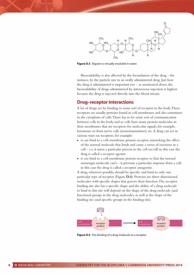
It can be seen from these examples that it is not always straightforward to predict whether or not a substance will be soluble. Digoxin, a drug used to treat heart problems (Figure D.3), is virtually insoluble in water despite having a large number of OH groups – as for griseofulvin, the polar interactions are not enough to o� set the non-polar ones.
‘Parenteral’ administration means any route other than via the gut – it includes injection, the pulmonary route and the topical route.
any route other than via the ‘Parenteral’ administration means any route other than via the ‘Parenteral’ administration means any route other than via the
Exam tipWhen asked to de� ne bioavailability in the exam you should de� ne it according to the syllabus de� nition: the fraction of the administered dosage that reaches the target part of the human body.
Bioavailability is quite a vague term and is de� ned (incorrectly) in the syllabus as the fraction of the administered drug that reaches the target part of the human body.
When a drug reaches the general circulation it will be distributed around the body – not all the drug that reaches the general circulation will reach the target site.
8 CHEMISTRY FOR THE IB DIPLOMA © CAMBRIDGE UNIVERSITY PRESS 2014D MEDICINAL CHEMISTRY
Bioavailability is also a� ected by the formulation of the drug – for instance, by the particle size in an orally administered drug. Just how the drug is administered is important too – as mentioned above, the bioavailability of drugs administered by intravenous injection is highest because the drug is injected directly into the blood stream.
Drug–receptor interactionsA lot of drugs act by binding to some sort of receptor in the body. These receptors are usually proteins found in cell membranes and also sometimes in the cytoplasm of cells. There has to be some sort of communication between cells in the body, and so cells have many protein molecules in their membranes that are receptors for molecular signals, for example, hormones or from nerve cells (neurotransmitters) etc. A drug can act in various ways on receptors, for example:
• it can bind to a cell-membrane protein receptor, mimicking the e� ect of the normal molecule that binds and cause a series of reactions in a cell – i.e. it turns a particular process in the cell on/o� ; in this case the drug is called a receptor agonist
• it can bind to a cell-membrane protein receptor so that the normal messenger molecule can’t – it prevents a particular response from a cell; in this case the drug is called a receptor antagonist.
A drug, wherever possible, should be speci� c and bind to only one particular type of receptor (Figure D.4). Proteins are three-dimensional molecules with speci� c shapes that govern their function. The receptor binding site also has a speci� c shape and the ability of a drug molecule to bind to this site will depend on the shape of the drug molecule (and functional groups in the drug molecule), as well as the shape of the binding site (and speci� c groups in the binding site).
CH3 CH3H CH3 H
CH3OH
H
HO HO
HO
CH3H H
HH
O
O OO
OO O
HO
Figure D.3 Digoxin is virtually insoluble in water.
receptorprotein
cellmembrane
cellmembrane receptor
protein
drug
drug
Figure D.4 The binding of a drug molecule to a receptor.

D MEDICINAL CHEMISTRY 9CHEMISTRY FOR THE IB DIPLOMA © CAMBRIDGE UNIVERSITY PRESS 2014
Nature of scienceScientists often have to make decisions about how much data they require to be sure about a conclusion. For instance, they must decide, based on the results of clinical trials and other evidence, whether or not a drug is safe to administer to the public. They must also sometimes consider whether the bene� ts outweigh the risks for a particular drug. However, the data available from clinical trials are limited and in many countries post-marketing surveillance of approved drugs, which evaluates a drug’s long-term safety in the wider patient population, is in operation. In some cases, a drug that has been on the market for a number of years may be withdrawn because of serious side e� ects reported after widespread use.
D2 Aspirin and penicillin
Analgesics
Analgesics are drugs that reduce pain.
There are two main types of analgesics: mild analgesics and strong analgesics. They exert their pain-relief action in di� erent ways. Strong analgesics will be discussed in the next section.
Mild analgesics, such as aspirin and ibuprofen, prevent the production of prostaglandins in the body by inhibiting an enzyme known as cyclooxygenase (COX), which is a key enzyme in the synthesis of prostaglandins.
Prostaglandins cause a number of physiological e� ects in the body, including the induction of pain, in� ammation and fever.
When an injury to a tissue occurs, prostaglandins are synthesised in the damaged tissue cells and bind to receptors – this stimulates sensory nerve � bres at the site of the injury to send signals to the brain, which then interprets them as pain. They also cause dilation (widening) of the blood vessels in the damaged tissue, leading to an in� ammatory response (swelling, redness, heat and pain at the site of injury) and can also stimulate the hypothalamus in the brain to cause an increase in body temperature (fever).
Mild analgesics act at the source of pain by inhibiting the production of chemical messengers that causes the sensation of pain, swelling and fever.
AspirinAs long ago as the 5th century BCE, it was known that chewing willow bark could give pain relief. Willow bark contains a compound called salicin, which is a sugar derivative of salicylic acid (2-hydroxybenzoic acid) that gets converted to salicylic acid in the body. Salicylic acid (Figure D.5) is a good analgesic but causes severe irritation of the
Learning objectives
• Understand the mode of action of aspirin
• Understand why aspirin is used
• Understand that ethanol has a synergistic e� ect with aspirin
• Understand how aspirin is synthesised from salicylic acid
• Understand how aspirin can be puri� ed
• Understand the characterisation of aspirin by melting point and infrared spectroscopy
• Understand how the chemical modi� cation of aspirin can a� ect its bioavailability
• Understand that penicillin is an antibiotic produced by fungi
• Understand that penicillins have a β-lactam ring
• Understand how penicillins work and why the β-lactam ring is important
• Understand why modifying the side-chain in penicillin is important
• Discuss the causes of bacterial resistance to penicillin
The systematic name of aspirin is 2-ethanoyloxybenzenecarboxylic acid.
Drugs that have been licensed and then subsequently withdrawn include terfenadine and sertindole.
Drugs that have been licensed

10 CHEMISTRY FOR THE IB DIPLOMA © CAMBRIDGE UNIVERSITY PRESS 2014D MEDICINAL CHEMISTRY
stomach lining resulting in vomiting and gastric bleeding. In the 1890s, a derivative of salicylic acid, called acetylsalicylic acid (Figure D.5), began to be used medically and, over 100 years on, it is still in widespread use. Acetylsalicylic acid is the chemical name for aspirin – it is an ester of salicylic acid and is far less irritating to the stomach than salicylic acid.
Aspirin is used all over the world as an analgesic and anti-in� ammatory agent. It belongs to a group of drugs known as non-steroidal anti-in� ammatory drugs (NSAIDs), of which ibuprofen is also a member. It is useful in treating painful conditions such as headache, fever, and also conditions in which both pain and in� ammation are present, such as arthritis.
Aspirin is also taken in low doses to help prevent recurrent heart attacks or strokes in patients who have previously su� ered a heart attack or stroke – the protection is through its anti-blood-clotting e� ect – it is acting as an anticoagulant. Some studies have also indicated that low-dose aspirin may prevent certain cancers, in particular colorectal cancer. However, further research is needed in this area. These examples illustrate the use of aspirin as a prophylactic – something taken to try to prevent a disease happening in the � rst place.
C
OHhydroxyl / phenol
carboxyl group
salicylic acid
O
COHOH
COH
3C
carboxyl group
acetylsalicylic acid
O
ester
O
Figure D.5 The structures of salicylic acid and acetylsalicylic acid (aspirin).
As we have already seen, aspirin exerts its e� ects through the inhibition of an enzyme called COX which plays a key role in prostaglandin synthesis. As well as mediating pain, fever and in� ammation, prostaglandins also have a number of other roles in the body, one of which is maintaining the mucous layer in the stomach. Therefore, one of the side e� ects of taking aspirin is gastric irritation, both directly by the drug itself but mainly indirectly through its inhibition of prostaglandin synthesis and therefore depletion of the protective mucous layer. This can lead to peptic ulcers and possibly stomach bleeding in some patients.
Another disadvantage of using aspirin is that some people may be sensitive to it (known as hypersensitivity), especially those who su� er from asthma in whom aspirin can trigger an asthma attack. Another drawback of aspirin is that it is not recommended to be taken by children younger than 16 because it has been associated with Reye’s syndrome – a potentially fatal condition that a� ects all organs of the body, but especially the brain and liver.
What is pain? When we burn a � nger is the pain in your � nger or
in your brain? When you go to the doctor, you are often asked to describe the pain – what language do we use to describe pain? Can one person ever understand another person’s pain?

D MEDICINAL CHEMISTRY 11CHEMISTRY FOR THE IB DIPLOMA © CAMBRIDGE UNIVERSITY PRESS 2014
The synergistic eff ect of ethanol Ethanol is an example of a drug that can increase the e� ects of other drugs, so care must be taken when alcoholic drinks are taken by people on certain types of medication. The increase in e� ect may be harmful to the body, and in some cases fatal.
Synergism can happen when two or more drugs, given at the same time, have an e� ect on the body that is greater than the sum of their individual e� ects. In other words, certain drugs can increase the e� ects of other drugs when given at the same time.
When alcohol is taken with aspirin there is an increased risk of hemorrhage (bleeding) in the stomach.
Synthesis of aspirinAspirin can be made from 2-hydroxybenzoic acid (salicylic acid) by warming with excess ethanoic anhydride (Figure D.6).
ethanoic acid
+ + C
H
H
CHO
O H
H
H
H
CC
O
OH3C
H3C
C
C
O
O
O
ethanoicanhydride
COO
H
O H
2-hydroxybenzoic acid
COO
H
aspirin
Figure D.6 Synthesis of aspirin from salicylic acid (2-hydroxybenzoic acid).
The type of reaction is addition–elimination (the CH3CO group is added to aspirin and ethanoic acid is eliminated) and happens in the presence of a small amount of concentrated phosphoric (or sulfuric) acid catalyst.
Aspirin is not very soluble in water and so the addition of water to the reaction mixture causes a precipitate of aspirin to form (white solid), as well as breaking down any unreacted ethanoic anhydride to ethanoic acid. The white solid can be � ltered o� and washed with some cold water (to remove any soluble impurities) and left to dry (in a desiccator or warm oven) to give the crude product. The mass of the product is recorded and the yield can be worked out.
Calculation of the yield of aspirinThis is best explained using an example.
Worked exampleD.1 In an experiment to synthesise aspirin, 5.60 g of salicylic acid (Mr 138.13) was reacted with 8.00 cm3 of
ethanoic anhydride (density 1.08 g cm−3) in the presence of a concentrated phosphoric acid catalyst. 5.21 g of a white solid was obtained at the end of the reaction. Calculate: a which reagent was in excessb the yield of aspirin.

12 CHEMISTRY FOR THE IB DIPLOMA © CAMBRIDGE UNIVERSITY PRESS 2014D MEDICINAL CHEMISTRY
a The equation for the reaction is shown in Figure D.6.
massdensity =
volume
mass of ethanoic anhydride that reacted = 1.08 × 8.00 = 8.64 g
relative molecular mass of ethanoic anhydride = 102.10
8.64number of moles of ethanoic anhydride =
102.10 = 0.0846 mol
5.60number of moles of salicylic acid =
138.13 = 0.0405 mol
This is a 1 : 1 reaction and so the ethanoic anhydride is in excess.
b To work out the yield of aspirin, we must use the number of moles of the limiting reactant, i.e. salicylic acid. From the equation, 0.0405 mol salicylic acid will produce 0.0405 mol aspirin.
relative molecular mass of aspirin = 180.17
theoretical yield of aspirin = 0.0405 × 180.17 = 7.30 g
percentage yield = ⎛ actual yield ⎞
× 100 ⎝ theoretical yield ⎠
⎛5.21⎞= ⎝7.30⎠ × 100 = 71.4%
Acid anhydridesThe basic structure of an acid anhydride is:
This can be regarded as being formed from two molecules of carboxylic acid with water removed (Figure D.7), although acid anhydrides are not actually made like this.
Acid anhydrides react when warmed with water to form carboxylic acids (Figure D.8). When water is added to the reaction mixture in the synthesis of aspirin, ethanoic acid is formed from excess ethanoic anhydride:
R C
R C
O
O
O
O
O
H
CH
H
CH
O
O
H
CH
H
CH
Figure D.7 Where the name ‘acid anhydride’ comes from.
H3C
H3C
C
C
O
OO
O
ethanoic anhydride ethanoic acid
+ +
H
H
H
C CO
OO
HH H
ethanoic acid
H
H
H
C CO
O H
Figure D.8 Hydrolysis (breaking apart with water) of ethanoic anhydride.

D MEDICINAL CHEMISTRY 13CHEMISTRY FOR THE IB DIPLOMA © CAMBRIDGE UNIVERSITY PRESS 2014
Purifi cation of aspirinThe crude sample of aspirin contains impurities and must be puri� ed – the main impurities are unreacted salicylic acid, and possibly water if the sample is not completely dry. Recrystallisation can be used to purify the aspirin.
The basic principles of recrystallisation are that a solid is dissolved in a solvent in which it is soluble at raised temperatures but much less soluble at lower temperatures. Any impurities are present in much smaller amounts and so remain in solution at the lower temperature.
The procedure for recrystallisation is:
• The product is dissolved in the minimum amount of hot solvent to form a close-to-saturated solution.
• The solution is � ltered while still hot to remove any insoluble impurities. Vacuum � ltration is used because it is much faster – the product may start to crystallise while � ltering if the solution cools too much.
• As the solution cools, the product becomes less soluble in the solvent and comes out of solution as solid crystals – less of the solid dissolves at lower temperatures. It may be necessary to cool in ice or scratch the inside of the beaker to initiate crystallisation.
• Any solid product is separated from the solvent by vacuum � ltration.
• Any impurities also dissolve in the hot solvent, but because they are present in much smaller amounts they do not exceed their solubility, even at lower temperatures, and remain in solution.Aspirin can be recrystallised from ethyl ethanoate or ethanol
(usually a 95% ethanol/water mixture). Water is generally not used for recrystallisation because aspirin tends to decompose in hot water.
Characterisation of aspirinThe full characterisation of an organic compound involves determining its purity, molecular formula, physical properties, structure etc. Here we will look at how the purity of the compound can be estimated and the determination of the functional groups present in the molecule.
Determination of the purity of aspirinHow pure a sample of aspirin is can be determined by chromatography or by measuring its melting point. A pure substance will melt at a well-de� ned temperature but the presence of impurities lowers the melting point and causes the solid to melt over a range of temperatures. The melting point of aspirin is reported as 138 –140 °C – so if a sample is tested and its melting range is found to be 125 –132 °C it can be concluded that the sample is quite impure.
The infrared spectrum of aspirinInfrared spectroscopy can be used to determine which bonds/functional groups are present in a molecule and also, by comparison with spectra in databases, to determine whether or not a particular compound has been made.
The infrared spectrum of aspirin is shown in Figure D.9.

14 CHEMISTRY FOR THE IB DIPLOMA © CAMBRIDGE UNIVERSITY PRESS 2014D MEDICINAL CHEMISTRY
There are two peaks in the carbonyl (C=O) region due to the two di� erent C=O groups present – an ester and a a carboxyl group (carboxylic acid). Consultating of more advanced tables of infrared data allows us to assign each peak as shown. The peaks at 1600 cm−1 and just below 1500 cm−1 are due to the vibrations of C–C bonds in the benzene ring.
If the infrared spectrum of aspirin is compared with that of salicylic acid (Figure D.10), the spectra are very similar but the C=O stretch from the ester at just above 1700 cm−1 is missing.
04000 3000 2000
carboxyl groupC=O
ester C=O
characteristic ofcarboxylic acids
very broad O-H stretch
CO
O
O
CH3C
OH
1500Wavenumber / cm–1
% T
rans
mitt
ance
1000
100
04000 3000 2000
carboxyl groupC=O
very broadO-H stretch
CO
O H
OH
15001700Wavenumber / cm–1
% T
rans
mitt
ance
1000
100
Figure D.9 The infrared spectrum of aspirin.
Figure D.10 The infrared spectrum of salicylic acid.
Solubility of aspirin and other drugsAspirin is administered orally and therefore must � rst be absorbed from the gastrointestinal tract before reaching the blood circulation to be distributed to the various body tissues. For a drug to enter the blood circulation after oral administration, it must � rst dissolve in the aqueous environment of the intestines before it can be absorbed across the lipid membranes of the intestinal wall. If the rate at which the drug dissolves is
D MEDICINAL CHEMISTRY 15CHEMISTRY FOR THE IB DIPLOMA © CAMBRIDGE UNIVERSITY PRESS 2014
slower than the rate at which it gets absorbed, this can a� ect the amount of drug that gets absorbed – and hence its bioavailability. Once in the bloodstream, the drug has to travel through the aqueous blood plasma and be distributed through the body to reach its site of action.
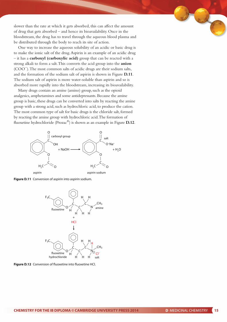
One way to increase the aqueous solubility of an acidic or basic drug is to make the ionic salt of the drug. Aspirin is an example of an acidic drug – it has a carboxyl (carboxylic acid) group that can be reacted with a strong alkali to form a salt. This converts the acid group into the anion (COO−). The most common salts of acidic drugs are their sodium salts, and the formation of the sodium salt of aspirin is shown in Figure D.11. The sodium salt of aspirin is more water-soluble than aspirin and so is absorbed more rapidly into the bloodstream, increasing its bioavailability.
Many drugs contain an amine (amino) group, such as the opioid analgesics, amphetamines and some antidepressants. Because the amine group is basic, these drugs can be converted into salts by reacting the amine group with a strong acid, such as hydrochloric acid, to produce the cation. The most common type of salt for basic drugs is the chloride salt, formed by reacting the amine group with hydrochloric acid. The formation of � uoxetine hydrochloride (Prozac®) is shown as an example in Figure D.12.
COH
carboxyl group
+ NaOH + H2O
aspirin aspirin sodium
O
CO– Na+
C
salt
O
O
COH
3C OH
3C
O
Figure D.11 Conversion of aspirin into aspirin sodium.
C C
F3C
O CH
HCl+
Nfluoxetine amine
CH3
C C
F3C
O C
H H
Cl–
H H
HH H
H
H
HH
N
H
fluoxetinehydrochloride salt
CH3
+
Figure D.12 Conversion of fl uoxetine into fl uoxetine HCl.

16 CHEMISTRY FOR THE IB DIPLOMA © CAMBRIDGE UNIVERSITY PRESS 2014D MEDICINAL CHEMISTRY
PenicillinAntibacterial drugs are some of the most frequently prescribed medicines. These drugs are toxic to bacteria while being relatively safe to the patients who take them. They achieve this by acting on sites in the bacterial cells that are either di� erent from those in our cells or that do not exist in our cells at all.
There are many di� erent types of antibacterial drugs (commonly called antibiotics), but the most commonly prescribed are the penicillins. They were discovered by chance in 1928 by a Scottish physician and microbiologist called Alexander Fleming. Penicillins are produced by some fungi of the Penicillium strain, such as Penicillium chrysogenum. One of the most important natural penicillins is benzylpenicillin (penicillin G) and this is manufactured by fermentation of a mixture of corn-steep liquor (a byproduct of corn-starch manufacture), sugars, minerals and phenylethanoic acid using a penicillin fungus in a carefully controlled environment.
Penicillin has a bicyclic structure (Figure D.13) containing a β-lactam ring (a cyclic amide that is part of a four-membered ring). This β-lactam ring is essential for the antibacterial activity of penicillin; if the ring is broken in any way, such as by acid or bacterial enzymes (see below), the penicillin is no longer active.
a bcarboxyl group
carboxamidegroup
β-lactam ring
C
H
H
CO
H
SCH3
CH3
COOH
N
RO
N
Figure D.13 All penicillins have the same basic bicyclic structure, but diff erent penicillins have diff erent side-chains. a The general structure of penicillins; b the side-chain for benzylpenicillin (penicillin G).
Cyclic amides are named using Greek letters to indicate the size of the ring. So a γ-lactam has a � ve-membered ring and a δ-lactam has a six-membered ring. The Greek letter refers to which carbon, going round the ring from the C=O group, the N atom is joined to – for example, the second or β-carbon in a 4-membered ring.
1 2
C
α β
ONH
Action of penicillin on bacterial cell wallsBacterial cells di� er from our own cells in that they contain a cell wall which contains a polymer made up of sugar chains cross-linked with peptides (short stretches of amino acids). This polymer has a mesh-like structure and gives strength to the cell wall, allowing the bacteria to withstand high osmotic pressures. Penicillin acts by irreversibly inhibiting an enzyme (transpeptidase) involved in the cross-linking of this polymer, resulting in a weakened cell wall and causing the bacterial cell to burst due to the high osmotic pressure caused by water from the surroundings entering the bacterial cell. Penicillin is not the only antibacterial that works by inhibiting cell-wall synthesis – cephalosporins and carbapenems work in a similar way.
The β-lactam ring is essential to the mode of action of penicillin (Figure D.14). An OH group on the side-chain of an amino acid (serine) in the transpeptidase-enzyme active site reacts with the β-lactam ring of the penicillin instead of its normal substrate. A covalent bond is formed between the enzyme and penicillin as the β-lactam ring opens – the complex formed prevents any substrate molecules entering the active site and reacting, therefore the enzyme is deactivated.

D MEDICINAL CHEMISTRY 17CHEMISTRY FOR THE IB DIPLOMA © CAMBRIDGE UNIVERSITY PRESS 2014
The � rst penicillin to be isolated and puri� ed was penicillin G (benzylpenicillin) (Figure D.13). However, this penicillin has a number of disadvantages, one of which is that it is easily broken down by stomach acid and must be given by injection. Scientists have overcome this problem by making derivatives of penicillin G that have modi� ed side-chains (R in the general penicillin structure in Figure D.13a) that can resist stomach acid and be given by the oral route.
Bacterial resistanceThe widespread use of penicillins has resulted in the development of bacteria that have become resistant to their antibacterial e� ects – this is known as bacterial resistance and arises because of mutations in the DNA of bacteria to aid their survival. Some strains of bacteria have developed ways of counteracting the e� ects of certain penicillins by producing an enzyme known as penicillinase (a β-lactamase), which opens the β-lactam ring of the penicillin, rendering it inactive. Penicillin G is an example of a penicillin that is inactivated by penicillinase. However, scientists have now developed penicillins that are less sensitive to the e� ects of this enzyme by modifying the side-chain in the penicillin structure (Figure D.15).
Bacterial resistance has developed not just for penicillins, but for most other types of antibacterials too. Some bacteria are resistant to more than one type, making them extremely di� cult to kill, so it is important to carry out research into the discovery and development of new antibacterial agents.
It is extremely important that antibacterials are taken according to a doctor’s instructions (called patient compliance) and that the whole course of treatment is taken. Otherwise failure to kill all the bacteria in the infection can lead to development of resistance in those bacteria that survive.
active site blocked
active site of enzyme
penicillin molecule
β-lactam ring opensCR
HH H
SCH3
CH3
N
C
C
O
CR
HH
H
H S CH3
CH3
N
CC
ONO
HOO HO
NO
OO
HO
Figure D.14 The mode of action of penicillin.
Penicillin G can be used to treat diseases caused by bacteria that do not produce penicillinase, such as meningitis and gonorrhea.
Modifying the side-chain in penicillins makes them more resistant to the penicillinase enzyme.
Figure D.15 Methicillin has a diff erent R group and is resistant to penicillinase enzymes. However, some strains of bacteria have become resistant to methicillin. MRSA, one of the so-called ‘superbugs’, stands for ‘methicillin-resistant Staphylococcus aureus’.
C
HH H
SCH3
CH3
N
O C
C
OH3C N
O
O HO
O
CH3

18 CHEMISTRY FOR THE IB DIPLOMA © CAMBRIDGE UNIVERSITY PRESS 2014D MEDICINAL CHEMISTRY
Such widespread bacterial resistance is also due to the extensive use of antibacterials, both for human use and for animals. Overprescribing of antibacterials for minor infections has increased the exposure of bacteria to the antibacterial agents and has increased the number of resistant bacteria. Antibacterials are also used extensively in animal feeds to lower the occurrence of infections in livestock. These antibacterials are given to healthy animals and can result in the development of resistant bacteria that can be passed on to humans via meat and dairy products.
Bacterial resistance is a widespread problem – it has developed because of the innate ability of bacteria to mutate DNA in order to survive in hostile environments, as well as the overuse and misuse of antibacterials. Improving the way that antibiotics are prescribed and taken by humans or used for livestock is essential if the development and spread of resistant bacteria is to be controlled.
Nature of scienceMany scienti� c discoveries come about following a systematic approach to research but some discoveries can be the result of a chance set of conditions and serendipity. The discovery of penicillin was one such situation but the genius of the scientist who discovered penicillin was in recognising that he was seeing something di� erent – not everyone would have made the connections required.
D3 Opiates
Strong analgesicsWhereas mild analgesics, such as aspirin, are used for relatively mild pain, such as headache or toothache, opiate/opioid analgesics are strong analgesics used for moderate to severe pain, such as in terminally ill patients. Mild analgesics may be combined with strong analgesics in some preparations – for example, paracetamol and codeine are often used together.
Opiates
Opiates are natural narcotic (sleep-inducing) analgesics derived from the opium poppy.
Opiates are derived from the juice of the unripe seed pods of the poppy Papaver somniferum. This juice is known as opium (the Greek word for ‘juice’) and contains a mixture of approximately 25 di� erent nitrogen-containing compounds (known as alkaloids), the most important of which is morphine. Morphine was � rst isolated in 1803 and is chie� y responsible for the biological e� ects of opium – it accounts for approximately 10% of the opium mixture. Codeine, a milder analgesic than morphine, is also found naturally in opium, although in smaller proportions.
Learning objectives
• Understand what is meant by an opiate
• Understand the mode of action of strong analgesics such as morphine and codeine
• Compare the structures of morphine, codeine and diamorphine
• Explain why diamorphine is more potent than morphine
• Understand how diamorphine and codeine can be synthesised from morphine
• Explain the advantages and disadvantages of using opiates
The term ‘narcotic’ can be used in di� erent ways. It is used here to describe analgesic drugs derived from opium, but nowadays it is often used in everyday language to indicate any illicit/strictly controlled drug.

D MEDICINAL CHEMISTRY 19CHEMISTRY FOR THE IB DIPLOMA © CAMBRIDGE UNIVERSITY PRESS 2014
Strong analgesics work by temporarily binding to opioid receptors in the brain, which block the transmission of pain signals in the brain.
Morphine and codeine are strong analgesics, which act by temporarily binding to opioid receptors in the brain. This blocks the transmission of pain signals in the brain and increases the pain perception threshold – even though pain in the a� ected tissue is still occurring and being transmitted via the peripheral nervous system, the patient is not as aware of it. Also, opioids increase the tolerance to pain, which means that even if pain is felt by the patient they are more able to tolerate it.
Opiates cause a number of e� ects on the body through binding to opioid receptors. These include analgesia, sedation, a feeling of well-being and suppression of the cough re� ex. They are used medically for pain relief and the treatment of coughs and diarrhea.
Opioid receptors in the brain are essential for the action of opiates such as morphine. These opioid receptors are proteins and there are various types in the brain. However, the opioid receptor that causes the greatest analgesic e� ect when opiates bind to it is also the one responsible for the greatest side e� ects, such as euphoria, addiction etc.
Both the medicinal e� ects of opiates and their addictive properties are caused by binding to the same opioid receptors in the brain.
Structures of morphine and its derivativesThe chemical structures of codeine, morphine and diamorphine are shown in Figure D.16. As can be seen, they are very similar in structure – all have a tertiary amine group and benzene ring, which are essential for analgesic activity.
The only di� erence between codeine and morphine is a methoxyl (–OCH3) group (ether functional group) on the benzene ring in codeine instead of a hydroxyl (–OH) group (an OH group attached directly to a benzene ring gives rise to a phenol) in morphine. When codeine enters the body, some of it is acted on by enzymes, which remove the methyl group to give a hydroxyl group; thus codeine is converted to morphine.
Exam tipWhen asked about the mode of operation of strong analgesics in the examination you should use the de� nition given on the syllabus: ‘strong analgesics work by temporarily bonding to receptor sites in the brain, preventing the transmission of pain impulses without depressing the central nervous system’.
A tertiary amine has N joined to three C atoms (three alkyl groups).
It states on the syllabus that opiates do not depress the central nervous system. However, the brain is part of the central nervous system and opiates are CNS depressants.
N-CH3
OH3C
O
HOhydroxyl
ether
tertiaryamine
codeine
benzene ring
N-CH3
HO
O
HOhydroxyl
hydroxyl / phenol
tertiaryamine
morphine
benzene ring
tertiaryamine
N-CH3
O
diamorphine
benzene ring
O
O
O
O
H3C
H3C
ester
ester
Figure D.16 Structures of codeine, morphine and diamorphine.
do not depress the central nervous It states on the syllabus that opiates do not depress the central nervous It states on the syllabus that opiates do not depress the central nervous

20 CHEMISTRY FOR THE IB DIPLOMA © CAMBRIDGE UNIVERSITY PRESS 2014D MEDICINAL CHEMISTRY
It is this conversion to morphine that accounts for the therapeutic properties of codeine, which suggests that the phenol group is also essential for the analgesic activity of opiates.
Diamorphine (heroin) (Figure D.16) is a semi-synthetic morphine derivative. The di� erence between the structures is that diamorphine contains two ester (CH3COO) groups, whereas morphine contains two OH groups.
Diamorphine is a more potent analgesic than morphine because it is better able to cross the blood–brain barrier.
Diamorphine is more lipid-soluble than morphine because of the replacement of the OH groups (which can take part in hydrogen bonding) by the ester groups (which cannot) and therefore is able to cross the blood–brain barrier and enter the brain more easily. The blood–brain barrier is essentially a lipid barrier that prevents the entry of potentially toxic substances from the capillaries into the brain – it allows small, lipid-soluble molecules across and hinders large, polar molecules. Once diamorphine has entered the brain, it is hydrolysed by enzymes to the monoester (only one ester group) and to morphine; these bind to opioid receptors and produce an analgesic e� ect.
Synthesis of derivatives of morphine
DiamorphineDiamorphine is synthesised from morphine by heating it with ethanoic anhydride (Figure D.17). This converts the two hydroxyl groups in morphine to ester groups. The type of reaction that occurs is addition–elimination (as in the synthesis of aspirin on page 11) – it could also be called esteri� cation. CH3COO– is the ethanoate group and so two ethanoate esters are formed.
Diamorphine is not a naturally occurring substance derived from poppies – it is made from a product derived from opium, so it does not � t the de� nition of an opiate given above. The de� nition of an opiate is, however, usually extended to include semi-synthetic morphine-like substances derived from morphine. In some de� nitions, diamorphine is described rather as an opioid, which is a wider class of compounds exhibiting morphine-like e� ects on the body – opiates are opioids, but not all opioids are opiates. The terms ‘opioid’ and ‘opiate’ are often used interchangeably.
hydroxyl / phenol
ethanoicanhydride
hydroxyl
morphine diamorphine
ethanoicacid
ester
ester
H3C
2
N
O
O H
O H
+H3C C
H3C CO
O
OO H
+ 2 H3C CO
O CH3C
O
O CH3C
O
CH2C
H H
H3C N
O +
CH2C
H H
Figure D.17 Synthesis of diamorphine from morphine.
Codeine synthesisCodeine can also be synthesised from morphine (Figure D.18). In the original process, morphine was reacted with iodomethane (the methylating agent) in the presence of a base. Phenols are slightly acidic and so the presence of a strong base converts the OH of the phenol to O−. The reaction is nucleophilic substitution, with the O− attacking the δ+ carbon atom of the CH3I.

D MEDICINAL CHEMISTRY 21CHEMISTRY FOR THE IB DIPLOMA © CAMBRIDGE UNIVERSITY PRESS 2014
Advantages and disadvantages of opiate analgesicsOpiates such as morphine and diamorphine are used medically for the relief of severe pain – they are especially e� ective in visceral pain (pain in internal organs, such as the liver and lungs). They are commonly used to relieve the pain associated with cancer in terminally ill patients. Morphine may also be used for the short-term control of diarrhea due to its constipating e� ect, and to control distressing coughing by lung cancer patients, due to its cough-suppressant e� ect. Milder opiates such as codeine are used to relieve moderate pain. Codeine is also used as a cough suppressant for dry coughs and as an antidiarrhea drug.
Opiate analgesics have a number of side e� ects associated with their use – in the short term they can cause nausea and vomiting, constipation, respiratory depression (slowed or shallow breathing), drowsiness and euphoria; in the long term they cause dependence and tolerance, chronic constipation and decrease in sex drive.
There are two types of dependence:
• psychological dependence, in which the drug-taker craves the drug if deprived of it for a short time and must get further supplies in order to satisfy their need
• physical dependence, in which the body cannot function without the drug and deprivation results in withdrawal symptoms.Illicit drug users su� er both physical and psychological dependence,
whereas patients taking opioids for medical reasons generally su� er only physical dependence. Tolerance occurs in both types of user, requiring higher doses to be taken to cause the same e� ect (therapeutic or euphoric).
etherO CH3
+ Kl + H2OH3C+ + KOHl
phenolmorphine
H3C N
O
O H
O H
CH2C
H H
codine
H3C N
O
O H
CH2C
H H
+ H3C + C2H5OH
C6H5
CH3
N+ CH3H3C + C2H5O–
C6H5
H3C
N+
etherO CH3
phenolmorphine
H3C N
O
O H
O H
CH2C
H H
codine
H3C N
O
O H
CH2C
H H
Figure D.18 Synthesis of codeine from morphine.
Figure D.19 A variation on the synthesis of codeine from morphine.
The synthesis is more usually carried out nowadays using a more complicated methylating agent – a salt of C6H5N(CH3) such as C6H5N(CH3)+(C2H5O−) – Figure D.19.

22 CHEMISTRY FOR THE IB DIPLOMA © CAMBRIDGE UNIVERSITY PRESS 2014D MEDICINAL CHEMISTRY
Abuse of opiatesOpiates have been taken for non-medical reasons for centuries. As well as dulling pain, they cause a pleasant, dreamy and relaxed state known as euphoria, with heroin also causing a feeling of warmth and thrill when injected intravenously. Because heroin is lipophilic, it enters the brain quickly and so causes a ‘euphoric rush’. However, dependence and tolerance develop quickly, and the user soon starts to need larger and larger doses to retain this ‘rush’. If the user is denied the drug withdrawal symptoms occur, including anxiety, cold sweats, vomiting and jerking of the legs. Treating opiate dependence is di� cult – it may involve a gradual reduction of the dose of the drug and the administration of a substitute called methadone which also binds to opioid receptors but has a prolonged action and reduces the craving and prevents withdrawal symptoms.
Opiate dependence is a worldwide problem and is associated with a signi� cant amount of crime. Users may � nd that they can no longer a� ord to pay for the increasing doses needed and so resort to criminal activity to pay for their drugs. Users who inject heroin intravenously are also at increased risk of infection from hepatitis or HIV/AIDS by sharing needles.
Nature of scienceScienti� c knowledge is continually developing. Although opium has been known and used for thousands of years it is only now that our knowledge of biochemistry has developed su� ciently for us to understand its mode of action on the molecular level.
D4 pH regulation of the stomachNormally the pH in the stomach is between 1 and 2, owing to the production of hydrochloric acid by the millions of gastric glands that line the stomach. The stomach is maintained at such a low pH for two main reasons:
• the acidic environment is not tolerated by the majority of microorganisms (e.g. bacteria) that may enter the digestive system with food – the low pH plays a role in the body’s natural defence against disease-causing microorganisms
• the digestive enzymes in the stomach (e.g. pepsin, which breaks down proteins) require a low pH for optimum catalytic activity.A layer of mucus lines the stomach, and protects the stomach wall
from damage by the acid. However, irritation to the stomach lining can occur by the production of excess acid – for example, caused by drinking too much alcohol, eating large (especially fatty) meals, smoking or stress. Certain drugs can irritate the stomach lining directly, whereas drugs such as aspirin can lower the production of mucus in the stomach making the stomach lining more susceptible to acid attack. This can result in the following:
• indigestion – irritation of the stomach lining caused by excess acid producing pain or discomfort in the upper abdomen and/or nausea
• heartburn (acid re� ux) – acid from the stomach rising up into the esophagus causing a burning sensation
• peptic ulcer – erosion of part of the gut lining, caused by acid
Learning objectives
• Understand that antacids can be used to reduce the amount of excess acid in the stomach
• Understand that the action of antacids is non-speci� c
• Write equations for neutralisation reactions involving di� erent antacids
• Understand how ranitidine (Zantac®) works
• Understand how omeprazole (Prilosec®) and esomeprazole (Nexium®) work
• Understand what is meant by an active metabolite
• Solve problems involving bu� er solutions

D MEDICINAL CHEMISTRY 23CHEMISTRY FOR THE IB DIPLOMA © CAMBRIDGE UNIVERSITY PRESS 2014
penetrating the mucous layer. This can be a serious condition if left untreated because internal bleeding can occur. Aspirin and other related anti-in� ammatory drugs can cause ulcers in some patients.Antacids are used to treat these conditions. They are weakly basic
compounds that neutralise acids, relieving the pain, discomfort or burning sensation and allowing repair of the mucous layer. In the case of peptic ulcers, neutralisation of the acid prevents further erosion of the gut lining allowing ulcers to heal.
The most commonly used antacids are metal hydroxides, carbonates and hydrogencarbonates (bicarbonates):
• magnesium hydroxide
• aluminium hydroxide
• calcium carbonate
• sodium hydrogencarbonate (also called sodium bicarbonate).Some antacid preparations contain mixtures of two di� erent antacids,
such as magnesium compounds and aluminium compounds (usually magnesium and aluminium hydroxides). The rationale for using these two di� erent antacids is that magnesium salts are faster acting and so work quickly to neutralise the acid, but aluminium salts have a slower and more prolonged e� ect, so the time interval between doses is increased. Also, magnesium salts in repeated doses can cause a laxative e� ect, but this is o� set by aluminium salts which can induce constipation.
Unlike the other drugs that have been discussed above,
antacids are non-speci� c and do not bind to protein receptors. They work by simply neutralising excess stomach acid.
The neutralising reactions for hydroxides are:
Al(OH)3(s) + 3HCl(aq) → AlCl3(aq) + 3H2O(l)
Mg(OH)2(s) + 2HCl(aq) → MgCl2(aq) + 2H2O(l)
Ca(OH)2(s) + 2HCl(aq) → CaCl2(aq) + 2H2O(l)
Metal carbonates and hydrogencarbonates also react with the acid to give a salt along with water and carbon dioxide:
CaCO3(s) + 2HCl(aq) → CaCl2(aq) + H2O(l) + CO2(g)
NaHCO3(s) + HCl(aq) → NaCl(aq) + H2O(l) + CO2(g)
Na2CO3(s) + 2HCl(aq) → 2NaCl(aq) + H2O(l) + CO2(g)
Exam tipCalcium hydroxide and sodium carbonate are also mentioned on the syllabus as antacids but these are not generally given in antacid preparations – presumably because they are also irritants.
Because carbon dioxide can cause bloatedness and � atulence, antifoaming agents may sometimes be included in a preparation – for example, activated dimeticone (dimethicone), which relieves � atulence.
Alginates may also be present in some antacid preparations. These form a ‘raft’ that � oats on top of the stomach contents reducing re� ux into the esophagus, which causes heartburn.
The term dyspepsia is often used interchangeably with indigestion but it is de� ned more generally as pain or discomfort in the upper abdomen.
interchangeably with indigestion dyspepsia
interchangeably with indigestion dyspepsia is often used
interchangeably with indigestion

24 CHEMISTRY FOR THE IB DIPLOMA © CAMBRIDGE UNIVERSITY PRESS 2014D MEDICINAL CHEMISTRY
Worked exampleD.2 Compare the volume of stomach acid (hydrochloric acid) of pH 1.50 that is neutralised by taking
one indigestion tablet containing 1.00 g of calcium carbonate with one containing 1.00 g of sodium hydrogencarbonate.
A pH of 1.50 corresponds to a concentration of H+(aq) of 10−1.50 = 0.0316 mol dm−3
Because HCl is a strong acid it completely dissociates and the concentration of H+(aq) is equal to the original concentration of the acid.
The equation for the reaction with calcium carbonate is:
CaCO3(s) + 2HCl(aq) → CaCl2(aq) + H2O(l) + CO2(g)
1.001.00 g of CaCO3 is 100.09
= 9.99 × 10⁻³ mol
9.99 × 10⁻³ mol CaCO3 reacts with 2 × 9.99 × 10−3 moles of HCl
i.e. 0.0200 mol hydrochloric acid
volume of hydrochloric acid = 0.0200
= 0.632 dm3 or 632 cm3
0.0316
The equation for the reaction with sodium hydrogencarbonate is:
NaHCO3(s) + HCl(aq) → NaCl(aq) + H2O(l) + CO2(g)
1.001.00 g of NaHCO3 is 84.01
= 0.0119 mol
0.0119 mol NaHCO3 react with 0.0119 mol hydrochloric acid
0.0119Volume of hydrochloric acid =
0.0316 = 0.377 dm3 or 377 cm3
1.00 g of calcium carbonate therefore reacts with signi� cantly more hydrochloric acid. This is because the molar masses are fairly similar and each mole of calcium carbonate reacts with twice as many moles of hydrochloric acid as sodium hydrogencarbonate does.
1 Work out the volume of hydrochloric acid of pH 2.00 that reacts with:
a 1.00 g of aluminium hydroxide b 1.00 g of magnesium hydroxide
? Test yourself
volume = number of moles
concentration

D MEDICINAL CHEMISTRY 25CHEMISTRY FOR THE IB DIPLOMA © CAMBRIDGE UNIVERSITY PRESS 2014
Treatment of peptic ulcersStomach acid is produced by parietal cells, which are cells in the lining of the stomach. The treatment of peptic ulcers involves regulating the acid levels in the stomach. There are two main approaches to this – stopping the production of the acid and preventing the release of the acid into the stomach.
RanitidineRanitidine or Zantac® (Figure D.20) is a drug that inhibits the production of acid. It does this by binding to a receptor protein (histamine H2-receptor) in the membrane of the parietal cells, which stops the normal chemical messenger (histamine) from binding to turn on the chain of events for producing acid. Ranitidine therefore prevents the production of stomach acid.
‘H2’, in this instance, has nothing to do with hydrogen gas.
Figure D.20 The structure of ranitidine.
Figure D.21 The structure of omeprazole. Esomeprazole is a stereoisomer of this – the atoms are joined together in the same order but arranged diff erently in space.
CH3
HC
O2NCH3
NH3C
SO N
H
N
H
C
CH3O
O CH3 omeprazole
zanamivir
H3C
H3C
NS
NN
HOO
ON
N
N
H
H
H
O
OHOH
HO
HO
H3CNH2
Ranitidine can be described as an H2-receptor antagonist because when it binds to an H2-receptor it does not cause activation of the receptor, but rather stops the naturally occurring molecule that does cause activation (the agonist) from binding.
Omeprazole and esomeprazole Omeprazole (Losec®, Prilosec®) and esomeprazole (Nexium®) (Figure D.21) are proton pump inhibitors and work by preventing the release of acid from the parietal cells into the stomach. Protons are released from the parietal cell by the action of a proton pump. This is a protein complex that moves protons through cell membranes – being charged, protons cannot di� use normally through a cell membrane made of mainly non-polar lipid molecules.
These drugs are weak bases but are mainly in the un-ionised form at the pH of blood plasma. They are also mostly non-polar and therefore lipid-soluble so they can pass through the cell membrane of the parietal

26 CHEMISTRY FOR THE IB DIPLOMA © CAMBRIDGE UNIVERSITY PRESS 2014D MEDICINAL CHEMISTRY
Active metabolitesWe have already seen examples of drugs that are converted into a di� erent form in the body – the form that causes the desired action of the drug. So, for instance:
• codeine is converted into morphine in the body and it is the morphine that binds much more strongly to the opioid receptors than codeine, producing an analgesic e� ect
• omeprazole/esomeprazole are converted into di� erent forms that are able to bind to proton pumps
• aspirin is converted into the active form – salicylic acid. Salicylic acid cannot be taken orally because it causes severe irritation of the stomach lining, resulting in vomiting and gastric bleeding. Therefore it is taken in ester form; this causes much less gastric irritation but is converted back into the active analgesic in the body.
Active metabolites are the active forms of drugs after they have been processed in the body.
There are many reasons for making a drug in a di� erent form to that of the active metabolite and these include:
• to avoid side e� ects – e.g. aspirin
• to allow the drug to pass through cell membranes – the active form of omeprazole is charged and would not pass through the cell membrane into the parietal cells; diamorphine is another drug that � ts into this category
• to allow the drug to dissolve in water more easily – e.g. fosphenytoin
• to target drugs to a particular area – for example, omeprazole again, where the active drug is formed only in the highly acidic conditions of the cells in the stomach lining.From this it can be seen that a knowledge of the biochemical processes
that occur in the body is essential when designing drugs that are to be converted to an active metabolite in the body.
Figure D.22 The active form of omeprazole.
CH3
CH3+
H3C
O
O
H3C
NN
NS
cells. Inside the parietal cells the medium is much more acidic and the basic molecules get protonated. Protonation starts a series of reactions that changes the structure of the drug molecule into one that can bind irreversibly to the proton pump (Figure D.22) and so stop it from carrying out its function. The drugs are e� ective for an extended period of time – until the cell is able to make new proton pumps.
Esomeprazole (Nexium®) is one of the biggest-selling prescription drugs
in the world, and at times has been the biggest-selling prescription drug in the US.

D MEDICINAL CHEMISTRY 27CHEMISTRY FOR THE IB DIPLOMA © CAMBRIDGE UNIVERSITY PRESS 2014
Buff er solutions Bu� ers are important both in the formulation of certain drugs and also most of the reactions that occur in the body do so in aqueous environments where the pH is carefully controlled.
A bu� er solution is one that resists changes in pH when small amounts of acid or alkali are added.
The graph in Figure D.23 shows the result of adding 10 cm3 of 0.100 mol dm−3 hydrochloric acid in stages to 100 cm3 of water (blue line) and to 100 cm3 of a bu� er solution (orange line).
A bu� er solution consists of two components – an acid and a base. The base reacts with any acid added and the acid reacts with any base added. There must be reasonably large amounts of each present for the solution to function as a bu� er.
Consider a general bu� er containing acid, HA and base A−. The equilibrium that exists in this solution is:
HA(aq) A−(aq) + H+(aq)
If some hydrochloric acid is added to this solution, the extra H+ added reacts with the A− (base) in the solution:
A−(aq) + H+(aq) → HA(aq)
The H+ added is ‘mopped up’ by reaction with the base and therefore the pH changes very little.
If some sodium hydroxide is added to the solution, the extra OH− added reacts with the HA (acid) in the solution:
HA(aq) + OH−(aq) → A−(aq) + H2O(l)
The OH− added is ‘mopped up’ by reaction with the acid and, once again, the pH changes very little.
Bu� ers can only be made from a weak acid and its conjugate base or a weak base and its conjugate acid – the acid and base present in the bu� er must always be a conjugate pair. Bu� ers cannot be made from a strong acid and its conjugate base or a strong base and its conjugate acid. The strong acid, for example, will be completely dissociated in solution and its conjugate base will have very little tendency to pick up protons when more acid is added.
Bu� ers contain weak acids – a weak acid is one that dissociates partially in aqueous solution. pKa provides a measure of how much it dissociates – the smaller the value of pKa, the more the acid dissociates and the stronger it is. pKa is di� erent for di� erent acids and also varies with temperature. pKa is discussed in more detail in the Higher Level section of Topic 8 (Subtopic 8.7).
How to calculate the pH of a buff er solutionFor a bu� er solution made up of a mixture of HA(acid) and A–(base), the pH of the bu� er can be worked out by using the Henderson–Hasselbalch equation:
⎛ [A−] ⎞pH = pKa + log10 ⎝[HA]⎠
Higher Level students will have already met the idea of a bu� er solution in Topic 8.already met the idea of a bu� er Higher Level students will have already met the idea of a bu� er Higher Level students will have already met the idea of a bu� er
pH
0
4
5
6
7
8
3
2
1
0
Volume of hydrochloric acid added / cm3
bu�er solution
water
2 64 108
Figure D.23 The orange line shows the eff ect of adding hydrochloric acid to 100 cm3 of buff er solution formed by mixing 50 cm3 of 1.00 mol dm−3 ethanoic acid and 50 cm3 of 0.100 mol dm−3 sodium ethanoate.
pKa works a little like pH (the ‘p’ has the same meaning) – the higher [H+(aq)], the lower the pH.
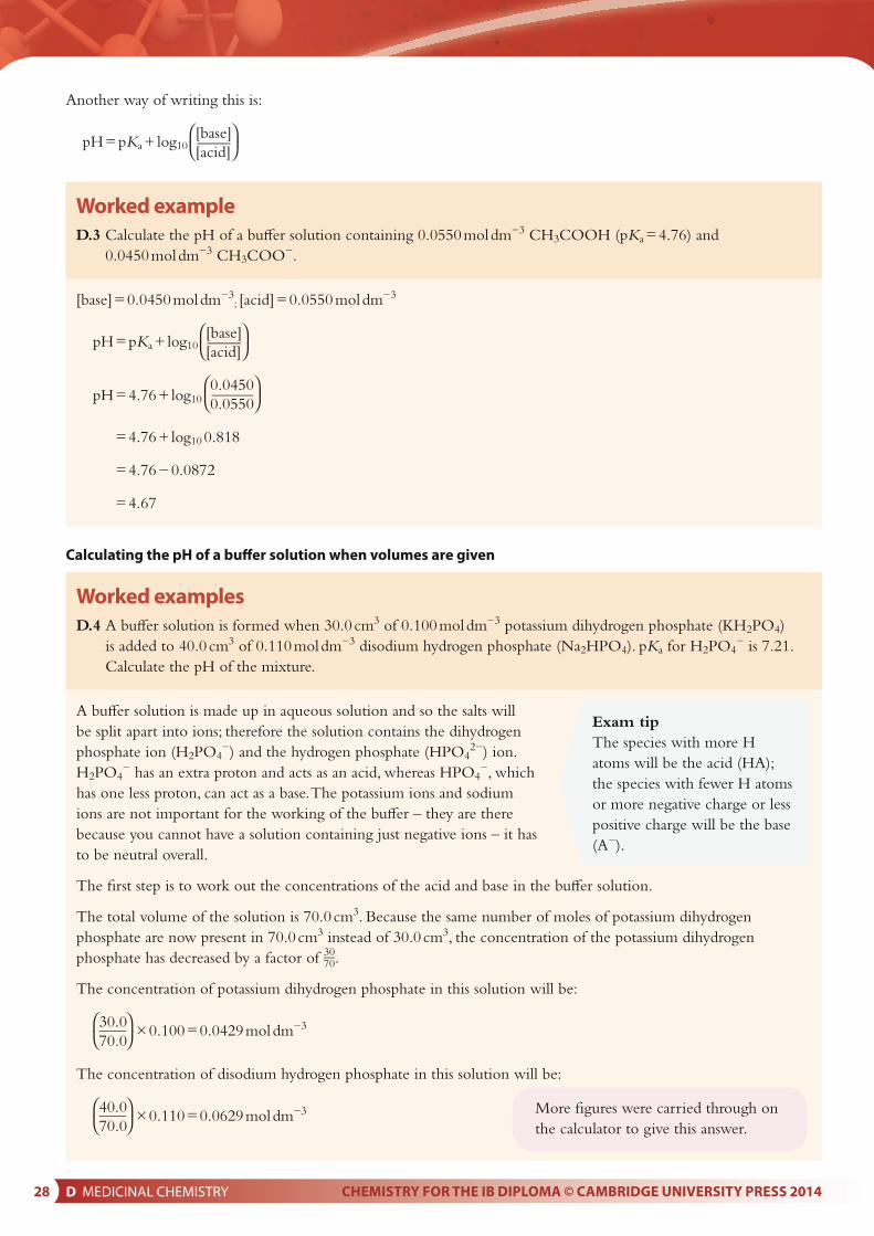
28 CHEMISTRY FOR THE IB DIPLOMA © CAMBRIDGE UNIVERSITY PRESS 2014D MEDICINAL CHEMISTRY
Another way of writing this is:
⎛[base]⎞pH = pKa + log10 ⎝[acid]⎠
Worked exampleD.3 Calculate the pH of a bu� er solution containing 0.0550 mol dm−3 CH3COOH (pKa = 4.76) and
0.0450 mol dm−3 CH3COO−.
[base] = 0.0450 mol dm−3; [acid] = 0.0550 mol dm−3
⎛[base]⎞pH = pKa + log10 ⎝[acid]⎠
pH = 4.76 + log10 ⎛0.0450⎞
⎝0.0550⎠
= 4.76 + log10 0.818
= 4.76 − 0.0872
= 4.67
Calculating the pH of a buff er solution when volumes are given
Worked examplesD.4 A bu� er solution is formed when 30.0 cm3 of 0.100 mol dm−3 potassium dihydrogen phosphate (KH2PO4)
is added to 40.0 cm3 of 0.110 mol dm−3 disodium hydrogen phosphate (Na2HPO4). pKa for H2PO4− is 7.21.
Calculate the pH of the mixture.
A bu� er solution is made up in aqueous solution and so the salts will be split apart into ions; therefore the solution contains the dihydrogen phosphate ion (H2PO4
−) and the hydrogen phosphate (HPO42−) ion.
H2PO4− has an extra proton and acts as an acid, whereas HPO4
−, which has one less proton, can act as a base. The potassium ions and sodium ions are not important for the working of the bu� er – they are there because you cannot have a solution containing just negative ions – it has to be neutral overall.
The � rst step is to work out the concentrations of the acid and base in the bu� er solution.
The total volume of the solution is 70.0 cm3. Because the same number of moles of potassium dihydrogen phosphate are now present in 70.0 cm3 instead of 30.0 cm3, the concentration of the potassium dihydrogen phosphate has decreased by a factor of 30
70.
The concentration of potassium dihydrogen phosphate in this solution will be:
⎛30.0⎞⎝70.0⎠ × 0.100 = 0.0429 mol dm−3
The concentration of disodium hydrogen phosphate in this solution will be:
⎛40.0⎞⎝70.0⎠ × 0.110 = 0.0629 mol dm−3
Exam tipThe species with more H atoms will be the acid (HA); the species with fewer H atoms or more negative charge or less positive charge will be the base (A−).
More � gures were carried through on the calculator to give this answer.

D MEDICINAL CHEMISTRY 29CHEMISTRY FOR THE IB DIPLOMA © CAMBRIDGE UNIVERSITY PRESS 2014
So [base] = 0.0629 mol dm−3; [acid] = 0.0429 mol dm−3
⎛[base]⎞pH = pKa + log10 ⎝[acid]⎠
pH = 7.21 + log10 ⎛0.0629⎞
⎝0.0429⎠
= 7.21 + log10 1.47
= 7.21 + 0.166
= 7.38
D.5 HEPES is used in some biological bu� ers. A bu� er solution can be made by dissolving sodium hydroxide in a HEPES solution.
Calculate the pH of the bu� er solution formed when 20.0 g of sodium hydroxide is added to 1.00 dm3 of a 1.00 mol dm−3 solution of HEPES (pKa = 7.5). Assume that there is no change in volume when the sodium hydroxide is added.
HEPES has an ‘extra’ proton and is therefore an acid. Reaction with sodium hydroxide converts some of it into a base.
Mr for sodium hydroxide is 40.00
20.0So the number of moles of sodium hydroxide =
40.00 = 0.500 mol.
From the equation, there is a 1 : 1 reaction with sodium hydroxide and therefore 0.500 mol HEPES reacts with 0.500 mol NaOH to form 0.500 mol of the anion.
Exam tipYou can check whether your answer for working out the pH of a bu� er solution is reasonable – if the solution contains a higher concentration of acid than base, the pH of the solution will be lower than the pKa of the acid; if there is a higher concentration of base than acid, the pH will be higher than the pKa.
O H
HEPES
N
N
S
O O
HO
O–N
N
S
O O
HO+ OH– + H2O
The concentration of each species in the bu� er solution can also be worked out using a moles calculation.The number of moles of potassium dihydrogen phosphate in 30.0 cm3:
⎛ 30.0 ⎞⎝1000⎠ × 0.100 = 0.00300 mol
So the concentration of potassium dihydrogen phosphate in the bu� er solution is:
⎛0.00300 ⎞ × 1000 = 0.0429 mol dm−3
⎝ 70 ⎠

30 CHEMISTRY FOR THE IB DIPLOMA © CAMBRIDGE UNIVERSITY PRESS 2014D MEDICINAL CHEMISTRY
In 1.00 dm3 of a 1.00 mol dm−3 solution of HEPES there is 1.00 mol of HEPES. So if 0.500 mol react there will be 0.500 mol remaining. Therefore the concentration of HEPES and the anion in the bu� er solution are both equal at 0.500 mol dm−3.
[base] = 0.500 mol dm−3; [acid] = 0.500 mol dm−3
⎛[base]⎞pH = pKa + log10 ⎝[acid]⎠
pH = 7.5 + log10 ⎛0.500⎞
⎝0.500⎠
= 7.5 + log10 1
= 7.5 + 0
= 7.5
Determining the composition of a buff er solution given its pH
Worked examplesD.6 A student wants to make up a bu� er solution of pH 7.7 using 0.100 mol dm−3 solutions of HEPES (pKa = 7.5)
and its sodium salt. Calculate how much of each solution must be used to make 500 cm3 of a bu� er of pH 7.7.
We need to calculate the ratio of the acid and base in the bu� er solution – this can be worked out using the Henderson–Hasselbalch equation.
⎛[base]⎞pH = pKa + log10 ⎝[acid]⎠
7.7 = 7.5 + log10 ⎛[base]⎞
⎝[acid]⎠
log10 ⎛[base]⎞
⎝[acid]⎠ = 0.2
baseacid
= 100.2 = 1.58
Therefore the ratio [base] : [salt] is 1.58 : 1
Because the concentrations of the solutions are the same, the amount of each solution required to make 500 cm3 of bu� er can be worked out as:
⎛1.58⎞volume of base = ⎝2.58⎠ × 500 = 306 cm3
⎛1.00⎞volume of acid = ⎝2.58⎠ × 500 = 194 cm3
Therefore the volume of the HEPES solution required is 194 cm3 and that of the solution of its sodium salt is 306 cm3.
This could also be worked out using 500 − 306.
If all � gures are carried through on the calculator the answers 193 cm3 and 307 cm3 are obtained.
2.58 is 1.58 + 1 from the ratio
10x is the inverse function of log10 – use the key combinations ‘shift log’ or ‘2nd log’ on your calculator.

D MEDICINAL CHEMISTRY 31CHEMISTRY FOR THE IB DIPLOMA © CAMBRIDGE UNIVERSITY PRESS 2014
D.7 What mass of solid sodium ethanoate must be added to 100.0 cm3 of 0.200 mol dm−3 ethanoic acid to produce a bu� er solution of pH 4.00? Assume there is no change in volume when the sodium ethanoate is added. The pKa for ethanoic acid is 4.76.
⎛[base]⎞pH = pKa + log10 ⎝[acid]⎠
The base is the ethanoate ion (CH3COO−) and the acid is ethanoic acid (CH3COOH)
So, 4.00 = 4.76 + log10 ⎛ [CH3COO−] ⎞
⎝[CH3COOH]⎠
log10 ⎛ [CH3COO−] ⎞
⎝[CH3COOH]⎠ = 4.00 − 4.76 = −0.76
⎛ [CH3COO−] ⎞⎝[CH3COOH]⎠ = 10−0.76 = 0.174
The concentration of ethanoic acid is 0.200 mol dm−3. Substituting this into the above equation we get:
[CH3COO−] = 0.174 × 0.200 = 0.0348 mol dm−3
So, the concentration of the sodium ethanoate in the solution must be 0.0348 mol dm−3, but because we are making up 100 cm3 of the bu� er solution, the number of moles of sodium ethanoate that must be used is given by:
no. moles = concentration × volume (in dm3)
= 0.0348 × ⎛100.0⎞
⎝ 1000 ⎠
= 0.003 48 mol
The molar mass of sodium ethanoate is 82.04 g. Therefore:
mass = no. moles × molar mass
= 0.003 48 × 82.04
= 0.285 g
So 0.285 g of sodium ethanoate must be dissolved in the ethanoic acid to produce a bu� er solution of pH = 4.00.

32 CHEMISTRY FOR THE IB DIPLOMA © CAMBRIDGE UNIVERSITY PRESS 2014D MEDICINAL CHEMISTRY
Calculating the change in pH of a buff er solution when acid or alkali is added
Worked exampleD.8 TRIS is used as a bu� er in biochemistry. A bu� er solution is prepared by adding hydrochloric acid to TRIS
to form a mixture of TRIS and its protonated form (TRIS-acid). The equilibrium that exists in the bu� er solution is:
a Calculate the pH of a bu� er solution containing 0.750 mol dm−3 TRIS-acid (pKa = 8.30) and 0.750 mol dm−3 TRIS.
b What is the pH of the solution formed when 10.0 cm3 of 0.100 mol dm−3 hydrochloric acid is added to 50.0 cm3 of the bu� er solution in part a?
a [base] = 0.750 mol dm−3; [acid] = 0.750 mol dm−3
⎛[base]⎞pH = pKa + log10 ⎝[acid]⎠
⎛0.750⎞= 8.30 + log10 ⎝0.750⎠
= 8.30
b When some acid is added to the bu� er solution, the following reaction occurs:
This means that the concentration of TRIS decreases and that the concentration of TRIS-acid increases. To work out by how much they change we need to work out the initial number of moles of TRIS and TRIS-acid and how many moles of acid were added.
The number of moles of TRIS in 50.0 cm3 of 0.750 mol dm−3 solution is given by:
no. moles = concentration × volume in dm3
⎛ 50.0 ⎞= 0.750 × ⎝1000⎠
= 0.0375 mol
This is the same as the number of moles of TRIS-acid.
TRIS TRIS-acid
+ H+
HO
HO
HO
NH2
HO
HO
HO
NH+3
Exam tipWhen [base] = [acid], the pH of the bu� er is equal to the pKa of the acid.
TRIS TRIS-acid
+ H+
HO
HO
HO
NH2
HO
HO
HO
NH+3

D MEDICINAL CHEMISTRY 33CHEMISTRY FOR THE IB DIPLOMA © CAMBRIDGE UNIVERSITY PRESS 2014
The number of moles of hydrochloric acid in 10.0 cm3 of 0.100 mol dm−3 solution is given by:
⎛ 10.0 ⎞no. moles = 0.100 × ⎝1000⎠
= 1.00 × 10⁻³ mol.
We will assume that the H+ from the hydrochloric acid reacts with the equivalent amount of TRIS and that there is no further change in the number of moles as equilibrium is established.
So the number of moles of TRIS thus decreases by 1.00 × 10⁻³ mol and the number of moles of TRIS-acid increases by 1.00 × 10⁻³ mol.
Initial amounts / mol: 0.0375 0.0375 molAfter HCl added / mol: 0.0375 − 1.00 × 10⁻³ 0.0375 + 1.00 × 10−3
= 0.0365 = 0.0385
The concentration of each species can be worked out by dividing the number of moles by the total volume in dm3, which is 50.0 + 10.0 = 60.0 cm3 or 0.0600 dm3
Concentration / mol dm−3: 0.0365 0.0385 0.0600 0.0600
= 0.608 = 0.642
So [base] = 0.608 mol dm−3; [acid] = 0.642 mol dm−3
⎛[base] ⎞pH = pKa + log10 ⎝[acid] ⎠
⎛0.608⎞= 8.30 + log10 ⎝0.642⎠
= 8.28
So, upon addition of 10.0 cm3 of the hydrochloric acid, the pH of the bu� er solution falls by 0.02 to 8.28.
TRIS TRIS-acid
+ H+
HO
HO
HO
NH2
HO
HO
HO
NH+3

34 CHEMISTRY FOR THE IB DIPLOMA © CAMBRIDGE UNIVERSITY PRESS 2014D MEDICINAL CHEMISTRY
D5 Antiviral medications
VirusesViruses are parasites – they invade host cells and use the materials and processes within those cells to produce new viruses – they cannot replicate outside host cells.
Viruses di� er greatly in shape and size from one type to the next but, in general, they have a core consisting of their genetic information (carried in the form of either DNA or RNA) which is surrounded by a protein coat known as a capsid. This capsid consists of identical protein subunits, called capsomeres, and its role is to protect the genetic information in the core. The capsid and genetic material together are called a nucleocapsid. Some viruses, such as the human immunode� ciency virus (HIV), also have a lipid envelope that surrounds the nucleocapsid (Figure D.24).
2 Calculate the pH values of the following bu� er solutions:
a A solution containing 0.0200 mol dm−3 butanoic acid (pKa = 4.82) and 0.0200 mol dm−3
sodium butanoate. b A solution containing 0.0500 mol dm−3
propanoic acid (pKa = 4.87) and 0.0200 mol dm−3 sodium propanoate.
c A solution containing 0.300 mol dm−3 ethanoic acid (pKa = 4.76) and 0.500 mol dm−3 sodium ethanoate.
d A solution made up by mixing together 25.0 cm3 of 0.200 mol dm−3 ethanoic acid (pKa = 4.76) and 50 cm3 of 0.100 mol dm−3 sodium ethanoate.
e A solution obtained when 10.0 cm3 of 0.100 mol dm−3 sodium hydroxide is added to 20.0 cm3 of 0.100 mol dm−3 ethanoic acid (pKa = 4.76).
f A solution obtained when 20.0 cm3 of 0.100 mol dm−3 ammonia solution is added to 40.0 cm3 of 0.100 mol dm−3 ammonium chloride solution (pKa = 9.25).
3 a A bu� er solution contains ethanoic acid (pKa = 4.76) at a concentration of 1.00 mol dm−3 and sodium ethanoate. If the pH of the bu� er solution is 4.20, what is the concentration of the sodium ethanoate?
b 20.0 cm3 of 0.0100 mol dm−3 hydrochloric acid is added to 50 cm3 of the bu� er solution in part a. Calculate the new pH of the bu� er solution.
4 What mass of solid sodium propanoate must be added to 50.0 cm3 of 0.100 mol dm−3 propanoic acid (pKa = 4.87) to produce a bu� er solution of pH 5.00? Assume there is no change in volume when the sodium propanoate is added.
? Test yourself
Learning objectives
• Describe the main di� erences between viruses and bacteria
• Understand the various modes of action of antivirals
• Understand how oseltamivir (Tami� u®) and zanamivir (Relenza™) work
• Compare the structures of oseltamivir and zanamivir
• Discuss why it is di� cult to solve the global AIDS problem
Nature of scienceScience is a highly collaborative enterprise and the development of a drug involves scientists with many di� erent specialisms. It is also important that individual scientists have knowledge outside their particular specialism – for instance, a knowledge of both organic chemistry and biochemistry was essential in the development of drugs such as omeprazole and esomeprazole.
Science is an ever-changing body of knowledge and collection and analysis of data is an important part of developing new theories. It used to be thought that stress and increased stomach acid were major causes of stomach ulcers until Warren and Marshall discovered that most stomach ulcers are actually caused by a bacterial infection. They were awarded a Nobel Prize in 2005.

D MEDICINAL CHEMISTRY 35CHEMISTRY FOR THE IB DIPLOMA © CAMBRIDGE UNIVERSITY PRESS 2014
Viruses are not considered to be living cells – they do not feed, excrete or grow and they consist only of what is necessary to invade the host cell and then take over that cell to produce copies of themselves. On the other hand, bacteria are living cells and are far more complex in structure and function than viruses – they are also able to reproduce outside host cells by cell division.
Some di� erences between viruses and bacteria are summarised in Table D.1.
Bacteria Viruses
living not living
larger than viruses smaller than bacteria
have a cellular structure – cell wall, cytoplasm, nucleus etc.
do not have a cellular structure
contain DNA and RNA contain either DNA or RNA
can reproduce independently need a host cell to reproduce
Table D.1 Diff erences between viruses and bacteria.
The lack of cell structure makes it much more di� cult to design drugs to target viruses than bacteria.
When designing drugs, there are far fewer enzymes/receptor proteins to target in viruses – viruses mostly use enzymes belonging to the host cell. Bacteria can be targeted, for instance by inhibiting the enzymes that make cell walls, but viruses don’t have cell walls.
To gain entry into host cells, viruses must � rst attach to the surface of a host cell. The genetic material of the virus is released into the cytoplasm and is then incorporated into the host cell’s DNA (if the virus contains RNA this must � rst be converted into DNA before it is inserted). The cell then starts producing viral proteins and viral DNA or RNA which get assembled into functional new viruses that leave the cell to go on to infect other cells.
Treatment and prevention of viral diseasesViruses cause a number of illnesses and diseases ranging from mild infections, such as the common cold, to potentially fatal diseases, such as acquired immunode� ciency syndrome (AIDS). It can be di� cult to � nd e� ective methods of preventing and treating viral infections for a number of reasons:
• Once inside a host cell, a virus can multiply very quickly and can have already spread throughout the body by the time that symptoms have appeared.
• Viruses can mutate their DNA or RNA resulting in a slight change in viral structure – this can make them resistant to drugs and can prevent vaccinations from being e� ective; this is particularly true of viruses such as HIV.
• Viruses use the host cell’s own processes and materials to produce new viruses, so it can be di� cult to design drugs that target only the virus and do not a� ect the host cell.However, despite these di� culties, several vaccines and antiviral
drugs have been developed and used to prevent and treat viral infections successfully.
Figure D.24 The HIV virus.

36 CHEMISTRY FOR THE IB DIPLOMA © CAMBRIDGE UNIVERSITY PRESS 2014D MEDICINAL CHEMISTRY
Vaccines stimulate the body’s natural defences (the immune system) to produce antibodies against a virus – so if infection does occur, the immune system is prepared and can stop the infection before it takes hold. Vaccines have been used successfully against a number of viruses including measles, mumps and polio.
Antiviral drugs work in a number of ways.
• Some alter the genetic material within cells – once inside a cell, the drug is converted into an active metabolite that becomes incorporated into the growing DNA strand (needed for viral replication) halting its synthesis. An example of a drug that acts in this way is aciclovir (acyclovir), which is used to treat cold sores; it stops viral DNA replication and so stops the virus from multiplying.
• Some inhibit the activity of enzymes within the host cell that are necessary for the formation of new viruses. An example is indinavir, which is used in AIDS treatment; it inhibits the HIV enzyme protease, which is essential to the assembly of functional new HIV viruses.
• Some stop the viruses from infecting host cells by preventing them from binding to the host cell surface and gaining access into the cell. Some drugs used to treat AIDS work in this way.
• Some prevent the virus from leaving the host cell so that it cannot infect other cells – see oseltamivir and zanamivir below.
Infl uenza antiviralsIn� uenza (‘� u’) is a viral disease that most people are familiar with – its symptoms include fever, headache, aching joints and fatigue. In severe cases it can cause death and there have been many cases of � u pandemics such as the outbreak of so called ‘Spanish � u’ in 1918, which killed millions of people, and ‘swine � u’ in 2009, which killed thousands.
The in� uenza virus has RNA as its genetic material rather than DNA. The capsid contains RNA and RNA polymerase (an enzyme that produces a type of viral RNA which is used by the cell to make viral proteins) and surrounding this it has a lipid envelope (derived from the host cell membrane) which has two very important proteins in it – neuraminidase (NA) and hemagglutinin (HA). Both proteins stick out of the membrane surrounding the virus. Hemagglutinin allows viruses to bind to receptors on the surface of cells to be infected – it binds to sialic acid residues on glycoproteins (proteins joined to sugar groups). Neuraminidase is an enzyme that hydrolyses the bond between a sialic acid residue and the rest of the glycoprotein – it essentially adds water across the bond to break it. Neuraminidase is important for helping the viruses to infect cells in that it breaks down the mucous surrounding cells in the upper respiratory tract, allowing the virus access to the target cell. Neuraminidase also plays an essential role at the end of the virus lifecycle – the virus enters the cell, replicates its RNA and essential proteins, but then the new viruses have to get out of the cell if they are to infect other cells. They do this by budding (Figure D.25). The virus will, however, remain bound to the surface of the cell (because the HA protein binds to receptors on the surface) until the NA breaks the link between the sialic acid and the rest of the membrane glycoprotein, allowing the virus to break free from the cell.

D MEDICINAL CHEMISTRY 37CHEMISTRY FOR THE IB DIPLOMA © CAMBRIDGE UNIVERSITY PRESS 2014
zanamivir
hydroxyl /alcohol ether
ether
carboxyl / carboxylic
acid
alkene / alkenyl groupalkene / alkenyl group
primaryamine
secondaryamide /
carboxamide
secondary amide /
carboxamide
primaryamine
ester
imine
oseltamivir
HO
HOH
H3C
HN
N
N
HNH2
OO
O
OH OH
secondary amine
guanidineH
N
NH
NH2
H3C
H2N
H3C
CH3
N
H
O
H3C
O
O
O
In� uenza antiviral drugs such as oseltamivir (Tami� u) and zanamivir (Relenza) are neuraminidase inhibitors. They bind to the active site of the neuraminidase enzyme, which prevents it from catalysing the hydrolysis of the sialic acid residue from the glycoproteins in the cell membrane, so that viruses remain anchored to the cell and cannot infect other cells.
Oseltamivir/zanamivir may be taken when � u symptoms develop but they are also taken as a prophylactic (a preventative measure) for people who have been exposed to the virus or are in high-risk groups (such as very young children).
Oseltamivir is administered orally as oseltamivir phosphate but zanamivir has very poor bioavailability when given orally and so is formulated as a dry powder for oral inhalation. Zanamivir is highly polar (see below) and cannot pass across cell membranes.
Comparison of the structures of oseltamivir and zanamivirThe structures of oseltamivir and zanamivir are compared in Figure D.26. There are certain similarities between the structures – e.g. the six-membered ring at the centre (in oseltamivir this is a cyclohexene ring but in zanamivir it could be called a dihydropyran ring). They also both contain a secondary carboxamide (amide) group. The ester and carboxyl (carboxylic acid) groups are highlighted in the same colour in Figure D.26 because the ester group in oseltamivir is hydrolysed in the body, converting it to a carboxyl group (see ‘active metabolites’ on page 26). Similarities between the general structures would be expected because they both bind to the same active site in the viral neuraminidase enzyme. There are also lots of di� erences between the structures highlighted in Figure D.26. The group highlighted grey in the zanamivir structure has been broken down into its constituent parts because they are more familiar from earlier work in the Coursebook but it actually constitutes a guanidine group (much as an ester is not really a ketone group and an ether group).
virus
NA
budding
nucleo-capsid
nucleo-capsid
nucleo-capsid
virus receptor
INFECTEDCELL
cell membrane
HA
Figure D.25 Viruses must escape from the host cell.
Figure D.26 Comparing the structures of oseltamivir and zanamivir.

38 CHEMISTRY FOR THE IB DIPLOMA © CAMBRIDGE UNIVERSITY PRESS 2014D MEDICINAL CHEMISTRY
AIDSAIDS was � rst recognised in 1981 and was found to be caused by the HIV virus a few years later. There are now believed to be more than 34 million people infected with HIV worldwide, and approximately 2 million deaths occur each year from AIDS.
HIV is a retrovirus and its genetic information is carried in the form of RNA, not DNA. HIV infection is lethal if left untreated because the HIV virus invades cells that form part of the immune system – these cells are white blood cells known as T-cells and they play a vital role in the body’s natural defence against infection. The HIV virus is able to infect these T-cells because they have speci� c receptor proteins on their surface to which the virus attaches to gain entry into the cell. Once inside, the viral enzyme reverse transcriptase converts the viral RNA into DNA so that it can be integrated into a T-cell’s DNA. The viral genes contained in the DNA are used to produce viral proteins and viral RNA within the cell, and these get assembled into new HIV viruses. The T-cell stops carrying out its role as an immune cell and instead becomes a factory for HIV viruses. When the newly formed viruses leave the T-cell, some of the T-cell membrane forms an envelope around the HIV virus.
Death of T-cells can occur when viruses exit a cell and this results in a decrease in the number of T-cells in the body and a weakened immune system. This is why people with AIDS are susceptible to potentially fatal infections and also some types of cancer – their immune systems are not strong enough to � ght against them.
Developing a method of eradicating HIV is di� cult because the virus is able to mutate rapidly, and also because the virus uses many of the host cell’s processes and materials to replicate – so it is di� cult to target the virus without a� ecting the host cell too. However, there are some di� erences that can be targeted. The HIV virus uses certain viral enzymes in the replication process that are di� erent to those found in the host cell. One of these enzymes is called reverse transcriptase, and the � rst antiretroviral drug that came onto the market to treat AIDS, azidothymidine (AZT), acts by targeting this enzyme. AZT inhibits reverse transcriptase and gets incorporated into the DNA strand that is being synthesised by the enzyme – this results in termination of DNA synthesis, and so the virus cannot replicate. Other viral enzymes that are inhibited by drugs include the one that integrates the DNA into the host cell’s DNA (called integrase) and the one that assembles the viral proteins to produce new viruses (called protease). Drugs are also available that stop the virus from binding to T-cell receptor proteins and gaining entry into the host cell. However, all these drugs only delay the progression of AIDS, they do not destroy the virus – nevertheless they have saved the lives of millions of people since their introduction.
Much research is being undertaken to � nd an e� ective vaccine that can be used against HIV to try to stop the spread of the virus. The ability of the virus to mutate and change its structure has made it di� cult to � nd a suitable vaccine that can prime the host’s natural immunity against such variations in structure. Promising results are being reported by researchers, both in animal studies and human trials, where a reduction in infection rates has been shown.
One problem with antiretroviral therapy is that it used to be
expensive, so AIDS su� erers in poor countries (where the majority of AIDS cases are found) did not generally have access to these life-saving drugs. However, the prices of the most commonly used antiretroviral treatments have decreased signi� cantly over the last few years and, together with a global commitment to make these treatments universally available, more and more patients in poorer countries are now receiving treatment. However, more work still needs to be done to ensure that prevention measures (such as education and condoms) and antiretroviral treatments are available to all.

D MEDICINAL CHEMISTRY 39CHEMISTRY FOR THE IB DIPLOMA © CAMBRIDGE UNIVERSITY PRESS 2014
Nature of scienceScience can be used for good and for bad and there are many ethical issues that face scientists working in certain � elds. As well as medical programs that have developed vaccines for smallpox and cured many other diseases, there are many rumours about biological weapons employing viruses or bacteria in top-secret programs in various countries around the world. There have also been cases of terrorist attacks using anthrax bacteria.
Science is a highly collaborative activity. Advances in our understanding of how viruses infect cells and the development of drugs to treat viral infections have involved many di� erent people with a variety of specialisms working together.
D6 Environmental impact of some medications
Nuclear wasteThere are many applications of radioactive isotopes in medicine – they are used for both treatment and diagnosis. Radioactive isotopes undergo radioactive decay by the emission of alpha particles, beta particles or gamma rays (there are other forms of radioactive decay). These are called ionising radiation because they cause the formation of ions (by ejection of electrons) when they interact with matter. Ionising radiation can damage cells and the main e� ect comes from damage caused to DNA.
Numerous radioisotopes are used or produced in various medical processes and these have half-lives that vary enormously – 131I, used to treat thyroid cancer, has a half-life of just 8 days; 60Co, used to treat other forms of cancer, has a half-life of 5.3 years.
Radioactive waste is a byproduct of the use of radioisotopes in medicine and, of course, it must be disposed of in some way. The classi� cation of radioactive waste is important in determining how it can be disposed of and the safety measures that must be used in its transport and handling.
Radioactive waste can be divided into di� erent categories – low-level and high-level. The category ‘intermediate-level radioactive waste’ is also sometimes used. The criteria used for the classi� cation are quite complex and low-level waste can be divided into sub-categories (A, B, C and >C) depending on the level of activity.
• Low-level waste has a low activity (not many radioactive nuclei decay each second to produce ionising radiation) and usually contains isotopes with short half-lives (ionising radiation is given of f for a shorter period of time).
• High-level waste has a high activity (many radioactive nuclei decay each second to produce ionising radiation) and usually contains isotopes with longer half-lives (ionising radiation is given of f for a long time).
One of the ways of reducing the environmental impact of nuclear medicine is to choose the radioactive sources very carefully – they should
Learning objectives
• Distinguish between high-level and low-level nuclear waste
• Discuss the environmental e� ects of medical nuclear waste
• Explain the dangers of antibiotic waste
• Understand the basics of green chemistry
• Understand how green chemistry was involved in the production of a precursor of oseltamivir
• Discuss the environmental issues associated with the use of solvents in the pharmaceutical industry
Half-life is the time it takes for the number of radioactive nucleipresent in a sample at any given time to fall to half its value.

40 CHEMISTRY FOR THE IB DIPLOMA © CAMBRIDGE UNIVERSITY PRESS 2014D MEDICINAL CHEMISTRY
have the minimum possible activity and the shortest possible half-lives, but still be suitable for the job.
Radioactive materials have the potential to be a serious hazard both to people and to the environment – therefore the disposal of medical nuclear waste must be controlled carefully. Governments set strict limits on the release of radioactivity into the environment and monitoring is essential to ensure that these regulations are adhered to. The main approaches to the disposal of nuclear waste are ‘dilute and disperse’, ‘delay and decay’ or ‘con� ne and contain’. ‘Con� ne and contain’ is always used for nuclear waste that has a high level of activity.
Health e� ects that can result from long-term exposure to increased low-level radioactivity, for example, from the disposal of medical nuclear waste, include cancer and mutations in DNA.
Although the waste from medical processes is generally classi� ed as low-level, some radioisotopes that are used in medicine are produced in nuclear reactors, which generate high-level waste.
Low-level wasteThis includes items that have been contaminated with radioactive material or have been exposed to radioactivity. Examples are gloves and other protective clothing, tools, syringes and excreta from patients treated with radioisotopes.
Low-level waste may be stored on site until it has decayed to such an extent that it can be disposed of as ordinary waste (e.g. in land� ll sites or released into the sewage system) or shipped to a central site for more specialised disposal.
Some low-level waste is incinerated, which reduces its volume considerably and distributes the radioisotopes over a wide area – ‘dilute and disperse’. The ash from incineration is assessed for activity and disposed of appropriately. Low-level waste with higher activity is often just buried underground (‘near-surface’ disposal) – for example, in individual concrete canisters or in concrete-lined vaults. Low-level waste may need to be contained underground for up to 500 years depending on its activity and half-life.
High-level wasteThis includes spent fuel rods and other materials from nuclear reactors. High-level waste will remain hazardous to humans and other living things for thousands of years.
High-level liquid waste can be converted to glass (vitri� cation) to make storage easier. High-level waste is � rst kept in storage pools (cooling ponds) under water, usually for a minimum of nine months, but sometimes spent fuel rods are stored in this way for decades. After su� cient cooling, fuel rods may be transferred to dry storage casks –these have very thick walls and are made of steel and concrete. The dry casks are then stored in concrete bunkers.
Permanent storage of high-level radioactive waste is a major problem and various solutions have been suggested – such as burying the waste deep underground in stable geological areas. Over thousands of years, however, it is di� cult to predict what processes could occur to cause release of the radioactive material. Many people argue that there is no suitable solution for the disposal of high-level waste.

D MEDICINAL CHEMISTRY 41CHEMISTRY FOR THE IB DIPLOMA © CAMBRIDGE UNIVERSITY PRESS 2014
The release of antibiotics into the environmentThe presence of antibiotics and other pharmaceuticals in waste water is becoming an increasing problem. Antibiotics can enter the water supply by several routes. These include:
• incorrect disposal of unwanted medicines – for example, by � ushing old medicines down the toilet
• agriculture – drugs given to animals will be present in animal waste (urine and feces) and can � nd their way into groundwater, rivers and lakes.Treating water to produce drinking water does remove some of these
chemicals but there is still a variety of pharmaceuticals present in the water we drink. Although these pharmaceuticals are found in drinking water in only very small amounts (typically nanograms per dm3) there are concerns that long-term exposure could result in damage to human health.
The release of antibiotics into the environment is regarded as a particular problem because not only can they cause damage to aquatic organisms, but they can also result in increased resistance of bacteria to antibiotics. Antibiotics (antibacterials) are used to treat a variety of conditions but if bacteria develop resistance to antibiotics such as penicillin, these diseases can become much more di� cult to cure. Antibiotic resistance is discussed more fully on page 17.
Green chemistry
Green chemistry (or sustainable chemistry) is an approach to chemical research and chemical industrial processes that seeks to minimise the production of hazardous substances and their release into the environment.
There are 12 principles of green chemistry, an idea developed by Paul Anastas and John C. Warner:1 Prevention – it is better to prevent waste than to treat or clean up waste
after it has been created.2 Atom economy – synthetic methods should be designed to maximise the
incorporation of all materials used in the process into the � nal product.3 Less hazardous chemical syntheses – wherever practicable, synthetic
methods should be designed to use and generate substances that possess little or no toxicity to human health and the environment.
4 Designing safer chemicals – chemical products should be designed to a� ect their desired function, while minimising their toxicity.
5 Safer solvents and auxiliaries – the use of auxiliary substances (solvents, separation agents etc.) should be made unnecessary wherever possible and innocuous when used.
6 Design for energy e� ciency – the energy requirements of chemical processes should be recognised for their environmental and economic impacts and should be minimised. If possible, synthetic methods should be conducted at ambient temperature and pressure.
7 Use of renewable feedstocks – a raw material or feedstock should be renewable, rather than depleting, whenever technically and economically practicable.

42 CHEMISTRY FOR THE IB DIPLOMA © CAMBRIDGE UNIVERSITY PRESS 2014D MEDICINAL CHEMISTRY
8 Reduce derivatives – unnecessary derivatisation (use of blocking groups, protection/ deprotection, temporary modi� cation of physical/chemical processes) should be minimised or avoided if possible because such steps require additional reagents and can generate waste.
9 Catalysis – catalytic reagents (as selective as possible) are superior to stoichiometric reagents.
10 Design for degradation – chemical products should be designed so that at the end of their function they break down into innocuous degradation products and do not persist in the environment.
11 Real-time analysis for pollution prevention – analytical methodologies need to be further developed to allow for real-time, in-process monitoring and control prior to the formation of hazardous substances.
12 Inherently safer chemistry for accident prevention – substances, and the form of a substance used in a chemical process, should be chosen to minimise the potential for chemical accidents including releases, explosions and � res.
(Green Chemistry: Theory and Practice by Paul Anastas & John Warner (1998) Figure 4.1 from p. 30. By permission of Oxford University Press www.oup.com)
As can be seen above, there are many things that have to be considered when making a pharmaceutical. The best synthetic routes to a drug:
• use readily available and safe materials
• have the minimum number of steps
• convert as much of the starting materials as possible into the required product at each step – good atom economy and good yield
• use as little solvent as possible
• use as little energy as possible.An important consideration in green chemistry is the concept of atom
economy. Atom economy can be used as a measure of how e� cient a particular reaction is in terms of converting as much of the starting materials as possible into useful products.
molar mass of desired productatom economy =
total molar mass of all reactants × 100%
We can use the preparation of 1-phenylethanone, which could be investigated as an enzyme inhibitor, as an example to illustrate how the equation is used.
Consider two di� erent ways of making 1-phenylethanone (C6H5COCH3), from 1-phenylethanol:
3C6H5CH(OH)CH3 + 2CrO3 + 3H2SO4 → 3C6H5COCH3 + Cr2(SO4)3 + 6H2O method 1
C6H5CH(OH)CH3 + 12O2 → C6H5COCH3 + H2O method 2
The atom e� ciency for each process can be worked out as follows.Method 1:
total molar mass of all reactants = (3 × 122.18) + (2 × 100.00) + (3 × 98.09) = 860.81 g mol−1
molar mass of desired product = 3 × 120.16 = 360.48 g mol−1
⎛360.48⎞atom economy = ⎝860.81⎠ × 100 = 41.88%

D MEDICINAL CHEMISTRY 43CHEMISTRY FOR THE IB DIPLOMA © CAMBRIDGE UNIVERSITY PRESS 2014
Method 2:total molar mass of all reactants = 122.18 + (0.5 × 32.00) = 138.18 g mol−1
molar mass of desired product = 120.16 g mol–1
⎛120.16⎞atom economy = ⎝138.18⎠ × 100 = 86.96%
It can be seen that method 2 has a much higher atom economy and is, therefore, much more e� cient. However, many other things must be considered when assessing these reactions in terms of green chemistry principles – the temperature used in each reaction, the solvents used and how much of them, disposal of the solvents, the nature of the catalyst required for the second reaction etc.
Synthesis of oseltamivirOseltamivir has been discussed earlier (page 37) as a treatment for in� uenza. Total synthesis (from petrochemical starting materials) of oseltamivir involves huge amounts of materials and can generate thousands of kilograms of waste per mole of oseltamivir made. Therefore it was essential to develop greener routes to the drug.
The current commercial synthetic route uses a naturally occurring material, shikimic acid, as the starting material – this cuts out several steps in the synthesis and makes it greener. Shikimic acid is a renewable material that can either be extracted from Chinese star anise or obtained from glucose by fermentation using genetically modi� ed bacteria. Even starting from shikimic acid, a further ten steps are required to make oseltamivir, so more work is required to make this synthesis even greener!
Although shikimic acid can be obtained from star anise, this in itself causes a problem – the yield is not very high and the production of shikimic acid is linked to the availability of star anise. The use of GM bacteria is likely to provide a better long-term solution to producing shikimic acid – fermentation uses relatively low temperatures and an aqueous medium, so it is a reasonably green process.
Waste solventsThere are many environmental issues with the use of solvents in the pharmaceutical industry. Solvents are used as the medium in which many reactions occur, in the extraction and puri� cation of compounds and so on. Solvents contribute typically about 80 –90% of the mass of substances used in the production of a pharmaceutical and also make a large contribution to the amount of energy used and the cost. Many of the solvents used
Atom economy is not the same as the yield of a reaction. Atom economy is a theoretical quantity based on a chemical equation and allows an evaluation of how much waste is produced. The yield of a reaction is an experimental quantity worked out from how much of the desired product is actually made in a chemical reaction.
In the calculation of atom economy above, it has been assumed that all reactions have 100% yield which will not be the case in practice.
When evaluating how green/environmentally friendly a particular process is both atom economy and yield must be considered as well as several other factors.
From the point of view of the pharmaceutical industry there is also the problem of residual solvents in drug samples, which need to be removed as far as possible before the drugs can be administered to humans – especially if they are toxic.

44 CHEMISTRY FOR THE IB DIPLOMA © CAMBRIDGE UNIVERSITY PRESS 2014D MEDICINAL CHEMISTRY
are organic because of the nature of the compounds being used and made – usually mostly non-polar and therefore more soluble in organic solvents – and many will be toxic to humans and other organisms.
The � rst aspect to be considered when trying to make a process greener is prevention – can a di� erent solvent be used that is less harmful to the environment? If a greener solvent cannot be found then the amount used should be reduced as far as possible.
The next consideration is the possibility of a solvent being recycled and reused. This avoids the need to dispose of the solvent. The energy considerations of purifying the solvent for reuse must be considered at this stage.
If the solvent cannot be reused, then it must be disposed of as safely as possible. There are many methods of disposal including incineration and injection underground. The nature of the solvents that are to be injected underground is controlled strictly because the practice could introduce potentially toxic chemicals into the environment. Incineration produces carbon dioxide, a greenhouse gas, which can contribute to climate change and can also produce toxic substances such as dioxins.
Nature of scienceAn understanding of science is essential if the public are to make informed judgements about the advantages and disadvantages of using antibiotics. It is important that scientists explain the issues in a way that is as complete as possible but also objective so that, for instance, farmers can make informed decisions about adding antibiotics to animal feed etc. and patients can understand the importance of disposing of unwanted medicines in an appropriate way.
When considering whether a particular drug should be licensed for medical use the bene� ts and risks to patients are considered by regulatory authorities. However, there are also environmental factors that should be considered and scientists are involved in developing processes to reduce the environmental impact of drug development and production.
D7 Taxol® – a chiral auxiliary case study (HL)
ChiralityWhen a molecule contains a carbon atom bonded to four di� erent groups, it is said to be chiral and two mirror images (known as enantiomers) exist. These enantiomers can behave very di� erently in the body as a result of their di� erent shapes. For example, one of the enantiomers may be able to bind e� ectively to an enzyme or receptor protein because its functional groups are in the correct orientation to form bonds with the protein, whereas the other may not be able to bind as strongly because the groups are in the wrong orientation to form bonds (Figure D.27). In some cases, one enantiomer may produce a therapeutic e� ect by binding to its target, whereas the other may produce a toxic e� ect by binding elsewhere.
Thalidomide is an example of a chiral drug that was given as a mixture of enantiomers (a racemic mixture) to pregnant women to combat morning sickness. It was later discovered that one of the enantiomers (the S-enantiomer – Figure D.28) was responsible for producing a teratogenic e� ect and caused limb deformities in the fetus. Pharmaceutical companies generally now either synthesise or separate out the active single enantiomer of a drug and develop this instead of the racemic mixture.
Learning objectives
• Understand what is meant by a chiral auxiliary
• Understand how chiral auxiliaries can be used for stereoselective syntheses
• Understand that paclitaxel (Taxol®) is used in chemotherapy
• Understand how paclitaxel is produced
• Understand how a polarimeter can be used to identify enantiomers

D MEDICINAL CHEMISTRY 45CHEMISTRY FOR THE IB DIPLOMA © CAMBRIDGE UNIVERSITY PRESS 2014
enzyme active site
A
BC
c
a
bD
a
enzyme active site
no bondformation
b
A
BD
c
a
bC
A
BC
D
mirror
A
BC
D
Figure D.27 Representation of a chiral drug binding to a theoretical enzyme active site:a one enantiomer can form three bonds with groups at the active site and is active; b the other enantiomer can only form two bonds with groups at the active site and is inactive.
(S)-thalidomide(teratogenic)
O
N
NH
H
OO
O
(R)-thalidomide
O
N
NH
H
OO
O
(S)-thalidomide(teratogenic)
O
N
NH
H
OO
O
(R)-thalidomide
O
N
NH
H
OO
O
Figure D.28 Enantiomers of thalidomide.
Administering the single enantiomer would not have helped in the case of thalidomide because the enantiomers interconvert when in the body, producing the racemic mixture. Nowadays, if a new drug is going to be marketed as the racemic mixture, testing for toxicity and e� ectiveness must be carried out on each enantiomer separately and also on the racemic mixture.
Chiral auxiliaries in asymmetric synthesisMany drugs have chiral centres and so can exist as two enantiomers, but it is usual for pharmaceutical companies to develop just one enantiomer of a drug for reasons already explained. Synthesis reactions normally produce a mixture of both enantiomers (racemic mixture). Consider Figure D.29, which shows the reaction between a Grignard reagent (C2H5MgBr) and pentan-2-one. Neither of the starting materials is chiral but the product has a chiral centre (shown green).
Figure D.29 The reaction of ethylmagnesium bromide with pentan-2-one.
Mg
C CH
Br
H
H
H
HGrignard reagent
(i)
H
C C
H
H
C
H
H
O
C
H
H
C
H
H
C
H
H
C
H
H
H
HH C H
H
H
C CH
H
(ii) H+/H2O
O
C
H
C
H
H
C
H
H
H
H
+ Mg(OH)Br
The Grignard reagent reacts as if it is C2H5− and can attack the pentan-
2-one (planar about the C=O group) from either the top or the bottom. Attack from either side is equally probable and so an equimolar mixture of the two enantiomers is formed – a racemic mixture (Figure D.30).
There are various ways of obtaining the required single enantiomer of a drug. For example, the synthesis of the racemic mixture may be carried out, followed by separation using chiral chromatography (normal
Figure D.30 A racemic mixture is formed because the reagent can attack from above or below.
C
C3H7
racemic mixture of both enantiomers formed
chiral centreH3C
CH2CH3–
C
OH
C2H5
CH3
C3H7
C
OH
C3H7
H3CC2H5
H+/H2OO
CH2CH3–

46 CHEMISTRY FOR THE IB DIPLOMA © CAMBRIDGE UNIVERSITY PRESS 2014D MEDICINAL CHEMISTRY
A chiral auxiliary is one enantiomer of an optically active substance that is temporarily incorporated into a non-chiral molecule to produce a single enantiomer of a product in an organic synthesis reaction.
PaclitaxelOne process in which chiral auxiliaries have been used successfully is in the semi-synthesis (from a natural precursor) of the anticancer drug paclitaxel (Taxol) (Figure D.32).
Paclitaxel is used to treat several forms of cancer – mainly breast, ovarian and lung cancer. It is usually given intravenously as part of a course of chemotherapy to treat cancer. Paclitaxel acts by preventing cell division – it does this by binding to microtubules in the cytoplasm, preventing them from breaking down during cell division.
C
C
rest ofmolecule CH2CH3
–
C2H5CH3
HOH+/H2O
reaction
chiral auxillaryadded
chiral auxillary
chiral auxillaryremoved
OC
C
rest ofmolecule
CH H
O
CH3
CH2CH3
C2H5
HO
chiral auxillary
C
C
rest ofmolecule
one enantiomer produced
C
OH
CH2CH3
C
C
rest ofmolecule
OH
Figure D.31 A chiral auxiliary favours the formation of one enantiomer.
chromatography does not separate enantiomers), or a synthetic reaction may be used that selectively produces one of the enantiomers of the product – this is known as stereoselective (asymmetric) synthesis.
One method of achieving stereoselective synthesis involves the use of a chiral auxiliary. This chiral auxiliary is a pure enantiomer and combines with the non-chiral reactant to form a chiral intermediate. The physical presence of the chiral auxiliary allows the reagent in the next stage of the synthesis to approach from one side of the molecule only, so forcing the reaction to follow a certain path that favours the production of one of the possible enantiomers (Figure D.31). Once the reaction is complete, the chiral auxiliary is removed to leave the desired enantiomer. The chiral auxiliary can then be recycled for use in other reactions.
H3COC–O
CH3
OH
OH
HO
O
O
O
O
CONH
O
C
H
OO
COCH3
Figure D.32 The structure of paclitaxel.
Chemotherapy is the treatment of cancer with drugs. These drugs destroy cancer cells or stop them from dividing.

D MEDICINAL CHEMISTRY 47CHEMISTRY FOR THE IB DIPLOMA © CAMBRIDGE UNIVERSITY PRESS 2014
Paclitaxel was originally obtained from Paci� c yew tree bark – however, it took the bark from more than one tree to provide enough paclitaxel to treat just one patient and so semi-synthetic processes were developed that involved making paclitaxel from another natural product derived from the needles of yew trees. Semi-synthesis of the drug allowed it to be made on a larger scale and reduced the environmental impact – extracting the drug from its natural source results in killing of the trees. Nowadays paclitaxel is also made by fermentation using plant cell cultures.
Using a polarimeter to distinguish between enantiomersThe enantiomers of an optically active substance can be distinguished using a polarimeter because they rotate the plane of plane-polarised light in opposite directions. A simple polarimeter consists of a source of light (usually a sodium lamp producing one speci� c wavelength), two polarising � lters, a sample tube and a scale to measure the degree of rotation of the plane-polarised light (Figure D.33).
Polarimetry is usually carried out on samples in solution. First, the solvent in which the test substance is to be dissolved is put in the sample tube, then the polarising � lters are rotated until the maximum amount of light passes through. At this point the two polarising � lters are exactly parallel.
The solvent is then replaced by a solution of the sample in the same solvent and the polarising � lter rotated again until the light coming through is of maximum brightness again. The direction in which the � lter must be rotated and the angle through which the light is rotated are recorded.
An enantiomer that rotates plane-polarised light clockwise (to the right – dextrorotatory) is called the (+)-enantiomer and one that rotates the plane anticlockwise (to the left – levorotatory) is called the (−)-enantiomer.
Just which con� guration (arrangement of groups around the chiral centre) of an enantiomer corresponds to the direction in which light is rotated can be worked out only by determining the absolute con� guration using X-ray crystallography and the rotation of plane-polarised light using a polarimeter. We cannot just look at a particular enantiomer’s three-dimensional structure and say that it rotates plane-polarised light to the right or to the left.
lightsource
sample tube
scalepolarising
�lter
polarising�lter
Figure D.33 A simple polarimeter.

48 CHEMISTRY FOR THE IB DIPLOMA © CAMBRIDGE UNIVERSITY PRESS 2014D MEDICINAL CHEMISTRY
An enantiomer can be identi� ed from its speci� c rotation. The speci� c rotation, [α], is worked out from the angle though which the plane-polarised light is rotated in degrees (α), the path length of solution that the light passes through (l) in dm and the concentration of the solution (c) in g cm−3:
α[α] =
cl
The speci� c rotation also depends on the wavelength of light used, the temperature (usually 20 °C) and the solvent. The speci� c rotation of an enantiomer under a given set of conditions is characteristic of the compound and can be used to identify it by comparing with tables of literature values. However, this method does rely on having a pure sample of one enantiomer because contamination with the other enantiomer, or any other optically active compounds, will a� ect the angle through which the plane-polarised light is rotated.
Nature of scienceScience is both systematic and creative. Systematic screening of a large number of plant extracts identi� ed paclitaxel as a possible anticancer drug. When the demand for paclitaxel exceeded the supply from natural sources scientists had to develop ways of making this important drug. A great deal of creativity was involved in designing ways of making paclitaxel.
D8 Nuclear medicine (HL)
Radioactive decay processesRadioactive decay involves changes in the nucleus of an atom resulting in particles and sometimes also electromagnetic radiation being emitted from the nucleus. The two main particles that are emitted are α particles (42He2+, helium nuclei – two protons and two neutrons) and β particles (electrons formed when a neutron turns into a proton and an electron). The emission of γ rays (high-energy electromagnetic radiation) may also accompany radioactive decay. Other particles that can be emitted from nuclei include positrons, which are positively charged electrons produced when a proton is converted to a neutron in the nucleus.
Examples of radioactive decay processes are:
α-decay: 22688Ra → 222
86Rn + 42α
β-decay: 125B → 12
6C + 0 −1e
Equations for nuclear decay must balance in terms of the total mass number and the total atomic number on each side.
When a nucleus undergoes α decay, the mass number will go down by four and the atomic number will go down by two. The element formed will be two places to the left in the periodic table.
Learning objectives
• Understand what is meant by radioactive decay and balance equations involving α and β particles
• Calculate the amount of material remaining after a certain period of time when an isotope decays
• Understand what particles are emitted when nuclear medicine is used
• Understand that radiotherapy can be external or internal
• Understand the reasons for the use of certain radioisotopes
• Understand that targeted alpha therapy and boron-neutron-capture therapy are used in the treatment of cancer
42α could also have been shown as 42He.
0 −1e could also have been shown as 0
−1β.
Beta particles are sometimes written as β− to distinguish them from positrons, which are written β+.

D MEDICINAL CHEMISTRY 49CHEMISTRY FOR THE IB DIPLOMA © CAMBRIDGE UNIVERSITY PRESS 2014
Because the emission of a β particle results from a neutron in the nucleus becoming a proton, the mass number does not change but the atomic number goes up by one (the atomic number is the number of protons). The element formed will be one place to the right in the periodic table.
Worked examplesD.9 Astatine-211 can undergo α decay by emitting α particles. Determine the identity of the isotope formed.
The equation for the reaction is:
21185At → AZ X + 42α
The total mass number on the right-hand side must equal 211; therefore A + 4 = 211 and A = 207
The total atomic number on the right-hand side must equal 85; therefore Z + 2 = 85 and Z = 83
The atomic number de� nes the element and the element with atomic number 83 is bismuth.
The isotope formed is 20783Bi.
D.10 Yttrium-90 can undergo β decay by emitting β particles. Determine the identity of the isotope formed.
The equation for the reaction is:
9039Y → AZ Q + 0
−1β
The total mass number on the right-hand side must equal 90; therefore A + 0 = 90 and A = 90
The total atomic number on the right-hand side must equal 39; therefore Z − 1 = 39 and Z = 40
The atomic number de� nes the element and the element with atomic number 40 is zirconium.
The isotope formed is 9040Zr.
5 Determine the identity of the isotope formed in each of the following decay processes:
a indium-115 undergoes β decay b radium-224 undergoes α decay c nickel-63 undergoes β decay d thorium-229 undergoes α decay
6 Work out whether each of the following processes involves emission of an α particle or a β particle:
a 17771Lu → 177
72Hf + ? b 173
79Au → 16977Ir + ?
c 66Cu → 66Zn + ? d 104Mo → 104Tc + ? e 278Rg → 274Mt + ?
? Test yourself

50 CHEMISTRY FOR THE IB DIPLOMA © CAMBRIDGE UNIVERSITY PRESS 2014D MEDICINAL CHEMISTRY
Half-lifeRadioactive decay is a random process and it is impossible to predict when any one nucleus will decay, but on average a sample of a particular radioisotope containing a very large number of atoms will decay with a constant half-life.
Half-life is the time it takes for the number of radioactive nuclei present in a sample at any given time to fall to half its value.
Half-life (t12) varies from isotope to isotope – for example, the half-life of
226Ra is 1600 years but that of 224Ra is 3.7 days. Half-life is independent of the mass of a radioactive sample – the half-life is the same whether 1 g or 1 kg of a particular isotope is present.
Figure D.34 shows a graph of the decay of an isotope with half-life 2 s. Every 2 s the number of nuclei remaining decreases by half.
Figure D.34 The decay of a radioisotope with half-life 2 s – this is exponential decay.
Time / s
half-life half-life half-life
Num
ber
of n
ucle
i rem
aini
ng /
1023
0 1 2 3 4
8
7
6
5
3
4
2
0
1
5 6 7 8 9 10
Calculations involving whole numbers of half-livesRadium-226 is an α-emitter with a half-life of approximately 1600 years – so if we start with 1 g of pure 226Ra, after 1600 years there will be 0.5 g present, after a further 1600 years there will be 0.25 g present and after a total of 4800 years (three half-lives) there will only be 0.125 g of radium-226 left:
half-life half-life half-life1 g ⎯⎯⎯⎯→ 0.5 g ⎯⎯⎯⎯→ 0.25 g ⎯⎯⎯⎯→ 0.125 g 1600 years 1600 years 1600 years
Half-life may also be expressed in terms of the activity of a sample (the number of nuclei that decay per second). The half-life is then the time taken for the activity to drop to half its original value.

D MEDICINAL CHEMISTRY 51CHEMISTRY FOR THE IB DIPLOMA © CAMBRIDGE UNIVERSITY PRESS 2014
Worked examplesD.11 Germanium-71 has a half-life of 11 days. If there were originally 2.00 mg of this isotope present in a sample,
calculate the mass remaining after 44 days.
It takes 11 days for the amount to fall by half, therefore there will be 1.00 mg present after 11 days. This will drop to 0.500 mg after another 11 days, 0.250 mg after another 11 days and 0.125 mg after a further 11 days:
2.00 mg ⎯⎯⎯→ 1.00 mg ⎯⎯⎯→ 0.500 mg ⎯⎯⎯→ 0.250 mg ⎯⎯⎯→ 0.125 mg 11 days 11 days 11 days 11 days
Therefore the mass of germanium-71 remaining will be 0.125 mg.
D.12 The half-life of uranium-238 is 4.5 × 10⁹ years. Calculate how long it would take 32 g of uranium-238 to decay to 1 g.
This decay involves � ve half-lives:
half-life half-life half-life half-life half-life32 g ⎯⎯⎯→ 16 g ⎯⎯⎯→ 8 g ⎯⎯⎯→ 4 g ⎯⎯⎯→ 2 g ⎯⎯⎯→ 1 g
So the total time is 5 × 4.5 × 109 = 2.3 × 10¹⁰ years.
D.13 Calculate the half-life of protoactinium-233 if it takes 108 days for 100 mg of the element to decay to 6.25 mg.
This decay will take four half-lives:
half-life half-life half-life half-life100 mg ⎯⎯⎯→ 50 mg ⎯⎯⎯→ 25 mg ⎯⎯⎯→ 12.5 mg ⎯⎯⎯→ 6.25 mg
So 108 days is equivalent to four half-lives. One half-life is therefore 108
= 27 days. 4
D.14 Calculate the time taken for the activity of a sample of rubidium-83 to fall to 12.5% of its original value given that its half-life is 86 days.
The data here are given in terms of activity instead of mass, but the answer is worked out in the same way:
86 days 86 days 86 days100% ⎯⎯⎯→ 50% ⎯⎯⎯→ 25% ⎯⎯⎯→ 12.5%
Three half-lives are required for this decay and so the total time is 3 × 86 = 258 days.

52 CHEMISTRY FOR THE IB DIPLOMA © CAMBRIDGE UNIVERSITY PRESS 2014D MEDICINAL CHEMISTRY
Calculations involving non-integral numbers of half-livesRadioactive decay is a � rst-order process, so the rate of decay is proportional to the number of undecayed nuclei remaining. We normally discuss the rate of decay in terms of the activity (A), which is the number of nuclei which decay per second, and so we can write a rate equation for radioactive decay as:
A = λN
where λ is the decay constant and N is the number of undecayed nuclei present. The unit of activity is the becquerel (Bq), which is equivalent to one disintegration (decay) per second.
This can be compared to a � rst-order rate equation for a chemical reaction:
rate = k[X]
as described in Topic 6. It can be seen that the decay constant is equivalent to a rate constant.
Integration of the rate equation for radioactive decay in calculus form produces the equation:
N = N0 e−λt
where N0 is the initial number of undecayed nuclei present and N is the number of undecayed nuclei present at time t.
Further mathematical manipulation produces an equation that relates the decay constant to the half-life:
λ = ln 2 t1
2
It can be seen from these equations that, like the � rst-order rate constant, the units of the activity constant are time−1. If the activity is measured in becquerels then the activity constant should have units of s−1.
7 In each of the following questions, calculate the amount remaining from a 100 mg sample of the given radioisotope after the speci� ed time.
a 105Rh has a half-life of 35 h. Calculate the mass remaining after 70 h.
b 209Po has a half-life of 105 y. Calculate the mass remaining after 420 y.
c 219Rn has a half-life of 3.96 s. Calculate the mass remaining afer 39.6 s.
8 Calculate the half-lives of each of the following radioisotopes.
a It takes 180 days for 80 mg of iron-59 to decay to 5 mg.
b It takes 2.1 × 10¹² years for 60 mg of platimum-190 to decay to 7.5 mg.
9 Calculate how long it will take for each of the following decay processes.
a 32 mg of silicon-32 (half-life 160 y) to decay to 1 mg.
b 56 mg of mendelevium-258 (half-life 56 d) to decay to 7 mg.
? Test yourself
This equation could also be written in the form ⎛ N ⎞ln
⎝N0⎠ = −λt
The rate equation for radioactive decay using calculus notation is:dN
= –λN dt
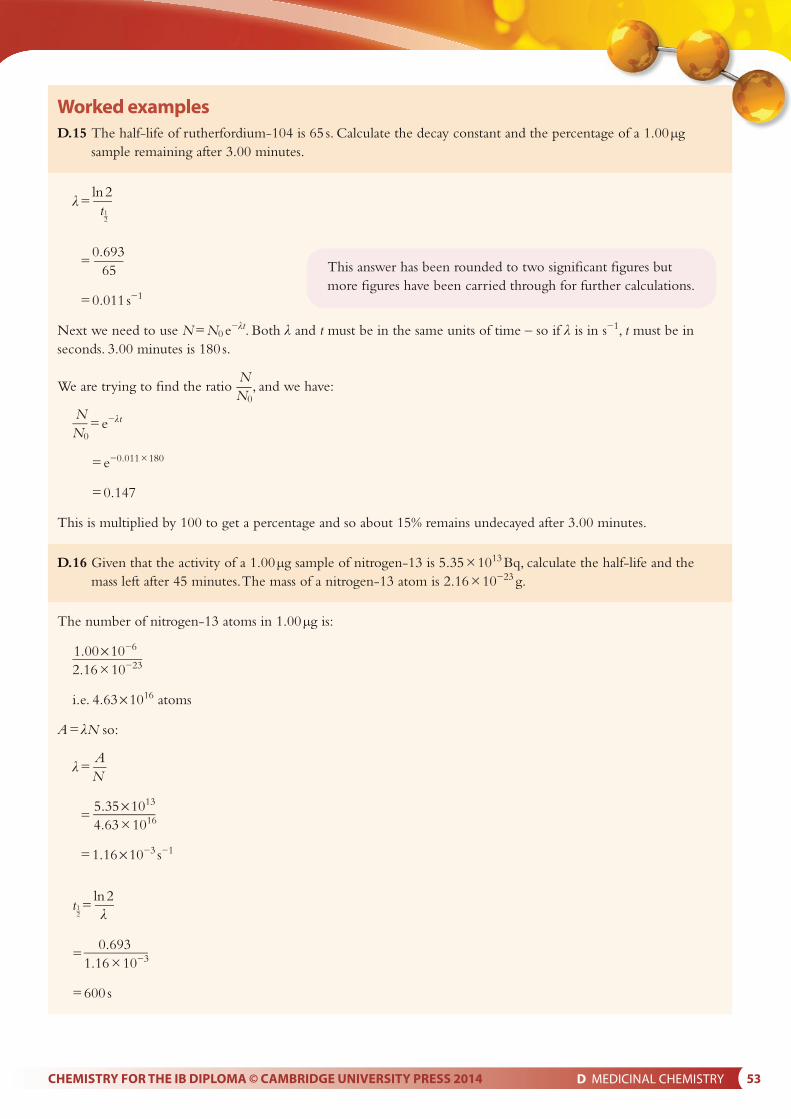
D MEDICINAL CHEMISTRY 53CHEMISTRY FOR THE IB DIPLOMA © CAMBRIDGE UNIVERSITY PRESS 2014
Worked examplesD.15 The half-life of rutherfordium-104 is 65 s. Calculate the decay constant and the percentage of a 1.00 μg
sample remaining after 3.00 minutes.
λ = ln 2 t1
2
= 0.693
65
= 0.011 s−1
Next we need to use N = N0 e−λt. Both λ and t must be in the same units of time – so if λ is in s−1, t must be in seconds. 3.00 minutes is 180 s.
NWe are trying to � nd the ratio
N0, and we have:
NN0
= e−λt
= e−0.011 × 180
= 0.147
This is multiplied by 100 to get a percentage and so about 15% remains undecayed after 3.00 minutes.
D.16 Given that the activity of a 1.00 μg sample of nitrogen-13 is 5.35 × 10¹³ Bq, calculate the half-life and the mass left after 45 minutes. The mass of a nitrogen-13 atom is 2.16 × 10⁻²³ g.
The number of nitrogen-13 atoms in 1.00 μg is:
1.00 × 10⁻⁶
2.16 × 10⁻²³
i.e. 4.63 × 10¹⁶ atoms
A = λN so:
Aλ = N
=
5.35 × 10¹³
4.63 × 1016
= 1.16 × 10⁻³ s−1
t12 =
ln 2 λ
0.693=
1.16 × 10−3
= 600 s
This answer has been rounded to two signi� cant � gures but more � gures have been carried through for further calculations.

54 CHEMISTRY FOR THE IB DIPLOMA © CAMBRIDGE UNIVERSITY PRESS 2014D MEDICINAL CHEMISTRY
To calculate the mass left after 45 minutes we use:
NN = N0 e−λt in the form
N0 = e−λt
The decay constant is in units of s−1 so we must convert the time to seconds:
t = 45 × 60 = 2700 s
NN0
= e−λt
= e−1.16 × 10−3 × 2700
= 0.0442
This is the proportion of the sample undecayed after 45 minutes, so the mass of nitrogen-13 left at the end is 0.0442 times the original mass:
mass = 0.0442 × 1.00
= 0.0442 μg, or 4.42 × 10⁻⁸ g
D.17 1.00 kg of a particular rock was believed to contain 0.120 g of potassium-40 when it was originally formed. Analysis of the rock has determined that it now contains 0.0500 g of potassium-40. Calculate the age of the rock given that the half-life of potassium-40 is 1.25 × 10⁹ years.
NWe will need to use
N0 = e−λt and so must � rst work out the decay constant:
λ = ln 2 t1
2
0.693 =
1.25 × 109
= 5.54 × 10⁻¹⁰ y−1
We can use mass instead of number of atoms in the following equation because any conversion factor will simply cancel out in the ratio.
NN0
= e−λt
0.0500 = e−5.54 × 10⁻¹⁰t
0.120
So e−5.54 × 10⁻¹⁰t = 0.4167
Taking the natural log of both sides:
−5.54 × 10⁻¹⁰t = ln 0.4167
–5.54 × 10⁻¹⁰t = −0.875
It is important to be consistent with units in these questions. The decay constant in the above example could also have been written as 0.0693 min−1. This is just 60 times the decay constant given.
Using the decay constant in this form means that time
can be used in minutes. Substituting in N
we get: N0
= e−λt
NN0
= e−0.0693 × 45, which gives the same answer as above.
Exam tipNN0
= e−λt can be used more conveniently in the form
⎛ N ⎞ln ⎝N0⎠
= –λt, which makes the mathematical manipulation
simpler in this type of problem.

D MEDICINAL CHEMISTRY 55CHEMISTRY FOR THE IB DIPLOMA © CAMBRIDGE UNIVERSITY PRESS 2014
0.875So t =
5.54 × 10−10
= 1.58 × 10⁹ y
The decay constant was expressed in y−1 so the time will come out in years.
10 Calculate the decay constant for each of the following:
a the half-life of nobelium-259 is 58 minutes b the half-life of rubidium-83 is 86.2 days c the half-life of neodymium-144 is
2.1 × 10¹⁵ years
11 Assuming that you start in each case with 5.00 μg of the isotopes in question 10, calculate how long it will take for the amount of the isotope to drop to 1.00 μg.
12 Calculate what percentage of the stated isotope is left after the stated time.
a carbon-14 (half-life 5730 years) after 10 000 years
b iodine-131 (half-life 8.04 days) after 3 weeks
13 Given that the activity of a 2.00 μg sample of osmium-191 is 3.27 × 10⁹ Bq, calculate its half-life and the mass left after 6 weeks. The mass of an osmium-191 atom is 3.19 × 10⁻²² g.
? Test yourself
Radioactivity in medicine
Alpha, beta, gamma, proton, neutron and positron emissions are used in nuclear medicine.
Radioactivity is used in the diagnosis and treatment of disease. In diagnostic applications, radioactive atoms are incorporated in pharmaceutical molecules or biochemical molecules (such as hormones) and injected into the body. These molecules travel round the body and their progress and interaction with cells and organs can be monitored using a detector that picks up the radiation emitted. Radioisotopes commonly used in imaging are gamma and/or positron (β+) emitters, such as technetium-99m (γ) and � uorine-18 (β+).
Radiotherapy (radiation therapy) refers to the treatment of a disease, usually cancer, using radiation. Radioisotopes for radiotherapy commonly emit α particles, β particles and γ rays. Proton-beam therapy, using protons from a particle accelerator, has also been used to treat some cancers. A related technique is neutron therapy, where a beam of protons from a particle accelerator strikes a beryllium target to produce a beam of neutrons which can be e� ective against tumours. Positron emitters are commonly used in the diagnosis of cancer but there has been some interest in using them in therapy as well.
Exam tipAs a � nal check we can consider if this answer seems reasonable. The half-life of potassium-40 is 1.25 × 10⁹ years, therefore 0.120 g should decay to 0.0600 g in this time. We are trying to � nd the time that it takes to decay to 0.0500 g and therefore would expect it to be slightly longer than one half-life, which our answer is.

56 CHEMISTRY FOR THE IB DIPLOMA © CAMBRIDGE UNIVERSITY PRESS 2014D MEDICINAL CHEMISTRY
Radiotherapy
α, β and γ rays are called ionising radiation because they cause the formation of ions (by ejection of electrons) when they interact with matter. Ionising radiation can damage cells and the main e� ect comes from damage caused to DNA.
Ionising radiation can either interact directly with the DNA, causing ionisation and a change of structure, or there can be indirect e� ects due to the formation of free radicals from other species such as water. The most common substance in our body is water and when ionising radiation interacts with water molecules they can become ionised. The ion and electron generated can go on to react further to produce free radicals such as the hydroxyl radical (HO•). Of the free radicals generated in these processes, the hydroxyl radical is probably the most dangerous – when it interacts with DNA it can trigger a series of reactions which results in damage to the DNA. Free radicals can also cause damage to proteins (enzymes) and lipids in cells.
Most cells in the body divide and replicate themselves – for example, to replace dead cells – and this happens in a controlled manner. Cancer involves cells that have been changed in some way(s) so that they replicate continuously – this happens in an uncontrolled manner. Because radiation mainly a� ects DNA, it has the greatest e� ect on cells that are replicating (using DNA), that is cancer cells. Healthy cells, which are not replicating, are more able to repair any damaged DNA and recover from the e� ect of the radiation. However, there are some cells in the body that do replicate more frequently and these include cells in hair follicles, red blood cells and tissue that is growing (as in children). These cells are also a� ected more by radiation and this explains why the side e� ects of radiotherapy can include hair loss and anemia.
The aim of radiotherapy is to kill cancer cells using radiation – but cause the minimum amount of damage to surrounding tissue and other cells in the body.
Radiotherapy can involve an external or internal source of radiation.
External radiotherapy involves targeting radiation from a machine that generates a beam of radiation onto a speci� c area of the body (Figure D.35). Di� erent machines produce beams of γ rays, (e.g. from cobalt-60), protons, electrons or X-rays.
There are two forms of treatment involving internal sources of radiation:
• brachytherapy – this involves putting a solid source of radioactivity into or near the tumour within the body. This is used to treat several types of cancer including prostate cancer and cancers of the head, neck, womb or cervix. Radioisotopes used in brachytherapy include palladium-103 (γ-emitter) and cobalt-60 (γ-emitter). Implants may be temporary (inserted and then removed later) or permanent.
• radioisotope therapy – using a liquid that is injected intravenously or taken orally. For instance, a patient with thyroid cancer may be treated
Figure D.35 A person undergoing radiotherapy using an external source of radiation.

D MEDICINAL CHEMISTRY 57CHEMISTRY FOR THE IB DIPLOMA © CAMBRIDGE UNIVERSITY PRESS 2014
with iodine-131 (β-emitter) by being given a capsule or solution containing radioactive iodine-131 (as sodium iodide) to take orally. The iodine is taken up by cancerous cells in the thyroid gland and the radiation kills them (and also healthy cells).
Side eff ects of radiotherapyRadiotherapy does not just a� ect cancer cells but also damages healthy cells in the area of treatment.
Common side e� ects of radiotherapy include:
• hair loss – this can occur where the beam enters the body and where it leaves the body; it is usually a temporary e� ect
• nausea – this is most likely when the treatment area is near the stomach; it is a temporary e� ect
• fatigue – tiredness can be caused by anemia due to red blood cells being destroyed during the treatment; a temporary e� ect
• sterility – this can occur if the treatment area includes the ovaries and testes; a permanent e� ect.
Radioisotopes used in nuclear medicine
Technetium-99m is the most common radioisotope used in medicine.
Technetium-99m is used widely as a radioactive tracer in medical imaging to diagnose illnesses. A radioactive tracer is introduced into the body and taken up by cells. It emits γ-radiation (or positrons, which produce γ-rays) that passes out through the body, and is detected using a γ-camera, which is designed to pick up such radiation.
There are several reasons why technetium-99m is especially suitable for this use:
• It is very close to being a pure emitter of γ-rays – γ-rays can pass through the body but α and β particles are not su� ciently penetrating. 99mTc decays by a process called ‘isomeric transition’ – the ‘m’ in ‘99m’ means that its nuclei are in an metastable state – a reasonably long-lived excited nuclear state – and it can decay to 99Tc by giving out this excess energy in the form of γ-ray photons. The energy of the γ-rays produced is similar to that of X-rays used in X-ray machines – the γ-rays are penetrating enough to pass out through the body, but not so penetrating that they are excessively dangerous.
• It has a half-life of six hours – this is long enough to allow it to travel round the body, but short enough that the patient does not remain radioactive for long.
• Technetium has a reasonably extensive chemistry and so its radioactive atoms can be incorporated into compounds that are soluble and can be transported round the body. Radioactive 99mTc can be made into the technetate(VII) ion – (TcO4
−), which is water-soluble but not as strong an oxidising agent as the manganate(VII) ion (Mn is in the same group as Tc).Internal radiotherapy procedures usually use β-emitters rather than
γ-emitters – β particles interact with matter more e� ectively than γ-rays and lose all their energy within a few millimetres in the body, causing a great deal of damage to cells in a localised area but little damage to cells

58 CHEMISTRY FOR THE IB DIPLOMA © CAMBRIDGE UNIVERSITY PRESS 2014D MEDICINAL CHEMISTRY
outside that area. γ-rays are able to pass out of the body without losing much energy and so γ-emitters are more useful for imaging purposes. Gamma rays pass through more tissue and cause damage to cells over a greater area but less damage to individual cells.
Yttrium-90 and lutetium-177 are used in radiotherapy – they are both β-emitters with relatively short half-lives.
Yttrium-90 is a β-emitter with a half-life of 64 hours and is used for the treatment of liver cancer. In the treatment, tiny beads (about 30 μm in diameter) containing 90Y are injected into the artery carrying blood to the liver and act locally on the tumour there, killing cells within a very short range.
Lutetium-177 is a β-emitter and γ-emitter with a half-life of 6.71 days. It is used for targeted radiotherapy by being incorporated into molecules that can bind to receptors on certain types of cell. The radiation then destroys only a particular type of cells within a very limited area. 177Lu can be used for treatment of neuroendocrine cancers – the neuroendocrine system is the system of nerves and glands that has the role of producing hormones in the body. The fact that 177Lu is a γ-emitter as well as a β-emitter means that it can also be used for imaging purposes.
Targeted alpha therapyAlpha particles are relatively large and highly charged and cause a great deal of damage to cells in a very small area. They are not very penetrating and so cannot be used as an external source of radioactivity – they are not able to penetrate skin.
Targeted alpha therapy (TAT) is a relatively new form of treatment that is still very much in the research stage. It involves using some way of bringing the source of α particles speci� cally to the cancer cells. The range of α particles in the body is typically 50 –100 μm (about the size of a human cell) and because they lose all their energy in a very small space they cause a great deal of damage to cells (more than β particles).
TAT can be designed to attack, as far as possible, just cancer cells by using monoclonal antibodies – antibodies that are all the same shape. Antibodies are proteins of the immune system that bind to speci� c receptors on certain cells (or foreign particles) to target them for destruction in the body. Monoclonal antibodies can be made to target a speci� c type of cancer cell (bearing a speci� c receptor on its surface) and can be labelled with an α-emitting radioisotope. The antibodies travel through the body and attach to just this one type of cell, carrying the radioisotope with them. Decay of the radioisotopes produces α particles, which destroy the cancerous cell. Up to about 20 α particles may be required to kill one cell. The antibodies will target one particular type of cell anywhere in the body and so TAT has the potential to treat cancers that have spread throughout the body.
Various radioisotopes have been suggested for this type of radiotherapy, including astatine-211(half-life 7.2 hours) and lead-212 (half-life 10.6 hours)
A di� erent type of TAT involves the use of radioactive radium chloride (radium-223, an α- and γ-emitter with a half-life of 11.4 days) to treat certain cancers that have spread to the bones. Radium is in the same group in the periodic table as calcium, which makes up a large proportion of bones, and is treated in the same way as calcium in the body. After

D MEDICINAL CHEMISTRY 59CHEMISTRY FOR THE IB DIPLOMA © CAMBRIDGE UNIVERSITY PRESS 2014
injection, the radium travels through the bloodstream to the bones, where it is taken up by cancerous cells in the bone (these are dividing more rapidly and will need a bigger supply of calcium/radium) and destroys the cells in the immediate vicinity (i.e. the cancerous cells) but healthy cells receive only small amounts of radiation or none at all. In this way it can target areas of cancer throughout the bones in the body.
Boron-neutron-capture therapyBoron-neutron-capture therapy (BNCT) has the potential to be a promising from of radiotherapy for the treatment of head and neck cancers.
BNCT relies on the fact that when non-radioactive boron-10 atoms, which have been taken up by cancer cells, are irradiated with a neutron beam from outside the body, they can capture neutrons to produce a high-energy form of boron-11, which can undergo � ssion to produce α particles and lithium nuclei:
105B + 10n → 11
5B* → 73Li + 42He
These massive, charged particles have a very short path length in the body and get rid of their energy in a very small space causing a great deal of damage to cells.
An essential part of BNCT is to make sure that tumour cells take up su� cient amounts of boron-10 and various drugs have been developed to achieve this.
Magnetic resonance imagingMagnetic resonance imaging (MRI) involves the use of nuclear magnetic resonance (NMR) to produce three-dimensional images of the internal organs. Although the word ‘nuclear’ is in the name, the process involved in this technique is very di� erent to those described above. NMR involves no changes to the nucleus of atoms but rather involves the change in orientation of a spinning nucleus relative to an external magnetic � eld.
Figure D.36 An MRI scanner.

60 CHEMISTRY FOR THE IB DIPLOMA © CAMBRIDGE UNIVERSITY PRESS 2014D MEDICINAL CHEMISTRY
An MRI body scanner (Figure D.36) is an NMR spectrometer in which a patient can be placed. The scanning takes 15–45 minutes, and the patient is required to lie still for this length of time. MRI interacts with the protons in water molecules (and other molecules such as fat) in cells in the body. Water molecules in cells in di� erent organs are in slightly di� erent environments, so the various organs in the body can be di� erentiated.
MRI is a safe, non-invasive technique for scanning organs in the body, and when the data are analysed using a computer, it is possible to obtain a three-dimensional scan of the body (Figure D.37). The only radiation involved is that in the radiofrequency part of the electromagnetic spectrum – side e� ects are rare and very minor.
Nature of scienceIn many medical processes, such as the use of radioactivity, scientists must consider whether the bene� ts outweigh the risks of the procedure. There will be many factors involved in such an analysis and an objective approach is essential.
Learning objectives
• Understand that, after synthesis, a drug must be extracted from the reaction mixture and puri� ed
• Describe the processes of extraction and puri� cation
• Understand some factors that a� ect the solubility of organic compounds
• Understand what is meant by Raoult’s law
• Understand the use of fractional distillation in the puri� cation of an organic compound
• Understand that a combination of spectroscopic techniques can be used to identify an organic compound
• Understand that steroids can be detected in urine samples using gas chromatography–mass spectrometry
• Understand that alcohol can be detected in the breath using a breathalyser
Figure D.37 A coloured MRI scan of a whole human body.
D9 Drug detection and analysis (HL)
Extraction of drugsAfter making a drug, the next thing to do is extract it from the reaction mixture – this is often called the work-up by organic chemists and usually relies on the solubility of the product varying from one solvent to another.
The � rst step in extraction often involves adding the reaction mixture to ice-water. If the product is a solid and insoluble in water it might precipitate at this stage and can be removed from the reaction mixture by � ltration.
If the product does not precipitate out then it can be extracted from the aqueous mixture by solvent extraction. This involves shaking the aqueous mixture with a solvent that is immiscible with water in a separating funnel – common solvents include ethoxyethane and ethyl ethanoate. A separating funnel is shown in Figure D.38.
D.38 A separating funnel.
organiclayer
separatingfunnel
tap
aqueouslayer

D MEDICINAL CHEMISTRY 61CHEMISTRY FOR THE IB DIPLOMA © CAMBRIDGE UNIVERSITY PRESS 2014
OC
HC
ethanoic acid
H
H
H
OO
OC
CH
methyl methanoate
H
H
H
OC
CC
H
H
HC
ethyl ethanoate
H
H
H
H
H
O
The separating funnel is shaken and the various components of the mixture partition between the two solvents according to their solubility in each – the pharmaceutical is likely to be an organic, non-polar compound that will dissolve much better in the organic solvent. Any inorganic, polar substances will dissolve much better in the aqueous layer. The mixture is allowed to settle and two layers should form – an organic layer and an aqueous layer. The tap is opened and the layers separated.
Better separation is obtained by using several small portions of organic solvent rather than one large one. The samples of organic solution are then combined and a drying agent (such as anhydrous magnesium sulfate) added to remove any water. The drying agent is � ltered o� and the organic solvent removed, usually by using a rotary evaporator (Figure D.39) which takes the solvent o� under reduced pressure – this means that the solution does not have to be heated so much which is important if the product is thermally unstable.
SolubilityIn order to identify suitable solvents for extraction, it is necessary to understand the factors that a� ect the solubility of organic compounds. The general rule is ‘like dissolves like’, which means that molecules will tend to dissolve in solvents with similar intermolecular forces – polar molecules will tend to dissolve in polar solvents such as water; and non-polar substances will dissolve in non-polar, organic solvents and generally be insoluble in water.
The ability to form hydrogen bonds often makes substances more soluble in water. As a general rule, small, highly polar molecules that are able to participate in hydrogen bonding will be soluble in water but larger, less polar molecules will not be.
We can look at some factors that a� ect solubility by considering some examples.
The structures of ethanoic acid and methyl methanoate, which are isomers of each other, are shown in Figure D.40. Both molecules are fairly small and polar but ethanoic acid has an H attached directly to an O and is therefore able to hydrogen bond to water, making it more soluble in water.
The structure of ethyl ethanoate is shown in Figure D.41. This is also polar but has more non-polar regions (CH3 groups) than methyl methanoate and is therefore considerably less soluble in water.
Figure D.39 A rotary evaporator.
Figure D.40 The structures of ethanoic acid and methyl methanoate.
Figure D.41 The structure of ethyl ethanoate.

62 CHEMISTRY FOR THE IB DIPLOMA © CAMBRIDGE UNIVERSITY PRESS 2014D MEDICINAL CHEMISTRY
Figure D.43 The structures of zanamivir and omeprazole.
Figure D.44 The experimental set-up for fractional distillation.
CH3O
O CH3 omeprazole
zanamivir
H3C
H3C
NS
NN
HOO
ON
N
N
H
H
H
O
OHOH
HO
HO
H3CNH2
factionatingcolumn
waterin
puresample
magneticstirrer bar
pig adapter
to vacuum pump
short-path vigreux apparatus
quickfit thermometerwater
out
condenser
crude sample
HEAT
benzene phenol benzene-1,2,3-triol
OH OH
OHHO
Figure D.42 The structures of benzene, phenol and benzene-1,2,3-triol.
Generally, the presence of more OH groups or NH2/NH groups in a molecule increases water solubility – phenol is about 45 times more soluble in water than benzene is, and benzene-1,2,3-triol is about � ve times more soluble than phenol (Figure D.42). The OH and NH/NH2 groups are able to form hydrogen bonds to water.
Zanamivir (Figure D.43), with lots of polar groups, is soluble in water, but omeprazole is about 70 times less soluble and is regarded as being only slightly soluble in water.
Substances containing ions tend to be more soluble in water because of the formation of ion–dipole interactions. Phenol is therefore more soluble in sodium hydroxide solution than in water because it forms the phenoxide ion (C6H5O−). Similarly, aspirin is less soluble in acidic solutions than in alkaline solutions because of the formation of ions in alkaline solution – the conversion of aspirin to its sodium salt to increase its solubility was discussed on page 15. Oseltamivir tablets (see page 37) contain the phosphate salt, which is much more soluble in water.
Purifi cationOnce the organic compound has been extracted from the reaction mixture it must be puri� ed. There are several methods for purifying compounds but the most common are recrystallisation, distillation/fractional distillation and chromatography.
Recrystallisation of solid compounds has already been described on page 13 and here we will concentrate on fractional distillation. This can be used to separate liquids that have quite similar boiling points – whereas simple distillation is used to separate liquids with very di� erent boiling points or for separating a liquid from a non-volatile residue. The experimental set-up for fractional distillation is shown in Figure D.44. The column is made of moulded glass or is packed with small beads to give a very large surface area. The mixture of liquids is heated and the liquid with the lower boiling point is collected by condensing the vapour.
To understand how fractional distillation works you need to understand a bit more about liquid–vapour equilibria.

D MEDICINAL CHEMISTRY 63CHEMISTRY FOR THE IB DIPLOMA © CAMBRIDGE UNIVERSITY PRESS 2014
Raoult’s lawPhase equilibrium was introduced in Topic 7. The vapour above a liquid in a closed container exerts a pressure on the walls of the container. If the vapour is in equilibrium with the liquid, this pressure is called the vapour pressure (or ‘equilibrium vapour pressure’ or ‘saturated vapour pressure’) (Figure D.45).
Vapour pressure is the pressure exerted by a vapour in equilibrium with a liquid (or a solid).
For a pure liquid, the vapour pressure depends only on the nature of the liquid and the temperature. For a mixture of liquids, the vapour pressure also depends on how much of each liquid is present. Raoult’s law states that the partial vapour pressure of any volatile component of an ideal solution is equal to the vapour pressure of the pure liquid multiplied by the mole fraction of that liquid in the solution. To understand what this means we need to introduce a few terms:
• Volatile – something that evaporates readily.
• Ideal solution – a mixture of liquids in which the intermolecular forces are the same as in the pure liquids – i.e. the tendency for a liquid to evaporate is the same in the pure liquid as in the solution. An example of a fairly-close-to-ideal solution is a mixture of hexane (C6H14) and heptane (C7H16).
• The mole fraction of a component, A, in a mixture is given by:
number of moles of A
mole fraction of A =
total number of moles in the mixtureMole fraction is given the symbol X, so in a mixture containing nA moles of A and nB moles of B, the mole fraction of A is given by: nAXA =
nA + nB
Note that mole fraction is a ratio and therefore has no units.
• Partial pressure – refers to the pressure exerted by a particular gas in a mixture of gases. If the gases behave ideally, the partial pressure is the same as the pressure that the same amount of that particular gas would exert if it were in the container by itself. If the pressure exerted by a mixture of 80% nitrogen and 20% oxygen is 100 kPa, the partial pressure of nitrogen is 80 kPa and that of oxygen is 20 kPa. Partial pressure is calculated from:
partial pressure of A = mole fraction of A × total pressure
or
PA = XA × Ptot
Dalton’s law of partial pressure states that for a mixture of ideal gases the total pressure is equal to the sum of the partial pressures:
Ptot = PA + PB + PC + ….Returning to Raoult’s law, it basically says that the contribution of each
component of a mixture to the total vapour pressure depends on what the vapour pressure of the pure liquid is and how much is present in the mixture. It can be written:
PA = XA × P 0A
where PA is the partial vapour pressure of A, XA is the mole fraction of A in the mixture and P 0A is the vapour pressure of pure A.
liquid
vapour
vapourpressure
Figure D.45 A vapour in equilibrium with its liquid exerts a vapour pressure.
The sum of the mole fractions of the various components in a mixture is 1.
Note: ‘mole fraction of A’ here refers to the mole fraction in the vapour.

64 CHEMISTRY FOR THE IB DIPLOMA © CAMBRIDGE UNIVERSITY PRESS 2014D MEDICINAL CHEMISTRY
Figure D.46 The variation of vapour pressure with composition for an ideal solution.
10
Mole fraction of A
vapour pressureof pure A
par
tial v
apou
rp
ress
ure
of B
par
tial v
apou
rp
ress
ure
of A
partial vapour pressure of B
partial vapour pressure of A
total vapour pressure
Vap
our p
ress
ure
Vap
our p
ress
ure
vapourpressureof pure B
01
Mole fraction of B
Figure D.46 shows how the vapour pressure of an ideal mixture varies with the composition of the mixture at constant temperature. At any point, the total vapour pressure is the sum of the partial vapour pressures of the components.
Worked exampleD.18 Given that the vapour pressures of pure hexane and heptane at 25 °C are 20.5 kPa and 6.10 kPa respectively,
calculate the vapour pressure of an ideal solution containing 4.00 mol hexane and 4.00 mol heptane at 25 °C. Then calculate the mole fraction of each in the vapour.
Using Raoult’s Law:
PA = XA × P 0A
nAXA = nA + nB
4.00Xhexane =
4.00 + 4.00 = 0.500
4.00Xheptane =
4.00 + 4.00 = 0.500
Phexane = 0.500 × 20.5 = 10.25 kPa
Pheptane = 0.500 × 6.10 = 3.05 kPa
Using Dalton’s Law of partial pressures:
Ptot = Phexane + Pheptane
= 10.25 + 3.05
= 13.3 kPa
So the total vapour pressure of the solution is 13.3 kPa.

D MEDICINAL CHEMISTRY 65CHEMISTRY FOR THE IB DIPLOMA © CAMBRIDGE UNIVERSITY PRESS 2014
If we assume that the gases behave ideally, the pressure exerted by a gas is proportional to the number of its molecules present, therefore the partial pressure of each gas is proportional to the number of moles of that gas present in the vapour phase. So, the mole fraction of hexane in the vapour phase is given by:
PhexaneXvap hexane =
Phexane + Pheptane
= 10.25
13.3
= 0.771
3.05Xvap
hexane = 13.3
= 0.229
It can be seen from this that the vapour is richer in the more volatile component (hexane) than the original mixture was – the more volatile component has a greater tendency to enter the vapour phase. This is important for the separation of liquids using fractional distillation.
Raoult’s law can also be applied to dilute solutions containing small amounts of non-volatile solutes to calculate by how much the vapour pressure is lowered. The calculation is carried out in the same way assuming that the vapour pressure of the non-volatile solute is zero.
Worked exampleD.19 The vapour pressure of water at 80 °C is 47.375 kPa. Calculate by how much the vapour pressure is lowered
by dissolving 10.00 g of glucose (Mr 180.18) in 1.00 dm3 of water (density at 80 °C is 0.971 79 g cm−3).
Mass of water = 1000 × 0.971 79 = 971.79 g
Number of moles of water = 971.79
= 53.93 mol 18.02
10.00Number of moles of glucose =
180.18 = 0.05550 mol
Total number of moles = 53.93 + 0.055 50 = 53.98 mol
53.93Mole fraction of water =
53.98 = 0.9990
Vapour pressure of water = 0.9990 × 47.375 = 47.33 kPa
The vapour pressure of pure water was 47.375 kPa, therefore the vapour pressure decreases by 0.05 kPa when the glucose is dissolved.
This answer could also have been worked out using the fact that the sum of the mole fractions in a mixture always adds up to 1.
The glucose is non-volatile and makes no contribution to the vapour pressure above the solution.

66 CHEMISTRY FOR THE IB DIPLOMA © CAMBRIDGE UNIVERSITY PRESS 2014D MEDICINAL CHEMISTRY
14 For each of the following ideal solutions, calculate the vapour pressure of the solution and the mole fraction of each component in the vapour:
a mole fraction of A = 0.400, vapour pressure of pure A = 20.0 kPa
mole fraction of B = 0.600, vapour pressure of pure B = 16.0 kPa
b number of moles of C = 1.20, vapour pressure of pure C = 10.0 kPa
number of moles of D = 0.800, vapour pressure of pure D = 7.00 kPa
c number of moles of E = 2.50, vapour pressure of pure E = 5.00 kPa
number of moles of F = 1.60, vapour pressure of pure F = 8.00 kPa
d mole fraction of G = 0.200, vapour pressure of pure G = 12.0 kPa
vapour pressure of pure H = 14.0 kPa
15 Calculate the decrease in the vapour pressure of water when 20.00 g of sucrose (Mr = 342.34) is dissolved in 500.0 cm3 of water at 40 °C. The vapour pressure and density of water at this temperature are 7.3812 kPa and 0.992 22 g cm−3 respectively.
? Test yourself
The boiling point of a liquidBoiling occurs when bubbles of vapour form in a liquid and escape. Bubbles of vapour cannot be formed until the vapour pressure equals the external pressure.
A liquid boils when its vapour pressure equals the external pressure. So the normal boiling point of a liquid is the temperature at which the vapour pressure of the liquid is one atmosphere.
The more volatile a liquid is, the higher its vapour pressure will be at a certain temperature – and therefore the lower its boiling point will be.
Dissolving a non-volatile solute in a solvent will increase the boiling point of the solvent (boiling point ‘elevation’) because the vapour pressure is lowered and therefore the liquid must be heated more so that its vapour pressure is equal to atmospheric pressure (see the calculation above).
Because the vapour pressure of a mixture of two liquids varies with the composition, the boiling point also varies with the composition of the mixture. The richer a mixture is in the more volatile component, the lower the boiling point.
Figure D.47 is a boiling point–composition diagram for an ideal solution with two components – liquid A and liquid B. The red line shows how the boiling point changes with composition and the blue line shows the vapour with which a particular mixture is in equilibrium. A is the more volatile component (higher vapour pressure in Figure D.46) and has the lower boiling point.
Consider a mixture in which the mole fraction of A is 0.3 (and that of B is 0.7) – the green dashed line (in Figure D.47) is drawn up from this composition to the liquid line and it can be seen that the boiling point of this mixture is 81 °C. The horizontal line across to the vapour curve (black dashed line) indicates the composition of the vapour with which this liquid is in equilibrium. It can be seen that the mole fraction of A in the vapour is 0.6, compared to 0.3 in the liquid – the vapour is richer in the more volatile component.

D MEDICINAL CHEMISTRY 67CHEMISTRY FOR THE IB DIPLOMA © CAMBRIDGE UNIVERSITY PRESS 2014
0.1 0.2 0.3 0.4 0.5 0.6 0.7 0.8 0.9 10
Mole fraction of A
boiling point of pure A
90
85
80
75
Boili
ng p
oint
/ °C
Boili
ng p
oint
/ °C
70
65
60
90
85
80
75
70
65
60
boiling point of pure B
0.9 0.8 0.7 0.6 0.5 0.4 0.3 0.2 0.1 01
Mole fraction of B
vapourliquid
81
Fractional distillationThe above discussion can be used to explain how fractional distillation separates a mixture of liquids.
If a mixture containing 0.1 mol A and 0.9 mol B is heated in the � ask, it will boil at 87 °C (Figure D.48). The mole fractions in the vapour will be XA = 0.3 and XB = 0.7 – so the vapour is richer in the more volatile component. The fractionating column is hot at the bottom and cooler at the top so, as the vapour rises up the column to the cooler parts, it condenses on the beads in the column to form a liquid with composition XA = 0.3 and XB = 0.7.
This liquid will then trickle down the column, where it is heated by hotter vapour coming up from below and will boil again, but this time at a lower temperature (81 °C) because it is richer in the more volatile
Figure D.47 A boiling point–composition diagram for an ideal solution.
0.1 0.2 0.3 0.4 0.5 0.6 0.7 0.8 0.9 10
Mole fraction of A
boiling point of pure A
90
85
80
75
Boili
ng p
oint
/ °C
Boili
ng p
oint
/ °C 70
65
60
90
85
80
75
70
65
60
boiling point of pure B
87
0.9 0.8 0.7 0.6 0.5 0.4 0.3 0.2 0.1 01
Mole fraction of B
vapourliquid
81
74
Figure D.48 Fractional distillation allows separation of a mixture of liquids.

68 CHEMISTRY FOR THE IB DIPLOMA © CAMBRIDGE UNIVERSITY PRESS 2014D MEDICINAL CHEMISTRY
component. This liquid will boil to form a vapour with composition XA = 0.6 and XB = 0.4. This vapour is richer in the more volatile component and rises higher up the column before it condenses to form a liquid of the same composition.
The liquid trickles down the column where it is heated again and boils at 74 °C to form a vapour of composition XA = 0.84 and XB = 0.16. Each time the liquid boils and condenses it becomes richer in the more volatile component and rises higher up the column until, if the column is long enough, essentially pure A will be obtained from the top of the column.
The closer the boiling points of the liquids, the more series of boiling and condensing processes are required and therefore the longer the column required for good separation of the liquids.
Not all liquids can be completely separated by fractional distillation and, for instance, ethanol and water form a constant-boiling-point mixture (95.6% ethanol).
Spectroscopic identifi cation of organic compoundsOnce an organic compound has been synthesised, puri� ed and extracted it can be identi� ed using spectroscopic techniques.
The structure of many organic molecules can be determined using a combination of infrared spectroscopy, mass spectrometry and proton NMR.
The structures of pharmaceutical compounds are usually very complicated and their spectra are extremely di� cult to interpret – here we look at some simpler examples.
Secondary amidesA secondary carboxamide (amide) group (RNH) is present in the structure of many drugs – oseltamivir and paclitaxel for example. The structure of a simple secondary amide is shown in Figure D.49.
The � rst step in identifying a compound is usually to run an infrared spectrum – this is a very simple and quick technique. The infrared spectrum of the secondary amide is shown in Figure D.50. The absorptions shaded red can be ignored – they are due to a di� erent vibrational motion in the molecule (N–H bend). We look at the region above 1500 cm−1 to assign the bands in the spectrum:
04000 3000 2000
fingerprint regionC-H
N-H C=O
CH
H
HO
N
1500Wavenumber / cm–1
% T
rans
mitt
ance
1000 500
100
H
CC
H
H
C H
H
H
Figure D.50 The infrared spectrum of N-methylpropanamide.
NC
CC
H
C
H
O
H
H
H
H
H
H
H
Figure D.49 The structure of a secondary amide; N-methylpropanamide.

D MEDICINAL CHEMISTRY 69CHEMISTRY FOR THE IB DIPLOMA © CAMBRIDGE UNIVERSITY PRESS 2014
due to a 13Catom in somemolecules
molecularion
loss of C2H5
Rela
tive
abun
danc
e / %
0
20
40
60
80
100
20 30 40 50
57582915
CH3+
C2H5+
8760 70 80
m / z
90
C
O
C
H
H
C
H
H
H+
C
H
H
N
H
H C
O
+
Figure D.51 The mass spectrum of N-methylpropanamide.
• the band at about 1650 cm−1 is due to C=O – this is a bit lower than we normally expect for this bond but is fairly typical for amides
• the band at 3000 cm−1 is found in all organic spectra – it is due to the C–H stretch
• the band at 3300 cm−1 is due to the N–H bond.Some key peaks are highlighted in the mass spectrum of the secondary
amide in Figure D.51. The molecular ion peak occurs at 87 and this gives us the relative molecular mass of the molecule. The peak at 88 is due to the presence of a carbon-13 atom in some of the molecules. Loss of an ethyl group (mass 29) from the molecular ion results in the formation of the CH3NHCO+ ion (m/z = 58), whereas loss of the CH3NH group forms CH3CH2CO+, at m/z = 57. Some other fragments that are formed are shown in Figure D.51.
The high-resolution NMR spectrum of N-methylpropanamide is shown in Figure D.52. There are four di� erent chemical environments for protons. Coupling between the protons in the ethyl group gives rise to a triplet (two H atoms on adjacent C) and a quartet (three H atoms on adjacent C). There is coupling between the H on the nitrogen and the Hs of the methyl group, resulting in the signal for the methyl Hs being split into a doublet (one H atom on adjacent N). However, the splitting of the signal due to the H on the nitrogen into a quartet (three H atoms on adjacent C) is not seen because of broadening due to other e� ects.
CH N
O
C
H
HC
H
H
C H
H
H
H
Chemical shift (δ) / ppm7 6 5 4 3 2 1
Figure D.52 The NMR spectrum of N-methylpropanamide.

70 CHEMISTRY FOR THE IB DIPLOMA © CAMBRIDGE UNIVERSITY PRESS 2014D MEDICINAL CHEMISTRY
CH3
O C
O
Phenyl ethanoateEster groups and benzene rings are components of the structure of many drugs – for instance, the structure shown in Figure D.53 is present in aspirin and diamorphine.
The infrared spectrum of phenyl ethanoate is shown in Figure D.54. The absorption at around 1750 cm−1 is due to the C=O bond, but an ester also contains a C– O single bond and the band for this is in the � ngerprint region (1050 –1410 cm−1). The peaks at 1600 cm−1 and just below 1500 cm−1 are due to the vibrations of C–C in the benzene ring – these bands are very characteristic of benzene rings.
The mass spectrum of phenyl ethanoate is shown in Figure D.55. Mass spectra can be di� cult to interpret because of rearrangement reactions. For instance, in the mass spectrum of phenyl ethanoate, the large peak at m/z = 94 is due to a rearrangement to produce C6H5OH+. A characteristic peak in the mass spectrum of monosubstituted aromatic compounds is the C6H5
+ peak at m/z = 77. Loss of CH3CO from the molecular ion results in the peak at 93.
04000 3000 2000
fingerprint region
C-H
C-OC=O
1500Wavenumber / cm–1
% T
rans
mitt
ance
1000 500
100
Figure D.54 The infrared spectrum of phenyl ethanoate.
Figure D.53 The structure of phenyl ethanoate.
due to a 13Catom in somemolecules
molecularion
loss of CH3CO
Rela
tive
abun
danc
e
0
20
40
60
80
100
25 75509377
C6H5+
136100
m / z
Figure D.55 The mass spectrum of phenyl ethanoate.

D MEDICINAL CHEMISTRY 71CHEMISTRY FOR THE IB DIPLOMA © CAMBRIDGE UNIVERSITY PRESS 2014
The NMR spectra of compounds containing benzene rings usually contain peaks around 7 ppm due to the hydrogen atoms attached to the benzene ring. These peaks may be resolved into separate signals or could just appear as one complex signal. An example of the NMR signal from the protons attached to a benzene ring is shown in Figure D.56.
Steroid detection in sportSteroids are mostly non-polar molecules having a common structural feature known as the steroid backbone (Figure D.57). This is made up of three six-membered rings (labelled A, B and C) and a � ve-membered ring (D) fused together. Steroids vary depending on the type and position of substituents on this steroid backbone. There is also usually a carbon–carbon double bond in either ring A or ring B. Oestrogens are di� erent to the other steroids in that they have an aromatic A ring.
Testosterone (Figure D.58) is an androgen (a male sex hormone). Androgens are also called anabolic steroids because they promote tissue growth, of muscle in particular. They are taken by sportsmen and sportswomen to enhance performance because they increase muscle mass and are also believed to improve endurance. The three most common anabolic steroids that are abused are testosterone and the synthetic derivatives nandrolone (Figure D.58) and stanozolol. They have been used in disciplines such as athletics, weightlifting and cycling.
The ethical implications of taking anabolic steroids are clear – it gives users an unfair advantage over their competitors and is simply cheating. It is a major concern to sporting bodies worldwide and drug screening in major sporting events is routinely employed to detect abuse.
CH3 CH
3CH2
CH3
CH
CH
HO
CH2
H3C
CH2
H3C
cholesterolA B
C D
testosterone
H H
CH3 H
CH3OH
OH
O
nandrolone
H H
HH
CH3
O
Figure D.57 Structure of the steroid backbone.
Figure D.58 The structures of testosterone and nandrolone.
Abusing anabolic steroids can have a major impact on the body – their use can cause a number of side e� ects such as breast growth in men, acne, infertility, mood swings and aggressiveness. They can also cause high blood pressure, liver disease (including cancer), heart attacks or strokes. Psychologically, abusers of anabolic steroids can become addicted to them, developing an increased desire to keep taking them, even if unwanted side e� ects occur.
Chemical shift ( ) / ppm7
Figure D.56 Part of a typical NMR spectrum of an aromatic compound showing the signals due to the H atoms attached to the benzene ring.
Steroids and their metabolites (substances they are broken down into in the body) can be detected using the combined techniques of gas chromatography–mass spectrometry. The organic chemicals are extracted from a urine sample and then separated into their various components using gas chromatography (gas–liquid chromatography). Each band, as it comes out of the chromatography column, is passed directly into a mass spectrometer where it is analysed. The mass spectrum of a compound is generally characteristic of that compound and so, by comparison with the mass spectra in a database, the compound can be identi� ed.

72 CHEMISTRY FOR THE IB DIPLOMA © CAMBRIDGE UNIVERSITY PRESS 2014D MEDICINAL CHEMISTRY
Gas chromatography Gas chromatography (GC) is used for the separation of mixtures, such as drugs in urine. Figure D.59 is a schematic diagram of a gas chromatograph. The sample is injected into a heated chamber, where it is vaporised.
• An inert gas (usually nitrogen or helium), called the carrier gas, carries the sample through the column.
• The column contains a non-volatile liquid (the stationary phase) spread onto the surface of � nely divided solid particles. As the mixture is carried through the column, it separates into its components. Separation is by partition – so how quickly each component travels depends on its solubility in the liquid stationary phase. Those with higher solubility in the liquid stationary phase are slowed down in the column relative to those that are less soluble in the stationary phase. The retention time, the time between when the mixture is injected and the time it is detected, also depends (among other things) on how volatile the substance is – the most volatile compound generally has the greatest tendency to enter the gaseous phase and therefore the shortest retention time.
• Each component is fed into a mass spectrometer as it emerges from the column.
carrier gas
inject sample
oven
colu
mn
detector
ampli�er computer
chromatogram
Figure D.59 A gas chromatograph.
Detection of alcohol in the breath
The presence of ethanol/alcohol in the breath can be detected using redox reactions – either using an oxidising agent in a chemical reaction or in a fuel cell.
A breathalyser test is a common test carried out at the roadside and involves the driver breathing into a device that detects the amount of alcohol in the breath. In the lungs, an equilibrium is established between alcohol dissolved in blood plasma and alcohol in the breath. Therefore, the amount of alcohol in the breath can be used to determine the amount of alcohol in the blood plasma.

D MEDICINAL CHEMISTRY 73CHEMISTRY FOR THE IB DIPLOMA © CAMBRIDGE UNIVERSITY PRESS 2014
There are three main ways that alcohol can be measured in the breath.The � rst uses a chemical test in a breathalyser, which either contains
dichromate(VI) (Cr2O72−) ions in crystals or solution. Dichromate(VI)
ions are orange and any ethanol present in the motorist’s breath will cause a change in colour to green as they are reduced to chromium(III) ions – the ethanol is � rst oxidised to ethanal and then to ethanoic acid in the process. The degree of colour change is directly related to the level of alcohol in the breath. In the simple, disposable devices using potassium dichromate(VI) crystals, the amount of alcohol in the breath is determined by how many crystals change colour. In devices using potassium dichromate(VI) solution the amount of colour change can be measured using a colorimeter/photocell.
The equations for the chemical reactions involved are:
C2H5OH(g) + H2O(l) → CH3COOH(l) + 4H+(aq) + 4e− oxidation
Cr2O72−(aq) + 14H+(aq) + 6e− → 2Cr3+(aq) + 7H2O(l) reduction
2Cr2O72−(aq) + 3C2H5OH(l) + 16H+(aq) → 4Cr3+(aq) + 3CH3COOH(l) + 11H2O(l) overall
orange green
The oxidation of ethanol to ethanoic acid may also be shown as:
C2H5OH + 2[O] → CH3COOH + H2O
where [O] represents oxygen from the oxidising agent.These chemical-based breathalysers have now largely been replaced by
a newer, more accurate type of hand-held analyser that uses a fuel cell to detect and measure alcohol in the breath. In this type of device, the breath enters a fuel cell � tted with two platinum electrodes and any alcohol in the breath gets oxidised. Instead of the energy being released as heat, it is converted directly to electrical energy and an electric current is generated – the more alcohol present, the higher the current. The reactions that occur in the fuel cell are:
C2H5OH(g) + H2O(l) → CH3COOH(l) + 4H+(aq) + 4e− oxidation
O2(g) + 4H+(aq) + 4e− → 2H2O(l) reduction
C2H5OH(g) + O2(g) → CH3COOH(l) + H2O(l) overall
The third method for detecting alcohol in breath involves infrared spectroscopy.
Nature of scienceTaking drugs to enhance performance in sport is as old as sport itself. What has changed, however, is the ability of scientists to detect drugs. Advances in technology and better understanding of metabolic pathways have allowed the development of ever-more-sophisticated testing protocols for drugs in sport and their metabolites. But science is always improving and so blood/urine samples from the Olympics are kept for eight years so that future advances might be able to detect previously undetectable substances.
occurs at the anode
occurs at the cathode

74 CHEMISTRY FOR THE IB DIPLOMA © CAMBRIDGE UNIVERSITY PRESS 2014D MEDICINAL CHEMISTRY
Exam-style questions
1 Drugs and medicines can have a number of physiological e� ects on the body.
a Explain the meaning of the following terms: i therapeutic e� ect [1] ii side e� ect [1] iii tolerance [1] iv bioavailability. [1]
b Explain the term ‘therapeutic window’. [1]
c Drugs can be administered to a patient via a number of di� erent routes. i There are three main ways of giving a drug by injection. Which of these gives the fastest response,
and why? [2] ii Which route would be chosen to treat a lung condition, such as asthma, locally? [1]
2 Analgesics and antibiotics are very important classes of drugs.
a Describe how a mild analgesic, such as aspirin, causes an analgesic e� ect. [1]
b i The structure of aspirin is shown in Figure D.5 and the IB Chemistry data booklet. Write an equation, showing structural formulas, for the synthesis of aspirin from salicylic acid. [2]
ii In an experiment to synthesise aspirin, 5.00 g of salicylic acid (Mr 138.13) was reacted with excess of the other reactant in the presence of a concentrated phosphoric acid catalyst. 5.02 g of a white solid was obtained at the end of the reaction. Calculate the yield of aspirin. [3]
iii Explain why it is possible to have a yield of aspirin greater than the initial mass of salicylic acid. [1] iv State the name of the process by which aspirin can be puri� ed and explain how it is carried
out in the laboratory. [4]
c Brie� y outline the mechanism by which penicillins carry out their antibacterial activity. [2]
d Some bacteria have developed resistance to penicillins by producing an enzyme that deactivates the penicillin.
i Name the enzyme produced by penicillin-resistant bacteria. [1] ii Explain how this enzyme deactivates the penicillin. [1] iii Which part of the penicillin structure can be modi� ed to make the penicillin less sensitive to
the actions of this enzyme? [1]
3 Analgesics are used to reduce pain.
a Morphine and diamorphine are both strong analgesics. Describe how they carry out their analgesic e� ect. [1]
b The structures of morphine and diamorphine can be found in the IB Chemistry data booklet (and in Figure D.16).
i Describe how the structure of diamorphine di� ers from morphine, with respect to the functional groups present. [1]
ii State the type of reaction used to convert morphine to diamorphine. [1] iii Write an equation for the reaction in part ii using the structure below to represent the structure
of morphine. [2]
O
O H
O H
iv Describe two possible social problems that can occur through heroin addiction. [2]

D MEDICINAL CHEMISTRY 75CHEMISTRY FOR THE IB DIPLOMA © CAMBRIDGE UNIVERSITY PRESS 2014
4 a Name the acid found in the gastric juice in the stomach. [1]
b Calcium carbonate is an antacid used to neutralise excess acid in the stomach. Write the equation for the reaction between calcium carbonate and this acid. [1]
c Explain the mode of action of ranitidine (Zantac®). [3]
d Explain, using an example, what is meant by an ‘active metabolite’. [2]
e Bu� ers are important in some drug formulations. A bu� er solution was made by dissolving a mixture containing 15.00 g of KH2PO4 and 15.00 g of K2HPO4 in water and making up to a total volume of 1.00 dm3. Given that the pKa for H2PO4
− is 7.21, calculate the pH of the bu� er. [3]
5 a How do viruses and bacteria di� er in the way that they replicate? [2]
b Describe two di� erent ways in which antiviral drugs work. [2]
c Oseltamivir (structure shown below) is an important drug used to treat in� uenza. Identify the functional groups that are highlighted in the structure. [3]
H3C
I
IIIII
H2N
H3C
CH3
N
H
O
H3C
O
O
O
d Explain why AIDS is so di� cult to eradicate on a global scale. [2]
6 a Explain how antibiotics could enter the environment and a problem caused by this. [4]
b Radioisotopes are used extensively in medicine. i Distinguish between high-level nuclear waste and low-level nuclear waste. [2] ii State two sources of low-level nuclear waste in medicine. [2] iii Explain how low-level nuclear waste from medicine may be disposed of. [2]
7 Taxol® is an important drug with several chiral centres.
a State which disease Taxol is used to treat. [1]
b State the natural source of Taxol. [1]
c The semi-synthesis of Taxol uses a chiral auxiliary. Describe the use of chiral auxiliaries in asymmetric synthesis. [3]
d Describe how enantiomers can be identi� ed by polarimetry. [3]
HL

76 CHEMISTRY FOR THE IB DIPLOMA © CAMBRIDGE UNIVERSITY PRESS 2014D MEDICINAL CHEMISTRY
8 a Lutetium-177 is a radioisotope that is used in medicine. Lutetium-177 undergoes β decay. i Write a nuclear equation for the decay of lutetium-177. [2] ii A patient is given a 10.0 mg dose of lutetium-177. Given that the half-life of lutetium-177 is 6.71 days,
calculate how much of the lutetium is present in the patient’s body after four weeks. [4]
b State three side e� ects of radiotherapy. [3]
c Targeted alpha therapy is a relatively new form of radiotherapy. Explain how the technique works. [3]
9 After a drug has been synthesised it must be extracted from the reaction mixture and then puri� ed.
a In a procedure, a student poured the reaction mixture into a beaker containing 50 cm3 of ice-cold water and then carried out solvent extraction using three separate 50 cm3 portions of ethanol. Explain what is wrong with this procedure and suggest an improvement. [3]
b i Assuming that benzene and methylbenzene form an ideal solution, calculate the vapour pressure of a mixture containing 1.00 g of benzene and 10.00 g of methylbenzene at 20 °C. The vapour pressures of benzene and methylbenzene at 20 °C are 10.00 kPa and 2.93 kPa respectively. [4]
ii Explain how the composition of the vapour in part i compares to that of the original liquid. [2] iii Explain the principles of a method for extracting benzene from the benzene–methylbenzene mixture
in part i. [4]
c The structure of nandrolone, an anabolic steroid, is shown below:
HL
HL
testosterone
H H
CH3 H
CH3OH
OH
O
nandrolone
H H
HH
CH3
O
i Identify three functional groups present in nandrolone. [3] ii The infrared spectrum of nandrolone is shown below. Identify the absorption bands in the region
above 1500 cm–1 in this spectrum. [4]
iii Explain how nandrolone could be identi� ed in a sample of urine from an athlete. [2]
04000 3000 2000 1500
Wavenumber / cm–1
% T
rans
mitt
ance
1000 500
100