optimal spherical focus in the peripheral retina
TRANSCRIPT
Optimal spherical focus in the peripheralretina
W. N. Charman1 and D. A. Atchison2
1Faculty of Life Sciences, University of Manchester, Moffat Building, Manchester, UK, and 2School of
Optometry and Institute of Health and Biomedical Innovation, Queensland University of Technology,
Kelvin Grove, Brisbane, QLD, Australia
Abstract
It is well known that, in most eyes, astigmatism increases with the field angle. A simple reduced-eye
model is used to demonstrate that, for point imagery in the peripheral retina, the combination of
oblique astigmatism with elliptical entrance and exit pupils means that the retinal image with the
optimal rotational symmetry is not necessarily at a focus corresponding to a �best-sphere� correction.
Equations are derived for the position of focus at which a circularly symmetric blur patch is obtained
and for the dimensions of the patch in this and other image planes. Ray tracing through a wide-angle
schematic eye is used to explore the validity of the simple model. It is shown that although the latter
gives good predictions of retinal imagery for very small entrance pupils, it becomes less valid for
larger, more realistic pupil diameters, due to the increasing importance of the effects of higher-order
aberrations. Nevertheless, the simple model can still yield useful insights into through-focus,
peripheral optical imagery.
Keywords: astigmatism, model eyes, peripheral refraction, pupil, retinal image
Introduction
In recent years, interest has grown in the possibility thatthe state of focus in the peripheral retina might play somerole in myopia development (e.g. Hoogerheide et al.,1971; Stone and Flitcroft, 2004; Wallman and Winawer,2004; Charman, 2005). Numerous studies have shownthat astigmatism increases with the field angle (see reviewsby Jennings and Charman, 1997; Atchison and Smith,2000, pp. 147–150; Gustafsson et al., 2001). Althoughthere is considerable inter-subject variation, the dioptricform of the image shells varies with the ametropia (e.g.Millodot, 1981; Seidemann et al., 2002; Atchison et al.,2006) and may also change with accommodation (Smithet al., 1988; Walker and Mutti, 2002) and age (Millodot,1984; Atchison et al., 2005; Charman and Jennings,2006). While most eyes lack rotational symmetry and
may display some astigmatism on the visual axis, obliqueastigmatism tends to dominate in the peripheral field.Other higher-order monochromatic aberrations (Navar-ro et al., 1998; Guirao and Artal, 1999; Atchison andScott, 2002; Atchison, 2004) and chromatic aberration(Thibos, 1987; Thibos et al., 1990) further degradeperipheral image quality.
When conventional refractive techniques reveal thepresence of astigmatism on the visual axis, it is conven-tional to assume, on the basis of simple geometricaloptics, that the optimal purely spherical correction (the�best� or �mean� sphere) will be that which brings the�circle of least confusion� onto the retina, i.e. S + C/2,where S and C are the spherical and cylindrical dioptriccomponents of the refraction. Image quality will then bethe same for all orientations of object detail. Correctingto bring one of the line foci onto the retina improvesimage quality for edges or gratings oriented in the samedirection as the focal line but only at the expense ofmuch reduced image quality for other orientations.
The same �best-sphere� concept can be applied toastigmatic, off-axis imagery. It is found that, on average,the resultant focus lies quite close to the retina (within0.75 D) for field angles up to 60� (Atchison and Smith,2000, p. 150). The question arises, however, as to
Received: 17 October 2007
Revised form: 4 January 2008, 16 January 2008
Accepted: 18 January 2008
Correspondence and reprint requests to: W.N.Charman.
Tel.: +44 (0)161 306 8736; Fax: +44 (0)161 200 3887.
E-mail address: [email protected]
Ophthal. Physiol. Opt. 2008 28: 269–276
ª 2008 The Authors. Journal compilation ª 2008 The College of Optometrists doi: 10.1111/j.1475-1313.2008.00552.x
whether the �best� (in the sense of being the smallest andmost symmetrical) retinal image of a point as a functionof spherical focus would still be expected to be foundwith a refraction corresponding to the local �best-sphere�, when off-axis, rather than axial, imagery isbeing considered. This uncertainty arises because, as thefield angle increases, the entrance and exit pupils of theeye change from circular shapes to ellipses with theirmajor axes perpendicular to the meridian in which thefield angle is measured. Whereas with a circular pupilthe best-sphere correction yields a retinal �circle of leastconfusion�, with an elliptical pupil it gives an �ellipse ofleast confusion� having the same eccentricity as that ofthe pupil, so that there is a loss of rotational symmetryin the point image. Rather than using the �best-sphere�for an elliptical pupil, might it be better to choose thespherical correction such that the image of a point is arotationally symmetrical blur patch, even though thediameter of this circular patch is larger than the minoraxis of the �ellipse of least confusion�?
In this paper, an attempt will be made to evaluate theapproximate magnitude of the differences in focusinvolved, using both a simple geometrical optics modeland rigorous ray tracing with a model eye havingaspheric surfaces (Navarro et al., 1985; Escudero-Sanzand Navarro, 1999).
Simple geometric model
Position of focus for a symmetrical blur patch
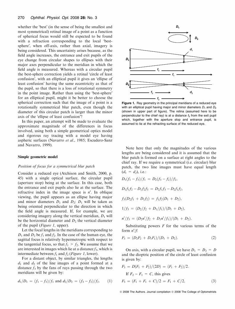
Consider a reduced eye (Atchison and Smith, 2000, p.45) with a single optical surface, the circular pupil(aperture stop) being at the surface. In this case, boththe entrance and exit pupils also lie at the surface. Therefractive index in the image space is n¢. In obliqueviewing, the pupil appears as an ellipse having majorand minor diameters D1 and D2: D1 will be taken asbeing oriented perpendicular to the direction in whichthe field angle is measured. If, for example, we areconsidering imagery along the vertical meridian, D1 willbe the horizontal diameter and D2 the vertical diameterof the pupil (Figure 1, upper).
Let the focal lengths in the meridians corresponding toD1 and D2 be f1 and f2. In the case of the human eye, thesagittal focus is relatively hypermetropic with respect tothe tangential focus, so that f1 > f2. We assume that weare interested in images which lie at a distance f3, which isintermediate between f1 and f2 (Figure 1, lower).
For a distant object, by similar triangles, the lengthsd1 and d2 of the line images of a point formed at adistance f3 by the fans of rays passing through the twomeridians will be given by:
d1=D1 ¼ ðf1 � f3Þ=f1 and d2=D2 ¼ ðf3 � f2Þ=f2: ð1Þ
Note here that only the magnitudes of the variouslengths are being considered and it is assumed that theblur patch is formed on a surface at right angles to thechief ray. If we require a symmetrical (i.e. circular) blurpatch, the two line images must have equal length(d1 = d2), i.e.:
D1ðf1 � f3Þ=f1 ¼ D2ðf3 � f2Þ=f2;
D1f1f2 � D1f2f3 ¼ D2f1f3 � D2f1f2;
f3ðD2f1 þ D1f2Þ ¼ f1f2ðD1 þ D2Þ;
1=f3 ¼ ðD2=f2 þ D1=f1Þ=ðD1 þ D2Þ;
n0=f3 ¼ ðD2n0=f2 þ D1n0=f1Þ=ðD1 þ D2Þ:
Substituting powers F for the various terms of theform n¢/f:
F3 ¼ ðD2F2 þ D1F1Þ=ðD1 þ D2Þ: ð2Þ
On axis, with a circular pupil, we have D1 = D2 = Dand the dioptric position of the circle of least confusionis given by:
F3 ¼ DðF1 þ F2Þ=ð2DÞ ¼ ðF1 þ F2Þ=2:
If F2 ) F1 = C, this gives
F3 ¼ ðF1 þ F1 þ CÞ=2 ¼ F1 þ C=2; ð3Þ
Figure 1. Ray geometry in the principal meridians of a reduced eye
with an elliptical pupil having major and minor diameters D1 and D2
(shown in upper part of figure). The retina (assumed here to be
perpendicular to the chief ray) is at a distance f3 from the exit pupil
which, together with the aperture stop and entrance pupil, is
assumed to lie at the refracting surface of the reduced eye.
270 Ophthal. Physiol. Opt. 2008 28: No. 3
ª 2008 The Authors. Journal compilation ª 2008 The College of Optometrists
i.e. the blur circle lies at the dioptric midpoint betweenthe foci corresponding to the two meridians, whichdemands the familiar best-sphere correction.To a first approximation, however, as the field angle h
is increased for the eye, the minor diameter of theelliptical pupil is only cosh times that of the majordiameter (Jay, 1962; Jennings and Charman, 1978;Atchison and Smith, 2000, p. 26), so:
D2 ¼ D1 cos h: ð4Þ
We then have, from Equation (2):
F3 ¼ ðD1F2 cos h þ D1F1Þ=½D1ð1 þ cos hÞ�¼ ½F1 þ ðF1 þ CÞ cos h�=ð1 þ cos hÞ¼ F1 þ C cos h=ð1 þ cos hÞ ð5Þ
It can be seen that the symmetrical blur circle now liesat a dioptric distance of Ccosh/(1 + cosh) from theposterior (sagittal) line focus, rather than simply C/2, or)C/(1 + cosh) from the anterior (tangential) line focus.Thus as h increases we would expect the �symmetrical�blur patch to move relatively closer to the posterior linefocus in comparison to its �best-sphere� position.In fact C will increase with the field angle. We now
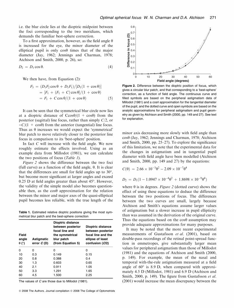
roughly estimate the effects involved. Using as anexample data from Millodot (1981), we can calculatethe two positions of focus (Table 1).Figure 2 shows the difference between the two foci
(full curve) as a function of the field angle, h. It is clearthat the differences are small for field angles up to 30�,but become more significant at larger angles and exceed0.25 D at field angles greater than about 45�. However,the validity of the simple model also becomes question-able then, as the cosh approximation for the relationbetween the minor and major axes of the quasi-ellipticalpupil becomes less reliable, with the true length of the
minor axis decreasing more slowly with field angle thancosh (Jay, 1962; Jennings and Charman, 1978; Atchisonand Smith, 2000, pp. 25–27). To explore the significanceof this limitation, we note that the experimental data forthe changes in astigmatism and in tangential pupildiameter with field angle have been modelled (Atchisonand Smith, 2000, pp. 149 and 27) by the equations:
CðhÞ ¼ 2:66� 10�3h2 � 2:09� 10�7h4
D2 ¼ D1ð1� 1:0947� 10�4h2 þ 1:8698� 10�9h4Þ
where h is in degrees. Figure 2 (dotted curve) shows theeffect of using these equations to deduce the differencebetween the two positions of focus. The differencesbetween the two curves are small, largely becauseAtchison and Smith�s equations assume larger valuesof astigmatism but a slower increase in pupil ellipticitythan was assumed in the derivation of the original curve.Thus the equations based on the cosh assumption mayprovide adequate approximations for many purposes.
It may be noted that the more recent experimentalmeasurements of Gustafsson et al. (2001), based ondouble-pass recordings of the retinal point-spread func-tion in emmetropes, give substantially larger meanvalues for peripheral astigmatism than those of Millodot(1981) and the equations of Atchison and Smith (2000,p. 149). For example, the mean of the nasal andtemporal with-the-rule astigmatism measured at a fieldangle of 60� is 8.9 D, when compared with approxi-mately 4.5 D (Millodot, 1981) and 6.9 D (Atchison andSmith, 2000, p. 149). The figure from Gustafsson et al.(2001) would increase the mean discrepancy between the
Table 1. Estimated relative dioptric positions giving the most sym-
metrical blur patch and the best-sphere correction
Field
angle
h (�)Astigmatic
error C (D)
Dioptric distance
between posterior
focal line and
the symmetrical
blur patch
(from Equation 5)
Dioptric distance
between posterior
focal line and the
ellipse of least
confusion (C/2)
0 0 0 0
10 0.3 0.149 0.15
20 0.8 0.388 0.4
30 1.3 0.603 0.65
40 2.1 0.911 1.05
50 3.3 1.291 1.65
60 4.5 1.500 2.25
The values of C are those due to Millodot (1981).
0
0.1
0.2
0.3
0.4
0.5
0.6
0.7
0.8
0 10 20 30 40 50 60
Field angle (degrees)
Dif
fere
nce
in f
ocu
s (D
)
Figure 2. Difference between the dioptric position of focus, which
gives a circular blur patch, and that corresponding to a �best-sphere�correction, as a function of field angle. The continuous curve and
filled symbols are based on the peripheral astigmatism data of
Millodot (1981) and a cosh approximation for the tangential diameter
of the pupil, and the dotted curve and open symbols are based on the
analytic approximations for peripheral astigmatism and pupil geom-
etry as given by Atchison and Smith (2000, pp. 149 and 27). See text
for explanation.
Optimal spherical focus: W. N. Charman and D.A. Atchison 271
ª 2008 The Authors. Journal compilation ª 2008 The College of Optometrists

two positions of focus at this field angle in Figure 2 from0.75 to approximately 1.5 D. In any case, the results atany field angle will scale with the individual�s obliqueastigmatism.
Dimensions of the circular blur patch and the �ellipse ofleast confusion�
As the circular patch lies a little closer to one of the linefoci than the focus corresponding to the best-spherecorrection, it may be expected that its diameter (d3) willbe intermediate between those of the major (d1) andminor (d2) diameters of the �ellipse of least confusion�.
From Equations (1) and (4):
d1 ¼ D1ðf1 � f3Þ=f1 and d2 ¼ D1 cos hðf3 � f2Þ=f2:
Using the cosh approximation for the changes intangential pupil diameter (D2 = D1cosh) and introduc-ing powers instead of image distances:
d1 ¼ D1ðF3 � F1Þ=F3 and d2 ¼ D2ðF2 � F3Þ=F3: ð6Þ
For the �ellipse of least confusion�, corresponding to a�best-sphere� correction, F3 ) F1 = F2 ) F3 = C/2, andF3 = (F1 + F2)/2. Substituting these expressions inEquation (6) gives:
d1 ¼ D1C=ðF1 þ F2Þ; d2 ¼ D1C cos h=ðF1 þ F2Þ: ð7Þ
For the rotationally symmetric blur patch case, fromEquation (5) F3 ) F1 = Ccosh/(1 + cosh). As for thissymmetrical case:
d3 ¼ d1 ¼ d2;
we can substitute in Equation (6) for (F3 ) F1) to find:
d3 ¼ d1 ¼ D1C cos h=½F3ð1 þ cos hÞ�: ð8Þ
Substituting the right-hand side of Equation (5) for F3
into Equation (8) gives:
d3 ¼ D1C cos h=½F1ð1 þ cos hÞ þ C cos h�
and since F2 = F1 + C
d3 ¼ D1C cos h=ðF1 þ F2 cos hÞ: ð9Þ
Figure 3 shows how the three diameters change as afunction of field angle, assuming that F1 is constant at60 D, the pupil diameter is 1 mm and that C changeswith field angle in the way found by Millodot (1981).Note that the optimal blur circle diameter is alwaysintermediate between the major and minor diameters ofthe ellipse of least confusion but that it does not exactlyequal the mean of the two ellipse diameters [the latterbeing, from Equation (7), D1C(1 + cosh)/[2(F1 + F2)].As would be expected from the behaviour of the focus
differences in Figure 2, there is very little differencebetween the various image diameters for field anglesh < 30�. The relative differences only start to becomesubstantial when h > 45�.
To give a sense of the approximate angular scale ofthe blur patches corresponding to the linear dimensionsof Figure 3, a blur patch of diameter 0.01 mm corre-sponds to a subtense of approximately 2.05 min arc ifthe posterior nodal distance is assumed to be 16.67 mm.If the pupil is larger than the 1 mm assumed forFigure 3, the dimensions of the blur patches will increaselinearly with the pupil diameter. Using the angulardimensions of the elliptical or circular blur patches, it isstraightforward to calculate the corresponding geomet-ric approximations to the MTF as a function oforientation and field angle (Jennings and Charman,1997).
It should again be emphasised that these results arebased on a simple reduced eye model with the pupil atthe refracting surface. It is further assumed that thereceiving retinal surface is perpendicular to the chief rayand that F1 is independent of the field angle. This is, ofcourse, not the case in the peripheral retina, where theprojection is nonlinear and the retinal angles ofincidence of the chief rays are not zero (Drasdo andFowler, 1974; Frisen and Scholdstrom, 1977). Theestimates are also based on geometrical optics, blurdue to diffraction being ignored. To give some indica-tion of the scale of the latter, the angular radius of thefirst dark ring of the Airy diffraction pattern for acircular pupil (/ = 1.22k/D radians for a diameter Dand a wavelength k), corresponds in green light toapproximately 2.3 min arc for a pupil diameter of 1 mmand 0.6 min arc for a 4 mm pupil. Finally, and perhapsmost importantly, aberrations other than astigmatismhave been ignored (see below). Nevertheless, the result isinteresting in raising the possibility that the best-sphere
0
5
10
15
20
25
30
35
40
Field angle (degrees)
Blu
r p
atch
dia
met
er(m
icro
ns)
d1 d2 d3
0 10 20 30 40 50 60
Figure 3. Dimensions of the retinal blur patches as a function of
field angle for an axial pupil diameter of 1 mm. The dotted (top) and
chain-dashed (bottom) curves give, respectively, the major and
minor diameters (d1 and d3) of the elliptical blur patch corresponding
to a �best-sphere� correction. The solid (middle) curve gives the
diameter of the circular blur patch (d3). The astigmatism data of
Millodot (1981) have been used.
272 Ophthal. Physiol. Opt. 2008 28: No. 3
ª 2008 The Authors. Journal compilation ª 2008 The College of Optometrists

correction does not give the most symmetrical blurpatch.
Ray tracing results
To test the equations, the optical design softwareprogram Zemax EE (Zemax Development Corporation,Bellevue, WA, USA) was used to trace rays through awell-regarded, wide-angle model of the eye incorporat-ing aspheric surfaces (Navarro et al., 1985; Escudero-Sanz and Navarro, 1999). The Navarro model hasrealistic peripheral refractive errors and its higher-orderaberrations also approximate well to those of real eyes(Escudero-Sanz and Navarro, 1999; Atchison andSmith, 2000, pp. 174–176, 255–256). It has four refract-ing surfaces, a homogeneous lens, and a retina with aradius of curvature of )12 mm. It comes in monochro-matic and chromatic versions, and is adaptive in thatsome parameters change with accommodation. Themonochromatic version of the model eye for theunaccommodated state was used in the present calcula-tions. The axial position of the retina was made tocorrespond to the on-axis, paraxial focus by setting thevitreous chamber to a length of 16.404 mm.An object point at a field angle of 55� in the vertical
meridian was chosen to explore the general effectsinvolved. This object field angle corresponded to 44.02�in image space. For this situation, the height of the chiefray on the retina was 11.47 mm and its angle ofincidence on the retina was 28.69�.Thin correcting lenses were placed along the axis of
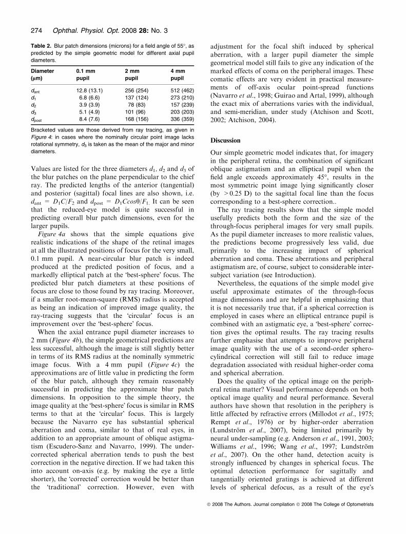
the chief ray in the corneal plane. Spot diagrams weredetermined for two possible image surfaces, using ahexapolar pattern and a ray density of 10. The imagesurfaces were either a plane perpendicular to the chiefray and coinciding with the retina on the chief ray (asused in the simple model), or the retinal surface itself.The results for pupil diameters of 0.1, 2 and 4 mm are
shown in Figure 4. For each pupil diameter, results areshown for the two image surfaces at positions of focuscorresponding to the paraxial tangential (anterior) linefocus, the best-sphere focus, the circular blur patchfocus and the sagittal (posterior) line focus, as calculatedby ray tracing for the 0.1 mm pupil. The two interme-diate foci were obtained using Equations (3) and (5).Not surprisingly in this vertical meridian, the non-zeroangle of incidence (approximately 29�) of the chief rayon the retina tends to stretch the actual retinal images inthe vertical direction compared with the images on thesurface perpendicular to the chief ray.For comparison with the ray-tracing image dimen-
sions given in Figure 4, the predictions of Equations (7)and (8) of the simple model, using a value of 60 D for F1
and the Navarro model�s value of 8.8 DC for theoblique astigmatism, yield the values shown in Table 2.
(a)
(b)
(c)
Figure 4. Spot diagrams for a Navarro model eye and an object
point at a field angle of 55� in the vertical meridian when different
correcting lenses are placed in front of eye in the plane of cornea
and centred along the off-axis chief ray. (a) 0.1 mm diameter
entrance pupil; (b) 2.0 mm diameter entrance pupil; (c) 4.0 mm
diameter entrance pupil. The top rows in each case show the
situation when the imaging plane is at the retina but perpendicular
to the chief ray. The bottom rows show the situation when the
imaging surface is the retina of the model eye. The scales at the
left indicate the sizes of the spot diagrams – notice that the scales
are different for different pupil sizes. The numbers at the bottom
indicate the powers of compensating lenses: )3.73 D places the
tangential focus at the retina, +0.67 D places the circle of least
confusion at the retina when no allowance is made for pupil
obliquity, +1.90 D places the pupil-compensated circle of least
confusion at the retina, and +5.07 D places the sagittal focus at the
retina. In each case, the sets of three numbers indicate, in order,
the horizontal extent of the spot diagram (lm), the vertical extent of
the spot diagram (lm), and the root-mean-square (RMS) radius of
the spot diagram (lm). The large spots indicate the chief ray
location.
Optimal spherical focus: W. N. Charman and D.A. Atchison 273
ª 2008 The Authors. Journal compilation ª 2008 The College of Optometrists
Values are listed for the three diameters d1, d2 and d3 ofthe blur patches on the plane perpendicular to the chiefray. The predicted lengths of the anterior (tangential)and posterior (sagittal) focal lines are also shown, i.e.dant = D1C/F2 and dpost = D1Ccosh/F1. It can be seenthat the reduced-eye model is quite successful inpredicting overall blur patch dimensions, even for thelarger pupils.
Figure 4a shows that the simple equations giverealistic indications of the shape of the retinal imagesat all the illustrated positions of focus for the very small,0.1 mm pupil. A near-circular blur patch is indeedproduced at the predicted position of focus, and amarkedly elliptical patch at the �best-sphere� focus. Thepredicted blur patch diameters at these positions offocus are close to those found by ray tracing. Moreover,if a smaller root-mean-square (RMS) radius is acceptedas being an indication of improved image quality, theray-tracing suggests that the �circular� focus is animprovement over the �best-sphere� focus.
When the axial entrance pupil diameter increases to2 mm (Figure 4b), the simple geometrical predictions areless successful, although the image is still slightly betterin terms of its RMS radius at the nominally symmetricimage focus. With a 4 mm pupil (Figure 4c) theapproximations are of little value in predicting the formof the blur patch, although they remain reasonablysuccessful in predicting the approximate blur patchdimensions. In opposition to the simple theory, theimage quality at the �best-sphere� focus is similar in RMSterms to that at the �circular� focus. This is largelybecause the Navarro eye has substantial sphericalaberration and coma, similar to that of real eyes, inaddition to an appropriate amount of oblique astigma-tism (Escudero-Sanz and Navarro, 1999). The under-corrected spherical aberration tends to push the bestcorrection in the negative direction. If we had taken thisinto account on-axis (e.g. by making the eye a littleshorter), the �corrected� correction would be better thanthe �traditional� correction. However, even with
adjustment for the focal shift induced by sphericalaberration, with a larger pupil diameter the simplegeometrical model still fails to give any indication of themarked effects of coma on the peripheral images. Thesecomatic effects are very evident in practical measure-ments of off-axis ocular point-spread functions(Navarro et al., 1998; Guirao and Artal, 1999), althoughthe exact mix of aberrations varies with the individual,and semi-meridian, under study (Atchison and Scott,2002; Atchison, 2004).
Discussion
Our simple geometric model indicates that, for imageryin the peripheral retina, the combination of significantoblique astigmatism and an elliptical pupil when thefield angle exceeds approximately 45�, results in themost symmetric point image lying significantly closer(by >0.25 D) to the sagittal focal line than the focuscorresponding to a best-sphere correction..
The ray tracing results show that the simple modelusefully predicts both the form and the size of thethrough-focus peripheral images for very small pupils.As the pupil diameter increases to more realistic values,the predictions become progressively less valid, dueprimarily to the increasing impact of sphericalaberration and coma. These aberrations and peripheralastigmatism are, of course, subject to considerable inter-subject variation (see Introduction).
Nevertheless, the equations of the simple model giveuseful approximate estimates of the through-focusimage dimensions and are helpful in emphasizing thatit is not necessarily true that, if a spherical correction isemployed in cases where an elliptical entrance pupil iscombined with an astigmatic eye, a �best-sphere� correc-tion gives the optimal results. The ray tracing resultsfurther emphasise that attempts to improve peripheralimage quality with the use of a second-order sphero-cylindrical correction will still fail to reduce imagedegradation associated with residual higher-order comaand spherical aberration.
Does the quality of the optical image on the periph-eral retina matter? Visual performance depends on bothoptical image quality and neural performance. Severalauthors have shown that resolution in the periphery islittle affected by refractive errors (Millodot et al., 1975;Rempt et al., 1976) or by higher-order aberration(Lundstrom et al., 2007), being limited primarily byneural under-sampling (e.g. Anderson et al., 1991, 2003;Williams et al., 1996; Wang et al., 1997; Lundstromet al., 2007). On the other hand, detection acuity isstrongly influenced by changes in spherical focus. Theoptimal detection performance for sagittally andtangentially oriented gratings is achieved at differentlevels of spherical defocus, as a result of the eye�s
Table 2. Blur patch dimensions (microns) for a field angle of 55�, as
predicted by the simple geometric model for different axial pupil
diameters.
Diameter
(lm)
0.1 mm
pupil
2 mm
pupil
4 mm
pupil
dant 12.8 (13.1) 256 (254) 512 (462)
d1 6.8 (6.6) 137 (124) 273 (210)
d2 3.9 (3.9) 78 (83) 157 (239)
d3 5.1 (4.9) 101 (96) 203 (203)
dpost 8.4 (7.6) 168 (156) 336 (359)
Bracketed values are those derived from ray tracing, as given in
Figure 4: in cases where the nominally circular point image lacks
rotational symmetry, d3 is taken as the mean of the major and minor
diameters.
274 Ophthal. Physiol. Opt. 2008 28: No. 3
ª 2008 The Authors. Journal compilation ª 2008 The College of Optometrists
oblique astigmatism (Wang et al., 1997). Measurementsof detection thresholds with small targets (Fankhauserand Enoch, 1962; Anderson et al., 2001), luminance/time reciprocity (Ronchi, 1971), motion detectionthresholds (Johnson and Leibowitz, 1974) and criticalfusion frequency (Jennings and Charman, 1981) are alsoall markedly affected by defocus. Thus it appears thatoptical quality is more relevant to dynamic than tostatic visual performance in the periphery. It may be,then, that any influence of the pattern of peripheralrefraction on myopia development (e.g. Hoogerheideet al., 1971; Stone and Flitcroft, 2004; Wallman andWinawer, 2004; Charman, 2005) is exercised through itseffects on dynamic performance rather than on spatialresolution.Correction of peripheral errors has obvious clinical
utility in allowing better visualisation of the fundus withophthalmoscopic devices (Wang et al., 1983) and inobtaining more reliable perimetric measurements withsmall targets (Fankhauser and Enoch,1962; Andersonet al., 2001). The present results give some indication ofthe benefits to image quality which might be achievablewith purely spherical corrections.
Acknowledgement
This work was supported by ARC Discovery grantDP0558209.
References
Anderson, S. J., Mullen, K. T. and Hess, R. F. (1991) Human
peripheral spatial resolution for achromatic and chromaticstimuli: limits imposed by optical and retinal factors.J. Physiol. (London) 442, 47–64.
Anderson, R. S., McDowell, D. R. and Ennis, F. A. (2001)Effect of localized defocus on detection thresholds fordifferent sized targets in the fovea and periphery. Acta
Ophthalmol. Scand. 79, 60–63.Anderson, R. S., Coulter, E., Zlatkova, M. B. and Demirel, S.(2003) Short-wavelength acuity: optical factors affecting
detection and resolution of blue-yellow sinusoidal gratingsin foveal and peripheral vision. Vision Res. 43, 101–107.
Atchison, D. A. (2004) Anterior corneal and internal contri-butions to peripheral aberrations in human eyes. J. Opt.
Soc. Am. A 21, 355–359.Atchison, D. A. and Scott, D. H. (2002) Monochromaticaberrations of human eyes in the horizontal visual field.
J. Opt. Soc. Am. A 19, 2180–2184.Atchison, D. A. and Smith, G. (2000) Optics of the HumanEye. Butterworth Heinemann, London.
Atchison, D. A., Pritchard, N., White, S. D. and Griffiths, A.M. (2005) Influence of age on peripheral refraction. VisionRes. 45, 715–720.
Atchison, D. A., Pritchard, N. and Schmid, K. L. (2006)
Peripheral refraction along the horizontal and vertical visualfields in myopia. Vision Res. 46, 1450–1458.
Charman, W. N. (2005) Aberrations and myopia. Ophthal.
Physiol. Opt. 25, 285–301.Charman, W. N. and Jennings, J. A. M. (2006) Longitudinalchanges in peripheral refraction with age. Ophthal. Physiol.
Opt. 26, 447–455.Drasdo, N. and Fowler, C. W. (1974) Non-linear projection ofthe retinal image in a wide-angle schematic eye. Br.
J. Ophthalmol. 58, 709–714.Escudero-Sanz, I. and Navarro, R. (1999) Off-axis aberrationsof a wide-angle schematic eye model. J. Opt. Soc. Am. A 16,
1881–1891.
Fankhauser, F. and Enoch, J. M. (1962) The effects of blurupon perimetric thresholds. Arch. Ophthalmol. 68, 240–251.
Frisen, L. and Scholdstrom, G. (1977) Relationship between
perimetric eccentricity and retinal locus in a human eye.Acta Ophthalmol. 55, 63–68.
Guirao, A. and Artal, P. (1999) Off-axis monochromatic
aberrations estimated from double-pass measurements inthe human eye. Vision Res. 39, 207–217.
Gustafsson, J., Terenius, E., Buckheister, J. and Unsbo, P.(2001) Peripheral astigmatism in emmetropic eyes. Ophthal.
Physiol. Opt. 21, 393–400.Hoogerheide, J. F., Rempt, F. and Hoogenboom, W. P. H.(1971) Acquired myopia in young pilots. Ophthalmologica
163, 209–215.Jay, B. S. (1962) The effective pupillary area at varyingpupillometric angles. Vision Res. 1, 418–428.
Jennings, J. A. M. and Charman, W. N. (1978) Optical imagequality in the peripheral retina. Am. J. Optom. Physiol. Opt.55, 582–590.
Jennings, J. A. M. and Charman, W. N. (1981) The effects ofcentral and peripheral refraction on critical fusion fre-quency. Ophthal. Physiol. Opt. 1, 91–96.
Jennings, J. A. M. and Charman, W. N. (1997) Analytic
approximation of the off-axis modulation transfer functionof the eye. Vision Res. 37, 697–704.
Johnson, C. A. and Leibowitz, H. W. (1974) Practice,
refractive error and feedback as factors influencingperipheral motion thresholds. Percept. Psychophys. 15,
276–280.
Lundstrom, L., Manzanera, S., Pietro, P. M., Ayala, D. B.,Gorceix, N., Gustafsson, J., Unsbo, P. and Artal, P. (2007)Effect of optical correction and remaining aberrations onperipheral resolution acuity in the human eye. Opt. Express,
15, 12654–12661.Millodot, M. (1981) Effect of ametropia on peripheralrefraction. Am. J. Physiol. Opt. 58, 691–695.
Millodot, M. (1984) Peripheral refraction in aphakic eyes. Am.J. Optom. Physiol. Opt. 61, 586–589.
Millodot, M., Johnson, C. A., Lamont, A. and Leibowitz, H.
W. (1975) Effect of dioptrics on peripheral visual acuity.Vision Res. 15, 1357–1362.
Navarro, R., Santamarıa, J. and Bescos, J. (1985) Accommo-
dation-dependent model of the human eye with aspherics.J. Opt. Soc. Am. A 2, 1273–1281.
Navarro, R., Moreno, E. and Dorronsoro, C. (1998) Mono-chromatic aberrations and point-spread functions of the
human eye across the visual field. J. Opt. Soc. Am. A 15,
2522–2529.
Optimal spherical focus: W. N. Charman and D.A. Atchison 275
ª 2008 The Authors. Journal compilation ª 2008 The College of Optometrists
Rempt, F., Hoogerheide, J. and Hoogenboom, W. P. H. (1976)
Influence of correction of peripheral refractive errors onperipheral static vision. Ophthalmologica 173, 128–135.
Ronchi, L. (1971) Absolute threshold before and after correc-
tion of oblique-ray astigmatism. J. Opt. Soc. Am. 61, 1705–1709.
Seidemann, A., Schaeffel, F., Guirao, A., Lopez-Gil, N. and
Artal, P. (2002) Peripheral refractive errors in myopic,emmetropic, and hyperopic young subjects. J. Opt. Soc. Am.A 19, 2363–2373.
Smith, G., Millodot, M. and McBrien, N. (1988) The effect of
accommodation on oblique astigmatism and field curvatureof the human eye. Clin. Exp. Optom. 71, 119–125.
Stone, R. A. and Flitcroft, D. I. (2004) Ocular shape and
myopia. Ann. Acad. Med. Singap. 33, 7–15.Thibos, L. N. (1987) Calculation of the influence of lateralchromatic aberration on image quality across the visual
field. J. Opt. Soc. Am. A 4, 1673–1680.
Thibos, L. N., Bradley, A., Still, D. L., Zhang, X. and
Howarth, P. A. (1990) Theory and measurement of ocularchromatic aberration. Vision Res. 30, 33–49.
Walker, T. W. and Mutti, D. O. (2002) The effect of
accommodation on ocular shape. Optom. Vis. Sci. 79,
424–430.Wallman, J. and Winawer, J. (2004) Homeostasis of eye
growth and the question of myopia. Neuron 43, 447–468.Wang, G. J., Pomarentzeff, O. and Pankratov, M. M. (1983)
Astigmatism of oblique incidence in the human model eye.Vision Res. 23, 1079–1085.
Wang, Z.-Y., Thibos, L. N. and Bradley, A. (1997) Effectsof refractive error on detection acuity and resolutionacuity in peripheral vision. Invest Opthalmol. Vis. Sci. 38,
2134–2143.Williams, D. R., Artal, P., Navarro, R., McMahon, M. J. and
Brainard, D. H. (1996) Off-axis optical quality and retinal
sampling in the human eye. Vision Res. 36, 1103–1114.
276 Ophthal. Physiol. Opt. 2008 28: No. 3
ª 2008 The Authors. Journal compilation ª 2008 The College of Optometrists