optical properties of gold nanoparticle assemblies on a
TRANSCRIPT

NANO EXPRESS Open Access
Optical Properties of Gold NanoparticleAssemblies on a Glass SurfaceM. O. Stetsenko1*, S. P. Rudenko1, L. S. Maksimenko1, B. K. Serdega1, O. Pluchery2 and S. V. Snegir2,3
Abstract
The assemblies of cross-linked gold nanoparticles (AuNP) attract lot of scientific attention due to feasibleperspectives of their use for development of scaled contact electrodes. Here, we developed and testedmethod of solid-state formation of dimers created from small AuNP (~18 nm) cross-linked with 1.9-nonadithiol (NDT) molecules. The morphology of created coating of a glass surface and its optical-polarizationproperties have been studied in detail by combination of scanning electron microscopy, atomic forcemicroscopy, UV-visible spectroscopy, and modulation-polarization spectroscopy.The modification of localized surface plasmon resonance (LSPR) of single AuNP and their assemblies werestudied by measuring of the spectral characteristics of polarization difference at all stages of synthesis. Theradiative and nonradiative modes of LSPR have been analyzed in detail at different angles of incidence light.This allowed establishing relation between surface morphology of the coating and its optical properties.
Keywords: Gold nanoparticles, Dimers, Localized surface plasmon resonance, Coupling, Modulation-polarizationspectroscopy
BackgroundThin films of cross-linked gold nanoparticle (AuNP)have received considerable scientific attention during thepast two decades. Two- and three-dimensional arrays ofmetallic nanoparticles allowed to create downscaled con-tact electrodes [1–5]; touch sensors [6, 7]; and optically[8–10], chemically [11], and mechanically controlled[12] resistors. The architecture of nanoparticle assem-blies can be controlled by employing of AuNP withvarious size, shape [1, 13], and by modification of the in-terparticle spacing. This distance can be changed byselecting of appropriate cross-linking agent, for instancealkyldithiol- [2–4] or thiol-terminated molecules withspecific functional properties [9, 10]. To achieve self-organization of AuNP, the DNA chains [1, 5] can beemployed also.A variation of the interparticle spacing allows to tune
optical properties of entire material by controlling ampli-tude and position of a localized surface plasmon reson-ance (LSPR) of the cross-linked nanoparticles at nanoscale
[7–9, 14]. Therefore, a lot of attention is paid for thisissue. An understanding of relation between morphologyand optical properties of AuNP assemblies can provide afundamental basis for further development of the scalednanoelectronic devices based on gold nanoparticles cross-linked with organic molecules with various functionalproperties.However, developing and engineering of the corre-
sponding nanostructures with reproducible architec-ture and preassigned optical properties is a subject ofcontinuous efforts [15, 16]. A lot of attentions is paidto create AuNP assemblies with controllable architec-ture especially AuNP dimers, since they can be con-sidered as the model of two gold nanoelectrodeslinked by molecules with required functionality. Dif-ferent pathways of dimer synthesis in liquid mediawere employed. Some of them proposes to controlrelative concentration of ethanol in water colloidal so-lution of AuNP [15, 17], while others to use variouspassivation agents [16, 18], multivalent thiol ligands[19, 20], and dithiol molecules [16, 21]. However, thefurther application of these AuNP assemblies in a mo-lecular electronics becomes infeasible since it requireintegration into electronic circuits. Thus, two main
* Correspondence: [email protected] Institute of Semiconductor Physics, National Academy ofSciences of Ukraine, 45, Av. Nauky, Kyiv 03028, UkraineFull list of author information is available at the end of the article
© The Author(s). 2017 Open Access This article is distributed under the terms of the Creative Commons Attribution 4.0International License (http://creativecommons.org/licenses/by/4.0/), which permits unrestricted use, distribution, andreproduction in any medium, provided you give appropriate credit to the original author(s) and the source, provide a link tothe Creative Commons license, and indicate if changes were made.
Stetsenko et al. Nanoscale Research Letters (2017) 12:348 DOI 10.1186/s11671-017-2107-8

challenges exist. First is to develop suitable method ofsolid-state synthesis which would occur directly on asolid surface of a conductive electrode. The secondone is to achieve required optical properties of anelectrode covered by AuNP assemblies. Therefore, theAuNP surface density, orientation toward incidentlight, and interparticle distance should be controlledduring synthesis. In this relation, the LSPR which ismonitored by UV-visible optical spectroscopy is expectedto scale with the amount of AuNP and interparticle dis-tance. When AuNP dimers are deposited on a glass slide,the transmission spectrum exhibits two distinct extinctionbands: one at a wavelength of the LSPR band of singleAuNP used in the assembly and the other at a greatly red-shifted wavelength due to the plasmon coupling along theinterparticle axis for AuNP dimers [22–24]. The firstexperimental observations of dependence of the plasmonoscillation modes from interparticle spacing of a dimerand its orientation [25, 26] toward the incident lightpolarization are found in a good agreement with the pro-posed theory [27].Here, we report about effective and facile method of
formation of AuNP dimers cross-linked with 1.9-nona-dithiol (NDT) which occurs directly on a solid surface.The glass surface was chosen to model a surface of anindium tin oxide (ITO) with electrical conductivity prop-erties. Moreover, we have used commercially availableglass slides to assemble AuNP which were synthesizedusing Turkevich method. With this, we brought to lightsome experimental asperities of solid-state synthesis ofdimers and difficulties of their identification. With scan-ning electron microscopy (SEM), atomic force micros-copy (AFM), UV-visible spectroscopy, and modulation-polarization spectroscopy (MPS), we provided a practical
guide about how to control functionalization of a glass sur-face with AuNP to create their assemblies and to study theoptical properties of created material. The MPS is an effect-ive optical technique for diagnostics and characterization ofthe LSPR modification at nanoscale within the films ofnoble metals and metal-dielectric nanocomposites [28–30].We will show that the plasmonic effects are stronglydependent on surface morphology, i.e., dispersion of singlenanoparticles on the surface, their aggregation, and di-mers formation. The comparison of LSPR parametersand optical-polarization properties for correspondingnanostructures will be demonstrated in the features ofthe spectral characteristics of the polarization differ-ence, ρ(λ) and the angle of isotropic reflection θρ=0(λ),which are measured by MPS technique.
MethodsSample PreparationSynthesis of AuNP occurred following the Turkevichmethod [31–33]. An aqueous solution of HAuCl4 (2.5 ×10⁻⁴ mol.L−1) was heated to the boiling point in anErlenmeyer flask. Then 1 ml of an aqueous sodium citratesolution (1.7 × 10⁻⁴ mol.L−1) was added with vigorousmagnetic stirring. The obtained colloidal water solution ofgold nanoparticles was stored at 4 °C in a refrigerator toavoid nanoparticle aggregation. The average size of AuNPwas ~18 nm (Fig. 1a) as evidenced by transmission elec-tron microscopy (TEM). AuNP have a round shape withdistinguishable facets on their surface which is monitoredby high-resolution transmission electron microscopy (h-TEM) (Fig. 1a, insert). Optical spectra of colloidal solution(Fig. 1b) exhibit the LSPR at a wavelength of 520 nm(Fig. 1b, red curve) which is responsible for ruby color ofwater colloidal solution of AuNP.
Fig. 1 TEM image of dried colloidal solution of AuNP (a).The inset on a is the image of single AnNPs obtained by h-TEM with triangular-like dark/bright regions revealing the presence of facets on the surface AuNP. The optical spectra (b) of AuNP colloidal solution (red solid curve) and ofAuNP chemically attached to a glass surface (black dashed curve) by means of (3-aminopropyl)-triethoxysilane (APTES) monolayer
Stetsenko et al. Nanoscale Research Letters (2017) 12:348 Page 2 of 10
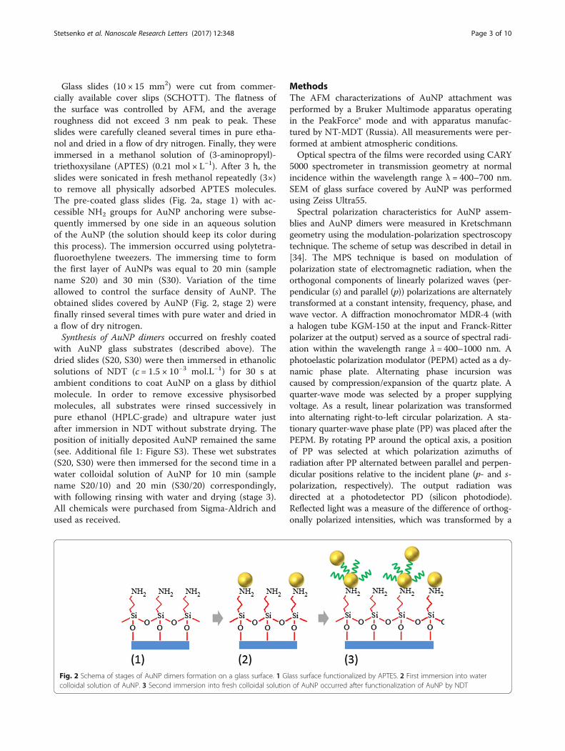
Glass slides (10 × 15 mm2) were cut from commer-cially available cover slips (SCHOTT). The flatness ofthe surface was controlled by AFM, and the averageroughness did not exceed 3 nm peak to peak. Theseslides were carefully cleaned several times in pure etha-nol and dried in a flow of dry nitrogen. Finally, they wereimmersed in a methanol solution of (3-aminopropyl)-triethoxysilane (APTES) (0.21 mol × L−1). After 3 h, theslides were sonicated in fresh methanol repeatedly (3×)to remove all physically adsorbed APTES molecules.The pre-coated glass slides (Fig. 2a, stage 1) with ac-cessible NH2 groups for AuNP anchoring were subse-quently immersed by one side in an aqueous solutionof the AuNP (the solution should keep its color duringthis process). The immersion occurred using polytetra-fluoroethylene tweezers. The immersing time to formthe first layer of AuNPs was equal to 20 min (samplename S20) and 30 min (S30). Variation of the timeallowed to control the surface density of AuNP. Theobtained slides covered by AuNP (Fig. 2, stage 2) werefinally rinsed several times with pure water and dried ina flow of dry nitrogen.Synthesis of AuNP dimers occurred on freshly coated
with AuNP glass substrates (described above). Thedried slides (S20, S30) were then immersed in ethanolicsolutions of NDT (c = 1.5 × 10⁻³ mol.L−1) for 30 s atambient conditions to coat AuNP on a glass by dithiolmolecule. In order to remove excessive physisorbedmolecules, all substrates were rinsed successively inpure ethanol (HPLC-grade) and ultrapure water justafter immersion in NDT without substrate drying. Theposition of initially deposited AuNP remained the same(see. Additional file 1: Figure S3). These wet substrates(S20, S30) were then immersed for the second time in awater colloidal solution of AuNP for 10 min (samplename S20/10) and 20 min (S30/20) correspondingly,with following rinsing with water and drying (stage 3).All chemicals were purchased from Sigma-Aldrich andused as received.
MethodsThe AFM characterizations of AuNP attachment wasperformed by a Bruker Multimode apparatus operatingin the PeakForce® mode and with apparatus manufac-tured by NT-MDT (Russia). All measurements were per-formed at ambient atmospheric conditions.Optical spectra of the films were recorded using CARY
5000 spectrometer in transmission geometry at normalincidence within the wavelength range λ = 400–700 nm.SEM of glass surface covered by AuNP was performedusing Zeiss Ultra55.Spectral polarization characteristics for AuNP assem-
blies and AuNP dimers were measured in Kretschmanngeometry using the modulation-polarization spectroscopytechnique. The scheme of setup was described in detail in[34]. The MPS technique is based on modulation ofpolarization state of electromagnetic radiation, when theorthogonal components of linearly polarized waves (per-pendicular (s) and parallel (p)) polarizations are alternatelytransformed at a constant intensity, frequency, phase, andwave vector. A diffraction monochromator MDR-4 (witha halogen tube KGM-150 at the input and Franck-Ritterpolarizer at the output) served as a source of spectral radi-ation within the wavelength range λ = 400–1000 nm. Aphotoelastic polarization modulator (PEPM) acted as a dy-namic phase plate. Alternating phase incursion wascaused by compression/expansion of the quartz plate. Aquarter-wave mode was selected by a proper supplyingvoltage. As a result, linear polarization was transformedinto alternating right-to-left circular polarization. A sta-tionary quarter-wave phase plate (PP) was placed after thePEPM. By rotating PP around the optical axis, a positionof PP was selected at which polarization azimuths ofradiation after PP alternated between parallel and perpen-dicular positions relative to the incident plane (p- and s-polarization, respectively). The output radiation wasdirected at a photodetector PD (silicon photodiode).Reflected light was a measure of the difference of orthog-onally polarized intensities, which was transformed by a
Fig. 2 Schema of stages of AuNP dimers formation on a glass surface. 1 Glass surface functionalized by APTES. 2 First immersion into watercolloidal solution of AuNP. 3 Second immersion into fresh colloidal solution of AuNP occurred after functionalization of AuNP by NDT
Stetsenko et al. Nanoscale Research Letters (2017) 12:348 Page 3 of 10

PD into alternating signal. This signal was registered by aselective amplifier equipped with a phase-lock detector(lock-in-voltmeter) tuned to the modulation frequency off = 50 kHz. The registered signal is the polarization differ-ence ρ(λ,θ) = rs
2–rp2, which is a magnitude of difference be-
tween the intensities of the internal reflection coefficientsof s- and p-polarized light (rs
2 and rp2, respectively). The
parameter ρ is a Q component of the Stokes vector [35].The refractive index of the quartz half-cylinder n = 1.456determines the value of the critical angle of total internalreflection (TIR) as θcr = 43.6°.When the reflection coefficients of s- and p-polarized
radiation have equal amplitude values, i.e. rs2(θ) = rp
2(θ),and the magnitude of polarization difference ρ(θ) equalszero, the light reflection occurs regardless of polarizationstate at the angle of isotropic reflection θρ=0 [36]. Thecondition of the isotropic reflection of electromagneticradiation can be occured in the following cases: the firstis a normal transmission/reflection of non-polarizedradiation; the second is an attenuated internal reflection,when according to the Fresnel equations, the internalreflection coefficients of s- and p-polarized radiation arenot equal at angles smaller than the critical angle (rs
2 <rp2). The last case was realized in the present work. Eachof these coefficients does not necessarily need to bezero. The equality of their magnitude is important.Both ρ and θρ=0 parameters of MPS technique are mutu-
ally supportive and exhibit the features of spectral depend-encies that caused by the optical-polarization properties ofnanostructures with AuNP and characterize their resonantproperties and features of surface morphology [29].
ResultsMorphology and Optics StudyThe AFM and SEM of S20 and S30 revealed that AuNPare randomly dispersed on a glass surface with some in-clusion of aggregated nanoparticles (Additional file 1:Figure S1, S2). The coverage of the surface by single
AuNP was calculated from AFM images and is equal toabout 20/μm2 and 60/μm2 AuNP for sample S20 andS30, respectively. An analysis of the height profile (insetof Additional file 1: Figure S1b) of single AuNP confirmsthat they have an average diameter of ~18 nm. The colorof the glass covered by AuNP becomes light pink(Fig. 1b, inset) due to LSPR of AuNP with λmax =521 nm. The color of the slides did not change with thetime as well as position of AuNP during AFM measure-ments revealing stability of AuNP coating.Among single AuNP separated by a distance larger
than one diameter (~18 nm), a few nanoparticles exhibita coupling of their LSPR. This appears in the spectrumas shoulder in the long wavelength region at 550–650 nm (Fig. 1b). These nanoparticles are arranged inthe objects without any distinct shape in which AuNPare separated from each other by a distance smaller thanone diameter of AuNP (Additional file 1: Figures S1, S2).Therefore, such assemblies are responsible for the for-mation of broad light absorption band which is shiftedto the longer wavelength region compared to LSPR ofsingle AuNP. Noteworthy, these aggregates appear firstafter glass immersion into colloidal solution of AuNPgiving stronger light absorption band after 20 min ofimmersion than LSPR of single AuNP (Fig. 3a). We sus-pect that at initial stage of AuNP assembling on a glass,they are attracted more intensively by some locale de-fects of the surface. Such aggregates were systematicallypresent on the surface in spite of multiple cleaning ofthe glass slides after APTES adsorption. The nature ofthis effect requires additional attention and studies.However, with increasing of immersion time from 20 to30 min, the ratio of single AuNP to their aggregates isprogressively rising up. This leads to increasing of theLSPR band of single AuNP (Fig. 3a).After functionalization of the AuNP surface with NDT
molecules (Fig. 2), the position of LSPR adsorption bandis changed due to modification of dielectric constant of
Fig. 3 Optical spectra for S20 (bottom curve), S30 (top curve) with characteristic LSPR band of single AuNP (λmax = 521 nm) (a) and for S30covered by NDT with following second immersion into AuNP solution to form dimers (S30/S20) (b)
Stetsenko et al. Nanoscale Research Letters (2017) 12:348 Page 4 of 10

AuNP surrounding media [37, 38]. The position of LSPRband of single AuNP consequently shifts to ~10 nm tolonger wavelengths (Fig. 3b). Meantime, the position ofλmax for AuNP aggregates did not change.The optical spectra of the glass surface with AuNP di-
mers (S30/20) revealed strong increase of overall intensity(Fig. 3b). The position of λmax of LSPR of single AuNP isundergoing minor changes also. The small shift to longerwavelength is attributed to appearance of longitude opticalexcitation mode in AuNP dimers. Similar results were ob-served for AuNP colloidal solution with ~20% of AuNPsdimers linked by various dithiol molecules. However, sim-ultaneously with the formation of dimers, the number ofnew single AuNP attached to a glass as well as AuNPlinked to existed aggregates is growing also.Thus, we performed AFM and SEM characterization
of the sample S30/S20. The AFM measurements (Fig. 4a)allowed observing AuNP dimers, longitudinal axis ofwhich is tilted to a plane of glass surface. Thus, theexpected height of these dimers was in the range of19.6–37.6 nm if considered that the length of NDT mol-ecule and diameter of AuNP are equal to ~1.6 and18 nm, correspondently. The quantity of such dimers, asseen from Fig. 4a, is very low. The closed-packed dimersand the ones with parallel axis to the surface were diffi-cult to recognize due to limited resolution of AFM. Thiswe overcame by the use of SEM (Fig. 4b). The red circlespoint to the positions of AuNP dimers. The measuredspacing between the nanoparticles of the dimers is equalto ~20 nm. An analysis of SEM and AFM images re-vealed that the overall percentage of synthesized dimersis equal to about 11.6% (Table 1).
MPS StudyOptical-polarization properties and particularly plas-monic effects have been studied for the single AuNP in
comparison with AuNP dimers using MPS technique bymeasuring the spectral characteristics of polarization dif-ference ρ(λ) in the different angular regions relatively tothe angle of total internal reflection θcr = 43.6° (Fig. 5).We have analyzed radiative (Fig. 5a) and non-radiative(Fig. 5b) modes of LSPR and paid attention to the di-mers contribution in spectral characteristics ρ(λ).In Fig. 5a, the spectral characteristic of ρ(λ) are shown
in radiative region at the incident angle of θ = 35° < θcr.All curves of ρ(λ) have difference in amplitude and ex-hibit their minima with a peak position at a wavelengthof 520, 544, 542, and 535 nm for sample S30, S20, S20/10, and S30/20, respectively. This difference we analyzedfurther with respect to the stage of glass surface functio-nalization (Fig. 2) and the time of immersion of glassslides in AuNP colloidal solution.The shortest time of the first immersion of a glass into
AuNP colloidal solution was equal to 20 min (Fig. 2)and led mainly to formation of the aggregates withoutany distinct internal packing structure. Consequently, weobserved the LSPR at 544 nm (Fig. 5a, curve S20). Withthe longer immersion time (S30), the number of singleAuNP grew up, as it clearly indicated by UV-visiblespectra on Fig. 3a. Thus, MPS revealed shift of minimafrom 544 nm (S20) to 520 nm (S30) (Fig. 5a) since theFrohlich frequency for small AuNP gave stronger impacton formation of the band [39, 40]. Changing of pack-aging density of AuNP simultaneously with formation ofAuNP dimers possessing a transverse plasmon couplingmode can lead to this spectral shift [22, 23]. Amongshifting, the overall amplitude value of ρ(λ) is increasing.Thus, the sample S30 demonstrates the highest reflec-tion properties of single AuNP (Fig. 5a).The correspondent growing of the amplitude values
for samples S20/10 and S30/20 was also observed (Fig. 2),when second immersion occurred after covering of
Fig. 4 AFM (a) and SEM (b) images of the sample S30/20. The inset on (a) is the cross section along A-B highlighting orientation of AuNP dimersaccording to measured height
Stetsenko et al. Nanoscale Research Letters (2017) 12:348 Page 5 of 10
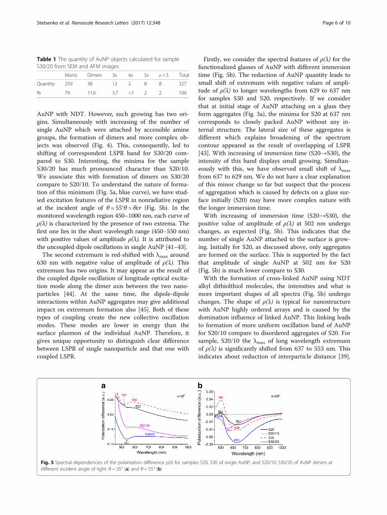
AuNP with NDT. However, such growing has two ori-gins. Simultaneously with increasing of the number ofsingle AuNP which were attached by accessible aminegroups, the formation of dimers and more complex ob-jects was observed (Fig. 4). This, consequently, led toshifting of correspondent LSPR band for S30/20 com-pared to S30. Interesting, the minima for the sampleS30/20 has much pronounced character than S20/10.We associate this with formation of dimers on S30/20compare to S20/10. To understand the nature of forma-tion of this minimum (Fig. 5a, blue curve), we have stud-ied excitation features of the LSPR in nonradiative regionat the incident angle of θ = 55°θ > θcr (Fig. 5b). In themonitored wavelength region 450–1000 nm, each curve ofρ(λ) is characterized by the presence of two extrema. Thefirst one lies in the short wavelength range (450–550 nm)with positive values of amplitude ρ(λ). It is attributed tothe uncoupled dipole oscillations in single AuNP [41–43].The second extremum is red-shifted with λmax around
630 nm with negative value of amplitude of ρ(λ). Thisextremum has two origins. It may appear as the result ofthe coupled dipole oscillation of longitude optical excita-tion mode along the dimer axis between the two nano-particles [44]. At the same time, the dipole-dipoleinteractions within AuNP aggregates may give additionalimpact on extremum formation also [45]. Both of thesetypes of coupling create the new collective oscillationmodes. These modes are lower in energy than thesurface plasmon of the individual AuNP. Therefore, itgives unique opportunity to distinguish clear differencebetween LSPR of single nanoparticle and that one withcoupled LSPR.
Firstly, we consider the spectral features of ρ(λ) for thefunctionalized glasses of AuNP with different immersiontime (Fig. 5b). The reduction of AuNP quantity leads tosmall shift of extremum with negative values of ampli-tude of ρ(λ) to longer wavelengths from 629 to 637 nmfor samples S30 and S20, respectively. If we considerthat at initial stage of AuNP attaching on a glass theyform aggregates (Fig. 3a), the minima for S20 at 637 nmcorresponds to closely packed AuNP without any in-ternal structure. The lateral size of these aggregates isdifferent which explains broadening of the spectrumcontour appeared as the result of overlapping of LSPR[43]. With increasing of immersion time (S20→S30), theintensity of this band displays small growing. Simultan-eously with this, we have observed small shift of λmax
from 637 to 629 nm. We do not have a clear explanationof this minor change so far but suspect that the processof aggregation which is caused by defects on a glass sur-face initially (S20) may have more complex nature withthe longer immersion time.With increasing of immersion time (S20→S30), the
positive value of amplitude of ρ(λ) at 502 nm undergochanges, as expected (Fig. 5b). This indicates that thenumber of single AuNP attached to the surface is grow-ing. Initially for S20, as discussed above, only aggregatesare formed on the surface. This is supported by the factthat amplitude of single AuNP at 502 nm for S20(Fig. 5b) is much lower compare to S30.With the formation of cross-linked AuNP using NDT
alkyl dithiolthiol molecules, the intensities and what ismore important shapes of all spectra (Fig. 5b) undergochanges. The shape of ρ(λ) is typical for nanostructurewith AuNP highly ordered arrays and is caused by thedomination influence of linked AuNP. This linking leadsto formation of more uniform oscillation band of AuNPfor S20/10 compare to disordered aggregates of S20. Forsample, S20/10 the λmax of long wavelength extremumof ρ(λ) is significantly shifted from 637 to 553 nm. Thisindicates about reduction of interparticle distance [39].
Table 1 The quantity of AuNP objects calculated for sampleS30/20 from SEM and AFM images
Mono Dimers 3x 4x 5x x > 5 Total
Quantity 259 38 12 2 8 8 327
% 79 11.6 3.7 <1 2 2 100
Fig. 5 Spectral dependencies of the polarization difference ρ(λ) for samples S20, S30 of single AuNP, and S20/10, S30/20 of AuNP dimers atdifferent incident angle of light: θ = 35° (a) and θ = 55° (b)
Stetsenko et al. Nanoscale Research Letters (2017) 12:348 Page 6 of 10

However, this shift cannot be attributed to significantincreasing of density of single AuNP. This assumptionis proved by minor changes of corresponding band atλmax = 502 nm for S20/10 compare to S20. Therefore,we suspect that such progressive shift could be associ-ated with the rearrangement of internal structure ofAuNP aggregates when sample S20 was successivelyimmersed into NDT and fresh AuNP colloidal solution.The formation of more uniformed structure of aggre-gates led to formation of more narrow absorption band(Fig. 5b) since the oscillation mode of cross-linkedAuNP became similar (by energy) in all aggregates. Wesuggest following explanation of this effect. Initially, allaggregates have no distinct internal structure. As soonas aggregates became in contact with NDT solution, thesurface of some AuNP in aggregates became partiallymodified by NDT molecules. Therefore, it is possiblethat due to minor mobility of these AuNP with NDTon a surface, they can become cross-linked. In addition,after the second immersion into fresh colloidal solu-tion, these aggregates can bind even more AuNP. Con-sequently, the shell of aggregated object can containAuNP separated on a distance equivalent to NDTlength leading to formation of narrow light absorptionband. Similar variation of the position of λmax wasobserved in ref [46].The behavior of the curve for sample S30/20 is differ-
ent compare to S20/10. The position of the extremum ofρ(λ) has minor changed. An additional small reflex atλmax = 553 nm might be observed. This reveals aboutsimilar rearranging of the aggregated structure like forS20/10, as discussed above. However, the amplitudevalue of the curve for S30/20 is essentially increased. Thisis caused by growing numbers of cross-linked AuNP ob-jects (Fig. 4) including 11.6% of dimers (Table 1). Thebroadening of the absorption band is remained the same.Obviously, it might be expected that dimers would give asingle and more pronounced reflex in the spectra com-pared to disordered aggregated objects since longitudinal
plasmon coupling mode is sensitive to the interparticledistance [24]. However, the correspondent band is nothighly pronounced since the long axis of dimers has di-verse orientation toward the incident light as it shown onFig. 4. Moreover, the longer wavelength edge of ρ(λ) aboveλ > 700 nm is possible associated with overlapping of plas-mon’s modes of oligomers and dimers [25, 26]. This rangechanged only for sample S30/20 whereas for sample S20/10, it remained the same as for S20 where formation of di-mers was not expected. This observation is consistentwith the fact that the sample S20/10 contains mainlyAuNP aggregates which give the strongest impact on for-mation of correspondent spectra of ρ(λ).
DiscussionA similar experimental studies of the spectral characteris-tic of polarization difference were performed for differentincident angles θ that allowed to summarize the evolutionof peak positions (Fig. 6a) and values of FWHM parame-ters (full width at a half maximum) (Fig. 6b) of LSPR forinvestigated samples. An increase of incident angle θ leadsto long-wavelength shift of LSPR and increasing ofFWHM. This is observed for sample S20 due to the pres-ence of a large number of different AuNP aggregates on aglass surface. The peak position of LSPR for S20/10 isindependent on the incident angle at the reduction of cor-responding values of FWHM with increasing of incidentangle θ. In this case, the surface electromagnetic wavedoes not develop, and its propagation is similar to a stand-ing wave. This is typical for nanostructure with a largenumber of individual AuNP and AuNP aggregates thatare not interact with each other’s.For the second type of sample S30/20 with dimers, the
LSPR peak positions exhibit a weak dependency on theincident angle near a wavelength of ~ 626 nm. This re-sult agrees with a studied of Hoon Cha and co-workersthat was similarly observed a plasmon coupling in theAuNP dimers [44]. The values of FWHM parameters fordimer are increased, but on the other hand, with a weak
Fig. 6 Parameters of LSPR in dependence on the incident angle θ for samples S20, S30, S20/10, S30/20: a the peak positions of resonance andb values of FWHM parameters
Stetsenko et al. Nanoscale Research Letters (2017) 12:348 Page 7 of 10

dependency on the incident angle. It can be caused bythe growing number of AuNP dimers and aggregatesrelatively to the single AuNP. Hence, among all investi-gated nanostructures, the optical-polarization featuresfor sample S30/20 exhibited significant plasmon coup-ling, which shifted toward longer wavelength with in-creasing of the incident angle.Note that the spectral characteristic of polarization dif-
ference includes the features of interaction between in-vestigated nanostructures and simultaneously bothparallel and orthogonal polarizations of electromagneticradiation. They make a new contribution to the featuresof the spectra of ρ(λ) for single AuNP and their dimers.Optical properties of single AuNP and their dimers alsocan depend on surface morphology and expressed direc-tion of dimer axis relative to a glass surface [22, 23].The size of nanoparticles and interparticle spacing can
lead to existence of several mechanisms of the plasmon’sinteractions with electromagnetic waves on single non-interacting AuNP, their aggregates, dimers, and betweenthem as a result of the dipole’s field interactions betweenadjacent AuNP [47]. Dipole plasmons of individual NPscan hybridize to form the bonding dipole plasmon modeat lower energies, giving rise to enormous electromag-netic field enhancement at the nanogap, i.e., the “hot-spot” [48, 49]. For dimer’s structure the role of gap sizebetween nanoparticles an important and if this value isless than 2.8 nm the quantum size effect is influenced
[50]. The plasmon coupling band for AuNP dimers shiftsto blue wavelength region and becomes drasticallybroader due to disturbing of the plasmon coupling inthe subnanometer regime by the electron tunnelingeffect [44, 51].Thus, an investigation of resonant-optical properties of
nanostructures with AuNP arrays depend on their sizes,shape and their mutual arrangement have been performedby measuring the spectral characteristics of the angle ofisotropic reflection θρ=0(λ) [29]. For nanostructures withsingle AuNP and AuNP dimers, the spectral characteris-tics of the angle of isotropic reflection θρ=0(λ) are shownin Fig. 7 next to the appropriate spectral characteristics ofpolarization difference ρ(λ) at the incident angle of θ = 43°.All curves θρ=0(λ) and ρ(λ) exhibit the resonant characterwith extrema that coincide at appropriate wavelength po-sitions. The extrema of θρ=0(λ) resonances at λ = 542 and560 nm for samples S20/10 and S30/20, respectively, forthe AuNP dimers are blue shifted relatively to λ = 600 nmfor both samples S20 and S30 of single AuNP. Moreover,these bands become broader with decreasing of interparti-cle distance of AuNP due to formation of AuNP dimersand increase of interaction between adjacent nanoparticlesinto AuNP aggregates. As a result of AFM measurements(Fig. 4a), the longitudinal axis of AuNP dimers is tilted toglass surface plane (Fig. 4). Obviously, the shape of dimersand aggregates that differs from spherical can be the rea-son for shifting of plasmon resonances. Similar shifting of
Fig. 7 Spectral dependencies of the angles of isotropic reflection θρ=0(λ) in comparison with the spectral dependencies of the polarizationdifference ρ(λ) at incident angle of light θ = 43° for samples S20 (a), S30 (b) of single AuNP, and S20/10 (c), S30/20 (d) of AuNP dimers
Stetsenko et al. Nanoscale Research Letters (2017) 12:348 Page 8 of 10

plasmon resonances in both red and blue wavelengthdirections in dependence on the changing of the nanopar-ticle shape was observed earlier [52].In spite of the fact that the dipole-dipole interaction is
attractive for p-polarization, which results in the reductionof the plasmon frequency (red shift of the plasmon band),while that for the s-polarization is repulsive, resulting inthe increase in the plasmon frequency (blue shift) [41], thestudying features of the isotropic reflection is unique be-cause it reflects the change in the condition of equivalentinteraction for both s- and p-polarization states ofradiation that simultaneously interact with a nanostruc-ture supported by MPS technique. It is known that naturaloscillations of the conduction electrons or plasma oscilla-tions of electrons in Au NPs are radiative modes. Correl-ation in extremum of the spectra of θρ=0(λ) with thespectra of ρ(λ) is observed in the vicinity to the criticalangle of TIR θcr and in closeness to the radiative region (θ= 43° < θcr), which is caused by small mass thickness of allinvestigated samples and is generally associated with at-tenuated internal reflection of electromagnetic radiation.Apparently, the nature of the existing extrema of θρ=0(λ)can be caused by average oscillations of plasmons in theAuNP due to generation of higher-order interactionsbetween nanoparticles (quadrupole, etc.) when NP size in-creases and interaction transition mode from quasi-staticto radiation is observed [53]. Moreover, even smallintensity of exciting, electromagnetic wave can lead tostrong oscillations, provided that the frequency of theincident radiation and the frequency of collective os-cillations of conduction electrons in Au nanostructureare in resonance.
ConclusionsThus, solid-state synthesis of AuNP dimers cross-linkedwith NDT molecules occurred directly on a glass surfacecoated by amine terminated molecules. These dimers haveinterparticle spacing determined by the length of NDTmolecule. The orientation of the dimers on a glass surfaceis different. Major numbers of dimers are parallel to thesurface of glass plane, while others have tilted orientation.This morphology of chemically attached dimers led tobroadening of UV-visible absorption spectra and appearingof features in spectra of polarization difference ρ(λ) underdifferent angles of incident light. The growing number ofAuNP dimers leads to broadening of LSPR band due todominating influence of interparticle plasmon couplingthat caused by decreasing of interparticle distance.A detailed analysis of optical-polarization characteristics
of LSPR in radiative and non-radiative wavelength regionsallowed distinguishing between single AuNP, AuNP aggre-gates, and AuNP dimers in dependence on surface densityof gold nanoparticles. A comparative study revealed thatminor number of AuNP aggregates on a glass surface gave
stronger optical absorbance compared to single AuNP.The initially disordered structure of these aggregatescan undergo ordering when they are becoming incontact with NDT molecules in solution. This leads toformation of collective oscillation modes which aresimilar to AuNP dimers but oriented arbitrarily on thesame surface. Therefore, study of the optical propertiesof AuNP dimers by MPS and UV-visible spectroscopyremains a complex task even when the complementarymethods like AFM and SEM are used.
Additional File
Additional file 1: Figure S1. SEM images of AuNps chemically attachedto glass surface for S30 (a) and AFM image of the same surface (b) with insetfor cross-section along A-B. The image was obtained using commerciallyavailable AFM (Bruker, Germany) Immesion time of a glass in AuNP colloidalsolution was 30 min. Figure S2. AFM images of the glass immersed intowater colloidal solution of AuNP for 20 min (S20) and for 30 min (S30).Images were obtained using commercially available AFM (NT MDT, Russia).Figure S3. AFM image of the sample S30 after immersion into NDT.(DOC 790 kb)
AcknowledgementsSVS acknowledges the support of the Ministry of Education and Science ofUkraine and Ministry of Foreign Affairs of France (project DNIPRO 2015–2016,no. 34846NB).
Authors’ ContributionsThe manuscript was written through contributions of all authors. All authorshave given approval to the final version of the manuscript.
Competing InterestsThe authors declare that they have no competing interests.
Publisher’s NoteSpringer Nature remains neutral with regard to jurisdictional claims inpublished maps and institutional affiliations.
Author details1V.Lashkaryov Institute of Semiconductor Physics, National Academy ofSciences of Ukraine, 45, Av. Nauky, Kyiv 03028, Ukraine. 2Institut desNanosciences de Paris, Sorbonne Universités, UPMC Univ Paris-06, CNRS-UMR7588, 4 place Jussieu, Paris, France. 3Chuiko Institute of Surface Chemistry ofNational Academy of Sciences of Ukraine, Gen. Naumov str.17, Kyiv 03164,Ukraine.
Received: 31 December 2016 Accepted: 25 April 2017
References1. Lu F et al (2015) Superlattices assembled through shape-induced directional
binding. Nat Commun 6:69122. Rurack K (2012) Nanoparticle-based sensors: striped cation-trappers. Nat
Mater 11(11):913–9143. Bose SK et al (2015) Evolution of a designless nanoparticle network into
reconfigurable Boolean logic. Nat Nanotechnol 10(12):1048–10524. Liao J et al (2015) Ordered nanoparticle arrays interconnected by molecular
linkers: electronic and optoelectronic properties. Chem Soc Rev 44(4):999–10145. Ross MB, Ku JC, Vaccarezza VM, Schatz GC, Mirkin CA (2015) Nanoscale form
dictates mesoscale function in plasmonic DNA–nanoparticle superlattices.Nat Nanotechnol 10(5):453–458
6. Segev-Bar M, Landman A, Nir-Shapira M, Shuster G, Haick H (2013) Tunabletouch sensor and combined sensing platform: toward nanoparticle-basedelectronic skin. ACS Appl Mater Interfaces 5(12):5531–5541
Stetsenko et al. Nanoscale Research Letters (2017) 12:348 Page 9 of 10

7. Segev-Bar M, Haick H (2013) Flexible sensors based on nanoparticles. ACSNano 7(10):8366–8378
8. Kim Y et al (2013) Stretchable nanoparticle conductors with self-organizedconductive pathways. Nature 500(7460):59–63
9. Nishi H et al (2012) Plasmonic enhancement of gold nanoparticles onphotocycloreversion reaction of diarylethene derivatives depending onparticle size, distance from the particle surface, and irradiation wavelength.Phys Chem Chem Phys 14(14):4898
10. Fruhnert M et al (2015) Synthesis, separation, and hypermethodcharacterization of gold nanoparticle dimers connected by a rigid rod linker.J Phys Chem C 119(31):17809–17817
11. Ibañez FJ, Zamborini FP (2012) Chemiresistive sensing with chemicallymodified metal and alloy nanoparticles. Small 8(2):174–202
12. Olichwer N, Leib EW, Halfar AH, Petrov A, Vossmeyer T (2012) Cross-linkedgold nanoparticles on polyethylene: resistive responses to tensile strain andvapors. ACS Appl Mater Interfaces 4(11):6151–6161
13. Borsley S, Flook S, Kay ER (2015) Binary nanoparticle planet–satelliteassemblies †. Chem Commun 1:2–5
14. Lindquist NC, Nagpal P, Mcpeak KM, Norris DJ, Oh S (2012) Engineeringmetallic nanostructures for plasmonics and nanophotonics. Rep Prog Phys75:036501 (61pp)
15. Tisserant J, Reissner PA, Beyer H, Fedoryshyn Y, and Stemmer A (2015)Water-mediated assembly of gold nanoparticles into aligned one-dimensional superstructures. Langmuir 31 (26), pp 7220–7227
16. Hussain I, Brust M, Barauskas J, Cooper AI (2009) Controlled step growth ofmolecularly linked gold nanoparticles : from metallic monomers to dimersto polymeric nanoparticle chains. Langmuir 8:1934–1939
17. Liao J, Zhang Y, Yu W, Xu L, Ge C, Liu J (2003) Linear aggregation of goldnanoparticles in ethanol. Colloids Surfaces A Physicochem Eng Asp 223:177–183
18. Alivisatos AP (2010) Coupling of optical resonances in a compositionallyasymmetric plasmonic nanoparticle dimer. Nano Lett 10:2655–2660
19. Song S, Kuang Y, Luo L, Sun X (2014) Asymmetric hetero-assembly ofcolloidal nanoparticles through “crash reaction” in a centrifugal field. DaltTrans 1:5994–5997
20. Hofmann A, Schmiel P, Stein B, and Graf C (2011) Сontrolled formation ofgold nanoparticle dimers using multivalent thiol ligands 27(24):15165-75.
21. Ranguwar NBR (2013) Discriminative response of aliphatic and aromaticdithiol in the self-assembly of gold nanoparticles. RSC Adv 3:15622–15625
22. Nordlander P, Oubre C, Prodan E, Li K, Stockman MI (2004) Plasmonhybridization in nanoparticle dimers. Nano Lett 4(5):899–903
23. Hao E, Schatz GC (2004) Electromagnetic fields around silver nanoparticlesand dimers. J Chem Phys 120(1):357–366
24. Jung H, Cha H, Lee D, Yoon S (2015) Bridging the nanogap with light:continuous tuning of plasmon coupling between gold nanoparticles. ACSNano 9(12):12292–12300
25. Brandl DW, Mirin NA, Nordlander P (2006) Plasmon modes of nanospheretrimers and quadrumers. J Phys Chem B 110(25):12302–12310
26. Chuntonov L, Haran G (2011) Trimeric plasmonic molecules: the role ofsymmetry. Nano Lett 11(6):2440–2445
27. Djuris AB (2013) UV–VIS and photoluminescence spectroscopy fornanomaterials characterization
28. Matyash IE, Serdega BK, Rudenko SP, Maksimenko LS (2011) Plasmonicoptical properties and the polarization modulation technique. In:Mishchenko M (ed) Polarimetric detection, characterization, and remotesensing., pp 473–500
29. Stetsenko MO, Maksimenko LS, Rudenko SP, Krishchenko IM, Korchovyi AA,Kryvyi SB, Kaganovich EB, and Serdega BK (2016) Surface Plasmon’sDispersion Properties of Porous Gold Films. Nanoscale Res. Lett 11:116. doi:10.1186/s11671-016-1327-7.
30. Grynko D a et al (2012) Modulation polarimetry of the topological effect ingold-organic nanocomposite films. Phys Solid State 54(11):2301–2308
31. Kimling J, Maier M, Okenve B, Kotaidis V, Ballot H, Plech A (2006) Turkevichmethod for gold nanoparticle synthesis revisited. J Phys Chem B 110:15700–15707
32. Turkevich J, Enustun B (1963) Coagulation of colloidal gold. J Am Chem Soc85(21):3317–3328
33. Hillier J, Turkevich J, Stevenson PC (1951) A study of the nucleation andgrowth processes in the synthesis of colloidal gold. Discuss Faraday Soc 11:55–75
34. Berezhinsky LJ, Maksimenko LS, Matyash IE, Rudenko SP, Serdega BK (2008)Polarization modulation spectroscopy of surface plasmon resonance. OptSpectrosc 105(2):257–264
35. Born M, Wolf E (1980) Principles of optics (sixth edition): electromagnetictheory of propagation, interference and diffraction of light
36. Rudenko SP, Maksimenko LS, Matyash IE, and Mischuk OM (2016) Diagnosticof surface plasmons resonances in nanosized gold films by modulationpolarization spectroscopy. Plasmonics 11(2):557–563
37. Bossard-Giannesini L, Cruguel H, Lacaze E, Pluchery O (2016) Plasmonicproperties of gold nanoparticles on silicon substrates: understanding Fano-like spectra observed in reflection. Appl Phys Lett 109(11):111901
38. Snegir S, Mukha I, Sysoiev D, Lacaze E, Huhn T, Pluchery O (2016) Opticallycontrolled properties of nanoparticles stabilised by photochromicdifurylethene-base diarylethenes. Materwiss Werksttech 47(2–3):229–236
39. Maier S (2007) Plasmonics: fundamentals and applications. Springer, UK40. Strizhevskii L, Dmitruk NL, Litovchenko VG (1989) Surface polaritons in
semiconductors and insulators. Naukova Dumka, Kyiv41. Jain PK, Huang W, El-Sayed MA (2007) On the universal scaling behavior of
the distance decay of plasmon coupling in metal nanoparticle pairs: aplasmon ruler equation. Nano Lett 7(7):2080–2088
42. Rudenko SP, Stetsenko MO, Krishchenko IM, Maksimenko LS, Kaganovich EB,Serdega BK (2016) Surface plasmons in porous gold films. Opt Spectrosc120(4):540–545
43. Losurdo M et al (2010) Size dependence of the dielectric function of silicon-supported plasmonic gold nanoparticles. Phys Rev B 82:23–25
44. Cha H, Yoon JH, Yoon S (2014) Probing quantum plasmon coupling usinggold nanoparticle dimers with tunable interparticle distances down to thesubnanometer range. ACS Nano 8(8):8554–8563
45. Diaz-Egea C, Abargues R, Martínez-Pastor JP, Sigle W, van Aken PA, Molina SI(2015) High spatial resolution mapping of individual and collective localizedsurface plasmon resonance modes of silver nanoparticle aggregates:correlation to optical measurements. Nanoscale Res Lett 10(1):310
46. Waldeisen JR, Wang T, Ross BM, Lee LP (2011) Disassembly of a core–satellite nanoassembled substrate for colorimetric biomolecular detection.ACS Nano 5(7):5383–5389
47. Song J-H KY-Y, Hong S-Y, Kim Y-G, Lee K-W (2007) Observation of plasmonhybridization in gold nanoparticle pairs. J Korean Phys Soc 50(3):558–562
48. Ma L, Zhang Z, Huang Y, Zhou Q, Hou M (2015) Nanogap effects on near-and far-field plasmonic behaviors of metallic nanoparticle dimers. Phys.Chem. Chem. Phys 7, 29293-29298
49. Huang Y, Ma L, Hou M, Li J, Xie Z, Zhang Z (2016) Hybridized plasmonmodes and near-field enhancement of metallic nanoparticle-dimer on amirror. Sci Rep 6:1–9
50. L Yang, H Wang, Y Fang, Z Li (2016) Polarization State of Light Scatteredfrom Quantum Plasmonic Dimer Antennas. ACS Nano 10(1):1580–1588
51. Zuloaga J, Prodan E, Nordlander P (2009) Quantum description of theplasmon resonances of a nanoparticle dimer. NANO Lett 9(2):887–891
52. Gupta G et al (2009) Absorption spectroscopy of gold nanoisland films :optical and structural characterization. Nanotechnology 20:025703 (9pp)
53. Khlebtsov NG (2008) Optics and biophotonics of nanoparticles with aplasmon resonance. Quantum Electron 38(6):504–529
Submit your manuscript to a journal and benefi t from:
7 Convenient online submission
7 Rigorous peer review
7 Immediate publication on acceptance
7 Open access: articles freely available online
7 High visibility within the fi eld
7 Retaining the copyright to your article
Submit your next manuscript at 7 springeropen.com
Stetsenko et al. Nanoscale Research Letters (2017) 12:348 Page 10 of 10