ophthalmology 101 - rose conference · light perception (lp) ... 3 months post-op ... . title:...
TRANSCRIPT

9/5/2017
1
Josh Olson, M.D.
Assistant Professor of Ophthalmology
No financial disclosures to report
Who am I/What do I do?
Basics of Ophthalmic Anatomy/Physiology
Basics of Ophthalmic Exam
Recording an Ophthalmic Exam
Common ocular diseases causing visual impairment
Example Cases
Low Vision Services and Adaptive Devices

9/5/2017
2
General Ophthalmology
M.D. specialized in the study and treatment of disorders/diseases of the eye and visual system
Associate Residency Director
Organize, coordinate, recruit, and implement training of future ophthalmologists
Orbit and Adnexa

9/5/2017
3
Orbit and Adnexa
Orbit and Adnexa
Orbit and Adnexa

9/5/2017
4
Orbit and Adnexa
Orbit and Adnexa
Orbit and Adnexa
9/5/2017
5
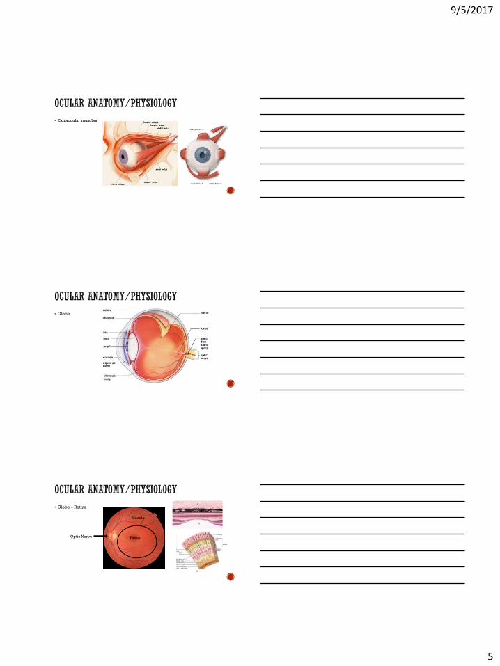
Extraocular muscles
Globe
Globe – Retina
Fovea
Macula
Optic Nerve

9/5/2017
6
Neuro pathway
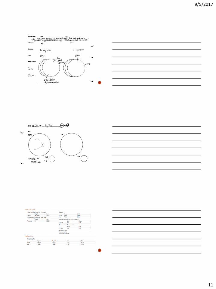
Basic goals of the eye exam
1) Determine a patient’s uncorrected visual ability
2) Determine if optical correction will improve vision
3) Identify any structural abnormality
4) Identify disease states that may lead to vision loss
5) Screen for ocular evidence of systemic disease
6) Address patients’ ocular symptoms or visual complaints

9/5/2017
7
1) Assessing Vision
Assessing Vision (Low Vision)
What happens when patients can’t see the “Big E?”
Bring them closer to the chart (changing numerator)
< 3 feet, assess for ability to count fingers (CF)
Hand Motion (HM)
Light Perception (LP)
Non-Light Perception (NLP)
Uncooperative patients?
Visual Field Exam
Confrontational Visual Fields
Manual Perimetry
Automated Perimetry

9/5/2017
8
Visual Field Exam
Confrontational Visual Fields
Manual Perimetry
Automated Perimetry
Visual Field Exam
Confrontational Visual Fields
Manual Perimetry
Automated Perimetry
Pupils
CNIII lesion
Adie’s
Horner’s
Pons stroke
Brain injury

9/5/2017
9
Eye alignment and movement
Refraction
Intraocular pressure

9/5/2017
10
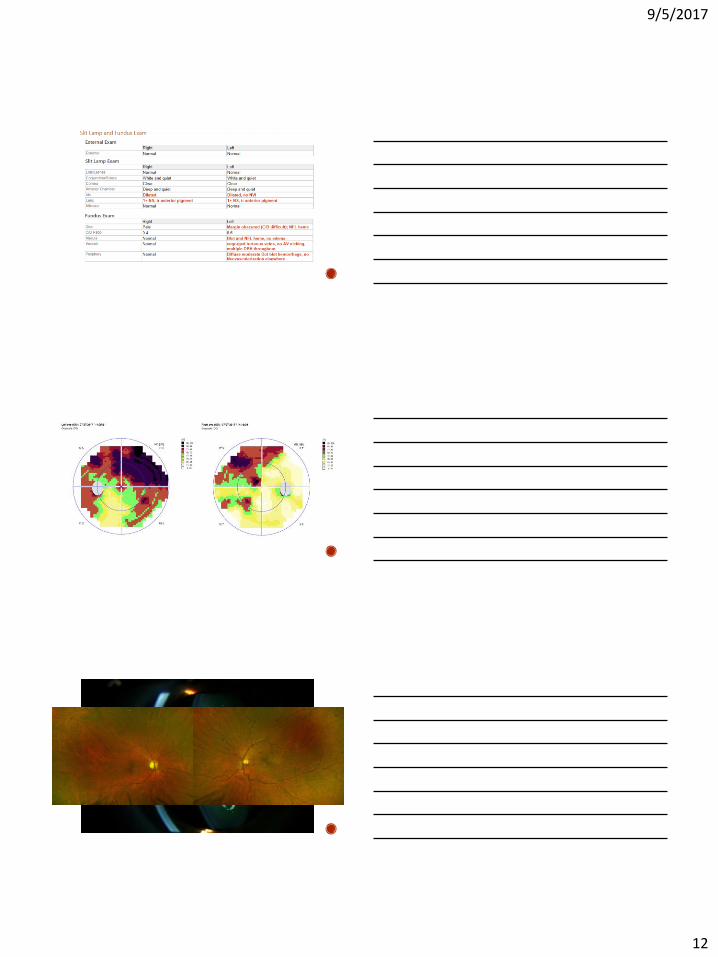
Anterior Exam
Posterior Exam
9/5/2017
11
9/5/2017
12

9/5/2017
13
Visual Impairment
Physical condition of the eyes (Best level of acuity)
Visual Disability
Condition of the individual
Dependent on ability of person to adapt/compensate to level of impairment
Adapted from Basic Ophthalmology
8th ed. AAO 2004

9/5/2017
14
Low vision
Vision between 20/70 and 20/400 with the best possible correction, or a visual field of 20 degrees or less in the better eye
Blindness
Legal blindness in the US means visual acuity of 20/200 or worse with the best possible correction, or a visual field of 20 degrees or less in the better eye
Diabetic eye disease
Macular degeneration
Cataracts
Glaucoma
Many eye manifestations:
Refractive changes, cataracts, dry eyes, macular edema, retinopathy
Retinal changes are the greatest cause of permeant vision loss
Chronic blood glucose variability and elevation leads to microvascular damage
Areas of the retina become ischemic and vessels become incompetent
The retina sends SOS signals to help grow new blood vessels (Neovascularization)

9/5/2017
15

9/5/2017
16
22 million patients worldwide
Estimated >30 million by 2020
3 million U.S. surgeries each year

9/5/2017
17

9/5/2017
18
Patient with significant visual impairment and mild visual disability
Discussed impaired vision likely to result in driving restriction if not treated
Potential permanent blinding disease states if left untreated
Recommended to undergo intravitreal anti-VEGF injection
Cataract extraction to follow in both eyes
PRP laser and periocular steroid in both eyes
Improve systemic disease with primary physician
With surgery and new glasses, vision expected to improve to 20/20
Needs multimodal therapy to prevent worsened, untreatable vision loss
Glaucoma = 2nd leading cause of blindness
60M cases worldwide in 2010 Estimated to grow to 78M by 2020
Bilateral blindness = 7.5% Growing from 4.4M to 6M patients from 2010 to 2020
Optic nerve disease most commonly due to high pressure

9/5/2017
19
⬆Pressure⬆

9/5/2017
20

9/5/2017
21
9/5/2017
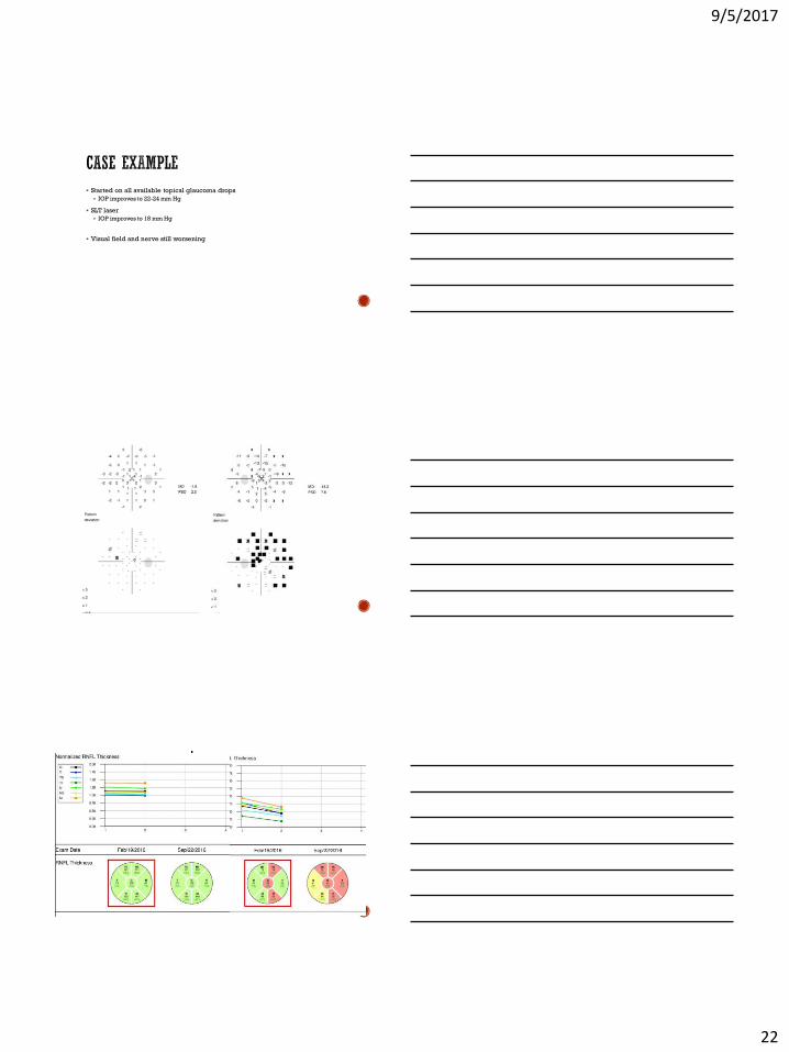
22
Started on all available topical glaucoma drops
IOP improves to 22-24 mm Hg
SLT laser
IOP improves to 18 mm Hg
Visual field and nerve still worsening

9/5/2017
23
Proceed to trabeculectomy surgery
3 months post-op
Now off all drops and doing well
IOP 10 mm Hg
Patient with significant visual impairment but no functional visual disability
Works in an office without need for visual aides
Leading cause of central vision loss in the U.S.
Degenerative build up of metabolic waste over time. Collection of drusen, resultant retinal breakdown, +/- neovascular membrane
80% Dry – slowly progressive atrophy
20% Wet – leaking/bleeding vessels with fibrotic scar formation

9/5/2017
24
Risk: Age, Smoking, UV exposure, genetic predisposition
Dry
Smoking cessation, AREDS vitamins
Wet
Anti-VEGF injections, Laser
Both
Low vision aids
9/5/2017
25
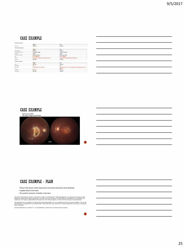
Last seen in 2005
VA: Right-CF@3’ Left-20/200
20042005
Patient with severe visual impairment and severe functional visual disability
Legally blind in both eyes
No medical treatment available at this time
9/5/2017
26
I can’t see
anything in
these glasses?!
General inquiries:
Debbie Hannem - [email protected]
Appointments:
612-625-4400
General information:
https://www.ophthalmology.umn.edu