open access isolation of milk oligosaccharides … glycoscience, 2009, 2, 9-15 9 1875-3981/09 2009...
TRANSCRIPT

Open Glycoscience, 2009, 2, 9-15 9
1875-3981/09 2009 Bentham Open
Open Access
Isolation of Milk Oligosaccharides using Solid-Phase Extraction
Robert E. Ward*
Department of Nutrition and Food Sciences, Utah State University, Logan, UT, 84322, USA
Abstract: A method was developed to isolate mono- and disaccharide-free oligosaccharides from human and bovine milk
using a combination of enzymatic digestion of lactose and solid-phase extraction. In the initial trial, 2.5 g of oligosaccha-
rides were isolated from one liter of human milk. In subsequent trials this was increased to over 5 g of oligosaccharides
per liter. Compared to filtration-based extraction methods, this procedure allows for further isolation of oligosaccharide
fractions via modulation of the column rinsing solvent. Neutral monosaccharide composition of the oligosaccharide poly-
mers was investigated using gas chromatographic analysis of the monosaccharides as alditol acetate derivatives. Results
indicate oligosaccharides are approximately made up of 24% fucose, 41% galactose, 22% glucose and 13% glucosamine.
Isolated bovine and human milk oligosaccharides were compared to lactose as fermentation substrates for Bifidobacterium
longum biovar infantis. Lactose fermentation yielded the greatest production of biomass followed by bovine and human
milk oligosaccharides.
INTRODUCTION
Human milk is rich in complex soluble carbohydrate polymers, and little is understood of the function of these molecules. Some analytical studies have indicated that there may be as much as 20 g/L of oligosaccharides in breast milk which would make them the third most concentrated compo-nent after lactose and fat [1, 2]. However, unlike lactose and fat, these molecules do not appear to provide energy to in-fants as they resist the action of human digestive enzymes [3, 4]. Most human milk oligosaccharides (HMO) are elongation products of lactose, and are synthesized from glucose, galac-tose, glucosamine, fucose and sialic acid [2]. In addition to being concentrated, HMO are also diverse and at least 200 individual structures have been detected using mass spec-trometry [5]. The compositions of HMO are also different among women, and any proposed function they may serve must address this heterogeneity [6].
As the composition of milk is the product of millennia of selective pressure, it stands to reason that individual con-stituents, especially those present in abundant quantities, would be expected to provide some sort of benefit to the infant or the mother or they would tend to be selected against over subsequent generations [7]. Several interesting bioactiv-ities have been attributed to HMO. For example, they an-tagonize the binding of some strains of bacteria to epithelial cells [8], and a large epidemiologic study indicated that sugar epitopes resulting from an individual genotype appear to protect against diarrhea in breast fed infants [9]. Further-more, those containing sialic acid may serve as immune modulators [10, 11]. It has also been suggested that HMO may serve as growth factors for colonic microbiota [2], as it has long been known that breast feeding results in increased levels of fecal bifidobacteria with respect to infant formula. Based on their structure and composition, HMO may affect
*Address correspondence to this author at the Department of Nutrition and
Food Sciences, Utah State University, Logan, UT, 84322, USA;
Tel: +1 435 797 2153; Fax: +1 435 797 4758; E-mail: [email protected]
the gut microbiota consortium either by selectively providing growth factors and energy substrates to some members, or alternatively via binding and eliminating others, or both. However, to begin to address whether HMO might be modu-lating this activity, it is first necessary to isolate them in large enough quantities to serve as fermentation and or growth factor substrates in microbiological media, and to insure that they are entirely free of milk mono and disaccha-rides.
Prior to the initiation of this work, it was noted in the literature that several groups have undertaken the task of isolating HMO from other constituents in milk, although mostly for quantification as opposed to the production of substrates for biochemical assays. The general scheme has been to first remove the fat from milk by centrifugation, and then the protein by precipitation with organic solvents [12]. To separate the HMO from lactose, gel filtration has often been used. This is a slow method that requires long separa-tion times and has a low capacity. In contrast, two methods have been published for obtaining HMO in large quantities, one using charcoal column chromatography [13], and the other nanofiltration [14]. In each case the milk fat was first removed using centrifugation, and protein precipitated with organic solvents. Both groups then enzymatically converted lactose to glucose and galactose to facilitate separation. Brand-Miller et al. [13] used charcoal column adsorption to retain oligosaccharides on the column, while monosaccha-rides were eluted. However, little methodological informa-tion was provided, and the resulting product was not well characterized. Using nanofiltration, Sarney et al. [14] iso-lated HMO from other milk constituents, and compared the resulting HMO produced with gel filtration. Their yield with nanofiltration was 6.7 grams of HMO from 1L of milk, yet there did appear to be residual lactose in the oligosaccharide fraction produced with nanofiltration, but not in that pre-pared using gel filtration. Nanofiltration is an attractive method for HMO isolation due to the speed with which sepa-rations can be performed and it does not require the use of organic solvents. Nonetheless, nanofiltration does not allow
10 Open Glycoscience, 2009, Volume 2 Robert E. Ward
for further separation of oligosaccharide fractions and it is unclear if it can be effective in removing all mono and disac-charides.
This report details the development and optimization of an isolation scheme for HMO using solid-phase extraction with graphitized carbon. It is low cost and, allows for com-plete removal of lactose and monosaccharides, and can be further optimized for isolation of selected fractions of HMO.
MATERIALS AND METHODOLOGY
Unless otherwise noted, all chemicals were obtained from Thermo Fisher Scientific (Waltham, MA). In the de-velopment and optimization of this method, frozen, pooled human milk was provided by the Mother’s Milk Bank of San Jose, CA, by Dr. Jimi Francis, University of Nevada, Reno and by Prolacta Bioscience. Milk samples were kept at -80°C until use. A sialic-acid rich Bovine milk fraction was pro-vided by Fonterra (Auckland, NZ).
The process for isolation of HMO from milk involves six steps which are outlined in Fig. (1). Milk is thawed at room temperature, well mixed, and centrifuged at 5000 g for 30 min at 4ºC (Step 1). Solid fat is removed from the top, and subsequently mixed with one volume of distilled water at 37º. The lipid/water suspension is gently mixed by swirling for several minutes, and centrifuged at 4°C. This is repeated a second time, and all 3 aqueous phases are pooled prior to the next step.
Fig. (1). Isolation scheme for obtaining mono and disaccharide free
oligosaccharides from milk.
In Step 2, two volumes of ethanol are mixed with defat-ted milk, and kept overnight at 4ºC. This process precipitates protein, which is removed by centrifugation at 5000 g for 30 min at 4ºC. The precipitate removed from the centrifuge tubes is resolubilized in two volumes of 2:1 ethanol:water, swirled to facilitate mixing, and then subjected to centrifuga-tion at 5000 x g for 30 minutes at 4°C. This is repeated a third time, the supernatants of the three precipita-tion/centrifugations are pooled, and ethanol removed by ro-tary evaporation.
To digest lactose (Step 3), milk extract is buffered by the addition of 40% v/v 0.123 M phosphate buffer, pH 6.5, an appropriate amount of -galactosidase of Kluyveromyces fragilis (Sigma Aldrich, St. Louis, MO) is added based on the estimated lactose concentration and quoted enzyme activ-ity, and the sample incubated for 3 h at 37ºC. After lactose digestion, particulates are removed via filtration with vac-uum through a Whatman #1 and #541 filters in a series.
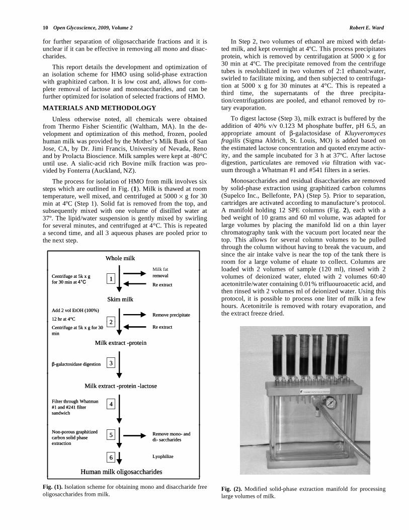
Monosaccharides and residual disaccharides are removed by solid-phase extraction using graphitized carbon columns (Supelco Inc., Bellefonte, PA) (Step 5). Prior to separation, cartridges are activated according to manufacture’s protocol. A manifold holding 12 SPE columns (Fig. 2), each with a bed weight of 10 grams and 60 ml volume, was adapted for large volumes by placing the manifold lid on a thin layer chromatography tank with the vacuum port located near the top. This allows for several column volumes to be pulled through the column without having to break the vacuum, and since the air intake valve is near the top of the tank there is room for a large volume of eluate to collect. Columns are loaded with 2 volumes of sample (120 ml), rinsed with 2 volumes of deionized water, eluted with 2 volumes 60:40 acetonitrile/water containing 0.01% trifluouroacetic acid, and then rinsed with 2 volumes ml of deionized water. Using this protocol, it is possible to process one liter of milk in a few hours. Acetonitrile is removed with rotary evaporation, and the extract freeze dried.
Fig. (2). Modified solid-phase extraction manifold for processing
large volumes of milk.
Whole milk
Milk fat
Whole milk
Milk fat
Skim milk
Centrifuge at 5k x g for 30 min at 4°C
Milk fat removal
Add 2 vol EtOH (100%)
12 hr at 4°CRemove precipitate
1
2
Re extract
Skim milk
Centrifuge at 5k x g for 30 min at 4°C
Milk fat removal
Add 2 vol EtOH (100%)
12 hr at 4°CRemove precipitate
1
2
Re extract
Centrifuge at 5k x g for 30 min
Milk extract -protein
β-galactosidase digestion
2
3
Re extractCentrifuge at 5k x g for 30 min
Milk extract -protein
β-galactosidase digestion
2
3
Re extract
Milk extract -protein -lactose
4Filter through Whatman#1 and #241 filter sandwich
Milk extract -protein -lactose
4Filter through Whatman#1 and #241 filter sandwich
Non-porous graphitized carbon solid phase extraction
Remove mono- and di- saccharides
Human milk oligosaccharides
Lyophilize
5
6
Non-porous graphitized carbon solid phase extraction
Remove mono- and di- saccharides
Human milk oligosaccharides
Lyophilize
5
6

Milk Oligosaccharide Isolation Open Glycoscience, 2009, Volume 2 11
A similar strategy was utilized for isolation of oligosac-charides from a bovine colostrum powder. One hundred grams was diluted in 1L deionized water, and otherwise the method was the same.
Carbohydrate Measurements
Where indicated, total carbohydrate was measured col-orimetrically by the method of Dubois [15], and lactose and free glucose were measured in the HMO using an enzymatic kit (R-biopharm, So. Marshall, MI).
High Performance Thin Layer Chromatography
High performance Thin Layer Chromatography (HPTLC) was performed by the method described by Kunz et al. (1999) using 10 20 cm plates from Whatman Ltd. ( Maid-stone, UK). In short, samples containing oligosaccharides (2- l) were spotted on plates and developed with a buta-nol-ethanol-water-acetic acid-pyridine (5:50:15:1.5:5, v/v/v/v/v) solvent system. To detect carbohydrates, plates were sprayed with orcinol solution (2 mg/ml 70% H2SO4 ) and placed in a 100°C oven for 15 min.
2D SPE and HPTLC Separation
The interaction of HMO with the graphitized carbon ma-trix of the SPE columns was investigated by loading a col-umn (500 mg adsorbant, 6 ml volume) with milk extract (post lactose digestion), and then eluting the adsorbed carbo-hydrates with an aqueous solution containing an increasing concentration of butanol. Oligosaccharides were eluted by sequentially rinsing the column with 3 volumes (15ml) of deionized water containing an increasing concentration of butanol. The percentage of butanol in the column rinse was 0.25%, 0.33%, 0.5%, 0.67%, 1%, 1.33%, 2%, 4%. The last rinse contained 0.1 % TFA in addition to the 4 % butanol. The carbohydrate concentration of elution fractions was measured using the phenol-sulfuric acid method, and 5 ml aliquots were lyophilized. Each fraction was brought up to a concentration of roughly 1.75% carbohydrate, 2 l aliquots were spotted on TLC plates, and were run according to stan-dard procedure.
Gas Chromatography of Alditol Acetate Derivatives
The monosaccharide composition of HMO was deter-mined by gas chromatography of alditol acetate derivatives. The general method for GC analysis was taken from York et al. [16]. In short, approximately 250 ng of HMO was added to a 4 ml vial and hydrolyzed by the addition of 250 l 2 M triflouoroacetic acid (TFA) for 1 h at 121 °C. Vials were cooled to room temperature, and TFA evaporated at 40 °C under a stream of nitrogen gas. To facilitate complete re-moval of TFA, 0.3 ml of isopropyl alcohol was added and sample reevaporated. Monosaccharides were reduced by 250
l 1M ammonia containing 10 mg/ml sodium borohydride, at room temperature for 1 h. Excess borohydride was con-verted to borate by the addition of 2-3 drops of glacial acetic acid, and then 250 l acetic acid-methanol 1:9 (v/v) added to each vial and the resulting solution evaporated at 40 °C un-der nitrogen. Three additional evaporations were conducted in the same manner, and then three further evaporation were conducted with 250 l methanol. The alditols were then O-acetylated by the addition of 50 l of acetic anhydride and 50 l pyridine, and incubated at 121 °C for 20 minutes. Vials were cooled, and 200 l of toluene added, and evaporated at room temperature. This was repeated a second time, and then the alditol acetates partitioned between 0.5 ml of dicholor-methane and 0.5 ml of water. The water was then extracted two more times with 0.5 ml dicholormethane, and the or-ganic phases combined and dried. The samples were resolu-bilized in 100 l acetone, and transferred to a GC vial insert. Free monosaccharides were prepared in the same way, ex-cept the hydrolysis step with TFA was not performed.
Per-O-acetylated monosaccharides were analyzed on a Hewlett-Packard 6890 Series gas chromatograph equipped with a flame ionization detector (FID) and a 30 m DB-225MS capillary column (0.25 mm i.d.;0.25 m film thickess). Hydrogen was used as the carrier gas at 36.5 ml min
-1. Injector temperature was maintained at 225 °C, and
the split ratio was 1:25. Injection volume was 1 l, oven temp was kept at 220 °C for entire run. Nitrogen was used as the make-up gas at 30 ml/min, and detector was operated at 250 °C. Peak identification was made by comparing reten-
Fig. (3). Ratio of total carbohydrates to lactose in human milk fractions during oligosaccharide purification. Total sugars were measured
colorimetrically and lactose was measured enzymatically. Insets A and B: HPTLC profiles of fractions from the initial and second purifica-
tion 1) whole milk 2) skim milk 3) after protein precipitation 4) after lactose digestion 5) after solid phase extraction 6) standards (from top)
glucose, lactose, raffinose, stachyose.

12 Open Glycoscience, 2009, Volume 2 Robert E. Ward
tion time to individual monosaccharide standards deritivat-ized using the same process.
Oligosaccharide Fermentation Assay
The fermentation of human and bovine milk oligosaccha-rides was measured using Bifidobacterium longum b.v. in-fantis according to the method of Ward et al. [17]. In short, MRS media was made up with isolated oligosaccharides instead of glucose at 1% (m/v). Growth was measure with a Klett-Summerson colorimeter (Klett Manufacturing Co., Inc., New York, NY) at specific time points using the no. 45 (green filter).
RESULTS
In three subsequent extractions the yield of oligosaccha-rides per liter of breast milk was approximately 2.5 g, 5.5 g, and 5.2 grams. The ratio of lactose (measured enzymatically) to total sugars (measured colometrically) at various stages in the procedure is shown in Fig. (3), and inset photos show HPTLC carbohydrate profiles resulting from these steps. It is worth noting when interpreting this figure, and the other data presented in this study, that of the five monosaccharide con-stituents of HMO, only glucose and galactose are detected by the colorimetric Dubois method, and only glucose, galactose and fucose stain with the orcinol reagent on TLC plates. From the figure it is clear that the majority of carbohydrate in breast milk is lactose, and approximately 5% of glucose and galactose is bound up in oligosaccharides which is con-sistent with previous estimates [10].
After the initial extraction with the 2.5 g/L HMO yield, it was reasoned that the process might be improved, and changes to the process outlined in Fig. (1) included rextrac-tion of the removed fat (Step 1) and protein (Step 2) to pre-vent any loss of HMO. Inset A of Fig. (3) shows the carbo-hydrate profiles after various stages of the first extraction, and from the image it appears there is some undigested lac-tose and some residual monosaccharides in the final HMO (Lane 5). For the second trial this was addressed by optimiz-ing the enzymatic digestion of lactose, and modifying the SPE column loading protocol. According the manufacturer,
-galactosidase from Kluyveromyces fragilis has relatively sharp pH and temperature optima (pH 6.5 and 37º C). Care was taken to insure that the reaction was conducted under these conditions, and the amount of enzyme added to the milk extract was doubled. In the second round of purification the progress of hydrolysis was monitored and determined to be complete after 3 hours (data not shown).
According to Redmond and Packer [18], the capacity of graphitized carbon SPE columns to adsorb the trisaccharide raffinose is approximately 27% of its mass. In the first trial, six columns, each with 10 g adsorbent beds and 60 ml vol-umes, were used to extract 1L of milk. The theoretical capac-ity of this system for oligosaccharides is over 16 g, which is in excess of the level of HMO in milk, and yet only 2.5 g were isolated. To better understand the interaction of HMO with the adsorbent, a binding study was conducted with a small SPE column (500 mg bed, 6 ml volume) and 5, 10, 20, 25 and 30 ml of milk. The findings (data not shown) indi-cated that graphitized carbon SPE columns could bind up to approximately 20% of their weight in HMO, and all the HMO in the 5 and 10 ml of milk. As the loading of the col-
umn was further increased up 25 ml of milk, no more than 20% of the weight of the column was bound, and the per-centage bound per ml of milk went down. Thus, from 10 ml of milk approximately 97 mg of HMO were bound, and 101 mg were extracted from 25 ml of milk. However, when 30 ml was applied to the column, only 39 mg of HMO were adsorbed to the column, which indicates that overloading the column washes off HMO that had been previously bound. In the second and third trial this was addressed by increasing the number of columns on the manifold to 12, by diluting the HMO extract prior to loading columns by doubling the vol-ume, and by alternating rinse steps with deionized water in between column loadings. These changes led to a 50% in-crease in yield, however, it would appear that the overall yield could still be improved via a better understanding of the factors that limit HMO binding as column loading is in-creased.
To investigate the potential of this method to isolate spe-
cific fractions of HMO a second binding study was con-
ducted with a small column (500 mg bed, 6 ml volume). Af-ter loading the column, it was rinsed with solutions contain-
ing an increasing percentage of butanol to probe the interac-
tion of the carbohydrates with the graphitized carbon adsorb-ent, and to get a better understand the size distribution of the
HMO. This experiment was conducted with butanol, rather
than acetonitrile, which was used in the bulk purification, as prior work had shown this solvent system to be effective in
isolating specific oligosaccharides [18]. In Fig. (4), the car-
bohydrates in Lane 1 consist of the material that eluted from the column during the application of the sample. Comparing
this lane to the standards at the other end of the plate indi-
cates that the eluate consists primarily of glucose and galac-tose. Lane 2 contains material that was rinsed off the loaded
column with water, and consisted primarily of monosaccha-
rides, as well. Lanes 3-9 show the effects of increasing the concentration of butanol in the column rinse, and it is clear
that as the concentration increases, different fractions of
HMO are released. From this data it appears HMO elute in three broad groups, one near the top (Lanes 3-6) with a simi-
lar migration distance to the disaccharides lactose (Lane 11)
and sucrose (Lane 12) and the trisaccharide raffinose, a sec-ond group (Lanes 3-8) with a similar mobility to the tet-
rasaccharide stachyose (Lane 12), and a group that remains
at the origin (Lanes 6-10). On the bottom of the figure a bar graph of the relative concentration of sugars in each fraction
is superimposed, and summing the contribution of Lanes 3-6
indicates that greater than 2/3 of the HMO appear to be tri- and tetrasaccharides. This appears to be in agreement with
the observation by LoCascio et al. [19] that B. longum bv.
infantis preferentiall uses small HMO for fermentation sub-strates, which they find to represent 63.9% of the total. Start-
ing in Lane 6, a band at the origin appears and becomes
darker in the subsequent four fractions (Lanes 6-10). , and Lanes 9 and 10 consisted solely of this band. In Lane 10,
0.1% Trifluoroacetic acid (TFA) was added to the 4% buta-
nol in the rinse. According to Redmond et al. [18], acidic sugars (including phosphorylated and sulfated monosaccha-
rides) are strongly adsorbed to graphitized carbon, and will
not be eluted by the addition of an organic modifier unless a volatile acid, such as TFA, is added. Thus, it would appear
that the band at the bottom of Lane 10 represents acidic

Milk Oligosaccharide Isolation Open Glycoscience, 2009, Volume 2 13
HMO, although this was not confirmed, and sialic acid is not
visualized by the orcinol spray applied to the HPTLC plate.
Fig. (4). HPTLC plate showing interaction of HMO with
graphitized carbon as a function of elution solvent percentage.
Lanes: 1) Eluate from column loading 2) column rinse with DI
water 3) 0.25% butanol 4) 0.33 % butanol 5) 0.5% butanol 6)
0.67% butanol 7) 1% butanol 8) 1.33% butanol 9) 2% butanol 10)
4% butanol + 0.1% TFA 11) lactose 12) from top: glucose, sucrose,
raffinose, stachyose 13) galactose.
HMO isolated in Trial 1 and Trial 2 were characterized by gas chromatographic analysis of their constituent mono-saccharides as alditol acetate derivatives, and the results are shown in Fig. (5). Data for sialic acid is not shown, as the method does not detect this monosaccharide. The first step in this procedure is to hydrolyze polymers to monomers using TFA, and by omitting this treatment free sugars were de-tected. Comparison of free and bound sugars confirms that HMO isolated in the first trial contained a significant propor-tion of free monosaccharides, and these were removed by modifications to the method in subsequent trials. In the inset table the percentage of bound monosaccharides in HMO
from each trial is compared and the results show the compo-sition to be very similar. To the best of the author’s knowl-edge, this is the first such report on the monosaccharide composition of a bulk HMO isolate.
While it has long been speculated that HMO are used as energy sources for colonic microbiota, definitive evidence for this activity was only provided recently [17, 19, 20]. Bo-vine milk also contains oligosaccharides (BMO), yet they are present in much lower levels than in human milk. Nonethe-less, due to the large volume of bovine milk produced each year, it was of interest to determine if these oligosaccharides are fermentable, and if so, how they compared to HMO as a substrate. To investigate this activity, they were isolated from a powdered milk fraction and used as the sole carbohy-drate in an adapted MRS media. Due to the low yield, there was not sufficient BMO to conduct trials with multiple strains, so the bacterium with the highest activity on HMO, Bifidobacterium longum biovar infantis, was chosen to test this assay. The results of the growth assay is shown in Fig. (6), and it is clear that not only can B. longum bv. infantis use the BMO as an energy source, they appear to be more effective than HMO. There are several differences between HMO and BMO which might explain this finding. For ex-ample, fewer individual oligosaccharide species have been detected (40 for BMO vs. >200 for HMO), and over 70% contain at least one sialic acid [21] . Furthermore, in general they are dominated by tri- and tetrasaccharides, do not con-tain fucose, and the largest one detected to date is a heptamer [22]. Previous studies investigating prebiotic effects of HMO have indicated bifidobacteria preferentially consume small HMO as well as small oligofructose polymers [19, 20].
DISCUSSION
Although oliogsaccharides are a prominent component of human milk, this compositional feature is not shared among all mammals. Levels in ruminant milks are low [23], whereas primate milks typically contain more and more di-verse oligosaccharides [24], as do elephants [25]. Nonethe-less, there does not seem to be any clear indication of their functional importance that can be deduced from phylogenetic
Fig. (5). Monosaccharide composition of HMO from two separate purifications determined by gas chromatography of alditol acetate deriva-
tives. Peak areas are normalized to surrogate concentration. Free monosaccharides were measured by omitting hydrolysis step. Inset table:
percent monsaccharide composition of HMO polymers from two separate trials after removing contribution of free sugars.

14 Open Glycoscience, 2009, Volume 2 Robert E. Ward
comparisons. In this context, intensive biochemical charac-terizations of the molecules and their various bioactivities may facilitate an understanding of their importance in infant, and perhaps adult, nutrition, and may suggest individual oli-gosaccharide structures to target for industrial production.
To facilitate such investigations it is crucial to obtaining oligosaccharides in a pure form, free from lactose. The first time this extraction was attempted, roughly 2.5 grams were isolated from 1 L of breast milk. By modifying three steps from that procedure, this was increased to approximately 5.5 g/L of milk. Although this was a substantial increase in yield, the initial concentration of HMO in the respective milks was not known. Due to the fact that HMO varies sub-stantially across women and time of lactation, it was not pos-sible to make a direct comparison.
While previous studies have indicated that HMO are not degraded by human digestive enzymes, the -galactosidase used in this study was of microbial origin and may have af-fected the resulting purified HMO in at least two ways. There may have been some degradation of specific HMO structures by enzymatic hydrolysis, and some galactooligo-saccharides (GOS) may have been formed via transglycosy-lation activity of the enzyme. However, in bacterial growth studies conducted with HMO purified by this method, simi-lar HMO profiles were observed as in unprocessed milk, and masses consistent with GOS polymers were not detected [19].
The method presented here is suitable for routine prepa-rative isolation of experimental quantities of oligosaccha-rides, free of contaminating lactose and monosaccharides. Furthermore, this method allows for the separation of sub-fractions, such as the either only neutral or only acidic HMO. This method should enable subsequent studies designed to facilitate understanding the molecular basis for the presence of these molecules in human milk.
Analysis of the monosaccharide composition of HMO from two individual purification trials indicates that the rela-tive proportions of individual sugars was quite similar. This
is perhaps surprising, as there appears to be a lot of hetero-geneity in HMO composition among women during lacta-tion, as well as between women [26], and the pooled milk used in these studies was typically contributed by fewer than 5 individual donors.
It has long been speculated that HMO may serve as fer-mentation substrates for intestinal microbiota, and using oli-gosaccharides isolated by this method it was recently shown that some strains can these polymers as an energy source [17, 20]. One surprising feature of this activity is that B. longum bv. infantis consistently reaches higher cell densities using HMO as a substrate than other gut species, and a re-cent genomic analysis indicates that this bacterium contains approximately 700 unique genes many of which are specifi-cally for this activity that other bifidobacterium strains do not possess [27]. It was of interest, therefore, to determine if oligosaccharides isolated from bovine milk using this method could support the growth of this strain. The results indicate that BMO can be fermentation substrates for micro-biota, and on a mass to mass basis, are more effective than HMO. It should be kept in mind, however, that the levels in bovine milk are very low.
In sum, this method is effective in facilitating the isola-tion of oligosaccharides from milk, and large quantities can be produced on the bench-top. The method is reproducible across trials, and allows for the further separation of oligo-saccharide fractions.
ACKNOWLEDGEMENTS
Financial support for this project was provided by the Utah Agricultural Experiment Station. This paper was ap-proved by the Utah Agricultural Experiment Station as paper # 8031.
REFERENCES
[1] Coppa GV, Pierani P, Zampini L, Carloni I, Carlucci A, Gabrielli
O. Oligosaccharides in human milk during different phases of lac-tation. Acta Paediatr Suppl 1999; 88(430): 89-94.
Fig. (6). Growth curves for Bifidobacterium longum biovar infantis using lactose, human milk and bovine milk oligosaccharides as the sole
carbohydrate source.

Milk Oligosaccharide Isolation Open Glycoscience, 2009, Volume 2 15
[2] Kunz C, Rudloff S, Baier W, Klein N, Strobel S. Oligosaccharides
in human milk: structural, functional, and metabolic aspects. Annu Rev Nutr 2000; 20: 699-722.
[3] Chaturvedi P, Warren CD, Buescher CR, Pickering LK, Newburg DS. Survival of human milk oligosaccharides in the intestine of in-
fants. Adv Exp Med Biol 2001; 501: 315-23. [4] Gnoth MJ, Kunz C, Kinne-Saffran E, Rudloff S. Human milk oli-
gosaccharides are minimally digested in vitro. J Nutr 2000; 130(12): 3014-20.
[5] German JB, Freeman SL, Lebrilla CB, Mills DA. Human milk oligosaccharides: evolution, structures and bioselectivity as sub-
strates for intestinal bacteria. Nestle Nutr Workshop Ser Pediatr Prog 2008; 62: 205-22.
[6] Stahl B, Thurl S, Henker J, Siegel M, Finke B, Sawatzki G. Detec-tion of four human milk groups with respect to Lewis-blood-group-
dependent oligosaccharides by serologic and chromatographic analysis. Adv Exp Med Biol 2001; 501: 299-306.
[7] German JB, Morgan CJ, Ward RE. Milk: a model for nutrition in the 21st Century. Aust J Dairy Technol 2003; 58(2): 49-54.
[8] Morrow AL, Ruiz-Palacios GM, Jiang X, Newburg DS. Human-milk glycans that inhibit pathogen binding protect breast-feeding
infants against infectious diarrhea. J Nutr 2005; 135(5): 1304-7. [9] Morrow AL, Ruiz-Palacios GM, Altaye M, et al. Human milk
oligosaccharides are associated with protection against diarrhea in breast-fed infants. J Pediatr 2004; 145(3): 297-303.
[10] Bode L, Kunz C, Muhly-Reinholz M, Mayer K, Seeger W, Rudloff S. Inhibition of monocyte, lymphocyte, and neutrophil adhesion to
endothelial cells by human milk oligosaccharides. Thromb Hae-most 2004; 92(6): 1402-10.
[11] Bode L, Rudloff S, Kunz C, Strobel S, Klein N. Human milk oligo-saccharides reduce platelet-neutrophil complex formation leading
to a decrease in neutrophil beta 2 integrin expression. J Leukoc Biol 2004; 76(4): 820-6.
[12] Egge H, Dell A, Von Nicolai H. Fucose Containing oligosaccha-rides from human milk. Arch Biochem Biophys 1983; 224(1): 235-
53. [13] Brand-Miller JC, McVeagh P, McNeil Y, Messer M. Digestion of
human milk oligosaccharides by healthy infants evaluated by the lactulose hydrogen breath test. J Pediatr 1998; 133(1): 95-8.
[14] Sarney DB, Hale C, Frankel G, Vulfson EN. A novel approach to the recovery of biologically active oligosaccharides from milk us-
ing a combination of enzymatic treatment and nanofiltration. Bio-technol Bioeng 2000; 69(4): 461-7.
[15] Dubois M, Gilles K, Hamilton JK, Rebers PA, Smith F. A col-orimetric method for the determination of sugars. Nature 1951;
168(4265): 167.
[16] York WS, Darvill AG, McNeil M, Stevenson TT. Isolation of char-
acterization of plant cell walls and cell wall component. Method Enzymol 1985; 119: 3-30.
[17] Ward RE, Ninonuevo M, Mills DA, Lebrilla CB, German JB. In vitro fermentation of breast milk oligosaccharides by Bifidobacte-
rium infantis and Lactobacillus gasseri. Appl Environ Microbiol 2006; 72(6): 4497-9.
[18] Redmond JW, Packer NH. The use of solid phase extraction with graphitized carbon for the fractionation and purification of sugars.
Carbohydr Res 1999; 319: 74-9. [19] LoCascio RG, Ninonuevo MR, Freeman SL, et al. Glycoprofiling
of bifidobacterial consumption of human milk oligosaccharides demonstrates strain specific, preferential consumption of small
chain glycans secreted in early human lactation. J Agric Food Chem 2007; 55(22): 8914-9.
[20] Ward RE, Ninonuevo M, Mills DA, Lebrilla CB, German JB. In vitro fermentability of human milk oligosaccharides by several
strains of bifidobacteria. Mol Nutr Food Res 2007; 51(11): 1398-405.
[21] Tao N, DePeters EJ, Freeman S, German JB, Grimm R, Lebrilla CB. Bovine milk glycome. J Dairy Sci 2008; 91(10): 3768-78.
[22] Chaturvedi P, Warren CD, Ruiz-Palacios GM, Pickering LK, New-burg DS. Milk oligosaccharide profiles by reversed-phase HPLC of
their perbenzoylated derivatives. Anal Biochem 1997; 251(1): 89-97.
[23] Martinez-Ferez A, Rudloff S, Guadix A, et al. Goats' milk as a natural source of lactose-derived oligosaccharides: Isolation by
membrane technology. Int Dairy J 2006; 16(2): 173-81. [24] Warren CD, Chaturvedi P, Newburg AR, Oftedal OT, Tilden CD,
Newburg DS. Comparison of oligosaccharides in milk specimens from humans and twelve other species. Adv Exp Med Biol 2001;
501: 325-32. [25] Kunz C, Rudloff S, Schad W, Braun D. Lactose-derived oligosac-
charides in the milk of elephants: comparison with human milk. Br J Nutr 1999; 82(5): 391-99.
[26] Ninonuevo MR, Perkins PD, Francis J, et al. Daily variations in oligosaccharides of human milk determined by microfluidic chips
and mass spectrometry. J Agric Food Chem 2008; 56(2): 618-26. [27] German JB, Freeman SL, Lebrilla CB, Mills DA. Human milk
oligosaccharides: evolution, structures and bioselectivity as sub-strates for intestinal bacteria. Nestle Nutr Workshop Ser Pediatr
Prog 2008; 62: 205-18; discussion 218-22.
Received: November 26, 2008 Revised: January 02, 2009 Accepted: January 05, 2009
© Robert E. Ward; Licensee Bentham Open.
This is an open access article licensed under the terms of the Creative Commons Attribution Non-Commercial License
(http://creativecommons.org/licenses/by-nc/3.0/) which permits unrestricted, non-commercial use, distribution and reproduction in any medium, provided the work is properly cited.