opa1mutations induce mitochondrial dna instability and - brain
TRANSCRIPT
OPA1mutations induce mitochondrial DNA instabilityand optic atrophy lsquoplusrsquo phenotypesPatrizia Amati-Bonneau12 Maria Lucia Valentino3 Pascal Reynier12 Maria Esther Gallardo4
Belecurren n Bornstein4 Anne Boissie re5 Yolanda Campos6 Henry Rivera6 Jesucurren s Gonzacurren lez de la Aleja6
Rosanna Carroccia3 Luisa Iommarini3 Pierre Labauge7 Dominique Figarella-Branger8
Pascale Marcorelles9 Alain Furby10 Katell Beauvais10 Franck Letournel11 Rocco Liguori3 ChiaraLa Morgia3 Pasquale Montagna3 Maria Liguori12 Claudia Zanna13 Michela Rugolo13 Andrea Cossarizza14
Bernd Wissinger15 ChristopheVerny16 Robert Schwarzenbacher17 Miguel Acurren ngel Martin6 Joaquin Arenas6
Carmen Ayuso18 Rafael Garesse4 Guy Lenaers5 Dominique Bonneau12 and Valerio Carelli3
1Decurren partement de Biochimie et Gecurren necurren tique Centre Hospitalier Universitaire drsquoAngers 2INSERMU694 Angers France3Dipartimento di Scienze Neurologiche Universita di Bologna Bologna Italy 4Departamento de Bioquimica Instituto deInvestigaciones Biomedicas lsquoAlberto Solsrsquo CSIC-UAM Facultad de Medicina Universidad Autonoma de Madrid CIBERERISCIII Madrid Spain 5INSERMU583 Institut des Neurosciences de MontpellierUniversitecurren s de Montpellier I et II MontpellierFrance 6Centro de Investigaciocurren n and Servicio de Neurologia Hospital Universitario12 de Octubre CIBERER ISCIII MadridSpain 7Service de Neurologie Centre Hospitalier Universitaire de Nimes Nimes France 8Service drsquoAnatomie Pathologiqueet Neuropathologie Centre Hospitalier UniversitaireccedilHopital de laTimone Marseille France 9Service drsquoAnatomiePathologique Centre Hospitalier Universitaire de Brest Brest France 10Service de Neurologie Centre Hospitalier deSaint-Brieuc Saint-Brieuc France 11Laboratoire de Neurobiologie et Neuropathologie Centre Hospitalier UniversitairedrsquoAngers Angers France 12Institute of Neurological Sciences National Research Council - Mangone Cosenza Italy13Dipartimento di Biologia Evoluzionistica Sperimentale Universita di Bologna Bologna Italy 14Dipartimento di ScienzeBiomediche Sezione di Patologia Generale Universita di Modena e Reggio Emilia Italy 15Molecular Genetics LaboratoryUniversity Eye Hospital TuebingenGermany 16Decurren partement de Neurologie Centre Hospitalier Universitaire drsquoAngersAngers France 17Structural Biology University of Salzburg Austria and 18Servicio de Genecurren tica Fundaciocurren n Jimecurren nez Dicurren azCIBERER ISCIII Madrid Spain
These authors contributed equally to this study
Correspondence to Valerio Carelli MD PhD Laboratorio di Neurogenetica Dipartimento di Scienze NeurologicheUniversita di BolognaVia Ugo Foscolo 7 40123 Bologna ItalyE-mail valeriocarelliuniboit
Mutations in OPA1 a dynamin-related GTPase involved in mitochondrial fusion cristae organization and con-trol of apoptosis have been linked to non-syndromic optic neuropathy transmitted as an autosomal-dominanttrait (DOA)We here report on eight patients from six independent families showing that mutations in the OPA1gene can also be responsible for a syndromic form of DOA associated with sensorineural deafness ataxiaaxonal sensory-motor polyneuropathy chronic progressive external ophthalmoplegia and mitochondrial myo-pathy with cytochrome c oxidase negative and Ragged Red Fibres Most remarkably we demonstrate thatthese patients all harboured multiple deletions of mitochondrial DNA (mtDNA) in their skeletal muscle thusrevealing an unrecognized role of the OPA1protein inmtDNA stabilityThe fiveOPA1mutations associated withthese DOAlsquoplusrsquo phenotypes were allmis-sense pointmutations affecting highly conserved amino acid positionsand the nuclear genes previously known to inducemtDNAmultiple deletions such as POLG1 PEO1 (Twinkle) andSLC25A4 (ANT1) were ruled outOur results show that certainOPA1mutations exert a dominant negative effectresponsible for multi-systemic disease closely related to classical mitochondrial cytopathies by a mechanisminvolving mtDNA instability
Keywords mitochondria mtDNA multiple deletions dominant optic atrophy mitochondrial encephalomyopathy chronicprogressive external ophthalmoplegia
doi101093brainawm298 Brain (2008) 131 338^351
2007 The Author(s)This is an Open Access article distributed under the terms of the Creative Commons Attribution Non-Commercial License (httpcreativecommonsorglicensesby-nc20uk) whichpermits unrestricted non-commercial use distribution and reproduction in any medium provided the original work is properly cited
Dow
nloaded from httpsacadem
icoupcombrainarticle1312338403807 by guest on 19 D
ecember 2021
Abbreviations BAEPs=brainstem auditory evoked potentials BDLP=bacterial dynamin-like protein CT=computerizedtomography CMT=Charcot^Marie-Tooth COX=cytochrome c oxidase CPEO=chronic progressive external ophthalmo-plegia DOA=dominant optic atrophy FBS= fetal bovine serum LHON=Leberrsquos hereditary optic neuropathyMEPs=motor evoked potentials mtDNA=mitochondrial DNA MRI=magnetic resonance imaging MRS=MR spectro-scopy nDNA=nuclear DNA OXPHOS=oxidative phosphorylation PVEPs=pattern visual evoked potentialsRRFs=Ragged Red Fibres SDH=succinate dehydrogenase SEPs= somatosensorial evoked potentials TP= thymidinephosphorylase
Received October 1 2007 Revised November 7 2007 Accepted November 16 2007 Advance Access publication December 24 2007
Mitochondrial disorders can be due to genetic defects inboth the small mitochondrial double-stranded circulargenome (mtDNA) and the nuclear DNA (DiMauro andSchon 2003) A good example of this double geneticdetermination is represented by the two most frequent non-syndromic hereditary optic neuropathies Leberrsquos hereditaryoptic neuropathy (LHON OMIM535000) and dominantoptic atrophy (DOA OMIM165500) LHON is due in thelarge majority of worldwide cases to one of three mtDNApoint mutation at positions 11778ND4 3460ND1 and14484ND6 all affecting different subunits of complex I(Carelli et al 2004) DOA in about 60ndash70 of cases isdue to mutations in the nuclear gene encoding for theOPA1 protein (Alexander et al 2000 Delettre et al 2000Cohn et al 2007) a dynamin-related GTPase targeted tomitochondria which locates mostly on the mitochondrialinner membrane (Delettre et al 2002 Olichon et al 2006)OPA1 has been involved in multiple functions the key rolebeing the fusion of mitochondria and thus the mitochon-drial network organization (Olichon et al 2006) FurtherOPA1 functions are oxidative phosphorylation (OXPHOS)and membrane potential maintenance (Olichon et al 2003Lodi et al 2004 Amati-Bonneau et al 2005) as well ascristae organization and control of apoptosis through thecompartimentalization of cytochrome c (Olichon et al2003 Frezza et al 2006)The large majority of mutations in the OPA1 gene
described to date are predicted to lead to a truncatedprotein and to haploinsufficiency (see httplbbmauniv-angersfr) (Ferre et al 2005) These mutations areinvariably associated with a non-syndromic slowly pro-gressive form of optic neuropathy as originally describedby Kjer (1959) Classic DOA usually begins before 10 yearsof age with a large variability in the severity of clinicalexpression which may range from non-penetrant unaf-fected cases up to very severe early onset cases even withinthe same family carrying the same molecular defect(Delettre et al 2002 Carelli et al 2004 Olichon et al2006 Cohn et al 2007) However there is at least one clearexample standing out of this paradigm This is a mutationin the OPA1 gene ie the c1334G4A leading to pR445Hamino acid change being associated with a syndromic formof optic neuropathy and sensorineural deafness (Amati-Bonneau et al 2003 Shimizu et al 2003) and in some of
the reported cases with chronic progressive externalophthalmoplegia (CPEO) ptosis and myopathy (Treftet al 1984 Meire et al 1985 Payne et al 2004)
CPEO isolated or variably associated with a widersyndromic clinical expression is the most frequent featureof mitochondrial myopathy and has a heterogeneousgenetic basis again driven by both primary defects in themtDNA ie single deletions and point mutations (DiMauroand Schon 2003) or by mutations in nuclear genesresulting in multiple deletions of mtDNA (Zeviani et al1989) At least four nuclear genes are now known to beinvolved in CPEO associated with mtDNA multipledeletions and autosomal recessive or dominant inheritanceThese are POLG1 (Van Goethem et al 2001) the enzymereplicating mtDNA the mitochondrial replicative DNAhelicase Twinkle (PEO1) (Spelbrink et al 2001) the heartmuscle-specific adenine nucleotide translocator ANT1(SLC25A4) (Kaukonen et al 2000) and finally thethymidine phosphorylase (TP) involved in the nucleosidepool maintenance (Nishino et al 1999) Among thesegenes mutations in at least two of them ie POLG1 andTP may present with a combination of deletions anddepletion of mtDNA in skeletal muscle (Hirano et al 2004Hudson and Chinnery 2006)
The association of CPEO and mitochondrial myopathywith optic atrophy is not frequent (Treft et al 1984 Meireet al 1985) and never reported as due to mutations in theabove-mentioned genes Thus the clinical phenotype asso-ciated with the OPA1R445H mutation is somehow a novelcombination bridging autosomal-dominant CPEO and DOA(Payne et al 2004) Recent studies showed that thebiochemical phenotype of the OPA1R445H mutationconsists in a defective OXPHOS in fibroblasts (Amati-Bonneau et al 2005) A defect in muscle bioenergeticefficiency was also documented by MR spectroscopy (MRS)in patients with the c2708delTTAG microdeletion and classicDOA (Lodi et al 2004) Furthermore slight reduction ofmtDNA copy number was reported in blood cells from DOApatients (Kim et al 2005) overall supporting the notion thatOPA1 may be involved in control of mtDNA content andultimately in OXPHOS efficiency
We here report the association of different mis-sensepoint mutations in the OPA1 gene in six families affectedwith lsquoplusrsquo phenotypes of optic atrophy and wider
OPA1mutations and mtDNA instability Brain (2008) 131 338^351 339
Dow
nloaded from httpsacadem
icoupcombrainarticle1312338403807 by guest on 19 D
ecember 2021
neuromuscular involvement including sensorineural deaf-ness cerebellar ataxia axonal sensory-motor polyneuro-pathy and mitochondrial myopathy frequently complicatedby CPEO Most remarkably we provide evidence thatmultiple deletions of mtDNA are accumulated in theskeletal muscle of these patients thus revealing anunrecognized role of the OPA1 protein in maintainingmtDNA integrity
Patients and methodsCase reportsWe here present the clinical histories of eight patients belonging tothe six families investigated (Fig 1) and a summary of the clinicaland laboratory findings is reported in Table 1
Case 1 (Family 1 I-2)This family is fully described elsewhere and we here detail againthe clinical histories of the two affected subjects (Liguori et al inpress) The proband is a 38-year-old man from Italy who wasnoted for poor vision at 4 years of age At 6 years of age a rapiddeterioration of his visual acuity led this patient to legal blindnessAt 9 years of age he also suffered a progressive hearing lossneeding acoustic prosthesis At 30 years of age he developed gaitdifficulties with frequent falls We observed this patient for thefirst time when he was 38 years old and his neurological exam
showed bilateral ophthalmoplegia and optic atrophy severe
deafness pes cavus hypopallestesia at lower limbs weak deep
tendon reflexes positive Romberg sign and ataxic gait Laboratory
investigations showed mild elevation of AST (45Ul normal value
538) and ALT (65Ul normal value541) Serum lactic acid after
standardized exercise was abnormally elevated (545mgdl normal
value 522) Muscle biopsy was positive for Ragged Red Fibres
(RRFs) and cytochrome c oxidase (COX) negative fibres (Fig 2
panels A B and C) Electron microscopy of skeletal muscle
showed mitochondria with morphologically abnormal cristae and
accumulation of lipid droplets Nerve conduction studies revealed
a mild sensory-motor axonal neuropathy Somatosensorial evoked
potentials (SEPs) showed absent cortical responses from the lower
limbs and increased latencies from the upper limbs suggestive of a
posterior column involvement Motor evoked potentials (MEPs)
were normal Pattern visual evoked potentials (PVEPs) showed
absent cortical responses bilaterally whereas electroretinogram was
unremarkable Brainstem auditory evoked potentials (BAEPs)
showed absent responses on left ear and increased latencies of
the IV and V response with absence of II and III response on right
ear Audiometric exam showed a severe bilateral sensorineural
hearing loss A brain MRI showed variable degrees of atrophy
affecting cerebral cortex brainstem and cerebellum (Fig 3
panel A) Bilateral hypointensity of basal ganglia was detected at
the gradient echo MRI scan which at CT scan was compat-
ible with bilateral calcifications (Fig 3 panels B and C)
Electrocardiogram (EKG) was normal
Fig 1 Genealogical trees of the families investigated Arrows indicate the probands of each pedigree for which geographic origin andthe OPA1mis-sense point mutation exon and amino acid change are also provided Asterisks indicate the individuals for which a clinicalhistory has been provided in the textWith the exception of Family 3 which is a sporadic case with a de novo mutation all the otherfamilies showed a pattern compatible with autosomal-dominant inheritance
340 Brain (2008) 131 338^351 P Amati-Bonneau et al
Dow
nloaded from httpsacadem
icoupcombrainarticle1312338403807 by guest on 19 D
ecember 2021
Case 2 (Family 1 II-1)This 7-year-old girl is the only daughter of the proband of Family1 being born by caesarean delivery of non-consanguineousparents She had initial feeding difficulties and a slight delay inher motor and language development At 6 years of age she wasnoted to have difficulties in watching the television At this time amarked pallor of the optic disc was reported and an audiometricexam revealed a sensorineural hearing loss Cerebral MRI and CTscan were normal as well as nerve conduction studies Weobserved the patient when she was 7 years old and herneurological examination was essentially normal except for theoptic atrophy and hearing loss Basal serum lactic and pyruvic acidwere normal A new audiometric exam confirmed the senso-rineural hearing loss Electroencephalogram (EEG) and EKG werenormal Except for her father there are no other family membersbeing affected by visual and hearing loss nor other neurologicalsymptoms
Case 1 (Family 2 IV-2)This 59-year-old proband belongs to an Italian family with at leastother six affected individuals with autosomal-dominant transmis-sion of optic atrophy He has suffered a slowly progressive visualloss since childhood initially prevalent on the right eye Weobserved this patient when he was 57 years old and hisneurological examination was remarkable only for bilateral opticatrophy and diffusely weak deep tendon reflexes Serum lactic acidafter standardized exercise was abnormally elevated (376mgdlnormal values 522) Muscle biopsy showed non-specific signs ofmyopathy (Fig 2 panels D E and F) but electron microscopyrevealed paracristalline inclusions in mitochondria Audiometricexam was essentially normal as well as BAEPs PVEPs showedmarkedly reduced amplitudes and increased latencies of corticalresponses The electroretinogram was unremarkable Ophthal-mologic investigations showed reduced visual acuity (210 OD610 OS) pale optic discs at fundus and a central scotoma atvisual field exam
Case 1 (Family 3 II-1)The case of this 39-year-old French woman has already beenreported (Amati-Bonneau et al 2003 2005) but at that time wehad no information on the muscular pathology Briefly shepresented with optic atrophy at the age of 6 years and withmoderate sensorineural hearing impairment since the age of 15years Neurophysiological studies (BAEPs and evoked otoacusticemissions) suggested that deafness was caused by auditiveneuropathy At a recent neurological examination she showedsevere optic atrophy and deafness and mild ataxia with positiveRomberg sign She underwent muscle biopsy that showed evidenceof RRFs and COX negative fibres (Fig 2 panels G H and I)
Case 1 (Family 4 I-2)This patient and her daughter were also previously reported(Amati-Bonneau et al 2005) Briefly she is a 57-year-old Spanishwoman who has been diagnosed with optic atrophy at age 13 yearsand a moderate sensorineural hearing loss was found when shewas 30 years old The first audiogram at 33 years of age showed apredominant high-frequency hearing loss and she displayed aprogressive worsening of auditory function over the followingyears At age 56 years she complained of exercise intolerance andTa
ble1Summaryof
clinicalandlabo
ratory
finding
s
FamilyCase
SexAge
Optic
atrophy
Ptosis
CPE
ODeafness
Ataxia
Rom
berg
sign
Clinicalmyopathy
Periph
eral(axo
nal)
neurop
athy
atelectrop
hysio
logy
MuscleBiop
symtD
NA
multip
ledeletio
ns
Mitocho
ndrial
netw
ork
infibroblasts
OPA
1mutation
Family
1Case1M38
++
+++
+
RRFsCOX-
+Fragmented
c1316G4T(pG439V
)Family
1Case2F7
+
+
NA
NA
NA
c1316G4T(pG439V
)Family
2Case1M59
+
Non
specific
myopathicchanges
+NA
c2729
T4A(pV910D
)
Family
3Case1F39
+
+++
NA
RRFsCOX-
+Fragmented
(Amati-B
onneau
etal20
05)
c1334G4A(pR455H
)
Family
4Case1F57
+++
+
Exercise
intolerance
+RRFsCOX
+
Fragmented
(Amati-B
onneau
etal20
05)
c1334G4A(pR455H
)
Family
4Case2F9
+
+
NA
NA
NA
Fragmented
(Amati-B
onneau
etal20
05)
c1334G4A(pR455H
)
Family
5Case1M43
+++
+++
Diffusemyalgia
+RRFsCOX-
+NA
c1635C4G
(pS545R
)Family
6Case1M67
+++
+
Muscleweakn
ess
+RRFsCOX-
+NA
c1069G4A(pA357T
)
M=male
F=female
CPE
O=chronicprogressiveex
ternalop
hthalm
oplegia
RRF
s=ragged
redfibresCOX-=
cytochromecox
ydasenegativ
efibresNA=no
tavailable
OPA1mutations and mtDNA instability Brain (2008) 131 338^351 341
Dow
nloaded from httpsacadem
icoupcombrainarticle1312338403807 by guest on 19 D
ecember 2021
EMG documented both myopathy and peripheral neuropathy Sheunderwent muscle biopsy which revealed RRFs and COX negativefibres Muscle respiratory chain enzyme activities were essentiallynormal even if on the low side of the control range (data notshown) She now suffers of bilateral ptosis and ophthalmoplegiahypotiroidism dysphagia and slight cognitive impairment
Case 2 (Family 4 II-1)The daughter of the proband of Family 4 was also diagnosed withoptic atrophy and hearing loss at age 9 years Her clinicalcondition is now slowly progressing
Case 1 (Family 5 II-1)This 43-year-old man from southern France suffered visualimpairment since childhood and diffuse myalgia in both legssince adolescence At age 39 years he experienced gait difficultiesand ataxia was reported at neurological examination At age 42years he was admitted to a gastroenterology unit for an episode ofcolic occlusion without any evident mechanical cause and wasthereafter admitted in the neurology unit At this time his visualacuity was reduced to counting fingers in both eyes and fundusexamination showed bilateral optic atrophy At neurological
examination he also had bilateral ptosis and ophthalmoplegia
impaired sensation to all modalities predominantly in the lower
limbs marked gait ataxia and positive Romberg sign
Neurophysiological studies (BAEPs and evoked otoacustic emis-
sions) showed auditive neuropathy EMG revealed an axonal
sensory-motor neuropathy without clear evidence of myopathy
Muscular biopsy evidenced RRFs and COX negative fibres Brain
MRI revealed an atrophy of the corpus callosum and brainstem
and a mild cerebellar atrophy (Fig 3 panel D) Furthermore
bilateral basal ganglia hypointensity was detected at T2-weighted
MRI scan (Fig 3 panels E and F) Interestingly brain MRS was
normal notably the lactate content (data not shown) The family
history of this proband was remarkable for optic atrophy in his
mother and brother but we did not obtain further details from
these cases
Case 1 (Family 6 II-8)This 67-year-old man from western France had bilateral ptosis and
moderate hearing loss since childhood At age 60 years he
developed bilateral cataract requiring surgery At age 67 years he
was admitted in a neurological unit complaining muscular
weakness At neurological examination he had bilateral ptosis
Fig 2 Muscle histopathology (Gomori modified trichrome COXSDH and SDH stain) (A) (B) and (C) refer to the proband of Family 1In panel A two fibres displaying increased eosinofilic material with subsarcolemmal distribution which resemble RRFs are shown (asterisks)In panel B at the double COXSDH stain some COX-deficient fibres are recognized by the prevalent SDH violet stain (arrows) and onehyperintense SDH fibre is also shown (asterisk) In panel C a section serial to the previous in panel B shows numerous fibres with increasedSDH stain in particular in the subsarcolemmal region (arrows) (D) (E) and (F) refer to the proband of Family 2 In panel D a hypertrophicfibre is shown with numerous centralized nuclei (arrows) whereas this patient did not present RRFs Panels E and F also show the greatvariability of fibre size but no clear COX-deficient or hyperintense SDH fibres were presentHowever a prevalent SDH stain was frequentin some fibres at COXSDH double stain as well as some parcellar increase of SDH only stain was evident in a few fibres (G) (H) and (I)refer to the proband of Family 3 In panel G a typical RRFs is shown (asterisk) In panel H frequent COX-deficient fibres are seen (arrows)and in panel I increased subsarcolemmal staining of SDH is present in numerous fibres (arrows)
342 Brain (2008) 131 338^351 P Amati-Bonneau et al
Dow
nloaded from httpsacadem
icoupcombrainarticle1312338403807 by guest on 19 D
ecember 2021
and ophthalmoplegia mild optic atrophy and areflexia at fourlimbs Muscular CPK as well as blood and CSF lactate levels werenormal EMG revealed an axonal sensory-motor neuropathy andmyopathic involvement of orbicularis oculi muscle Muscularbiopsy evidenced numerous RRFs and COX negative fibres andelectron microscopy revealed paracristalline inclusions in mito-chondria The family history of this patient was remarkable forptosis in his father sister and brother but no further clinicaldetails were available on these patients
Muscle histopathology and ultrastructureQuadriceps deltoid or tibialis anterior muscle biopsies either byneedle or open surgery were performed under local anaesthesiaand after informed consent of the patient Muscle specimens werefrozen in cooled isopentane and stored in liquid nitrogen forhistological and histoenzymatic analysis including Gomori mod-ified trichrome staining COX activity succinate dehydrogenase(SDH) activity and double COXSDH staining according tostandard protocols A fragment was also fixed in 2 glutaralde-hyde and processed for ultrastructural analysis
Fibroblasts cultureFibroblasts culture was established from skin biopsies havingobtained informed consent of the patient Fibroblasts were grownin DMEM medium supplemented with 10 fetal bovine serum(FBS) 2mM l-glutamine and antibiotics For the experimentsfibroblasts were grown in DMEM glucose medium or DMEM
glucose-free medium containing 5mM galactose 5mM pyruvate(DMEM galactose medium) Mitochondrial morphology wasassessed after cell staining with 10 nM Mitotracker (MolecularProbes) for 30min at 37C Fluorescence was visualized with adigital imaging system using an inverted epifluorescence micro-scope with 6314 oil objective (Diaphot Nikon Japan) Imageswere captured with a back-illuminated Photometrics Cascade CCDcamera system (Roper Scientific Tucson AZ USA) andMetamorph acquisitionanalysis software (Universal ImagingCorp Downingtown PA USA)
Molecular investigationsInformed consent for genetic investigations was obtained from allpatients after approval of the study by the board of the localethical committee in the different institutions participating to thisproject Total DNA was extracted from the plateletlymphocytefraction and skeletal muscle by the standard phenolchloroformmethod
Sequencing of the OPA1 geneFor the OPA1 gene analysis genomic DNA was amplified by PCRwith specific primers designed to amplify all exons and flankingintronic regions as previously described (Pesch et al 2001)PCR reactions were carried out in a 50 ml volume with 50ndash100 nggenomic DNA 10mM TrisndashHCL pH 89 50mM KCL 15-3mMMgCl2 and 200 mM of each dNTP 10 pmol of primers and 1UAmpliTaq polymerase (Applied Biosystems Weiterstadt Germany)
A A B C
D E F
Fig 3 Brain MRI and CTscan (A) (B) and (C) refer to the proband from Family 1 In panel A a mid-sagittal T1-weighted brain MRI scanshows variable degrees of atrophy affecting cerebral cortex brainstem and cerebellum In panel B the axial gradient echo MRI scan showsbilateral hypointensity within the globi pallidi (arrows) which is detected as depositions of calcium in the CTscan (arrows) shown inpanel C (D) (E) and (F) refer to the proband from Family 5 In panel D a mid-sagittal T1-weighted brain MRI scan shows a thin corpuscallosum as well as brainstem and cerebellar atrophy In panel E the axial T2-weighted scan shows bilateral hypointensity within the globipallidi which are also detected in the coronal scan (arrows) shown in panel F
OPA1mutations and mtDNA instability Brain (2008) 131 338^351 343
Dow
nloaded from httpsacadem
icoupcombrainarticle1312338403807 by guest on 19 D
ecember 2021
PCR products were purified by ExoSAP treatment (Amersham)and sequenced employing BigDye Terminator chemistry (AppliedBiosystems)
Analysis of mtDNA deletionsSouthern blot analysis was performed as previously reported(Moraes et al 1989) on the linearized mtDNA molecule afterdigestion with the restriction enzyme PvuII separated by agaroseelectrophoresis (08) transferred onto nitrocellulose membranesand hybridized with the entire human mtDNA probe labelledwith digoxigenin-alkaline phosphatase (Roche DiagnosticsSwitzerland)Long-range PCR on mtDNA was also performed by two
different protocols One method is essentially as reported byNishigaki et al (2004) The set of primers used is as followsF1482-1516 and R1180-1146 (wild-type mtDNA fragment of16267 bp) F3485-3519 and R14820-14786 (wild-type mtDNAfragment of 11335 bp) F5459-5493 and R735-701 (wild-typemtDNA fragment of 11845 bp) The PCR conditions were onecycle at 94C for 1min 30 cycles at 98C for 10 s and 68C for11min a final superextension cycle at 72C for 10min The PCRwas performed using Takara LA Taq DNA polymerase for the firstpair of primers and Takara Ex Taq DNA polymerase for the otherset of primers (Takara Shuzo Corp Japan) The PCR productswere separated by a 08 agarose gel The second method is justsimilar to the one previously described the PCR being performedby using Takara LA Taq DNA polymerase (Takara Shuzo CorpJapan) and two set of primers F8285-8314 and R15600-15574(wild-type mtDNA fragment of 7315 bp) F8285-8314 and R13705-13677 (wild-type mtDNA fragment of 5420 bp) The PCRconditions were one cycle at 94C for 2min 30 cycles at 98Cfor 5 s and 68C for 15min a final superextension cycle at 72Cfor 10min
Sequencing of mtDNAThe complete mtDNA was amplified in 24 overlapping PCRfragments using specifically designed primers (available uponrequest) based on the revised human mtDNA Cambridge referencesequence (wwwmitomaporgmitoseqhtml) (Andrews et al1999) The PCR fragments were sequenced in both directionsusing a dye terminator cycle sequencing kit (Applera RockvilleMD) Assembling and identification of variations in the mtDNAwas carried out using the Staden Package (Staden et al 2000)Sequencing across the junction points of some mtDNA
deletions was achieved by amplifying specific mtDNA fragmentsto detect the 5 kb deletion using the set of primer F8287-8306 andR13590-13571 and the 81 and 76 kb deletion using the set ofprimer F5651-5670 and R14268-14249 The PCR conditionswere one first cycle at 94C for 5min 30 cycles at 94C for1min 55C for 1min 72C for 1min a final superextension cycleat 72C for 7min The PCR products isolated from the agarosegels by QIAquick gel extraction kit (Qiagen Valencia CA)were sequenced in an ABI Prism 310 Genetic Analyzer using BigDye Terminator Cycle Sequencing Reaction Kits (AppliedBiosystems)
Evaluation of mtDNA copy numberQuantitation of mtDNA relative to nuclear DNA (nDNA)was performed by two real-time PCR-based different methods
Both were multiplex assays based on hydrolysis probe chemistryIn the first method the target genes were the 12S ribosomal geneof mtDNA (primers and probe sequences and PCR reactionconditions are available on request) and the RNAseP nuclear gene(TaqMan RNAseP Control Reagent Kit Applied BiosystemsFoster City CA USA) Calibration curves were used to quantifymtDNA and nDNA copy number which were based on the linearrelationship between the crossing points cycle values and thelogarithm of the starting copy numberThe second method was as previously described (Cossarizza
et al 2003) Briefly a mtDNA fragment (nt 4625-4714) and anuclear DNA fragment (FasL gene) were co-amplified by multi-plex polymerase chain reaction PCR reaction conditions primersand probes are as previously detailed (Cossarizza et al 2003)A standard curve for mtDNA and nuclear DNA was generatedusing serial known dilutions of a vector (provided by GenemoreModena Italy) in which the regions used as template for the twoamplifications were cloned tail to tail to have a ratio of 11 of thereference moleculesFor both methods the data are means of at least three
independent measurements
Sequencing of nuclear genes involved in mtDNAmultiple deletionsDirect sequencing of the complete coding region and the exonintron boundaries of the genes POLG1 PEO1 (Twinkle) andSLC25A4 (ANT1) were carried out as previously described(Gonzalez-Vioque et al 2006) in an ABI 3730 (AppliedBiosystems Foster City CA USA) sequencer
OPA1protein homology modellingThe OPA1 sequence (gi18860834NM_1308331) was submittedto profilendashprofile sequence searches with the FFAS (Jaroszewskiet al 2002) server (httpffasljcrfedu) Bacterial dynamin likeprotein (BDLP GI122920796) was identified as the mostsignificant hit (FFAS score -441) with a sequence identity of13 in the region of 220-940 of OPA1 The BDLP coordinates(PDB-ID 2j68) (Low and Lowe 2006) were used as a template formodelling the OPA1 structure with the SCWRL-Server (httpwww1jcsgorgscriptsprodscwrl) using default settings withconformations of conserved residues retained Manual inspectionmutagenesis and figures were done using program PyMOL(DeLano Scientific)
StatisticsData were analysed by one-way ANOVA using the softwareSigmaStat Ver 35 (Systat Software Inc) Data were consideredsignificantly different when P-values5005
ResultsMutation analysis of OPA1 geneAll six probands with optic atrophy lsquoplusrsquo clinicalphenotypes underwent complete sequence analysis of theOPA1 gene and in each case a mis-sense pathogenicmutation was found Sequencing of the mutated exon wasperformed in other members of the family when availableand revealed a full penetrance of the mutated alleles in
344 Brain (2008) 131 338^351 P Amati-Bonneau et al
Dow
nloaded from httpsacadem
icoupcombrainarticle1312338403807 by guest on 19 D
ecember 2021
other affected patients The mutation c1316 G4T(pG439V) in exon 14 found in Family 1 was previouslyreported and molecular analysis of multiple familymembers demonstrated that the mutation was presentonly in the proband and her daughter suggesting a de novoevent (Liguori et al in press) The mutation of Family 1 isclose to the previously reported mutation c1334 G4A(pR445H) in exon 14 (Treft et al 1984 Meire et al 1985Shimizu et al 2003 Amati-Bonneau et al 2003 Payneet al 2004) which was found in Family 3 and Family 4 ofour series Both these mutations as well as the othersidentified in Family 5 (c1635 C4G pS545R exon 17) andFamily 6 (c1069 G4A pA357T exon 11) introduceamino-acid changes in highly conserved positions of theGTPase domain and were not found in a panel of 460control chromosomes The mutation found in Family 5which is of French ancestry has also been detected in anunrelated pedigree from the United Kingdom displayinga similar clinical phenotype (Hudson et al in this issue)Furthermore this same mutation has also been previouslyfound in a study on a series of DOA patients from Japan(Nakamura et al 2006) Finally the c2729 T4A(pV910D) mutation in exon 27 found in Family 2stands out because it affects the GTPase effector domainand is associated with a milder phenotype compared to allother mutations (Table 1)
Muscle histochemistry and ultrastructureAll probands from the six families here reported underwentskeletal muscle biopsy and in all occasions except for case 1from Family 2 RRFs andor COX negative fibres weredetected at Gomori-modified trichrome and double COXSDH staining (Table 1 and Fig 2 panels A B G and H)SDH staining also showed different degrees of mitochon-drial proliferation ranging from increased subsarcolemmalstaining to full SDH positive fibres (Fig 2 panels C and I)Case 1 from Family 2 again stands out because he did notdisplay any clear sign of mitochondrial dysfunction atmuscle histochemistry even if a frequent prevalence of theSDH stain was observed at double COXSDH stainingpossibly indicative of a partially depleted COX activity(Fig 2 panel E) However this patient had non-specifichistological changes such as marked variability of fibres sizewith both hypo and hypertrophic changes splitting fibrescentral nuclei and sporadic subsarcolemmal rimmedvacuoles all suggestive of myopathy (Fig 2 panels Dand F) He underwent muscle biopsy because of pathol-ogical serum lactate levels after standardized muscle exercise(see case report) Overall the patients of our case seriesranged between age 38 and 67 years and with the exceptionof case 1 from Family 2 RRF were 25 to 8 SDHhyperintense fibres were 6 to 35 and finally COXnegative fibres were 7 to 35 In all cases who underwentelectron microscopy there was ultrastructural evidence ofmitochondrial pathology including the proband from
Family 2 most frequently mitochondrial proliferationaltered morphology of mitochondria and cristae organiza-tion and paracristalline inclusions (Fig 4)
Mitochondrial DNA analysisThe finding of clear signs of mitochondrial myopathy witha mosaic distribution of RRFs and COX negativeSDHhyperintense fibres pointed to the possible occurrence ofmtDNA defects and prompted various molecular investiga-tions on mtDNA Initially due to the apparent maternalinheritance the index case of Family 4 underwent acomplete sequence analysis of muscle mtDNA which didnot reveal any candidate pathogenic mutation All changesdetected were well established population-specific poly-morphic variants defining haplogroup J1 and the completesequence has been deposited in GenBank [httpwwwncbinlmnihgovGenbankGenbankOverviewhtml(accession number is EU151466)] None of the otherprobands underwent complete mtDNA sequence analysismostly because of unequivocal evidence of dominanttransmission of the disease as in Families 1 2 and 6
All probands underwent mtDNA analysis to screen forthe presence of large-scale rearrangements either bySouthern blot analysis followed by long PCR or directlyby long-range PCR when the availability of muscle mtDNAwas limited All probands who underwent Southern blotanalysis (Families 3 4 5 and 6) showed variable levels ofmtDNA multiple deletions (one example is given in Fig 5panel A) The presence of multiple deletions was confirmedin all probands by long-range PCR performed with differentsets of primers (Fig 5 panels B and C) We did notperform quantitative analysis but the abundance of multi-ple deletions was quite variable ranging from patients withvery low levels (Fig 5 line 1 in panel C) to cases with
Fig 4 Ultrastructure of skeletal muscle At electron microscopy acollection of aberrant subsarcolemmal mitochondria is recogniz-able with lsquoparking lotrsquo-like paracristallin inclusions in the musclebiopsy from the proband of Family 6
OPA1mutations and mtDNA instability Brain (2008) 131 338^351 345
Dow
nloaded from httpsacadem
icoupcombrainarticle1312338403807 by guest on 19 D
ecember 2021
higher abundance (Fig 5 line 4 in panel C) To confirmthat the bands detected by long-range PCR were trulydeleted molecules of mtDNA we specifically re-amplifiedby PCR with appropriate primers some of the putativedeletions purified the band and performed sequenceanalysis detecting the junction point of the deleted molecule(Fig 6) The proband from Family 4 was not investigatedby this approach because muscle DNA was no longeravailable We detected the mtDNA 49 kbp (commondeletion) in all the other probands investigated (the same ofpanel C in Fig 5)The mtDNA 81 kbp was particularlyabundant in proband 1 from Family 2 but present only atlow level in all other cases whereas the mtDNA 76 kbpwas easily detected in all probands except the one fromFamily 2The absolute quantitation of mtDNA copy number
was performed on DNA samples extracted from skeletal
muscle of the six probands and of 14 normal individuals(Fig 7) As negative and positive controls we respec-tively used total DNA extracted from 143BTK-derived209 Rho 0 cells (a kind gift of Giuseppe Attardi)completely devoid of mtDNA and from skeletal muscleof a patient with mitochondrial encephalomyopathy lacticacidosis and stroke-like (MELAS) syndrome with abundantRRFs and over 80 heteroplasmy of the 3243A4GtRNALeu mtDNA point mutation (Valerio Carelli datanot shown)
The mean values of mtDNA copy number in controlgroups were 1949 948 (age-matched controls) and2008 927 (total controls) We found similar values inaffected individuals except for the probands of Families 3and 6 that showed a non-significant increase of mtDNAcopy number compared to the control group respectively3185 1431 and 2954 1033 (Fig 7)
mtDNA wild type
1 2 43
A
mtDNAsingle deletion
mtDNAmultipledeletions
B
1 2 31 2 4 43
5420 bp
mtDNA wild type
DNAwild type7315 bp
mtDNAmultipledeletions
mtDNAmultipledeletions
C
mtDNA wild-type11335 bp
mtDNAmultipledeletions
1 2 3 4 5 MWMW 1 2 3 4 5 6 7
1636
2036
3054
4072
12216
1636
2036
3054
4072
12216 mtDNAwild-type11845 bp
mtDNAmultipledeletions
Fig 5 Molecular investigation (A) Southern blot analysis (proband from Family 4) Lanes1 and 2 are muscle DNAs from two age-matchedhealthy controls who show the presence of a single band correspondent to wild-type mtDNA Lane 3 is muscle DNA from the proband ofFamily 4 showing multiple bands the wild-type mtDNA and at least other two lighter bands of very low intensity corresponding todeleted mtDNA molecules Lane 4 is muscle DNA from a patient with a single large-scale mtDNA deletion Arrows indicated mtDNAdeleted molecules (B) Long PCR (proband from Family 4) This panel shows an agarose electrophoresis separation of the wild-typemtDNA long-PCR amplified fragment of 5420bp on the left and the fragment of 7315bp on the right (see lsquoMethodsrsquo section for details)Lane1 shows wild-type and deleted molecules of muscle mtDNA from a patient known to carry mtDNAmultiple deletions as detected bySouthern-blot (positive control) Lane 2 shows a single wild-type mtDNA band amplified from muscle DNA of a healthy individual(negative control) Lane 3 shows a single wild-type mtDNA band amplified from fibroblast DNA of the proband from Family 4 Lane 4shows wild-type and deleted molecules in the muscle mtDNA of the proband from Family 4 (C) Long PCR (probands from all otherfamilies)This panel shows an agarose electrophoresis separation of the wild-type mtDNA long-PCR amplified fragment of 11335bp on theleft and the fragment of 11845bp on the right (see lsquoMethodsrsquo section for details)Variably abundant extrabands due to mtDNA deletedmolecules frommuscle DNA of all probands except the one from Family 4 are present Lane1 is proband from Family 2 Lane 2 is probandfrom Family 1 Lane 3 is proband from Family 5 Lane 4 is proband from Family 3 Lane 5 is proband from Family 6 in both left and rightelectrophoresis in the right electrophoresis Lane 6 is a positive control (a CPEO patient previously diagnosed with mtDNA multipledeletions) and Lane 7 is a negative control Molecular weight is marker X (Roche) and the size of some reference bands is indicatedThe presence of mtDNA deletions in all these probands has been further confirmed using the other set of primers described in themethods (not shown)
346 Brain (2008) 131 338^351 P Amati-Bonneau et al
Dow
nloaded from httpsacadem
icoupcombrainarticle1312338403807 by guest on 19 D
ecember 2021
Mitochondrial network in fibroblastsConsidering that OPA1 function is thought to be relevantfor mitochondrial network organization we investigated themitochondrial morphology of fibroblasts bearing the OPA1mis-sense mutation c1316 G4T (pG439V) in exon 14(proband from Family 1) (Liguori et al in press) comparedwith fibroblasts obtained from normal individuals inglucose medium and after 24 h incubation in glucose-freemedium containing galactose Under this latter conditioncells are forced to rely predominantly on oxidativephosphorylation for ATP synthesis given the low efficiencyof this carbon source to feed the glycolytic pathway (Ghelliet al 2003) After loading with Mitotracker Red and
examination by fluorescence microscopy fibroblasts fromcontrol subjects displayed a typical filamentous intercon-nected network in both growth conditions with glucosemedium and after 24 h switch on galactose medium(Fig 8) The OPA1 mutant fibroblasts presented withfilamentous mitochondria but less interconnected andoccasionally with balloon-like enlargements in glucosemedium (Fig 8) After 24 h growth in galactose mediumthe mitochondrial network of most OPA1 mutant fibro-blasts underwent a complete fragmentation resulting in onlynon-interconnected discrete organelles (Fig 8)
Fig 6 mtDNA junction points The deletion junctions of three different mtDNA deletions (49kbp or common deletion in the probandfrom Family 1 81kbp in the proband from Family 2 76kbp in the proband from Family 5) amplified from muscle DNA and directlysequenced are shownThe arrows on the sequence pherograms delimitate the repeat location at the boundaries of each mtDNA deletionand corresponding nucleotide positions are also indicated
0
2000
4000
6000
8000
10000
12000
mtD
NA
co
py
nu
mb
ern
ucl
eus
Age-matched controls (n=5) Controls (n=14) C2729 TgtA (pV910D)
C1316 GgtT (pG439V) C1635 CgtG (pS545R) C1334 GgtA (pR445H)
C1069 GgtA (pA357T) Positive control (MELAS patient)
Negative control (Rho 0 cell line)
Fig 7 mtDNA content assay The assessment of muscle mtDNAcopy number of OPA1patients is shown in comparison with normalcontrols (age-matched and not) and with positive (a patient withMELAS syndrome with abundant RRFs) and negative (143BTK-derived 209 Rho 0 cells arrow) controls Represents a P-value5005 the arrow indicates the negative control
Fig 8 Mitochondrial network in fibroblasts The mitochondrialnetwork of fibroblasts as visualized by the Mitotracker Red dyeis shown comparing a control subject with the proband fromFamily 1at time 0 in glucose medium and after 24h growth ingalactose mediumThe two conditions do not influence theinterconnected tubular organization of the filamentousmitochondrial network in the control fibroblasts The patientrsquosfibroblasts present less interconnected filamentous mitochondriain glucose medium and after 24h growth in galactose mediumthe mitochondrial network is completely fragmented
OPA1mutations and mtDNA instability Brain (2008) 131 338^351 347
Dow
nloaded from httpsacadem
icoupcombrainarticle1312338403807 by guest on 19 D
ecember 2021
Fibroblast cells from Families 3 and 4 patients (Table 1)were previously investigated and shown to display similarpropensity to hyperfragmentation of mitochondrial network(Amati-Bonneau et al 2005)
Exclusion of nuclear genes involved inmtDNA multiple deletionsTo rule out the contribution of the genes known to beinvolved in mtDNA multiple deletion formation weperformed their sequence analysis No sequence changesof pathogenic significance were identified in the codingregions and flanking exonndashintron boundaries for POLG1PEO1 and SLC25A4 excluding any involvement of thesegenes in the pathogenic mechanisms
OPA1 protein modellingSequence searches indicate that OPA1 is a 961 amino acidresidue protein belonging to a family of highly conservedGTPases related to Dynamin and the closest structuralhomologue identified to date is the bacterial dynamin-likeprotein (BDLP) (Low and Lowe 2006) The BDLP structureprovides an OPA1 homology model for the C terminal regionof the transmembrane helix and the PARL cleavage site(residues 220ndash960) Currently OPA1 is considered amechano-enzyme that uses GTP hydrolysis to switch betweendistinct conformations implicated in membrane fusionExcept one all OPA1 mis-sense mutations found in theDOA patients here investigated reside in the highly conservedGTPase domain (Fig 9) GTPase activity is critical for OPA1function and these miss-sense mutations (A357T G439VR445H S545R) affect the GTPase domain just adjacent to itsactive site potentially impairing GTP hydrolysis by lockingthe protein in an lsquoonrsquo or lsquooff rsquo state Thus these mutationsmay interfere with nucleotide binding and alter the affinityand hydrolysis rate of the GTPase domain Overall thesemutations possibly impair the fine tuned conformationalstates of the activendashinactive balance of OPA1 directlyimpacting on its properties The only mis-sense mutationdifferently located (V910D) resides outside the GTPasedomain at the interface of the two effector domainsperforming the conformational change (Fig 9) This muta-tion replaces a hydrophobic valine with a negatively chargedaspartate and may impact the integrity of the interface bydestabilizing the lsquooff rsquo state leading to an activated conforma-tion of the protein
DiscussionIn this study we show the unprecedented finding thatmutations in the OPA1 gene not predicted to produce pro-tein truncation and haploinsufficiency as patho-mechanismfor DOA are associated with mtDNA instability and resultin complicated clinical phenotypes that we propose todefine as OPA1 lsquoplusrsquo syndromes These clinical phenotypesseem to be invariably defined at least by the association of
optic atrophy and muscular involvement which may rangefrom non-specific myopathy to classical mitochondrialmyopathy with RRFs and COX negative fibres and CPEOand in all cases except for Family 2 also by the occurrenceof sensorineural deafness Central and peripheral nervoussystem may also be variably involved with frequentoccurrence of cerebellar or spinocerebellar ataxia andperipheral axonal neuropathy The common molecularfeature we have found in all cases is the accumulation ofmultiple mtDNA deletions in the skeletal muscle from thesepatients The age of the patients we have investigatedranged between 38 and 67 years thus excluding that theamount of RRFCOX negative fibres and the levels ofmtDNA multiple deletions we observed could be ascribedto their age-related somatic accumulation (Johnston et al1995 Bua et al 2006) Our findings link for the first timeOPA1 protein function with mtDNA integrity maintenancemaking OPA1 the fifth gene involved in mtDNA multipledeletion pathologies together with POLG1 PEO1(Twinkle) SLC25A4 (ANT1) and TP
OPA1 is a 960 amino acid residue protein that belongs toa family of highly conserved GTPases related to Dynamin(Praefcke and McMahon 2004 Hoppins et al 2007)OPA1 is anchored to the mitochondrial inner membraneand has an important role in the mitochondrial fusion
Fig 9 OPA1model and DOA mutations Homology model ofhuman OPA1 (gi|18860834|ref |NM_1308331|) residues 220^960based on the crystal structure of bacterial dynamin like protein(PDB-ID 2j68 (Low and Lowe 2006) FFAS (Jaroszewski et al2002) score -441 sequence identity 13)OPA1 is shown as a greycartoon with the GDP molecule bound to the GTPase domaindepicted in sticks The DOA mutations A357TG439V R445HS545R and V910D are depicted in sphere mode with carbon atomsin yellow
348 Brain (2008) 131 338^351 P Amati-Bonneau et al
Dow
nloaded from httpsacadem
icoupcombrainarticle1312338403807 by guest on 19 D
ecember 2021
process and in protection from apoptosis Indeed down-regulation of OPA1 using specific small interference RNAleads to fragmentation of the mitochondrial networkconcomitantly to dissipation of the mitochondrial mem-brane potential and to a drastic disorganization of thecristae (Olichon et al 2003) Moreover OPA1 is alsoinvolved in protection from and regulation of the apoptoticprocess by dealing with cytochrome c storage and release(Olichon et al 2003 Frezza et al 2006) Recent studiespoint to a mounting evidence that OPA1 is also involved inOXPHOS efficiency (Lodi et al 2004 Amati-Bonneauet al 2005) even if details of its mechanism and role inmitochondrial respiratory functions are lacking Onepossibility is the involvement of OPA1 in regulating theamount of mtDNA as suggested by a study showing thatDOA patients may have slightly reduced mtDNA copynumber in blood lymphocytes (Kim et al 2005) It is alsoknown that mutant Mgm1 protein the homologous proteinof OPA1 in yeast may lead to loss of mtDNA and petitphenotype (Herlan et al 2003) However our currentresults on the possible involvement of OPA1 also inmtDNA maintenance in human subjects failed to revealmtDNA depletion in the skeletal muscle of our probandsbesides the documented occurrence of mtDNA instabilitywith multiple deletions On the contrary in two cases themtDNA copy number was increased compared withcontrols even if without reaching statistical significancein accordance with the presence of RRFs and compensatoryenhancement of mitochondrial biogenesisIt is remarkable that contrary to the majority of the
OPA1 mutations associated with DOA to date all themutations investigated in the present study are mis-sensepoint mutations changing amino acid residues in the highlyconserved GTPase domain with only one exception(V910D) This rules out haploinsufficiency as a patho-mechanism in our cases and suggests more likely that gainor loss of function of the protein activity is responsible formtDNA instability The current hypothesis states that OPA1is a mechano-enzyme that uses GTP hydrolysis to switchbetween distinct conformations that either facilitate mem-brane fusion directly or recruit machinery for it (Praefckeand McMahon 2004 Olichon et al 2006 Hoppins et al2007) GTPase activity is critical for OPA1 function andDOA miss-sense mutations found in the GTPase domainadjacent to its active site (A357T G439V R445H S545R)may impair GTP hydrolysis locking the protein in an lsquoonrsquoor lsquooff rsquo state Thus these mutations have the potential tointerfere with nucleotide binding and affinity possiblyaffecting the hydrolysis rate of the GTPase domain Hencethese mutations may alter the finely tuned conformationalstates of the activendashinactive balance of OPA1 protein and havea direct impact on its properties The other DOA miss-sensemutation ie V910D in the GTPase effector domainpoints to the existence of further such critical residues inthe OPA1 protein Knowing the possible involvement ofdNTP pools in the mtDNA deletion formation in TP
(Nishino et al 1999) and possibly in ANT1 (Kaukonenet al 2000) related syndromes a scenario we mayhypothesize based on the currently reported OPA1 defectsis that differences in GTPase activity of OPA1 may affect thedGTP pool ultimately leading to mtDNA instability
However OPA1 is attached to the inner mitochondrialmembrane pointing towards the intermembrane space witha crucial well-established role in cristae conformation(Olichon et al 2003 2006 Frezza et al 2006) mtDNAnucleoids (Malka et al 2006) are also anchored to the sameinner mitochondrial membrane but on the matrix sideThus the other possible mechanism through which OPA1mis-sense mutations may lead to mtDNA instability iseither the indirect interaction through changes in cristaemorphology or the direct interaction of OPA1 proteinthrough its N-terminal matrix-tail with mtDNA nucleoidsand its possible role in stabilizing them It is reasonable topredict that shifting the mitochondrial network organiza-tion towards a more fragmented conformation as shown byour investigation on fibroblasts from four patients reportedin the current and previous studies (Amati-Bonneau et al2005) may also imply changes in the cristae organizationand stabilization of mtDNA nucleoids as well as theiractual amount Comparatively to other tissues the skeletalmuscle mitochondrial network is differently organized andspecific studies investigating the fissionfusion activity inthis tissue are lacking Muscle cells are a syncytium withmitochondria being intercalated among the myofibres andabundantly located subsarcolemmally the site wheremitochondria increase in numbers when compensatoryproliferation occurs in mitochondrial disorders Ourelectron microscopy images of muscle mitochondria showsome of the classical changes previously reported inmitochondrial myopathies but these are most probablydue to the accumulation of mtDNA deletions and notprimarily generated by the OPA1 mutations
The clinical presentation of our patients is similar towhat is usually seen in other mitochondrial encephalomyo-pathies and more specifically in syndromes related tomtDNA multiple deletions These cases widen the pheno-types that are associated with molecular defects in theOPA1 gene The association of CPEO mitochondrialmyopathy with RRFs and COX negative fibres cerebellaror spino-cerebellar involvement and peripheral neuropathyhas been all previously seen in patients with mutations inthe POLG1 gene (Hudson and Chinnery 2006) The onlyremarkable difference from these clinical phenotypes is theconsistent presence of optic atrophy as the core clinicalmanifestation of any OPA1-related phenotype We proposethat screening of the OPA1 gene in families withdominantly inherited CPEO and optic atrophy is manda-tory and only further screening of familial CPEO inheritedas a mendelian trait without optic atrophy will provideevidence if the optic atrophy is a pathognomonicmanifestation of OPA1-related disorders It is of note thatthe only patient with a mutation lying outside the GTPase
OPA1mutations and mtDNA instability Brain (2008) 131 338^351 349
Dow
nloaded from httpsacadem
icoupcombrainarticle1312338403807 by guest on 19 D
ecember 2021
domain (V910D Family 2) had the milder phenotypecharacterized essentially by only optic atrophy as in classicDOA but had evidence of myopathy The latter had noclear morphologic signs of mitochondrial dysfunction atmuscle histoenzymatic staining such as RRFs or COXnegative fibres but had pathologically increased lactic acidafter exercise mitochondria with paracristalline inclusionsat electron microscopy and the lowest amount of mtDNAmultiple deletions Thus contrary to the truncativemutations in the OPA1 gene predicted to lead tohaploinsufficiency which do not present a tight geno-typendashphenotype correlation we suggest that with OPA1mis-sense mutations the genotype may be associated withspecific clinical phenotypes However it must be noted thata number of other mis-sense mutations in the OPA1 genehave been described and listed in the eOPA1 website(httplbbmauniv-angersfr) (Ferre et al 2005) includingthe exons building up the GTPase domain Concerningthese latter mutations there is no report of lsquoplusrsquo features inthese patients which seem to be affected by classic DOAThus it is reasonable to assume that mis-sense mutationsin the GTPase domain not necessarily lead to lsquoOPA1 plusrsquophenotypes which may be strictly dependent on amino acidlocation andor changeOverall the involvement of OPA1 in mtDNA stability
opens a wide and unexpected scenario where all the otherproteins involved in the machinery of mitochondrialfissionfusion may be implicated (Chen and Chan 2005)The field of human disorders related to molecular defects insuch proteins is rapidly growing having at least other threeexamples besides OPA1 mutations in DOA mutations inthe Mfn2 gene causing CharcotndashMarie-Tooth (CMT) type2A dominant peripheral neuropathy (Zuchner et al 2004)mutations in the GDAP1 gene causing CMT4A (Niemannet al 2005) and the most recently reported first mutation inthe DLP1 gene associated with a lethal infantile neurologicalsyndrome (Waterham et al 2007) all these disorders beingfrequently if not always associated with optic atrophyA further confirmation that fissionfusion machinery maybe very relevant for both mtDNA maintenance andintegrity comes from recent studies on Mfn1 and Mfn2as well as OPA1 null cells suggesting loss of mtDNAnucleoids and defective oxidative phosphorylation (Chenet al 2007)In conclusion we report the novel implication of specific
mis-sense mutations in the OPA1 gene in the maintenanceof mtDNA integrity and the association with optic atrophylsquoplusrsquo syndromes similar to classic mitochondrial encepha-lomyopathies The mechanism leading to mtDNA multipledeletions is unclear and needs further investigationsHowever the obvious consequence of this report is toconsider OPA1 gene analysis in patients with unexplainedmitochondrial diseases in particular if optic atrophy ispresent and mtDNA multiple deletions are recognized inthe skeletal muscle
AcknowledgementsThis study has been supported by Telethon-Italy (grantGGP06233 to VC) fondazione Gino Galletti (grant toVC) and progetto di ricerca sanitaria finalizzata (grant toVC and MR) PAB PR DB AB and GL weresupported by INSERM the University Hospital of Angers(PHRC 04-12) the University of Angers and Montpellier Iand II France and by grants from Retina France andlsquoOuvrir les yeuxrsquo patients Association Further financialsupport comes from the Fondo de InvestigacionesSanitarias Instituto de Salud Carlos III Spain (PI060205to BB and PI060547 to MAM) and Ministerio deEducacion y Ciencia Spain (BFU2004-04591 to RG)We would like to thank Dr Luca Scorrano and Dr ArturoCarta for referring the Italian patients and Dr FrancescaFalzone Dr Giulia Barcia and Prof Antonia Parmeggianifor their help in clinical management of the Italian patientsWe are deeply indebted to all patients and their families forparticipating in this project Funding to pay the OpenAccess publication charges for this article was provided bythe RFO University of Bologna 2006 grant
ReferencesAlexander C Votruba M Pesch UE Thiselton DL Mayer S Moore A
et al OPA1 encoding a dynamin-related GTPase is mutated in
autosomal dominant optic atrophy linked to chromosome 3q28 Nat
Genet 2000 26 211ndash5
Amati-Bonneau P Odent S Derrien C Pasquier L Malthiery Y Reynier P
et al The association of autosomal dominant optic atrophy and
moderate deafness may be due to the R445H mutation in the OPA1
gene Am J Ophthalmol 2003 136 1170ndash1
Amati-Bonneau P Guichet A Olichon A Chevrollier A Viala F Miot S
et al OPA1 R445H mutation in optic atrophy associated with
sensorineural deafness Ann Neurol 2005 58 958ndash63
Andrews RM Kubacka I Chinnery PF Lightowlers RN Turnbull DM
Howell N Reanalysis and revision of the Cambridge reference sequence
for human mitochondrial DNA Nat Genet 1999 23 147
Bua E Johnson J Herbst A Delong B McKenzie D Salamat S et al
Mitochondrial DNA-deletion mutations accumulate intracellularly to
detrimental levels in aged human skeletal muscle fibers Am J Hum
Genet 2006 79 469ndash80
Carelli V Ross-Cisneros FN Sadun AA Mitochondrial dysfunction as a
cause of optic neuropathies Prog Retin Eye Res 2004 23 53ndash89
Chen H Chan DC Emerging functions of mammalian mitochondrial
fusion and fission Hum Mol Genet 2005 14 R283ndash9
Chen H McCaffery JM Chan DC Mitochondrial fusion protects against
neurodegeneration in the cerebellum Cell 2007 130 548ndash62
Cohn AC Toomes C Potter C Towns KV Hewitt AW Inglehearn CF
et al Autosomal dominant optic atrophy penetrance and expressivity in
patients with OPA1 mutations Am J Ophthalmol 2007 143 656ndash62
Cossarizza A Riva A Pinti M Ammannato S Fedeli P Mussini C et al
Increased mitochondrial DNA content in peripheral blood lymphocytes
from HIV-infected patients with lipodystrophy Antiviral Therapy 2003
8 51ndash7
Delettre C Lenaers G Griffoin JM Gigarel N Lorenzo C Belenguer P
et al Nuclear gene OPA1 encoding a mitochondrial dynamin-related
protein is mutated in dominant optic atrophy Nat Genet 2000 26
207ndash10
Delettre C Lenaers G Pelloquin L Belenguer P Hamel CP OPA1 (Kjer
type) dominant optic atrophy a novel mitochondrial disease Mol Genet
Metab 2002 75 97ndash107
350 Brain (2008) 131 338^351 P Amati-Bonneau et al
Dow
nloaded from httpsacadem
icoupcombrainarticle1312338403807 by guest on 19 D
ecember 2021
DiMauro S Schon EA Mitochondrial respiratory-chain diseases N Engl J
Med 2003 348 2656ndash68
Ferre M Amati-Bonneau P Tourmen Y Malthiery Y Reynier P eOPA1
an online database for OPA1 mutations Hum Mutat 2005 25 423ndash8
Frezza C Cipolat S Martins de Brito O Micaroni M Beznoussenko GV
Rudka T et al OPA1 controls apoptotic cristae remodeling indepen-
dently from mitochondrial fusion Cell 2006 126 177ndash89
Ghelli A Zanna C Porcelli AM Schapira AH Martinuzzi A Carelli V
et al Leberrsquos hereditary optic neuropathy (LHON) pathogenic muta-
tions induce mitochondrial-dependent apoptotic death in transmito-
chondrial cells incubated with galactose medium J Biol Chem 2003
278 4145ndash50
Gonzalez-Vioque E Blazquez A Fernandez-Moreira D Bornstein B
Bautista J Arpa J et al Association of novel POLG mutations and
mtDNA multiple deletions with variable clinical phenotypes in a
Spanish population Arch Neurol 2006 63 107ndash11
Herlan M Vogel F Bornhovd C Neupert W Reichert AS Processing of
Mgm1 by the rhomboid-type protease Pcp1 is required for maintenance
of mitochondrial morphology and of mitochondrial DNA J Biol Chem
2003 278 27781ndash8
Hirano M Nishigaki Y Marti R Mitochondrial neurogastrointestinal
encephalomyopathy (MNGIE) a disease of two genomes Neurologist
2004 10 8ndash17
Hoppins S Lackner L Nunnari J The machine that divide and fuse
mitochondria Ann Rev Biochem 2007 76 331ndash30
Hudson G Chinnery PF Mitochondrial DNA polymerase-gamma and
human disease Hum Mol Genet 2006 15 (Spec No 2) R244ndash52
Hudson G Amati-Bonneau P Blakely EL Stewart JD He L Schaefer AM
et al Mutation of OPA1 causes dominant optic atrophy with external
ophthalmoplegia ataxia deafness and multiple mitochondrial DNA
deletions a novel disorder of mtDNA maintenance Brain 2007
Jaroszewski L Li W Godzik A In search for more accurate alignments in
the twilight zone Protein Sci 2002 11 1702ndash13
Johnston W Karpati G Carpenter S Arnold D Shoubridge EA Late-onset
mitochondrial myopathy Ann Neurol 1995 37 16ndash23
Kaukonen J Juselius JK Tiranti V Kyttala A Zeviani M Comi GP et al
Role of adenine nucleotide translocator 1 in mtDNA maintenance
Science 2000 289 782ndash5
Kim JY Hwang JM Ko HS Seong MW Park BJ Park SS Mitochondrial
DNA content is decreased in autosomal dominant optic atrophy
Neurology 2005 64 966ndash72
Kjer P Infantile optic atrophy with dominant mode of inheritance a
clinical and genetic study of 19 Danish families Acta Ophthalmol Scand
1959 37 1ndash146
Liguori M La Russa A Manna I Andreoli V Caracciolo M Spadafora P
et al A phenotypic variation of dominant optic atrophy and deafness
(ADOAD) due to a novel OPA1 mutation J Neurol (in press)
Lodi R Tonon C Valentino ML Iotti S Clementi V Malucelli E et al
Deficit of in vivo mitochondrial ATP production in OPA1-related
dominant optic atrophy Ann Neurol 2004 56 719ndash23
Low HH Lowe J A bacterial dynamin-like protein Nature 2006 444
766ndash9
Malka F Lombes A Rojo M Organization dynamics and transmission of
mitochondrial DNA focus on vertebrate nucleoids Biochim Biophys
Acta 2006 1763 463ndash72
Meire F De Laey JJ de Bie S van Staey M Matton MT Dominant optic
nerve atrophy with progressive hearing loss and chronic progressive
external ophthalmoplegia (CPEO) Ophthalmic Paediatr Genet 1985 5
91ndash7
Moraes CT DiMauro S Zeviani M Lombes A Shanske S Miranda AF
et al Mitochondrial DNA deletions in progressive external ophthalmo-
plegia and Kearns-Sayre syndrome N Engl J Med 1989 320 1293ndash9
Nakamura M Lin J Ueno S Asaoka R Hirai T Hotta Y et al Novel
mutations in the OPA1 gene and associated clinical features in Japanese
patients with optic atrophy Ophthalmology 2006 113 483ndash8
Niemann A Ruegg M La Padula V Schenone A Suter U Ganglioside-
induced differentiation associated protein 1 is a regulator of the
mitochondrial network new implications for Charcot-Marie-Tooth
disease J Cell Biol 2005 170 1067ndash78
Nishigaki Y Marti R Hirano M ND5 is a hot-spot for multiple atypical
mitochondrial DNA deletions in mitochondrial neurogastrointestinal
encephalomyopathy Hum Mol Genet 2004 13 91ndash101
Nishino I Spinazzola A Hirano M Thymidine phosphorylase gene
mutations in MNGIE a human mitochondrial disorder Science 1999
283 689ndash92
Olichon A Baricault L Gas N Guillou E Valette A Belenguer P et al
Loss of OPA1 perturbates the mitochondrial inner membrane structure
and integrity leading to cytochrome c release and apoptosis J Biol
Chem 2003 278 7743ndash6
Olichon A Guillou E Delettre C Landes T Arnaune-Pelloquin L
Emorine LJ et al Mitochondrial dynamics and disease OPA1 Biochim
Biophys Acta 2006 1763 500ndash9
Payne M Yang Z Katz BJ Warner JE Weight CJ Zhao Y et al Dominant
optic atrophy sensorineural hearing loss ptosis and ophthalmoplegia a
syndrome caused by a missense mutation in OPA1 Am J Ophthalmol
2004 138 749ndash55
Pesch UE Leo-Kottler B Mayer S Jurklies B Kellner U Apfelstedt-Sylla E
et al OPA1 mutations in patients with autosomal dominant optic
atrophy and evidence for semi-dominant inheritance Hum Mol Genet
2001 10 1359ndash68
Praefcke GJK McMahon HT The dynamin superfamily universal
membrane tubulation and fission molecules Nat Rev 2004 5 133ndash47
Shimizu S Mori N Kishi M Sugata H Tsuda A Kubota N A novel
mutation in the OPA1 gene in a Japanese patient with optic atrophy
Am J Ophthalmol 2003 135 256ndash7
Spelbrink JN Li FY Tiranti V Nikali K Yuan QP Tariq M et al Human
mitochondrial DNA deletions associated with mutations in the gene
encoding Twinkle a phage T7 gene 4-like protein localized in
mitochondria Nat Genet 2001 28 223ndash31
Staden R Beal KF Bonfield JK The Staden package 1998 Methods Mol
Biol 2000 132 115ndash30
Treft RL Sanborn GE Carey J Swartz M Crisp D Wester DC et al
Dominant optic atrophy deafness ptosis ophthalmoplegia dystaxia
and myopathy A new syndrome Ophthalmology 1984 91 908ndash15
Van Goethem G Dermaut B Lofgren A Martin JJ Van Broeckhoven C
Mutation of POLG is associated with progressive external ophthalmo-
plegia characterized by mtDNA deletions Nat Genet 2001 28 211ndash2
Waterham HR Koster J van Roermund CW Mooyer PA Wanders RJ
Leonard JV A lethal defect of mitochondrial and peroxisomal fission
N Engl J Med 2007 356 1736ndash41
Zeviani M Servidei S Gellera C Bertini E DiMauro S DiDonato S An
autosomal dominant disorder with multiple deletions of mitochondrial
DNA starting at the D-loop region Nature 1989 339 309ndash11
Zuchner S Mersiyanova IV Muglia M Bissar-Tadmouri N Rochelle J
Dadali EL Mutations in the mitochondrial GTPase mitofusin 2
cause Charcot-Marie-Tooth neuropathy type 2A Nat Genet 2004 36
449ndash51
OPA1mutations and mtDNA instability Brain (2008) 131 338^351 351
Dow
nloaded from httpsacadem
icoupcombrainarticle1312338403807 by guest on 19 D
ecember 2021
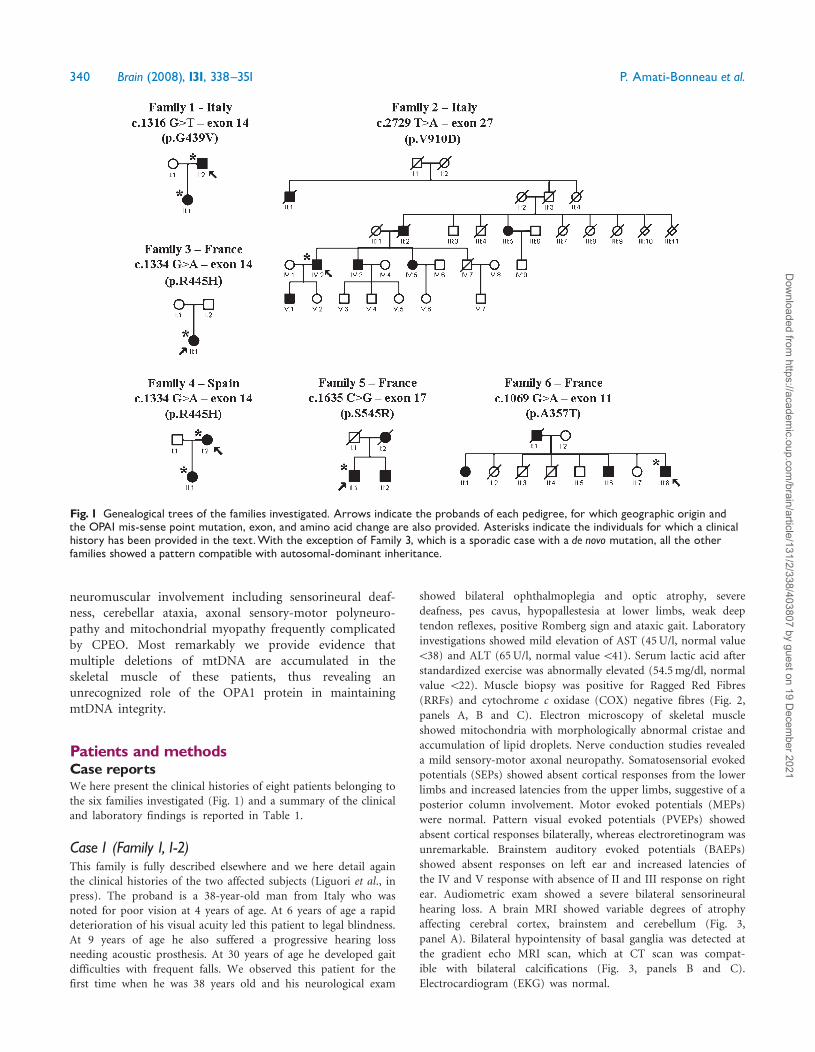
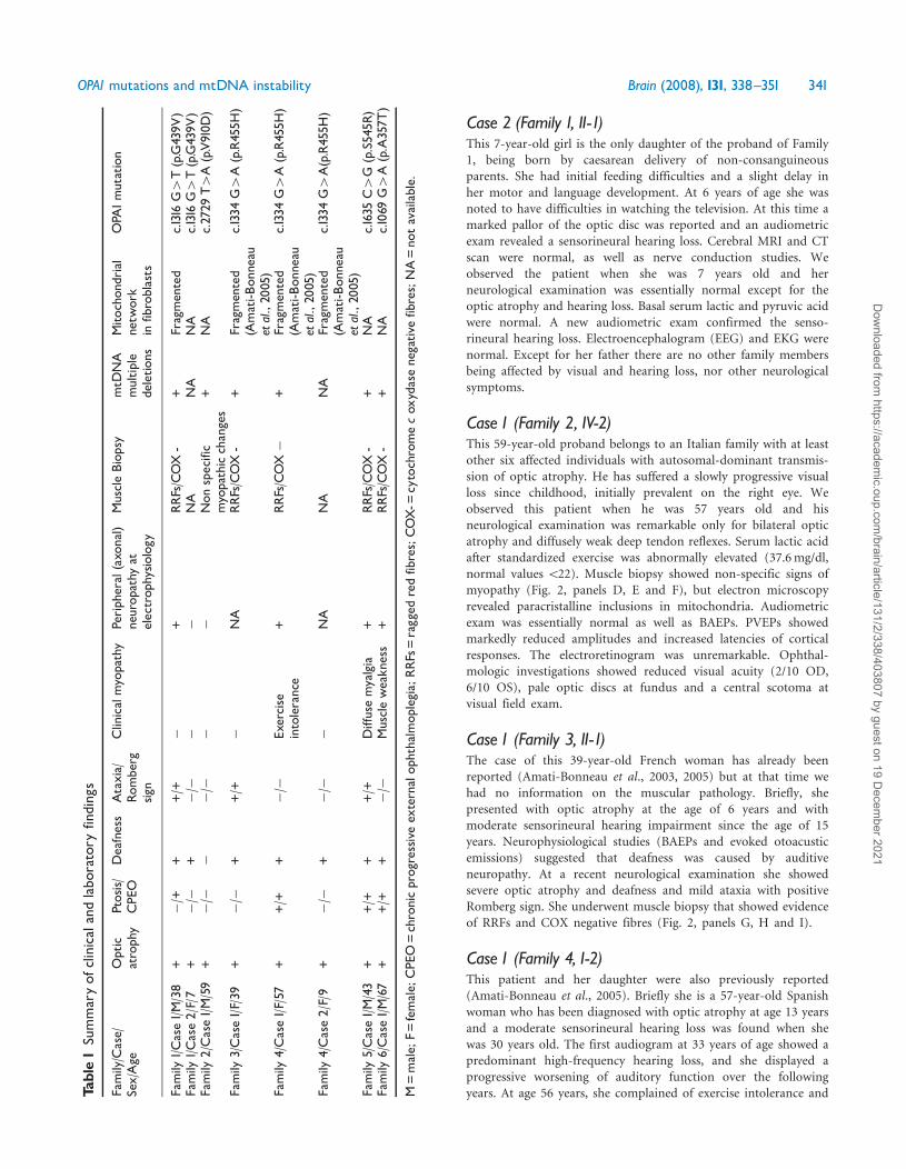
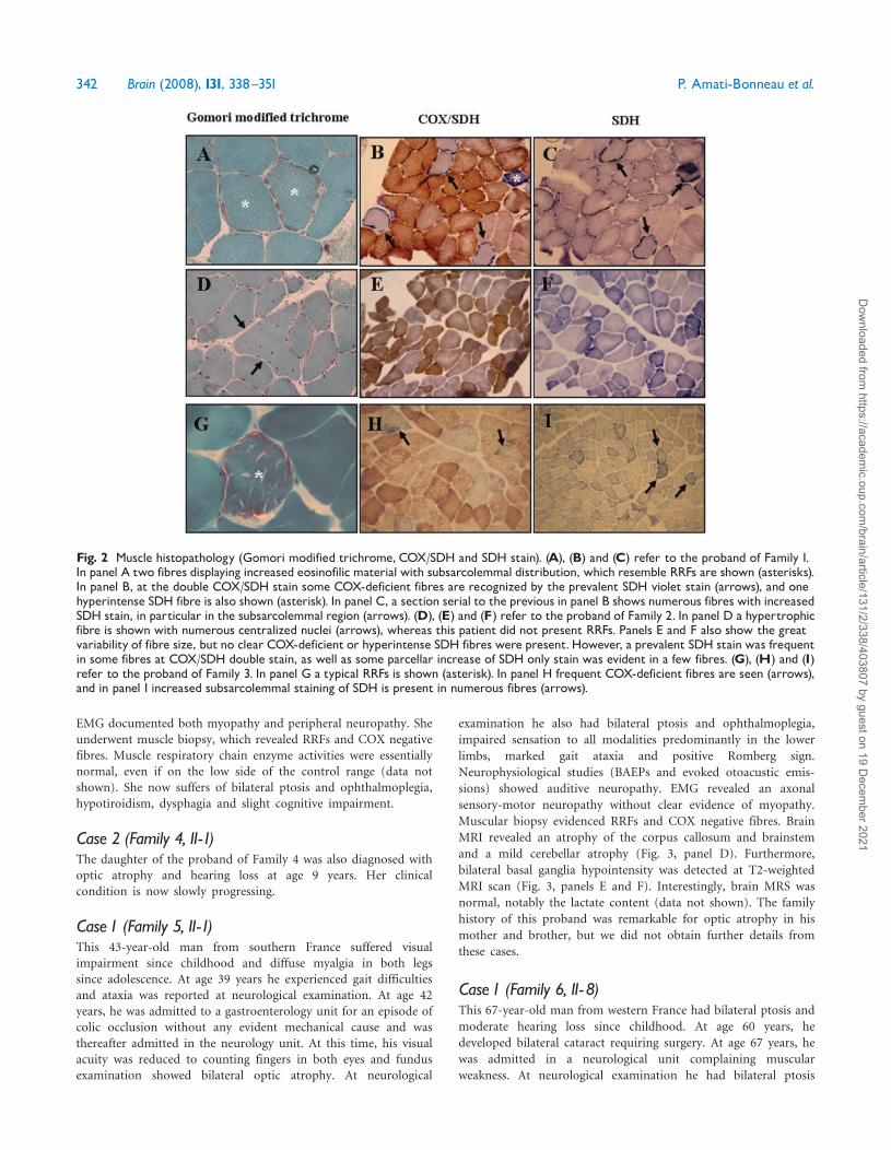
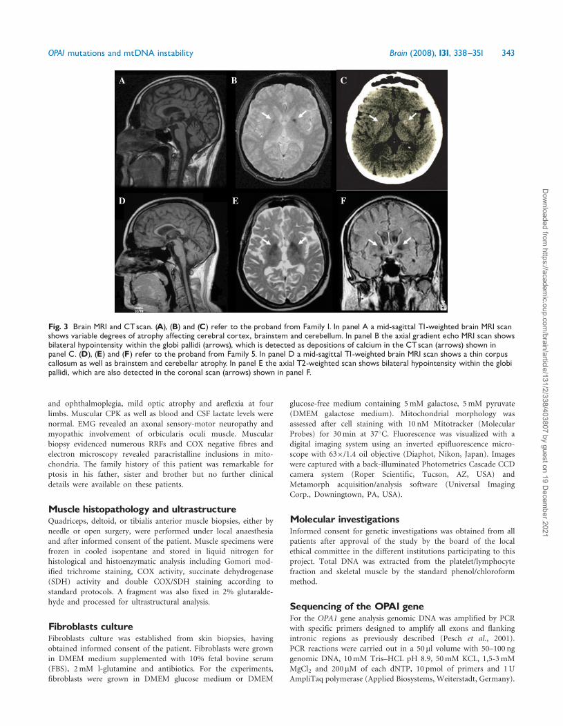
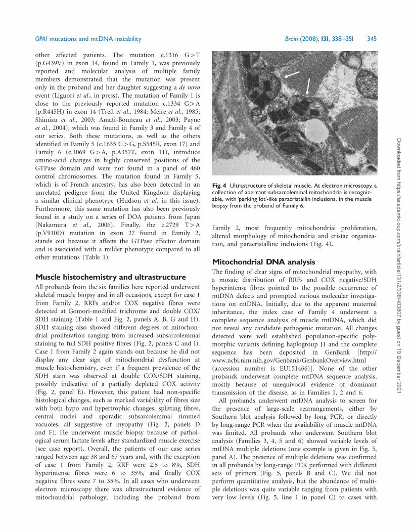
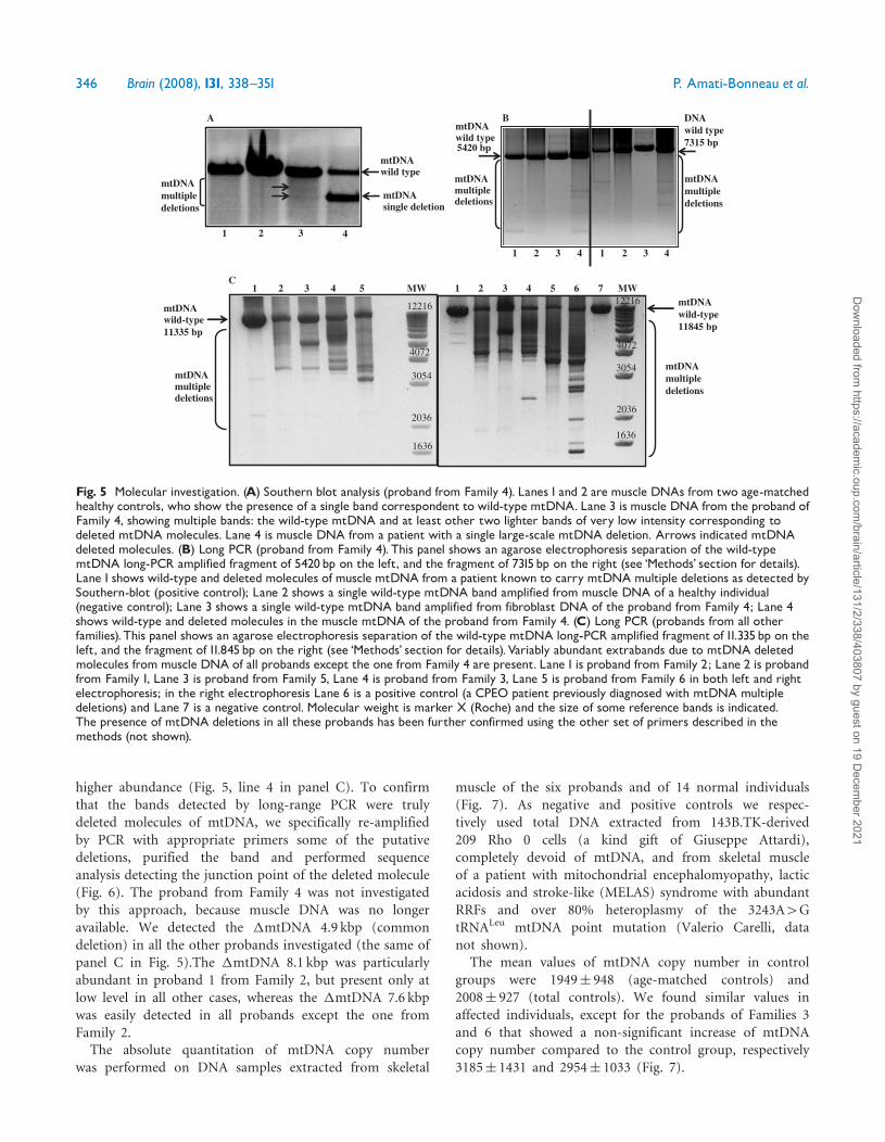
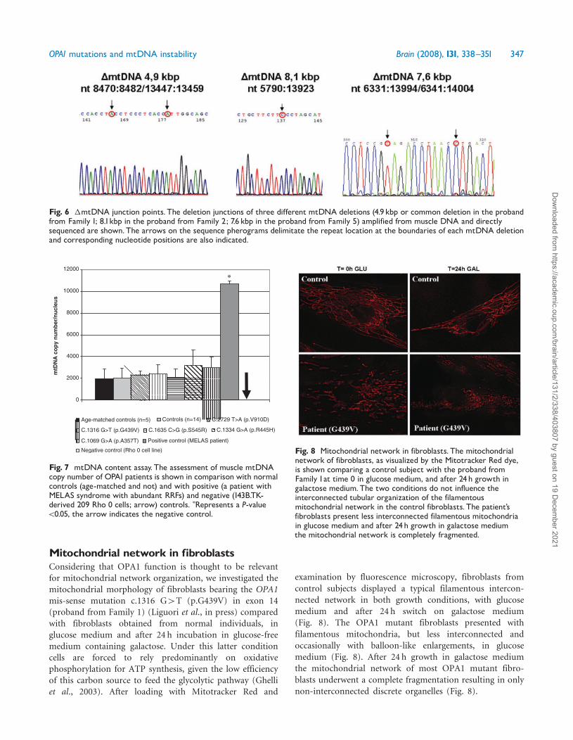
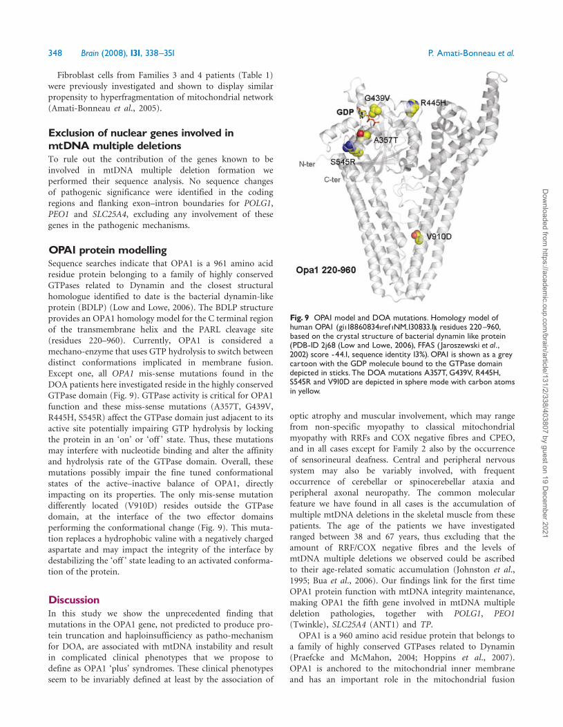
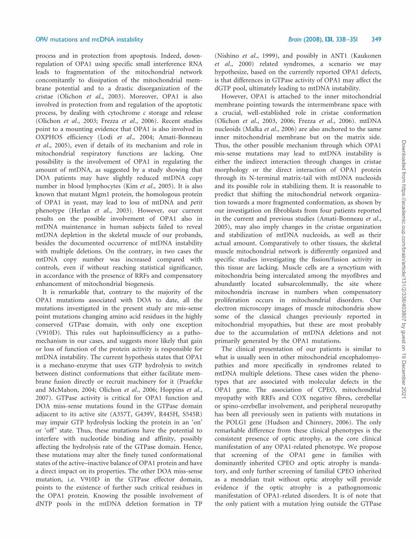