oogenesis and egg development in triatomines: a ... · pdf fileoogenesis and egg development...
TRANSCRIPT

Anais da Academia Brasileira de Ciências (2005) 77(3): 405-430(Annals of the Brazilian Academy of Sciences)ISSN 0001-3765www.scielo.br/aabc
Oogenesis and egg development in triatomines:a biochemical approach
GEORGIA C. ATELLA1, KATIA C. GONDIM1, EDNILDO A. MACHADO2
MARCELO N. MEDEIROS2, MÁRIO A.C. SILVA-NETO1 and HATISABURO MASUDA1
1Instituto de Bioquímica Médica, Centro de Ciências da Saúde, Bloco HUniversidade Federal do Rio de Janeiro, UFRJ, Av. Bauhinia s/n, Cidade Universitária, Ilha do Fundão
21944-590 Rio de Janeiro, RJ, Brasil2Instituto de Biofísica Carlos Chagas Filho, Centro de Ciências da Saúde, Bloco G
Universidade Federal do Rio de Janeiro, UFRJ, Av. Bauhinia s/n, Cidade Universitária, Ilha do Fundão21944-590 Rio de Janeiro, RJ, Brasil
Manuscript received on March 3, 2005; accepted for publication on March 30, 2005;presented by Lucia Mendonça Previato
ABSTRACT
In triatomines, as well as in other insects, accumulation of yolk is a process in which an extra-ovarian tissue,
the fat body, produces yolk proteins that are packed in the egg. The main protein, synthesized by the fat
body, which is accumulated inside the oocyte, is vitellogenin. This process is also known as vitellogenesis.
There are growing evidences in triatomines that besides fat body the ovary also produces yolk proteins. The
way these yolk proteins enter the oocyte will be discussed. Yolk is a complex material composed of proteins,
lipids, carbohydrates and other minor components which are packed inside the oocyte in an organized manner.
Fertilization triggers embryogenesis, a process where an embryo will develop. During embryogenesis the
yolk will be used for the construction of a new individual, the first instar nymph. The challenge for the
next decade is to understand how and where these egg proteins are used up together with their non-protein
components, in pace with the genetic program of the embryo, which enables cell differentiation (early phase
of embryogenesis) and embryo differentiation (late phase) inside the egg.
Key words: oogenesis, vitellogenesis, embryogenesis, triatomines, Rhodnius prolixus.
INTRODUCTION
During the evolution of species a very important
achievement, by Mother Nature, was the develop-
ment of multicellular organisms starting from a
unique cell, the fertilized oocyte. This strategy was
adopted by most multicellular organisms and among
them we can recognize oviparous and non-oviparous
Dedicated to the memory of Prof. Dr. Herman Lent byinitiative of Pedro L. Oliveira, Instituto de Bioquímica Médica.Universidade Federal do Rio de Janeiro (UFRJ), RJ, Brasil.Correspondence to: Hatisaburo MasudaE-mail: [email protected]
living organisms. In oviparous organisms, including
insects, embryonic development occurs apart from
the maternal body. Therefore, egg survival relies on
the utilization of previously stored material for em-
bryo’s growth. This material, the yolk, is composed
of proteins, lipids, sugars and other minor compo-
nents, and is stored in a highly organized manner
inside the egg. Following fertilization the egg will
give rise to an entire new organism. During devel-
opment, yolk will be gradually used in accordance
with the needs of specific cells imposed by the ge-
netic program of the embryo.
An Acad Bras Cienc (2005) 77 (3)

406 GEORGIA C. ATELLA ET AL.
The accumulation of yolk, or vitellogenesis,
is a process in which extra-ovarian tissues produce
yolk protein precursors such as vitellogenin (VG)
the principal protein of the oocyte (Postlethwait and
Giorgi 1985, Raikhel and Dhadiala 1992). Due to
the heterosynthetic nature of vitellogenesis in in-
sects, their oocytes are specialized for the specific
accumulation of yolk proteins. Therefore, insect
oocytes present a whole set of structures designed
to select, internalize and store specific proteins, such
as microvilli, coated pits, coated vesicles and yolk
granules. In most insects, a single phospholipogly-
coprotein, now named Vitellin (VT) to differentiate
it from VG, is the main component of the eggs and
serves both for embryonic and, in some cases, early
larval development (Postlethwait and Giorgi 1985,
Zhu et al. 1986, Oliveira et al. 1989).
Triatomines were the first group of insects
where the accumulation of an extra-ovarian protein
by vitellogenic oocytes was reported. The paper of
Sir V.B. Wigglesworth in 1943 described the pres-
ence in the egg of Rhodnius prolixus of a pink pig-
ment that was originated from a hemolymphatic pre-
cursor. The pigment was thought to be partially
degraded hemoglobin “katahemoglobin”. The pig-
ment described by Wigglesworth was isolated fifty
two years later, and characterized as RHBP (Rhod-
nius Heme-Binding Protein) in our laboratory (Oli-
veira et al. 1995), a protein that functions as an an-
tioxidant while in the hemolymph (Dansa-Petretski
et al. 1995) and which is accumulated inside the
oocytes (Machado et al. 1998). The remarkable
paper of Wigglesworth was, in fact, the very first re-
port on the accumulation of an extra-ovarian protein
by vitellogenic oocytes, opening the trail that led to
the characterization of VG uptake by Telfer (1954).
William Telfer (1960) was the first person to asso-
ciate endocytosis with yolk deposition in oocytes
of Hyalophora cecropia. He reported a selective
concentration of hemolymph-borne proteins and, in
1961, the route of entry in yolk granules. Soon af-
ter, Roth and Porter (1964) and Roth et al. (1976)
observed, in oocytes of the mosquito Aedes aegypti,
the presence of coated vesicles, the cellular struc-
tures associated with selective endocytosis. These
were the pioneering reports that were followed by an
enormous amount of other reports that corroborated
the initial investigations, as reviewed by Telfer et
al. (1982), Raikhel and Dhadiala (1992), and Sni-
girevskaya and Raikhel (2004). These studies led
to major milestones in our understanding of how
macromolecules are taken up from surrounding ex-
tracellular fluids into both mammalian and insect
cells.
The adaptation for blood feeding behavior,
among arthropods, evolved independently in sev-
eral groups so, during the course of evolution, dif-
ferent metabolic adaptations were used to obtain,
digest and use this special source of food (Lehane
1991). The majority of blood-feeding insects, in-
cluding triatomines, ingest a large amount of blood
in a single meal, comprising several times the ani-
mal’s own weight (Friend et al. 1965). The diges-
tion of blood brings to the insect a special problem
related to the release of an enormous amount of free
heme upon degradation of hemoglobin. Heme is a
toxic molecule due to its ability to generate reactive
oxygen species (Aft and Mueller 1983, Vincent et al.
1988, Vincent 1989, Smith 1990). Thus, heme toxi-
city must be counteracted with very efficient defense
systems. In R. prolixus, several lines of defense have
been characterized such as Rhodnius Heme-Binding
Protein (RHBP) (Dansa-Petretski et al. 1995); uric
acid (Souza et al. 1997); hemozoin formation in
the midgut (Oliveira et al. 1999); and antioxidant
enzymes (Paes and Oliveira 1999, Paes et al. 2001).
The digested blood, on the other hand, also
contains a large amount of amino acids and lipids
that can be used to produce molecules such as Vitel-
logenins (VGs) that are secreted to the hemolymph
to be taken up by the growing oocytes. The ma-
jor hemolymph lipoprotein of insects is called lipo-
phorin, and its most studied role is the inter organ
transport of lipids (Chino et al. 1977, 1981), thus
lipids can be carried and delivered to the ovaries
(Kawooya et al. 1988). Lipophorin is also stored
inside the oocytes of moths (Telfer et al. 1991).
Besides proteins, carbohydrates and lipids, the
An Acad Bras Cienc (2005) 77 (3)

OOGENESIS AND EGG DEVELOPMENT IN TRIATOMINES 407
A B
CTrophic Cord
Oocyte Vitellarium Tropharium
Ovary Ovariole
Trophic cord
Fig. 1 – The ovary of Rhodnius prolixus: The ovary was dissected at day 5 after blood meal and spread onto a glass slide and
photographed. The ovary (A) is composed of two hemi-ovaries that are connected by a common oviduct. Each hemi-ovary is
composed of seven structures named ovariole. From the hemi-ovary depicted at the right side of Fig. 1A one ovariole was removed to
be shown in detail (B). A line drawing of the ovariole is shown in (C), in order to represent the internal structures (After Huebner and
Anderson 1972).
oocytes of triatomines need other components such
as heme, calcium, and also mechanical protection
of the egg and protection against microorganisms,
since embryo development occurs outside maternal
body. Once fertilized, the egg is laid and the em-
bryo development is initiated. So, components nec-
essary for embryo development such as DNA, pro-
teins, lipids, carbohydrates and enzymes must all be
packed inside the egg.
THE OVARIES OF TRIATOMINES
A broad spectrum of studies with hemipterans, in-
cluding triatomines, has established that they are
telotrophic (Bonhag 1955, 1958, Davis 1956, Mas-
ner 1968, Brunt 1971, Huebner and Anderson 1972,
Schreiner 1977, Couchman and King 1979, Bun-
ing 1981). Telotrophic ovary is a quite complex
structure; as shown in Fig. 1A R. prolixus ovary is
composed of two hemi-ovaries. Each hemi-ovary
is composed of seven structures named ovarioles
which are depicted in Fig. 1B. Internal structures
of the ovariole are depicted in Fig. 1C. The cell bi-
ology and structures of telotrophic ovaries were re-
viewed by Telfer et al. (1982) and Huebner (1984).
A female of R. prolixus is able to produce around
40 to 42 eggs in 15 days after a single blood meal,
thus each ovariole is able to produce approximately
three eggs, since Rhodnius ovary contains 14 ovari-
oles. The way these eggs are produced will be com-
mented here. The purpose of this review is to bring
into discussion the oocyte proteins of triatomines,
especially because these insects feed on blood and
use them as substrates to build up their eggs. We
present a brief survey of the main proteins and try
to describe their mode of entry, their accumulation
inside the oocyte and their use by the growing em-
bryo. Among the proteins that will be commented
are VGs, lipophorin, Rhodnius Heme-binding pro-
tein (RHBP) and Rhodnius Calcium-binding Protein
(RCBP). The involvement of enzymes such as phos-
phatases, proteases and kinases in the mobilization
of yolk, during embryogenesis, will also be com-
mented.
VITELLINS AND VITELLOGENINS
VT is derived fromVG, which in most insects is syn-
thesized in the fat body (Pan et al. 1969, Engelmann
1979, Harnish et al. 1982) as a single or multiple
precursors that are processed before secretion to the
hemolymph (Raikhel et al. 1990, Tufail et al. 2004).
An Acad Bras Cienc (2005) 77 (3)

408 GEORGIA C. ATELLA ET AL.
Synthesis and accumulation of yolk proteins are un-
der hormonal control as reviewed by Hagedorn and
Kunkel (1979), Davey (1981), Engelmann (1990),
Raikhel and Dhadiala (1992) and Bellés (2004). In
some insects, complementary synthesis of VGs by
the ovaries has been described (Postlethwait et al.
1980, Bianchi et al. 1985, Rousset et al. 1988,
Melo et al. 2000). VG expression and secretion
to the hemolymph by fat body and uptake by the
ovary are regulated at the hormonal and sexual lev-
els. Since the first report on R. prolixus, involv-
ing corpus allatum (juvenile hormone) in the regu-
lation of vitellogenesis (Wigglesworth 1936), it has
become apparent that juvenile hormone influences
vitellogenesis in the majority of insects (Brookes
1969, Engelmann 1970). The replacement of juve-
nile hormone following the removal of corpora allata
restores vitellogenesis.
Among hemipterans, the process of vitelloge-
nesis is, in fact, controlled by juvenile hormone as
demonstrated by Coles (1965) in R. prolixus, and by
Mundall and Engelmann (1977) in Triatoma pro-
tracta. On the other hand, it was also demonstrated
in R. prolixus that oogenesis is inhibited by ecdysone
in a dose-dependent fashion (Garcia et al. 1979).
In Triatoma protracta and R. prolixus, vitellogene-
sis is stimulated by mating. The effect of mating
on egg production of R. prolixus is such that dur-
ing the early gonadotrophic cycle the rate of vitel-
logenesis is the same in both mated and virgin fe-
males, but at some point onward vitellogenesis is
strongly inhibited (Pratt and Davey 1972a). Mating
activates corpus allatum to produce juvenile hor-
mone through ventral nervous cord (Mundall and
Engelmann 1977, Davey 1987). It is generally ac-
cepted that in R. prolixus oogenesis is controlled by
hormones, being released in response to abdominal
stretch during blood feeding (Davey 1987). Besides
that, it was suggested that stretching alone is not
enough to induce oogenesis, which is also depen-
dent on the presence of proteins in the diet (Garcia
and Azambuja 1985). Insects fed with protein-free
diet failed to induce oogenesis although abdominal
stretch had occurred. It was also observed that the
origin of the blood has strong influence on R. pro-
lixus oogenesis (Lima Gomes et al. 1990, Valle et
al. 1987, 1992).
In R. prolixus, juvenile hormone acts directly
on the ovary, controlling the enlargement of inter-
follicular channels, a condition known as patency,
allowing macromolecules from the hemolymph,
such as VG, lipophorin, RHBP etc to reach the sur-
face of the oocyte. This enlargement is thought to be
the consequence of Na+/ K+ ATPase activity present
in follicular cells. This effect is antagonized by
antigonadotropins (Pratt and Davey 1972b, Davey
1981). It has been suggested that the function of
antigonadotropins is to assure the cessation of vitel-
logenesis.
Recently, Medeiros et al. (2002, 2004) have
provided evidences that eicosanoids control insect
oogenesis, modulating the levels of cAMP on the
ovarian follicles. In many vertebrate processes, in-
cluding endocytosis, eicosanoids modulate the level
of second messengers (Smith 1989). Eicosanoids
receptors are coupled to changes in the intracel-
lular levels of cAMP (Stanley-Samuelson and
Pedibhotla 1996). So, events controlled by cAMP
and protein phosphorylation, such as receptor re-
cycling, vesicle fusion and vesicles recycling can
be coordinated by eicosanoids (Fallon and Schwartz
1988, Griffiths and Hollinshead 1990, Colombo et
al. 1994, Fallon et al. 1994, Mellman 1996, Foti et
al. 1997). In R. prolixus, lipoxygenase products
down-regulate cAMP production, thus increasing
the rate of yolk uptake while cyclooxygenase prod-
ucts upregulate cAMP levels inhibiting yolk accu-
mulation. In Hyalophora cecropia, it was reported
that cyclic nucleotides (cAMP and cGMP) induce
termination of VG uptake (Wang and Telfer 1996).
Among triatomines R. prolixus is the most stud-
ied insect with respect to VG and VT. Scarce in-
formation is available in other triatomines species
such as Triatoma infestans and Triatoma protracta.
The native molecular mass of VG of Triatoma pro-
tracta is 437 kDa (Mundall and Engelmann 1977)
and 220 kDa for VG and VT of Triatoma infestans
(Salomón and Stoka 1986). VT and VG of Triatoma
An Acad Bras Cienc (2005) 77 (3)

OOGENESIS AND EGG DEVELOPMENT IN TRIATOMINES 409
infestans show partial immunological identity with
VG and VT of Panstrongylus megistus and R. pro-
lixus (Salomón and Stoka 1986).
In R. prolixus, VT native molecular mass is 430
and 260 kDa (Chalaye 1979). The native particles
dissociate in four subunits under denaturing con-
ditions. The molecular mass of the subunits are
180 kDa, 158 kDa, 50 kDa and 46 kDa (Masuda
and Oliveira 1985). Similar estimates for VT sub-
units were obtained by Valle et al. (1987). The VG
of R. prolixus is synthesized in the fat body as a high
molecular mass precursors of 205 kDa and 190 kDa
(Valle et al. 1993). VG is secreted to the hemolymph
and it is taken up by the growing oocytes at the
ovary by receptor mediated endocytosis (Oliveira et
al. 1986). Inside the eggs VG is referred to as VT
to differentiate it from VG from the hemolymph.
Despite this nomenclature, slight physical-chemical
modifications occur on VG molecule during its up-
take and storage, and scarce information is available
for most insect groups. Upon fertilization,VT which
was previously accumulated inside the oocyte dur-
ing the process of oogenesis is used by the new em-
bryo and ends up with the eclosion of the first instar
nymph (Oliveira et al. 1989). Embryonic develop-
ment occurs initially as an embryonic differentiation
that is followed by nymphal differentiation inside
the egg. During the initial phase VT is not signifi-
cantly utilized. In R. prolixus around day 10, when
katatrepsis occurs (Huebner 1984), only 15% of the
total VT are used (Oliveira et al. 1989). From this
point onward, VT is rapidly used by the embryo and
at day 15 the first instar nymph hatches. Around
50% of the total content of VT are still present in
the gut of the new insect, which disappears in the
following five days (Oliveira et al. 1989). So, pos-
sibly this strategy is used by these insects to improve
their chance to survive in nature, since the first instar
nymphs have approximately five days to find a new
host from where they will suck blood for the first
time in their lives.
It was very intriguing the fact that only 15% of
total VT are used to build up a new embryo (kata-
trepsis). The remaining VT is utilized inside the
gut of the embryo. This fact suggests that part of
the VT content is used for embryonic differentiation
and another part for nymph differentiation inside and
outside of the egg. Looking more carefully to the
characteristics of VT, it was possible to recognize
and separate three different populations of VT in R.
prolixus (Salerno et al. 2002). These populations
were named VT1, VT2 and VT3 according to the
order of elution in an ion exchange column. One in-
teresting feature about them is that they seem to be
segregated inside the oocyte (Salerno, unpublished
data). Another interesting feature about them is that
the content of phosphate varies. VT1 is the less
phosphorylated population and the first to be eluted
from the column (Salerno et al. 2002). This varia-
tion can eventually be explained by the presence of
two enzymes present in R. prolixus oocytes, an acid
phosphatase (Nussenzveig et al. 1992) and casein
kinase II (CK II) (Silva-Neto and Oliveira 1993).
Masuda and Oliveira (1985) have shown the pres-
ence of phosphate associated with protein moiety of
VT, specifically at serines residue. CK II recognizes
clusters of phosphorylated serine residues, so the
participation of this enzyme in the modification of
phosphorylation status of this protein is a good pos-
sibility. Melo et al. (2000) demonstrated in R. pro-
lixus that epithelial cells derived from large follicles
are able to synthesize ovarian VG (O-VG). O-Vg
elutes at the same position of VT1, suggesting that
VT1 is of different origin from other populations.
Salerno (2001) demonstrated that VG, synthesized
at the fat body, gives rise to the VT3 population as
consequence of its post-endocytic modification. Re-
cently, we have collected evidences that VT2 pop-
ulation is synthesized by trophocytes (Salerno, un-
published data). So, it looks like the different pop-
ulations found inside the R. prolixus oocytes are the
result of a concerted effort of different cells such
as fat body cells, follicular cells and trophocytes.
The origin of different populations may explain the
differential behavior of each population in ion ex-
change column.
No significant differences were observed in
neutral lipid composition among all different popu-
An Acad Bras Cienc (2005) 77 (3)

410 GEORGIA C. ATELLA ET AL.
lations of VT, but some differences in the glycosila-
tion status of proteins were observed. The presence
of glucose associated with the oligossacharide of
VG purified from the hemolymph and also associ-
ated with the oligossacharide of ovarianVG suggests
that VGs from fat body and ovary are secreted after
their oligossacharide is processed at the Golgi appa-
ratus. The decrease in the content of glucose of VT3
must be attributed to the post-endocytic processing
of VG. Post-endocytic processing of VT has already
been reported by Giorgi et al. (1993). The lack of
glucose in the oligossacharide associated with VT2
can be attributed to its different origin.
It is intriguing that in some insects VG is syn-
thesized in the fat body, in others also by
follicle cells and in the case of Rhodnius also by the
tropharium cells characterizing its triple origin. The
timing of O-VG synthesis of R. prolixus resembles
that of egg-specific protein of Bombyx mori (Irie
and Yamashita 1983). This protein is also synthe-
sized in follicle cells and appears in the late phase of
oocyte maturation. It is tempting to speculate that
O-Vg (or VT1) of R. prolixus plays a similar role,
as egg specific protein in Bombyx mori. In this in-
sect, egg-specific protein is located at the outer layer
of the oocyte and utilized even before VG (Irie and
Yamashita 1980).
VT1 is localized at the outer layer of oocyte
(Salerno, unpublished data), so this population of
VT will possibly be the first one to be used by the
embryo, similar to what occurs with egg-specific
protein of Bombyx mori. The proportion of VT1
population from the total is around 15%, approxi-
mately the amount used by the embryo up to day 10
after oviposition (Oliveira et al. 1989) when yolk is
enclosed by the embryo (Kelly and Huebner 1986).
A very important feature of Rhodnius VTs is
the fact that they are produced and stored inside the
egg in different periods of oocyte maturation. VT2
is probably the first one to be stored inside the egg
since it is synthesized by the trophocytes (Coelho
and Gomes, personal communication), followed by
the deposition of VT3 from the hemolymph. The
last VT to be deposited is VT1 which is synthesized
by follicle cells derived from large follicles (Melo
et al. 2000).
NON-VITELLIN YOLK PROTEINS
As VTs can account for 60 to 90% of the protein
content of the mature oocyte, other oocyte proteins
have been taken out of the spotlight, independent of
their being also abundant, varying from 1-20% of
total proteins. Although VTs are known to serve
as source of amino acids, lipids, phosphate, and
carbohydrates for the growing embryo (Yamashita
and Indrasith 1988), non-VT proteins carry other
non-proteic components to be used by the embryo
(biliverdin, carotenoids, iron, calcium, heme and an
extra amount of amino acids, phosphate and lipids)
as recently reviewed by Masuda et al. (2004).
RHODNIUS HEME-BINDING PROTEIN (RHBP)
It has recently been discovered that the presence of a
pink pigment in the eggs of R. prolixus described in
the work of Sir V.B. Wigglesworth (1943) as being
partially degraded hemoglobin (“katahemoglobin”),
is in fact a new protein (Oliveira et al. 1995) now
known as Rhodnius Heme-Binding Protein (RHBP).
Wigglesworth also presented data suggesting that
a similar phenomenon was occurring in the cattle
tick Boophilus microplus, where thousands of deep-
brown colored eggs are laid after a blood meal.
The hypothesis of Wigglesworth that heme
from vertebrate was being recycled by the arthro-
pods was also adopted by O’Hagan (1974), who
showed that an amount of heme equivalent to 10%
of that found in the blood ingested by the tick was
found in the eggs of this arachnid. However, the
suggestion that the heme from the diet was being re-
used was not demonstrated directly and was openly
in disagreement with the well-established concept
that all nucleated cells make their own heme (Ponka
1997). This apparent conflict was directly investi-
gated by Braz et al. (1999), showing Boophilus as
the first multicellular organism to be unable to make
its own heme. Therefore a hallmark in the adapta-
tion of ticks to feed on blood during evolution may
An Acad Bras Cienc (2005) 77 (3)

OOGENESIS AND EGG DEVELOPMENT IN TRIATOMINES 411
have been the acquisition of mechanisms to absorb
and transport heme through the hemolymph and de-
liver it to cells, especially to the growing oocytes,
where a huge amount of heme is accumulated dur-
ing oogenesis.
We isolated and characterized the pink pigment
(RHBP) from both hemolymph and oocytes of R.
prolixus, showing that it was in fact a hemeprotein
composed of a single 15-kDa polypeptide chain.
cDNA of RHBP was cloned and sequenced. The
full-length RHBP cDNA codes for a pre-protein of
128 amino acids and the deduced amino acids se-
quence present no significant homology to any
known proteins (Paiva-Silva et al. 2002). Studies
of RHBP structure are under progress (Nagem et
al. 2001).
The presence of apoRHBP in the hemolymph
functions as an antioxidant defense to protect Rhod-
nius from the challenge of dealing with the excess
of heme formed upon hemoglobin digestion. RHBP
in the hemolymph promptly binds one heme
per polypeptide chain non-covalently. The bind-
ing of heme by RHBP suppresses the generation
of activated oxygen species, thus protecting the in-
sect against oxidative stress (Dansa-Petretski et al.
1995). As mentioned above, in spite of the heme
detoxification mechanisms (hemozoin formation),
part of the heme, originated from the digestion of
hemoglobin, crosses the gut wall (Dansa-Petretski et
al. 1995), and binds to RHBP, which led to the idea
that heme was being recycled in Rhodnius. The sit-
uation in Rhodnius is more complex than that of the
tick, because the hemipteran has a fully active heme
biosynthesis pathway (Braz et al. 1999). Hence,
possibly Rhodnius synthesize its own heme but also
recycles part of the heme from the digestion of host
hemoglobin.
An important function of RHBP in the hemo-
lymph is to transport heme to the heart where it is de-
graded to biliverdin (Paiva-Silva, unpublished data).
This “unloading” of RHBP in the heart is probably
important to help the insect to maintain a safe level
of apo-RHBP in the hemolymph.
RHBP isolated from the oocyte is fully satu-
rated with heme and cannot bind additional heme.
So, it is not possible to attribute any anti-oxidant
role to this protein inside the oocyte. The function
of RHBP in the egg must be of a different nature from
that in the hemolymph. The presence of RHBP as-
sociated with heme in the oocyte is responsible for
the characteristic pink color of R. prolixus’ oocytes.
RHBP is accumulated by growing oocytes
through receptor-mediated endocytosis and is di-
rected to the yolk granules (Machado et al. 1998).
Although several organs and tissues are able to in-
corporate RHBP, the ovary is by far the most active
site of RHBP accumulation.
Limiting the availability of heme-RHBP, but
maintaining normal levels of VG and lipophorin in
the hemolymph, the females of R. prolixus produce
only a few but normal eggs. Experimental maneu-
vers to increase the concentration of heme-RHBP
in the hemolymph resulted in an increased num-
ber of eggs produced in a dose-dependent manner
(Machado et al. 1998). This is an indication that
a balanced amount of heme-RHBP and other yolk
proteins such as VT is required to build up the yolk
granules.
VG and lipophorin do not compete with RHBP
during the process of RHBP uptake, suggesting ei-
ther the existence of different receptors or different
binding sites for the same receptor. Recently, the
presence of a lipophorin receptor has been discov-
ered, independent of the Vg receptor in Aedes ae-
gypti oocyte (Cheon et al. 2001). Independently of
the mechanism used for their internalizations, it is
clear that a cross information is required in order to
obtain a balanced amount of RHBP and VG inside
the mature egg. When RHBP is limiting, the strategy
used by the female is to reduce the number of viable
eggs. All attempts to induce R. prolixus females to
produce eggs without heme-RHBP, using diets free
of heme, failed (Machado, unpublished data).
The expression of RHBP is induced by a blood
meal (Paiva-Silva et al. 2002) and is temporally cor-
related with the appearance in the hemolymph of
other yolk proteins such as VG (Oliveira et al. 1986)
and lipophorin (Gondim et al. 1989), providing the
An Acad Bras Cienc (2005) 77 (3)

412 GEORGIA C. ATELLA ET AL.
balanced need of VG RHBP and lipophorin for egg
production. Besides that, RHBP secretion by fat
bodies is also dependent on intracellular levels of
heme (Paiva-Silva et al. 2002). The amount of heme
originated from hemoglobin digestion is not enough
to guarantee normal oogenesis in vitellogenic Rhod-
nius females. De novo heme biosynthesis is neces-
sary to obtain a normal vitellogenesis. Using suc-
cynil acetone, a specific inhibitor of ALA dehy-
dratase (Lindblad et al. 1977), a substantial decrease
in the number of eggs laid is observed (Braz et al.
2001), which is reversed by feeding the insects with
porphobilinogen, an intermediate downstream in the
pathway.
Very recently, it has been shown that the entry
of RHBP in the oocyte is controlled by eicosanoids
(Medeiros et al. 2002, 2004). PGE2 (one of the
products of the cyclooxygenase pathway) decreased
RHBP uptake, while indomethacin (a cyclooxyge-
nase inhibitor) increased uptake in vitro and in vivo.
Upon treatment with indomethacin, two ovarian
polypeptides (18 and 25 kDa) were dephosphory-
lated, suggesting that a protein phosphorylation sig-
nal transduction pathway is involved. In fact, there is
a growing set of evidences relating endocytosis and
cell signaling pathways (Di Fiore and De Camilli
2001).
The tight control observed between the uptake
of RHBP and the other yolk proteins such as VT
required to build up the yolk granules, can be ex-
plained in part by the following observations: a clear
decline in RHBP content is observed during embryo-
genesis without reduction in heme content (Braz et
al. 2002), thus suggesting that heme from RHBP is
recycled. So, a minimum amount of heme, inside
the egg, must be provided in order to obtain a normal
development.
In several arthropods including Rhodnius, a
large fraction of the yolk remains unused in the crops
of first instar nymphs (Oliveira et al. 1989). In the
first five days following hatching, Rhodnius nymphs
completely degrade the remaining yolk proteins and
a dark material can be observed in the lumen of
midgut of these non-fed insects, possibly hemozoin.
Hemozoin formation can be promoted by perimi-
crovillar membranes of the midgut (Oliveira et al.
2000), but it can also be an autocatalytic process,
dependent on the existence of pre-formed hemozoin
(Dorn et al. 1995). The presence of hemozoin in the
midgut of the non-fed nymphs will possibly seed the
formation of more hemozoin as the result of blood
digestion that will occur when the insect feeds on
vertebrate blood for the first time in its life. This is
also an important reason for the presence of RHBP in
the eggs. Taken together, these observations demon-
strate that RHBP utilization during embryogenesis
can provide heme to de novo synthesis of embryo
hemeproteins. Also that is a source of food for the
newly hatched nymph and seeds the formation of
hemozoin in the gut of the newly hatched nymph,
to prepare the insect to receive its first ingestion
of blood in nature. All these events are important
enough for the embryo development so that the fe-
males of R. prolixus do not produce a single egg
without RHBP in it.
LIPOPHORIN
During oogenesis in insects, oocytes accumulate a
great amount of lipids, which are supplied by lipo-
phorin, a major hemolymphatic lipoprotein (Chino
et al. 1981, Kawooya and Law 1988, Sun et al.
2000, Ziegler and Ibrahim 2001). Lipophorin trans-
ports different classes of lipids, such as diacylglyc-
erol, phospholipids, cholesterol, hydrocarbons, free
fatty acids, between insect various tissues, accord-
ing to physiological demand (Soulages and Wells
1994, Blacklock and Ryan 1994, Ryan and Van der
Horst 2000). Hematophagous insects, such as the
triatomines R. prolixus and Panstrongylus megis-
tus, ingest large amounts of blood at each meal and,
like it occurs in other insects, digestion causes the
release of free fatty acids in midgut lumen which,
after absorption, are used in the midgut epithe-
lium for the synthesis of other lipids, as phospho-
lipids, cholesteryl esthers, tri- and diacylglycerol
(Tsuchida and Wells 1988, Turunen and Crailsheim
1996, Canavoso et al. 2004, Grillo and Gondim,
An Acad Bras Cienc (2005) 77 (3)

OOGENESIS AND EGG DEVELOPMENT IN TRIATOMINES 413
unpublished data). Lipids are then transferred to
circulating lipophorin that transports them to the or-
gans where they are stored and/or utilized (Atella et
al. 1995, Coelho et al. 1997, Canavoso et al. 2004).
In the adult females of R. prolixus, during the
days following a blood meal, lipophorin transports
phospholipids (Gondim et al. 1992) to the ovaries,
and the rate of incorporation of these lipids varies
according to oocyte developmental stage (Gondim
et al. 1989), in the same way that it was observed
for the rate of VT uptake (Oliveira et al. 1986).
Considering the fact that during oogenesis a huge
amount of membranes must be synthesized to cope
with oocyte growth and accumulation of yolk gran-
ules (Telfer et al. 1982), phospholipids incorporated
by vitellogenic oocytes are possibly used to build up
these membranes. In fact, when the fate of radioac-
tive phospholipids was followed after their transfer
from lipophorin to oocytes, a great part of radioac-
tivity was found associated with membranes of yolk
bodies (Gondim 1992).
Chino et al. (1977) addressed the question of
lipid accumulation in the ovaries of the silkmoth
Philosamia cynthia, and concluded that there was
more lipid in the oocytes than it was expected due
to the present amounts of lipophorin and VT. It was
proposed, then, that lipophorin delivered lipids, but
was not incorporated by the ovaries.
In R. prolixus, oocytes store triacylglycerol
(Santos and Gondim, unpublished data), possibly
in lipid droplets, that will be used during embryo-
genesis, as discussed below. So, in triatomines,
lipophorin probably delivers not only phospholipids,
but also diacylglycerols to the growing oocytes, as it
does in Manduca sexta, where the participation of a
membrane-associated lipoprotein lipase may be in-
volved (Van Antwerpen et al. 1998). In this moth,
where lipophorin of two density classes (Beenakkers
et al. 1988) can be found in adult hemolymph, HDLp
(high density lipophorin) is taken up, associated
lipids are removed and the lipoprotein is converted
to a very-high density lipophorin (VHDLp) inside
the oocytes, and stored there (Kawooya et al. 1988).
However, in this same insect, most lipids are sup-
plied to the ovaries by circulating LDLp (low den-
sity lipophorin), that transfers lipids to the oocytes
without protein incorporation (Kawooya and Law
1988). In another lepidopteran, Hyalophora ce-
cropia, lipophorin is also accumulated by the ovaries
(Telfer et al. 1991), and in the mosquito Aedes ae-
gypti this lipoprotein can be found in oocytes (Sun
et al. 2000).
So, lipophorin may have its apoproteins incor-
porated or not, depending on insect species and also
on the organ under analysis, and when lipophorin
is not accumulated by the cells to which it deliv-
ers lipids, the particle can be reloaded, acting as a
reusable lipid shuttle (Downer and Chino 1985, Van
Heusden et al. 1987, Van Antwerpen et al. 1988,
Kawooya and Law 1988). This is the case of the
developing ovaries of R. prolixus, where lipophorin
apoproteins are not accumulated by the oocytes
(Gondim et al. 1989, Machado et al. 1996), and
it was demonstrated that it can be loaded with lipids
at the fat body and the midgut (Atella et al. 1992,
1995). The midgut shows a greater capacity to trans-
fer lipids to lipophorin soon after feeding, while the
fat body shows an increased rate of lipid transfer
later on, when midgut is almost empty (Coelho et
al. 1997).
Although lipophorin is not incorporated by the
developing oocytes of R. prolixus, for delivering
lipids it interacts with specific binding sites at cell
surface. Receptors for this lipoprotein were studied
in some insects, as the flight muscles and fat body
of Locusta migratoria (Hayakawa 1987, Dantuma
et al. 1996), the fat body and midgut of Manduca
sexta (Tsuchida and Wells 1990, Gondim and Wells
2000) and the ovaries of Aedes aegypti (Cheon et
al. 2001). In R. prolixus, lipophorin binding to spe-
cific sites at membranes from fat body and midgut
was characterized (Pontes et al. 2002, Grillo et al.
2003), and a receptor from oocyte membrane is un-
der investigation.
Later on, during egg development, imported
lipids will propel embryo formation. The role
played by these macromolecules should include fu-
eling of metabolic process, shaping and plasticity
An Acad Bras Cienc (2005) 77 (3)

414 GEORGIA C. ATELLA ET AL.
of cell boundaries, as well as intracellular signaling.
Despite the presumed role of lipid and lipid-derived
molecules on cell development, the number of stud-
ies addressing such matter is scarce. Most of the
available studies have focused on non-mammalian
vertebrates’ models such as chicken, lizards, alli-
gators and turtles (Rowe et al. 1995, Speake et al.
1998, Speake and Thompson 1999, 2000). As re-
gards insects, most studies have focused on the role
played by lipophorin in lipid storage. Once the num-
ber of studies where triatomine species were em-
ployed as models is still scarce we provide here a
brief account of data including a discussion of sim-
ilar aspects found in other insect orders.
The first study providing an analysis on lipid
dynamics during insect development was conducted
by Kinsella (1966). In this study a qualitative and
quantitative picture of sterol and sterol esters dy-
namics during Periplaneta americana egg develop-
ment is depicted. Total sterol content of develop-
ing embryos is kept constant whereas the propor-
tion of esterifed sterol increased. By the time of
nymphal eclosion the total content of esterified sterol
increased from 20 to 42%. Also, fatty acid compo-
sition of the sterol esters is quite altered. Sterol es-
ters isolated in early embryogenesis are appreciably
more saturated due to an increase on the content of
palmitic (16) and stearic (18) acids. Sterol esters iso-
lated late on embryogenesis are more unsaturated,
due to an increase on the content of oleic (18:1) and
linoleic acid (18:2). The significance of such shift on
fatty acid saturation is unclear and is probably linked
to their metabolic function during embryogenesis. A
similar shift on the composition of fatty acids asso-
ciated with neutral lipids was verified by Lipsitz et
al. (1969) in the cricket Acheta domesticus. Dur-
ing larval development the content of myristic acid
on monoacylglycerols changed from 34.5% of to-
tal sterified fatty acids at day 12 to 10.9 at day 30.
Such data demonstrate that the profile fatty acid ster-
ification is probably following punctual adjustments
during either embryo/larval development. The role
played by such adjustments, as well as the mecha-
nism underlying such changes, are still unknown.
Kinsella and Smyth (1966) showed that the
amount of mono- and diacylglycerols remained con-
stant throughout egg development. However, by
the time of nymphal emergence the amount of such
lipids increased sharply as a consequence of tria-
cylglycerol hydrolysis. Total extractable lipid de-
creased from 39.5 to 23.2% of the dry weight of the
oothecae. Triacylglycerol fraction solely decreased
by 75%. In the blood sucking-bug R. prolixus a
similar profile was found by our group. Triacyl-
glycerol content decreased to half the value found
in oocytes by the end of egg development period, al-
though triacylglycerol percentage was kept constant
during embryogenesis (Santos and Gondim, unpub-
lished data). Free fatty acid (FFA) metabolism was
also followed during the same period by Kinsella
(1967). Data indicated a rather different profile with
a surge on FFA content from 0.2 mg% of dry matter
to 1.4% in the last day of egg development. Such
result is a clear indication of triacylglycerol hydrol-
ysis mediated by lipase activity particularly before
nymph hatching. Therefore, measurements of FFA
levels are a direct measure of respiratory quotient
once such molecules are catabolized to provide res-
piratory energy for both invertebrate and vertebrate
tissue (Kinsella 1967).
Dynamics of larval to pupa transition was fol-
lowed by Pant et al. (1973). Total lipids and neu-
tral lipids increased through larval development and
attained the maximum concentration at fifth instar
stage. After pupation there was a sudden decrease
in pupal fat body lipids, which suggests their uti-
lization on metabolism. FFA and phospholipids are
also accumulated during the larval development in
the insect body. Once after ecdysis a decrease on
either free fatty acid or phospholipids is noticed in
pupal fat body.
The above mentioned studies employed sepa-
ration and quantification techniques to follow lipid
dynamics during insect development. Such strategy
implies in the homogenization of the whole tissue
and therefore assumptions could not be made regard-
ing the intracellular localization of such molecules.
In order to fulfill this gap, Atella and Shahabuddin
An Acad Bras Cienc (2005) 77 (3)

OOGENESIS AND EGG DEVELOPMENT IN TRIATOMINES 415
(2002) employed fluorescently labeled lipids to in-
vestigate maternal lipid uptake and distribution in
mosquitoes. Such strategy demonstrated for the first
time that maternal fatty acid and phospholipids seg-
regate differently in tissues of newly hatched mos-
quito larvae. Inside the adult ovary lipids were found
to be stored in yolk granules of developing oocytes
and distributed evenly. In neonate larvae, however,
maternal fatty acids are stored along the side of the
body. They are located inside structures that resem-
ble the larval fat body, especially at the base of the
body hair and in the thorax, where the muscles are lo-
cated. Such distribution implies a role in energy for
the food intake after birth. Most maternal phospho-
lipids are concentrated in the motile intestinal gastric
cecae and are probably released into the gut lumen to
act as food emulsifiers during larval feeding. Such
distribution was found in Aedes aegypti and also in
Anopheles gambiae suggesting that it may be a con-
sequence of a general segregation pathway common
to several mosquito species. Now, we are testing the
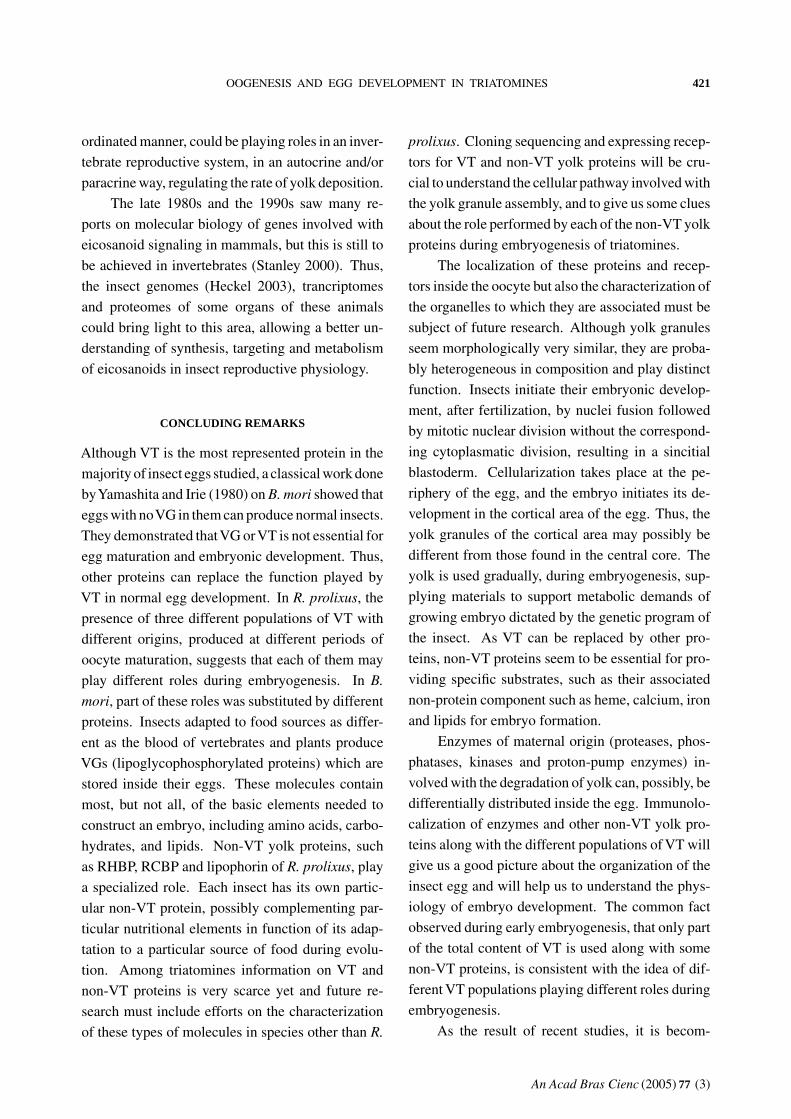
lipid segregation in R. prolixus embryogenesis. As
preliminary data show that maternal derived lipids
remained in the newborn nymphs, then it is likely
that a similar mechanism is taking place in R. pro-
lixus (Fig. 2) (Silva-Neto, Masuda and Atella, un-
published data). Fatty acid sterification shift and
the differential partitioning of lipid classes during
egg development described above imply that dur-
ing insect egg development a unique and complex
cell biology process is taking place. The extent and
uniqueness of such process should receive the at-
tention of investigators working in the area in the
future.
CALCIUM-BINDING PROTEINS
Calcium ion is involved in a variety of functions in
living organisms as allosteric activator of some en-
zymes, modulator of cytoskeletal rearrangements,
muscle contraction, hormone action, membrane fu-
sion, transmembrane passage and also as a second
messenger. So, the metabolism of calcium must be
precisely regulated inside the cells in order to attend
all the metabolic demands of individual cells. Thus
calcium is an essential component of the egg, since
it will be required during development of a new em-
bryo. These observations led to the conclusion that
this ion must be packed inside the egg during the
process of oogenesis, in order to allow a normal de-
velopment of the insect.
Calmodulin (CAM)
One possible way to pack calcium ion inside
the oocyte is through the calcium-binding protein,
calmodulin. Calmodulin (CaM) is a ubiquitous,
acidic, calcium-binding protein of 20 kDa with four-
high affinity Ca2+-binding sites present in eukary-
otic cells. When intracellular calcium concentration
rises above 1µM, the binding of calcium drives a
conformational change in calmodulin. The calcium-
bound state of calmodulin associates with a variety
of proteins and modulates their activities which ex-
plain why it is involved in numerous cellular reg-
ulatory processes. In the cockroach Blatella ger-
manica, a large amount of CaM is stored in growing
oocytes, where it comprises 0.9% of the total solu-
bilized protein (Zhang and Kunkel 1992). Isolation
of B. germanica CaM followed by detailed molecu-
lar characterization demonstrated close similarity to
vertebrate CaMs (Zhang and Kunkel 1992). Small
structural differences distinguish it from all other
animal CaMs. The demonstration of CaM synthe-
sis by vitellogenic follicles, and its absence in the
hemolymph of the female, together with the demon-
stration of its synthesis suggest that most of the
CaM incorporated by the egg is of ovarian origin. It
was demonstrated in Oncopeltus fasciatus that a gap
junctionally transmitted epithelial cell signal was
necessary for yolk uptake (Anderson and Woodruff
2001). Similar signal is also necessary in insects
representing other orders suggesting that possibly
this phenomenon was conserved during evolution
(Wakmonski and Woodruff 2002). The epithelial
cell signal, transmitted through gap junction stimu-
lating the incorporation of yolk, is calmodulin
(Brooks and Woodruff 2004). CaM possibly plays a
dual function: during oogenesis and embryogenesis.
An Acad Bras Cienc (2005) 77 (3)

416 GEORGIA C. ATELLA ET AL.
A B C
Fig. 2 – Maternal lipid localization in R. prolixus neonate nymphs. Purified lipophorin was double labeled with Texas Red PE and
Bodipy FA injected into vitellogenic females; typically four days after injection, eggs were laid. They were kept at 28◦C until hatching.
Nymphs were immediately collected and kept on ice. Fluorescence associated with newborn nymph was analyzed by fluorescent
microscopy. (A) Phase-contrast image. (B) Localization of Bodipy FA in newborn kissing-bug nymph. (C) Localization of Texas Red
PE in newborn kissing-bug nymph.
CaM stored in the egg is present in the cytoplasmic
compartment surrounding yolk granules and is virtu-
ally absent from yolk granules (Zhang and Kunkel
1994). The storage of a huge amount of CaM in
developing eggs has in turn led to different specula-
tions regarding its role in the programmed degrada-
tion of VT during embryogenesis. VT is able to bind
CaM and after degradation of VT, the CaM-binding
capacity is retained by the 53-kDa fragment gener-
ated by limited proteolysis. Besides that, the amount
of CaM in developing eggs dramatically decreases
after the beginning of limited proteolysis of the yolk.
Therefore, CaM can be involved with limited pro-
teolysis of VT. The stoichiometry between VT and
CaM is (1:1), which reinforces this possibility.
Rhodnius Calcium-Binding Protein (RCBP)
In addition to CaM, another calcium-associated pro-
tein yolk was isolated and characterized from oocyte
and hemolymph from R. prolixus. This protein ca-
pable of binding calcium and storing it inside the
oocyte (Silva-Neto et al. 1996) is a phosphoprotein.
It exhibits an apparent molecular mass of 18 kDa
on gel filtration, but it migrates as an 8 kDa band
on Tris-Glycine SDS-polyacrylamide gels because
of its high degree of phosphorylation. 24% of the
total number of residues are serine, being phospho-
serine the sole phosphorylated amino acid found.
A protein that has a similar amino-terminal and re-
sembles the oocyte form of the protein was also
found in the hemolymph. Radioactive phosphopro-
tein was isolated from the hemolymph and oocytes
of insects fed with blood containing 32Pi, and it was
injected into vitellogenic females. Only the ovary
was able to incorporate the protein. Both radioac-
tive phosphoproteins were detected inside the oocyte
16 hours following injection. The phosphoprotein
binds 50 mol of 45Ca/mol of protein with a K0.5 of
10-3M. Based on these characteristics, it was named
Rhodnius calcium-binding phosphoprotein (RCBP).
RCBP resembles phosvitin, one of the most phos-
phorylated proteins found in nature, isolated from
non-mammalian vertebrates. While phosvitin is a
product of the post-endocytic cleavage ofVG, RCBP
is not, because it is already present as such in the
hemolymph. As phosvitins (28-35 kDa) are associ-
ated with yolk granules (Byrne et al. 1989) it is also
possible that RCBP is involved with yolk granule
formation.
The exact function of RCBP in the egg is not
known, but preliminary data suggest that RCBP is
used by developing embryos during the initial 10
days of post-oviposition (Silva-Neto, unpublished
data) during which katatrepsis is completed and the
yolk is enclosed by the embryo (Kelly and Huebner
1986). The role of RCBP is still not known, and
requires further investigation.
YOLK ASSOCIATED ENZYMES
Female gamete formation in oviparous organisms
occurs synchronously with another biochemical
process which is yolk deposition. Huge amounts
An Acad Bras Cienc (2005) 77 (3)

OOGENESIS AND EGG DEVELOPMENT IN TRIATOMINES 417
of carbohydrates, proteins and lipids are gradually
deposited into growing oocytes during oogenesis.
Such process will allow embryo development out-
side the maternal body. Yolk platelets are special-
ized organelles where yolk components are stored.
Nevertheless the composition of yolk platelets has
been already described in detail for many insects
but the mechanisms which underlie the utilization
of yolk components, by developing embryo, remain
to be understood in detail. A recurrent strategy has
been the identification and biochemical analysis of
yolk platelet-associated enzymes. Such analysis has
paved the way for our current understanding of yolk
platelet physiology in R. prolixus.
R. prolixus egg development is a fertilization
triggered event which occurs continuously over a
period of 15 days. The major biochemical event, tak-
ing place in yolk granules, is the gradual processing
ofVT. Oliveira et al. (1989) showed a continuous al-
teration in VT molecule following oviposition. The
authors divided this process into two phases: limited
and extensive proteolysis. The first phase spans for
10 days as embryogenesis proceeds while the main
VT bands, detected on native-PAGE gels, progres-
sively migrate further into the gel. The second phase
occurs inside the digestive system following day 10
and VT content declines to half of its initial value at
hatching. The remaining material is used up by the
first instar nymph during its initial five days of life
outside the eggshell. In the following paragraphs
we describe yolk-associated enzymes studied in our
laboratory, whose role was assigned by examination
of their biochemical properties during egg develop-
ment.
Nussenzveig et al. (1992) were the first to de-
scribe a set of acid hydrolases associated to yolk
platelet in R. prolixus. The presence of such en-
zymes was first detected incubating purified yolk
platelets in different pHs, which induced a differ-
ential profile of VT proteolysis. Protease activity
towards VT was maximal at pH values between 3.5
and 4.5. This reaction was solely blocked by the
classical cathepsin D (CD) inhibitor, pepstatin A, in
a dose dependent-manner. Yolk platelets were sub-
jected to freezing and thawing followed by ultra-
centrifugation. This strategy led to the obtainment
of a pellet which contained almost all enzyme activ-
ity. It also demonstrated a close association between
the protease and the membrane of the yolk platelet.
Further experiments showed that R. prolixus (CD) is
a 45 kDa protein whose activity towards a specific
fluorescent peptide designed for aspartyl proteases
increases over 15-fold during egg development (Fi-
alho et al. 2005).
A second enzyme associated to yolk platelets
was a lysosomal acid phosphatase. Such enzyme
is able to hydrolyze p-nitrophenyl phosphate with
a peak of activity around pH 5.0. This reaction is
strongly blocked in the presence of sodium fluoride
and tartrate but not by p-chloromercuriobenzoate
(Nussenzveig et al. 1992). Later on, it was demon-
strated that Rhodnius acid phosphatase (AP) activity
is mediated by a single enzyme of 94 kDa (Fialho
et al. 2002). CD and AP were therefore physically
associated to yolk platelet and we then hypothesized
that they should play a role in VT proteolysis during
egg development. Such hypothesis was investigated
as follows.
Non-mated females of R. prolixus produce and
lay non-fertilized eggs (Davey 1967). Such eggs
were assayed for AP activity and the enzyme ac-
tivity obtained was compared with the one from
fertilized eggs homogenized in different days fol-
lowing oviposition (Fialho et al. 2002). AP activ-
ity is 5-fold increased compared to oocytes in the
first days following fertilization. In contrast non-
fertilized but oviposited eggs showed no activation
of this enzyme. This result indicated for the first
time a close link betweenAP activity and oocyte fer-
tilization. Once VT is a phosphorylated molecule,
we followed the fate of metabolic labeled phospho-
serines with the aid of radioactive inorganic phos-
phate in both set of eggs. Surprisingly, fertilized
eggs showed a decrease of 60% of their phospho-
serine content. A similar profile was not observed
in non-fertilized eggs where a decrease of only 20%
was detected. Such results were confirmed by the
analysis of phosphoserine content with the aid of
An Acad Bras Cienc (2005) 77 (3)

418 GEORGIA C. ATELLA ET AL.
monoclonal antibodies raised against phosphoserine
(Fialho et al. 2002). We next analyzed the ability
of phosphorylated molecules to inhibit pNPPase ac-
tivity catalyzed by partially purified AP. VT but not
isolated phosphoserine, mannose 6-phosphate and
ATP could block such reaction. This result was a
clear demonstration that VT is a substrate specifi-
cally recognized by eggAP. Therefore, in R. prolixus
egg fertilization triggers a cascade of signaling event
which culminates in the dephosphorylation ofVT by
AP. This finding implied that removal of covalently
bound phosphate groups is a part of a programmed
pathway for appropriate embryo utilization of yolk
reserves. Such biochemical pathway appears to be
unique to R. prolixus, once B. germanica developing
eggs, where the same phenomenon was investigated,
do not present such alterations (Nordin et al. 1990).
Efforts from many laboratories working world-
wide with developing eggs of different arthropods
have provided a huge amount of information about
the physical-chemical properties, amino acid and
gene sequences of several proteases involved in
VT degradation. Some of the mechanisms, which
regulate VT proteolysis, have been described as
well. These mechanisms include for instance the
expression of genes coding for proteases, inacti-
vation of protease activity by inhibitory peptides,
or most commonly, pH-induced activation of self-
processing proteases. Proteolytic systems described
in developing eggs of arthropods usually rely on the
acidification of YPs. Such mechanism was demon-
strated in several models such as developing eggs
of Xenopus, sea urchin and tick and therefore its
correlation to yolk utilization is clearly established
(Fagotto 1990, 1991, Fagotto and Maxfield 1994).
Furthermore, the presence of a unique acidification
system in R. prolixus composed by an H+-PPase and
also by an H+-ATPase has been recently demon-
strated (Motta et al. 2004). Although these studies
have shed light on a recurrent strategy for the mod-
ulation of protease activity, they do not provide any
information about the susceptibility of VT molecule
to the proteolytic attack.
Once VT, AP and CD share the same intracel-
lular compartment, i.e. yolk platelets, we hypoth-
esized that these enzymes might have a concerted
action against VT. Fialho et al. (2005) followed the
fate of yolk-associated CD activity originally de-
scribed in R. prolixus oocytes during egg develop-
ment. They have shown that CD activity, in total egg
homogenates, is blocked by the classical aspartyl
protease inhibitor, pepstatin A. Surprisingly, AP in-
hibitors such as NaF, Na+/K+ tartrate and inorganic
phosphate also block VT proteolysis. This block-
age is not observed when tyrosine phosphatase in-
hibitors such as vanadate and phenylarsine oxide or
levamisole were used in VT proteolysis assay. NaF
concentrations able to block isolated AP activity do
not affect the activity of partially purified CD. There-
fore, a specific repressor of VT proteolysis should
be dephosphorylated by AP in vivo. The identity of
this molecule should be addressed further. The surge
of phosphoproteome techniques will allow a close
look to the transient nature of phosphate groups at-
tached to phosphoproteins during egg development
in the future. Combination of such strategy with
a complete survey on non-proteic phosphorylated
molecules able to be dephosphorylated byAP should
provide some detail on the mechanism of CD regu-
lation in this system. In conclusion, the above data
demonstrated for the first time that AP, besides its
simple hydrolytic function of low molecular weight
substrates, plays a role in the regulation of the initial
steps of protein digestion inside yolk platelets.
Dephosphorylation-coupled digestion of pro-
teic substrates is a recurrent theme in cell biology.
The presence of covalently bound phosphate on VTs
was soon recognized as a remarkable feature of those
proteins (Allerton and Perlmann 1965). Nowadays,
data concerning the physical-chemical properties,
amino acid sequencing and cDNA sequences are
available for VTs in different insect groups. Thus
it became clear that every VT molecule from any
organism contains some degree of covalently bound
phosphate in its structure (Byrne et al. 1989, Sap-
pington and Raikhel 1998). Extensive phosphoryla-
tion is particularly found in non-mammalian verte-
brate VGs within a domain named phosvitin (Byrne
An Acad Bras Cienc (2005) 77 (3)

OOGENESIS AND EGG DEVELOPMENT IN TRIATOMINES 419
et al. 1989). Nearly half of its amino acid com-
position is made up of serines and most of them
are phosphorylated. The presence of such motif on
VTs provides an anionic surface where some non-
phosphorylated serines are clustered among phos-
phorylated serines. This region is a consensus se-
quence for further phosphorylation by a protein ki-
nase named casein kinase II (CK II).
CK II is a cyclic nucleotide-independent cal-
cium/calmodulin-insensitive protein kinase (Silva-
Neto et al. 2002) strongly inhibited in vitro by hep-
arin and which is able to utilize either ATP or GTP
as phosphate donors in the phosphorylation reac-
tion (Pinna and Meggio 1997). The naturally oc-
curring consensus sequences of CK II on VTs make
these proteins a suitable substrate for the identifica-
tion of such enzyme on insect eggs. Silva-Neto and
Oliveira (1993) demonstrated the presence of CK II
on fully grown oocytes of R. prolixus. Such enzyme
is 3-fold activated after oocyte fertilization andVT is
its main substrate throughout the whole egg develop-
ment (Fialho et al. 1999). Limited proteolysis takes
place on VT molecule in the third day after ovopo-
sition. From this point on CK II activity towards
VT gradually disappears. Such result demonstrated
a specific structural requirement for the recognition
of Rhodnius VT by CK II (Fialho et al. 1999). Non-
fertilized eggs do not display this transient CK II
activation profile. This result was a strong indica-
tion that CK II modulation is also included in the
programming of egg development. Isolation of such
enzyme allowed a close look into the dynamic of VT
phosphorylation by such enzyme (Silva-Neto et al.
2002). Incubation of purified VT with purified CK
II led to the incorporation of 2 mol of phosphate/mol
VT. However, the total number of phosphorylation
sites available can be altered by previous incubation
of VT with alkaline phosphatase. Under such con-
dition limited dephosphorylation increased the total
number of phosphorylation sites. However, exten-
sive dephosphorylation led to a dramatic decrease
on the total phosphorylation sites. Such results led
us to speculate that the presence of CK II in yolk
platelets assures the high degree of VT phosphory-
lation. This would allow the interaction of VT with
cations at certain regions which might be important
for two processes: first, to keep VT in a paracrys-
taline state inside the yolk platelet where its “pack-
ing” degree would require a lower number of wa-
ter molecules; second, to prevent the availability of
cations such as calcium from being able to trigger
lipid-peroxidation catalyzed by neighboring heme-
proteins in the yolk platelet such as RHBP. Such
hypotheses are currently under investigation in our
laboratory.
EICOSANOIDS AS LOCAL REGULATORSOF OOGENESIS
The control of insect oogenesis is associated with
hormones such as juvenile hormone and ecdysone
(Engelmann 1979, Kunkel and Nordin 1985). These
hormones control the synthesis of yolk protein and
its receptors (Raikhel and Dhadiala 1992); the pa-
tency of follicular cells (Sevala and Davey 1989,
Raikhel and O’Lea 1991); and uptake of yolk pro-
teins (Stoffolano et al. 1992). The great complexity
and variety of cells that compose insect ovaries sug-
gest the participation of local mediators that also
control and coordinate oogenesis. However, local
mediators which could help in the control of ooge-
nesis are little understood.
Eicosanoids are local lipid mediators produced
from the oxygenation of C20 polyunsaturated fatty
acids, most notably arachidonic acid (C 20:4ω-6).
Once released from membrane phospholipids, ara-
chidonate can be oxygenated by prostaglandin G/H
synthase (cyclooxygenase) or (5-, 8-, 12-, 15-) lipo-
xygenases to form prostaglandins (PGs), leuko-
trienes (LTs) or related hydroxy acids, which can
play a role as intra or extracellular signals (Smith
1989, Stanley 2000, Funk 2001). These compounds
regulate many physiological and physiopathological
processes in addition to modulating inflammatory
and immunological responses in mammals (Samuel-
son 1983, O‘Neill and Ford-Hutchinson 1993, Mur-
doch et al. 1993, Funk 2001, Pai et al. 2003), but
their production is not restricted to these animals,
as several invertebrate species have been shown to
An Acad Bras Cienc (2005) 77 (3)

420 GEORGIA C. ATELLA ET AL.
produce PGs and LTs (Meijer et al. 1986, Stanley-
Samuelson et al. 1991, Petzel et al. 1993, Stanley-
Samuelson and Pedibhotla 1996, Stanley 2000,
Reddy et al. 2004).
Eicosanoids have several actions in reproduc-
tive biology of vertebrates (Murdoch et al. 1993,
Priddy and Killick 1993, Funk 2001), which have
encouraged many research groups to study their rel-
evance in insect reproduction. The first report on
the biosynthesis of eicosanoids in the insect repro-
ductive system was in the house cricket Acheta do-
mesticus by Destephano and Brady (1977). They
showed that male testes and seminal vesicles were
able to produce PGs, and a PG synthesizing com-
plex was transferred from male to female via sper-
matophore during mating. That study also showed
that PGs have had a stimulating effect on egg-laying
behavior. Since then, the prostaglandin biosynthesis
has been recognized in reproductive tracts of sev-
eral insects as Bombyx mori (Yamaja-Setty and Ra-
maiah 1979), Teleogryllus commodus (Loher et al.
1981), Trichoplusia ni (Hagan and Brady 1982), Lo-
custa migratoria (Lange 1984), Musca domestica
(Wakayama et al. 1986), Triatoma infestans (Bren-
ner and Bernasconi 1989) and R. prolixus (Medeiros
et al. 2002).
However, the model of PG synthesizing com-
plex transfer and egg laying behavior release showed
to be not conserved, even among the crickets. In
other models such as L. migratoria, Trichoplusi ni,
Musca domestica, PG injections are not able to in-
duce oviposition (Stanley 2000). It has also been
described that there is a transfer of PGs or arachi-
donic acid rather than enzymes in insects such as
Bombyx mori and Musca domestica during mating
(Stanley-Samuelson and Pedibhotla 1996, Stanley
2000). The role of PGs in these systems remains
to be elucidated, but Loher (1979) showed a net in-
crease in the egg number in unmated Teleogryllus
commodus females after PGE2 injection. Among
triatomines, Medeiros et al. (2002) showed in R.
prolixus that derivatives of cyclooxygenase pathway
decrease the yolk protein uptake, suggesting that
these compounds can act as regulators of oogene-
sis, as seen in other arthropod models (Spaziani et
al. 1993, 1995, Sagi et al. 1995).
Lipoxygenase products are described in the
reproductive tract of some invertebrates, e.g. four
starfish species (Meijer et al. 1986), the sea squirt
Ciona intestinalis, and the surf clam Spisula so-
lidissima (Hada et al. 1997). Pagés et al. (1986)
demonstrated lipoxygenase activity in Drosophila
melanogaster extracts and also the presence of lipo-
xygenase-like immunoreactivity in ejaculatory bulb
of male flies. The reproductive tract of mated fire-
brat Thermobia domestica has lipoxygenase activity
(Ragab et al. 1987, 1991), and the egg laying be-
havior is dependent of phospholipase A2 activity,
which seems to be augmented after mating (Bitsch
et al. 1995). In triatomines, Medeiros et al. (2004)
showed, by a pharmacological approach, that the
lipoxygenase pathways products act as positive ef-
fectors of yolk uptake.
The eicosanoids signaling in vertebrate in-
volves cyclic nucleotide modulation. The partici-
pation of second messengers involved in eicosanoid
signaling in insects or even in invertebrates is scarce
but there are some evidences of modulation by cyclic
AMP levels in these systems. Meijer et al. (1986)
showed a reduction of cAMP levels in oocytes of
starfish Methasterias sp. after treatment with 8(R)-
HETE, which induced oocyte maturation. Sagi et
al. (1995) demonstrated an increase in cAMP lev-
els following treatment of ovarian tissue with PGE2
in the prawn Macrobrachium rosembergii. Spaziani
et al. (1995) also showed, in the crayfish Procam-
barus paeninsulanus, a correlation among ovarian
tissue contraction, PGF2α levels and tissue cAMP
levels. The role of PGE2, which was also augmented
during oogenesis progression (Spaziani et al. 1993),
remains to be elucidated. Medeiros et al. (2004)
showed, in R. prolixus, that cyclooxygenase prod-
ucts (inhibitors of yolk uptake) increase the ovar-
ian cAMP levels, while the lipoxygenase products
(stimulators of yolk uptake) act in an opposite way
decreasing the level of this second messenger (Wang
and Telfer 1996). Worth mentioning is that this is
the first time that products of both pathways, by co-
An Acad Bras Cienc (2005) 77 (3)
OOGENESIS AND EGG DEVELOPMENT IN TRIATOMINES 421
ordinated manner, could be playing roles in an inver-
tebrate reproductive system, in an autocrine and/or
paracrine way, regulating the rate of yolk deposition.
The late 1980s and the 1990s saw many re-
ports on molecular biology of genes involved with
eicosanoid signaling in mammals, but this is still to
be achieved in invertebrates (Stanley 2000). Thus,
the insect genomes (Heckel 2003), trancriptomes
and proteomes of some organs of these animals
could bring light to this area, allowing a better un-
derstanding of synthesis, targeting and metabolism
of eicosanoids in insect reproductive physiology.
CONCLUDING REMARKS
Although VT is the most represented protein in the
majority of insect eggs studied, a classical work done
byYamashita and Irie (1980) on B. mori showed that
eggs with noVG in them can produce normal insects.
They demonstrated thatVG orVT is not essential for
egg maturation and embryonic development. Thus,
other proteins can replace the function played by
VT in normal egg development. In R. prolixus, the
presence of three different populations of VT with
different origins, produced at different periods of
oocyte maturation, suggests that each of them may
play different roles during embryogenesis. In B.
mori, part of these roles was substituted by different
proteins. Insects adapted to food sources as differ-
ent as the blood of vertebrates and plants produce
VGs (lipoglycophosphorylated proteins) which are
stored inside their eggs. These molecules contain
most, but not all, of the basic elements needed to
construct an embryo, including amino acids, carbo-
hydrates, and lipids. Non-VT yolk proteins, such
as RHBP, RCBP and lipophorin of R. prolixus, play
a specialized role. Each insect has its own partic-
ular non-VT protein, possibly complementing par-
ticular nutritional elements in function of its adap-
tation to a particular source of food during evolu-
tion. Among triatomines information on VT and
non-VT proteins is very scarce yet and future re-
search must include efforts on the characterization
of these types of molecules in species other than R.
prolixus. Cloning sequencing and expressing recep-
tors for VT and non-VT yolk proteins will be cru-
cial to understand the cellular pathway involved with
the yolk granule assembly, and to give us some clues
about the role performed by each of the non-VT yolk
proteins during embryogenesis of triatomines.
The localization of these proteins and recep-
tors inside the oocyte but also the characterization of
the organelles to which they are associated must be
subject of future research. Although yolk granules
seem morphologically very similar, they are proba-
bly heterogeneous in composition and play distinct
function. Insects initiate their embryonic develop-
ment, after fertilization, by nuclei fusion followed
by mitotic nuclear division without the correspond-
ing cytoplasmatic division, resulting in a sincitial
blastoderm. Cellularization takes place at the pe-
riphery of the egg, and the embryo initiates its de-
velopment in the cortical area of the egg. Thus, the
yolk granules of the cortical area may possibly be
different from those found in the central core. The
yolk is used gradually, during embryogenesis, sup-
plying materials to support metabolic demands of
growing embryo dictated by the genetic program of
the insect. As VT can be replaced by other pro-
teins, non-VT proteins seem to be essential for pro-
viding specific substrates, such as their associated
non-protein component such as heme, calcium, iron
and lipids for embryo formation.
Enzymes of maternal origin (proteases, phos-
phatases, kinases and proton-pump enzymes) in-
volved with the degradation of yolk can, possibly, be
differentially distributed inside the egg. Immunolo-
calization of enzymes and other non-VT yolk pro-
teins along with the different populations of VT will
give us a good picture about the organization of the
insect egg and will help us to understand the phys-
iology of embryo development. The common fact
observed during early embryogenesis, that only part
of the total content of VT is used along with some
non-VT proteins, is consistent with the idea of dif-
ferent VT populations playing different roles during
embryogenesis.
As the result of recent studies, it is becom-
An Acad Bras Cienc (2005) 77 (3)

422 GEORGIA C. ATELLA ET AL.
ing clear that yolk granules are not merely digestive
vesicles but rather sites where a complex metabolism
occurs, finely regulated in relation to both time and
space. The central issue of developmental biol-
ogy is the identification of a diversity of cells that
were once indistinguishable from one another. With
the progress in the fields of biochemistry, cell biol-
ogy, and molecular biology new technologies be-
came available to explore these developmen-
tal mechanisms. Reliable knowledge of the fate
map of the embryo and of the cell lineage of sin-
gle precursor cells has been achieved, in contrast to
the relative lack of information on several aspects
of the metabolism of maternally inherited compo-
nents of insect eggs. Thus, the challenge for the
next decade is to collect enough data to understand
how and where these egg proteins are used up to-
gether with their non-protein components to support
cell differentiation. Besides that, it is also important
to understand how their degradation is controlled in
pace with the genetic program of the embryo, which
enables cell differentiation (early phase of embryo-
genesis) and embryo differentiation (late phase) in-
side the egg.
Finally, it is necessary to recognize that this
review is a consequence of a lifetime effort by Dr.
Lent who encouraged young Brazilian students and
scientists to engage in the study of triatomines. We
were all stimulated by reading his papers, books and
notes, and also by the scientists he trained.
ACKNOWLEDGMENTS
We express our gratitude to Heloísa S. L. Coelho,
Lilian S.C. Gomes, José S.L. Junior and Litiane M.
Rodrigues for their excellent technical assistance;
and to Dr. Tereza Marques de Oliveira Lima for the
English support in the reading of the manuscript.
This work was supported by Conselho Nacional de
Desenvolvimento Científico e Tecnológico (CNPq),
Coordenação de Aperfeiçoamento de Pessoal de
Nível Superior (CAPES), Financiadora de Estudos
e Projetos (Finep), Programa de Núcleo de Excelên-
cia (PRONEX), Programa de Apoio ao Desenvolvi-
mento Científico e Tecnológico (PADCT) and Fun-
dação Carlos Chagas Filho de Amparo à Pesquisa
do Estado do Rio de Janeiro (FAPERJ).
RESUMO
Em triatomíneos, assim como em outros insetos, o acú-
mulo de vitelo é um processo no qual um tecido extra-
ovariano, o corpo gorduroso, produz proteínas que são
empacotadas no interior de um ovo. A principal proteína,
sintetizada pelo corpo gorduroso, que é acumulada no in-
terior de um ovócito, é a vitelogenina. Este processo é
também conhecido por vitelogênese. Existem crescentes
evidências em triatomíneos, que além do corpo gorduroso,
o ovário também produz proteínas de vitelo. A forma
como estas proteínas de vitelo entram nos ovócitos será
aqui comentada. O vitelo é um material complexo com-
posto por proteínas, lipídeos, carboidratos e outros com-
postos minoritários que são empacotados de uma maneira
organizada no interior dos ovócitos. A fertilização dis-
para a embriogênese, um processo que culmina com o
desenvolvimento do embrião. Durante a embriogênese
o vitelo será utilizado para a construção de um novo in-
divíduo, a ninfa de primeiro estádio. O desafio para a
próxima década é entender onde e como estas proteínas
de vitelo são utilizadas junto com os seus componentes
não protéicos, em compasso com o programa genético do
embrião, que comanda a diferenciação celular (fase inicial
da embriogênese) e diferenciação do embrião (fase final
da embriogênese) no interior do ovo.
Palavras-chave: ovogênese, vitelogênese, embriogêne-
se, triatomíneos, Rhodnius prolixus.
REFERENCES
Aft RL and Mueller GC. 1983. Hemin mediated DNA
strands scission. J Biol Chem 258: 12069–12072.
Allerton SE and Perlmann GE. 1965. Chemical
characterization of the phosphoprotein phosvitin. J
Biol Chem 240: 3892–3898.
Anderson KL and Woodruff RI. 2001. A gap junction-
ally transmitted epithelial cell signal regulates endo-
cytic yolk uptake in Oncopeltus fasciatus. Dev Biol
239: 68–78.
Atella GC and Shahabuddin M. 2002. Differential
partitioning of maternal fatty acid and phospho-
lipid in neonate mosquito larvae. J Exp Biol 205:
3623–3630.
An Acad Bras Cienc (2005) 77 (3)

OOGENESIS AND EGG DEVELOPMENT IN TRIATOMINES 423
Atella GC, Gondim KC and Masuda H. 1992. Trans-
fer of phospholipids from fat body to lipophorin in R.
prolixus. Arch Insect Biochem Physiol 19: 133–144.
Atella GC, Gondim KC and Masuda H. 1995. Load-
ing of lipophorin particles with phospholipids at the
midgut of R. prolixus. Arch Insect Biochem Physiol
30: 337–350.
Beenakkers AMTh, Chino H and Law JH. 1988.
Lipophorin Nomenclature. Insect Biochem 18:
1–2.
Bellés X. 2004. Vitellogenesis Directed by Juvenile
Hormone. In: Raikhel AS and Sappington TW
(Eds), Reproductive Biology of Invertebrates,
vol. 12, Part B: Progress in Vitellogenesis, Science
Publishers, Inc., Enfield, USA / Plymouth, UK,
p. 157–198.
Bianchi AG, Coutinho M, Pereira SD, Marinotti O
and Targa HJ. 1985. VG and VT of Musca domes-
tica: Quantification and synthesis by fat bodies and
ovaries. Insect Biochem 15: 77–84.
Bitschi C, Ragab A and Chap H. 1995. Inhibition of
phospholipase A2 modulates fecundity in the primi-
tive insect Thermobia domestica (Thysanura). J In-
sect Physiol 41: 209–216.
Blacklock BJ and Ryan RO. 1994. Hemolymph lipid
transport. Insect Biochem Mol Biol 24: 855–873.
Bonhag PF. 1955. Histochemical studies of the ovar-
ian nurse cell tissues and oocytes of the milkweed
bug, Oncopeltus fasciatus (Dallas). II Sudanophilia,
phospholipids and cholesterol. J Morphol 97: 283–
311.
Bonhag PF. 1958. Ovarian structure and vitellogenesis
in insects. Annu Rev Entomol 3: 137–160.
Braz GRC, Coelho HSL, Masuda H and Oliveira
PL. 1999. A missing metabolic pathway in the cattle
tick Boophilus microplus. Curr Biol 9: 703–706.
Braz GRC, Abreu L, Masuda H and Oliveira PL.
2001. Heme biosynthesis and oogenesis in the blood-
sucking bug, Rhodnius prolixus. Insect Biochem Mol
Biol 31: 359–364.
Braz GRC, Moreira MF, Masuda H and Oliveira
PL. 2002. Rhodnius heme-binding protein (RHBP)
is a heme source for embryonic development in the
blood-sucking bug R. prolixus. Insect Biochem Mol
Biol 32: 709–717.
Brenner RR and Bernasconi A. 1989. Prostaglandin
biosynthesis in the gonads of the hematophagous
insect Triatoma infestans. Comp Biochem Physiol
93B: 1–4.
Brooks RA and Woodruff RI. 2004. Calmodulin trans-
mitted through gap junctions stimulates endocytic in-
corporation of yolk precursors in insect oocytes. Dev
Biol 271: 339–349.
Brookes VJ. 1969. The induction of yolk protein syn-
thesis in the fat body of an insect Leucophea maderae
by an analog of the juvenile hormone. Dev Biol 20:
459–471.
Brunt A. 1971. The histology of the first batch of eggs
and their associated tissue in the ovariole of Dysder-
cus fasciatus (Heteroptera: Pyrrhocoridae) as seen
with the light microscope. J Morphol 134: 105–130.
Buning J. 1981. Cluster formation in telotrophic merois-
tic ovarioles. IX Congr Int Soc Dev Biol Basle
(Abstract).
Byrne BM, Gruber M and Ab GB. 1989. The evolution
of egg yolk proteins. Prog Biophys Mol Biol 53:
33–69.
Canavoso LE, Frede S and Rubiolo ER. 2004.
Metabolic pathways for dietary lipids in the midgut of
hematophagous Panstrongylus megistus (Hemiptera:
Reduviidae). Insect Biochem Mol Biol 34: 845–854.
Chalaye D. 1979. Étude immunochimique des pro-
teins hémolymphatiques et ovocytaires de R. prolixus
(Stal). Can J Zool 57: 329– 336.
Cheon HM, Seo SJ, Sun J, Sappington TW and
Raikhel AS. 2001. Molecular characterization of
the VLDL receptor homolog mediating binding of
lipophorin in oocyte of the mosquito Aedes aegypti.
Insect Biochem Mol Biol 31: 753–760.
Chino H, Downer RGH and Takahashi K. 1977. The
role of diacylglycerol-carrying lipoprotein I in lipid
transport during insect vitellogenesis. Biochim Bio-
phys Acta 487: 508–516.
Chino H, Downer RGH, Wyatt GR and Gilbert LI.
1981. Lipophorin, a major class of lipoprotein of
insect haemolymph. Insect Biochem 11: 491.
Coelho HSL, Atella GC, Moreira MF, Gondim KC
and Masuda H. 1997. Lipophorin density variation
during oogenesis in R. prolixus. Arch Insect Biochem
Physiol 35: 301–313.
An Acad Bras Cienc (2005) 77 (3)

424 GEORGIA C. ATELLA ET AL.
Coles GC. 1965. Haemolymph proteins and yolk forma-
tion in R. prolixus (Stal). J Exp Biol 43: 425–431.
Colombo MI, Mayorga LS, Nishimoto I, Ross EM and
Stahl P. 1994. GS regulation of endosome fusion
suggests a role for signal transduction pathway in
endocytosis. J Biol Chem 269: 14919–14923.
Couchman J and King PE. 1979. Germarial struc-
ture and oogenesis in Brevicoryne brassicae (L)
(Hemiptera:Aphiidae). Int J Insect Morphol Embryol
8: 1–10.
Dansa-Petretski M, Ribeiro JMC, Atella GC, Ma-
suda H and Oliveira PL. 1995. Antioxidant role of
R. prolixus heme-binding protein. J Biol Chem 270:
10893–10896.
Dantuma NP, Van Marrewijk WJA, Wynue HJ and
van der Horst DJ. 1996. Interaction of an insect
lipoprotein with its binding site at the fat body. J
Lipid Res 37: 1345–1355.
Davey KG. 1967. Some consequences of copulation in
Rhodnius prolixus. J Insect Physiol 13: 1629–1636.
Davey KG. 1981. Hormonal control of VG uptake in R.
prolixus Stal. Am Zool 21: 701–705.
Davey KG. 1987. Inputs to the hormonal control of egg
development in R. prolixus. Mem Inst Oswaldo Cruz
82: 103–108.
Davis NT. 1956. The morphology and functional
anatomy of the male and female reproductive systems
in Cimex lecularius L (Heteroptera; Cimicidae). Ann
Entomol Soc Am 49: 466–493.
Destephano DB and Brady VE. 1977. Prostaglandin
and prostaglandin synthetase in the cricket Acheta
domesticus. J Insect Physiol 23: 905–911.
Di Fiore PP and De Camilli P. 2001. Endocytosis and
signaling: An inseparable partnership. Cell 106: 1–4.
Dorn A, Stoffel R, Matile H, Bubendof A and Ri-
dley R. 1995. Malarial haemozoin beta-haematin
supports haem polymerization in the absence of pro-
tein. Nature 374: 269–271.
Downer RGH and Chino H. 1985. Turnover of pro-
tein and diacylglycerol components of lipophorin in
insect haemolymph. Insect Biochem 15: 627–630.
Engelmann F. 1970. The Physiology of Insect Repro-
duction. Oxford, New York: Pergamon Press.
Engelmann F. 1979. Insect VG: Identification biosyn-
thesis, and role in vitellogenesis. Adv Insect Physiol
14: 49–109.
Engelmann F. 1990. Hormonal control of arthropod re-
production: Progress in comparative endocrinology.
Prog Clin Biol Res 342: 357–364.
Fagotto F. 1990. Yolk degradation in tick eggs: II. Evi-
dence that catepsin L-like proteinase is stored as a la-
tent, acid-activable proenzyme. Arch Insect Biochem
Physiol 14: 237–252.
Fagotto F. 1991. Yolk degradation in tick eggs: III.
Developmentally regulated acidification of the yolk
spheres. Develop Growth and Differ 33: 57–66.
Fagotto F and Maxfield FR. 1994. Changes in yolk
platelet pH during Xenopus laevis development cor-
relate with yolk utilization. A quantitative confocal
microscopy study. J Cell Science 107: 3325–3337.
Fallon RJ and Schwartz AL. 1988. Asialoglycopro-
tein receptor phosphorylation and receptor-mediated
endocitosis in hepatoma cells. J Biol Chem 263:
13159–13166.
Fallon RJ, Danaher M, Saylors RL and Saxena A.
1994. Defective asialoglycoprotein receptor endocy-
tosis mediated by tyrosine kinase inhibitors. J Biol
Chem 269: 11011–11017.
Fialho E, Masuda H and Silva-Neto MAC. 1999. Pro-
tein phosphorylation during Rhodnius prolixus em-
bryogenesis: protein kinase casein kinase II activity.
Insect Biochem Mol Biol 29: 215–223.
Fialho E, Silveira AB, Masuda H and Silva-Neto
MAC. 2002. Oocyte fertilization triggers acid phos-
phatase activity during Rhodnius prolixus embryoge-
nesis. Insect Biochem Mol Biol 32: 871–880.
Fialho E, Nakamura A, Juliano A, Masuda H
and Silva-Neto MAC. 2005. Cathepsin D-mediat-
ed yolk protein degradation is blocked by acid
phosphatase inhibitors. Arch Biochem Biophys 46:
246–253.
Foti M, Carpentier JL, Aiken C, Trono D, Lew DP
and Krause KH. 1997. Second messenger regu-
lation of receptors association with clathrin coated
pits: a novel and selective mechanism in the control
of CD4 endocytosis. Mol Biol Cell 8: 1377–1389.
Friend WG, Choy CTH and Cartwright E. 1965. The
effect of nutrient intake on the development and egg
production of R. prolixus (Stal) (Hemiptera reduvi-
idae). Can J Zool 43 : 892–903.
Funk CD. 2001. Prostaglandins and leukotrienes: Ad-
vances in eicosanoid biology. Science 294: 1871–
1875.
An Acad Bras Cienc (2005) 77 (3)

OOGENESIS AND EGG DEVELOPMENT IN TRIATOMINES 425
Garcia ES and Azambuja P. 1985. A protein diet ini-
tiates oogenesis in R. prolixus. J Med Biol Res 18:
195–199.
Garcia MLM, Mello RP and Garcia ES. 1979. Ecdy-
sone, juvenile hormone and oogenesis in R. prolixus.
J Insect Physiol 25: 695–700.
Giorgi F, Masetti M, Ignacchiti V, Cecchettini A
and Bradley JT. 1993. Post-endocytic VT process-
ing in ovarian follicles of the stick insect Carausius
morosus (Br.). Arch Insect Biochem Physiol 24: 93–
111.
Gondim KC. 1992. Transporte de fosfolipídeos pela lipo-
forina de R. prolixus e seu papel na ovogênese. PhD
thesis presented to Instituto de Biofísica Carlos Cha-
gas Filho, Universidade Federal do Rio de Janeiro,
RJ, Brasil.
Gondim KC and Wells MA. 2000. Characterization of
lipophorin binding to the midgut of larval Manduca
sexta. Insect Biochem Mol Biol 30: 405–413.
Gondim KC, Oliveira PL and Masuda H. 1989. Lipo-
phorin and oogenesis in R. prolixus: Transfer of phos-
pholipids. J Insect Physiol 35: 19–27.
Gondim KC, Atella GC, Kawooya JJK and Masuda
H. 1992. Role of phospholipids in the lipophorin
particles of R. prolixus. Arch Insect Biochem Physiol
20: 303–314.
Griffiths G and Hollinshead R. 1990. Ultrastruc-
tural localization of the regulatory (RII) subunit of
cyclic AMP-dependent protein kinase to subcellular
compartments active in endocytosis and recycling of
membrane receptor. J Cell Sci 96: 691–703.
Grillo LAM, Pontes EG and Gondim KC. 2003. Lipo-
phorin interaction with the midgut of R. prolixus:
characterization and changes in binding capacity. In-
sect Biochem Mol Biol 33: 429–438.
Hada T, Swift LL and Brash AR. 1997. Discovery of
5R-lipoxygenase activity in oocytes of the surf clam,
Spisula solidissima. Biochim et Biophys Acta 1346:
109–119.
Hagan DV and Brady UE. 1982. Prostaglandins in the
cabbage looper, Trichoplusia ni. J Insect Physiol 28:
761–765.
Hagedorn HH and Kunkel JG. 1979. VG and VT in
insects. Ann Rev Entomol 24: 475–505.
Harnish D, Wyatt G and White B. 1982. Insect VTs-
identification of primary products of translation. J
Exp Zool 220: 11–19.
Hayakawa Y. 1987. Characterization of lipophorin re-
ceptor in locust flight muscles. Biochim Biophys
Acta 919: 58–63.
Heckel D. 2003. Genomics in pure and applied ento-
mology. Annu Rev of Entomol 48: 235–260.
Huebner E. 1984. The Ultrastructure and Development
of the Telotrophic Ovary. In: King RC and Akai
H (Eds), Insect Ultrastructure, vol. 2, New York:
Plenum Press, p. 13–48.
Huebner E and Anderson E. 1972. A cytological study
of the ovary of R. prolixus. I. The ontogeny of follic-
ular epithelium. J Morphol 136: 459–493.
Irie K and Yamashita O. 1980. Changes in VT and
other yolk proteins during embryonic development
in the silkworm, Bombyx mori. J Insect Physiol 26:
811–817.
Irie K and Yamashita O. 1983. Egg-specific protein in
the silkworm, Bombyx mori: purification, properties,
localization and titre changes during oogenesis and
embryogenesis. Insect Biochem 13: 71–80.
Kawooya JK and Law JH. 1988. Role of lipophorin in
lipid transport to the insect egg. J Biol Chem 263:
8748–8753.
Kawooya JK, Osir EO and Law JH. 1988. Uptake of the
major hemolymph lipoprotein and its transformation
in the insect egg. J Biol Chem 25: 8740-8747.
Kelly GM and Huebner E. 1986. The effect of
insect growth regulator fenoxycarb on R. prolixus
(Insecta, Hemiptera) embryogenesis. Can J Zool 64:
2425–2429.
Kinsella JE. 1966. Sterol metabolism during embryoge-
nesis of Periplaneta Americana (L). J Insect Physiol
12: 435–438.
Kinsella JE. 1967. Free fatty acid metabolism during
embryogenesis of Periplaneta Americana (L). Nature
14: 211–212.
Kinsella JE and Smyth Jr T. 1996. Lipid metabolism
of Periplaneta Americana L. during embryogenesis.
Comp Biochem Physiol 17: 237–244.
Kunkel JG and Nordin JH. 1985. Yolk proteins. In:
Kerkut GA and Gilbert LI (Eds), Comprehensive
Insect Physiology, Biochemistry, and Pharmacology,
vol. 1, Oxford, UK: Pergamon Press, p. 83–111.
An Acad Bras Cienc (2005) 77 (3)

426 GEORGIA C. ATELLA ET AL.
Lange AB. 1984. The transfer of prostaglandin synthe-
sizing activity during mating in Locusta migratoria.
Insect Biochem 14: 551–56.
Lehane MJ. 1991. The importance of blood-sucking
insects. In: sc Biology of Blood-Sucking Insects.
UK: Harper Collins Academics, p. 1–5.
Lima Gomes JEP, Azambuja P and Garcia ES. 1990.
Comparative studies on the growth and reproductive
performances of R. prolixus reared on different blood
sources. Mem Inst Oswaldo Cruz 85: 299–304.
Lindblad B, Lindstedt S and Steen G. 1977. On the
enzymic defects in hereditary tyrosemia. Proc Natl
Acad Sci 74: 4641–4645.
Lipsitz EY, McFarlane JE and Henneberry GO.
1969. Developmental changes in the fatty acid com-
position of the larval lipid of the house cricket Acheta
domesticus (L.). Can J Biochem 48: 264–268.
Loher W. 1979. The influence of prostaglandin E2 on
oviposition in Teleogryllus commodus. Entomol Exp
Appl 25: 107–109.
Loher W, Ganjan I, Kubo I, Stanley-Samuelson
DW and TOBE SS. 1981. Prostaglandins: their role
in egg laying of the cricket Teleogryllus commodus.
Proc Natl Acad Sci 78: 7835–7838.
Machado EA, Atella GC, Gondim KC, Souza W and
Masuda H. 1996. Characterization and immunocy-
tochemical localization of lipophorin binding sites
in the oocytes of R. prolixus. Arch Insect Biochem
Physiol 31: 185–196.
Machado EA, Oliveira PL, Moreira MF, Souza W
and Masuda H. 1998. Uptake of Rhodnius Heme-
Binding Protein (RHBP) by the ovary of R. prolixus.
Arch Insect Biochem Physiol 39: 133–143.
Masner P. 1968. The inductors of differentiation of pre-
follicular tissue and the follicular epithelium in ovari-
oles of Pyrrhocoris apterus. J Embryol Exp Morphol
20: 1–13.
Masuda H and Oliveira PL. 1985. Characterization
of VT and VG from R. prolixus. Identification of
phosphorylated compounds in the molecule. Insect
Biochem 15: 543–550.
Masuda H, Braz GRC, Paiva-Silva GO, Silva-Neto
MAC and Oliveira PL. 2004. Non-VT Yolk Pro-
teins. In: Raikhel AS and Sappington TW (Eds),
Reproductive Biology of Invertebrates, vol. 12, Part
B: Progress in Vitellogenesis, Science Publishers,
Inc., Enfield, USA/Plymouth, UK, p. 289–314.
Medeiros MN, Oliveira DMP, Paiva-Silva GO, Silva-
Neto MAC, Romero A, Bozza M, Masuda H and
Machado EA. 2002. The role of eicosanoids on
Rhodnius heme-binding protein (RHBP) endocytosis
by R. prolixus ovaries. Insect Biochem Mol Biol 32:
537–545.
Medeiros MN, Mendonça LH, Hunter AL, Paiva-
Silva GO, Mello FG, Henze IP, Masuda H, Maya-
Monteiro CM and Machado EA. 2004. The Role
of Lipoxygenase Products on the Endocytosis ofYolk
Proteins in Insects: Participation of cAMP. Arch In-
sect Biochem Physiol 55: 178–187.
Meijer L, Brash AR, Bryant RW, NG K, MacLouf J
and Sprecher H. 1986. Stereospecific induction of
starfish oocyte maturation by (8R)-Hydroxyeicosa-
tetraenoic acid. J Biol Chem 261: 17040–17047.
Mellman I. 1996. Endocytosis and molecular sorting.
Ann Rev Cell Biol 12: 575–625.
Melo ACA, Valle D, Machado EA, Salerno AP,
Paiva-Silva GO, Cunha-e-Silva NL, Souza W
and Masuda H. 2000. Synthesis of VG by the fol-
licle cells of R. prolixus. Insect Biochem Mol Biol
30: 549–557.
Motta LS, Silva WS, Oliveira DMP, Souza W and
Machado EA. 2004. A new model for proton pump-
ing in animal cells; the role of pyrophosphate. Insect
Biochem Mol Biol 34: 19–27.
Mundall E and Engelmann F. 1977. Endocrine con-
trol of VG synthesis and vitellogenesis in Triatoma
protracta. J Insect Physiol 23: 825–836.
Murdoch WJ, Hansen TR and McPherson LA. 1993.
A review – role of eicosanoids in vertebrate ovulation.
Prostaglandins 46: 85–115.
Nagem RAP, Brandão Neto JR, Forrer VP, Menegh-
ini R, Paiva-Silva GO, Oliveira PL, Masuda H
and Polikarpov I. 2001. Crystallization and pre-
liminary X-ray study of heme binding protein from
the blood-sucking insect R. prolixus. Acta Crystal-
logr D 57: 860–861.
Nordin JH, Beaudoin EL and Liu XD. 1990. Pro-
teolytic processing of Blattella germanica VT dur-
ing early embryo development. Arch Insect Biochem
Physiol 15: 119–135.
An Acad Bras Cienc (2005) 77 (3)

OOGENESIS AND EGG DEVELOPMENT IN TRIATOMINES 427
Nussenzveig RH, Oliveira PL and Masuda H. 1992.
Identification of yolk platelet-associated hydrolases
in the oocytes of R. prolixus. Arch Insect Biochem
Physiol 21: 252–262.
O’Hagan JE. 1974. Boophilus microplus: Digestion
of hemoglobins by the engorged female tick. Expl
Parasitol 35: 110–118.
O’Neill GP and Ford-Hutchinson AW. 1993. Expres-
sion of mRNA for cyclooxygenase-1 and cyclooxy-
genase-2 in human tissues. FEBS Lett 330: 156–160.
Oliveira MF, Silva JR, Dansa-Petretski M, De
Souza W, Braga CMS, Masuda H and Oliveira
PL. 2000. Haemozoin formation in the midgut of the
blood-sucking insect R. prolixus. FEBS Lett 477:
95–98.
Oliveira MO, Silva JR, Dansa-Petretski M, De
Souza W, Lins U, Braga CS, Masuda H and
Oliveira PL. 1999. Haem detoxification by an in-
sect. Nature 400: 517–518.
Oliveira PL, Gondim KC, Guedes DS and Masuda H.
1986. Uptake of yolk protein in R. prolixus. J Insect
Physiol 12: 859–866.
Oliveira PL, Petretski MDA and Masuda H. 1989.
VT processing and degradation during embryogene-
sis in R. prolixus. Insect Biochem 19: 489–498.
Oliveira PL et al. 1995. A heme-binding protein from
hemolymph and oocytes of the blood-sucking insect,
R. prolixus. J Biol Chem 270: 10897–10901.
Paes MC and Oliveira PL. 1999. An extracellular
gluthatione peroxidase from the blood-sucking
bug, R. prolixus. Arch Insect Biochem Physiol 41:
171–177.
Paes MC, Borges OM and Oliveira PL. 2001. Hy-
drogen peroxide detoxification in the midgut of the
blood-sucking insect R. prolixus. Arch Insect Bio-
chem Physiol 48: 63–71.
Pagés M, Roselló J, Casas J, Gelpí E, Gualde N
and Rigauld M. 1986. Cyclooxygenase and lipo-
xygenase-like activity in Drosophila melanogaster.
Prostaglandins 32: 729–740.
Pai R, Nakamura T, Moon WS and Tarnawski AS.
2003. Prostaglandins promote colon cancer cell in-
vasion; signaling by cross-talk between two distinct
growth factor receptors. FASEB J 17: 1640–1647.
Paiva-Silva GO et al. 2002. On the biosynthesis of R.
prolixus heme-binding protein. Insect Biochem Mol
Biol 32: 1533–1541.
Pan ML, Bell WJ and Telfer WH. 1969. Vitellogenic
blood protein synthesis by insect fat body. Science
165: 393–394.
Pant R, Nautiyal GC and Singh JB. 1973. Varia-
tion in major lipid components during development
of Philosamia ricini Indian. J Biochem Biophys 10:
116–118.
Petzel DH, Parrish AK, Ogg CL, Witters NA,
Howard RW and Stanley-Samuelson DW. 1993.
Arachidonic acid and prostaglandin E2 in Malpighian
tubules of female yellow fever mosquitoes. Insect
Biochem Mol Biol 23: 431–437.
Pinna LA and Meggio F. 1997. Protein kinase CK2
(“casein kinase-2”) and its implication in cell division
and proliferation. Progr in Cell Cycle Res 3: 77–97.
Ponka P. 1997. Tissue-specific regulation of iron meta-
bolism and heme synthesis: Distinct control mecha-
nisms in erythroid cells. Blood 89: 1–25.
Pontes EG, Grillo LM and Gondim KC. 2002. Char-
acterization of lipophorin binding to the fat body of R.
prolixus. Insect Biochem Mol Biol 32: 1409–1417.
Postlethwait JH and Giorgi F. 1985. Vitellogenesis
in insects. In: Browder LW (Ed), Developmen-
tal Biology, a Comprehensive Synthesis, New York:
Plenum Press, p. 85–136.
Postlethwait JH, Bownes M and Jowett T. 1980.
Sexual phenotype and VG synthesis in Drosophila
melanogaster. Dev Biol 79: 379–387.
Pratt GE and Davey KG. 1972a. The corpus allatum
and oogenesis in R. prolixus (Stal) The effect of mat-
ing. J Exp Biol 56: 223–237.
Pratt GE and Davey KG. 1972b. The corpus allatum
and oogenesis in R. prolixus (Stal) I the effects of
allatectomy. J Exp Biol 56: 201–214.
Priddy AR and Killick SR. 1993. Eicosanoids and
ovulation. Prostaglandins Leukot Essent Fatty Acids
49: 827–831.
Ragab A, Bitsch C, Thomas JMF, Bitsch J and Chap
H. 1987. Lipoxygenase conversion of arachidonic
acid in males and inseminated females of the firebrat
Thermobia domestica (Thysanura). Insect Biochem
17: 863–870.
Ragab A, Durand J, Bitsch C, Chap H and Rigaud M.
1991.The lipoxygenase pathway of Arachidonic acid
metabolism in reproductive tissues of the firebrat,
An Acad Bras Cienc (2005) 77 (3)

428 GEORGIA C. ATELLA ET AL.
Thermobia domestica, (Thysanura). Insect Biochem
21: 321–326.
Raikhel AS and Dhadiala TS. 1992. Accumulation of
yolk proteins in insect oocytes. Annu Rev Entomol
37: 217–251.
Raikhel AS and O’Lea AO. 1991. Control of follicular
epithelium development and vitellin envelope forma-
tion in the mosquito: role of juvenile hormone and
20-hydroxyecdysone. Tissue and Cell 23: 577–591.
Raikhel AS, Dhadiala TS, Cho WL, Hays AR and
Koller CN. 1990. Biosynthesis and endocytosis of
yolk proteins in the mosquito. In: Hagedorn HH
(Ed), Molecular Insect Science, New York: Plenum
Press, p. 147–154.
Reddy PS, Reddy PR and Nagaraju GP. 2004. The
synthesis and effects of prostaglandins on the ovary
of the crab Oziotelphusa senex senex. Gen Comp
Endocr 135: 35–41.
Roth TF and Porter KR. 1964. Yolk protein uptake in
the oocyte of the mosquito Aedes aegypti. J Cell Biol
20: 313–332.
Roth TF, Cutting JA and Atlas JB. 1976. Protein
transport and selective membrane mechanism. J
Supramol Struct 4: 527–548.
Rousset A, Bitsch C and Bitsch J. 1988. Poly-
peptide composition and biosynthesis of the yolk
proteins in the firebrat Thermobia domestica (In-
secta, Thysanura). Arch Insect Biochem Physiol
9: 299–312.
Rowe JW, Holy L, Ballinger RE and Stanley-
Samuelson D. 1995. Lipid provisioning of turtle
eggs and hatchings: total lipid, phospholipids, tri-
acylglycerol and triacylglycerol fatty acids. Comp
Biochem Physiol B 112: 323–330.
Ryan RO and van der Horst DJ. 2000. Lipid transport
biochemistry and its role in energy production. Annu
Rev Entomol 45: 233–260.
Sagi A, Silkowsky J, Fleisher-Berkovish S, Danon
A and Chayoth R. 1995. Prostaglandin E2 in the
previtellogenic ovaries of the prawn Macrobrachium
rosembergii: synthesis and effect on the level of
cAMP. Gen Comp Endocr 100: 308–313.
Salerno AP. 2001. Origem e processamento das pop-
ulações de vitelina de R. prolixus: comparação com
vitelogenina. PhD thesis – Universidade Federal do
Rio de Janeiro, RJ, Brasil.
Salerno AP, Dansa-Petretski M, Silva-Neto MAC,
Coelho HSL and Masuda H. 2002. R. prolixus VT
is composed of three different populations: Compari-
son with VG. Insect Biochem Mol Biol 32: 709–717.
Salomón OD and Stoka A. 1986. VT and VG charac-
terization of Triatoma infestans and related species.
Acta Physiol Pharmacol Latinoam 36: 419–429.
Samuelson B. 1983. Leukotrienes: mediators of im-
mediate hypersensitivity reactions and inflammation.
Science 220: 279–281.
Sappington TW and Raikhel AS. 1998. Molecular
characteristics of insect vitellogenins and vitelloge-
nin receptors. Insect Biochem Mol Biol 28: 277–300.
Schreiner B. 1977. Vitellogenesis in the milkweed bug
Oncopeltus fasciatus: A light and electron micro-
scopic investigation. J Morphol 155: 81–110.
Sevala VL and Davey KG. 1989. Action of juvenile
hormone on follicle cells of Rhodnius prolixus: ev-
idence for a novel regulatory mechanism involving
protein kinase C. Experientia 45: 355–356.
Silva-Neto MAC and Oliveira PL. 1993. Protein
Phosphorylation in the Oocytes of R. prolixus – Iden-
tification of a Type II Casein Kinase. Insect Biochem
Mol Biol 23: 815–823.
Silva-Neto MAC, Atella GC, Fialho E, Paes MC,
Zingali RB, Petretski JH, Ales EW and Masuda
H. 1996. Isolation of a calcium-binding phospho-
protein from the oocytes and hemolymph of the
blood-sucking insect R. prolixus. J Biol Chem 271:
30227–30232.
Silva-Neto MAC, Fialho E, Paes MC, Oliveira PL
and Masuda H. 2002. Cyclic nucleotide-indepen-
dent phosphorylation of VT by casein kinase II puri-
fied from R. prolixus oocytes. Insect Biochem Mol
Biol 32: 847–857.
Smith A. 1990. Transport of tetrapyrroles: Mecha-
nisms and biological and regulatory consequences.
In: Dailey HA (Ed.), Biosynthesis of Heme and
Chlorophylls, New York: McGraw-Hill Publishing
Company, p. 435–490.
Smith WI. 1989. The eicosanoids and their biochemical
mechanism of action. Biochem J 259: 315–324.
Snigirevskaya ES and Raikhel AS. 2004. Receptor-
Mediated Endocytosis of Yolk Proteins in Insect
Oocytes. In: Raikhel AS and Sappington TW
(Eds), Reproductive Biology of Invertebrates, vol.
An Acad Bras Cienc (2005) 77 (3)

OOGENESIS AND EGG DEVELOPMENT IN TRIATOMINES 429
12, Part B: Progress in Vitellogenesis, Enfield, USA/
Plymouth, UK: Science Publishers, Inc., p. 199–228.
Soulages JL and Wells MA. 1994. Lipophorin: the
structure of an insect lipoprotein and its role in lipid
transport in insects. Adv Protein Chem 45: 371–415.
Souza AVG, Petretski JH, Demasi M, Bechara E and
Oliveira PL. 1997. Urate protects a blood-sucking
insect against hemin-induced oxidative stress. Free
Radical Bio Med 22: 209–214.
Spaziani EP, Hinsch GW and Edwards SC. 1993.
Changes in prostaglandin E2 and F2α during vitello-
genesis in the Florida crayfish Procambarus paenin-
sulanus. J Comp Physiol B 163: 541–545.
Spaziani EP, Hinsch GW and Edwards SC. 1995. The
effect of prostaglandin E2 and Prostaglandin F2α on
ovarian tissue in the Florida crayfish Procambarus
paeninsulanus. Prostaglandins 50:189–200.
Speake BK and Thompson MB. 1999. Comparative
aspects of yolk lipid utilization in birds and reptiles.
Poult Avian Biol Rev 10: 181–211.
Speake BK and Thompson MB. 2000. Lipids of the
eggs and neonates of ovoviviparous lizards. Comp
Biochem Physiol A 127: 453–467.
Speake BK, Murray AM and Noble RC. 1998. Trans-
port and transformations in birds and reptiles. Prog
Lipid Res 37: 1–32.
Stanley DW. 2000. Eicosanoids in invertebrate sig-
nal transduction systems. In: Eicosanoids in the
reproductive biology of invertebrates. Prince-
ton: Princeton University Press, p. 55–108.
Stanley-Samuelson DW and Pedibhotla VK. 1996.
What can we learn from prostaglandin and related
eicosanois in insects? Insect Biochem Mol Biol 26:
223–234.
Stanley-Samuelson DW, Gensen E, Nickerson KW,
Tiebl K, Ogg CL and Howard RW. 1991. Insect
immune response to bacterial infection is mediated
by eicosanoids. Proc Natl Acad Sci USA 88: 1064–
1068.
Stoffolano JG, Li NF, Zou BX and Yin CM. 1992.
Vitellogenin uptake, not synthesis is dependent on
juvenile hormone in adult of Phormia regina. J Insect
Physiol 38: 839–845.
Sun J, Hiraoka T, Dittmer NT, Cho K and Raikhel
AS. 2000. Lipophorin as a yolk protein precursor in
the mosquito, Aedes aegypti. Insect Biochem Mol
Biol 30: 1161–1171.
Telfer WH. 1954. Immunological studies of insect
metamorphosis: II. The role of a sex-limited blood
protein in the egg formation by the Cecropia silk-
worm. J Gen Physiol 37: 539–558.
Telfer WH. 1960. The selective accumulation of blood
proteins by the oocytes of saturniid moths. Biol Bull
118: 338–351.
Telfer WH. 1961. The route of entry and localization
of blood proteins in the oocytes of saturniid moths. J
Biophys Biochem Cytol 9: 747–59.
Telfer WH, Huebner E and Smith DS. 1982. The cell
biology of vitellogenic follicles in Hyalophora and
Rhodnius. In: King RC and Akai H (Eds), Insect
Ultrastructure, vol. 1, New York: Plenum Press, p.
118–149.
Telfer WH, Pan M and Law JH. 1991. Lipophorin in
developing adults of Hyalophora cecropia: Support
of yolk formation and preparation for flight. Insect
Biochem 21: 653–663.
Tsuchida K and Wells MA. 1988. Digestion, absorp-
tion, transport and storage of fat during the last larval
stadium of Manduca sexta. Changes in the role of
lipophorin in the delivery of dietary lipid to the fat
body. Insect Biochem 18: 263–268.
Tsuchida K and Wells MA. 1990. Isolation and charac-
terization of a lipoprotein receptor from the fat
body of an insect, Manduca sexta. J Biol Chem 265:
5761–5767.
Tufail M, Raikhel AS and Takeda M. 2004. Biosyn-
thesis and Processing of InsectVGs. In: Raikhel AS
and Sappington TW (Eds), Reproductive Biology
of Invertebrates, vol. 12, Part B: Progress in Vitello-
genesis, Enfield, USA/Plymouth, UK: Science Pub-
lishers, Inc., p. 1–32.
Turunen S and Crailsheim K. 1996. Lipid and sugar
absorption. In: Lehane MJ and Billingsley PF
(Eds), Biology of the Insect Midgut, London: Chap-
man & Hall, p. 293–320.
Valle D, Lima Gomes JEP, Goldenberg S and Garcia
ES. 1987. R. prolixus vitellogenesis: dependence
upon the blood source. J Insect Physiol 33: 249–
254.
Valle D, Garcia ES and Goldenberg S. 1992. Influ-
ence of the blood meal source on vitellogenin synthe-
An Acad Bras Cienc (2005) 77 (3)

430 GEORGIA C. ATELLA ET AL.
sis by isolated fat bodies of Rhodnius prolixus. Insect
Biochem Mol Biol 22: 159–166.
Valle D, Kun J, Linns J, Garcia ES and Goldenberg
S. 1993. R. prolixusVG is synthesized as high molec-
ular weight precursors. Isolation of a VG cDNA
clone. Insect Biochem Mol Biol 23: 457–465.
van Antwerpen R, Linnemans WAM, van der Horst
DJ and Beenakers AMTh. 1988. Immunocyto-
chemical localization of lipophorins in the flight mus-
cles of the migratory locust (Locusta migratoria) at
rest and during flight. Cell Tissue Res 252: 661–668.
van Antwerpen R, Salvador K, Tolman K and Gen-
try C. 1998. Uptake of lipids by developing oocytes
of the hawkmoth Manduca sexta. The possible role
of lipoprotein lipase. Insect Biochem Mol Biol 28:
399–408.
van Heusden MC, van der Horst DJ, Voshol J and
Beenakkers AMTh. 1987. Recycling of protein
component of the flight-specific lipophorin in Lo-
custa migratoria. Insect Biochem 17: 771–776.
Vincent SH. 1989. Oxidative effects of heme and por-
phyrins on proteins and lipids. Seminars in Hema-
tology 26: 105–113.
Vincent SH, Grady RW, Snider JM and Muller-
Eberhard U. 1988. The influence of heme-binding
proteins in heme-catalyzed oxidations. Arch Bio-
chem Biophys 265: 539–550.
Wakayama EJ, Dillwith JW and Blomquist GJ. 1986.
Occurrence and metabolism of prostaglandins in the
housefly, Musca domestica (L.). Insect Biochem 16:
895–902.
Wakmonski SL and Woodruff RI. 2002. For uptake of
yolk precursors, epithelial cell oocyte gap junctional
communication is required by insects representing
six different orders. J Insect Physiol 48: 667–675.
Wang Y and Telfer WH. 1996. Cyclic nucleotide-
induced temination of VG uptake by Hyalophora ce-
cropia follicles. Insect Biochem Mol Biol 26: 85–94.
Wigglesworth VB. 1936. The function of corpus al-
latum in the growth and reproduction of R. prolixus
(Hemiptera). Q J Microsc Sci 79: 91–121.
Wigglesworth VB. 1943. The fate of the hemoglobin
in R. prolixus and other blood-sucking arthropods.
Proc Roy Soc (London) Ser B 131: 313–339.
Yamaja-Setty BN and Ramaiah TR. 1979. Isolation
and identification of prostaglandins from the repro-
ductive organs of male silkmoth, Bombyx mori L.
Insect Biochem 9: 613–617.
Yamashita O and Indrasith LS. 1988. Metabolic fates
of yolk proteins during embryogenesis in arthropods.
Develop Growth and Differ 30: 347–359.
Yamashita O and Irie K. 1980. Larval hatching from
VG-deficient eggs developed in male hosts of the silk-
worm. Nature 283: 385–386.
Zhang Y and Kunkel JG. 1992. High abundance
calmodulin from Blatella germanica eggs binds to
VT subunits but disappears during VT utilization. In-
sect Biochem Mol Biol 22: 293–304.
Zhang Y and Kunkel JG. 1994. Most egg calmodulin is
a follicle cell contribution to the cytoplasm of Blatella
germanica oocyte. Dev Biol 161: 513–521.
Zhu J, Indrasith LS and Yamashita O. 1986. Char-
acterization of VT, egg-specific protein and 30 kDa
protein from Bombyx eggs, and their fates during oo-
genesis and embryogenesis. Biochim Biophys Acta
882: 427–436.
Ziegler R and Ibrahim MM. 2001. Formation of
lipid reserves in fat body and eggs of the yellow
fever mosquito, Aedes aegypti. J Insect Physiol 47:
623–627.
An Acad Bras Cienc (2005) 77 (3)