on the existence of the dna resonance code and its the existence of the d… · deciphering the dna...
TRANSCRIPT

NeuroQuantology | February 2019| Volume 17 | Issue 02 | Page 56-71| doi: 10.14704/nq.2019.17.02.1973Myakishev-Rempel M., On The Existence of The DNA Resonance Code and Its Possible Mechanistic Connection to The Neural Code
56
eISSN 1303-5150 www.neuroquantology.com
On The Existence of The DNA Resonance Code and Its Possible Mechanistic Connection to The Neural Code
Ivan V. Savelyev1,2, Nelli V. Zyryanova1,3, Oksana O. Polesskaya1,4,7, Max Myakishev-Rempel1,4,5,6*
Key Words: alu, transposon, microtubules, biofield, morphogenetic field, morphic resonance, biological development
DOI Number: 10.14704/nq.2019.17.02.1973 NeuroQuantology 2019; 17(02):56-71
Corresponding author: Max Myakishev-RempelAddress: 1DNA resonance lab, San Diego, CA, USA; 2currently in Yalta, Russia; 3currently in Moscow, Russia; 4Localized Therapeutics, San Diego, CA, USA; 5Transposon LLC, San Diego, CA, USA; 6Vaccine Research Institute of San Diego, San Diego, CA, USA; 7University of California - San Diego, San Diego, CA, USAe-mail [email protected] conflicts of interest/financial disclosures: The authors declare that the research was conducted in the absence of any commercial or financial relationships that could be construed as a potential conflict of interest. Received: 25 January 2019; Accepted: 18 February 2019
Abbreviations: LINE1 - Long interspersed nuclear element 1
NMR - Nuclear magnetic resonance
PHz - petahertz, 1015 hertz
bp – base pair
nm - nanometerIntroductionAlexander Gurwitsch (1874-1954) developed experimental models for the measurement of non-chemical communication between biological objects 96 years ago. He postulated the existence of the morphogenic field (Gurwitsch, 1922) responsible for creation of the shape of the body in 1922, proved the
existence of such a field (Alexander and Gurwitsch, 1925; Beloussov et al., 2004; Gurwitsch, 1923, 1924; Michael Lipkind, 1998; M. Lipkind, 1998) and characterized its spectral properties (Protoplasma, 1932). His results were reproduced by Anna Gurwitch (Gurwitsch, 1968; Gurwitsch and Gurwitsch, 1991) Burlakov (Burlakov et al., 2000) and over 100 works of others, reviewed in references (Quickenden and Que Hee, 1974; Volodyaev and Beloussov, 2015), and his scientific school continues now (Beloussov et al., 2004). Alexander Gurwitsch was nominated for the Nobel Prize 11 times.
Gurwitsch’s typical experiment used a growing onion root as a source of biologically active waves that he called mitogenic radiation since it accelerated
ABSTRACT
A possible role of DNA sequence-specific electromagnetic resonances in the formation of the morphogenic field is discussed. It is proposed that the morphogenic field is formed by resonant oscillations of delocalized electron and proton clouds in the base stack of the DNA. Models are proposed for DNA sequence-dependence of possible electromagnetic resonance patterns. It is proposed that genomic repeats act as universal resonators providing the bidirectional communication between the chromatin structure and the morphogenic field. It is proposed that genomic repeats participate in two major functions - the morphogenic function and the brain function. It is proposed that microtubules mediate the resonance communication between the action potential in axons and genomic repeats in the nucleus. The existence of an algorithm is proposed responsible for the conversion of genomic information into the shape of the body. Such an algorithm is named the DNA resonance code. It is proposed that the DNA resonance code can be deciphered by targeted efforts in biophysics, spectroscopy, molecular modeling, and experimental genomics. A possible resonance interaction between the DNA of neurons and neuronal firing is discussed and it is suggested that deciphering the DNA resonance code may be of help to deciphering the neuronal coding in the brain. It is suggested that the deciphering of the DNA resonance code would benefit medical applications related to morphogenesis and brain function.

57
eISSN 1303-5150 www.neuroquantology.com
NeuroQuantology | February 2019| Volume 17 | Issue 02 | Page 56-71| doi: 10.14704/nq.2019.17.02.1973Myakishev-Rempel M., On The Existence of The DNA Resonance Code and Its Possible Mechanistic Connection to The Neural Code
mitosis (Protoplasma, 1932). Another growing onion root or a petri dish with yeast culture was used as a receiving object. The sending and receiving objects were separated by a quartz prism allowing for spectral mapping of mitogenic irradiation. The further experiments of Burlakov with retroreflector prisms proved that such irradiation is not only capable of accelerating mitosis but also of producing developmental abnormalities, thus confirming the concept of the morphogenic properties of the field (Burkov et al., 2008; Burlakov, Kapranov, et al., 2012).
Basic experiments confirming the existence of biological fields involve two samples such as cell culture aliquots in sealed quartz cuvettes separated by optical filters. When one of the aliquots is perturbed, the second one may catch a signal that is transferred non-chemically and is blocked by light impermeable filters. Such effects are often referred to as “non-chemical cell-cell communication” (Cifra et al., 2011; Scholkmann et al., 2013; Trushin, 2004; Xu et al., 2017). Original experimental reports include communication between cell culture aliquots via a polystyrene petri dish (Farhadi et al., 2007; Rossi et al., 2011) and between plant roots through the air (Ciszak et al., 2012).
Among such models, the simplest and most robust seems a model of Burlakov (Burlakov et al., 2000) that uses fish embryos. Compared to cell culture and adult organisms, embryos are more sensitive, produce stronger biological fields, and their developmental abnormalities are easier to observe. In Burlakov’s model, 50 fish embryos are placed in each of two quartz cuvettes stacked on top of each other and incubated for several days in a metal box. It was observed that older embryos inhibit the development of the younger ones. A Germanium mirror accelerates the development when a single cuvette is placed on it, and a quartz retroreflector prism represses the development and causes developmental abnormalities (Burlakov et al., 2000). Burlakov’s lab has published a great many papers using this model (Beloussov et al., 2003; Burlakov, 2000; Burlakov, Burlakova, et al., 2012; Burlakov et al., 1999, 2000, 2002, 2006, 2009, 2010, 2017; Burlakov and Lebedeva, 2015) and is continuing the tradition of the scientific school of Alexander Gurwitsch.
The concept of a morphogenic field is a response to the need to explain biological development: how is the shape of the body, organs, and tissues
formed from a single fertilized egg cell? Current chemical explanations of development are correct but insufficient. Specifically, biomolecular gradients and neuronal signaling are very imprecise to explain the high spatial precision and reproducibility of body shapes and structures. There is a need for the cell to know their position in the body and molecular gradients and neuronal signals are insufficiently precise for that. On the other hand, the idea of morphogenic field is proposed by others to explain how the cells and tissues determine their position and coordinate their growth and shrinkage.
Understanding the fundamental mechanisms of development has an immediate practical application: controlling the shape of the body would help in curing obesity, growing new organs, bones, limbs, and teeth; remodeling scarred wounds, and rejuvenating aged joints.
The idea that the morphogenic field is holographic and created by the genomic DNA was first published by Richard Miller and Burt Webb (Miller and Webb, 2012). It was proposed that as a flat manmade holographic image contains the information about the volume of the photographed object, so the linearly recorded information of DNA would recreate a 3D blueprint for the body which the cells and tissues use to determine their position, build and sustain the shape and structures of the body. Luc Montagnier (Montagnier et al., 2011), continuing the work of Jacques Benveniste (Davenas et al., 1988), provided preliminary demonstrations that DNA sequences produce biologically active electromagnetic fields. Konstantin Meyl (Meyl, 2011) proposed that biological electromagnetic waves produced by DNA have an unusual field structure allowing them to transmit through tissues without loss. The waves predicted by Meyl are aperiodic, helical (not unlike DNA) and have a phase shift between magnetic and electric fields. Oscillations in Molecular StructuresEven though the background ideas for explaining the role of DNA in morphogenesis through a morphogenic field have been laid by Gurwitsch, Miller, Burlakov, Montagnier, Meyl and others, the specific mechanism for the creation and perception of morphogenic field by DNA has yet to be discovered. Here we outline specific approaches for the discovery of this mechanism.
Let us define “DNA resonance” as a wave interaction between identical DNA sequences or DNA

58
eISSN 1303-5150 www.neuroquantology.com
NeuroQuantology | February 2019| Volume 17 | Issue 02 | Page 56-71| doi: 10.14704/nq.2019.17.02.1973Myakishev-Rempel M., On The Existence of The DNA Resonance Code and Its Possible Mechanistic Connection to The Neural Code
sequences that are not identical, but have similar oscillatory properties. For example, it is a case of resonance, when a sound made by one musical instrument causes a string in another instrument to vibrate when the second string is tuned to the same exact tone. Similarly, we propose that specific DNA sequences or DNA-protein complexes of chromatin that have similar oscillatory patterns would resonate, allowing synchronization and signaling from one sequence to the other.
Although the idea that the morphogenic field is produced by the genomic sequence has been around since it was proposed by Richard Alan Miller in 1973 (Miller and Webb, 2012), expanded by Peter Gariayev (Gariaev, 1994; Gariaev et al., 2001) and Marco Bischof (Bischof, 1995), some 20 years later, there have been no published attempts to actually decipher the conversion algorithms of DNA sequences into wave patterns. Here we name this algorithm as “DNA resonance code” and define it as an algorithm which describes the conversion of a genomic DNA sequence into the structure of the morphogenic field and ultimately into the shape of the body. For example, although the initial sequence of the human genome was completed in 2001 (Lander and International Human Genome Sequencing Consortium, 2001; Venter, 2001), and of the mouse genome in 2002 (Mouse Genome Sequencing Consortium et al., 2002) , now, 16 years later, it is impossible to predict the shape of the body of these species based on their genomic sequence. Even more, science lacks an algorithm by which the sequence of the genome defines the shape of any species. Currently, there is no way for computational genomics to reconstruct the shape of a worm, a fly, a dog, or a human from their genomic sequence. We believe this is because genomics considers only molecular interactions and ignores the resonance language of DNA.
Since a large fraction of the genome remains chemically passive, it is referred to as “junk DNA”, even though a large portion of this so-called “junk DNA” bears a sign of functional significance: in addition to transcribed genes which are conserved in evolution, there are as many untranscribed sequences which are also conserved (Lander and International Human Genome Sequencing Consortium, 2001). We suggest that these conserved untranscribed sequences are involved in creating and sustaining the shape of the body, organs, and tissues, and guiding the biochemical factory of the cell via resonance signaling. We suggest that this resonance
signaling is bidirectional: the genome in each nucleus receives the information from the field created by all the nuclei of the body and in turn, contributes to the overall field of the body. We also suggest that the field drives condensation and decondensation of specific DNA sequences and thus specific DNA sequences convert the electromagnetic signals into chemical changes and conversely, chemical changes into electromagnetic signals.
The periodic nature of DNA’s double helix inspired many researchers to model its mechanical oscillations (Gariaev, 1994; Scott, 1985; Volkov and Kosevich, 1987). Since DNA is highly charged, bound by water and by proteins of chromatin and transcription factors, we doubt it can sustain mechanical oscillations independently of the surrounding water and proteins, although it should be able to vibrate together with them. Since we are looking for sustained oscillations which are defined by the DNA sequence, we favor not mechanical (sub-molecular) oscillations, but the oscillations of collective delocalized pi-electron resonance clouds of the base stack and similarly collective delocalized proton clouds of hydrogen bonds of DNA. We have suggested (Guschin, Polesskaya, Zyryanova, Tovmash, Mara, et al., 2018; Polesskaya, Guschin, et al., 2018) that it is more likely that these are the electron and proton cloud oscillations in the base stack of DNA which are responsible for DNA resonance and for the morphogenic field, for that reason that the base stack is hydrophobic inside and its core is separated from water, proteins and any other molecules of the surrounding milieu, thus making it an insulated wire and a linear (double-helical) crystal. This should allow electron and proton charges to oscillate in the base stack without disturbing the external milieu and therefore without much dissipation of energy.
The existence of the collective delocalized electron clouds in the base stack has been reasonably established by researchers of DNA’s electrical conductivity, typically called “DNA charge transfer”. The base stack of DNA was observed to be a good electric conductor and a semiconductor able to transmit either excess electrons or electron holes (Arnold et al., 2016). The unique properties of the electron clouds in the base stack are due to aromatic properties of those DNA bases and the fact that they are compressed together by the sugar-phosphate backbone. Since the bases are hydrophobic, water will push them together in order to minimize the contact with them, while the charges of the

59
eISSN 1303-5150 www.neuroquantology.com
NeuroQuantology | February 2019| Volume 17 | Issue 02 | Page 56-71| doi: 10.14704/nq.2019.17.02.1973Myakishev-Rempel M., On The Existence of The DNA Resonance Code and Its Possible Mechanistic Connection to The Neural Code
phosphates repel each other making the DNA as linear as possible. This combination of pulling and pushing is responsible for its perfect double helical structure and for the perfect structure of the base stack inside. The pi-electrons of the base stack are doubly delocalized into a collective cloud: first, they are dissociated from their host carbon and nitrogen atoms by the aromatic rings (akin to the hexagon nut of benzene), and second, these aromatic bases are stacked on top of each other into the double helical ladder, making their electron rings overlap in a periodic fashion. Note that this periodicity should also help oscillations in these distributed collective delocalized electron structures.
Importantly, the electrons in the base stack are delocalized via Heisenberg’s quantum uncertainty, which in chemistry is referred to as chemical resonance (Healy, 2011). The delocalization of electrons of aromatic bases in the base stack is thought to be the reason for lossless charge transfer over long stretches of the double helix (Kurnikov et al., 2002).
Richard Alan Miller wrote 45 years ago: “The formation of a certain type of chemical bond known as the resonance bond (which is most easily seen in the case of the benzene molecule) leads to a peculiar situation in which certain electrons are freed from a local or particular location in a molecule. These are then free to travel around the entire molecule. This means that the electrons occupy an energy shell of the whole molecule as opposed to any particular atom in the molecule. The existence of molecular systems with mobile electrons has been found to be of profound significance in the phenomena of life” (Miller et al., 1975).” All the essential biochemical substances, which perform the fundamental functions of living matter, are composed completely or partially of such mobile electrons. Molecules which contain these electrons are known as conjugated systems (Pullman and Pullman, 1963). “The essential fluidity of life may correspond with the fluidity of the electronic cloud in conjugated molecules. Such systems may best be considered as both the cradle and the main backbone of life” (Miller et al., 1975)). The importance of the conjugation of pi-electrons of aromatic bases and amino acids was also emphasized by Stuart Hameroff who observed a correlation between aromaticity of anesthetic compounds and their potency, and also proposed that conjugation of aromatic pi-electrons is the basis for the signal transduction via microtubules (Hameroff et al., 2014).
The protons creating the hydrogen bonds in DNA are known to be also in the state of chemical resonance, being delocalized between tautomeric states of the bases (Chatzidimitriou-Dreismann, 1993). In addition to the known delocalization of protons in basepairs, we suggest that the hydrogen-bond protons may be delocalized along the DNA length into a continuous proton cloud spreading as far as the unbroken base stack spreads. Although such conjugation of protons has not been shown for DNA, a similar conjugation has been described as “proton conductivity” or “proton highways” in protein solutions (Wraight, 2006).Magnetism of DNAThe idea that DNA can have magnetic properties goes back to the experimental works of Lev Blumenfeld in 1959 (Blumenfeld, 1959). The dispute about these properties continues to this day (Blois et al., 1963; Khomutov, 2004; Kwon, Choi, et al., 2012; Kwon et al., 2008, 2009; Lee et al., 2011; Muller et al., 1961; Shulman et al., 1961; Snipes and Gordy, 1964; WALSH jun. et al., 1961). All experiments on this topic are carried out in strong magnetic fields using purified DNA.
We approached this question from another angle. We have noticed (Guschin, Polesskaya, Zyryanova, Tovmash and Myakishev-Rempel, 2018) that two DNA strands in biological reactions such as ligation, transcription, replication, and formation of hairpins, behave very much like pairs of antiparallel magnets, Fig. 1. For example, in a ligation reaction of a plasmid vector, blunt ends find each other with high specificity. This is usually attributed to the high efficiency of the ligase enzyme, but we suspect that even in the absence of ligase, DNA ends could attract each other as pairs of antiparallel magnets, Fig. 1. Note that our hypothesis on the antiparallel magnetization of DNA strands did not come from experimental evidence but from DNA behavior in enzymatic reactions. We noticed that antiparallel magnetization of DNA strands would often greatly improve the specificity of enzymatic reactions and explain some of the observed specificity.
Figure 1. Two strands of natural DNA may be magnetized in an antiparallel fashion. Blunt ends of a vector may be magnetically attracted to each other.

60
eISSN 1303-5150 www.neuroquantology.com
NeuroQuantology | February 2019| Volume 17 | Issue 02 | Page 56-71| doi: 10.14704/nq.2019.17.02.1973Myakishev-Rempel M., On The Existence of The DNA Resonance Code and Its Possible Mechanistic Connection to The Neural Code
is in line with the idea of Blumenfeld that DNA can function as magnetic tape to record, store, and retrieve information in a magnetic form (Blumenfeld, 1963; Kiperman, 2015; Shnoll, 2003). Importantly, DNA has an advantage over the magnetic tape in that it has unique sequences which could be used as addresses for cataloging the information, very much like a formatted computer disk.
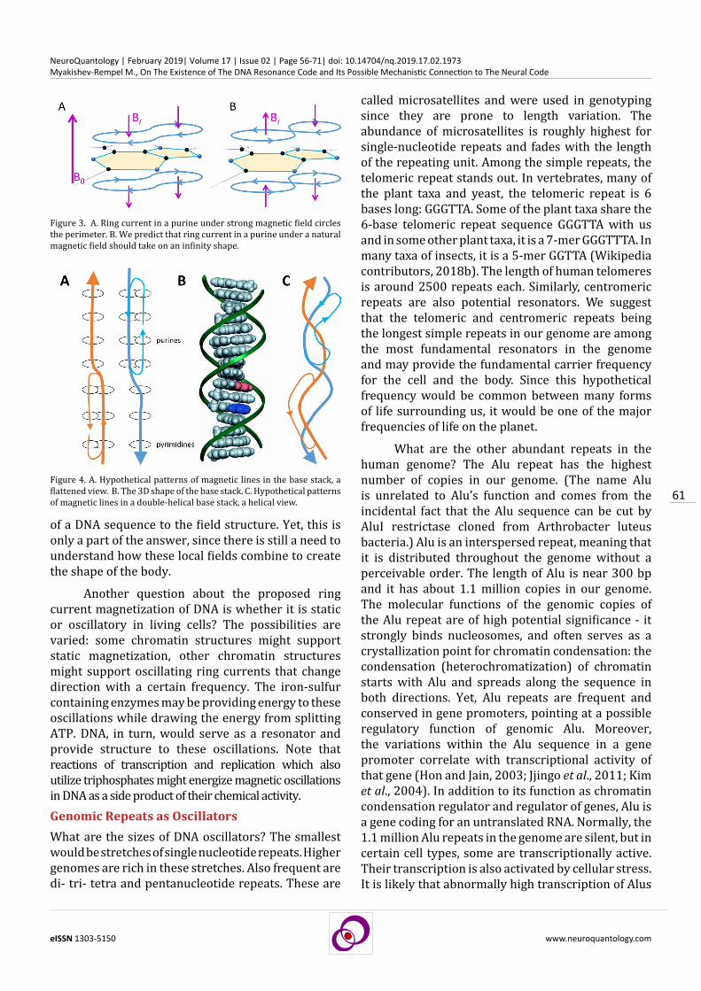
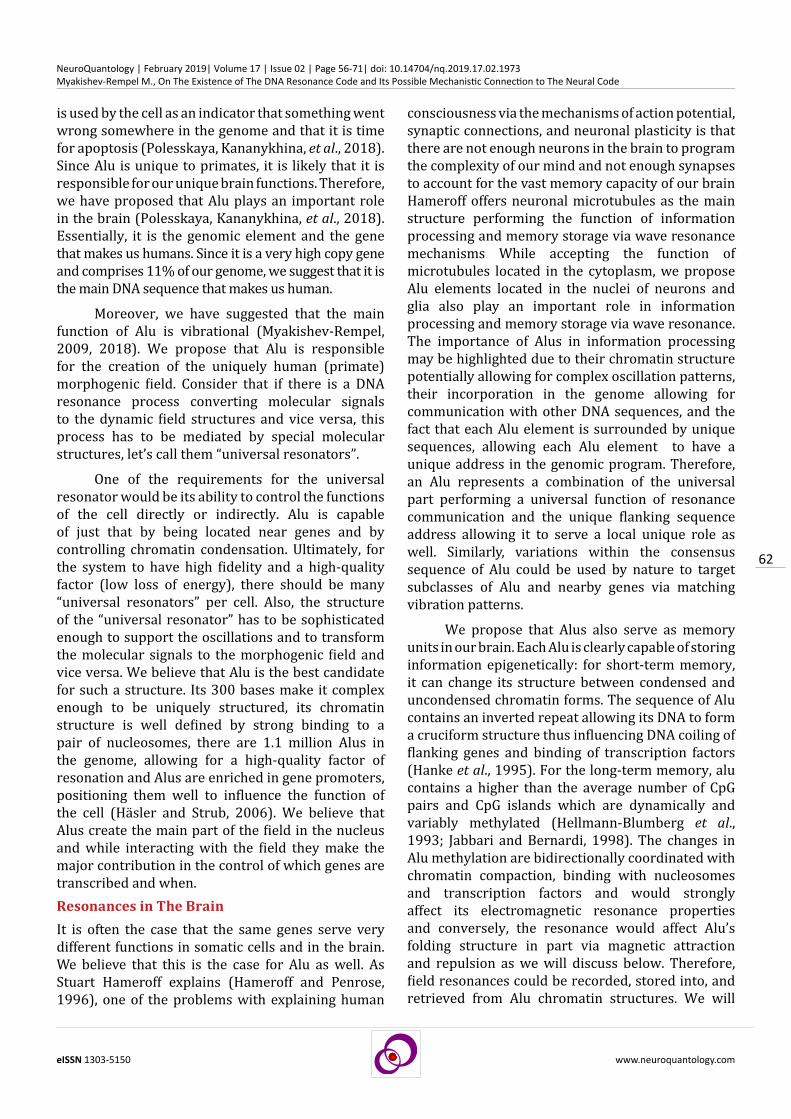
While ring currents in single ring molecules such as pyrimidines (C and T) have simple geometry, fused-ring molecules such as purines (A and G) are more complex. Although under a strong magnetic field the ring current in purines goes around their perimeter, Fig. 3A, we predicted (Guschin, Polesskaya, Zyryanova, Tovmash and Myakishev-Rempel, 2018) that in physiological weak magnetic conditions, the ring currents in purines would flip into an infinity shape (∞), Fig. 3B. The infinity shape of the ring current would be most optimal since it would create antiparallel magnetic vectors within a purine canceling each other. When combined in a base stack, the magnetic vectors of the bases would combine, creating patterns of magnetic lines as shown in Fig. 4. The stretches of purines would have double lines and the stretches of pyrimidines single lines.
Note that the magnetic field of the magnetized base stack would spill over outside of the double helix and should be readable by possible field sensing molecules from outside. Therefore, the sequence will indeed act as a magnetic tape and the DNA sequence will be converted to the field pattern outside of the molecule. This fits our definition of the DNA resonance code: the algorithm of conversion
How could DNA strands be magnetic? The idea that ring currents (Brandes et al., 1988; Dogra et al., 2014; Guelev et al., 2001; Lown and Hanstock, 1985; Peters et al., 1995; Webb, 2000) may be responsible (Kwon, Jin, et al., 2012) for magnetism in DNA comes from nuclear magnetic resonance (NMR) studies. It is textbook knowledge that in a strong magnetic field, a ring current is induced in an aromatic ring, which in turn induces a secondary magnetic field in the opposite direction of the primary field. This secondary field exposes the adjacent protons, observed as a shift of corresponding peaks in NMR (Fowler et al., 2007; Wikipedia contributors, 2018a).
Typically, NMR studies are done in solid substances, but in solution, aromatic rings turn their axis perpendicular to the initial field, thus disabling induction of the ring current, Fig. 2, A1-A4 (A. Hill et al., 2007; Sankarampadi Aravamudhan, 2015). The same is true for the DNA double helix, which turns perpendicular to the initial magnetic field, Fig. 2, B1-B2 (Van Winkle et al., 1997; Vavrinská et al., 2016; Zhang et al., 2017).
We proposed (Guschin, Polesskaya, Zyryanova, Tovmash and Myakishev-Rempel, 2018) that in the cell, in the absence of a strong magnetic field, ring currents may be induced by some of the enzymes using the energy of ATP. Specifically, we noticed that many of the DNA-associated proteins are iron-sulfur proteins (Fuss et al., 2015). We suggested (Guschin, Polesskaya, Zyryanova, Tovmash and Myakishev-Rempel, 2018) that iron-sulfur clusters in these proteins are utilized to magnetize specific sequences and create static and dynamic patterns of magnetization in the DNA sequence. This idea
Figure 2. Ring currents in DNA. A1-A3. The initial magnetic field induces a secondary magnetic field in a nucleobase. A3-A4. If the nucleobase is in solution, it turns along the initial field. B1-B2. If DNA is in solution, it turns perpendicular to the applied magnetic field.
61
eISSN 1303-5150 www.neuroquantology.com
NeuroQuantology | February 2019| Volume 17 | Issue 02 | Page 56-71| doi: 10.14704/nq.2019.17.02.1973Myakishev-Rempel M., On The Existence of The DNA Resonance Code and Its Possible Mechanistic Connection to The Neural Code
of a DNA sequence to the field structure. Yet, this is only a part of the answer, since there is still a need to understand how these local fields combine to create the shape of the body.
Another question about the proposed ring current magnetization of DNA is whether it is static or oscillatory in living cells? The possibilities are varied: some chromatin structures might support static magnetization, other chromatin structures might support oscillating ring currents that change direction with a certain frequency. The iron-sulfur containing enzymes may be providing energy to these oscillations while drawing the energy from splitting ATP. DNA, in turn, would serve as a resonator and provide structure to these oscillations. Note that reactions of transcription and replication which also utilize triphosphates might energize magnetic oscillations in DNA as a side product of their chemical activity. Genomic Repeats as OscillatorsWhat are the sizes of DNA oscillators? The smallest would be stretches of single nucleotide repeats. Higher genomes are rich in these stretches. Also frequent are di- tri- tetra and pentanucleotide repeats. These are
called microsatellites and were used in genotyping since they are prone to length variation. The abundance of microsatellites is roughly highest for single-nucleotide repeats and fades with the length of the repeating unit. Among the simple repeats, the telomeric repeat stands out. In vertebrates, many of the plant taxa and yeast, the telomeric repeat is 6 bases long: GGGTTA. Some of the plant taxa share the 6-base telomeric repeat sequence GGGTTA with us and in some other plant taxa, it is a 7-mer GGGTTTA. In many taxa of insects, it is a 5-mer GGTTA (Wikipedia contributors, 2018b). The length of human telomeres is around 2500 repeats each. Similarly, centromeric repeats are also potential resonators. We suggest that the telomeric and centromeric repeats being the longest simple repeats in our genome are among the most fundamental resonators in the genome and may provide the fundamental carrier frequency for the cell and the body. Since this hypothetical frequency would be common between many forms of life surrounding us, it would be one of the major frequencies of life on the planet.
What are the other abundant repeats in the human genome? The Alu repeat has the highest number of copies in our genome. (The name Alu is unrelated to Alu’s function and comes from the incidental fact that the Alu sequence can be cut by AluI restrictase cloned from Arthrobacter luteus bacteria.) Alu is an interspersed repeat, meaning that it is distributed throughout the genome without a perceivable order. The length of Alu is near 300 bp and it has about 1.1 million copies in our genome. The molecular functions of the genomic copies of the Alu repeat are of high potential significance - it strongly binds nucleosomes, and often serves as a crystallization point for chromatin condensation: the condensation (heterochromatization) of chromatin starts with Alu and spreads along the sequence in both directions. Yet, Alu repeats are frequent and conserved in gene promoters, pointing at a possible regulatory function of genomic Alu. Moreover, the variations within the Alu sequence in a gene promoter correlate with transcriptional activity of that gene (Hon and Jain, 2003; Jjingo et al., 2011; Kim et al., 2004). In addition to its function as chromatin condensation regulator and regulator of genes, Alu is a gene coding for an untranslated RNA. Normally, the 1.1 million Alu repeats in the genome are silent, but in certain cell types, some are transcriptionally active. Their transcription is also activated by cellular stress. It is likely that abnormally high transcription of Alus
Figure 4. A. Hypothetical patterns of magnetic lines in the base stack, a flattened view. B. The 3D shape of the base stack. C. Hypothetical patterns of magnetic lines in a double-helical base stack, a helical view.
Figure 3. A. Ring current in a purine under strong magnetic field circles the perimeter. B. We predict that ring current in a purine under a natural magnetic field should take on an infinity shape.
62
eISSN 1303-5150 www.neuroquantology.com
NeuroQuantology | February 2019| Volume 17 | Issue 02 | Page 56-71| doi: 10.14704/nq.2019.17.02.1973Myakishev-Rempel M., On The Existence of The DNA Resonance Code and Its Possible Mechanistic Connection to The Neural Code
is used by the cell as an indicator that something went wrong somewhere in the genome and that it is time for apoptosis (Polesskaya, Kananykhina, et al., 2018). Since Alu is unique to primates, it is likely that it is responsible for our unique brain functions. Therefore, we have proposed that Alu plays an important role in the brain (Polesskaya, Kananykhina, et al., 2018). Essentially, it is the genomic element and the gene that makes us humans. Since it is a very high copy gene and comprises 11% of our genome, we suggest that it is the main DNA sequence that makes us human.
Moreover, we have suggested that the main function of Alu is vibrational (Myakishev-Rempel, 2009, 2018). We propose that Alu is responsible for the creation of the uniquely human (primate) morphogenic field. Consider that if there is a DNA resonance process converting molecular signals to the dynamic field structures and vice versa, this process has to be mediated by special molecular structures, let’s call them “universal resonators”.
One of the requirements for the universal resonator would be its ability to control the functions of the cell directly or indirectly. Alu is capable of just that by being located near genes and by controlling chromatin condensation. Ultimately, for the system to have high fidelity and a high-quality factor (low loss of energy), there should be many “universal resonators” per cell. Also, the structure of the “universal resonator” has to be sophisticated enough to support the oscillations and to transform the molecular signals to the morphogenic field and vice versa. We believe that Alu is the best candidate for such a structure. Its 300 bases make it complex enough to be uniquely structured, its chromatin structure is well defined by strong binding to a pair of nucleosomes, there are 1.1 million Alus in the genome, allowing for a high-quality factor of resonation and Alus are enriched in gene promoters, positioning them well to influence the function of the cell (Häsler and Strub, 2006). We believe that Alus create the main part of the field in the nucleus and while interacting with the field they make the major contribution in the control of which genes are transcribed and when. Resonances in The BrainIt is often the case that the same genes serve very different functions in somatic cells and in the brain. We believe that this is the case for Alu as well. As Stuart Hameroff explains (Hameroff and Penrose, 1996), one of the problems with explaining human
consciousness via the mechanisms of action potential, synaptic connections, and neuronal plasticity is that there are not enough neurons in the brain to program the complexity of our mind and not enough synapses to account for the vast memory capacity of our brain Hameroff offers neuronal microtubules as the main structure performing the function of information processing and memory storage via wave resonance mechanisms While accepting the function of microtubules located in the cytoplasm, we propose Alu elements located in the nuclei of neurons and glia also play an important role in information processing and memory storage via wave resonance. The importance of Alus in information processing may be highlighted due to their chromatin structure potentially allowing for complex oscillation patterns, their incorporation in the genome allowing for communication with other DNA sequences, and the fact that each Alu element is surrounded by unique sequences, allowing each Alu element to have a unique address in the genomic program. Therefore, an Alu represents a combination of the universal part performing a universal function of resonance communication and the unique flanking sequence address allowing it to serve a local unique role as well. Similarly, variations within the consensus sequence of Alu could be used by nature to target subclasses of Alu and nearby genes via matching vibration patterns.
We propose that Alus also serve as memory units in our brain. Each Alu is clearly capable of storing information epigenetically: for short-term memory, it can change its structure between condensed and uncondensed chromatin forms. The sequence of Alu contains an inverted repeat allowing its DNA to form a cruciform structure thus influencing DNA coiling of flanking genes and binding of transcription factors (Hanke et al., 1995). For the long-term memory, alu contains a higher than the average number of CpG pairs and CpG islands which are dynamically and variably methylated (Hellmann-Blumberg et al., 1993; Jabbari and Bernardi, 1998). The changes in Alu methylation are bidirectionally coordinated with chromatin compaction, binding with nucleosomes and transcription factors and would strongly affect its electromagnetic resonance properties and conversely, the resonance would affect Alu’s folding structure in part via magnetic attraction and repulsion as we will discuss below. Therefore, field resonances could be recorded, stored into, and retrieved from Alu chromatin structures. We will

63
eISSN 1303-5150 www.neuroquantology.com
NeuroQuantology | February 2019| Volume 17 | Issue 02 | Page 56-71| doi: 10.14704/nq.2019.17.02.1973Myakishev-Rempel M., On The Existence of The DNA Resonance Code and Its Possible Mechanistic Connection to The Neural Code
address the electromagnetic connection between Alu elements in the nucleus, microtubules in the axons, and the action potential in the axons later in the article.
The idea that repetitive elements have a regulatory function and are controlling the activity of genes is older than genomic sequencing and the discovery of the double helix. This idea was proposed and experimentally proven at a time when the majority of scientists believed that genes are made of protein and not DNA. It was Barbara McClintock who used genetic methods to discover the repetitive (transposable) elements. She called them Control Elements and proved that they are universal genetic elements controlling nearby genes (Lippman et al., 2004; McClintock, 1951; Mcclintock, 1956). In 1951, she described the “elements in the heterochromatin concerned with differential control of the times at which certain genes may become reactive.” (McClintock, 1951) We suggest that in humans these are Alu elements and that these elements not only control the expression of nearby genes but also are resonators communicating electromagnetically with the morphogenic field which they help to create.
The literature on Alus is strongly dominated by research on their transposition properties. While respecting the importance of transposition in the evolutionary past, we have emphasized (Myakishev et al., 2008; Polesskaya, Kananykhina, et al., 2018) that the proposed function of Alus doesn’t have to be related to transposition in any way. Since the transposition rate of the majority of the 1.1 million copies of genomic Alus is extremely low, possibly one transposition per generation, we have proposed that at the present, Alu has important resonance, gene regulation, and memory functions unrelated to its transposability (Myakishev et al., 2008; Polesskaya, Kananykhina, et al., 2018). We suggest that the current function of Alus should be explored without the heavy weight of their past. Even the fact that in the distant past they selfishly conquered 11 percent of our genome may be redeemed by their proposed contribution to our morphogenic field and consciousness. Polynucleosomal OscillatorsWhat could be the molecular structure of the Alu resonator? Since each Alu strongly binds two nucleosomes, and since Alu sequences are typically compacted (heterochromatinized), it is likely that Alu resonators include nucleosomes. Since the base
stack behaves as an insulated conductor,(Holmlin et al., 1997) we proposed that nucleosomes serve as induction coils in such a resonator (Guschin, Polesskaya, Zyryanova, Tovmash and Myakishev-Rempel, 2018). The way nucleosomes are organized in living cells has been uncertain for many years. Solenoid and two-start zigzag packaging were obtained in vitro using periodic DNA templates (Chen et al., 2014; Maeshima et al., 2016; Widom and Klug, 1985; Wu et al., 2016). Recently, mapping nucleosomal packaging in living cells showed the irregular nature of nucleosomal packaging (Li et al., 2015; Maeshima et al., 2014; Ou et al., 2017). Most likely, since the genomic sequence is aperiodic, the nucleosomes in living cells are packaged in irregular structures (Li et al., 2015; Maeshima et al., 2014; Ou et al., 2017; Scheffer et al., 2011). Among these irregular structures, frequently are mono-, di-, and tetranucleosomes (Scheffer et al., 2011). We proposed that these structures may serve as resonant circuits (Polesskaya, Guschin, et al., 2018). For example, we proposed an oscillation model for a tetranucleosome, Fig. 5 (Polesskaya, Guschin, et al., 2018).
In this resonant circuit, the current circles the nucleosomes back and forth and the stacked pairs of the nucleosomes change their magnetic polarity from one phase of the cycle to another (Polesskaya, Guschin, et al., 2018). Since the length of the linker
Figure 5. One of the possible models of a tetranucleosomal oscillator. I – current, B – magnetic field. Top – the first phase, bottom – the second phase of the oscillation period.
64
eISSN 1303-5150 www.neuroquantology.com
NeuroQuantology | February 2019| Volume 17 | Issue 02 | Page 56-71| doi: 10.14704/nq.2019.17.02.1973Myakishev-Rempel M., On The Existence of The DNA Resonance Code and Its Possible Mechanistic Connection to The Neural Code
DNA sequences connecting the stacked nucleosome pairs will depend on the sequence, the geometry of the tetranucleosomes will vary accordingly, thus the frequency and the geometry of the generated electromagnetic waves will change as well. This is in agreement with the above idea that various sequences will have various resonation properties and that similar sequences will resonate. Moreover, this model is permissive to sequence variations: the resonance between the two sequences will occur as long as the overall geometry remains similar. This illustrates a certain redundancy of the resonance code and suggests ways for its experimental discovery.
Although Alu elements are distributed through our genome seemingly at random, there is one exception: there are frequent pairs of Alus, structured as inverted repeats, where the Alu sequence is followed by its complement (Chen and Carmichael, 2009; Lobachev et al., 2000). Usually, there is a high similarity between the halves of the inverted Alu repeat. Notably, between the two Alu sequences in the inverted repeat, there usually is a short unique bridge sequence, which we think may be used by nature as a sequence tag to tag each inverted Alu repeat. Since each Alu binds two nucleosomes, the inverted Alu repeat should form a tetranucleosomal structure, which we suggest is a special type of resonator. Since there are many inverted Alu repeats in the genome, these special resonators may resonate with each other. The length of the bridge sequence should then fine tune the resonant frequency and shape of the wave for specific groups of these inverted Alu resonators. FrequenciesWe believe that the methods of modern genomics will be sufficient for decoding the resonance code of DNA:
genetic engineering with subsequent measurement of spectral properties and full-genome mapping of the influence of waves on the openness and transcription of DNA. Toward that goal, we have shown the effect of red light on the expression of candidate genes in the murine epidermis (Myakishev-Rempel et al., 2015).
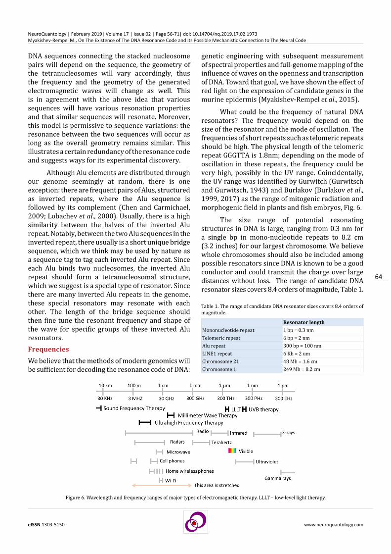
What could be the frequency of natural DNA resonators? The frequency would depend on the size of the resonator and the mode of oscillation. The frequencies of short repeats such as telomeric repeats should be high. The physical length of the telomeric repeat GGGTTA is 1.8nm; depending on the mode of oscillation in these repeats, the frequency could be very high, possibly in the UV range. Coincidentally, the UV range was identified by Gurwitch (Gurwitsсh and Gurwitsсh, 1943) and Burlakov (Burlakov et al., 1999, 2017) as the range of mitogenic radiation and morphogenic field in plants and fish embryos, Fig. 6.
The size range of potential resonating structures in DNA is large, ranging from 0.3 nm for a single bp in mono-nucleotide repeats to 8.2 cm (3.2 inches) for our largest chromosome. We believe whole chromosomes should also be included among possible resonators since DNA is known to be a good conductor and could transmit the charge over large distances without loss. The range of candidate DNA resonator sizes covers 8.4 orders of magnitude, Table 1.
Figure 6. Wavelength and frequency ranges of major types of electromagnetic therapy. LLLT – low-level light therapy.
Resonator lengthMononucleotide repeat 1 bp = 0.3 nmTelomeric repeat 6 bp = 2 nmAlu repeat 300 bp = 100 nmLINE1 repeat 6 Kb = 2 umChromosome 21 48 Mb = 1.6 cmChromosome 1 249 Mb = 8.2 cm
Table 1. The range of candidate DNA resonator sizes covers 8.4 orders of magnitude.

65
eISSN 1303-5150 www.neuroquantology.com
NeuroQuantology | February 2019| Volume 17 | Issue 02 | Page 56-71| doi: 10.14704/nq.2019.17.02.1973Myakishev-Rempel M., On The Existence of The DNA Resonance Code and Its Possible Mechanistic Connection to The Neural Code
The ideal tool for testing sequence-dependent DNA oscillations would be experimental genomics. So far, very little is published in this area. Some clues might be inferred from electromagnetic therapy practice. Electromagnetic therapy in the West is usually limited to transcutaneous electrical nerve stimulation (TENS), and red and near-infrared light therapy (low-level light therapy, LLLT). Other forms of electromagnetic therapy are used in Eastern Europe, Russia, and Asia including ultraviolet light, millimeter waves, ultrahigh frequency, and sound frequency electromagnetic waves, Fig. 6. These waves have the capacity to be effective at very low doses, suggesting that they tap into existing signaling in the body, i.e., they influence the morphogenic field. We suggest that live chromatin may support oscillations at every one of these frequencies: smaller DNA resonators would resonate at higher frequencies and larger ones at lower frequencies.
For example, individual aromatic rings of DNA bases are well known to resonate at 260 nm (1.2 PHz) in the UV range. These are small structures, 0.3-0.7 nm size. On the other hand, chromatin is known also to oscillate at a very slow rate of 1 oscillation every 40 minutes (1.5 cycles per hour, 0.0004 Hz) (Métivier et al., 2003; Pliss et al., 2013). The frequency difference between these two oscillations (1.2 PHz of UV absorption and 0.0004 Hz of chromatin oscillations) is 61 orders of magnitude. How could chromosomes be involved in such a wide range of oscillations? In addition to the size difference between the oscillators, consider the mass and the geometry of the oscillator. Above we mentioned 3 types of oscillations in DNA - oscillation of the parts of the DNA molecule (mechanical oscillations), oscillations of the delocalized electron cloud of the base stack (electron oscillations), and oscillations of the delocalized hydrogen bond protons of the base stack (proton oscillations). A proton is nearly 1900 times heavier than an electron and a base pair of DNA is 640 times heavier than the proton. Consider also that the DNA molecule is heavily hydrated and bound to chromatin which makes its mass much bigger. The geometry of oscillations would also strongly affect the frequency. Swinging oscillations would be much slower than stretching oscillations. Thus, we conclude that the variation in the size of oscillators within the chromosome, the nature of oscillating fields (electron, proton, molecule), and the geometry of oscillations should allow DNA to support a very wide range of frequencies.
Dissipation ProblemIn addition to classical electromagnetic waves, alternative electromagnetic wave geometries have been proposed by Konstantin Meyl (Meyl, 2011, 2012). These proposed electromagnetic waves are helical, longitudinal, and characterized by an unusual phase shift between electric and magnetic fields. Meyl proposes that these waves are produced by DNA, making the biofield and that these waves have special properties allowing them to work in the irregular milieu of biological tissues.
This brings us to the question of dissipation of electromagnetic waves in biological tissues. Dissipation is the strongest argument against the morphogenic field. For example, among the therapeutic frequencies, UV is strongly absorbed by DNA, red and near-infrared light are not absorbed by DNA but likely somewhat absorbed by chromatin and DNA binding proteins, and millimeter waves are strongly absorbed by water. How could the morphogenic field function and organize biological structures which are so complex, malleable, and irregular?
One answer could be that the nature of the waves is unusual, such as in the special waves of Meyl. Another possibility is that biomolecular events happen at the microscopic and nanoscale level where neither macroscopic laws nor quantum chemistry laws are applicable and new quantum-biophysical laws are working. In addition to the above possibilities, we propose that some of the signal scattering problems are solved in nature by waveguides. Specifically, consider the problem of signal scattering for DNA which is located in the nucleus and has to communicate across nuclear, cellular, and organelle membranes to the DNA in other nuclei. Since the cellular milieu contains many organelle membranes and uneven shapes, the scattering of light in the cells and in the biological tissue is high and should challenge the electromagnetic communication between the nuclei. A similar problem needs to be solved to explain the possible role of genomic resonance in the workings of the mind and consciousness. The problem is that the main sensory, movement, and thinking activities are mediated by the movement of action potentials along the axons of neurons, but DNA is located in the nucleus, where it is insulated and spatially removed from these action potentials. For example, the nuclei of the dorsal ganglia of touch sensing cells are located in our spine, while their nerve endings are located

66
eISSN 1303-5150 www.neuroquantology.com
NeuroQuantology | February 2019| Volume 17 | Issue 02 | Page 56-71| doi: 10.14704/nq.2019.17.02.1973Myakishev-Rempel M., On The Existence of The DNA Resonance Code and Its Possible Mechanistic Connection to The Neural Code
in the skin. The length of the axon innervating the foot is around 3 feet. How could electromagnetic communication take place at such large distances?Microtubular Signal TransductionWe suggest that it is microtubules which serve as waveguides to transfer the electromagnetic signal from the nucleus through the cytoplasm. The theory of microtubular signal transmission has been developed by Stuart Hameroff and coauthors for the last 30 years (Craddock et al., 2014; Hameroff, 1974; Hameroff et al., 2002; Sahu et al., 2013). Hameroff proposed that microtubules in neurons contribute to the creation of consciousness by transmitting, computing and storing information. He proposed that it is delocalized electrons of aromatic amino acids that are oscillating in microtubules. Stuart Hameroff and Anirban Bandyopadhyay proposed that these oscillations are of electron spins (Hameroff, 2007). Although the ideas of Hameroff and colleagues about microtubular signal transduction so far, have limited experimental support (Sahu et al., 2013), we find the indirect evidence for these ideas encouraging. Specifically, good dynamical health of microtubules is of high importance for neuronal function - a failure to keep the growth and degradation of microtubules in balance causes signaling dysfunction. The perfect straight cylindrical structure of microtubules makes them perfect as waveguides.
We utilized this understanding to theoretically resolve one of the major problems with DNA resonance: the problem of dissipation of the signal. Until now, it was believed by us and others that DNA is forming the morphogenic field by sending electromagnetic waves in all directions but in this model, the signal would be quickly lost in the of the thick organic milieu of the cytoplasm. Now, the fact, that microtubules are present in each eukaryotic cell, and are oriented radially connecting the nuclear and cellular membranes, allowed us to suggest that the electromagnetic waves travel between the nuclei via a network of microtubules serving as waveguides, Fig. 7 (Savelyev et al., 2018).
We propose that the microtubules and DNA communicate resonantly through the nuclear membrane and that the microtubules of neighboring cells communicate resonantly through the contact points of the cell membranes, thereby integrating all the body nuclei into one resonance network. Furthermore, we propose that this DNA-microtubular network of the nervous system and connective tissue is primarily responsible for uniting the organism
into one resonating system. This signal transduction model explains how nature could achieve high transduction efficiency for the DNA resonances across the tissues and solves the dissipation problem for the DNA-generated morphogenic field. Connecting DNA Resonance to Neuronal FunctionOnce such a DNA and microtubules are integrated into the model of the morphogenic field, it is pertinent to understand how the DNA-generated field of the body interacts with the electromagnetic field of the brain. We can see that the difference between the morphogenic field of the body and the electromagnetic field of the brain might be mechanistically negligible. In both somatic and neural cells, the electromagnetic oscillations from the DNA in the nuclei may be transmitted via the microtubules thus forming a brain-wide and body-wide network.
At this point, it becomes possible to tie the action potential propagation in neurons with the electromagnetic oscillations in DNA. Until recently, the electric activity in axons was thought to be spatially separated from the DNA in the nucleus. Now, a mechanism can be proposed electromagnetically linking DNA and axonal oscillations together via the microtubules as waveguides, Fig. 8.
Moreover, this model suggests that there is a direct electromagnetic conversation between the DNA code and axons, in other words, that DNA resonance code and neural codes are mechanistically linked to each other. The concept of neural code or neural coding recently came to the forefront with the widespread effort to develop the brain-computer interface. In spite of much effort, it is becoming clear
Figure 7. We propose that the electromagnetic oscillations in DNA within the nucleus and microtubules in cytoplasm are synchronized across the nuclear membrane and that the oscillations between microtubule networks of adjacent cells are synchronized across cellular membranes thus uniting all nuclei of the body in one oscillation network via waveguides of microtubules.

67
eISSN 1303-5150 www.neuroquantology.com
NeuroQuantology | February 2019| Volume 17 | Issue 02 | Page 56-71| doi: 10.14704/nq.2019.17.02.1973Myakishev-Rempel M., On The Existence of The DNA Resonance Code and Its Possible Mechanistic Connection to The Neural Code
that the scientists have a very limited understanding of the ways the brain thinks and therefore, it is very hard to interface with it. A better understanding of the neuronal syntax and the neuronal code would greatly help the brain-computer interface and ultimately establishing brain-technology-brain interface or synthetic telepathy. We suggest that the key to the neural code may, although unexpectedly, lie in deciphering the DNA resonance code. How to Decipher The DNA Resonance CodeTo our knowledge, although the holographic model of the genome-generated morphogenic field was proposed 45 years ago, very little has been done so far experimentally to decipher the code. We believe that standard approaches of modern science should be sufficient for cracking and deciphering it. Consider a couple of examples of cracking unknown codes from the past: the key to the deciphering of Egyptian hieroglyphs was the Rosetta stone which contained parallel inscriptions in Egyptian and two known languages. Comparing these three texts allowed Jean-François Champollion and Thomas Young to decipher the code some 200 years ago. In the case of the DNA resonance code, we suggest that the code can be deciphered by combining quantum chemical molecular modeling, biophysical experiments, genome-wide chromatin accessibility, and transcriptional activity mapping in response to electromagnetic waves, spectral analysis of synthetic DNA and gene-modified live tissue, and linguistic computational analysis.
Another historical example is the deciphering of the amino acid-coding genetic code by Marshall Nirenberg and others 57 years ago. The approach he used was to feed a synthetic nucleic sequence into a cellular extract and analyze the molecular outcome. We suggest that the same approach could be employed for the discovery of the DNA resonance code - by inserting a sequence into a cell, allowing
the cells to transfer the message via waves to another batch of cells, and using genome-wide analysis to map the resulting chromatin changes. In the case of Nirenberg, once the first letter of the code (UUU coding for Ala) was discovered, it took a very short time for the world community to crack the rest of the code. Note, that for the DNA resonance code we already proposed a set of candidate sequences including Alu and LINE1, the second most highly abundant repetitive element, 6000 bp long, represented by 0.5 million copies and comprising 17% of our genome. Applications And ImplicationsOnce the code is deciphered, the potential applications will stem from the two major roles which were proposed by us and others: the role of DNA in the creation and interaction with the morphogenic field and with the mind.
Since the proposed role of the morphogenic field is to drive biological development of tissues, organs and body structure; understanding its code will allow us to control body shape, reverse obesity, and induce the growth of tissues, organs, limbs, and new teeth. We believe that the progress in organ engineering is unimpressive because of our inability to speak the resonance language of DNA. Deciphering the DNA resonance code will enable organ engineering.
We also proposed that DNA resonance is linked to the work of the mind via resonance patterns and that deciphering the DNA resonance code will greatly facilitate the deciphering of electrophysiological patterns of the brain. In short, we propose that the brain code and DNA resonance codes are linked and speak the same language. The deciphering of the brain code will allow the development of brain-computer and of brain-technology-brain interfaces, also known as synthetic telepathy. We suggest that the path to the brain code via DNA (although counterintuitive) may be the shortest path, since DNA is digital, and the methods for genetic engineering and genome-wide analysis are exceptionally efficient.
The implications of deciphering the DNA resonance code should also affect the self-image of humanity since it will clearly demonstrate that all of us are linked via DNA resonance vibrations to each other and to all life on the planet and that should help our unity and ecology.ConclusionsWe reviewed the reasons to search for the morphogenic field, highlighted key experiments confirming its existence, and briefly reviewed publications highlighting and the key role of DNA in
Figure 8. We propose that in neurons, action potential propagation is electromagnetically linked to DNA via microtubules and via the nuclear membrane, allowing the genome to sense and control action potential propagation and thus actively participate in perception, movement control, memory, and other aspects of mind and consciousness.

68
eISSN 1303-5150 www.neuroquantology.com
NeuroQuantology | February 2019| Volume 17 | Issue 02 | Page 56-71| doi: 10.14704/nq.2019.17.02.1973Myakishev-Rempel M., On The Existence of The DNA Resonance Code and Its Possible Mechanistic Connection to The Neural Code
its creation and function. We reviewed the models of others and proposed new models for the involvement of delocalized electron and proton clouds of the base stack in the formation and function of the morphogenic field. We developed the idea that genomic repeats act as universal resonators providing the bidirectional communication between the chromatin structure and the morphogenic field. We proposed that DNA resonances of the genomic repeats perform two functions: create the morphogenic field and participate in the electrical activity of the brain. We proposed that microtubules mediate the resonance communication between the action potential in axons and genomic repeats in the nucleus. We postulated the existence of an algorithm responsible for the conversion of the genomic information into the shape of the body and named it the DNA resonance code. We proposed that the DNA resonance code can be deciphered by combining targeted efforts in biophysics, spectroscopy, molecular modeling, and experimental genomics. We concluded that the DNA resonance code may be directly linked to the neural code and that deciphering the former would help to decipher the later. Ultimately we suggest that deciphering the DNA resonance code would benefit a wide range of medical applications related to morphogenesis and brain function. Author ContributionsAll authors contributed to the discussion. MMR conceived and wrote the manuscript. OP suggested the resonance information transfer between the nucleus and the microtubules. MMR and IS developed the microtubule theme. MMR and NZ developed the magnetic theme.AcknowledgmentsWe thank Richard Alan Miller for drawing our attention to microtubules. We thank Richard Alan Miller, Dan Winter, Peter Gariayev, Dean Radin, Michal Cifra, Vadim Guschin, Alexei Tovmash, Abraham Mara, James Ernest Charles, Evgenia Golovina, Eugenia Kananykhina, Karina Tumanyants, Elena Tumanyants, Anatoly Yatsunenko, Nikolai Kondratev, Raymond Lanzafame, Istvan Stadler, Anna Baranova, Alex Vetcher, Olga Nazarenko, Vladimir Kornienko, Tatyana Shapiro, Elena Erdyneva, Dimitri Deheyn, Bor-Kai Hsiung, Kyle Thackston, Daniel Sievenpiper, Rae Anderson, Astrid Engel, Konstantin Kupriyanov, and Irina Garanina for the discussion of different aspects of the work and at different stages. MMR and OP thank NIH for 1R43MH110273 grant that supported them during a segment of the work.
ReferencesA. Hill RJ, Sedman VL, Allen S, Williams P, Paoli M, Adler-
Abramovich L, Gazit E, Eaves L, Tendler SJ. Alignment of aromatic peptide tubes in strong magnetic fields. Advanced Materials 2007; 19(24): 4474-4479.
Alexander and Gurwitsch L. Weitere Untersuchungen über mitogenetische Strahlungen. Archiv für mikroskopische Anatomie und Entwicklungsmechanik 1925; 104(1): 109–115.
Arnold AR, Grodick MA, Barton JK. DNA Charge Transport: from Chemical Principles to the Cell. Cell chemical biology 2016; 23(1): 183–197.
Beloussov LV, Burlakov AB, Louchinskaia NN (2003) Biophotonic patterns of optical interactions between fish eggs and embryos. Indian journal of experimental biology 2003; 41(5): 424–430.
Beloussov LV, Opitz JM, Gilbert SF. Life of Alexander G. Gurwitsch and his relevant contribution to the theory of morphogenetic fields. International Journal of Developmental Biology. 2004; 41(6): 771-777. Available at: http://www.ijdb.ehu.es/web/descarga/paper/9449452.
Bischof M. Biophotonen: Das Licht in Unseren Zellen. Zweitausendeins, 1995.
Blois MS Jr, Maling JE, Taskovich LT. On the electron spin resonances in DNA. Biophysical journal 1963; 3: 275–297.
Blumenfeld LA. Abnormal magnetic properties of nucleic acids. Biophysics 1959; 4(5): 515–519.
Blumenfeld LA. About ferromagnetism of organic structures. Doklady USSR Academy of Sciences 1963; 148(2): 361–364.
Brandes R, Kearns DR, Rupprecht A. A 2H-NMR study of the DNA hydration water in solid Li-DNA assembles. Biopolymers 1988; 27(5): 717–732.
Burkov VD, Burlakov AB, Perminov SV. Correction of Long Range Interaction Between Biological Objects Using Corner-Cube Reflectors. Biomedical Radioelectronics 2008; (8-9): 41–48.
Burlakov AB. Distant physical interactions between the developing fish embryos. Biophotonics and Coherent Systems Moscow Univ Press 2000; pp: 298–304.
Burlakov AB and Lebedeva NE. Adequacy of biological models while investigating effects of ultra low dose chemical and physical actions. Weak and ultraweak fields and emissions in biology and medicine 2015; pp: 21–22.
Burlakov AB, Burlakova OV, Golichenkov VA. The long range interactions of different age embryo loach Misgurnus fossilis. Doklady Biological Sciences 1999; 368(4): 562–564.
Burlakov AB, Burlakova OV, Golichenkov VA. Distant wave-mediated interactions in early embryonic development of the loach Misgurnus fossilis L. Russian Journal of Developmental Biology 2000; 31(5): 287–292.
Burlakov AB, Burlakova OV, Korolev YN, Golichenkov VA. Polarization effects in long range interaction of biological objects. Moscow University Biological Sciences Bulletin. Series 2002; 16(2): 3–8.
Burlakov AB, Medvedeva AA, Burlakova OV, Malakhov YI, Golichenkov VA. Special aspects of biological effects of loach’s ultraweak emissions spectral components in early ontogenesis. In Weak and ultraweak fields and emissions in biology and medicine 2006; pp: 34–40. biophys.ru.

69
eISSN 1303-5150 www.neuroquantology.com
NeuroQuantology | February 2019| Volume 17 | Issue 02 | Page 56-71| doi: 10.14704/nq.2019.17.02.1973Myakishev-Rempel M., On The Existence of The DNA Resonance Code and Its Possible Mechanistic Connection to The Neural Code
Burlakov AB, Burlakova OV, Golichenkov VA. Possible changes of individual biological time by weak electromagnetic emissions. In Weak and ultraweak fields and emissions in biology and medicine. St-Petersburg 2009; pp: 40–47.
Burlakov AB, Burlakova OV, Golichenkov VA. Ultraweak bioemissions role in development of lower vertebrates. Krasnodar: Krasnodar’s Center of Scientific and Technical Information. Physics, biophysics and information technology 2010; pp: 81–84.
Burlakov AB, Kapranov YS, Kufal GE, Perminov SV. About possible influence on biological object electromagnetic fields. Weak and Ultraweak Fields and Radiation in Biology and Medicine-Proceedings of IV International Congress 2012; pp: 111–112.
Burlakov AB, Burlakova OV, Golichenkov VA. The long range interactions of biological systems. In Weak and ultraweak fields and radiation in biology and medicine 2012; pp: 5–8. biophys.ru.
Burlakov AB, Chernova GB, Babkina VV, Sidorov VV, Andeber OP. Variability of morphological characters as a manifestation of the effects of radiation extremely high frequency and their evaluation in the context of bioinformatics. Biomedical Radioelectronics 2017; (1): 44–50. elibrary.ru.
Chatzidimitriou-Dreismann CA. Protonic delocalization and quantum correlations in the H-bond dynamics of G--C and κ--π DNA base pairs. International journal of quantum chemistry 1993; 46(3): 483–498.
Chen LL and Carmichael GG. Altered nuclear retention of mRNAs containing inverted repeats in human embryonic stem cells: functional role of a nuclear noncoding RNA. Molecular Cell 2009; 35(4), 467–478.
Chen P, Zhu P, Li G. New insights into the helical structure of 30-nm chromatin fibers. Protein & Cell 2014; 5(7): 489–491.
Cifra M, Fields JZ, Farhadi A. Electromagnetic cellular interactions. Progress in Biophysics and Molecular Biology 2011; 105(3): 223–246.
Ciszak M, Comparini D, Mazzolai B, Baluska F, Arecchi FT, Vicsek T, Mancuso S. Swarming behavior in plant roots. PloS One 2012; 7(1): e29759.
Craddock TJA, Friesen D, Mane J, Hameroff S, Tuszynski JA. The feasibility of coherent energy transfer in microtubules. Journal of the Royal Society, Interface / the Royal Society 2014; 11(100): 20140677.
Davenas E, Beauvais F, Amara J, Oberbaum M, Robinzon B, Miadonnai A, Tedeschi A, Pomeranz B, Fortner P, Belon P, Sainte-Laudy J. Human basophil degranulation triggered by very dilute antiserum against IgE. Nature 1988; 333(6176): 816-818.
Dogra S, Awasthi P, Tripathi S, Pradeep TP, Nair MS, Barthwal R. NMR-based structure of anticancer drug mitoxantrone stacked with terminal base pair of DNA hexamer sequence d-(ATCGAT)2. Journal of Biomolecular Structure & Dynamics 2014; 32(7): 1164–1183.
Farhadi A, Forsyth C, Banan A, Shaikh M, Engen P, Fields JZ, Keshavarzian A. Evidence for non-chemical, non-electrical intercellular signaling in intestinal epithelial cells. Bioelectrochemistry 2007; 71(2): 142–148.
Fowler PW, Lillington M, Olson LP. Aromaticity, π-electron delocalization, and ring currents. Journal of Macromolecular Science, Part A: Pure and Applied Chemistry 2007; 79(6): 969–979.
Fuss JO, Tsai CL, Ishida JP, Tainer JA. Emerging critical roles of Fe-S clusters in DNA replication and repair. Biochimica et Biophysica Acta 2015; 1853(6): 1253–1271.
Gariaev P, Birshtein BI, Iarochenko AM, Marcer PJ, Tertishny GG, Leonova KA, Kaempf U. The DNA-wave biocomputer. International Journal of Computing Anticipatory Systems. Ed. Daniel Dubois, Published by CHAOS 2001; 10. Retrieved from http://www.curealternative.net/bioelettr/memoria_acqua_DNA_wave_computer.pdf
Gariaev PP. Wave genome 1994; pp: 2–8. Public Benefit.
Guelev V, Lee J, Ward J, Sorey S, Hoffman DW, Iverson BL. Peptide bis-intercalator binds DNA via threading mode with sequence specific contacts in the major groove. Chemistry & Biology 2001; 8(5): 415–425.
Gurwitsch A. Über den Begriff des Embryonalen feldes. Wilhelm Roux’ Archiv Fur Entwicklungsmechanik Der Organismen 1922; 51(1): 383–415.
Gurwitsch A. Die Natur des spezifischen Erregers der Zellteilung. Archiv Für Mikroskopische Anatomie Und Entwicklungsmechanik 1923; 100(1): 11–40.
Gurwitsch AA. The problem of mitogenetic radiation as molecular biology aspect. Medicine, 1968. Retrieved from https://play.google.com/books/reader?id=A1v-AgAAQBAJ&lr&pg=GBS.PP1
Gurwitsch AG and Gurwitsch AA. Principles of analytical biology and cell fields theory. (L. V. Beloussov, Ed.). Moscow: Nauka, 1991.
Gurwitsch L. Untersuchungen über mitogenetische Strahlen. Archiv Für Mikroskopische Anatomie Und Entwicklungsmechanik 1924; 103(3): 483–489.
Gurwitsсh A and Gurwitsсh L. Twentieth anniversary of mitogenetic radiation (discovery, development, future directions). Biology Bulletin Reviews 1943; 16(3): 1–28.
Guschin VV, Polesskaya O, Zyryanova N, Tovmash A, Myakishev-Rempel M. On the function of DNA magnetism. VIXRA 2018; 1803.0075.
Guschin VV, Polesskaya O, Zyryanova N, Tovmash A, Mara A, Erdyneeva E, Myakishev-Rempel M. Possible Models of Ring Current Oscillations in DNA. VIXRA 2018; 1803.0176.
Hameroff S. Orchestrated Reduction of Quantum Coherence in Brain Microtubules: A Model for Consciousness. NeuroQuantology 2007; 5(1).
Hameroff S and Penrose R. Orchestrated objective reduction of quantum coherence in brain microtubules: The ‘Orch OR’ model for consciousness. Mathematics and Computers in Simulation 1996; 40: 453–480.
Hameroff S, Nip A, Porter M, Tuszynski J. Conduction pathways in microtubules, biological quantum computation, and consciousness. Bio Systems 2002; 64(1-3): 149–168.
Hameroff SR. Ch’i: A Neural Hologram? Microtubules, Bioholography, and Acupuncture. The American Journal of Chinese Medicine 1974; 02(02): 163–170.
Hameroff SR, Craddock TJA, Tuszynski JA. Quantum effects in the understanding of consciousness. Journal of Integrative Neuroscience 2014; 13(02): 229–252.
Hanke JH, Hambor JE, Kavathas P. Repetitive Alu elements form a cruciform structure that regulates the function of the human CD8 alpha T cell-specific enhancer. Journal of molecular biology 1995; 246(1): 63–73.

70
eISSN 1303-5150 www.neuroquantology.com
NeuroQuantology | February 2019| Volume 17 | Issue 02 | Page 56-71| doi: 10.14704/nq.2019.17.02.1973Myakishev-Rempel M., On The Existence of The DNA Resonance Code and Its Possible Mechanistic Connection to The Neural Code
Häsler J and Strub K. Alu elements as regulators of gene expression. Nucleic Acids Research 2006; 34(19): 5491–5497.
Healy EF (2011) Heisenberg’s chemical legacy: resonance and the chemical bond. Foundations of Chemistry 13(1). Springer: 39–49.
Hellmann-Blumberg U, Hintz MF, Gatewood JM, et al. (1993) Developmental differences in methylation of human Alu repeats. Molecular and cellular biology 13(8): 4523–4530.
Holmlin RE, Dandliker PJ, Barton JK. Charge Transfer through the DNA Base Stack. Angewandte Chemie 1997; 36(24): 2714–2730.
Hon LS and Jain AN. Compositional structure of repetitive elements is quantitatively related to co-expression of gene pairs. Journal of Molecular Biology 2003; 332(2): 305–310.
Jabbari K and Bernardi G. CpG doublets, CpG islands and Alu repeats in long human DNA sequences from different isochore families. Gene 1998; 224(1-2): 123–127.
Jjingo D, Huda A, Gundapuneni M, Mariño-Ramírez L, Jordan IK. Effect of the transposable element environment of human genes on gene length and expression. Genome Biology and Evolution 2011; 3: 259–271.
Khomutov GB. Possible role of iron ions in changes in composition of DNA complexes and their magnetic properties in cell cycle processes. Biofizika 2004; 49(1): 140–144.
Kim TM, Jung YC, Rhyu MG. Alu and L1 retroelements are correlated with the tissue extent and peak rate of gene expression, respectively. Journal of Korean Medical Science 2004; 19(6): 783–792.
Kiperman S. The scientific drama of professor Blumenfeld. ISRAGEO, 2015. Retrieved from http://www.isrageo.com/2015/11/10/dramy116/ (accessed 14 September 2018).
Kurnikov IV, Tong GS, Madrid M, Beratan DN. Hole size and energetics in double helical DNA: competition between quantum delocalization and solvation localization. The Journal of Physical Chemistry B 2002; 106(1): 7-10.
Kwon YW, Lee CH, Do ED, Choi DH, Jin JI, Kang JS, Koh EK. Hydration Effect on the Intrinsic Magnetism of Natural Deoxyribonucleic Acid as Studied by EMR Spectroscopy and SQUID Measurements. Bulletin of the Korean Chemical Society 2008; 29(6): 1233–1242.
Kwon YW, Lee CH, Choi DH, Jin JI. Materials science of DNA. Journal of Materials Chemistry 2009; 19(10): 1353–1380.
Kwon YW, Jin JI, Oh DK, Lee CH. Charge Transport in the DNA fiber: Effects of Magnetic Field and Microwave Irradiation. New Physics 2012; 62(9): 1035–1037.
Kwon YW, Choi DH, Jin JI, Lee CH, Koh EK, Grote JG. Comparison of magnetic properties of DNA-cetyltrimethyl ammonium complex with those of natural DNA. Science China. Chemistry 2012; 55(5): 814–821.
Lander ES. International Human Genome Sequencing Consortium. Initial sequencing and analysis of the human genome. Nature 2001; 409(6822): 860–921.
Lee CH, Kwon YW, Jin JI. Electrical and magnetic properties of DNA. Materials Science of DNA 2011; pp: 121–162.
Lipkind M. Alexander Gurwitsch and the Concept of the Biological Field, Part 1. 21st Century Science & Technology 1998; 11: 36–51.
Lipkind M. Alexander Gurwitsch and the Concept of the Biological Field, Part 2. 21st Century Science & Technology 1998; 11: 34–53.
Lippman Z, Gendrel AV, Black M, Vaughn MW, Dedhia N, McCombie WR, Lavine K, Mittal V, May B, Kasschau KD, Carrington JC. Role of transposable elements in heterochromatin and epigenetic control. Nature 2004; 430(6998): 471-476.
Li X, Feng H, Zhang J, Sun L, Zhu P. Analysis of chromatin fibers in Hela cells with electron tomography. Biophysics Reports 2015; 1: 51–60.
Lobachev KS, Stenger JE, Kozyreva OG, Jurka J, Gordenin DA, Resnick MA. Inverted Alu repeats unstable in yeast are excluded from the human genome. The EMBO Journal 2000; 19(14): 3822–3830.
Lown JW, Hanstock CC. High Field 1H-NMR Analysis of the 1:1 Intercalation Complex of the Antitumor Agent Mitoxantrone and the DNA Duplex [d(CpGpCpG)]2. Journal of Biomolecular Structure & Dynamics 1985; 2(6): 1097–1106.
Maeshima K, Imai R, Tamura S, & Nozaki T. Chromatin as dynamic 10-nm fibers. Chromosoma 2014; 123(3): 225–237.
Maeshima K, Rogge R, Tamura S, Joti Y, Hikima T, Szerlong H, Krause C, Herman J, Seidel E, DeLuca J, Ishikawa T. Nucleosomal arrays self-assemble into supramolecular globular structures lacking 30-nm fibers. The EMBO journal 2016; 35(10): 1115-1132.
McClintock B. Chromosome organization and genic expression. Cold Spring Harbor Symposia on Quantitative Biology 1951; 16: 13–47.
Mcclintock B. Controlling elements and the gene. Cold Spring Harbor Symposia on Quantitative Biology 1956; 21, 197–216.
Métivier R, Penot G, Hübner MR, Reid G, Brand H, Kos M, Gannon F. Estrogen receptor-alpha directs ordered, cyclical, and combinatorial recruitment of cofactors on a natural target promoter. Cell 2003; 115(6): 751–763.
Meyl K. DNA and Cell Resonance (Kindle). Villingen-Schwenningen, Germany: INDEL GMBH, 2011.
Meyl K. DNA and cell resonance: magnetic waves enable cell communication. DNA and Cell Biology 2012; 31(4): 422–426.
Miller RA and Webb B. Embronic Holography: An Application of the Holographic Concept of Reality. DNA Decipher Journal 2012; 2(2). Available at: http://www.dnadecipher.com/index.php/ddj/article/view/26 (accessed 11 September 2018).
Miller RA, Webb B, Dickson D. A holographic concept of reality. Psychoenergetic Systems, Gordon and Breach Science Pub Ltd 1975; 1: 55–62.
Montagnier L, Aissa J, Del Giudice E, Lavallee C, Tedeschi A, Vitiello G. DNA waves and water. Journal of Physics. Conference Series, IOP Publishing 2011; 306(1): 012007.
Mouse Genome Sequencing Consortium, Waterston RH, Lindblad-Toh K. Initial sequencing and comparative analysis of the mouse genome. Nature 2002; 420(6915): 520-562.
Muller A, Hotz G, Zimmer KG. Electron spin resonances in bacteriophage: alive, dead, and irradiated. Biochem. Biophys. Research Communs. 1961; 4. Nuclear Research Center, Karlsruhe, Ger. Available at: https://www.osti.gov/biblio/4075174.
Myakishev M, Polesskaya O, Kulichkova V, Baranova A, Gause L, Konstantinova I. PCR-based detection of Pol III-transcribed transposons and its application to the rodent model of ultraviolet response. Cell Stress & Chaperones 2008; 13(1): 111–116.

71
eISSN 1303-5150 www.neuroquantology.com
NeuroQuantology | February 2019| Volume 17 | Issue 02 | Page 56-71| doi: 10.14704/nq.2019.17.02.1973Myakishev-Rempel M., On The Existence of The DNA Resonance Code and Its Possible Mechanistic Connection to The Neural Code
Myakishev-Rempel M. Acute light exposure (670 nm) activates genes in mouse skin but does not significantly alter cancer growth in long term. Presented at the American Society for Photobiology, The science behind LLLT, University of Rochester, Rochester, NY, 2009.
Myakishev-Rempel M. DNA Resonance. In Teslatech extraordinary technology conference, Albuquerque, NM, USA, 10 August 2018.
Myakishev-Rempel M, Stadler I, Polesskaya O, Motiwala AS, Nardia FB, Mintz B, Baranova A, Zavislan J, Lanzafame RJ. Red Light Modulates Ultraviolet-Induced Gene Expression in the Epidermis of Hairless Mice. Photomedicine and laser surgery 2015; 33(10): 498-503.
Ou HD, Phan S, Deerinck TJ, Thor A, Ellisman MH, O’Shea CC. ChromEMT: Visualizing 3D chromatin structure and compaction in interphase and mitotic cells. Science 2017; 357(6349).
Peters R, King CY, Ukiyama E, Falsafi S, Donahoe PK, Weiss MA. An SRY mutation causing human sex reversal resolves a general mechanism of structure-specific DNA recognition: application to the four-way DNA junction. Biochemistry 1995; 34(14): 4569–4576.
Pliss A, Malyavantham KS, Bhattacharya S, Berezney R. Chromatin dynamics in living cells: identification of oscillatory motion. Journal of Cellular Physiology 2013; 228(3): 609–616.
Polesskaya O, Guschin V, Kondratev N, Garanina I, Nazarenko O, Zyryanova N, Tovmash A, Mara A, Shapiro T, Erdyneeva E, Zhao Y. On possible role of DNA electrodynamics in chromatin regulation. Progress in biophysics and molecular biology 2018; 134: 50–54.
Polesskaya O, Kananykhina E, Roy-Engel AM, Nazarenko O, Kulemzina I, Baranova A, Vassetsky Y, Myakishev-Rempel M. The role of Alu-derived RNAs in Alzheimer’s and other neurodegenerative conditions. Medical hypotheses 2018; 115: 29-34.
Protoplasma (1932) Die mitogenetische Strahlung. 16(1): 164–167.
Pullman B and Pullman A. Quantum biochemistry. New York: Interscience Publishers, 1963.
Quickenden TI, Hee SS. Weak luminescence from the yeast Saccharomyces cerevisiae and the existence of mitogenetic radiation. Biochemical and biophysical research communications 1974; 60(2): 764-770.
Rossi C, Foletti A, Magnani A, Lamponi S. New perspectives in cell communication: Bioelectromagnetic interactions. Seminars in Cancer biology 2011; 21(3): 207-214.
Sahu S, Ghosh S, Hirata K, Fujita D, Bandyopadhyay A. Multi-level memory-switching properties of a single brain microtubule. Applied Physics Letters 2013; 102(12): 123701.
Sankarampadi Aravamudhan. QM Chemical Shift Calculations to Infer on the Long-Range Aromatic Ring Current-Induced Field Contributions. Journal of Materials Science and Engineering A. 2015; 5(5-6): 181-196.
Savelyev I, Zyryanova N, Polesskaya O, Myakishev-Rempel M. Microtubular Transmission in Millimeter Wave Therapy. Vixra, 2018; 1805.0104.
Scheffer MP, Eltsov M, Frangakis AS. Evidence for short-range helical order in the 30-nm chromatin fibers of erythrocyte nuclei. Proceedings of the National Academy of Sciences 2011; 108(41): 16992-16997.
Scholkmann F, Fels D, Cifra M. Non-chemical and non-contact cell-to-cell communication: a short review. American journal of translational research 2013; 5(6):586-593.
Scott AC. Soliton oscillations in DNA. Physical Review A 1985; 31(5): 3518.
Shnoll SE. Lev Aleksandrovich Blumenfeld (23.XI.1921 - 3.IX.2002). Biophysics 2003; 48(6): 966–976. elibrary.ru.
Shulman RG, Walsh Jr WM, Williams HJ, Wright JP. Ferromagnetic resonance in DNA samples. Biochemical and Biophysical Research Communications 1961; 5(1): 52-56.
Snipes W and Gordy W. Electron Paramagnetic Resonance of Vanadyl Ions Trapped in RNA and DNA. The Journal of Chemical Physics 1964; 41(11): 3661-3662.
Trushin MV. Distant non-chemical communication in various biological systems. Rivista Di Biologia 2004; 97(3): 409–442.
Van Winkle DH, Chatterjee A, Link R, Rill RL. Magnetic-field alignment of cholesteric liquid-crystalline DNA. Physical Review E 1997; 55(4): 4354-4359.
Vavřinská A, Zelinka J, Šebera J, Sychrovský V, Fiala R, Boelens R, Sklenář V, Trantírek L. Impact of nucleic acid self-alignment in a strong magnetic field on the interpretation of indirect spin–spin interactions. Journal of biomolecular NMR 2016; 64(1): 53-62.
Venter JC, Adams MD, Myers EW, Li PW, Mural RJ, Sutton GG, Smith HO, Yandell M, Evans CA, Holt RA, Gocayne JD. The sequence of the human genome. science 2001; 291(5507): 1304-1351.
Volkov SN, Kosevich AM. Conformation oscillations of DNA. Molekuliarnaia biologiia 1987; 21(3): 797-806.
Volodyaev I, Beloussov LV. Revisiting the mitogenetic effect of ultra-weak photon emission. Frontiers in physiology 2015; 6: 241.
WALSH jun. WM, Shulman RG, Heidenreich RD (1961) Ferromagnetic Inclusions in Nucleic Acid Samples. Nature 192. Nature Publishing Group: 1041.
Webb GA. Nuclear Magnetic Resonance. Royal Society of Chemistry, 2000.
Widom J and Klug A. Structure of the 3000Å chromatin filament: X-ray diffraction from oriented samples. Cell 1985; 43(1): 207-213.
Wikipedia contributors. Aromatic ring current. Wikipedia, 2018a. . Available at: https://en.wikipedia.org/w/index.php?title=Aromatic_ring_current&oldid=836742995 (accessed 13 September 2018).
Wikipedia contributors. Telomere. Wikipedia, 2018b. https://en.wikipedia.org/w/index.php?title=Telomere&oldid=857271897 (accessed 14 September 2018).
Wraight CA. Chance and design--proton transfer in water, channels and bioenergetic proteins. Biochimica et biophysica acta 2006; 1757(8): 886–912.
Wu C, McGeehan JE, Travers A. A metastable structure for the compact 30-nm chromatin fibre. FEBS letters 2016; 590(7): 935–942.
Xu J, Yang F, Han D, Xu S. Wireless Communication in Biosystems, 2017; August 8. arXiv [physics.bio-ph]. . Available at: http://arxiv.org/abs/1708.02467.
Zhang L, Hou Y, Li Z, Ji X, Wang Z, Wang H, Tian X, Yu F, Yang Z, Pi L, Mitchison TJ. 27 T ultra-high static magnetic field changes orientation and morphology of mitotic spindles in human cells. Elife 2017; 6: e22911.