omm midterm
DESCRIPTION
ommTRANSCRIPT

Adductor Group Muscle Origin Insertion Action Innervation
Obturator Externus trochanteric fossa of femur obturator nerve (L3, L4)
Pectineus pecten pubis
Adductor Longus obturator nerve (L2-L4)
Adductor Brevis inferior pubic ramus obturator nerve (L2-L4)
Adductor Magnus
Adductor Minimus inferior pubic ramus medial lip of linea aspera obturator nerve (L2-L4)
Gracilis medial border of tibial tuberosity obturator nerve (L2, L3)
Thigh Muscles:Anterior Thigh Muscle Origin Insertion Action Innervation
Sartorius ASIS medial to tibial tuberosity femoral nerve (L2, L3)
Quadriceps Femoris: - Rectus Femoris AIIS, acetabular roof of hip
femoral nerve (L2-L4)
- Vastus Medialis
- Vastus Lateralis
- Vastus Intermedius anterior femoral shaft
Hip Muscles: (medial thigh - classified as hip bc they mainly act on hip joint)
outer surface of obturator membrane and bony boundaries
adduction & ext. rotation of hip; stabilizes pelvis in sagittal plane
pectineal line & prox. Linea aspea of femur
adduction, ext. rotation, slight flexion of hip; stabilizes pelvis in coronal and sagittal planes
femoral nerve, obturator nerve (L2, L3)
superior pubic ramus, ant. Side of symphysis
linea aspera: medial lip in middle third of femur
adduction & flexion (up to 70) of hip joint (extend hip past 80 flexion); stabilizes pelvis in coronal and sagittal plaes
linea aspera: medial lip in upper 1/3 of femur
adduction & flexion (up to 70) of hip joint (extend hip past 80 flexion); stabilizes pelvis in coronal and sagittal plaes
inferior pubic ramus, ischial ramus, ischial tuberosity
deep: medial lip of linea aspera superficial: medial epicondyle, femur
adduction, external rotation, extension of hip; stabilizes pelvis in coronal & sagittal planes
deep: obturator nerve (L2-L4) superficial: tibial nerve (L4)
adduction, ext. rotation, slight flexion of hip
inferior pubic ramus below symphysis
hip: adduction & flexion knee: flexion & internal rotation
hip: flexion, abduction, ext. rotation knee: flexion & internal rotation
entire muscle: on both sides of tibial tuberosity via patellar ligament vastus medialis & lateralis: both sides of tuberosity on medial & lateral condyles via medial and longitudinal patellar retinacula articularis genus: suprapatellar recess of knee joint capsule
entire group: knee extension rectus femoris: hip flexion articularis genus: prevent capsule entrapment
medial lip of linea aspera, distal part of intertrochanteric line
lateral lip of linea aspera, lateral surface of greater trochanter

femoral nerve (L2-L4)
Posterior Thigh Muscle Origin Insertion Action InnervationHamstring:
- Biceps Femoris head of fibula
- Semimembranosus ischial tuberosity tibial nerve (L5-S2)
- Semitendinosus tibial nerve (L5-S2)
Popliteus flexion & internal rotation of knee tibial nerve (L4-S1)
Leg Muscles:Anterior Compartment Origin Insertion Action Innervation
Tibialis Anterior deep fibular nerve (L4, L5)
Extensor Digitorum Longus deep fibular nerve (L5, S1)
Extensor Hallucis Longus deep fibular nerve (L5)
Fibularis Tertius anterior border of distal fibula base of 5th metatarsal deep fibular nerve (L5, S1)
Lateral Compartment Origin Insertion Action Innervation
entire muscle: on both sides of tibial tuberosity via patellar ligament vastus medialis & lateralis: both sides of tuberosity on medial & lateral condyles via medial and longitudinal patellar retinacula articularis genus: suprapatellar recess of knee joint capsule
entire group: knee extension rectus femoris: hip flexion articularis genus: prevent capsule entrapment
- Articularis Genus ("5th head)
anterior femoral shaft, at level of suprapatellar recess
long head: ischial tuberosity, sacrotuberous ligament short head: lateral lip of linea aspera in middle 1/3 of femur
entire muscle: knee flexion & external rotation long head: extend hip, stabilizes pelvis in sagittal plane
long head: tibial nerve (L5-S2) short head: common fibular nerve (L5-S2)
medial tibial condyle, oblique popliteal ligament, popliteus fascia
hip: extension, stabilizes pelvis in sagittal plane knee: flexion & internal rotation
ischial tuberosity, sacrotuberous ligament
medial to tibial tuberosity in pes anserinus
hip: extension, stabilizes pelvis in sagittal plane knee: flexion & internal rotation
lateral femoral condyle, posterior horn of lateral mensicus
posterior tibial surface (above soleus origin)
upper 2/3 of lateral tibial surface, crural interosseous membrane & highest part of superficial crural fascia
medial & plantar surface of medial cuneiform, medial base of 1st metatarsal
talocrural joint: dorsiflexion subtalar joint: inversion (supination)
lateral tibial condyle, fibular head, anterior fibular border, & crural interosseous membrane
by 4 slips to dorsal aponeurosis of the 2-5 toes & bases of the distal phalanges of 2-5 toes
talocrural joint: dorsiflexion subtalar joint: eversion (pronation) also: extends metatarsophalangeal & interphalangeal joints of 2-5 toes
middle 1/3 of medial surface of fibula, crural interosseous membrane
dorsal apopneurosis of big toe & base of its distal phalanx
talocrural joint: dorsiflexion subtalar joint: eversion or inversion depending on initial foot position also: extends metatarsophalangeal & interphalangeal joints of big toe
talocrural joint: dorsiflexion subtalar joint: eversion (pronation)

Fibuarlis Longus superficial fibular nerve (L5, S1)
Fibuarlis Brevis tuberosity at 5 metatarsal base superficial fibular nerve (L5, S1)
Posterior Compartment Origin Insertion Action InnervationSuperficial Part Muscles:Triceps Surae:
- Soleus
calcaneal tuberosity, Achilles t. tibial nerve (S1, S2)
- Gastrocnemius
Plantaris proximal to gastroc lateral head calcaneal tuberosity, Achilles t. negligible due to small cross section tibial nerve (S1, S2)
Deep Part Muscles:
Tibialis Posterior tibial nerve (L4, L5)
Flexor Digitorum Longus bases of 2nd-5th distal phalanges tibial nerve (L5-S2)
Flexor Hallucis Longus base of the distal phalanx of big toe tibial nerve (L5-S2)
fibular head, prox. 2/3 of lateral surface of fibula (partially from intramuscular septa)
plantar side of medial cuneiform, base of 1st metatarsal
talocrural joint: plantar flexion subtalar joint: eversion (pronation) also: supports foot's transverse arch
distal 1/2 of lateral fibular surface, intermuscular septa
talocrural joint: plantar flexion subtalar joint: eversion (pronation)
posterior surface of fibula head & neck; attached to soleal line of tibia via tendinous arch talocrural joint: plantar flexion
subtalar joint: inversion (supination) knee joint: flexion (gastroc)medial head: medial epicondyle of
femur lateral head: lateral epicondyle of femur
crural interosseous membrane & adjacent borders of tibia & fibula
tuberosity of navicular; medial, intermediate & lateral cuneiforms, bases of 2nd-4th metatarsals
talocrural joint: plantar flexion subtalar joint: inversion (supination) supports the longitudinal & transverse arches of the foot
middle 1/3 of posterior surface of tibia
talocrural joint: plantar flexion subtalar joint: inversion (supination) metatarsophalangeal & interphalangeal joints of 2nd-5th toes, plantar flexion
distal 2/3s of posterior surface of fibula, adjacent crural interosseous membrane
talocrural joint: plantar flexion subtalar joint: inversion (supination) - metatarsophalangeal & interphalangeal joints of big toe, plantar flexion - supports medial longitudinal arch of foot

Popliteus flexion & internal rotation of knee tibial nerve (L4-S1)lateral femoral condyle, posterior horn of lateral mensicus
posterior tibial surface (above soleus origin)

Shoulder Girdle Muscles:Migrated from Head Origin Insertion Action Innervation
Trapezius
Sternocleidomastoid
Omohyoid superior border of scapula body of hyoid bone
Post. Muscle - trunk & girdle Origin Insertion Action Innervation
Rhomboid major spinous processes of T1-T4 dorsal scapular nerve (C4-C5)
descending part: occipital bone (superior nuchal line & external occipital protuberance) transverse part: broad aponeurosis at level of T1-T4 spinous processes ascending part: spinous processes of T5-T12
- lateral third of clavicle (descending part) - acromion (transverse part) - scapular spine (ascending part)
descending part: draws scapula obliquely upward, rotates glenoid cavity inferiorly (acting w/ inferior part of serratus anterior); tilts head to same side & rotates it to opposite side (when shoulder girdle is fixed) transverse part: draws scapula medially ascending part: draws scapula medially downward (supports rotating action of descending part) entire muscle: steadies scapula on thorax
CN XI (accessory nerve) & cervical plexus (C2-C4)
sternal head: manubrium sterni clavicular head: medial third of clavicle
mastoid process & superior nuchal line
unilateral: tilts head to same side, rotates head to opposite side bilateral: extends head, assists in respiration when head is fixed
CN XI (accessory nerve) & direct branches form cervical plexus (C1-C2)
- depresses (fixes) hyoid bone - moves larynx & hyoid bone downward (for phonation & final phase of swallowing) - tenses cervical fascia w its intermediate tendon and maintains patency of internal jugular vein
ansa cervicalis of cervical plexus (C1-C4)
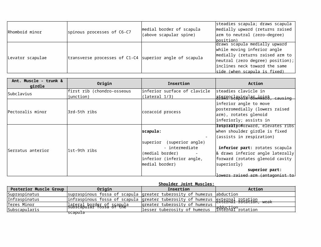
medial border of scapula (above scapular spine)
steadies scapula; draws scapula medially upward (returns raised arm to neutral (zero-degree) position)

Rhomboid minor spinous processes of C6-C7 dorsal scapular nerve (C4-C5)
Levator scapulae transverse processes of C1-C4 superior angle of scapula dorsal scapular nerve (C4-C5)
Ant. Muscle - trunk & girdle Origin Insertion Action Innervation
Subclavius first rib (chondro-osseous junction) nerve to subclavius (C5, C6)
Pectoralis minor 3rd-5th ribs coracoid process
Serratus anterior 1st-9th ribs long thoracic nerve (C5-C7)
Shoulder Joint Muscles:Posterior Muscle Group Origin Insertion Action Innervation
Supraspinatus supraspinous fossa of scapula greater tuberosity of humerus abduction suprascaular nerve (C4-C6)Infraspinatus infraspinous fossa of scapula greater tuberosity of humerus external rotation suprascaular nerve (C4-C6)Teres Minor lateral border of scapula greater tuberosity of humerus external rotation, weak adduction axilliary nerve (C5-C6)Subscapularis subscapular fossa of the scapula lesser tuberosity of humerus internal rotation subscapular nerve (C5-C6)
medial border of scapula (above scapular spine)
steadies scapula; draws scapula medially upward (returns raised arm to neutral (zero-degree) position)
draws scapula medially upward while moving inferior angle medially (returns raised arm to neutral (zero degree) position); inclines neck toward the same side (when scapula is fixed)
inferior surface of clavicle (lateral 1/3)
steadies clavicle in sternoclavicular joint
draws scapula downward, causing inferior angle to move posteromedially (lowers raised arm), rotates glenoid inferiorly; assists in respiration
medial & lateral pectoral nerves (C6-T1)
scapula: - superior (superior angle) - intermediate (medial border) - inferior (inferior angle, medial border)
entire muscle: draws scapula laterally forward, elevates ribs when shoulder girdle is fixed (assists in respiration) inferior part: rotates scapula & draws inferior angle laterally forward (rotates glenoid cavity superiorly) superior part: lowers raised arm (antagonist to inferior part)

Deltoid deltoid tuberosity on humerus axilliary nerve (C5-C6)
Latissimus dorsi thoracodorsal nerve (C6-C8)
Teres major inferior angle of scapula crest of lesser tuberosity of humerus subscapular nerve (C5-C6)
Anterior Muscle Group Origin Insertion Action Innervation
Pectoralis major
Coracobrachialis coracoid process musculocutaneous nerve (C6 C7)
clavicular part: lateral 1/3 clavicle acromial part: acromion spinal part: scapular spine
clavicular part: anteversion (moves arm & shoulder forward), internal rotation, adduction acromial part: abduction spinal part: retroversion (moves arm & shoulder backwards), external rotation, adduction (between 60-90 degreses of abduction, clavicular & spinal parts assist acromial part with abduction)
vertebral part: T7-T12 spinous processes, thoracolumbar fascia of spinous processes of all lumbar vertebrae and sacrum iliac part: posterior 1/3 of iliac crest scapular part: inferior angle of scapula
crest of lesser tuberosity (anterior view) of humerus
internal rotation, adduction, retroversion (moves arm backward), respiration (expiration, "cough muscle")
internal rotation, adduction, retroversion
clavicular part: clavicle medial half sternocostal part: sternum & 2nd-6th costal cartilages abdominal part: anterior layer of rectus sheath
crest of greater tuberosity of humerus
entire muscle: adduction & internal rotation clavicular & sternocostal part: anteversion assists in respiration when shoudler girdle is fixed
medial & lateral pectoral nerves (C5-T1)
humerus (in line w crest of lesser tuberosity)
anteversion, adduction, internal rotation

Arm Muscles:Posterior Muscle Group Origin Insertion Action Innervation
Triceps brachii olecranon of ulna radial nerve (C6-C8)
Anconeus olecranon of ulna (radial surface) extends elbow & tightens joint capsule radial nerve (C6-C8)
Anterior Muscle Group Origin Insertion Action Innervation
Brachialis ulnar tuberosity flexion at elbow joint
Biceps Brachii radial tuberosity musculocutaneous nerve (C5-C6)
Forearm Muscles:Posterior Muscle Group Origin Insertion Action Innervation
Superficial Extensors:
- Extensor digitorum radial nerve (C7, C8)
- Extensor digiti minimi radial nerve (C7, C8)
long head: infraglenoid tubercle of scapula medial head: posterior surface of humerus, distal to radial groove, medial intermuscular septum lateral head: humerus posterior surface, proximal to radial groove, lateral intermuscular septum
elbow joint: extension shoulder joint: long head - backward movement & adduction of arm
lateral epicondyle of humerus (& posterior joint capsule in some cases)
distal half of humerus anterior surface, also medial & lateral intermuscular septa
musculocutaneous n. (C5-C6) radial nerve (C7)
long head: supraglenoid tubercle of scapula short head: coracoid process of scapula
elbow joint: flexion, supination (with elbow flexed) shoulder joint: flexion (humerus forward motion), stabilization of humeral head during deltoid contraction, abduction & internal (medial) rotation of humerus
common head (lateral epicondyle of humerus)
dorsal digital expansion of 2nd-5th digits
wrist: extension MCP, PIP, DIP joints of 2nd-5th digits: extension & abduction of fingers
common head (lateral epicondyle of humerus)
dorsal digital expansion of 2nd-5th digits
wrist: extension, ulnar abduction of hand MCP, PIP, DIP joints of 5th digits: extension & abduction of 5th digit

- Extensory carpi ulnaris base of 5th metatarsal radial nerve (C7, C8)
Deep Extensors:
- Supinator supinates forearm radial nerve (C6, C7)
- Abductor pollicis longus base of 1st metacarpal radial nerve (C7, C8)
- Extensor pollicis brevis base of proximal phalanx of thumb radial nerve (C7, C8)
- Extensor pollicis longus base of distal phalanx of thumb radial nerve (C7, C8)
- Extensor indicis radial nerve (C7, C8)
Anterior Muscle Group Origin Insertion Action InnervationSuperficial Flexors:
- Pronator teres median nerve (C6, C7)
median nerve (C8, T1)
common head (lateral epicondyle of humerus), ulnar head (dorsal surface of ulna)
wrist: extension, adduction (ulnar deviation) of hand
olecranon of ulna, lateral epicondyle of humerus, radial collateral ligament, annular ligament of radius
radius (between radial tuberosity & insertion of pronator teres)
dorsal surfaces of radius & ulna, also interosseous membrane
radiocarpal joint: abduction (radial deviation) of hand carpometacarpal joint of thumb: abduction
posterior surface of radius & interosseous membrane (distal to abductor pollicis longus)
radiocarpal joint: abduction (radial deviation) of hand carpometacarpal & MCP joint of thumb: extension
posterior surface of ulna & interosseous membrane
wrist: extension & abduction (radial deviation) of thumb carpometacarpal joint of thumb: adduction MCP & interphalangeal joints of thumb: extension
posterior surface of ulna, interosseous membrane
posterior digital expansion of 2nd digit
wrist: extension MCP, PIP, DIP joints of 2nd digit: extension
humeral head: humerus medial epicondyle ulnar head: ulna coronoid process
lateral surface of radius (distal to supinator insertion)
elbow: weak flexor forearm: pronation
- Flexor digitorum superficialis
humeral head: humerus medial epicondyle ulnar head: ulna coronoid process radial head: distal to radial tuberosity
sides of the middle phalanges of 2nd-5th digits
elbow: weak flexor wrist, MCP, & PIP joints of 2nd-5th digits: flexion
- Flexor carpi radialis medial epicondyle of humerus median nerve (C6, C7)
- Flexor carpi ulnaris ulnar nerve (C7-T1)
- Palmaris longus medial epicondyle of humerus palmar aponeurosis median nerve (C7, C8)
Deep Flexors:
- Flexor digitorum profundus
- Flexor pollicis longus median nerve (C7, C8)
- Pronator quadratus distal 1/4 of ulna anterior surface distal 1/4 of radius anterior surface median nerve (C7, C8)
Radial Forearm Muscles Origin Insertion Action InnervationRadialis Group:
- Brachioradialis styloid process of radius radial nerve (C5, C6)
dorsal base of 2nd metacarpal radial nerve (C6, C7)
- Extensor carpi radialis brevis lateral epicondyle of humerus dorsal base of 3rd metacarpal radial nerve (C7, C8)
base of 2nd metacarpal (sometimes 3rd)
wrist: flexion & adduction of hand (ulnar deviation)
head of humerus: medial epicondyle head of ulna: olecranon
pisiform hook of hamate, base of 5th metacarpal
wrist: flexion & adduction of hand (ulnar deviation)
elbow: weak flexor wrist: palmar flexion, tightens palmar aponeurosis for gripping
proximal 2/3s of flexor surface of ulna & adjacent interosseous membrane
palmar surface of distal phalanges of 2nd-5th digits
wrist joints and MCP, PIP, DIP joints of 2nd-5th digits: flexion
median nerve (radial part, 2nd - 3rd digits) (C8, T1) ulnar nerve (ulnar part, 4th-5th digits) (C7-T1)
mid-anterior surface of radius and adjacent interosseous membrane
palmar surface of distal phalanx of thumb
wrist: flexion & radial abduction of hand carpometacarpal joint of thumb: opposition metacarpophalangeal & interphalangeal joints of thumb: flexion
pronates hand, stabilizes distal radioulnar joint
lateral surface of distal humerus, lateral intermuscular septum
elbow: flexion forearm: semipronation
- Extensor carpi radialis longus
lateral surface of distal humerus (lateral supracondylar ridge), lateral intermuscular septum
elbow: weak flexor wrist: dorsal extension (assists in fist closure), abduction (radial deviation) of hand
elbow: weak flexor wrist: dorsal extension (assists in fist closure), abduction (radial deviation) of hand

Hand Muscles:Metacarpal Muscles Origin Insertion Action Innervation
1st-4th lumbricals
1st-4th dorsal interossei ulnar nerve (C8, T1)
1st-3rd palmar interossei ulnar nerve (C8, T1)
Thenar Muscles Origin Insertion Action Innervation
Abductor pollicis brevis abduction of thumb median nerve (C8, T1)
Adductor pollicis ulnar nerve (C8, T1)
Flexor pollicis brevis
Opponens pollicis trapezium radial border of 1st metatarsal median nerve (C8, T1)
radial sides of tendons of flexor digitorum profundus (variable)
each one inserts on the dorsal digital expansion of the respective digit (1st - index finger; 2nd - middle finger; 3rd - ring finger; 4th - little finger)
metacarpophalangeal joint of 2nd-5th digits: flexion proximal & distal interphalangeal joints of 2nd-5th digits: extension
1st, 2nd lumbricals: median nerve (C8, T1) 3rd, 4th lumbricals: ulnar nerve (C8, T1)
by 2 heads from adjacent sides of 1st-5th metacarpals
dorsal digital expansion of 2nd-4th digits, base of proximal phalanx first interosseus: radial side of 2nd proximal phalanx (index) second interosseus: radial side of 3rd proximal phalanx (middle) third interosseus: ulnar side of 3rd proximal phalanx (middle) fourth interosseus: ulnar side of 4th proximal phalanx (right)
metacarpophalangeal joint of 2nd-4th digits: flexion proximal & distal interphalangeal joints of 2nd-4th digits: extension & abduction of fingers (abduction of index & ring fingers from middle finger)
first interosseus: ulnar side of 2nd metacarpal (index) second interosseus: radial side of 4th metacapral (ring) third interosseus: radial side of 5th metacarpal (little)
dorsal digital expansion & base of proximal phalanx of associated finger
metacarpophalangeal joints of 2nd, 4th, 5th digits: flexion proximal & distal interphalangeal joints of 2nd, 4th, 5th digits: extension & adduction of fingers (adduction of 2nd, 4th, 5th digits towards middle finger)
scaphoid bone & trapezium, flexor retinaculum
base of proximal phalanx of thumb (via radial sesamoid)
transverse head: palmar surface of 3rd metacarpal oblique head: capitate bone, base of 2nd metacarpal
base of proximal phalanx of thumb (via ulnar sesamoid)
carpometacarpal joint of thumb: opposition metacarpaophalangeal joint of thumb: flexion
superficial head: flexor retinaculum deep head: capitate bone, trapezium
base of proximal phalanx of thumb (via radial sesamoid)
carpometacarpal joint of thumb: flexion, opposition metacarpaophalangeal joint of thumb: flexion
superifical head: median nerve (C8, T1) deep head: ulnar nerve (C8, T1)
carpometacarpal joint of thumb: opposition

Hypothenar Muscles Origin Insertion Action Innervation
Abductor digiti minimi pisiform bone ulnar nerve (C8, T1)
Flexor digiti minimi hook of hamate, flexor retinaculum base of proximal phalanx of 5th digit ulnar nerve (C8, T1)
Opponens digiti minimi hook of hamate ulnar border of 5th metacarpal ulnar nerve (C8, T1)
Palmaris brevis ulnar border of palmar aponeurosis skin of hypothenar eminence ulnar nerve (C8, T1)
ulnar base of proximal phalanx & dorsal digital expansion of 5th digit
metacarpophalangeal joint of little finger: flexion & abduction of little finger PIP & DIP joints of little finger: extension
MCP joint of little finger: flexion
draws metacarpal in the palmar direction (opposition)
tightens palmar aponeurosis (protective function)