oculus pentacam /pentacam hr anterior segment tomography · oculus pentacam®/pentacam® hr ......
TRANSCRIPT

OCULUS Pentacam®/Pentacam® HRAnterior Segment Tomography

OCULUS Pentacam®/Pentacam® HRThe Gold Standard for Anterior Segment Tomography
Since its introduction in 2002 the OCULUS Pentacam® has proven to be an indispensable tool for ophthalmologists, clinicians and surgeons committed to providing precise diagnostics and successful treatment.
Efficient preliminary and follow-up examinationsIn a single delegable step the OCULUS Pentacam® measures the entire anterior eye segment independent of tear film. From the high-resolution Scheimpflug images it calculates a motion-corrected 3D model.
Comprehensive analysisAs the basis for precise keratometry, a prerequisite for IOL calculation, and for detection of ectasia and irregularities as well as wavefront calculation, the Pentacam® provides a complete description of the entire cornea (pachymetry as well as elevation and curvature data).
Scientifically reliable screeningsTo perform quick and reliable glaucoma screenings the OCULUS Pentacam® determines anterior chamber volume, angle and depth automatically and then compares the values in the Indices Report with a normal and a pathological population.
Reliable values for your diagnosesUsing blue light, the OCULUS Pentacam® makes opacities of the cornea, the crystalline lens and IOLs visible so that corneal diseases are detected reliably and cataract progression can be assessed objectively.The Pentacam® HR stands out by virtue of its bright optics, which make for extremely sharp, highest-quality Scheimpflug images.
Dr. Tobias Neuhann, MD, Germany
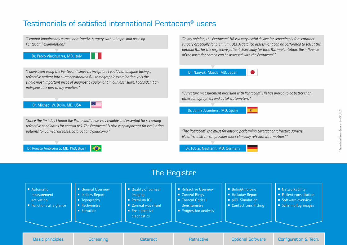
“The Pentacam® is a must for anyone performing cataract or refractive surgery. No other instrument provides more clinically relevant information.”*
Dr. Michael W. Belin, MD, USA
“I have been using the Pentacam® since its inception. I could not imagine taking a refractive patient into surgery without a full tomographic examination. It is the single most important piece of diagnostic equipment in our laser suite. I consider it an indispensable part of my practice.”
Dr. Paolo Vinciguerra, MD, Italy
“I cannot imagine any cornea or refractive surgery without a pre and post-op Pentacam® examination.”
Dr. Naoyuki Maeda, MD, Japan
“In my opinion, the Pentacam® HR is a very useful device for screening before cataract surgery especially for premium IOLs. A detailed assessment can be performed to select the optimal IOL for the respective patient. Especially for toric IOL implantation, the influence of the posterior cornea can be assessed with the Pentacam®.”
Dr. Renato Ambrósio Jr, MD, PhD, Brazil
“Since the first day I found the Pentacam® to be very reliable and essential for screening refractive candidates for ectasia risk. The Pentacam® is also very important for evaluating patients for corneal diseases, cataract and glaucoma.”
Dr. Jaime Aramberri, MD, Spain
“Curvature measurement precision with Pentacam® HR has proved to be better than other tomographers and autokeratometers.”
Testimonials of satisfied international Pentacam® users
* Tra
nsla
ted
from
Ger
man
by
OCU
LUS.
Basic principles Screening Cataract Refractive Optional Software Configuration & Tech.
� Automatic measurement activation
� Functions at a glance
� General Overview � Indices Report � Topography � Pachymetry � Elevation
� Quality of corneal imaging
� Premium IOL � Corneal wavefront � Pre-operative diagnostics
� Refractive Overview � Corneal Rings � Corneal Optical Densitometry
� Progression analysis
� Belin/Ambrósio � Holladay Report � pIOL Simulation � Contact Lens Fitting
� Networkability � Patient consultation � Software overview � Scheimpflug images
The Register

Anterior Segment TomographyFast, reproducible, delegable
Thanks to automatic measurement activation the Pentacam® gives you an overall view of the anterior eye segment in mere seconds. Measurements are made independent of tear film and examiner. The Pentacam® HR evaluates up to 138,000 real measuring points.
The most important functions at a glance:
� Topography of anterior and posterior corneal surface � Full-surface pachymetry � Early ectasia detection � 3D anterior chamber analysis � Indices Report with normal data for all crucial parameters
� Normative corneal wavefront data � Total Corneal Refractive Power (TCRP) � 3D-densitometry of the cornea and the crystalline lens � Contact lens fitting software � Anterior segment tomography
Anterior and posterior corneal surface
Anterior chamber
Anterior and posterior surface of crystalline lens
Iris

Before the surgery my doctor explained what the Pentacam® examination involved by showing me on the iPad. I was amazed that I was still even able to see at all with such a clouded lens!
The Pentacam®: Indispensable for you and your patients
Cataract
Benefit from simple, comprehensive corneal screenings. Indices Report, early detection of ectasia according to Belin/Ambrósio and evaluation of corneal optical densitometry – four clearly defined steps help you select the optimal IOL. The Pentacam® calculates Total Corneal Refractive Power (TCRP), supporting you in particular when it comes to selection, orientation and calculation of toric IOLs. The function for importing axis length from optical biometers and access to ray-tracing programs such as Phaco Optics and OKULIX make it possible to perform IOL calculations on any cornea.
Refractive assessment
Early detection of corneal ectasia using Belin/Ambrósio software is unique. A final parameter is calculated and represented in colour. This is how the Pentacam® software assists you in making diagnoses.Keratometry, asphericity and full-surface pachymetry assist you in planning refractive surgery. The Pentacam® measures irregular corneas with great precision, presenting the necessary parameters in a clear and application-oriented fashion. This ensures reliable planning for implantation of corneal rings, CXL and cornea transplantations.
Glaucoma
Glaucoma is one of the most common eye diseases. The Indices Report is a unique screening tool for detecting it. Make use of the evaluation of anterior chamber angle and volume based on normal data and clinical pictures. In particular, studies have identified automatically calculated anterior chamber volume as a sensitive parameter.
* Assessment of the anterior chamber parameters after laser iridotomy in primary angle close suspect using Pentacam® and gonioscopy; Alireza et al; Int J Ophthalmol, 2013, 6(5):680-684
Comparison of scheimpflug imaging and spectral domain anterior segment optical coherence tomography for detection of narrow anterior chamber angles; Grewal et al; Eye Vol.: 25
I simply didn’t want to wear spectacles anymore! During the pre-op examination the Pentacam® results gave me certainty.
My risk of developing glaucoma was detected by my ophthalmologist early on. He was able to initiate appropriate measures immediately.
Basic principles Screening Cataract Refractive Optional Software Config. & Technology

Screening: Filter, Evaluate, RepresentAll the important information at a glance
The great challenge in clinical routine is filtering, evaluating and representing data in a clear-cut way. This is exactly what the Indices Report does. The crucial data are represented in such a way that you can gain a comprehensive picture of your patient at a glance.
Indices Report
� Normative data are gathered from published studies and stored in the Pentacam® software.
� The distribution of normal values in a population is represented by the bars shaded in grey.
� The diagrams show the distribution for normal (green) and pathological (red) eyes.
� Sources which the evaluations are based on are cited to provide additional information.
� In cases of irregularities the individual displays for detailed findings appear in the interactive navigation bar.
Early detection of corneal ectasia
Distribution of a normal (green) and a pathological (red) population
Individual patient value
Interactive navigation bar
Distribution in normal population

After initial information has been provided by the Indices Report additional displays show further, case-specific information. Depending on the irregularities in question the interactive navigation bar recommends displays which lead you to suitable detailed analyses – custom-tailored to the individual patient.
Screening and follow-up exams ensure safety
General Overview
The Scheimpflug images allow you to make a qualitative assessment of the anterior eye segment. Opacities of the cornea or the crystalline lens are represented impressively. Keratometry, pachymetry and asphericity allow for an initial analysis of the corneal surface. The anterior chamber is defined by chamber volume, angle and depth. Intraocular pressure is corrected on the basis of central corneal thickness.
Superimposition of Scheimpflug images
For better patient consultation you can superimpose 2 Scheimpflug images. This way numerous changes in the anterior eye segment can be detected which you can explain to your patient with the help of visualization.
4 Maps Refractive
Topography and elevation maps make quantitative assessments of the corneal surface possible. Especially the posterior elevation map shows pathological changes very early on. The corneal thickness map clearly shows the thinnest position to help you plan refractive surgery, for example.Combining elevation data of the anterior and posterior corneal surface, topography and pachymetry will help you detect abnormalities early on.
Basic principles Screening Cataract Refractive Optional Software Config. & Technology

Cataract: IOL SelectionPremium IOL in 4 easy steps
Premium IOLs make it possible to improve your patients’ visual function considerably. Whether through toric, aspheric or multifocal geometry, the exact measuring data gathered by the Pentacam® will assist you in selecting the right lens.
Cataract Pre-OP Display
Cataract Pre-OP Display was developed in collaboration with Prof. Dr. Naoyuki Maeda from the University Medical School in Osaka, Japan. This display assists users in selecting the optimal premium IOL.
To do this, the following parameters are taken into consideration:1. overall corneal astigmatism,2. total corneal spherical aberrations,3. total corneal irregularities and4. the influence of the posterior corneal surface.
Detection and evaluation of the influence of the
posterior corneal surface
Total corneal irregularities
Total corneal spherical aberrations
Influence of the posterior corneal surface on overall corneal astigmatism

For documentation of cataract progression and optimal surgery planning the Pentacam® provides cataract surgeons with comprehensive analysis options.
Pre-operative diagnostics is your success concept
Refractive power distribution display
The table shows the refractive power of the cornea in various zones and rings. This allows for individual assessment of the influence of the posterior corneal surface on overall sphericity, toricity and irregularity of the cornea.
Normative Corneal Wavefront
The wavefront error of the cornea as a whole is calculated individually using ray tracing. Higher-order aberrations are calculated and represented in comparison to a normal population.
3D-Densitometry and PNS
Through blue-light illumination opacities of the natural lens become visible. Two- and three-dimensional quantification of lens opacification in the individual layers and of posterior capsular opacification is performed.The unique PNS (Pentacam® Nucleus Staging) function permits optimized ultrasonic energy output in phacoemulsification as well as optimized effective phaco time (EPT) in Femto assisted cataract surgery.
Basic principles Screening Cataract Refractive Optional Software Config. & Technology
Refractive ScreeningPlanning refractive laser surgery
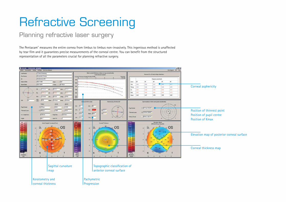
The Pentacam® measures the entire cornea from limbus to limbus non-invasively. This ingenious method is unaffected by tear film and it guarantees precise measurements of the corneal centre. You can benefit from the structured representation of all the parameters crucial for planning refractive surgery.
Keratometry and corneal thickness
Sagittal curvature map
Pachymetric Progression
Topographic classification of anterior corneal surface
Corneal asphericity
Position of thinnest pointPosition of pupil centrePosition of Kmax
Corneal thickness map
Elevation map of posterior corneal surface

Corneal assessment involves more than mere topography. In fact it calls for holistic evaluation. The Pentacam® represents pachymetric progression, allowing for structural evaluation in this way. Optical densitometry facilitates targeted slit lamp examinations so as to enable you to detect diseases early on.
Assess the entire cornea qualitatively and quantitatively
Pachymetric Progression
Starting at the thinnest point and moving in the direction of the periphery, corneal thickness progression is represented in the form of concentric rings. Using two diagrams, the display shows the patient’s corneal thickness progression as compared to normative data. From this data the individual progression index is calculated, permitting early detection of ectatic abnormalites.
Corneal Rings
This display shows all the parameters necessary for planning corneal ring implantation. Dependent on the selected surgical procedure – manual dissection technique or femtosecond laser – corneal thickness is represented in specific areas and segments.
Corneal Optical Densitometry
Panorama images of the cornea make corneal diseases visible. This allows for objective quantification and follow-up. Optical densitometry can be evaluated using a table or a colour chart. In the table the measured values are represented according to various zones and layers.Corneal optical densitometry results are displayed in reference to published age-related normative data. This provides a basis for the detection of other diseases such as Fuchs’ Dystrophy.
Basic principles Screening Cataract Refractive Optional Software Config. & Technology
Optional Software ModulesThe Pentacam®: individual and flexible
The flexibility of the software allows you to custom-configure your Pentacam®. You can add software supplements at any time, even after the first configuration has been carried out.
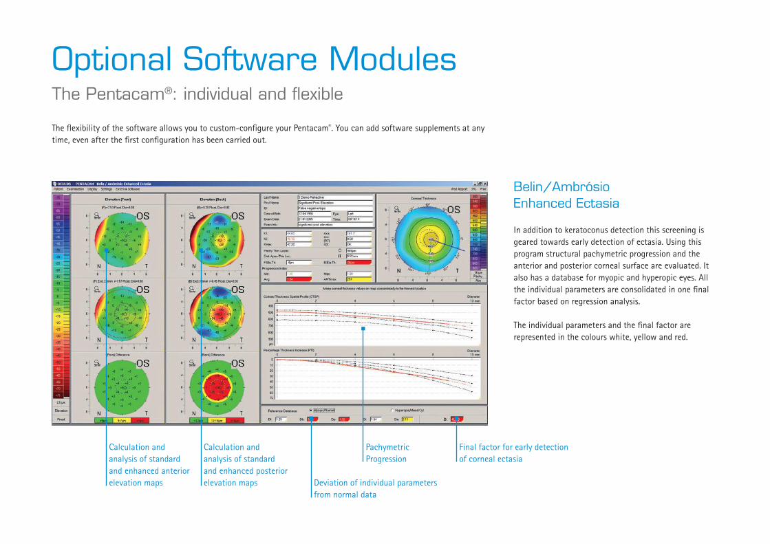
Belin/Ambrósio Enhanced Ectasia
In addition to keratoconus detection this screening is geared towards early detection of ectasia. Using this program structural pachymetric progression and the anterior and posterior corneal surface are evaluated. It also has a database for myopic and hyperopic eyes. All the individual parameters are consolidated in one final factor based on regression analysis.
The individual parameters and the final factor are represented in the colours white, yellow and red.
Calculation and analysis of standard and enhanced anterior elevation maps
Calculation and analysis of standard and enhanced posterior elevation maps
Final factor for early detection of corneal ectasia
Deviation of individual parameters from normal data
Pachymetric Progression
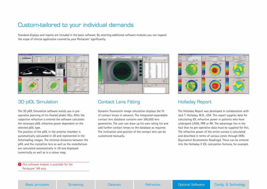
Standard displays and reports are included in the basic software. By selecting additional software modules you can expand the scope of clinical application covered by your Pentacam® significantly.
Custom-tailored to your individual demands
3D pIOL Simulation
The 3D pIOL Simulation software assists you in pre-operative planning of iris-fixated phakic IOLs. After the subjective refraction is entered the software calculates the necessary pIOL refractive power dependent on the selected pIOL type.The position of the pIOL in the anterior chamber is automatically calculated in 3D and represented in the Scheimpflug images. The minimal distances between the pIOL and the crystalline lens as well as the endothelium are calculated automatically in 3D and displayed numerically as well as in a colour map.
Contact Lens Fitting
Dynamic fluorescein image simulation displays the fit of contact lenses in advance. The integrated expandable contact lens database contains over 380,000 lens geometries. The user can draw up his own rating list and add further contact lenses to the database as required. The inclination and position of the contact lens can be customized manually.
Holladay Report
The Holladay Report was developed in collaboration with Jack T. Holladay, M.D., USA. This report supplies data for calculating IOL refractive power in patients who have undergone LASIK, PRK or RK. The advantage lies in the fact that no pre-operative data must be supplied for this. The refractive power of the entire cornea is calculated and described in terms of various zones through EKRs (Equivalent Keratometer Readings). These can be entered into the Holladay II IOL calculation formula, for example.
This software module is available for the Pentacam® HR only
Basic principles Screening Cataract Refractive Optional Software Config. & Technology
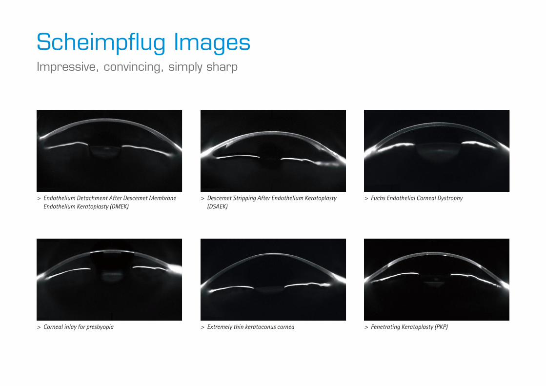
Scheimpflug ImagesImpressive, convincing, simply sharp
> Endothelium Detachment After Descemet Membrane Endothelium Keratoplasty (DMEK)
> Descemet Stripping After Endothelium Keratoplasty (DSAEK)
> Fuchs Endothelial Corneal Dystrophy
> Corneal inlay for presbyopia > Extremely thin keratoconus cornea > Penetrating Keratoplasty (PKP)
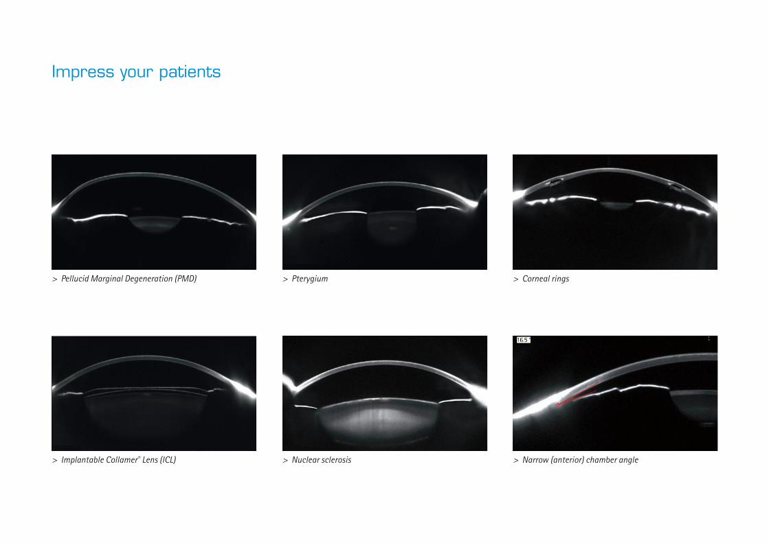
> Pellucid Marginal Degeneration (PMD) > Pterygium > Corneal rings
> Implantable Collamer® Lens (ICL) > Nuclear sclerosis > Narrow (anterior) chamber angle
Impress your patients

All Features at a GlanceCustomise the OCULUS Pentacam®/ Pentacam® HR to your own requirement
Software included
General Overview
Indices Report
Topographical Keratoconus Classification
Topography and Elevation Maps of the Anterior and Posterior Corneal Surface
Pachymetry Maps, absolute and relative
4 Maps Refractive
Iris Image and HWTW
Comparison and Differential Analysis of 2 Exams
Tomography
Scheimpflug Image Overview
Optional examination functions My wish list
Cataract Software Package
Cataract Pre-OP Display Corneal Power Distribution Zernike Analysis and Normative Corneal Wavefront PNS and 3D Cataract Analysis Total Corneal Refractive Power Automatic calculation of the anterior chamber angle in 360°, measurement based on Scheimpflug images 4 Maps Topometric and 4 Maps Chamber Show 2 Exams Compare 4 Exams
Refractive Software Package
Corneal Optical Densitometry Corneal Rings Fourier Analysis Refractive Pachymetric 4 Maps Selectable Show 2 Exams Compare 4 Exams
Belin/Ambrósio Enhanced Ectasia Display
Holladay Report
Contact Lens Fitting
Optional evaluation functions My wish list
DICOM Interface
Only available with the Pentacam® HR My wish list
3D pIOL Simulation Software and Aging Prediction These functions are unique and are only available with the OCULUS Pentacam® / Pentacam® HR

Server (with/without DICOM)
The Fascination of TechnologyIngenious yet simple
The Floating License Key – for maximum flexibility
This basic Pentacam® software is already available at all workplaces in your network. You decide which optional examination and evaluation functions you need in addition. You also choose the number of optional evaluation software features which should be available simultaneously. The Floating License Key (FLK) activates the corresponding licenses and makes them available on your network. To help you decide what you need all the optional evaluation functions can be accessed 20 times for demonstration purposes.
Efficiency and productivity through networking
The OCULUS Patient Data Management system (PDM) optimises your work processes. It is always included in the scope of delivery; it organises patient and examination data from all OCULUS instruments. The PDM is network-compatible and can be incorporated into many Electronic Medical Record (EMR) systems. Needless to say, the OCULUS PDM communicates with the DICOM environment and makes results available in DICOM format.
Patient consultation
By using the Pentacam® Viewer-App for iPads you underscore your competence. Colour representations generated by the Pentacam® and two- and three-dimensional tomographic images make it possible to explain abnormalities to your patients in a clearly illustrated way as well as to discuss treatment options.
Consultation
Registration
Examination room 1
Examination room 2
Examination room 3
Basic principles Screening Cataract Refractive Optional Software Config. & Technology

Technical DataPentacam®/Pentacam® HR
The
avai
labi
lity
of p
rodu
cts
and
feat
ures
may
var
y by
cou
ntry
. OCU
LUS
rese
rves
the
righ
t to
cha
nge
prod
uct
spec
ifica
tions
and
des
ign.
33/0
714/
EN/H
A
P/70
700/
EN
WWW.OCULUS.DE OCULUS Optikgeräte GmbHPostfach • 35549 Wetzlar • GERMANY Tel. +49-641-2005-0 • Fax +49-641-2005-295Email: [email protected] • www.oculus.de
• OCULUS USA, [email protected]• OCULUS Asia, [email protected]• OCULUS Czechia, [email protected]• OCULUS Iberia, [email protected]• OCULUS Poland, [email protected]
OCULUS is certified by TÜV according toDIN EN ISO 13485
272 mm(10.7 in)
203 mm(8.0 in)
505
-535
mm
(20.
0 - 2
1.0
in)
360 mm(14.0 in)
280 mm(11.0 in)
Scheimpflug Camera Pentacam® Pentacam® HR
Camera digital CCD camera digital CCD camera
Light source blue LED (475 nm UV-free) blue LED (475 nm UV-free)
Processor DSP with 400m operations/s DSP with 400m operations/s
Speed 50 images in 2 seconds 1) 100 images in 2 seconds 2)
Measurement range Pentacam® Pentacam® HRCurvature 3 to 38 mm
9 to 99 dpt3 to 38 mm 9 to 99 dpt
Precision ± 0.2 dpt ± 0.1 dpt
Reproducibility ± 0.2 dpt ± 0.1 dpt
Operating distance 80 mm (3.1 in) 80 mm (3.1 in)
Technical specifications Pentacam® Pentacam® HRDimensions (W x D x H) 280 x 360 x 505-535 mm
(11 x 14 x 20-21 in)280 x 360 x 505-535 mm (11 x 14 x 20-21 in)
Weight 10.5 kg (23.1 lbs) 11 kg (24.3 lbs)
Max. power consumption 50 W 50 WRecommended computer specifications Win 7 pro, 2.7 GHz, 8 GB RAM, 1 TB HDD,
NVIDIA graphic board (1024 x 768 true colour), USB interface
Win 7 pro, 2.7 GHz, 8 GB RAM, 1 TB HDD, NVIDIA graphic board (1024 x 768 true colour), USB interface
1) Scheimpflug image of the entire anterior segment2) Cornea fine scan
in accordance with Medical Products Directive 93/42/EEC