oculus pentacam pentacam hr - eyeoptics1).pdf · this combination is the basis for calculating...
TRANSCRIPT

OCULUS Pentacam®
Pentacam®HR
TheGoldStandardin AnteriorSegmentTomography
Wefocusonprogress

�
Providingthebestcaretoyourpatients
>Theeyeisextremelysensitive.Lossorimpairmentofsightisinconceivableforyourpatients.
>Yourpatientsappreciatethefast,non-contactmeasurement.
Youcanestablishagooddoctor-patientrelationshipbyshowingtheintuitivePentacam®displaystoyourpatientsanddiscussingtreatmentoptions.
>Yourstaffappreciatetheeaseofuseandtheseamlessconnectivitytotheelectronicmedicalrecords.
>TheOCULUSPentacam®winspatienttrust.

�
CataractsurgeryFollowing a Pentacam® examination, my doctor showed me my cloudy lens . I was surprised that I could see anything at all! My doctor explained the operation using his PC. This gave me the sense of assurance I needed before the interven-tion.
CornealsurgeryI was happy with the quick Pentacam® examina-tion and the competent consultation with my eye doctor. He showed me some Pentacam® images of my eye and explained the surgery in detail. I felt like I was in good hands with this physician.
GlaucomascreeningMy eye doctor used a Pentacam® and identified that I am a glaucoma risk patient. I am very grateful that he checked my eye and discovered that I have a very small anterior chamber and a small angle.
Establishthebasisforlongtermrelationshipwithyourpatients.

�
First introduced in 1999, the Pentacam® is commercially avail-able since �00�. It is the first automatically rotating Scheimpflug camera. During the rotating scan that takes max. � seconds, up to 50 Scheimpflug images of the anterior eye segment are captured. The examination is released automatically and is user indepen-dent. During the scanning process the patient’s eye motions are captured using a second camera and compensated mathemati-cally. Ray tracing is used to compensate for optical distortions. This combination is the basis for calculating solid data for further evaluation. More than 100 published studies and papers prove the efficiency of this concept.The Pentacam® measures the cornea from limbus to limbus. It supplies topographic data on elevation and curvature of the en-tire anterior and posterior corneal surface. The corneal thickness (pachymetry) is measured and presented graphically over its entire surface. A topography based keratoconus detection and quantifi-cation are performed. The anterior chamber depth, chamber vol-ume (size) and the chamber angles are calculated and presented for the Glaucoma screening. The illumination of the eye using blue LED light makes corneal and lens opacities (cataract) visible. For patients information the anterior chamber can be visualized and displayed with the virtual tomography model. After the examination, Pentacam® provides an indice report that summarizes the abnormalities found during the scan. This report is based on clinical published studies and articles that define ab-normalities. The Pentacam® can be customized with two software packages and several software modules to fit your exact needs. Basic software:
Pentacam®
Morethanatopographer
BasicSoftware:
Qualitative assessment of the corneaTopography and elevation data of the anterior and posterior corneal surfaceOverall pachymetry, absolute and relative
Glaucoma screeningPachymetry based IOP correctionChamber angle, -volume and –depth
Topography based keratoconus detection and classificationIndice Report to detect abnormalitiesComparison and differential displaysSuperimposition of Scheimpflug imagesTomographyAutomatic measurement of the corneal diameter HWTW
Nowavailablewithanewiriscameraoptictoprovidethe
HWTWmeasurementautomatically.

5
The Pentacam® HR produces sharp and brilliant images with its outstanding hardware. The high resolution CCD chip and opti-mized optic design measures up to 1�8,000 true elevation points. For a detailed analysis of the cornea up to 100 Scheimpflug imag-es can be captured during the rotating scan. A moveable fixation target insures high ametropic patients a comfortable exam.
The high resolution Scheimpflug images of the Pentacam® HR impressively show the IOLs and pIOLs, as well as corneal rings, corneal injuries and corneal opacities such as, Fuchs dystrophy.The crystalline lens is displayed from its anterior capsule to the posterior capsule, even if opacities are present.
The Pentacam® HR is equipped with basic software and can be customized with two software packages and several software modules.
The unique �D pIOL simulation software including aging prediction is available as an option.
Pentacam®HRTheProfessionalModel
ThisiswhatmakestheHRspecial:
Sharp Scheimpflug images to display corneal opacities, implants, rings etc.Sharp presentation of IOLs and pIOLs to determine position and detect PCOSpecial scan mode to display pIOLsPrecise measurement of the cornea with up to 100 Scheimpflug imagesMoveable fixation target�D pIOL simulation software including aging prediction
Nowavailablewithanewiriscameraoptictoprovidethe
HWTWmeasurementautomatically.

�
�
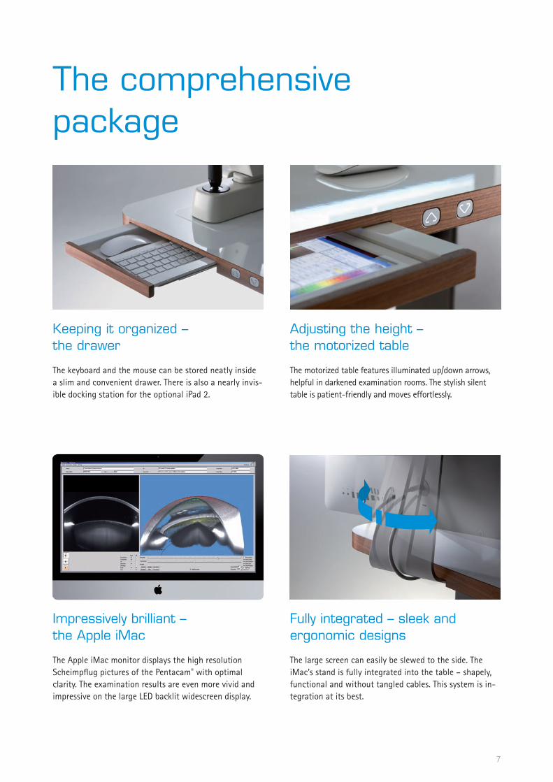
Thecomprehensivepackage
Keepingitorganized–thedrawer
The keyboard and the mouse can be stored neatly inside a slim and convenient drawer. There is also a nearly invis-ible docking station for the optional iPad �.
Adjustingtheheight–themotorizedtable
The motorized table features illuminated up/down arrows, helpful in darkened examination rooms. The stylish silent table is patient-friendly and moves effortlessly.
Impressivelybrilliant–theAppleiMac
The Apple iMac monitor displays the high resolution Scheimpflug pictures of the Pentacam® with optimal clarity. The examination results are even more vivid and impressive on the large LED backlit widescreen display.
Fullyintegrated–sleekandergonomicdesigns
The large screen can easily be slewed to the side. The iMac’s stand is fully integrated into the table – shapely, functional and without tangled cables. This system is in-tegration at its best.

DataNetwork
EMRcompatibility
The Pentacam® software is compatible with many com-mercially available EMR systems.
Physicianswantahighlyefficientpracticeandmoretimeforpatients.ThePentacam®offersbothandcomeswiththepatientdatamanage-mentsoftware.
Softwarelicenses
In order to fully read and evaluate the Pentacam® exams at remote desktops, software licenses are required. The software license includes the Pentacam® Basic software. It is customizable and can be expanded to include addi-tional software modules. With a software license, you can display and review any exam from any Pentacam® unit.
DICOMcompatibility
The Pentacam® software is fully DICOM compatible. The Pentacam® software receives information from the DICOM Modality Work List (MWL) from the Hospital Informa-tion System (HIS) and transmits the results to the Picture Archiving and Communication System (PACS) for storage and further evaluation.
9
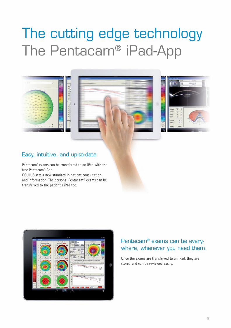
ThecuttingedgetechnologyThePentacam®iPad-App
Easy,intuitive,andup-to-date
Pentacam® exams can be transferred to an iPad with the free Pentacam®-App.OCULUS sets a new standard in patient consultation and information. The personal Pentacam® exams can be transferred to the patient’s iPad too.
Pentacam®examscanbeevery-where,wheneveryouneedthem.
Once the exams are transferred to an iPad, they are stored and can be reviewed easily.

10
BrilliantimagesPrecisemeasurements
Glaucomascreening
The Pentacam® provides a comprehensive and completely automatic analysis of the anterior chamber. Immediately after the eye has been examined, the instrument displays whether the patient has an increased risk for glaucoma. Post-operative evaluation of the anterior chamber shows alterations, for example, after an iridectomy or other sur-gical interventions.
Cataract
The Scheimpflug images produced by the Pentacam® sup-ply a clear representation of lens opacity. The �D cataract analysis combined with the PNS (Pentacam® Nucleus Stag-ing) is a unique feature. The center of the cornea and its anterior and posterior surfaces are measured very precisely for optimal calculation of the refractive corneal power. For your patients this means a perfect calculation of the IOL power – even after refractive surgery.
Cornea
The Pentacam® stands out by virtue of its high-resolution images and precise measurements for optimal surgical planning. Intelligent analysis programs help to substanti-ate your decisions. Surgical success is documented com-pletely from start to finish. Even the smallest irregulari-ties in the healing process are detected early. An exami-nation using Pentacam® offers your patients the highest degree of safety.
Cataract/Refractivesurgery
On the basis of measurements made by the Pentacam® one can determine whether the eye is suitable for surgical intervention or not. The surgery can be explained to the patient on the basis of the obtained results in clear and easy terms. In addition the Pentacam® offers the possibility to simulate in �D the position of iris fixated phakic IOLs, including simulation of age-related growth of the crystal-line lens. The selection of the aspheric IOL for purposes of reducing spherical aberrations is improved considerably by the unique corneal wavefront. The position of the im-planted IOL can be located on the Scheimpflug image and assessed in detail in terms of centering and tilting.

11
Refractivesoftwarepackage:
Freely selectable reference bodies for elevation mapsOverview display for refractive surgeonsCorneal thickness progression analysis for early keratoconus detectionFourier Analysis DisplayFreely selectable four maps displayExtended comparative and differential analysis of up to four examinationsSide-by-side comparison of two examinationsSide-by-side comparison of topometric and pachy-metric data
Cataractsoftwarepackage:
True measurements in the Scheimpflug imagesCorneal Wavefront and Zernike analysis of the total corneaCataract Pre-op display (developed in collaborati-on with Prof. Naoyuki Maeda, MD)
Detailed corneal assessment to select premium IOLs
Power Distribution Display / Total Corneal Refractive Power
Improved IOL calculationOrientation of toric IOls
True Net corneal powerPNS and �D Cataract AnalysisExtended comparative and differential analysis of up to four examinationsSide-by-side comparison of two examinationsSide-by-side comparison of topometric and pachymetric dataFour maps, anterior chamberFour maps, topometricAnterior chamber depth mapAnterior chamber angle in ��0°, automatically
CustomizableTheSoftwarePackages
These functions are unique and are only available with the OCULUS Pentacam®

Applications:Keratoconus detectionPre-surgical planning of refractive corneal surgeryFollow-up after corneal surgeryCalculation of IOL refractive powerPlanning of astigmatism-reducing incisions (LRI)Follow-up after refractive surgery(pre-post LASIK, PRK; PKP, LKP, DSEK)
Details:The rotating measurement principle guarantees high resolution of the measuring points in the central cornea. Topographic analysis of the anterior and posterior corneal surfaces is based on the measured real height data. These provide the basis for:
Sagittal (axial), tangential (local) curvature maps, refractive power maps of the anterior and posterior corneal surfaceElevation maps of the anterior and posterior corneal surfaceFour color coded maps display for refractive assessmentTopometric display for detailed corneal shape assessment including True Net PowerTopography based keratoconus detection and classificationComparison and differential displays
TopographyMapsoftheanteriorandposteriorcornealsurface
Applications:Pre-surgical planning of refractive corneal surgeryAbsolute and relative presentationGlaucoma screeningRelative pachymetry map for early keratoconus detectionIOP correction taking into account measured corneal thickness based on various correction formulas (for e.g. Ehler, Shah, Dresden etc.)
Details:An overview representation in color shows the corneal thickness from limbus to limbus. The measured values can be displayed in a pre-determined grid or represented manually at any point via mouse click.Automatic representation of:
corneal thickness in the centre of the pupilcorneal thickness in the apexthe thinnest point of the cornea
Pachymetrymaps
1�
BasicsoftwareDiscoverthePossibilities
1�These functions are unique and are only available with the OCULUS Pentacam®
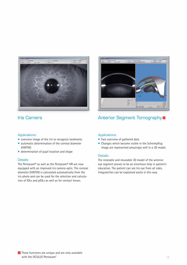
Applications:Fast overview of gathered dataChanges which become visible in the Scheimpflug image are represented amazingly well in a �D model.
Details:The rotatable and moveable �D model of the anterior eye segment proves to be an enormous help in patient’s education. The patient can see his eye from all sides. Irregularities can be explained easily in this way.
AnteriorSegmentTomography
Applications:overview image of the iris to recognize landmarksautomatic determination of the corneal diameter (HWTW)determination of pupil location and shape
Details:The Pentacam® as well as the Pentacam® HR are now equipped with an improved iris camera optic. The corneal diameter (HWTW) is calculated automatically from the iris photo and can be used for the selection and calcula-tion of IOLs and pIOLs as well as for contact lenses.
IrisCamera

1�
BasicsoftwareDiscoverthePossibilities
Applications:Glaucoma screeningPre- to post-operative comparison of changes in anterior chamber, e.g. after Iridectomy
Details:Tomographic representation, virtual model of anterior segmentAutomatic calculation of
Anterior Chamber Angle (ACA)Anterior Chamber Volume (ACV)Anterior Chamber Depth (ACD), internal or external
3DAnteriorChamberAnalysis
Applications:Comparison of the Scheimpflug images of two different examsVisualization of changes after surgical interventionPatient information – up-to-date
Details:The superimposition of the Scheimpflug images includes a blending function to qualitatively visualize and analyze the changes in the anterior chamber.Examples:
Cornea, before and after LASIKAnterior chamber, before and after iridotomyProgression of lens opacifications (cataract)
SuperimpositionofScheimpflugimages
Additionalsoftwaremodules
15
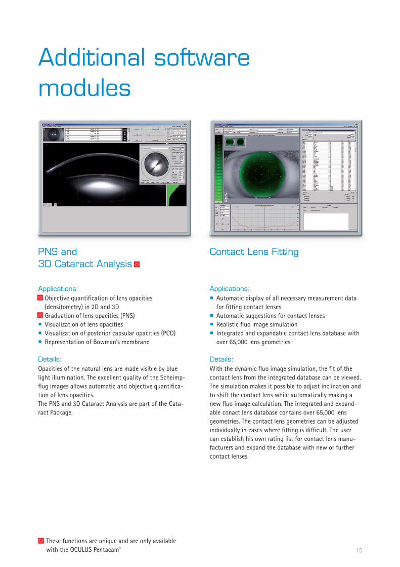
Applications:Automatic display of all necessary measurement data for fitting contact lensesAutomatic suggestions for contact lensesRealistic fluo image simulationIntegrated and expandable contact lens database with over �5,000 lens geometries
Details:With the dynamic fluo image simulation, the fit of the contact lens from the integrated database can be viewed. The simulation makes it possible to adjust inclination and to shift the contact lens while automatically making a new fluo image calculation. The integrated and expand-able conact lens database contains over �5,000 lens geometries. The contact lens geometries can be adjusted individually in cases where fitting is difficult. The user can establish his own rating list for contact lens manu-facturers and expand the database with new or further contact lenses.
ContactLensFitting
These functions are unique and are only available with the OCULUS Pentacam®
Applications:Objective quantification of lens opacities (densitometry) in �D and �DGraduation of lens opacities (PNS)Visualization of lens opacitiesVisualization of posterior capsular opacities (PCO)Representation of Bowman’s membrane
Details:Opacities of the natural lens are made visible by blue light illumination. The excellent quality of the Scheimp-flug images allows automatic and objective quantifica-tion of lens opacities.The PNS and �D Cataract Analysis are part of the Cata-ract Package.
PNSand3DCataractAnalysis

Additionalsoftwaremodules
Applications:Comprehensive clinical comparative representationEKRs (Equivalent Keratometer Readings) for optimized IOL-calculation for post-refractive patient eyes, if no pre-op data are available
Details:The Holladay Report was developed in collaboration with Jack T. Holladay, M.D.. It supplies data for calculating the optimal IOL refractive power for patients who have undergone refractive corneal surgeries such as LASIK and RK especially if no pre-op data are available. The Hol-laday Report calculates the real relationship of the pos-terior corneal surface to the anterior corneal surface. The overall refractive power of the cornea is calculated and described using the EKRs (Equivalent Keratometer Read-ings) in various zones. They can be used in IOL formulas, e.g. the Holladay �.
HolladayReportandHolladayEKRReport
Applications:Selection of aspheric IOLs for correction of corneal spherical aberrations (Z�.0)Fitting of corneal rings in reference to the axis of the comaDetermination of low and high order aberrations
Details:The Zernike Analysis of the Pentacam® consists of two parts:
The calculation of the corneal wavefront of the entire cornea (anterior and posterior surface) is performed via ray tracing – and is thus independent of the shape of the cornea (e.g. post LASIK, PRK, LKP, PKP etc).The surface based Zernike Analysis is performed using e.g. a theoretical, optimal corneal ellipse (ecc = 0.�51). It can be shown for the anterior and posterior surface of the cornea.
The Corneal Wavefront Analysis is part of the Cataract Package.
CornealWavefrontAnalysis
1�
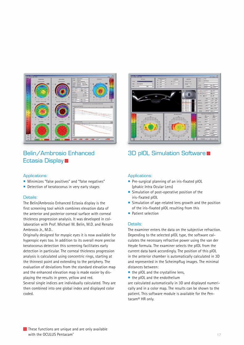
Applications:Pre-surgical planning of an iris-fixated pIOL (phakic Intra Ocular Lens)Simulation of post-operative position of the iris-fixated pIOLSimulation of age-related lens growth and the position of the iris-fixated pIOL resulting from thisPatient selection
Details:The examiner enters the data on the subjective refraction. Depending to the selected pIOL type, the software cal-culates the necessary refractive power using the van der Heyde formula. The examiner selects the pIOL from the current data bank accordingly. The position of this pIOL in the anterior chamber is automatically calculated in �D and represented in the Scheimpflug images. The minimal distances between:
the pIOL and the crystalline lens,the pIOL and the endothelium
are calculated automatically in �D and displayed numeri-cally and in a color map. The results can be shown to the patient. This software module is available for the Pen-tacam® HR only.
3DpIOLSimulationSoftware
These functions are unique and are only available with the OCULUS Pentacam®
Applications:Minimizes “false positives” and “false negatives“ Detection of keratoconus in very early stages
Details:The Belin/Ambrosio Enhanced Ectasia display is the first screening tool which combines elevation data of the anterior and posterior corneal surface with corneal thickness progression analysis. It was developed in col-laboration with Prof. Michael W. Belin, M.D. and Renato Ambrosio Jr., M.D..Originally designed for myopic eyes it is now available for hyperopic eyes too. In addition to its overall more precise keratoconus detection this screening facilitates early detection in particular. The corneal thickness progression analysis is calculated using concentric rings, starting at the thinnest point and extending to the periphery. The evaluation of deviations from the standard elevation map and the enhanced elevation map is made easier by dis-playing the results in green, yellow and red. Several single indices are individually calculated. They are then combined into one global index and displayed color coded.
Belin/AmbrosioEnhancedEctasiaDisplay
1�

Pentacam®Hardware
Pentacam®/Pentacam®HR
Basic software
Apple iMac �1“
Apple iMac ��“
Apple iPad �
Design lift table
included optional
Pentacam®HRPremium
Nothing left to be desired.
Software fully equipped (except pIOL)
Apple iMac ��“
Apple iPad �
Premium design lift table
� additional software licenses
included optional
18

Pentacam®Software
19
Basicsoftware Pentacam® Pentacam® HR Pentacam® HR Premium
Overview of all captured Scheimpflug images
Topography maps of the anterior and posterior corneal surface
Pachymetry maps, absolute and relative
Elevation maps of the anterior and posterior corneal surface
�D Anterior chamber analysis
Anterior segment tomography
General overview display
Keratoconus detection and classification, topometrically
Four maps, refractive
Comparative and differential analysis of two examinations
Comparison and super imposition of Scheimpflug images
Optionalsoftwaremodules Pentacam® Pentacam® HR Pentacam® HR Premium
Package Refractive
Package Cataract
Belin/Ambrosio Enhanced Ectasia
Holladay Report and Holladay EKR Detail Report
PNS and �D cataract analysis
Contact lens fitting
�D pIOL simulation software including aging prediction –
Optionalhardware Pentacam® Pentacam® HR Pentacam® HR Premium
�1“ iMac –
��“ iMac
iPad �
Softwarelicences Pentacam® Pentacam® HR Pentacam® HR Premium
Additional software licences included (�)
included optional – not available
Thisisincludedinthepackages:
Package Refractive:
Freely selectable reference bodies for elevation mapsOverview display for refractive surgeonsCorneal thickness progression analysis for early keratoconus detectionFourier Analysis DisplayFour maps, freely selectableSide-by-side comparison of two examinationsExtended comparative and differential analysis of up to four examinationsSide-by-side comparison of topometric and pachymetric data
Package Cataract:
Side-by-side comparison of two examinationsExtended comparative and differential analysis of up to four examinationsSide-by-side comparison of topometric and pachymetric dataPower Distribution Display / Total Corneal Refractive PowerCataract pre-op DisplayAnterior chamber angle in ��0°, automaticallyFour maps, anterior chamberFour maps, topometricAnterior chamber depth mapTrue measurements in the Scheimpflug imagesCorneal wavefront and Zernike analysis of the total corneaTrue Net PowerPNS and �D cataract analysis

Feature Pentacam® Pentacam® HR
Camera digital CCD camera digital CCD camera
Light source blue LEDs (��5 nm UV-free) blue LEDs (��5 nm UV-free
Processor DSP with �00 mil. operations/s DSP with �00 mil. operations/s
Speed 50 images in � seconds 1) 100 images in � seconds �)
Dimensions (HxWxD) 5�5 x �80 x ��0 mm 5�5 x �80 x ��0 mm
Weight 9 kg 9 kg
PC minimum requirements Pentium IV, 1.5 GHz, Windows XP, 1 GB RAM, VGA graphic card 10�� x ��8 true colour, SB interface
Pentium IV, 1.5 GHz, Windows XP, 1 GB RAM, VGA graphic card 10�� x ��8 true colour, SB interface
Measurement Range Pentacam® Pentacam® HRCurvature � – �8 mm
9 – 99 D� – �8 mm 9 – 99 D
Precision ± 0,� D ± 0,1 D
Reproducibility ± 0,� D ± 0,1 D
Operating distance 80 mm 80 mm
10.�1 inch��� mm
�.99 inch�0� mm
19.8
8 –
�1.0
� in
ch50
5-5
�5 m
m
1�.1� inch��0 mm
11.0� inch�80 mm
TechnicalDataAllPentacam®-Models
in accordance with Medical Products Directive 93/42/EEC
1) Scheimpflug image of the entire anterior segment�) Cornea fine scan
All s
tate
men
ts m
ade
in th
is b
roch
ure
are
corr
ect t
o th
e be
st o
f OCU
LUS‘
kno
wle
dge
as o
f the
prin
ting
date
.Sp
ecifi
catio
n, a
cces
sorie
s an
d de
sign
are
sub
ject
to c
hang
e w
ithou
t not
ifica
tion
and
may
var
y de
pend
ing
on re
gion
.
OCULUS Optikgeräte GmbHPostfach • �55�9 Wetzlar • GERMANY Tel. +�9-��1-�005-0 • Fax +�9-��1-�005-�95E-Mail: [email protected] • www.oculus.de
OCULUS Inc., USA#112 • 2125 196th Street SW • Lynnwood • WA 98036 Toll free 1-888-284-8004 • Fax +1-425-670-0742E-Mail: [email protected] • www.oculususa.com
OCULUS is certified by TÜV according toDIN EN ISO 1��85:�00�/DIN EN ISO 9001:�000
WWW.OCULUS.DE
26/0
411/
e/H
a