ocular trauma management of lid lacerations · ocular trauma management of lid lacerations ... dnb,...
TRANSCRIPT

www. dosonline.org l 33
Ocular Trauma
Management of Lid Lacerations
Ocular Trauma
Nitin VichareMS, DNB, FAICO
Nitin Vichare MS, DNB,FAICO
Dept. of Ophthalmology, Command Hospital, (Southern Command), Pune, Maharashtra
Eyelids are not only protective curtains in front of eyes but it also gives shape and beauty to the face. Beauty
of eyes lies in the perfectly contoured and aligned lids. Any defect or injury to eye lids due to trauma or surgical excision needs to be meticulously repaired for best possible cosmetic outcome. This requires in depth knowledge of lid anatomy and reconstruction techniques.
Surgical AnatomyEyelid is specialized tissue characterized by skin on anterior surface and mucous membrane – tarsal conjunctiva on its posterior. Eyelid skin is thinnest in the body. It has loose attachment and absence of fat in corium. Lid contains muscle, glands, blood vessels and nerves. The firmness to the lid is provided with tarsus which is dense fibrous tissue and not a cartilage.
Eyelid marginEyelid margin has slightly rounded anterior edge and sharp posterior edge. Anatomical structures in eyelid margin from posterior to anterior are (Figure 1).
• Mucocutaneous junction
• Meibomian gland orifices
• Gray line
• Eyelash follicles
Intermarginal strip is 2 mm flat strip lies between anterior and posterior edge. It is covered with stratified squamous epithelium which forms transition between skin and conjunctiva. The sharp posterior border lies in contact with the ocular surface and responsible for proper spreading of tears. Immediately anterior to posterior border, ducts of meibomian glands open in a single row. Gray line lies anterior to meibomian gland openings and represents
avascular plane between orbicularis and the tarsus plate. It is the plane along with lid can be spilt into two halves.
Anterior and Posterior lamellaFunctional anatomy of the lid can be simplified by dividing lid into two parts along gray line. Eyelid skin and orbicularis muscle forms anterior lamella while conjunctiva and tarsal plate forms posterior lamella.
While repairing the lid, meticulous reconstruction of anterior and posterior lamella is done to get properly aligned lid.
Medial cantus and lateral cantusFibrous extension from tarsal plate forma the canthal tendons. Medial canthal tendon has two limbs which get
Figure 1: Eyelid margin architecture
34 l DOS Times - Vol. 20, No. 8 February, 2015
attached to anterior and posterior lacrimal crest. Lateral cantal tendon attaches to lateral orbital tubercle at the inner aspect of lateral orbital rim. Cantal tendons maintain horizontal pull on the lids to maintain proper lid apposition.
Preoperative evaluationDetailed history is obtained to determine time, course and circumstances of injury. History of associated injury including head and limb injuries should be taken. Management of ocular injury starts after traumatized patient is stabilized and life threatening injuries are addressed. A history consistent with injuries from high-speed projectiles require appropriate imaging studies to determine the presence of intraocular or intraorbital foreign bodies. Injuries associated with animal and human bites are managed with the administration of appropriate antibiotics and prophylaxis.
Detailed ocular examination includes visual acuity, ocular movements, intra ocular pressure, pupillary reactions and posterior segment examination. Eyelid trauma can be associated with hyphema, angle recession or retinal detachment. Globe injuries should be attended before lid injuries.
Systemic antibiotics should be started. Intravenous antibiotics are preferable for severely contaminated wounds. Wounds are irrigated thoroughly to remove all debris. Tetanus toxoid must be given to non-immunized patients.
Timing of RepairEvery effort must be made to reconstruct the injured tissues as soon as possible. However management of life threatening injuries takes precedence. Primary repair can be done even after 24 -48 hrs after the patient is stabilized. Excellent blood supply in the eyelid region allows tissue to survive.
Anaesthesia Majority of lid lacerations can be repaired under local anaesthesia. Facial block can be supplemented. However
extensive injuries which need considerable time for reconstruction should preferably be under taken under general anaesthesia.
Principles of lid repair1
• Clean the wound at initial repair to remove dirt or foreign body to prevent subsequent tattooing.
• All wounds are examined carefully and any visible damage repaired.
• Reconstruction should be done in layers as per correct anatomical orientation.
• Skin incision given along line of tension. In lower lid line of incision should be oriented perpendicular to lid margin.
• Cut ‘Down hill to Up hill’ to prevent blood from obscuring line of incision.
• Wounds should not be extended to explore structures unless the exploration is for suspected foreign body.
• Extensive lid laceration can lead to damage to orbital septum. While repairing lid orbital septum should not be sutured.
• Lid has excellent blood supply which allows tissue to survive as free graft. During primary repair tissue should be preserved as much as possible. Do not unnecessarily cut or freshen the wound edges.
• Preferably do not add tissue at the time of primary repair unless cornea is at risk. Wait for wound to settle for 3, 6 or 9 months before repairing defects such as lid retraction scar or ptosis unless corneal exposure demands early intervention.
Techniques of Lid Repair
Anterior lamellar defects not involving lid margin2
Primary closure with undermining
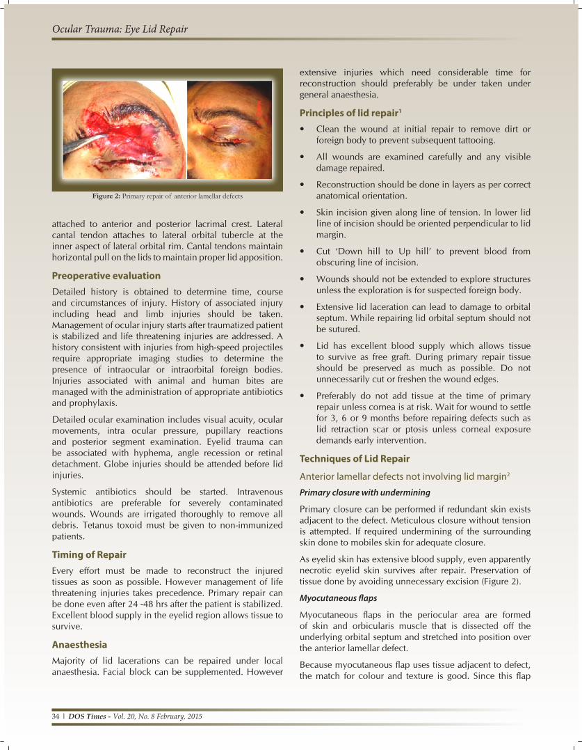
Primary closure can be performed if redundant skin exists adjacent to the defect. Meticulous closure without tension is attempted. If required undermining of the surrounding skin done to mobiles skin for adequate closure.
As eyelid skin has extensive blood supply, even apparently necrotic eyelid skin survives after repair. Preservation of tissue done by avoiding unnecessary excision (Figure 2).
Myocutaneous flaps
Myocutaneous flaps in the periocular area are formed of skin and orbicularis muscle that is dissected off the underlying orbital septum and stretched into position over the anterior lamellar defect.
Because myocutaneous flap uses tissue adjacent to defect, the match for colour and texture is good. Since this flap
Figure 2: Primary repair of anterior lamellar defects
Ocular Trauma: Eye Lid Repair
www. dosonline.org l 35
Ocular Trauma
brings its own blood supply, bare bones or free grafts can be covered (Figure 3).
Free skin grafts
Free skin grafts are harvested from a donor site and transferred to fill an anterior lamellar defect. Vascular supply to the free graft must be provided by recipient site for the graft to survive.
Full thickness skin graft (FTSG) employs entire thickness of epidermis and dermis harvested from donor site. Upper eyelid skin is the best choice for reconstruction of eyelid defects (Figure 4). Other sites for harvesting FTSG include retroauricular or pre auricular skin, supraclavicular skin and upper inner arm skin.
Split thickness skin graft (STSG) is seldom used in eyelid reconstruction. It is useful when a large area of skin needs to be covered and no myocutaneous flap can be mobilized. With STSG, the colour, texture and thickness are often a poor match for eyelid skin.
Eye lid margin repair3
Eyelid laceration involving lid margin requires meticulous approximation to avoid notching which cause functional and cosmetic problem. The wound should be carefully inspected to identify tarsus and lid margin landmarks like gray line, anterior lash line and posterior margin. If wound is ragged, freshening the edges with a scalpel blade may aid in structure recognition and apposition. Repair should be carried out preferably under operating microscope.
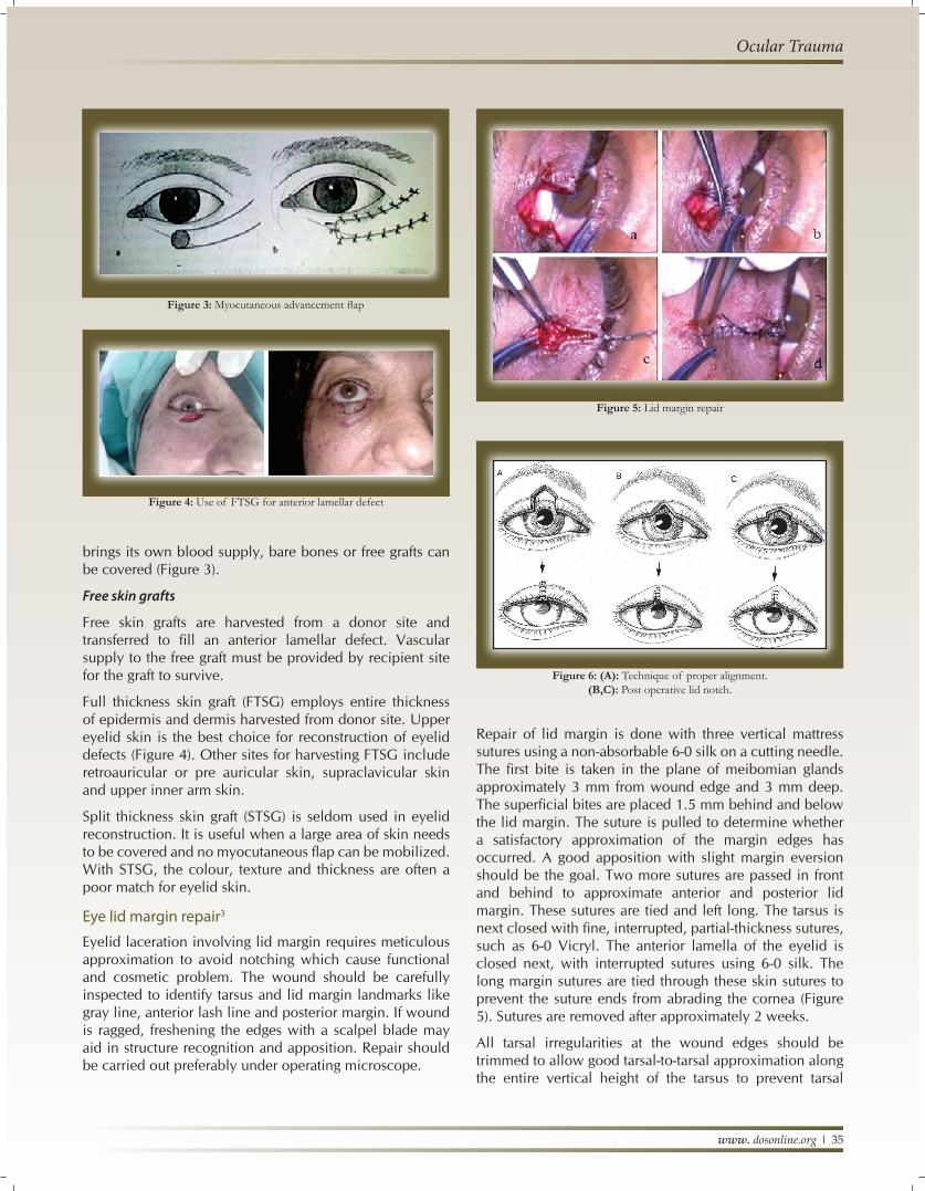
Repair of lid margin is done with three vertical mattress sutures using a non-absorbable 6-0 silk on a cutting needle. The first bite is taken in the plane of meibomian glands approximately 3 mm from wound edge and 3 mm deep. The superficial bites are placed 1.5 mm behind and below the lid margin. The suture is pulled to determine whether a satisfactory approximation of the margin edges has occurred. A good apposition with slight margin eversion should be the goal. Two more sutures are passed in front and behind to approximate anterior and posterior lid margin. These sutures are tied and left long. The tarsus is next closed with fine, interrupted, partial-thickness sutures, such as 6-0 Vicryl. The anterior lamella of the eyelid is closed next, with interrupted sutures using 6-0 silk. The long margin sutures are tied through these skin sutures to prevent the suture ends from abrading the cornea (Figure 5). Sutures are removed after approximately 2 weeks.
All tarsal irregularities at the wound edges should be trimmed to allow good tarsal-to-tarsal approximation along the entire vertical height of the tarsus to prevent tarsal
Figure 3: Myocutaneous advancement flap
Figure 4: Use of FTSG for anterior lamellar defect
Figure 5: Lid margin repair
Figure 6: (A): Technique of proper alignment. (B,C): Post operative lid notch.
36 l DOS Times - Vol. 20, No. 8 February, 2015
buckling. This converts the lid defect into a ‘pentagon’ which is sutured as described above. Failure to achieve proper alignment leads to post operative notching of lid margin (Figure 6).
Eyelid injuries with tissue loss4
Full thickness eyelid defects with tissue loss is classified depending on horizontal extent of defect into -
Small defects (< 1/3)
Medium defects (1/3 to 1/2)
Large defects (>1/2)
Repair of small defects (< 1/3 of horizontal length)
Direct closure
If either the upper or lower eyelid has sustained a full-thickness injury that results in less than 1/3 loss of tissue
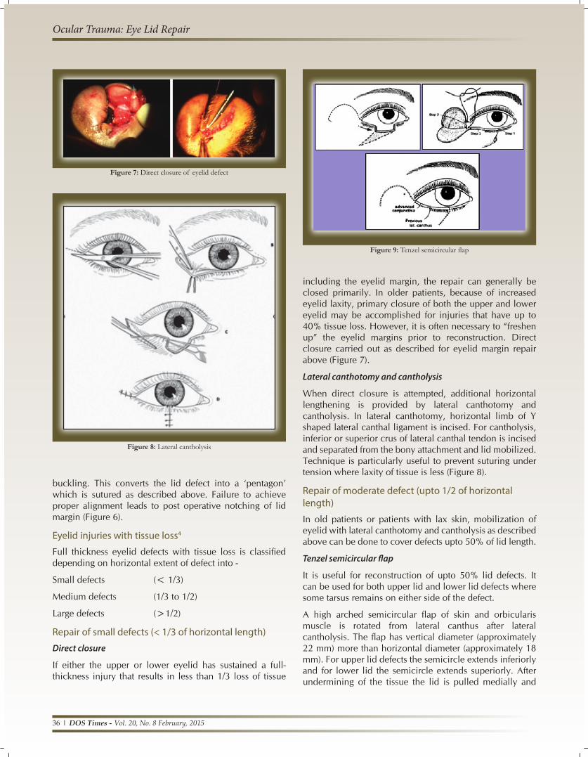
Figure 7: Direct closure of eyelid defect
Figure 8: Lateral cantholysis
including the eyelid margin, the repair can generally be closed primarily. In older patients, because of increased eyelid laxity, primary closure of both the upper and lower eyelid may be accomplished for injuries that have up to 40% tissue loss. However, it is often necessary to “freshen up” the eyelid margins prior to reconstruction. Direct closure carried out as described for eyelid margin repair above (Figure 7).
Lateral canthotomy and cantholysis
When direct closure is attempted, additional horizontal lengthening is provided by lateral canthotomy and cantholysis. In lateral canthotomy, horizontal limb of Y shaped lateral canthal ligament is incised. For cantholysis, inferior or superior crus of lateral canthal tendon is incised and separated from the bony attachment and lid mobilized. Technique is particularly useful to prevent suturing under tension where laxity of tissue is less (Figure 8).
Repair of moderate defect (upto 1/2 of horizontal length)
In old patients or patients with lax skin, mobilization of eyelid with lateral canthotomy and cantholysis as described above can be done to cover defects upto 50% of lid length.
Tenzel semicircular flap
It is useful for reconstruction of upto 50% lid defects. It can be used for both upper lid and lower lid defects where some tarsus remains on either side of the defect.
A high arched semicircular flap of skin and orbicularis muscle is rotated from lateral canthus after lateral cantholysis. The flap has vertical diameter (approximately 22 mm) more than horizontal diameter (approximately 18 mm). For upper lid defects the semicircle extends inferiorly and for lower lid the semicircle extends superiorly. After undermining of the tissue the lid is pulled medially and
Figure 9: Tenzel semicircular flap
Ocular Trauma: Eye Lid Repair

www. dosonline.org l 37
Ocular Trauma
direct closure of wound margins carried out. New lateral canthus created by suturing part of the new lid with intact limb of lateral canthal tendon (Figure 9).
Repair of large defects (> ½ of horizontal length)
Cutler Beard Bridge technique
Originally described for reconstruction of the upper lid, this technique can be used for reconstruction of the lower lid defect also, a procedure known as reverse Cutler- Beard.
Cutler Beard procedure is done in two stages. In first stage, after measuring the upper eyelid defect, a three-sided inverted U shaped incision is marked on the lower eyelid, about 5 mm below lid margin. After giving full thickness incision, the lower lid flap is pulled under the bridge of lower lid and sutured in layers to the upper lid defect. Since this flap is devoid of tarsus, autogenous cartilage from ear can be used. Separation of the flap is done 6 weeks to 3 months later as second stage surgery. After cutting the flap, lid margin of newly formed upper eyelid is sutured with conjuctiva covering the free margin (Figure 10).
Hughes tarsoconjunctival flap technique
It is partial thickness posterior lamellar flap harvested from upper lid to cover lower lid defects. After everting upper lid, incision is made through tarsus 4 mm above lid margin and flap is mobilized. Flap is sutured with lower lid tarsus to create posterior lamella. Sufficient skin to cover the anterior surface of the flap can be obtained either by harvesting a full-thickness skin graft or by advancing a myocutaneous flap from surrounding skin (Figure 11).
Mustarde cheek rotation flap
It is reserved for the reconstruction of very extensive
Figure 10: Cutler Beard procedure
Figure 11: Hughes Tarsoconjunctival flap
lower eyelid defects usually involving more than 75% of the eyelid. A large myocutaneous cheek flap is dissected and used in conjunction with an adequate mucosal lining posteriorly. A deep inverted triangle must be excised below the defect to allow adequate rotation of the flap. The side of the triangle nearest the nose should be practically vertical. The advantage of this procedure is that it is a one-stage, complete lower lid reconstruction (Figure 12).
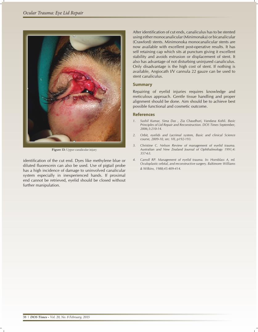
Repair of Canalicular laceration
Eyelid injuries involving medial canthal region can lead to canalicular injury (Figure 13). All canalicular lacerations need to be repaired whether upper or lower. Repair has to be undertaken under operating microscope.
Identification and retrieval of proximal end of canaliculus is most difficult step. Gentle traction at edges of wound with cotton applicator stick under high magnification will help. If it is difficult to identify, gentle irrigation of fluid or air injection through uninjured canalicular system can help in
Figure 12: Mustarde cheek rotation flap
38 l DOS Times - Vol. 20, No. 8 February, 2015
identification of the cut end. Dyes like methylene blue or diluted fluorescein can also be used. Use of pigtail probe has a high incidence of damage to uninvolved canalicular system especially in inexperienced hands. If proximal end cannot be retrieved, eyelid should be closed without further manipulation.
After identification of cut ends, canaliculus has to be stented using either monocanalicular (Minimonaka) or bicanalicular (Crawford) stents. Minimonoka monocanalicular stents are now available with excellent post-operative results. It has self retaining cap which sits at punctum giving it excellent stability and avoids extrusion or displacement of stent. It also has advantage of not disturbing uninjured canaliculus. Only disadvantage is the high cost of stent. If nothing is available, Angiocath I/V cannula 22 gauze can be used to stent canaliculus.
SummaryRepairing of eyelid injuries requires knowledge and meticulous approach. Gentle tissue handling and proper alignment should be done. Aim should be to achieve best possible functional and cosmetic outcome.
References1. Sushil Kumar, Sima Das , Zia Chaudhuri, Vandana Kohli. Basic
Principles of Lid Repair and Reconstruction. DOS Times September, 2006;3:210-14.
2. Orbit, eyelids and Lacrimal system, Basic and clinical Science course, 2009-10, sec. VII, p192-193.
3. Christine C. Nelson Review of management of eyelid trauma. Australian and New Zealand Journal of Ophthalmology 1991;4: 357-63.
4. Carroll RP. Management of eyelid trauma. In: Hornblass A, ed. Oculoplastic orbital, and reconstructive surgery. Baltimore: Williams & Wilkins, 1988;45:409-414.
Figure 13: Upper canalicular injury
Ocular Trauma: Eye Lid Repair