ocular inserts: a novel controlled drug delivery system
TRANSCRIPT

ISSN: 2277- 7695
CODEN Code: PIHNBQ
ZDB-Number: 2663038-2
IC Journal No: 7725
Vol. 1 No. 12 2013 Online Available at www.thepharmajournal.com
THE PHARMA INNOVATION - JOURNAL
Vol. 1 No. 12 2013 www.thepharmajournal.com Page | 1
Ocular Inserts: A Novel Controlled Drug Delivery System
K. P. Sampath Kumar1*, Debjit Bhowmik2, G.Harish3, S.Duraivel3, B. Pragathi kumar3
1. Department of Pharmaceutical Sciences, Coimbatore Medical College, Coimbatore, India. 2. Karpagam University, Coimbatore, India. [E-mail: [email protected]] 3. Nimra College of Pharmacy, Vijayawada, Andhra Pradesh, India.
Ocular drug delivery is one of the most challenging tasks faced by Pharmaceutical researchers. Major barriers in ocular medication are the ability to maintain a therapeutic level of the drug at the site of action for a prolonged duration. The ophthalmic preparations are available as sterile, buffered, isotonic solution. Several types of dosage forms are applied as the delivery system for the ocular delivery of drugs. The most prescribed dosage form is the eye drop solution as drops are easier to administer. Suspensions, gelled systems, ointment are also used for prolonged therapeutic action. Characteristics of ophthalmic preparations should be non-irritating to the ocular tissue. homogenous i.e., particles uniformly dispersed, smooth & free from lumps or agglomerates. Relatively non-greasy. Should not cause blurred vision. Should not cause intolerable foreign body sensation. Sterile and adequately preserved. Physically & chemically stable. Efficacious. New ocular drug delivery systems: Using eye drops to administer drugs needs frequent application. Prolonged drug release can be achieved using ophthalmic inserts, solid devices placed in the eye, but the inserts must then be removed when they are no longer needed. Ocuserts are the new drug delivery systems which are designed in such a way that they release the drug at predetermined and predictable rates thus eliminating the frequent administration of the drug. The systems generally include controlled, delayed and or sustained release bioerodible implantable elements having multiple layers of different materials and/or different concentrations of materials. The elements generally include an inner layer, or core, including a therapeutic agent, and one or more outer layers made of polymeric materials, for example substantially pure polymeric materials. In the area of topical ocular administration, important efforts concern the design and the conception of new ophthalmic drug delivery systems able to prolong the residence time. Keyword: Ocular Inserts, Bioerodible Implantable Elements, Predetermined Bioavailability.
1. INTRODUCTION: In developing a drug delivery strategy, issues of absorption, distribution, metabolism, elimination (ADME) must be considered.1 The eye presents unique opportunities and challenges when it comes to delivery of pharmaceuticals. Ophthalmic drug
delivery is one of the most interesting and challenging endeavors facing the pharmaceutical scientists.2 The anatomy, physiology and biochemistry of the eye render this organ exquisitely impervious to foreign substances. The challenge in front of formulator
The Pharma Innovation - Journal
Vol. 1 No. 12 2013 www.thepharmajournal.com Page | 2
is to circumvent the protective barriers of the eye without causing permanent tissue damage.3 The development of newer, more sensitive diagnostic techniques and therapeutic agents renders urgency to the development of maximum successful and advanced ocular drug delivery systems. The goal of pharmacotherapeutics is the attainment of an effective drug .concentration at the intended site of action for a desired period of time. Eye, as a portal for drug delivery is generally used for the local therapy as against systemic therapy in order to avoid the risk of eye damage from high blood concentrations of drug which are not intended for eye.4, 5 The conventional ocular dosage forms are eye drops, eye ointments, eye gels, eye solutions, eye injections, eye irritation solutions, eye suspensions, sol to gel systems.6 The most widely used are eye drops, eye ointments and gels, which constitute 80% of the total ophthalmic preparations. The eye drop dosage form is easy to instill but suffers from the inherent drawback, however these have limitations such as requirement of frequent administration, unpredictable doses7, rapid precorneal elimination, loss of drug by drainage, sticking of eye lids, poor patient compliance, blurred vision, no true sustained effect and even irritation.8, 9 It is generally agreed that the intraocular bioavailability of topically applied drugs is extremely poor ranging from 5-10% of total administered. 10, 11,12 The primitive ophthalmic solution, suspension etc are clearly no longer sufficient to combat some present virulent diseases.2 Effective treatment of ocular diseases is a formidable challenge for scientists in the field, especially because of the nature of diseases and presence of the ocular barriers especially in posterior ocular segments. Over last several years, attempts have been made to improve ocular bioavailability through manipulation of product formulation such as viscosity and application of mucoadhesive polymers. Thus far, these approaches to prolong corneal contact time have led to modest improvement in ocular bioavailability. Consequently, it seems logical to consider nonconventional approaches such as
nanotechnology, microspheres, liposomes, appropriate prodrug in situforming gel, and iontophoresis for effective delivery and to further enhance ocular absorption and reduce side effects. Because of poor ocular bioavailability, the frequent periodic instillation of eye drops becomes necessary to maintain a continuous sustained level of medication. This gives the eye massive and unpredictable dose of medication unfortunately higher drug concentrations this causes both ocular and systemic side effects.13 In order to remove the constraints placed by these conventional ocular therapies. A newer approach for ocular drug delivery systems are being explored to develop extended duration and controlled release strategy. Current ophthalmic drug delivery: Drops (95%+ of $8-12 billion market), ocular insert, gels and ointments Advantages of drops: Established, inexpensive manufacturing, Accepted standard of care Disadvantages: Variable, messy and difficult dosing, compliance issues, Short residence time, dilution and washout, require high drug concentration, systemic side-effects. Problems with conventional ophthalmic dosage forms Drainage of the instilled solution; Lacrimation and tear turnover; Metabolism; Tear evaporation; Non-productive absorption/ adsorption; Limited corneal area and poor corneal permeability; and Binding of the lachrymal proteins. The following recent trends are in vogue: Polymer based delivery plans Mucoadhesive dosage forms Ocular Inserts Collagen shields Drug presoaked hydrogel type contact lens and pledgets- Ocufit®, Minidisc®, SODI®,NODS®, Lacrisert® etc. Ocular Iontophoresis Phase Transition systems Microspheres and Nanoparticles Chemical delivery systems vesicular systems. In future, much of the emphasis will be given to achieve noninvasive sustained drug release for eye disorders in both segments. A clear understanding of the complexities associated with tissues in normal and pathological conditions, physiological barriers, and multicompartmental pharmacokinetics would greatly hasten further development in the field. An ideal system should be able to achieve an effective drug concentration at the target tissue for an extended period of time,

The Pharma Innovation - Journal
Vol. 1 No. 12 2013 www.thepharmajournal.com Page | 3
while minimizing systemic exposure. In addition, the system should be both comfortable and easy to use. Patient acceptance will continue to be emphasized in the design of future ophthalmic drug delivery systems. A reasonable strategy to circumvent the drawbacks of individual technologies is to combine technologies. Reported examples include liposomes and nanoparticles in droppable gels and liposomes and nanoparticles coated with bioadhesive polymers. The future challenges faced by topical ocular drug delivery systems are as follows: The ocular bioavailability must be increased from less than 1% to 15-20% of the administered dose. Most of the currently marketed ocular drugs were initially developed for non-ocular applications, hence their low or nonspecificity. So, there is a need to develop new drug candidates primarily intended for ocular use. Further studies to fully exploit the potential of noncorneal routes, especially for ionic/water-soluble moieties and also drug molecules with a preferential corneal absorption (and minimum absorption through nasal mucosa), should be explored. Appropriate design and packaging of these delivery systems needs further research. There are several scientific and technological advances that are driving the progress in this field. Especially the advances in nanotechnology and biomaterials science may provide new smart technologies to augment ophthalmic drug delivery. The core may include a polymeric material combined with an active agent beneficial in treating a condition of an eye. e.g. the pilocarpine inserts used in glaucoma therapy PILOCAR-20®, PILOCAR-40®. 2. RECENT TRENDS IN OCCULAR DRUG DELIVERY SYSTEM 2.1 The following recent trends are:-2, 9, 14, 15, 16
From the below newer approaches, the sensitive, successful extended duration and controlled release ocular delivery systems like ocular inserts, are being developed in order to attain better ocular bioavailability and sustained action of ocular drugs. Utilization of the principle of controlled release as embodied by ocular inserts
therefore offer an attractive alternative approach to the difficult problem of prolonging pre-corneal drug residence time.4
Mucoadhesive dosage forms. Ocular inserts. Collagen shields or corneal shields. Artificial tear inserts. Drug-presoaked hydrogel type contact
Lens. Ocular iontophoresis. Phase transition systems. Microspheres and nanoparticles.
3. COMMON EYE INFECTIONS.17, 18, 19
Bacteria are the causative pathogens for a large number of eye infections. In addition virus, fungus and protozoans also cause eye infections. As such, eyes are prone to number of diseases but more commonly found are mentioned here. o Conjunctivitis. o Blepharitis. o Keratitis. o Cataract. o Iritis (anterior uveitis). o Glaucoma. 3.1 Conjunctivitis. Conjunctivitis, commonly known as pink eye as shown in Fig 1, is an the clear membrane that covers the white part of the eye and lines the inner surface of the eyelids. The inflamed conjunctiva will usually make the eye appear red or pink because the tiny blood vessels that are normally within the conjunctiva get irritated and enlarged. It usually affects both eyes at the same time although it may start in one eye and spread to the other after a day or two days. It may be asymmetrical, affecting one eye more than the other. Pink eye can be infectious or non-infectious.

The Pharma Innovation - Journal
Vol. 1 No. 12 2013 www.thepharmajournal.com Page | 4
There are many causes for conjunctivitis, including.
Bacterial conjunctivitis – staphylococci, streptococci.
Viral conjunctivitis (often associated with the common cold) – adenovirus.
Chlamydial conjunctivitis – Chlamydia trachomatis.
Allergic conjunctivitis –allergic disease such as hay fever, asthma and eczema and by antigens like pollen, dust mites or cosmetics.
Reactive conjunctivitis or irritant conjunctivitis – chemicals, smoke, fumes etc.
3.1.1 Signs and Symptoms of conjunctivitis are:-
The blood vessels over the white of the eye are more visible and swollen.
The lining of the eyelids also looks red or pinker due to inflammation.
Eye is sticky, with a heavy discharge and tearing that may cause the lids to stick together, especially after sleeping.
Inflammed and swollen eyelids. Blurred vision.
4. OCULAR ANATOMY AND PHYSIOLOGY.9, 13, 17, 20
The human eye is a complex anatomical device that remarkably demonstrates the architectural wonders of the human body. The human eye is a challenging organ for topical administration of drugs. The basis of this can be found in the anatomical arrangement of the surface tissues and in the permeability of the cornea. The protective function of the eyelids and lachrymal system is such that there is rapid removal of material instilled into the eye unless the material is suitably small in volume and chemically and physiologically compatible with
surface tissues. The eye is referred as a globe and consists of two spheres, one set in the other, as shown in Fig 1. The front sphere is smaller and is bordered anteriorly by the sclera. The combined weight of both spheres has been given as 6.7-7.5gm, with a volume of approximately 6.5ml. The circumference of the eye is about 75mm. The eye is located in the bony orbital cavity of the head. 4.1 Eyeball: The wall of the human eyeball (globe) is composed of three concentric layers. 1. The outer fibrous layer. The fibrous layer is made up of two parts. a) Posterior (5/6th) is opaque and called the sclera. b) Anterior (1/6th) is transparent and called the cornea. 2. A middle vascular layer – the uvea or uveal tract consisting of the choroid, the ciliary body and the iris. 3. A nervous layer-the retina.
Fig 1:Anatomical structure of human eye ball. 4.2 Sclera Contains the microcirculation, which nourishes the tissues of this anterior segment and is usually white.

The Pharma Innovation - Journal
Vol. 1 No. 12 2013 www.thepharmajournal.com Page | 5
4.3 Vascular Layer consists of three parts:- The choroid – remains just behind the retina forming the posterior 5/6th of the middle coat, composed of numerous blood vessels and pigmented cells containing melanin. 4.4 The ciliary body – includes orbicularis ciliaris, ciliary processes, and ciliary muscle. 4.5 The Iris nervous coat is called retina, which contains photosensitive receptors. The eyeball houses an optical apparatus which consists, in sequences of the precorneal film, the cornea, the aqueous humor, the pupil, the crystalline lens, the vitreous humor and the retina. The aqueous and vitreous humors are layers of clear fluid or gel like material interposed between the solid structures. The crystalline lens is a refractive element with variable power controlled and supported by a muscle incorporated in the ciliary body. 4.6 Conjunctiva. The conjunctival membrane covers the outer surface of the white portion of the eye and the inner aspects of the eyelids. It is attached loosely and thereby permits free movement of the eyeball. Except for the cornea, the conjunctiva is the most exposed portion of the eye. 4.7 Lachrymal System. The conjunctival and corneal surfaces are covered and lubricated by a film of fluid secreted by the conjunctival and lachrymal glands. The secretion of the lachrymal glands, the tears are delivered through a number of fine ducts into the conjunctival fornix. The movement of the eye helps to spread the tears over the conjunctival surface. The excess fluid is directed into the lachrymal lake a small triangular area lying in the angle bound by the inner most portions of the lids. Tears are drained from the lachrymal lake, by two small tubes, the lachrymal canaliculi which lead into the upper part of the nasolacrimal duct. The act of blinking exerts a suction-force-pump action in removing tears from the
lachrymal lake and emptying them into the nasal cavity. Lacrimation is induced reflexly by stimulation of nerve ending of the cornea or conjunctiva, the turnover rate of nasolacrimal fluid is 16%. The eyeball is continually irrigated by a gentle stream of lacrimal fluid which prevents it from becoming dry and inflammed. 4.8. Composition of tear. The secretion is a clear watery fluid containing numerous salts, glucose, other organic compounds, approximately 0.7% protein and the enzyme, lysozyme. Water: 98.2% Solids: 1.8% Organic elements:- Protein-0.67%, Sugar-0.65%, Nacl-0.66%, NPN-0.05% Urea-0.03%. Other mineral elements sodium, potassium and ammonia-0.79%. 4.9. Precorneal Film. Part of the tear fluid provides the moist surface to cornea. The film, compatible with both aqueous and lipid ophthalmic preparations is composed of three layers, the thin outermost layer is lipid an d is secreted mainly by the meibomian glands. The lipid layer keeps the cornea moist by preventing evaporation of the underlying layers, a thicker middle aqueous layer, Secreted by the lacrimal gland, it helps in nourishing the cornea. It consists of water, salts, glucose, urea, proteins, lysozyme (an antibacterial enzyme) and immunoglobulin, and a thin inner mucous layer. It is secreted by the goblet cells of the tarsal conjunctiva. This layer is necessary for tear film stability. It smoothes the corneal epithelial surface, enhances tear spreading, lubricates the eye and helps to trap debris. It is renewed during each blink and when blinking is suppressed, it dries in patches.Although the tear film is typically only about 7µL in volume with fluid pH 7.4 and if blinking does not occur, the volume can go up to 30µL without spillage,the cul-de-
The Pharma Innovation - Journal
Vol. 1 No. 12 2013 www.thepharmajournal.com Page | 6
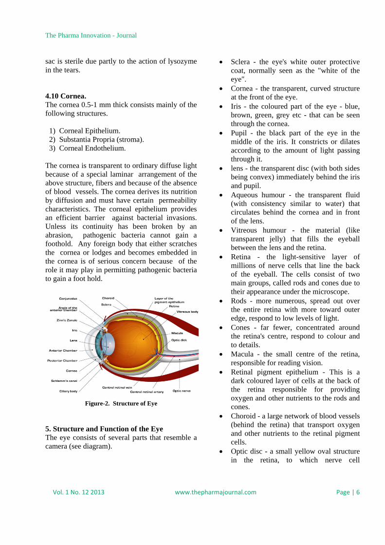
sac is sterile due partly to the action of lysozyme in the tears. 4.10 Cornea. The cornea 0.5-1 mm thick consists mainly of the following structures. 1) Corneal Epithelium. 2) Substantia Propria (stroma). 3) Corneal Endothelium. The cornea is transparent to ordinary diffuse light because of a special laminar arrangement of the above structure, fibers and because of the absence of blood vessels. The cornea derives its nutrition by diffusion and must have certain permeability characteristics. The corneal epithelium provides an efficient barrier against bacterial invasions. Unless its continuity has been broken by an abrasion, pathogenic bacteria cannot gain a foothold. Any foreign body that either scratches the cornea or lodges and becomes embedded in the cornea is of serious concern because of the role it may play in permitting pathogenic bacteria to gain a foot hold.
Figure-2. Structure of Eye
5. Structure and Function of the Eye The eye consists of several parts that resemble a camera (see diagram).
Sclera - the eye's white outer protective coat, normally seen as the "white of the eye".
Cornea - the transparent, curved structure at the front of the eye.
Iris - the coloured part of the eye - blue, brown, green, grey etc - that can be seen through the cornea.
Pupil - the black part of the eye in the middle of the iris. It constricts or dilates according to the amount of light passing through it.
lens - the transparent disc (with both sides being convex) immediately behind the iris and pupil.
Aqueous humour - the transparent fluid (with consistency similar to water) that circulates behind the cornea and in front of the lens.
Vitreous humour - the material (like transparent jelly) that fills the eyeball between the lens and the retina.
Retina - the light-sensitive layer of millions of nerve cells that line the back of the eyeball. The cells consist of two main groups, called rods and cones due to their appearance under the microscope.
Rods - more numerous, spread out over the entire retina with more toward outer edge, respond to low levels of light.
Cones - far fewer, concentrated around the retina's centre, respond to colour and to details.
Macula - the small centre of the retina, responsible for reading vision.
Retinal pigment epithelium - This is a dark coloured layer of cells at the back of the retina responsible for providing oxygen and other nutrients to the rods and cones.
Choroid - a large network of blood vessels (behind the retina) that transport oxygen and other nutrients to the retinal pigment cells.
Optic disc - a small yellow oval structure in the retina, to which nerve cell

The Pharma Innovation - Journal
Vol. 1 No. 12 2013 www.thepharmajournal.com Page | 7
connections travel from all the rods and cones.
Optic nerve and beyond - the "cord" of nerve cell connections that passes from the eyeball to destinations throughout the brain.
6. Function of the Eye When you see an object, the light travels from that object to the cornea, then passes through the aqueous humour, pupil, lens and vitreous humour to reach the retina. During this passage, the light becomes focused onto the macula.At the macula, the light causes chemical reactions in the cones, that consequently send electrical messages from the eye to the brain. The brain recognises these messages and indicates to you that this particular object has been seen. The cones are therefore responsible for you being able to recognise colours and to read.The rods are essential for you to see in the dark, and to detect objects to the sides, above and below the object on which you are directly focused. This function prevents you from bumping into obstacles when moving around.All the retinal cells (rods and cones) are provided with oxygen and other nutrients from the retinal pigment cells (epithelium), which are kept supplied by the rich network of blood vessels in the choroid. 7. MECHANISM OF CONTROL DRUG RELEASE INTO THE EYE The mechanism of controlled drug release into the eye is as follows: A. Diffusion, B. Osmosis, C. Bio-erosion. A. Diffusion In the Diffusion mechanism, the drug is released continuously at a controlled rate through the membrane into the tear fluid. If the insert is formed of a solid non-erodible body with pores and dispersed drug. The release of drug can take place via diffusion through the pores. Controlled release can be further regulated by gradual dissolution of solid dispersed drug within this matrix as a result of inward diffusion of aqueous solutions.In a soluble device, true dissolution
occurs mainly through polymer swelling. In swelling-controlled devices, the active agent is homogeneously dispersed in a glassy polymer. Since glassy polymers are essentially drug-impermeable, no diffusion through the dry matrix occurs. When the insert is placed in the eye, water from the tear fluid begins to penetrate the matrix, then swelling and consequently polymer chain relaxation and drug diffusion take place. The dissolution of the matrix, which follows the swelling process, depends on polymer structure: linear amorphous polymers dissolve much faster than cross-linked or partially crystalline polymers. Release from these devices follows in general Fickian 'square root of time' kinetics; in some instances, however, known as case II transport, zero order kinetics has been observed. B. Osmosis In the Osmosis mechanism, the insert comprises a transverse impermeable elastic membrane dividing the interior of the insert into a first compartment and a second compartment; the first compartment is bounded by a semi-permeable membrane and the impermeable elastic membrane, and the second compartment is bounded by an impermeable material and the elastic membrane. There is a drug release aperture in the impermeable wall of the insert. The first compartment contains a solute which cannot pass through the semi-permeable membrane and the second compartment provides a reservoir for the drug which again is in liquid or gel form.When the insert is placed in the aqueous environment of the eye, water diffuses into the first compartment and stretches the elastic membrane to expand the first compartment and contract the second compartment so that the drug is forced through the drug release aperture. C. Bioerosion In the Bioerosion mechanism, the configuration of the body of the insert is constituted from a matrix of bioerodible material in which the drug is dispersed. Contact of the insert with tear fluid results in controlled sustained release of the drug by bioerosion of the matrix. The drug may be dispersed uniformly throughout the matrix but it

The Pharma Innovation - Journal
Vol. 1 No. 12 2013 www.thepharmajournal.com Page | 8
is believed a more controlled release is obtained if the drug is superficially concentrated in the matrix.In truly erodible or E-type devices, the rate of drug release is controlled by a chemical or enzymatic hydrolytic reaction that leads to polymer solubilization, or degradation to smaller, water-soluble molecules. These polymers, as specified by Heller, [34] may undergo bulk or surface hydrolysis. Erodible inserts undergoing surface hydrolysis can display zero order release kinetics; provided that the devices maintain a constant surface geometry and that the drug is poorly water-soluble. 8. ABSORPTION OF DRUGS IN THE EYE. Topical delivery into the cul-de-sac is, by far, the most common route of ocular drug delivery, absorption from this site by.
1. Corneal 2. Non-corneal routes.
Maximum absorption takes place through the cornea, which leads the drug into aqueous humor. The non-corneal route involves absorption across the sclera and conjunctiva, this route is not productive as it restrains the entry of drug into the intraocular tissues. 8.1 Physicochemical properties of drug. Transcellular or the Paracellular pathway is the main route for drugs to penetrate across the corneal epithelium. Paracellular pathway involves the passive diffusion through the intercellular spaces. (Hydrophilic drugs). Transcellular pathway involves the partitioning of the drugs to cells (lipophilic drugs). For both pathways the passive diffusion along their concentration gradient is the main permeation mechanism. 8.2 OPTHALMIC INSERTS.9, 23, 24
Ophthalmic inserts are sterile preparations with a solid or a semisolid consistency, and whose size and shape are especially designed for ophthalmic application. The inserts are placed in the lower fornix and less frequently, in the upper fornix or
on the cornea. Ocular inserts can overcome the disadvantages reported with traditional. Ophthalmic systems like eye drops, suspensions and ointments. The typical pulse entry type drug release behavior observed with eye drops, suspensions and ointments is replaced by more controlled, sustained and continuous drug delivery using a controlled release ocular drug delivery system. In the recent years, there has been explosion of interest in the polymer based delivery devices, adding further dimension to topical drug delivery thereby promoting the use of polymers such as collagen and fibrin fabricated into erodible inserts for placement in cul-de-sac. Utilization of the principles of controlled release as embodied by ocular inserts offers an attractive approach to the problem of prolonging precorneal drug residence times. Ocular inserts also offer the potential advantage of improving patient compliance by reducing the dosing frequency. The main objective of the ophthalmic inserts is to increase the contact time between the preparation and the conjunctival tissue to ensure a sustained release suited to topical or systemic treatment. They are composed of polymeric support with or without drugs, the latter being incorporated as dispersion or a solution in the polymeric support.
Figure-3: Routes of Occular Drug Delivery

The Pharma Innovation - Journal
Vol. 1 No. 12 2013 www.thepharmajournal.com Page | 9
8.3 Classification of ophthalmic inserts:- Based upon their solubility behaviour. (1) Insoluble:- a) Diffusion b) Osmotic and c) Contact lens (2) Soluble:- a) Based on natural polymers e.g. collagen b) Based on synthetic or semi synthetic polymers e.g. cellulose derivatives like HPMC, HPC, MC etc 8.4 Bioerodible. a. Insoluble ocuserts. Only the insoluble types can usually deliver drugs by a variety of method satcontrolled, predetermined rate, but need removal from the eye when empty. b. Soluble ocuserts: Soluble(S) inserts generally defined as erodible (E), monolithic polymeric devices that undergo gradual dissolution while releasing the drug and do not need removal. True dissolution occurs mainly through polymer swelling, while erosion corresponds to a chemical or enzymatic hydrolytic process. In swelling-controlled devices the active agent is homogeneously dispersed in a glassy polymer, glassy polymers are essentially drug impermeable so no diffusion takes place through the dry matrix. When the insert is placed in the eye water from the tear fluid begins to penetrate the matrix, then swelling and consequently polymer chain relaxation and drug diffusion take place releasing their drug content. I. Insoluble ocular inserts Inserts made up of insoluble polymer can be classified into two categories: A. Reservoir systems; B. Matrix systems. A. Reservoir systems Each class of inserts shows different drug release profiles. The reservoir systems can release drug
either by diffusion or by an osmotic process. It contains, respectively, a liquid, a gel, a colloid, a semisolid, a solid matrix, or a carrier containing drug. Carriers are made of hydrophobic, hydrophilic, organic, natural or synthetic polymers. They have been sub-classified into: 1. Diffusional inserts, e.g., 'Ocuserts'; 2. Osmotic inserts. 1. Diffusional insert or Ocuserts Ocusert system is a novel ocular drug delivery system based on porous membrane. The release of drug from diffusional inserts/Ocusert is based on a diffusional release mechanism. It consists of a central reservoir of drug enclosed in specially designed microporous membrane allowing the drug to diffuse from the reservoir at a precisely determined rate. As pointed out by Urquhart, the Ocusert pilocarpine ocular therapeutic system, developed by Alza Corporation, is notable for several reasons. This product was the first rate-controlled, rate specified pharmaceutical for which the strength is indicated on the label by the rate(s) of drug delivery in vivo , rather than by the amount of contained drug. It provides predictable, time-independent concentrations of drug in the target tissues, a feat impossible to achieve with conventional, quantity-specified, pulse entry ophthalmic medications. The near-constant drug concentration in ocular tissues markedly improves the selectivity of action of pilocarpine. A major advantage is that two disturbing side effects of the drug, miosis and myopia, are significantly reduced, while reduction of intraocular pressure (IOP) in glaucoma patients is fully maintained.Two types of Ocusert are available: the Pilo-20 and Pilo-40. The former delivers the drug at a rate of 20 µg/h for 7 days, and the latter at a rate of 40 µg/h for 7 days. This device, which is certainly well familiar to the readers of this review, has been exhaustively described and discussed in a series of specialized papers. Briefly, it consists of a reservoir containing pilocarpine alginate enclosed above
The Pharma Innovation - Journal
Vol. 1 No. 12 2013 www.thepharmajournal.com Page | 10
and below by thin EVA (ethylene-vinyl acetate) membranes. The insert is encircled by a retaining ring of the same material, impregnated with titanium dioxide. The dimensions of the elliptical device are (for the 20 µg/h system): major axis-13.4 mm, minor axis-5.7 mm, thickness-0.3 mm. The membranes are the same in both systems, but to obtain a higher release rate, the reservoir of the 40 µg/h system contains about 90 mg of di (2-ethylhexyl) phthalate as a flux enhancer. 2. OSMOTIC INSERT The osmotic inserts are generally composed of a central part surrounded by a peripheral part and are of two types: Type 1: The central part is composed of a single reservoir of a drug with or without an additional osmotic solute dispersed throughout a polymeric matrix, so that the drug is surrounded by the polymer as discrete small deposits. The second peripheral part of these inserts comprises a covering film made of an insoluble semi-permeable polymer. The osmotic pressure against the polymer matrix causes its rupture in the form of apertures. Drug is then released through these apertures from the deposits near the surface of the device. Type 2: The central part is composed of two distinct compartments. The drug and the osmotic solutes are placed in two separate compartments, the drug reservoir being surrounded by an elastic impermeable membrane and the osmotic solute reservoir by a semi-permeable membrane. The second peripheral part is similar to that of type 1. The tear diffuse into the osmotic compartment inducing an osmotic pressure that stretches the elastic membrane and contracts the compartment including the drug, so that the active component is forced through the single drug release aperture. B. Matrix systems The second category, matrix system, is a particular group of insoluble ophthalmic devices mainly represented by contact lenses. It
comprises of covalently cross-linked hydrophilic or hydrophobic polymer that forms a three dimensional network or matrix capable of retaining water, aqueous drug solution or solid components. The hydrophilic or hydrophobic polymer swells by absorbing water. The swelling caused by the osmotic pressure of the polymer segments is opposed by the elastic retroactive forces arising along the chains or crosslinks are stretched until a final swelling (equilibrium) is reached. 1. Contact lenses Contact lenses are shaped structures and initially used for vision correction. Their use has been extended as potential drug delivery devices by presoaking them in drug solutions. The main advantage of this system is the possibility of correcting vision and releasing drug simultaneously. Refojo has proposed a subdivision of contact lenses into 5 groups.
a) Rigid b) Semi-rigid c) Elastomeric d) Soft hydrophilic e) Bio-polymeric
Rigid contact lenses have the disadvantage of being composed of polymers (e.g., poly methyl methacrylic acid) hardly permeable to moisture and oxygen, a problem which has been overcome by using gas permeable polymers such as cellulose acetate butyrate. However, these systems are not suitable for prolonged delivery of drugs to the eye and their rigidity makes them very uncomfortable to wear. For this reason, soft hydrophilic contact lenses were developed for prolonged release of drugs such as pilocarpine, chloramphenicol and tetracycline prednisolone sodium phosphate. The most commonly used polymer in the composition of these types of lenses is hydroxy ethyl methyl metacrylic acid copolymerized with poly (vinyl pyrrolidone) or ethylene glycol dimethacrylic acid (EGDM). Poly (vinyl pyrrolidone) is used for increasing water of hydration, while EGDM is used to decrease the water of hydration. The soft

The Pharma Innovation - Journal
Vol. 1 No. 12 2013 www.thepharmajournal.com Page | 11
hydrophilic contact lenses are very popular because they are easy to fit and are tolerated better. The drug incorporation into contact lenses depends on whether their structure is hydrophilic or hydrophobic. When contact lens (including 35 to 80% water) is soaked in solution, it absorbs the drug. Drug release depends markedly on the amount of drug, the soaking time of the contact lens and the drug concentration in the soaking solution.
II. Soluble ocular inserts These soluble inserts offer the advantage of being entirely soluble so that they do not need to be removed from their site of application, thus limiting the intervention to insertion only. They can be broadly divided into two types, the first one being based on natural polymers and the other on synthetic or semi-synthetic polymers. A. Natural polymers The first type of soluble inserts is based on natural polymer Natural polymer used to produce soluble ophthalmic inserts is preferably collagen. The therapeutic agent is preferably absorbed by soaking the insert in a solution containing the drug, drying, and re-hydrating it before use on the eye. The amount of drug loaded will depend on the amount of binding agent present, the concentration of the drug solution into which the composite is soaked as well as the duration of the soaking. As the collagen dissolves, the drug is gradually released from the interstics between the collagen molecules. B. Synthetic and semi-synthetic polymer The second type of soluble insert is usually based on semi-synthetic polymers (e.g., cellulose derivatives) or on synthetic polymers such as polyvinyl alcohol. A decrease of release rate can be obtained by using Eudragit, a polymer normally used for enteric coating, as a coating agent of the insert . Saettone et al . have observed in rabbits that Eudragit coated inserts containing pilocarpine induced a miotic effect of a longer duration, compared to the corresponding uncoated ones. However, the inherent problems encountered with these soluble inserts are the
rapid penetration of the lachrymal fluid into the device, the blurred vision caused by the solubilization of insert components and the risk of expulsion due to the initial dry and glassy consistency of the device. [4] Ethyl cellulose, a hydrophobic polymer, can be used to decrease the deformation of the insert and thus to prevent blurred vision. As for the risk of expulsion, several authors have incorporated carbomer, a strong but well tolerated bio-adhesive polymer. The soluble inserts offer the additional advantage of being of a generally simple design, of being based on products well adapted for ophthalmic use and easily processed by conventional methods. The main advantage is decreased release rate, but still controlled by diffusion. III. Bio-erodible ocular inserts These inserts are formed by bio-erodible polymers (e.g., cross-linked gelatin derivatives, polyester derivatives) which undergo hydrolysis of chemical bonds and hence dissolution. The great advantage of these bio-erodible polymers is the possibility of modulating their erosion rate by modifying their final structure during synthesis and by addition of anionic or cationic surfactants. A cross-linked gelatin insert was used by Attia et al . to increase bioavailability of dexamethasone in the rabbit eye. The dexamethasone levels in the aqueous humor were found to be four-fold greater compared to a dexamethasone suspension. However, erodible systems can have significantly variable erosion rates based on individual patient physiology and lachrimation patterns, while degradation products and residual solvents used during the polymer preparation can cause inflammatory reaction. In the following paragraphs, some important ocular inserts are discussed which are available commercially (SODI) or in advanced states of development (collagen shields, Ocufit, NODS, and Minidisc). Soluble ophthalmic drug insert Soluble ophthalmic drug insert (SODI) is a small oval wafer, which was developed by soviet scientists for cosmonauts who could not use eye

The Pharma Innovation - Journal
Vol. 1 No. 12 2013 www.thepharmajournal.com Page | 12
drops in weightless conditions. SODI is together with the collagen shields, the first modern revival of the gelatin 'lamellae', which disappeared from pharmacopoeias in the late forties. The SODIs are the result of a vast collaborative effort between eminent Russian chemists and ophthalmologists, and led eventually (in 1976) to the development of a new soluble copolymer of acrylamide, N -vinylpyrrolidone and ethyl acrylate (ratio 0.25: 0.25: 0.5), designated ABE. A comparison of medicated eye films prepared with different polymers, showed that ABE produced the highest concentration of drugs in rabbit ocular tissues. After large-scale preclinical and clinical testing, the ABE copolymer was used for the industrial manufacture of the SODI in the form of sterile thin films of oval shape (9 x 4.5 mm, thickness 0.35 mm), weighing 15-16 mg, and color-coded for different drugs (over 20 common ophthalmic drugs, or drug combinations). After introduction into the upper conjunctival sac, a SODI softens in l0-15 s, conforming to the shape of the eyeball. In the next l0-15 min the film turns into a polymer clot, which gradually dissolves within 1 h while releasing the drug. The sensation of an 'extraneous body' in the eye disappears in 5-15 min. COLLAGEN SHIELDS Collagen is the structural protein of bones, tendons, ligaments, and skin and comprises more than 25% of the total body protein in mammals. This protein, which is derived from intestinal collagen, has several biomedical applications, the main of which is probably catgut suture. Bloomfield et al . are credited for first suggesting, in 1977 and 1978, the use of collagen inserts as tear substitutes and as delivery systems for gentamicin. They compared the levels of gentamicin in tears, cornea, and sclera of the rabbit eye after application of a collagen insert, drops, an ointment or following subconjunctival administration. After 3 h, they found that the collagen insert gave the highest concentration of gentamicin in the tear film and in the tissue. Other treatments using collagen shields impregnated with gentamicin and dexamethasone
have been described. In rabbits, aqueous humor levels of dexamethasone and gentamicin achieved with collagen shields were compared to subconjunctival injections. The authors concluded that the use of collagen shields impregnated with gentamicin-dexamethasone was comparable to the subconjunctival delivery of these drugs over a 10-h period.Some drawbacks of these devices, however, need mentioning. To apply the collagen shield, the cornea is anaesthetized while the physician uses a blunt forceps to insert the hydrated or unhydrated shield. Contrary to medicated contact lenses, collagen shields often produce some discomfort and interfere with vision. In rabbits, collagen shields have been found to exacerbate ulcerations of alkali-burned corneas. [A new preparation referred to as collasomes consists of small pieces (1 mm x 2 mm x 0.1 mm) of collagen suspended in a 1% methylcellulose vehicle. Kaufman and co-workers recently reported that collasomes provide the same therapeutic advantages of the shields (high and sustained levels of drugs and/or lubricants to the cornea), while not presenting their disadvantages. OCUFIT The Ocufit is a sustained release, rod shaped device made of silicone elastomer, patented in 1992 and currently developed by Escalon Ophthalmics Inc. (Skillman, NJ). It was designed to fit the shape and size of the human conjunctival fornix. Accordingly, it does not exceed 1.9 mm in diameter and 25-30 mm in length, although smaller sizes for children and newborn babies are planned. The superiority of the cylindrical shape can be traced in an earlier paper by Katz and Blackman. They reported the effect of the size and shape of the inserts on tolerance and retention by human volunteers. These workers found that expulsion of rod shaped units was significantly ( P < 0.01) less frequent than expulsion of oval, flat inserts. A typical example of a rod-shaped insert is the Lacrisert (Merck and Co., Inc.), a cellulosic device used to treat dry-eye patients. The insoluble Ocufit reportedly combines two important features, long retention and sustained

The Pharma Innovation - Journal
Vol. 1 No. 12 2013 www.thepharmajournal.com Page | 13
drug release. When placed in the upper fornix of volunteers, placebo devices were retained for 2 weeks or more in 70% of the cases. Moreover, active disease (bacterial, allergic and adenoviral conjunctivitis, trachoma, episcleritis, anterior uveitis, cornea1 ulcers or scars) did not overtly affect the ability of the patients to retain the inserts. Tetracycline-loaded inserts released in vitro 45% of the drug over the 14-day period with an initial burst in the first day followed by a constant rate over the remaining period. 9. THE MINIDISC OCULAR THERAPEUTIC SYSTEM This monolytic polymeric device, originally described by Bawa et al . (Bausch and Lomb,
Rochester, New York) and referred to as Minidisc ocular therapeutic system (OTS), is shaped like a miniature (diameter 4-5 mm) contact lens, with a convex and a concave face, the latter conforming substantially to the sclera of the eye. The particular size and shape reportedly allow an easy placement of the device under the upper or lower lid without compromising comfort, vision or oxygen permeability. When compared with another standard insert, the Lacrisert, the Minidisc was reported to require less time and less manual dexterity for insertion. Different versions of the device have been evaluated, such as, non-erodible hydrophilic, non-erodible hydrophobic and erodible.
Table-1 Types of Occular Inserts
In vitro tests showed that the hydrophilic OTS (based on polyhydroxymethyl methacrylate) released sulfisoxazole for 118 h, while the hydrophobic unit (based on a proprietary Bausch
and Lomb pre-polymer) released gentamicin sulfate for more than 320 h. Clinical trials on placebo units demonstrated that the devices were well tolerated when placed either in the upper or

The Pharma Innovation - Journal
Vol. 1 No. 12 2013 www.thepharmajournal.com Page | 14
lower conjunctival sac. In the eyes of healthy volunteers, the hydrophilic OTS released sulfisoxazole continuously for 3 days. [19] Further studies conducted on the hydrophobic Minidisc showed that gentamicin sulfate was efficiently released in rabbit eyes for 14 days.
Table-2 OCULAR INSERTS DEVICES Name Description
Soluble ocular drug Insert
Small oval wafer, composed of soluble copolymers consisting of actylamide, N-venyl pyrrolidone and ethyl acetate, soften on insertion
New ophthalmic drug delivery system
Medicated solid polyvinyl alcohol flag that is attached to a paper- covered with handle. On application, the flag detaches and gradually dissolves, releasing the drugs
Collagen shields Erodible disc consist of cross-link porcine scleral collagen
Ocusert
Flat, flexible elliptical insoluble device consisting of two layers, enclosing a areservior, use commercially to deliver Pilocarpine for 7 days
Minidisc or ocular therapeutic
system 4-5 mm diameter contoured either hydrophilic or hydrophobic disc
Lacrisert
Rose-shape device made from Hydroxy propyl cellulose use for the eye syndrome as an alternative to tears
Bioadhesive ophthalmic eye insets
Adhesive rods based on a mixture of Hydroxy propyl cellulose, ethyl cellulose, Poly acrylic acid cellulosephthalate
Dry drops
A preservative free of hydrophilic polymer solution that is freeze dried on the tip of a soft hydrophobic carrier strip, immediately hydrate in tear strip
Gelfoam Slabs of Gelfoam impregnated with a mixture of drug and cetyl ester wax in chloroform
10. Advantages of ocuserts:25, 26, 27
Increased contact time and thus improved
bio-availability. Possibility of providing a prolonged drug
release and thus a better efficacy. Administration of an accurate dose in the
eye and thus a better therapy. Reduction of systemic side effects and
thus reduced adverse effects. Reduction of the number of
administrations and thus better patient. Compliance, Comfort. Lack of explosion. Ease of handling and insertion. Non-interference with vision and oxygen
permeability. Reproducibility of release kinetics. Sterility. Stability. Exclusion of preservatives. Increased shelf life with comparison to
aqueous solutions due to absence of water.
11. Disadvantages:
The insert may be lost immediately. Sometimes the insert twists to form ‘a
figure eight’, which diminishes the delivery rate.
A leakage may occur. Dislocation of the device in front of the
pupil. 12. CONCLUSION: The ocular insert represents a significant advancement in the therapy of eye disease. Ocular inserts are defined as sterile, thin, multilayered, drug-impregnated, solid or semisolid consistency devices placed into the cul-de-sac or conjuctival sac, whose size and shape are especially designed for ophthalmic application. They are composed of a polymeric support that may or may not contain a drug. Advantages with ocuserts such as, Accurate dosing Capacity to provide at constant rate and prolong drug release thus a better efficacy. Increasing contact time and thus improving

The Pharma Innovation - Journal
Vol. 1 No. 12 2013 www.thepharmajournal.com Page | 15
bioavailability. Possible reduction of systemic absorption and thus reduced systemic adverse effects.Reduced frequency of administrations and thus better patient compliance with lower incidence of visual side effects. Administration of an accurate in the eye and thus a better therapy Possibility of targeting internal ocular tissues through non-corneal conjuctival - scleral penetration routes; and Increased shelf life with respect to eye-drops due to the absence of water. Advantage of inserts as dosage form Ease of handling and insertion Lack of expulsion during wear Reproducibility of release kinetics Applicability to variety of drugs Non-interference with vision and oxygen permeability. Sterility. Stability. Ease of manufacture. 13. REFERENCE:
1. Chrai SS,MakoidMC,EriksonSP,Robinson JR.Dropsizeandinitialdosingfrequencyproblemsof topically applied ophthalmic drugs.J PharmSci.1974;64:333–8.
2. Zaki I, Fitzgerald P, Hardy JG, Wilson CG.Comparison of effect of viscosity on theprecorneal residence of solution in rabbit andman.JPharmPharmacol.1986;38:463–6.
3. Lee VH, Robinson JF. Review: Topical oculardrugdelivery; recentdevelopmentsand futurechallenges.JOculPharmacol.1976;2:67.[]
4. Saettone MF, Salminen L. Ocular inserts fortopicaldelivery.AdvDrugDelRev.1995;16:95–106.
5. Schoenwald RD. OcularPharmacokinetics/Pharmacodynamics. In:Mitra AK, editor.Ophthalmic drug deliverysystems.NewYork:MarcelDekker;1993.
6. Davis JL, Gilger BC, Robinson MR. Novelapproaches to ocular drug delivery.Curr OpinMolTher.2004;6:195–205.[]
7. Neefe CW. Contact lens for ocular drugdelivery.USPatent.1974;3:786–812.
8. GibaldiM,PerrierD.2nded.NewYork:MarcelDekker,Inc;1993.Pharmacokinetics.
9. Robinson JC. Ocular Anatomy and PhysiologyRelevanttoOcularDrugDelivery.In:MitraAK,editor.Ophthalmic drug delivery systems.NewYork:MarcelDekker;1993.
10. Chien YW. 2nd ed. New York: Marcel Dekker,Inc;1992.Noveldrugdeliverysystems.
11. Khar RK, Vyas SP. 1st ed. New Delhi: C.B.S.Publishers and Distributors, Inc; 2002.
Targeted and controlled drug delivery novelcarriersystems.
12. MainardesRM,UrbanMC,CintoPO,ChaudMV.Colloidal carriers for ophthalmic drugdelivery.CurrDrugTargets.2005;6:363–71.[]
13. Friedrich SW, Saville BA, Cheng YL, RootmanDS.Pharmacokineticdifferencesbetweenocularinserts and eyedrops.J Ocul PharmacolTher.1996;12:5–18.[]
14. MichaelsAS,GuilloidMS.Osmoticburstingdrugdeliverydevice.USPatent.1979;4:177–256.
15. Gurtler F, GurnyR. Patent literature review ofophthalmic inserts.Drug Dev IndPharm.1995;21:1.
16. Bloomfield SE,Miyata T, DunnMW, Bueser N,Stenzel KH, Rubin AL. Soluble gentamacinophthalmic inserts as a delivery system.ArchOpthalmol.1978;96:885–7.
17. Ahmed I, Gokhale RD, Shah MV, Patton TF.Physicochemicaldeterminantsofdrugdiffusionacross the conjunctiva, sclera and cornea.JPharmSci.1987;76:583–6.[]
18. EllerMG,SchoenwaldRD,DixsonJA,SegarraT,BarfknechtCF.Optimizationmodelsforcornealpenetration of ethoxyzolamide analogues.JPharmSci.1985;74:155–60.[]
19. Huang HS, Schoenwald RD, Lac JL. Cornealpenetration behavior of b blocking agents II.JPharmSci.1983;72:1272–9.[]
20. GrassGM,Robinson JR.Mechanismsof cornealdrugpenetrationII:Ultrastructuralanalysisofpotential pathways for drug movements.JPharmSci.1988;77:15–23.[]
21. Alvarez‐Lorenzo C, Hiratani H. Soft contactlensescapableofsustaineddeliveryoftimolol.JPharmSci.2002;91:2182–92.[]
22. PattonTF,RobinsonJR.Quantitativeprecornealdisposition of topically applied pilocarpinenitrate in rabbit eye.J PharmSci.1976;65:1295–301.[]
23. Himmelstein KJ, Guvenir I, Palton TP.Preliminary Pharmacokinetics model ofpilocarpineuptakeanddistributionintheeye.JPharmSci.1978;67:603–6.[]
24. Mitra AK. Opthalmic drug delivery. In: Tyle P,editor.Drug Delivery Devices.New York:MarcelDekker;1998.
25. Jain NK. New Delhi: C.B.S. Publisher anddistributer, Inc; 2004. Controlled and noveldrugdelivery.
26. Korsmeyer RW, Peppas NA. Macromolecularand modeling aspects of swelling‐controlledsystems. In: Roseman TJ, Mansdorf SZ,editors.Controlled Release DeliverySystems.NewYork:MarcelDekker;1983.
The Pharma Innovation - Journal
Vol. 1 No. 12 2013 www.thepharmajournal.com Page | 16
27. Darougar S. Patent literature review of ocularinserts.USPatent.1999;6:264–971.
28. Sahane NK, Banarjee SK, Gaikwad DD, JadhavSL, Throat RM. Ocular Inserts‐ A Review.DrugInvenTod.2010;2:57–64.
29. RallTW.Drugusedinthetreatmentofasthma.In:GoodmanGA,editor.GoodmanandGilman'sthe Pharmacological Basis of Therapeutics.9thed. New York: Palmer Taylor MaxwellMacmillan pergamon Publishing Corporation;1990.
30. Insel PA. Anlgesic‐antiphyretic and anti‐inflammatoryagentsanddrugsemployedinthetreatment of Gout. In: Goodman GA,editor.Goodman and Gilman's thePharmacological Basis of Therapeutics.9th ed.New York: McGraw‐Hill Health professiondivisions;1996.
31. Brogden RN, Heel RC, Pakes GE, Speight TM,Avery GS. Diclofenac sodium: A review of itspharmacological properties and therapeuticsuse in rheumatic diseases and pain of varyingorigin.Drugs.1980;20:48.