ocular changes in oi in a portuguese population changes in oi in a... · discussion •bcva was...
TRANSCRIPT

Ocular changes in OI in a Portuguese population
Rafael Barão, MD
Authors: Patrícia Firmino, Rafael Barão, Débora Pereira, Mariana de Sá, Patrícia Monteiro, Paula Silva, Ana C. Fonseca, Ilda M. Poças

Conflicts of interest
I, Rafael Barão, DO NOT have a financial interest/arrangement or affiliation with one or moreorganizations which could be perceived as a real or apparent conflict of interest in the context of thesubject of this presentation

Background
• OI can cause several ocular manifestations
• The most common (and noticeable) is the bluish coloration
of the sclerae (+ type I)1,2
• Other common manifestations include
• Reduced corneal thickness2,3, 4 and rigidity4
• High refractive errors5
• Keratoconus6,7
• Glaucoma (higher incidence de per se or higher risk?)2,8, 9, 10, 11
• Less common: scleral rupture12, retinal hemorrhages13, CNV14, among
others
1. Sillence et. al. 19932. Hald et al. 20183. Pedersen et. al. 19844. Lagrou et. al. 20185. Chau et. al. in Osteogenesis Imperfecta, 20146. Beckh et. Al. 19957. Zeri et. al. 20188. Congdon et. al. 20069. Rosbach et. al. 2011 10. Wallace et. al. 201411. Pirouzian et. al. 200712. Mauri et. al. 201613. Ganesh et. al. 2004
Objectives
• Assess and explore the ocular features of OI patients in an adult
Portuguese population
• Exploratory analysis of relationships between OI types and ocular phenotypes
• Exploratory analysis of relationships between several variables

Study design and setting
• An ongoing cross-sectional study on the ocular features in OI patients
• Protocol envolves coordinated visitations to several departments• Cardiology, Genetics, Rheumatology, Stomatology and Ophthalmology

Methods
• All patients underwent complete ophthalmological exam and extensive testing
• BCVA, ocular motility and alignment testing
• Slit-lamp biomicroscopy and fundus observation
• Automated refractometry
• Tonometry (GAT)
• Corneal tomography (Pentacam® HR)
• Non-mydriatic retinography

Sample
• 31 adult Portuguese patients have enrolled
• 24 ♀, 7 ♂
• Mean age: 43 ± 16y
• Height 146 ± 19 cm and weight 57 ± 16 kg 0
20
40
60
80
100
Ag
e, y

OI clinical type and genotype
CO
L1A1
CO
L1A2
SERPIN
F1
CRTA
PUC
0
5
10
15
20
9
19
Genotype
N
I III IV VI VII UC
0
5
10
15
20
54
18
Clinical type
N
I III IV VI VII UC
0
5
10
15
Clinical type
N
COL1A1
COL1A2
SERPINF1
CRTAP
UC
Genotype
14
3

Visual acuity and refractive errors
• Mean BCVA 0,09 ± 0,2 logMAR (similar OU)
• Mean Sph -1,3 ± 5,2 D, Cyl -1,3 ± 0,9 D
• 7 patients w/ high grade refractive errors
• High myopia (range -6,5 to -24D): 8 eyes
• High hyperopia (range 5,25 to 6,75D): 3 eyes
• Cyl: 52% WTR, 25% ATR, 23% Obl
• BCVA correlated with Sph error (r = -0,3; p = 0,01)
-30
-20
-10
0
10
Sp
he
ric
al
err
or,
D
-4
-3
-2
-1
0
Cyli
nd
ric
al
err
or,
D

Visual acuity and refractive errors
• No significant difference in BCVA or refractive errors between OI clinical or genetic types
I Non-I
-30
-20
-10
0
10
Clinical type
Sp
he
ric
al
err
or,
D
I Non-I
-4
-3
-2
-1
0
Clinical type
Cyli
nd
ric
al
err
or,
D

Visual acuity and refractive errors
I III IV VI VII
-30
-20
-10
0
10
Clinical type
Sp
he
ric
al
err
or,
D
I III IV VI VII
-4
-3
-2
-1
0
Clinical type
Cyli
nd
ric
al
err
or,
D
• No significant difference in BCVA or refractive errors between OI clinical or genetic types

Visual acuity and refractive errors
• No significant difference in BCVA or refractive errors between OI clinical or genetic types
I III IV VI VII
-0.5
0.0
0.5
1.0
1.5
2.0
Clinical type
BC
VA
lo
gM
AR
I Non-I
0.0
0.5
1.0
1.5
2.0
Clinical type
BC
VA
lo
gM
AR

Corneal thickness
• Avg CCT was 481 ± 54 µm (similar OU)
• Significantly reduced vs. reference range1
(p < 0,001)
• Significantly different between OI type I and
non-type I (p = 0,01)
• Types I vs. IV (p = 0,026)
• Types I vs. III (p > 0,05)
• 77% of patients had thin corneas
• 2 patients had thick corneas (avg 599 µm)
• 1 type IV and 1 thus far unclassified
• 5 patients had normal CCT
I Non-I
350
400
450
500
550
600
650
Clinical type
CC
T,
m
350
400
450
500
550
600
650
CC
T,
m
1. Hoffmann et. al 2013

Corneal thickness
• Avg CCT was 481 ± 54 µm (similar OU)
• Significantly reduced vs. reference range1
(p < 0,001)
• Significantly different between OI type I and
non-type I (p = 0,01)
• Types I vs. IV (p = 0,026)
• Types I vs. III (p > 0,05)
• 77% of patients had thin corneas
• 2 patients had thick corneas (avg 599 µm)
• 1 type IV and 1 thus far unclassified
• 5 patients had normal CCT
I Non-I
350
400
450
500
550
600
650
Clinical type
CC
T,
m
I III IV VI VII
Clinical type
1. Hoffmann et. al 2013

Sclerae
• 80% of patients had some form of (dis)coloration of the sclerae
• CCT was significantly lower in patients with blue sclerae (p < 0,001)
I III IV VI VII
0
5
10
15
20
25
Clinical type
N Blue
White
Blu
e sc
lera
e
White
scl
erae
350
400
450
500
550
600
650
CC
T,
m
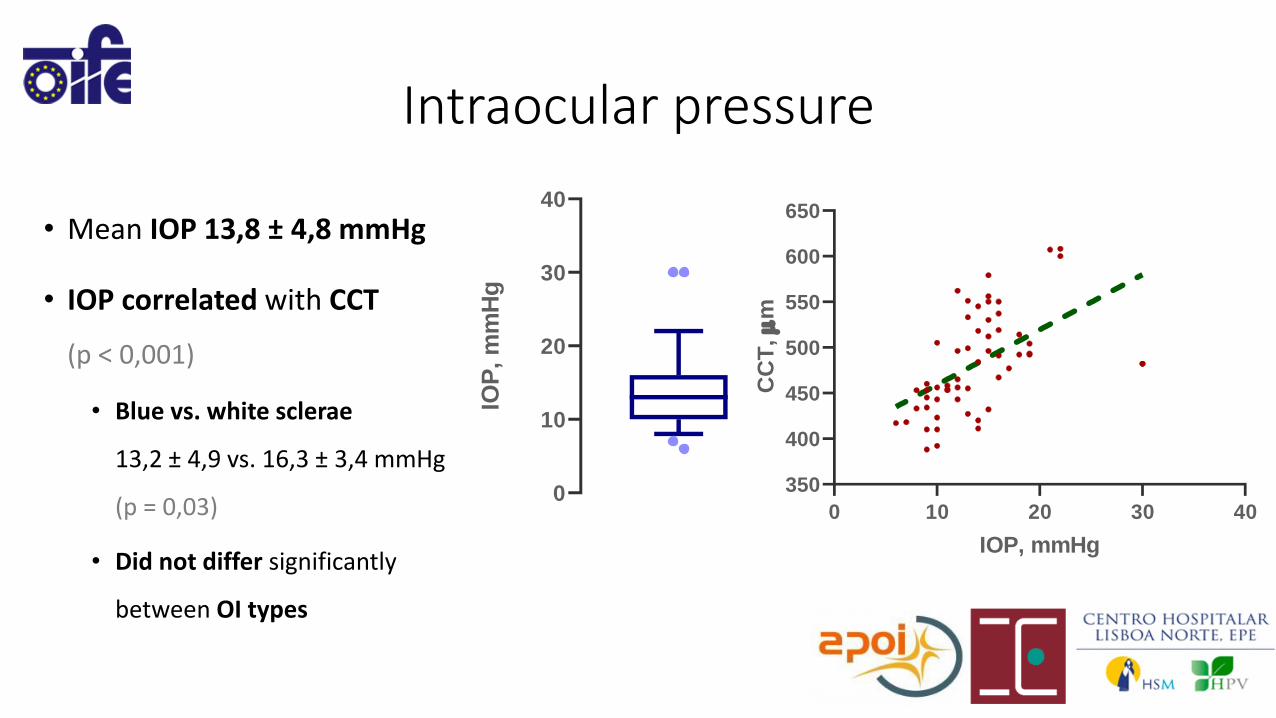
Intraocular pressure
• Mean IOP 13,8 ± 4,8 mmHg
• IOP correlated with CCT
(p < 0,001)
• Blue vs. white sclerae
13,2 ± 4,9 vs. 16,3 ± 3,4 mmHg
(p = 0,03)
• Did not differ significantly
between OI types
0 10 20 30 40
350
400
450
500
550
600
650
IOP, mmHg
CC
T,
m
0
10
20
30
40
IOP
, m
mH
g

Intraocular pressure
• Mean IOP 13,8 ± 4,8 mmHg
• IOP correlated with CCT
(p < 0,001)
• Blue vs. white sclerae
13,2 ± 4,9 vs. 16,3 ± 3,4 mmHg
(p = 0,03)
• Did not differ significantly
between OI types
I Non-I
Clinical type
Blu
e sc
lera
e
White
scl
erae
0
10
20
30
40
IOP
, m
mH
gI III IV VI VII
0
10
20
30
40
Clinical type

Corneal tomography
• Prevalence of corneal tomographic changes1,2: 77%
• Clinical bilateral keratoconus: 2 patients / Subclinical: 3 pts / Suspect corneas: 16 pts (10 bil.) bilateral)
• No statistical difference between type I and non-type I groups (p > 0,05)
1. Hashemi et. al 20162. Shetty et. al 2017
I III IV VI VII Non-I
0
10
20
30
40
Clinical type
N, eyes
Clinical keratoconus
Subclinical keratoconus
Suspect
Normal
Unk.

Corneal tomography


Glaucoma
• Only 1 patient has been diagnosed with glaucoma…
… But she has active Behçet’s disease-associated uveitis


Retinal changes
• No relevant segment changes were detected
… Other than chorioretinal atrophy associated with high grade myopia

Discussion
• The majority of patients were OI type I and COL1A1 was the most common causative gene
• Clinical validity of these findings is stronger in type I disease
• Type I and type III populations seem similar (IOP, CCT, blue sclerae)
I III IV VI VII
0
5
10
15
20
25
Clinical type
N Blue
White
I III IV VI VII
350
400
450
500
550
600
650
Clinical type
CC
T,
m
I III IV VI VII
0
10
20
30
40
Clinical type
IOP
, m
mH
g

Discussion
• BCVA was moderately reduced, and there was a high incidence of high-grade ametropia
• Do collagen anomalies affect the axial length of the eye?
• Reduced CCT is an hallmark of ocular disease in OI
• Correlates negatively with IOP and is more associated to type I disease and with blue sclerae
• What is its usefulness in diagnosis?1
• Is it a risk factor for the development of glaucoma?2
• The COL1A1 gene has been associated with several forms of glaucoma3
• How is the relationship between blue sclerae and CCT relevant?
• Is it dependent on the continuity of thinning of the sclerocorneal layer?
• Or is it best explained by the molecular changes particular to OI type I?
1. Hald et al. 20182. Congdon et. al. 20063. Mauri et. al. 2016

Discussion
• Diagnosis of glaucoma in OI patients presents a clinical challenge
• Lower IOPs and lower CCTs
• Do the collagen changes in the sclerocorneal layer affect the way cupping develops?
• Since we did not perform posterior segment OCT or SAP the epidemiology of glaucoma in
this population may be underestimated

Discussion
• There was a significantly high prevalence of tomographically abnormal corneas…
… However
• There was no significant difference between OI clinical types
• There was no significant difference in BCVA between abnormal and normal córneas
• Are current keratoconus tomographic screening indices1,2 applicable in this populations?
1. Hashemi et. al 20162. Shetty et. al 2017

THANK YOU FOR YOUR [email protected]