ocular and extra-ocular features of patients with leber ... and extra-ocular features of patients...
TRANSCRIPT

Ocular and extra-ocular features of patients with Leber congenitalamaurosis and mutations in CEP290
Suzanne Yzer,1 Anneke I. den Hollander,2 Irma Lopez,3 Jan-Willem R. Pott,4 Jan Tjeerd H.N. de Faber,1Frans P.M. Cremers,5,6 Robert K. Koenekoop,3 L. Ingeborgh van den Born1
1The Rotterdam Eye Hospital, Rotterdam, The Netherlands; 2Department of Ophthalmology, Radboud University Nijmegen MedicalCentre, Nijmegen, The Netherlands; 3McGill Ocular Genetics Laboratory, Montreal Children's Hospital Research Institute, McGillUniversity Health Centre, Montreal, QC, Canada; 4Department of Ophthalmology, University Medical Centre Groningen,Groningen, The Netherlands; 5Department of Human Genetics, Radboud University Nijmegen Medical Centre, Nijmegen, TheNetherlands; 6Nijmegen Centre for Molecular Life Sciences, Radboud University Nijmegen, Nijmegen, The Netherlands
Purpose: This study investigated the centrosomal protein, 290-KD (CEP290) associated genotype and ocular and extra-ocular phenotype in 18 patients with Leber congenital amaurosis (LCA).Methods: Eighteen patients with LCA from 14 families with mutations in the CEP290 gene were identified withsequencing or with heteroduplex analysis. Ophthalmic examinations were performed on all patients. Scans of the centralnervous system were reassessed in three patients and obtained in two. Renal function was evaluated in all patients.Ultrasonography of the kidneys was performed in six patients.Results: Eight patients (from five families) carried the c.2991+1655A>G mutation homozygously. Nine solitary patientscarried this variant combined with a nonsense, frameshift, or splice site mutation on the second allele. One new nonsensemutation was identified: c.1078C>T. Fourteen patients (from 12 families) had been completely blind from birth or hadlight perception. The best-recorded visual acuity was 20/200. Peripheral fundus changes appeared to be progressive witha relatively preserved posterior pole. Novel ophthalmic features for the CEP290 phenotype were Coats-like exudativevasculopathy in two patients, a small chorioretinal coloboma in one patient, and well defined, small, atrophic spots at thelevel of the retinal pigment epithelium causing a dot-like appearance in five patients. Some CEP290 patients exhibitedsystemic abnormalities. We found abnormal proprioception in two patients and mild mental retardation in one. One patientwas infertile due to immobile spermatozoa. No renal abnormalities were detected.Conclusions: CEP290-associated LCA has a severe, progressive, and clinically identifiable phenotype. Distinct extra-ocular findings were noted, which may be attributed to ciliary dysfunction.
Leber congenital amaurosis (LCA) represents a group ofclinically and genetically heterogeneous retinal dystrophiesdefined by severe visual dysfunction at birth, sensorynystagmus, a nondetectable electroretinogram (ERG), and anautosomal recessive inheritance pattern [1,2]. LCA has anestimated incidence of 1 in 30,000 newborns and a worldwideprevalence of approximately 200,000 patients [3,4].
Eighteen causative genes for LCA are known (see B-diseases), and mutations in these genes account forapproximately 70% of all patients with LCA [5,6]. To date,the most frequently mutated LCA gene is centrosomal protein,290-KD (CEP290) [7,8], which encodes a proteinubiquitously expressed in ciliated cells, including rods andcones [9]. In addition to LCA, mutations in CEP290 can alsolead to Joubert syndrome [10,11], Senior-Loken syndrome[10], the lethal disorder Meckel-Gruber syndrome [12,13],and Bardet-Biedl syndrome [14]. These diseases are termedciliopathies, which form a group of overlapping phenotypes
Correspondence to: Suzanne Yzer, The Rotterdam Eye Hospital,Rotterdam, The Netherlands, Phone 011-31-104017777, FAX:011-31-10 4111747; email: [email protected]
featuring developmental malformation of the brain, cystickidney disease or nephronophthisis, and retinal degeneration.
CEP290 mutations account for 6%–22% of non-syndromic LCA, depending on the population studied [5,7].The majority of patients with LCA with CEP290 mutationscarry the intronic mutation c.2991+1655A>G (p.Cys998X),which with lymphoblast RNA analysis was found to introducea cryptic exon into the CEP290 mRNA, creating a stop codonat the 5′ end of the insertion. However, only approximately50% of the mRNA contained the cryptic exon, rendering thisvariant a hypomorphic mutation, which subsequently mayinfluence the phenotypic outcome [7].
In the initial report on CEP290-associated LCA, wereported on intrafamilial variability with respect to visualacuity in four Canadian patients, and the absence of extra-ocular symptoms, except for seizures in two affected siblingsand abnormal proprioception in one patient [7]. Severalreports on the CEP290-associated phenotype have since beenpublished. Perrault et al. [8] described the phenotype as LCAtype 1 (cone-rod), characterized by some photo-aversion, highhyperopia, severely reduced visual acuity (<20/400), and asalt-and-pepper aspect of the fundus with macular
Molecular Vision 2012; 18:412-425 <http://www.molvis.org/molvis/v18/a44>Received 12 July 2011 | Accepted 7 February 2012 | Published 10 February 2012
© 2012 Molecular Vision
412

degeneration, resulting in a typical retinitis pigmentosa (RP)appearance by the second decade. Coppieters and coworkersalso observed the CEP290 LCA phenotype as one of a severecone-rod dystrophy with progressive peripheral funduschanges, but with a relatively spared macular region [15].Pasadhika et al. [16] studied the morphology of CEP290-related LCA with spectral domain–optical coherencetomography (SD-OCT). They showed preservation of theouter nuclear layer (ONL) with a poorly definedphotoreceptor inner/outer segment junction and with adistorted inner retina in the central macular area. The ocularphenotype was further expanded by Littink and colleagues,who reported on two patients with CEP290 mutations withrelatively well preserved visual acuity (VA) and withdetectable scotopic responses on ERG, leading the authors todiagnose these patients with early-onset severe retinaldystrophy rather than LCA [17].
Several groups studied associated extra-ocular signs inpatients with LCA with CEP290 mutations. Perrault et al.[8] observed transitory hypotonia, ataxia, mental retardation,and autistic behavior in a minority of their patients withsurprising intra-familiar discrepancies. McEwen andcoworkers [9] examined other ciliary-driven sensorydysfunctions, and discovered olfactory defects in retinaldegeneration 16 (rd16) mice, and subsequently in patientswith LCA with CEP290 mutations [18]. Coppieters et al.[15] reported on different degrees of kidney disease andneurologic involvement in patients carrying the sameCEP290 mutations. The researchers hypothesized that theneurologic phenotype was modified by the presence ofheterozygous Abelson Helper Integration sit 1 (AHI1)mutations. Papon and colleagues [19] showed ultrastructuraldefects in respiratory cilia in nasal biopsies and brushing onseven patients with LCA with CEP290 mutations.
The goal of this study was to further delineate the ocularphenotype of CEP290 LCA and to search for other extra-ocular symptoms in a cohort of Dutch and Canadian patients.In the current study of 18 patients, we were able to expand theophthalmic phenotype of CEP290-related LCA by Coats-likeexudative vasculopathy and chorioretinal coloboma as newophthalmic findings.
METHODSMutation analysis: DNA was extracted from peripheral bloodleukocytes using a salting-out procedure [20]. Patients weretested for the c.2991+1655A>G CEP290 mutation with allele-specific PCR [7] or with the newest version of the LCAmutation chip (Asper Ophthalmics, Tartu, Estonia). After oneheterozygous allele was identified, all 53 CEP290 codingexons were analyzed with bi-directional sequencing orheteroduplex analysis (ABI3730 and ABI3100 GeneticAnalyzers; Applied Biosystems, Inc. [ABI], Foster City, CA)to identify the second allele. Primer sequences and PCRconditions were described previously (Appendix 1) [13].
Briefly, PCR amplification consisted of a denaturizing step at95 °C for 5 min, followed by 35 cycles of amplification (95 °Cfor 30 s, X °C for 30 s, 72 °C for 45 s) and a final extensionat 72 °C for 5 min. In two unrelated affected siblings, no bloodsamples were drawn at request of the parents; however,samples were available for the probands of these families.Patients: Eighteen healthy patients were recruited at TheRotterdam Eye Hospital and the Montreal Children’sHospital. Written informed consent was obtained from thepatients with LCA or their legal representatives before bloodwas drawn. The procedures were approved by the EthicsCommittees of the Rotterdam Eye Hospital and McGillUniversity Health Centre and adhered to the Declaration ofHelsinki. Ten females and 8 males were included in this studyin an age range from 2 to 43 years. Eight patients of sixfamilies (Patients 1, 2, 3, 4, 5, 6, 12, and 14) were part of acohort of patients with LCA whose molecular data, but notclinical data, were described by den Hollander et al. [7]. Theother ten patients were suspected of carrying CEP290mutations based on their fundus appearance.
After mutations in CEP290 were identified, retrospectivedata were studied, and all 18 patients with LCA werereexamined. A medical history was taken and includedquestions about the age of onset of ocular and potential extra-ocular symptoms. The history also included questionsconcerning pregnancy, birth, post-natal period, development,consanguinity, and family history. Ophthalmologicexaminations included evaluation of the pupillary reactionand nystagmus, best-corrected visual acuity (BCVA), andobjective refractive error after cycloplegia. The anteriorsegments were examined with slit-lamp biomicroscopy, andfunduscopy was performed after pupillary dilation. In allpatients, fundus photography attempts were made. Opticalcoherence tomography (OCT) was obtained in four patients(OCT3 Stratus; Carl Zeiss Meditec, Inc., Dublin, CA, andSpectralis; Heidelberg Engineering, Heidelberg, Germany).ERGs were performed according to International Society forClinical Electrophysiology of Vision standards [21]. Renalfunction was studied with blood sample testing for potassium,sodium, creatinine levels, and creatinine clearance in allpatients. Renal ultrasounds were performed to evaluate kidneyarchitecture in six patients.
Signs of Joubert syndrome (e.g., lack of normaldecussation of superior cerebllar peduncular fiber tracts[molar tooth sign]) were specifically studied in five patients.In three patients, previously recorded images of the centralnervous system were reevaluated (magnetic resonanceimaging [MRI] for two and computed tomography [CT] forone), and in two other patients, MRIs were obtained.
RESULTSMutation analysis: The results of the molecular analyses aresummarized in Table 1. Fifteen patients were of Dutch descent
Molecular Vision 2012; 18:412-425 <http://www.molvis.org/molvis/v18/a44> © 2012 Molecular Vision
413

and three of French-Canadian descent. Three sibships wereincluded from three (Patients 1–3) and two affectedindividuals (Patients 8 and 9, 16, and 17). Eight patients (fromfive families) carried the c.2991+1655A>G mutationhomozygously. Nine patients were compound heterozygoteswith one c.2991+1655A>G mutation and a nonsense,frameshift or splice site mutation on the other allele. One newCEP290 nonsense mutations was identified: c.1078C>T(p.Arg360X; Patient 15). Patient 12 was from aconsanguineous marriage, but was compound heterozygous.Consanguinity was five generations ago, and segregationanalysis showed the mother was a heterozygous carrier of theR1272X mutation and the father of the c.2991+1655A>Gmutation. The genotype of Patients 1 (and his siblings 2 and3), 4, 5, 6, 12, and 14 and the co-segregation of four of thesefamilies were described by den Hollander et al. [7] as patients21365, 21393, 20152, 21918, 17971, and 12832, respectively.
Clinical findings: Clinical information is summarized inTable 2 except for the description of the retinal appearancewhich is separately summarized in Table 3.On medicalhistory, all patients had roving eye movements, or nystagmuswith sluggishly pupillary reactions since early childhood. Eyepoking was present in 17 patients and enophthalmos in sevenpatients (Patients 1, 2, 3, 5, 12, 13, and 17). Photophobia wasnot recorded as an early symptom in patients with vision. Twopatients (Patients 2 and 15) developed photophobia later inlife. Three patients (Patients 7, 14, and 15) had a clear historyof night blindness.
In seven patients, no light perception was recorded. Intwo, there was an indication of light perception in the past(Patients 4 and 6). Sixteen eyes of nine patients (Patients 1, 2,7, 8, 9, 13, 14, 15, and 18) perceived light only. In the other
patients, visual acuity varied from detecting hand motion (oneeye of Patient 13) to 20/200 (one eye of Patient 11).Deterioration of visual acuity was documented in threepatients over periods of 15 years or more (Patients 7, 11, and15). Mild to high hyperopia (ranging from +2.00 to +12.00diopters spherical equivalent) was a constant feature in themajority of the patients (11 patients). One patient showedmyopia (Patient 15), possibly due to her cataract. Six patients(Patients 6, 9, 10, 12, 16, and 18) had keratoconus. In five, thiswas a bilateral finding. In one patient (Patient 6), ophthalmicexamination was not possible due to severe ectatic corneaswith vascular in-growth and bullous changes (Figure 1A). Hislenses were luxated into the vitreous cavity (as seen onultrasonography). Posterior subcapsular cataracts wereobserved in two patients (Patients 14 and 15) and an anteriorsubcapsular cataract in one (Patient 7). Two patients werepseudophakic (Patient 8 and 9), and one aphakic (Patient 10).
Seventeen patients were available for funduscopy. Arelatively pink optic disc was observed in 13 patients (Patients1, 2, 3, 4, 8, 9, 10, 11, 12, 13, 16, 17, and 18; Figure 1B andFigure 2A). In six (Patients 1, 3, 11, 13, 16, and 18), a distinctyellowish scleral rim was observed (Figure 2A). Two patients(Patients 1 and 7) had pseudo-papillary edema withepipapillary fibrosis in one (Patient 7; Figure 3A). Anotherpatient (Patient 15) displayed a small chorioretinal colobomainferior to the optic disc unilaterally (Figure 4). Vessels weremildly to moderately attenuated in all but one patient whosevessels were dilated (Patient 13). One patient (Patient 7)showed vascular sheathing on the optic disc (Figure 3A).
All 17 patients had a preserved posterior pole. The maculaand fovea were recognizable in all patients but without normalreflexes. The right eye in Patient 18 was the only exception
TABLE 1. CEP290 MUTATIONS IN LEBER CONGENITAL AMAUROSIS PATIENTS.
Allele 1 Allele 2Patient number Origin Mutation Effect Mutation Effect
1$ Netherlands c.2991+1655A>G p.Cys998X c.2991+1655A>G p.Cys998X2$ Netherlands c.2991+1655A>G p.Cys998X c.2991+1655A>G p.Cys998X3$ Netherlands c.2991+1655A>G p.Cys998X c.2991+1655A>G p.Cys998X4 Netherlands c.2991+1655A>G p.Cys998X c.265dupA p.Thr89AsnfsX15 Netherlands c.2991+1655A>G p.Cys998X c.679_680delGA p.Glu227SerfsX16 Netherlands c.2991+1655A>G p.Cys998X c.180+1G>T splice defect7 Netherlands c.2991+1655A>G p.Cys998X c.5668G>T p.Gly1890X
8§ Canada c.2991+1655A>G p.Cys998X c.2991+1655A>G p.Cys998X9§ Canada c.2991+1655A>G p.Cys998X c.2991+1655A>G p.Cys998X10 Canada c.2991+1655A>G p.Cys998X c.2991+1655A>G p.Cys998X11 Netherlands c.2991+1655A>G p.Cys998X c.5587–1G>C splice defect12 Netherlands c.2991+1655A>G p.Cys998X c.3814C>T p.Arg1272X13 Netherlands c.2991+1655A>G p.Cys998X c.2991+1655A>G p.Cys998X14 Netherlands c.2991+1655A>G p.Cys998X c.2991+1655A>G p.Cys998X15 Netherlands c.5587–1G>C splice defect c.1078C>T p.Arg360X16¶ Netherlands c.2991+1655A>G p.Cys998X c.3175dup p.Ile1059fs17¶ Netherlands NT NT 18 Netherlands c.4661_4663del p.Glu1544del c.1645C>T p.Arg549X
*: Previously genotyped by den Hollander et al. [7]; $§¶: sibships; NT: not tested on request of parents.
Molecular Vision 2012; 18:412-425 <http://www.molvis.org/molvis/v18/a44> © 2012 Molecular Vision
414

TAB
LE 2
. CLI
NIC
AL
FIN
DIN
GS I
N L
EBER
CO
NG
ENIT
AL
AM
AU
RO
SIS P
ATI
ENTS
WIT
H C
EP29
0 M
UTA
TIO
NS.
Vis
ual a
cuity
Ref
ract
ion
Patie
nt (y
)Se
x
RE
LE
RE
LE
Ant
erio
r se
gmen
t and
mis
cella
neou
s*1
$ (1
9)M
LPLP
+9.2
5–1.
00x6
0+9
.00
−1.2
5x19
4iri
s tra
nslu
cenc
y, e
noph
thal
mos
*2$
(18)
FLP
LP+9
.25–
1.75
x72
+9.2
5–4.
50x1
09en
opht
halm
os*3
$ (1
3)M
NLP
NLP
+8.0
0–2.
75x6
7+8
.50–
3.50
x140
enop
htha
lmos
, nor
mal
fund
i at a
ge 1
y*4
(8)
FN
LPN
LPN
AN
Aab
norm
al p
ropr
ioce
ptio
n*5
(5)
FN
LPN
LP+1
2.00
–1.5
0x30
+12.
75–4
.50x
168
enop
htha
lmos
*6 (4
3)M
NLP
NLP
NA
NA
ecta
tic c
orne
as w
ith se
vere
ker
atoc
onus
, nor
mal
fund
i at a
ge (0
.5y)
, dro
pped
nuc
lei
(43y
), m
ildly
men
tally
reta
rded
, wal
ked
at a
ge 6
y, a
bnor
mal
pro
prio
cept
ion
7 (3
9)M
20/4
00LP
11.2
5+1
1.75
–1.0
0x15
0A
SC c
atar
act L
E, v
isua
l acu
ity 2
0/20
0 (8
y), i
mm
otile
sper
mat
ozoa
8§ (1
5)F
LPLP
pseu
doph
akia
pseu
doph
akia
pseu
doph
akic
, cat
arac
t ext
ract
ion
at a
ge 1
6y9§
(16)
MLP
LPps
eudo
phak
iaps
eudo
phak
iaps
eudo
phak
ic, k
erat
ocon
us B
E ca
tara
ct e
xtra
ctio
n at
age
10y
10 (2
5)F
CF
CF
apha
kia
apha
kia
kera
toco
nus R
E11
(34)
M20
/200
CF
+8.2
5–3.
50x1
65+7
.50–
4.25
x35
visu
al a
cuity
20/
100
(15y
), iri
doto
my
BE,
hyp
ofer
tility
*12
(9)
FN
LPN
LP+9
,75–
4.50
x37
+9.5
0–6.
25x1
45ke
rato
conu
s BE,
pig
men
t ant
erio
r len
s cap
sule
, eno
phth
alm
os13
(18)
FH
MLP
+8.5
0–2.
75x8
0+7
.50–
3.00
x100
enop
htha
lmos
, vitr
itis
*14
(45)
FLP
LP+3
.75–
0.75
x26
+3.5
0–1.
50x4
8PS
C c
atar
act B
E, a
ster
oid
hyal
osis
RE,
sist
er L
CA
: LP,
ker
atoc
onus
, dot
s, m
enta
lre
tard
atio
n, e
pile
psia
, psy
chia
tric
diso
rder
, nor
mal
CT
scan
15 (3
8)F
LPLP
−1.2
5–1.
75x1
78−3
–1.2
5x16
2PS
C c
atar
act R
E, v
isua
l acu
ity 2
0/40
0 (2
3y),
abno
rmal
EEG
, tem
pora
ry u
se o
f ant
i-ei
lept
ic d
rugs
, 2 si
bs w
ith S
aeth
re C
hotz
en sy
ndro
me
16¶
(4)
FN
LPN
LP+6
.25
+6.0
0ke
rato
conu
s BE
17¶
(2)
MN
LPN
LP+2
.00
+3.0
0en
opht
halm
os18
(17)
MLP
LPN
AN
Ake
rato
conu
s BE
*:
Pre
viou
sly
geno
type
d by
den
Hol
land
er e
t al.
[7];
$§¶:
sib
ship
s; A
SC: a
nter
ior s
ubca
psul
ar; B
E: b
oth
eyes
; CF:
cou
ntin
g fin
gers
; CT:
com
pute
d
tom
ogra
phy;
EEG
: ele
ctro
ence
phal
ogra
phy;
F:fe
mal
e; H
M: h
and
mot
ion;
LC
A: L
eber
con
geni
tal a
mau
rosi
s; L
E: le
ft ey
e; L
P: li
ght p
erce
ptio
n; M
: mal
e; N
A: n
ot
acce
ssib
le fo
r exa
min
atio
n; N
LP: n
o lig
ht p
erce
ptio
n; P
SC: p
oste
rior s
ubca
psul
ar; R
E: ri
ght e
ye; R
PE: r
etin
al p
igm
ent e
pith
eliu
m; S
E: sp
heric
al e
quiv
alen
t; y:
age
of
exa
min
atio
n in
yea
rs.
Molecular Vision 2012; 18:412-425 <http://www.molvis.org/molvis/v18/a44> © 2012 Molecular Vision
415

TAB
LE 3
. RET
INA
L FI
ND
ING
S IN
LEB
ER C
ON
GEN
ITA
L A
MA
UR
OSI
S PA
TIEN
TS W
ITH
CEP
290
MU
TATI
ON
S.Pa
tient
(y)
Rec
ent f
undu
scop
y
*1$
(19)
optic
dis
c: p
ink,
scle
ral r
im, p
seud
opap
illar
y ed
ema;
ves
sels
: mild
ly a
ttenu
ated
; pos
terio
r pol
e: p
rese
rved
RPE
, mild
wrin
klin
g IL
M, r
ecog
niza
ble
fove
a w
ithou
t ref
lexe
s; (m
id-)
perip
hery
: dot
-like
atro
phy
at R
PE le
vel,
subt
le R
PE c
hang
es, b
one-
spic
ules
*2$
(18)
optic
dis
c: p
ink;
ves
sels
: mod
erat
ely
atte
nuat
ed; p
oste
rior p
ole:
pre
serv
ed R
PE, r
ecog
niza
ble
fove
a, n
o re
flexe
s; (m
id-)
per
iphe
ry: d
ot-li
ke a
troph
y at
RPE
leve
l, so
me
bone
-sp
icul
es; f
ar p
erip
hery
: mor
e pr
onou
nced
RPE
atro
phy
*3$
(13)
optic
dis
c: p
ink,
scle
ral r
im; v
esse
ls: m
ildly
atte
nuat
ed; p
oste
rior p
ole:
rela
tivel
y hy
perp
igm
ente
d fo
vea,
no
refle
xes,
mild
wrin
klin
g IL
M; (
mid
-) p
erip
hery
: dot
-like
atro
phy
atR
PE le
vel,
subt
le R
PE c
hang
es, s
pora
dic
bone
-spi
cule
s*4
(8)
optic
dis
c: p
ink;
ves
sels
: mild
ly a
ttenu
ated
; pos
terio
r pol
e: re
lativ
ely
spar
ed R
PE, m
ild e
pire
tinal
mem
bran
e, re
cogn
izab
le fo
vea,
no
refle
xes;
(mid
-) p
erip
hery
: abn
orm
al re
flex
alon
g va
scul
ar a
rcad
e, su
btle
RPE
cha
nges
, gra
nula
r asp
ect
*5 (5
)op
tic d
isc:
mod
erat
ely
pale
; ves
sels
: mod
erat
ely
atte
nuat
ed; p
oste
rior p
ole:
pre
serv
ed R
PE, r
ecog
niza
ble
fove
a, n
o re
flexe
s; (m
id-)
per
iphe
ry: p
rese
rved
RPE
; far
per
iphe
ry:
rela
tivel
y hy
popi
gmen
ted,
gra
nula
r asp
ect
*6 (4
3)N
A7
(39)
optic
dis
c: p
ale,
pse
udo-
papi
llary
ede
ma,
vas
cula
r she
etin
g; v
esse
ls: m
oder
atel
y at
tenu
ated
; pos
terio
r pol
e: p
rese
rved
RPE
, sub
tle R
PE c
hang
es R
E, e
xuda
tive
deta
chm
ent L
E;(m
id-)
per
iphe
ry: p
rono
unce
d R
PE a
troph
y, b
one-
spic
ules
8§ (1
5)op
tic d
isc:
pin
k; v
esse
ls: v
ery
atte
nuat
ed; p
oste
rior p
ole:
nor
mal
; per
iphe
ry: g
ranu
lar a
spec
t9§
(16)
optic
dis
c: p
ink;
ves
sels
: atte
nuat
ed; p
oste
rior p
ole:
nor
mal
; per
iphe
ry: n
orm
al10
(25)
optic
dis
c: p
ink;
ves
sels
: atte
nuat
ed; p
oste
rior p
ole:
reco
gniz
able
fove
a, d
iffus
e hyp
opig
men
tatio
n; (m
id-)
per
iphe
ry: d
ot-li
ke at
roph
y at
RPE
leve
l; fa
r per
iphe
ry: p
igm
ent m
ottli
ng,
bone
-spi
cule
s11
(34)
optic
dis
c: p
ink,
scle
ral r
im; v
esse
ls: m
ildly
atte
nuat
ed; p
oste
rior p
ole:
pre
serv
ed R
PE m
acul
ar re
gion
, rec
ogni
zabl
e fo
vea,
no
refle
xes,
mild
epi
retin
al m
embr
ane
RE;
(mid
-)pe
riphe
ry: (
lobu
lar)
atro
phy
RPE
, som
e bo
ne-s
picu
les
*12
(9)
optic
dis
c: p
ink;
ves
sels
: mild
ly a
ttenu
ated
; pos
terio
r pol
e: p
rese
rved
RPE
, rel
ativ
ely
dark
, rec
ogni
zabl
e fo
vea,
no
refle
xes,
wrin
klin
g IL
M (B
E), y
ello
w d
epos
its te
mpo
ral t
om
acul
a R
E; (m
id-)
per
iphe
ry: r
elat
ivel
y no
rmal
13 (1
8)op
tic d
isc:
pin
k, sc
lera
l rim
; ves
sels
: tor
tuou
s and
dila
ted;
pos
terio
r pol
e: re
lativ
ely
norm
al, n
o re
flexe
s; (m
id-)
per
iphe
ry: R
PE a
troph
y, e
xuda
tive
retin
al d
etac
hmen
t inf
erio
rqu
adra
nts w
ith C
oat's
-like
vas
culo
path
y*1
4 (4
5)op
tic d
isc:
mod
erat
e pa
llor;
vess
els:
seve
rely
atte
nuat
ed; p
oste
rior p
ole:
pre
serv
ed R
PE, f
ovea
reco
gniz
able
, no
refle
xes;
(mid
-) p
erip
hery
: (lo
bula
r) R
PE a
troph
y, b
one-
spic
ules
15 (3
8)op
tic d
isc:
mild
pal
lor;
vess
els:
mod
erat
ely
atte
nuat
ed; p
oste
rior p
ole:
pre
serv
ed R
PE, s
ubtle
RPE
cha
nges
, ER
M; (
mid
-) p
erip
hery
: cho
riore
tinal
col
obom
a R
E, (l
obul
ar) a
troph
yR
PE, b
one-
spic
ules
, pav
ing-
ston
e de
gene
ratio
n 36
0 de
gree
s16
¶ (4
)op
tic d
isc:
pin
k, sc
lera
l rim
; ves
sels
: mild
ly a
ttenu
ated
; pos
terio
r pol
e: p
rese
rved
RPE
, mac
ula:
rela
tivel
y da
rk a
spec
t; (m
id-)
per
iphe
ry: t
apet
al re
flex,
subt
le h
ypo-
and
hype
rpig
men
ted
area
s17
¶ (2
)op
tic d
isc:
pin
k; v
esse
ls: m
ildly
atte
nuat
ed; p
oste
rior p
ole:
pre
serv
ed R
PE; (
mid
-) p
erip
hery
: tap
etal
refle
x, g
ranu
lar a
spec
t18
(17)
optic
dis
c: p
ink,
scle
ral r
im; v
esse
ls: m
oder
atel
y at
tenu
ated
; pos
terio
r pol
e: p
rese
rved
RPE
, sub
tle R
PE c
hang
es m
acul
a, p
rese
nt fo
veol
ar re
flex
RE,
abs
ent L
E; (m
id-)
per
iphe
ry:
RPE
atro
phy,
bon
e-sp
icul
es
*:
Pre
viou
sly
geno
type
d by
den
Hol
land
er e
t al.
[7].
$§¶:
sibs
hips
; ER
M: e
pire
tinal
mem
bran
e; IL
M: i
nter
nal l
imiti
ng m
embr
ane;
LE:
left
eye;
NA
: not
acc
essi
ble
fo
r exa
min
atio
n; R
E: ri
ght e
ye; R
PE: r
etin
al p
igm
ent e
pith
eliu
m; y
: age
of e
xam
inat
ion
in y
ears
.
Molecular Vision 2012; 18:412-425 <http://www.molvis.org/molvis/v18/a44> © 2012 Molecular Vision
416
because it showed a foveal reflex. The macular region wasrelatively darker pigmented compared to the surrounding RPEin three patients (Patients 3, 12, and 16) (Figure 2A). In fourpatients (Patients 7, 10, 15, and 18), the macular regiondisplayed subtle RPE changes. In six patients (Patients 1, 3,4, 11, 12, and 15), mild wrinkling of the inner limitingmembrane with or without epiretinal fibrosis was noted.
Two patients did not have peripheral RPE changes(Patient 9 aged 16 and Patient 12 aged 9). In five patients,including a sibship of three affected (Patients 1, 2, 3, 10, and
13), small atrophic spots at the level of the RPE that imposedas dot-like changes were noted in the periphery combined witha granular aspect of the RPE (Figure 2B). Three of thesepatients (Patients 2, 3, and 13) also displayed some intra-retinal pigmentations. A striking greyish white, patchy tapetalreflex was seen in the (mid-) periphery in three young patients(Patients 4, 16, and 17; Figure 5A,B). One (Patient 16) hadhypo- and hyper-pigmented areas in the far periphery whereasher younger brother (Patient 17) did not. In the remainingpatients (Patients 5, 7, 8, 11, 14, 15, and 18) ranging in age
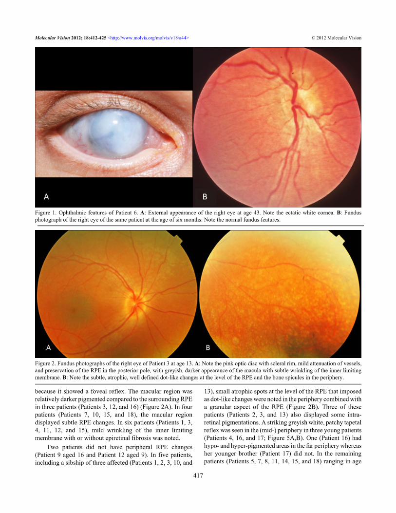
Figure 1. Ophthalmic features of Patient 6. A: External appearance of the right eye at age 43. Note the ectatic white cornea. B: Fundusphotograph of the right eye of the same patient at the age of six months. Note the normal fundus features.
Figure 2. Fundus photographs of the right eye of Patient 3 at age 13. A: Note the pink optic disc with scleral rim, mild attenuation of vessels,and preservation of the RPE in the posterior pole, with greyish, darker appearance of the macula with subtle wrinkling of the inner limitingmembrane. B: Note the subtle, atrophic, well defined dot-like changes at the level of the RPE and the bone spicules in the periphery.
Molecular Vision 2012; 18:412-425 <http://www.molvis.org/molvis/v18/a44> © 2012 Molecular Vision
417

from five to 39, RPE atrophy was more pronounced with bonespicule-like pigmentations.
Two patients (Patients 7 and 13) were treated for Coats-like exudative vasculopathy. Patient 7 presented with Coats-like exudative changes in the left eye at the age of 31 anddeveloped telangiectatic vessels in the inferior quadrants ofthe right eye at the age of 38 (Figure 3B). The other patient(Patient 13) returned a year after her last check-up because ofphotopsia and was found to have exudative detachments withtortuous, dilated vessels in the inferior quadrants of both eyesat the age of 18.
Goldmann perimetry was performed in two patients(Patients 7 and 11) and revealed central visual fields of lessthan 5° using the largest target (V-4e). ERG responses werenondetectable in all patients. OCT images revealed normal-
appearing retinal layers in Patient 12 (Figure 6), whereasthinned photoreceptor and outer nuclear layers were observedin two patients with deep foveal pits (Patients 7 and 14) andcyst-like changes in one of them (Patient 7; Figure 3C). Thesecysts were not noted on funduscopy. In another patient(Patient 11), the thickness of the retinal layers seemed normal,but foveal depression was lacking.
The motor and intellectual development in 16 patientswas reported and documented as normal when compared toother blind children. None of our patients had a hearing deficit,polydactyly, or obesity. Olfactory dysfunction was notreported. Two patients (Patients 4 and 6) had abnormalproprioception, and one had mild mental retardation. Noabnormalities suggestive of Joubert syndrome were presenton MRI, although minor anomalies were seen in one patient
Figure 3. Fundus photographs and OCT of Patient 7 at age 39. A: Note the pseudopapillary edema, vascular sheathing, preserved RPE in theposterior pole, subtle RPE changes in the macula, and the pronounced RPE and choriocapillary atrophy with bone spicule-like pigmentationsalong the vascular arcade in the right eye. B: Note the exudation in the posterior pole with shallow detachment in the left eye. C: Spectralisoptical coherence tomography (OCT) image of the macular region of the right eye showing cyst-like changes (*) in the outer nuclear layerwith preservation of the inner/outer segment junction under the fovea but absent in the parafoveal region. Note the thinning of the outer nuclearlayer at the center (arrow).
Molecular Vision 2012; 18:412-425 <http://www.molvis.org/molvis/v18/a44> © 2012 Molecular Vision
418

with normal intelligence (Patient 4). Neuro-imaging inanother four patients (Patients 1, 5, 7, and 12) did not revealany abnormalities of the central nervous system.Electroencephalography abnormalities were recorded in thepast in one patient (Patient 15) for which she receivedtreatment with antiepileptic drugs.
All patients had normal renal function on laboratorytesting. No signs of cystic kidney disease were detected onultrasonography in the patients (Patients 1, 4, 5, 6, 7, and 12)tested.
Three male patients were of reproductive age. Two didnot have offspring: one had mental retardation (Patient 6), andthe other one (Patient 7) was infertile due to immobilespermatozoa. Patient 11 had hypofertility but had conceivedoffspring naturally.
DISCUSSIONThe goal of the current study was to investigate ocular specificcharacteristics and extra-ocular features in patients withCEP290-related LCA.
In our cohort of 18 patients, we found one new nonsensemutation (Patient 15; c.1078C>T [p.Arg360X]) in CEP290.The most frequently encountered CEP290 mutation inpatients of European descent with LCA [7,8], the c.2991+1655A>G mutation, was detected in 22 of 34 alleles (of14 families) tested. Almost half of the patients carried ahomozygous c.2991+1655A>G mutation. One third of thepatients had this mutation combined with a nonsense,
frameshift, or splice site mutation on the other allele. In onlytwo patients, the founder mutation was not detected, but theocular phenotype of these patients was identical to the otherpatients.
Because of the relative large number of patients withLCA with CEP290 mutations in the present study spread overdifferent age groups, we were able to study different stages ofthe disease. Although there was some inter- and intrafamilialvariability, the patients showed a relatively homogenousdistinctive phenotype. We found frequently occurringsymptoms as in other forms of LCA, such as eye poking,enophthalmos, nystagmus, sluggish pupillary reflexes,hyperopia, keratoconus, and juvenile cataracts [22,23]. Visualacuity in the majority of the patients was either lightperception or no vision at all from birth. In the small group ofpatients with visual acuity equal to or more than hand motion,we were able to record progressive deterioration of visualacuity in succeeding years. Although we did not perform alongitudinal study, with retrospective data analysis (includingfundus color photographs), we established an increase indegenerative changes on funduscopy, from no changes tosmall, well defined, atrophic spots at the level of the RPE (dot-like) to more pronounced RPE atrophy with intraretinal bonespicule-like pigmentations and a preserved macular region asin RP. Based on VA loss and progressive retinal changes overtime, we conclude that LCA caused by CEP290 mutations isa progressive retinal dystrophy.
Figure 4. Fundus photograph of the righteye of Patient 15 at age 38. Note the mildpallor of the optic disc, the attenuationof the vessels, the chorioretinalcoloboma, the retinal pigmentepithelium atrophy (RPE) atrophy, andthe bone spicule-like pigmentations.
Molecular Vision 2012; 18:412-425 <http://www.molvis.org/molvis/v18/a44> © 2012 Molecular Vision
419

Perrault et al. [8] and Coppieters et al. [15] classifiedCEP290 associated LCA as a cone-rod retinal dystrophy.Whereas no ERG data were available in the first, the lattermentioned severe cone-rod disease on ERG testing in threeout of 27 patients with LCA and CEP290 mutations. Littinkand colleagues [17] observed two patients with CEP290mutations, relatively well preserved VA (up to 20/40), andonly detectable rod function and no cone function on ERG.Although we did not have recordable ERG responses in anyof the patients studied, our clinical data of progressiveperipheral retina changes and a relatively spared macula evenin late stages of the disease suggest that the patients in thiscohort had a progressive rod-cone type of disease rather thana cone-rod one.
We were able to get spectral domain–optical coherencetomography (SD-OCT) in four of our patients. Thinning ofthe ONL and the photoreceptor layer was found in two. Theother patients displayed normal retinal layers at the macula(Figure 6); however, the foveal pit was absent in one. OtherOCT studies in LCA CEP290 patients also showed thepresence of all retinal layers [17]. Pasadhika et al. [16] showedthat the photoreceptor inner/outer segment juncture waspoorly defined in the central macula whereas this junction wasinvisible in the periphery. A retained ONL was found in thecentral macular area of most patients that was more prominentat younger age. Inner retinal structures were distorted, andabout half of the studied patients had cystlike lesions in theinner retina, similar to one of our patients (Figure 3C).
Figure 5. Fundus photographs of the right eye (A) and left eye (B) of Patient 16 at age 4. The optic nerve is pink with mild attenuation of thevessels and a preserved macula. Note the grayish white, patchy, tapetal reflex in the midperiphery.
Figure 6. Spectralis OCT of the right eye of Patient 12 at age 9. Note the intact and normal thickness of all retinal layers.
Molecular Vision 2012; 18:412-425 <http://www.molvis.org/molvis/v18/a44> © 2012 Molecular Vision
420

Cideciyan et al. [24] performed a retinal micro-anatomic OCTstudy and revealed a relatively preserved foveal conephotoreceptor lamination and detectable subjacent RPE inpatients with LCA and CEP290 mutations, independent ofseverity of visual loss. The sparing of cones, however,decreased with aging, suggesting that CEP290 mutant centralretinas show slowly progressive degeneration of cones.Cideciyan et al. [24] also found that cone cell death seemedconsiderably slower than the loss of rods, suggesting differentroles of CEP290 in the two photoreceptor populations. Thesefindings all further support our impression that from amorphological perspective in fact this type of retinaldystrophy might be a rod-cone disease.
As for the retinal appearance, in three patients weobserved a striking tapetal reflex, consisting of intraretinalgreyish white marbled areas. In the younger siblings, thetapetal reflex was more pronounced than in the older patient,suggesting the possibility of a transient phenomenon, whichwas also documented by Littink et al. [17]. One of thesepatients carried the intron mutation homozygously whereasthe other was a compound heterozygote. Perrault et al. [8]depicted the same feature in one of their patients carrying thec.2991+1655A>G mutation homozygously.
The small atrophic spots at the RPE layer and the tapetalreflex–like changes seem specific to CEP290-associatedLCA, since they have not been described in other forms ofLCA. Another new observation was a distinct yellow scleralrim and pseudopapillary edema in a subset of patients. Ascleral ring is a relatively common finding in RP [25] and RP-related disorders but seems suggestive of this specific type ofLCA.
Two of our patients developed bilateral Coats-likeexudative vasculopathy, a rare complication in patients withRP (1.2%–3.6%) and frequently associated with juvenile RPcaused by Crumbs like 1 gene (CRB1) mutations [26].However, Coats-like exudative vasculopathy has never beendescribed in LCA. In a review paper, Kahn et al. [27] referredto a report on LCA and Coats-like disease. In retrospect, thisreport represents a family with early-onset RP. Our finding ofbilateral Coats reaction in two patients (both homozygous forthe c.2991+1655A>G mutation) suggests mutations inCEP290 might predispose for this type of complication.Senior-Loken syndrome, which can also be caused byCEP290 mutations [10], has also been shown to be associatedwith Coats-like disease [28].
Another new finding was the chorioretinal coloboma inone of our patients who did not carry the founder mutation.The occurrence of a developmental coloboma may becoincidental but was reported before in a Dutch patient withJoubert syndrome [29] for whom no molecular data areavailable.
Concerning the extra-ocular symptoms, one of our malepatients (Patient 7) was infertile due to immobile
spermatozoa, and one patient was hypofertile, presumably dueto asthenospermia. This finding was reported previously [19]in a compound heterozygous CEP290 LCA patient. It seemslogical to assume that CEP290 mutations may cause abnormalsperm tails and velocity. The observation that axonemes ofspermatozoa were abnormal in groups of patients with X-Linked RP [30] and Usher syndrome [31] was made more than20 years ago before the identification of ciliary genes, such asretinitis pigmentosa type 1(RP1), retinitis pigmentosa GTPaseregulator gene (RPGR), retinitis pigmentosa GTPaseregulator-interacting protein (RPGRIP1), and the Ushersyndrome genes [32]. Infertility in mice with overexpressionof RPGR has been reported recently [33]. Since other cilia-driven problems were proven, such as ultrastructuralrespiratory cilia defects and olfactory dysfunction in patientswith CEP290 mutations and heterozygote carriers [19],further investigation of sperm abnormalities in male patientsand male heterozygotes would be interesting.
Abnormalities of the central nervous system such as themolar tooth sign in Joubert syndrome were excluded in fiveof our patients including two patients with balance difficultiesone of whom had mild mental retardation. These two ataxicpatients had the c.2991+1655A>G mutation combined with asevere mutation (frame shift or a splice site) on the other allele.Perrault and coworkers also found mental retardation and/orautistic disorders in three CEP290-associated LCA familieswith homozygous or compound heterozygous frameshiftmutations. One family had the molar tooth sign, whereasneuro-imaging was not available for the other two families[8]. No abnormalities suggestive of renal disease weredetected in our cohort, and no renal cysts were found in thesix patients tested.
Altogether, in our study CEP290-LCA seemed to beassociated with a predominantly ocular phenotype. Although,we did not examine all our patients in depth for other possibleextra-ocular features such as anosmia and respiratorydysfunction, indicating aberrant ciliary functioning. Patientshomozygous for the hypomorphic c.2991+1655A>Gmutation might have more residual protein than patients withcompound heterozygous mutations, and therefore mightpresent with a less severe disease [7]. In this cohort of patients,however, visual acuity was best in patients who werecompound heterozygotes, and extra-ocular features occurredin homozygous (mental retardation) and in compoundheterozygous patients (ataxia, mental retardation, andinfertility). Therefore, other genetic variants may modify thephenotype as described by Leitsch et al. [14], who reportedon a Bardet-Biedl syndrome (BBS) patient with homozygousCEP290 mutations and one heterozygous mutation in anotherciliary BBS gene (Meckel syndrome type 3 [MKS3]).Recently, Coppieters and colleagues hypothesized theinfluence of heterozygous AHI1 mutations on the CEP290LCA phenotype. Khanna et al. [34] demonstrated that apolymorphic coding variant in retinitis pigmentosa GTPase
Molecular Vision 2012; 18:412-425 <http://www.molvis.org/molvis/v18/a44> © 2012 Molecular Vision
421

regulator-interacting protein-1 like (RPGRIP1L) influencesthe development of retinal degeneration in individuals withciliopathies caused by mutations in other genes. Therefore,the predictive value of the genotype seems more complicatedthan generally assumed and probably depends on the degreeof complex disruptions.
With the recent promising developments in human genereplacement therapy in LCA caused by RPE65 mutations[35-37], treatment options for LCA caused by CEP290mutations may become available in the future. Since this formof LCA occurs frequently, the accompanying phenotypeshould be recognized. We conclude that the LCA phenotyperesulting from CEP290 mutations in a Dutch and French-Canadian cohort is a severe, slowly progressive form of LCAwith a clinically identifiable ocular phenotype. The phenotypeconsists of a relative normal optic disc with scleral ring, mildor moderate attenuation of the vessels, a relatively preservedposterior pole, and some mild lobular RPE atrophy withoutobvious bone spicules in the periphery that progresses withaging. New clinical ocular findings were small, atrophic RPEspots, Coats-like exudative vasculopathy, and chorioretinalcoloboma. A tapetal reflex was observed as a possibletransient phenomenon. New interesting extra-ocular findingswere immotile spermatozoa and ataxia. Extra-ocular findingsin patients with LCA with CEP290 mutations are relativelyfrequent and mutation independent.
ACKNOWLEDGMENTSThe authors thank the patients for participating in this study.We acknowledge the help of Gerard de Graaf, Saskia van derVelde-Visser, and Christel Beumer for technical assistance.This study was supported by SWOO (StichtingWetenschappelijk Onderzoek Oogziekenhuis), theFoundation Fighting Blindness Canada (to R.K.K., A.I.dH.,and F.P.M.C.), Fonds de la Recherche en Santé du Québec(FRSQ award to R.K.K.), Toronto Financial Group (toR.K.K.), Canadian Institutes of Health Research (to R.K.K.),The Rotterdam Eye Hospital (to F.P.M.C., A.I.dH., L.I.vdB.),Foundation Fighting Blindness USA (to A.I.dH.), and theNetherlands Organisation for Scientific Research (to A.I.dH.).
REFERENCES1. Franceschetti A. L'importance diagnostique de
l'électrorétinogramme dans les dégénérescences tapéto-rétiniennes avec rétrécissement du champ visuel ethéméralopie. Conf Neurol 1954;14:184–6.
2. Leber T. Über Retinitis Pigmentosa und angeborene Amaurose.Graefes Arch Klin Exp Ophthalmol 1869; 15:1-25.
3. Stone EM. Leber congenital amaurosis - a model for efficientgenetic testing of heterogeneous disorders: LXIV EdwardJackson Memorial Lecture. Am J Ophthalmol 2007;144:791-811. [PMID: 17964524]
4. Koenekoop RK. An overview of Leber congenital amaurosis: amodel to understand human retinal development. SurvOphthalmol 2004; 49:379-98. [PMID: 15231395]
5. den Hollander AI, Roepman R, Koenekoop RK, Cremers FPM.Leber congenital amaurosis: genes, proteins and diseasemechanisms. Prog Retin Eye Res 2008; 27:391-419. [PMID:18632300]
6. Wang H, den Hollander AI, Moayedi Y, Abulimiti A, Li Y,Collin RW, Hoyng CB, Lopez I, Abboud EB, Al-Rajhi AA,Bray M, Lewis RA, Lupski JR, Mardon G, Koenekoop RK,Chen R. Mutations in SPATA7 cause Leber congenitalamaurosis and juvenile retinitis pigmentosa. Am J Hum Genet2009; 84:380-7. [PMID: 19268277]
7. den Hollander AI, Koenekoop RK, Yzer S, Lopez I, Arends ML,Voesenek KE, Zonneveld MN, Strom TM, Meitinger T,Brunner HG, Hoyng CB, van den Born LI, Rohrschneider K,Cremers FP. Mutations in the CEP290 (NPHP6) gene are afrequent cause of Leber congenital amaurosis. Am J HumGenet 2006; 79:556-61. [PMID: 16909394]
8. Perrault I, Delphin N, Hanein S, Gerber S, Dufier JL, Roche O,Defoort-Dhellemmes S, Dollfus H, Fazzi E, Munnich A,Kaplan J, Rozet JM. Spectrum of NPHP6/CEP290 mutationsin Leber congenital amaurosis and delineation of theassociated phenotype. Hum Mutat 2007; 28:416. [PMID:17345604]
9. Chang B, Khanna H, Hawes N, Jimeno D, He S, Lillo C,Parapuram SK, Cheng H, Scott A, Hurd RE, Sayer JA, OttoEA, Attanasio M, O'Toole JF, Jin G, Shou C, Hildebrandt F,Williams DS, Heckenlively JR, Swaroop A. In-frame deletionin a novel centrosomal/ciliary protein CEP290/NPHP6perturbs its interaction with RPGR and results in early-onsetretinal degeneration in the rd16 mouse. Hum Mol Genet 2006;15:1847-57. [PMID: 16632484]
10. Sayer JA, Otto EA, O'Toole JF, Nurnberg G, Kennedy MA,Becker C, Hennies HC, Helou J, Attanasio M, Fausett BV,Utsch B, Khanna H, Liu Y, Drummond I, Kawakami I,Kusakabe T, Tsuda M, Ma L, Lee H, Larson RG, Allen SJ,Wilkinson CJ, Nigg EA, Shou C, Lillo C, Williams DS,Hoppe B, Kemper MJ, Neuhaus T, Parisi MA, Glass IA, PetryM, Kispert A, Gloy J, Ganner A, Walz G, Zhu X, GoldmanD, Nurnberg P, Swaroop A, Leroux MR, Hildebrandt F. Thecentrosomal protein nephrocystin-6 is mutated in Joubertsyndrome and activates transcription factor ATF4. Nat Genet2006; 38:674-81. [PMID: 16682973]
11. Valente EM, Silhavy JL, Brancati F, Barrano G, KrishnaswamiSR, Castori M, Lancaster MA, Boltshauser E, Boccone L, Al-Gazali L, Fazzi E, Signorini S, Louie CM, Bellacchio E,International Joubert Syndrome Related Disorders StudyGroup. Bertini E, Dallapiccola B, Gleeson JG. Mutations inCEP290, which encodes a centrosomal protein, causepleiotropic forms of Joubert syndrome. Nat Genet 2006;38:623-5. [PMID: 16682970]
12. Baala L, Audollent S, Martinovic J, Ozilou C, Babron MC,Sivanandamoorthy S, Saunier S, Salomon R, Gonzales M,Rattenberry E, Esculpavit C, Toutain A, Moraine C, Parent P,Marcorelles P, Dauge MC, Roume J, Le Merrer M, MeinerV, Meir K, Menez F, Beaufrère AM, Francannet C, Tantau J,Sinico M, Dumez Y, MacDonald F, Munnich A, Lyonnet S,Gubler MC, Génin E, Johnson CA, Vekemans M, Encha-Razavi F, Attié-Bitach T. Pleiotropic effects of CEP290(NPHP6) mutations extend to Meckel syndrome. Am J HumGenet 2007; 81:170-9. [PMID: 17564974]
Molecular Vision 2012; 18:412-425 <http://www.molvis.org/molvis/v18/a44> © 2012 Molecular Vision
422

13. Frank V, den Hollander AI, Brüchle NO, Zonneveld MN,Nürnberg G, Becker C, Du Bois G, Kendziorra H, Roosing S,Senderek J, Nürnberg P, Cremers FP, Zerres K, Bergmann C.Mutations of the CEP290 gene encoding a centrosomalprotein cause Meckel-Gruber syndrome. Hum Mutat 2008;29:45-52. [PMID: 17705300]
14. Leitch CC, Zaghloul NA, Davis EE, Stoetzel C, Diaz-Font A,Rix S, Alfadhel M, Lewis RA, Eyaid W, Banin E, Dollfus H,Beales PL, Badano JL, Katsanis N. Hypomorphic mutationsin syndromic encephalocele genes are associated with Bardet-Biedl syndrome. Nat Genet 2008; 40:443-8. [PMID:18327255]
15. Coppieters F, Casteels I, Meire F, De Jaegere S, Hooghe S, vanRegemorter N, Van Esch H, Matuleviciene A, Nunes L,Meersschaut V, Walraedt S, Standaert L, Coucke P, HoebenH, Kroes HY, Vande Walle J, de Ravel T, Leroy BP, De BaereE. Genetic screening of LCA in Belgium: predominance ofCEP290 and identification of potential modifier alleles inAHI1 of CEP290-related phenotypes. Hum Mutat 2010;31:E1709-66. [PMID: 20683928]
16. Pasadhika S, Fishman GA, Stone EM, Lindeman M, Zelkha R,Lopez I, Koenekoop RK, Shahidi M. Differential MacularMorphology in Patients with RPE65-, CEP290-, GUCY2D-,and AIPL1-Related Leber Congenital Amaurosis. InvestOphthalmol Vis Sci 2010; 51:2608-14. [PMID: 19959640]
17. Littink KW, Pott JW, Collin RW, Kroes HY, Verheij JB,Blokland EA, de Castro Miró M, Hoyng CB, Klaver CC,Koenekoop RK, Rohrschneider K, Cremers FP, van den BornLI, den Hollander AI. A novel nonsense mutation in CEP290induces exon skipping and leads to a relatively mild retinalphenotype. Invest Ophthalmol Vis Sci 2010; 51:3646-52.[PMID: 20130272]
18. McEwen DP, Koenekoop RK, Khanna H, Jenkins PM, LopezI, Swaroop A, Martens JR. Hypomorphic CEP290/NPHP6mutations result in anosmia caused by the selective loss of Gproteins in cilia of olfactory sensory neurons. Proc Natl AcadSci USA 2007; 104:15917-22. [PMID: 17898177]
19. Papon JF, Perrault I, Coste A, Louis B, Gérard X, Hanein S,Fares-Taie L, Gerber S, Defoort-Dhellemmes S, Vojtek AM,Kaplan J, Rozet JM, Escudier E. Abnormal respiratory ciliain non-syndromic Leber congenital amaurosis with CEP290mutations. J Med Genet 2010; 47:829-34. [PMID: 20805370]
20. Miller SA, Dykes DD, Polesky HF. A simple salting outprocedure for extracting DNA from human nucleated cells.Nucleic Acids Res 1988; 16:1215. [PMID: 3344216]
21. Marmor MF, Holder GE, Seeliger MW, Yamamoto S. Standardfor clinical electroretinography (2004 update). DocOphthalmol 2004; 108:107-14. [PMID: 15455793]
22. Hanein S, Perrault I, Gerber S, Tanguy G, Barbet F, Ducroq D,Calvas P, Dollfus H, Hamel C, Lopponen T, Munier F, SantosL, Shalev S, Zafeiriou D, Dufier JL, Munnich A, Rozet JM,Kaplan J. Leber congenital amaurosis: comprehensive surveyof the genetic heterogeneity, refinement of the clinicaldefinition, and genotype-phenotype correlations as a strategyfor molecular diagnosis. Hum Mutat 2004; 23:306-17.[PMID: 15024725]
23. Yzer S, Leroy BP, De Baere E, de Ravel TJ, Zonneveld MN,Voesenek K, Kellner U, Ciriano JP, de Faber JT,Rohrschneider K, Roepman R, den Hollander AI, CruysbergJR, Meire F, Casteels I, van Moll-Ramirez NG, Allikmets R,
van den Born LI, Cremers FP. Microarray-based mutationdetection and phenotypic characterization of patients withLeber congenital amaurosis. Invest Ophthalmol Vis Sci 2006;47:1167-76. [PMID: 16505055]
24. Cideciyan AV, Aleman TS, Jacobson SG, Khanna H, SumarokaA, Aguirre GK, Schwartz SB, Windsor EA, He S, Chang B,Stone EM, Swaroop A. Centrosomal-ciliary gene CEP290/NPHP6 mutations result in blindness with unexpected sparingof photoreceptors and visual brain: implications for therapyof Leber congenital amaurosis. Hum Mutat 2007;28:1074-83. [PMID: 17554762]
25. Heckenlively JR. Retinitis Pigmentosa. Philadelphia: JBLippincott Company; 1988. p 86.
26. den Hollander AI, Heckenlively JR, van den Born LI, de KokYJ, van der Velde-Visser SD, Kellner U, Jurklies B, vanSchooneveld MJ, Blankenagel A, Rohrschneider K,Wissinger B, Cruysberg JR, Deutman AF, Brunner HG,Apfelstedt-Sylla E, Hoyng CB, Cremers FP. Leber congenitalamaurosis and retinitis pigmentosa with Coats-like exudativevasculopathy are associated with mutations in the crumbshomologue 1 (CRB1) gene. Am J Hum Genet 2001;69:198-203. [PMID: 11389483]
27. Khan JA, Ide CH, Strickland MP. Coats'-type retinitispigmentosa. Surv Ophthalmol 1988; 32:317-32. [PMID:2457260]
28. Schuman JS, Lieberman KV, Friedman AH, Berger M,Schoeneman MJ. Senior-Loken syndrome (familial renal-retinal dystrophy) and Coats' disease. Am J Ophthalmol 1985;100:822-7. [PMID: 4073180]
29. Lindhout D, Barth PG, Valk J, Boen-Tan TN. The Joubertsyndrome associated with bilateral chorioretinal coloboma.Eur J Pediatr 1980; 134:173-6. [PMID: 7439204]
30. Hunter DG, Fishman GA, Kretzer FL. Abnormal axonemes inX-linked retinitis pigmentosa. Arch Ophthalmol 1988;106:362-8. [PMID: 3345154]
31. Hunter DG, Fishman GA, Mehta RS, Kretzer FL. Abnormalsperm and photoreceptor axonemes in Usher's syndrome.Arch Ophthalmol 1986; 104:385-9. [PMID: 3954639]
32. Adams NA, Awadein A, Toma HS. The retinal ciliopathies.Ophthalmic Genet 2007; 28:113-25. [PMID: 17896309]
33. Brunner S, Colman D, Travis AJ, Luhmann UF, Shi W, Feil S,Imsand C, Nelson J, Grimm C, Rülicke T, Fundele R,Neidhardt J, Berger W. Overexpression of RPGR leads tomale infertility in mice due to defects in flagellar assembly.Biol Reprod 2008; 79:608-17. [PMID: 18579752]
34. Khanna H, Davis EE, Murga-Zamalloa CA, Estrada-CuzcanoA, Lopez I, den Hollander AI, Zonneveld MN, Othman MI,Waseem N, Chakarova CF, Maubaret C, Diaz-Font A,MacDonald I, Muzny DM, Wheeler DA, Morgan M, LewisLR, Logan CV, Tan PL, Beer MA, Inglehearn CF, Lewis RA,Jacobson SG, Bergmann C, Beales PL, Attié-Bitach T,Johnson CA, Otto EA, Bhattacharya SS, Hildebrandt F, GibbsRA, Koenekoop RK, Swaroop A, Katsanis N. A commonallele in RPGRIP1L is a modifier of retinal degeneration inciliopathies. Nat Genet 2009; 41:739-45. [PMID: 19430481]
35. Bainbridge JW, Smith AJ, Barker SS, Robbie S, Henderson R,Balaggan K, Viswanathan A, Holder GE, Stockman A, TylerN, Petersen-Jones S, Bhattacharya SS, Thrasher AJ, FitzkeFW, Carter BJ, Rubin GS, Moore AT, Ali RR. Effect of gene
Molecular Vision 2012; 18:412-425 <http://www.molvis.org/molvis/v18/a44> © 2012 Molecular Vision
423

therapy on visual function in Leber's congenital amaurosis. NEngl J Med 2008; 358:2231-9. [PMID: 18441371]
36. Maguire AM, Simonelli F, Pierce EA, Pugh EN Jr, Mingozzi F,Bennicelli J, Banfi S, Marshall KA, Testa F, Surace EM, RossiS, Lyubarsky A, Arruda VR, Konkle B, Stone E, Sun J, JacobsJ, Dell'Osso L, Hertle R, Ma JX, Redmond TM, Zhu X, HauckB, Zelenaia O, Shindler KS, Maguire MG, Wright JF, VolpeNJ, McDonnell JW, Auricchio A, High KA, Bennett J. Safetyand efficacy of gene transfer for Leber's congenital amaurosis.N Engl J Med 2008; 358:2240-8. [PMID: 18441370]
37. Hauswirth WW, Aleman TS, Kaushal S, Cideciyan AV,Schwartz SB, Wang L, Conlon TJ, Boye SL, Flotte TR, ByrneBJ, Jacobson SG. Treatment of leber congenital amaurosisdue to RPE65 mutations by ocular subretinal injection ofadeno-associated virus gene vector: short-term results of aphase I trial. Hum Gene Ther 2008; 19:979-90. [PMID:18774912]
Molecular Vision 2012; 18:412-425 <http://www.molvis.org/molvis/v18/a44> © 2012 Molecular Vision
424

Appendix 1. Sequences and PCR conditions of CEP290.
Abbreviations: °C represents degrees celcius; bprepresents base pairs; F represents forward; MgCl2 representsmagnesium; chloride; R represents reverse. To access the
data, click or select the words “Appendix 1.” This will initiatethe download of a compressed (pdf) archive that contains thefile.
Molecular Vision 2012; 18:412-425 <http://www.molvis.org/molvis/v18/a44> © 2012 Molecular Vision
Articles are provided courtesy of Emory University and the Zhongshan Ophthalmic Center, Sun Yat-sen University, P.R. China.The print version of this article was created on 7 February 2012. This reflects all typographical corrections and errata to thearticle through that date. Details of any changes may be found in the online version of the article.
425