ob/gyn risk management case studies - acoog · ob/gyn risk management case studies jay goldberg,...
TRANSCRIPT

OB/GYN Risk Management
Case Studies
Jay Goldberg, MD, MSCPEinstein Medical Center Philadelphia


Physicians Insurance Association of America
malpractice claims against OB/GYNs
62%
42%
29%
16%
0%
10%
20%
30%
40%
50%
60%
70%
1 Claim 2 Claims 3 Claims 4 Claims
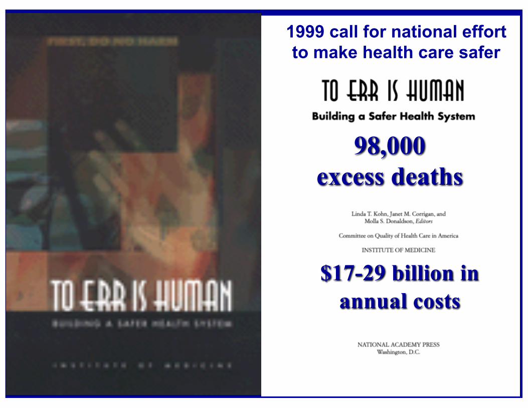
98,000excess deaths
$17-29 billion in annual costs
1999 call for national effort to make health care safer


IOM’s 1999 “To Err is Human” was followed by an increase in patient safety publications and awareness
Stelfox Qual Saf Health Care 2006

Reducing OB litigation through alterations in practice patterns
Clark Obstet Gynecol 2008
• 189 closed perinatal claims reviewed x 3• 70% of claims involved substandard care• Payments avoidable in 85% EFM, 80%
VBAC, 54% shoulder dystocia (SD) cases• Recommendations: 24-hour in-house
OB, adherence to medication protocols (Pitocin), conservative approach to VBAC, standardized procedure note for SD

Definitions
• Medical malpractice– Injury caused by a deviation of the standard of
care• Standard of Care
– Reasonable care provided by a reasonable provider
• Mal-occurrence v Malpractice• Two schools of thought

Why do patients file suit?
• Injury• Malpractice• Perceived malpractice
– Poor judgment– Poor documentation– Anger
• Lottery ticket

Keys to optimize litigation outcomes
• Practicing good medicine– Within the standard of care (if not, have to
guarantee a good outcome)• Informed consent• Good documentation
– Standardized forms• Good legal counsel• Good defense experts

Litigation Stepsmay vary from state to state
• Complaint• Certificate of Merit (injury, theory)• Depositions (fact and expert) preparation• Expert reports• Settlement negotiation• Trial
– Appearance to jury of defendant, plaintiff, attorneys, experts: Honesty & likeability
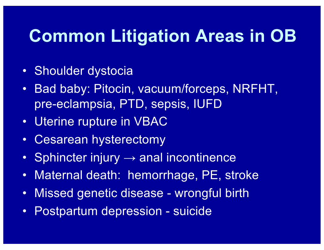
Common Litigation Areas in OB
• Shoulder dystocia• Bad baby: Pitocin, vacuum/forceps, NRFHT,
pre-eclampsia, PTD, sepsis, IUFD• Uterine rupture in VBAC• Cesarean hysterectomy• Sphincter injury → anal incontinence• Maternal death: hemorrhage, PE, stroke• Missed genetic disease - wrongful birth• Postpartum depression - suicide
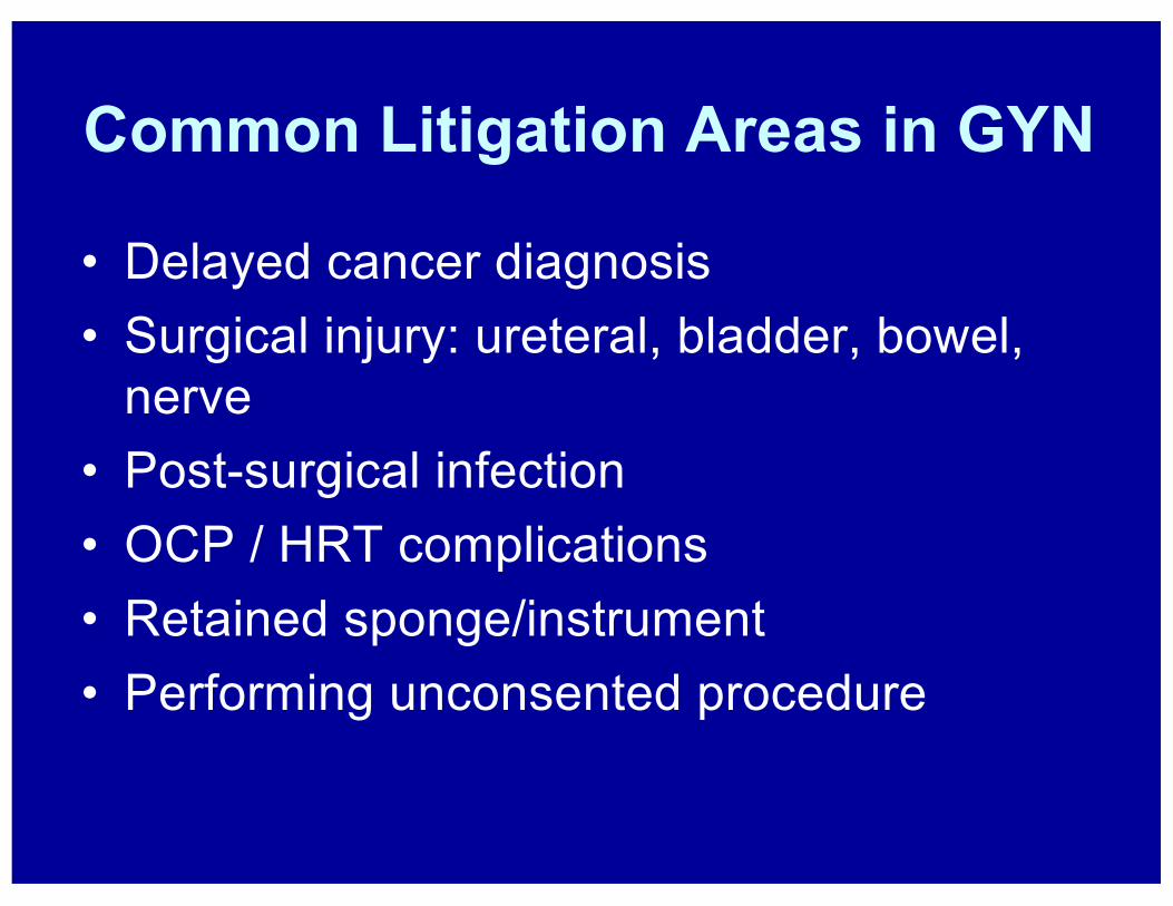
Common Litigation Areas in GYN
• Delayed cancer diagnosis• Surgical injury: ureteral, bladder, bowel,
nerve• Post-surgical infection• OCP / HRT complications• Retained sponge/instrument• Performing unconsented procedure

Case Studies
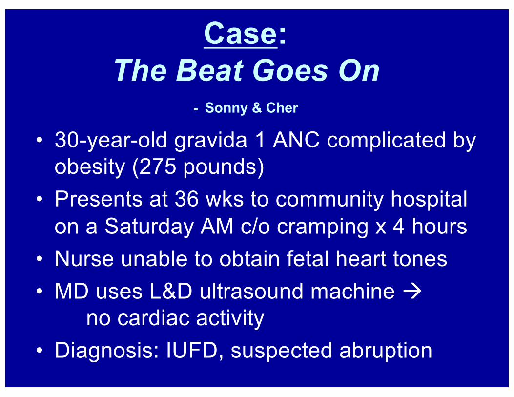
Case:The Beat Goes On
- Sonny & Cher
• 30-year-old gravida 1 ANC complicated by obesity (275 pounds)
• Presents at 36 wks to community hospital on a Saturday AM c/o cramping x 4 hours
• Nurse unable to obtain fetal heart tones• MD uses L&D ultrasound machine à
no cardiac activity• Diagnosis: IUFD, suspected abruption
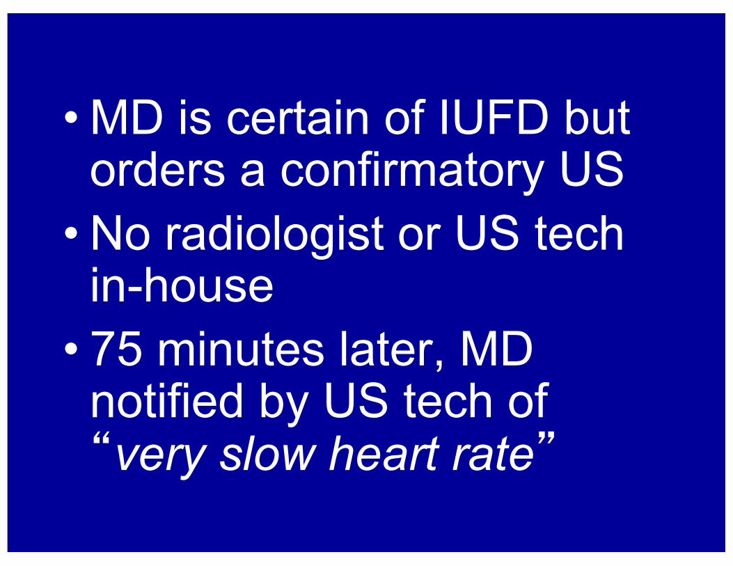
• MD is certain of IUFD but orders a confirmatory US
• No radiologist or US tech in-house
• 75 minutes later, MD notified by US tech of “very slow heart rate”

• Stat cesarean delivery• Complete placental
abruption• 2,477 gram female with
Apgars 0/6• Umbilical cord pH = 6.75

• Hypoxic ischemic encephalopathy (HIE)
• Child requires 24 hr care

• Plaintiff’s Allegations: –L&D US machine inferior–MD missed cardiac activity
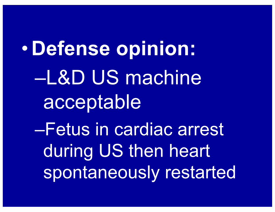
• Defense opinion: –L&D US machine acceptable
–Fetus in cardiac arrest during US then heart spontaneously restarted

Settle or go to Trial ?Plaintiff settlement
demand of $50 million
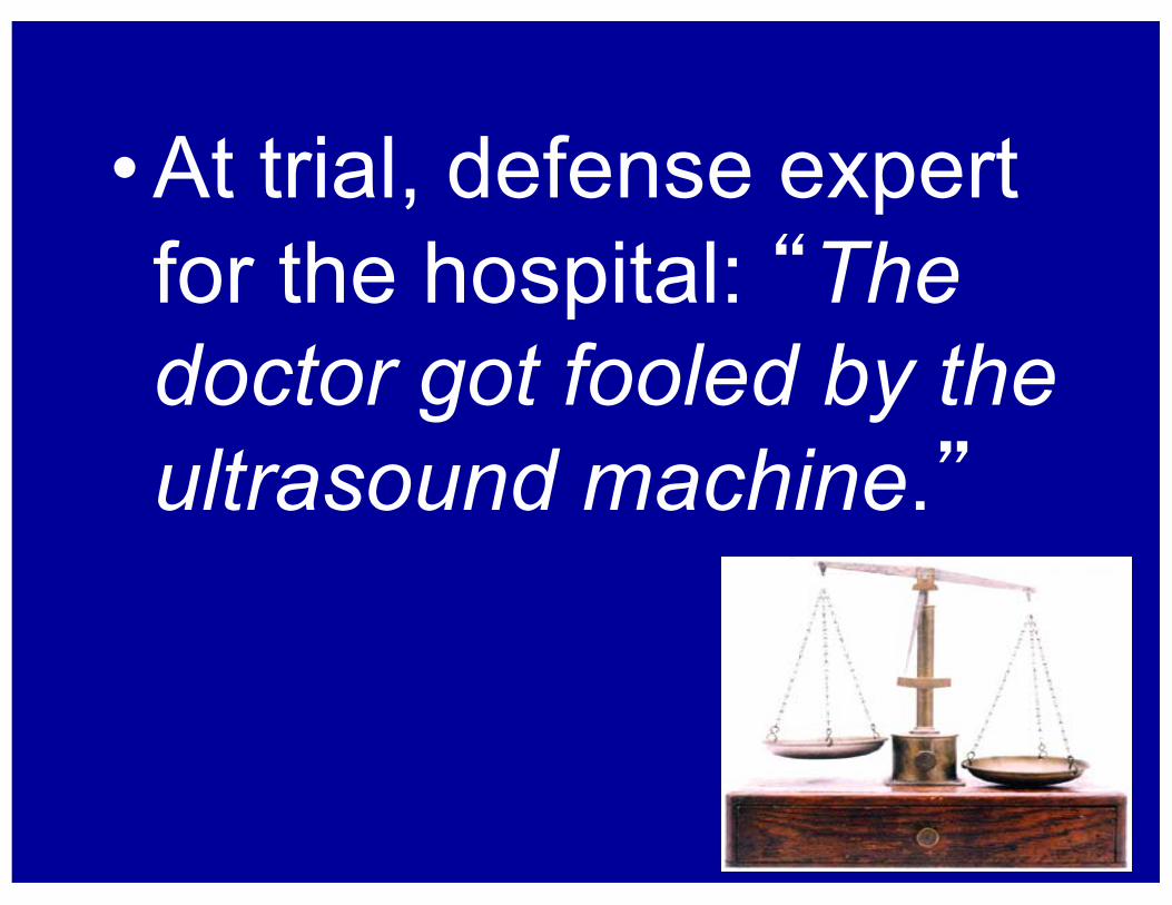
• At trial, defense expert for the hospital: “The doctor got fooled by the ultrasound machine.”

• Trial: Plaintiff verdict
Hospital: $78 millionMD: not negligent

Medico-legal Risk of an Undiagnosed Ureter Injury

Urinary Tract Injuries in Hysterectomy
• Complicate4.3%ofhysterectomies.(Ibeanu Obstet Gynecol 2009)- Bladderinjury2.8%- Ureteralinjury1.8%
• 75%ofinjurieswerenotsuspectedduringthehysterectomy.(Ibeanu 2009)
• Promptintra-operativediagnosisandtreatmentpreventstheriskofadditionalsurgery,urinoma,fistula,renalfailure,andlawsuits.
• Cystoscopyafterhysterectomymaydetectureteralandbladderinjuries.

Resident experience with and post-training plans for cystoscopy at the time
of hysterectomy
DinaVaynberg,MD,ChaseWhite,MD,DavidJaspan,DO,JayGoldberg,MD,MSCP
Women’sHealth2015

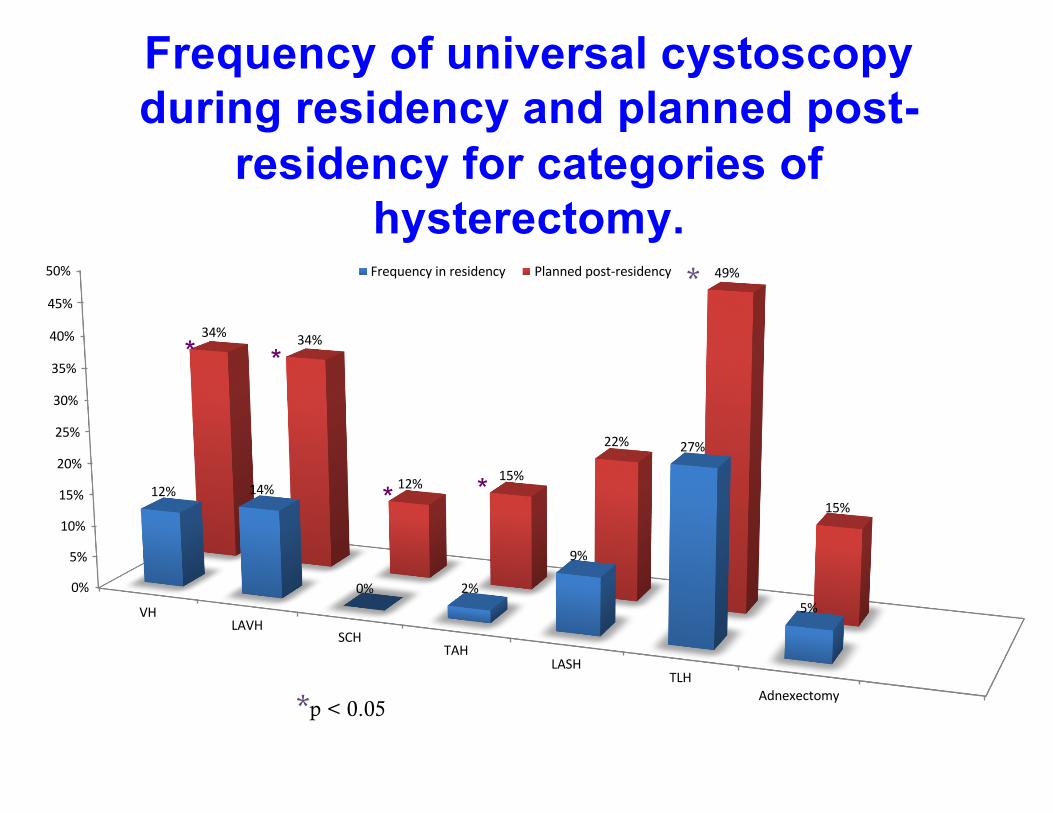
Frequency of universal cystoscopy during residency and planned post-residency for categories of hysterectomy.
TypeofHysterectomy
Universal CystoscopyResidency
Universal CystoscopyPost-residency p
VH12% 34% 0.005
LAVH 14% 34% 0.009
SCH 0% 12% 0.009
TAH 2% 15% 0.008
LASH 9% 22% 0.08
TLH 27% 49% 0.014
Adnexectomy 5% 15% 0.088
Frequency of universal cystoscopy during residency and planned post-
residency for categories of hysterectomy.
0%
5%
10%
15%
20%
25%
30%
35%
40%
45%
50%
VHLAVH
SCHTAH
LASHTLH
Adnexectomy
12% 14%
0% 2%
9%
27%
5%
34% 34%
12% 15%
22%
49%
15%
Frequencyinresidency Plannedpost-residency *
*p < 0.05
* *
* *

Case of an Undetected Ureteral Injury
• 43-year-oldG0 withchronicpelvicpain,endometriosis
• Normalsizeduterus• SomeimprovementwithLupron
• ConsentedforTAH/BSO

Intraoperative Course
•TAH/BSOperformed.•Nointra-operativeinjurysuspected.
• Identificationofuretersnotmentionedinoperativenote.
•Nocystoscopyperformed.•Path→benigndisease.

Postoperative Course• POD #1: decreased urine output and creatinine
elevated to 1.2. Urine output improved with IVF.
• POD #5: developed right flank pain
CT urogram à RIGHT URETERAL OBSTRUCTION
• POD #6: cystoscopy, unsuccessful stenting. Right percutaneous nephrostomy.
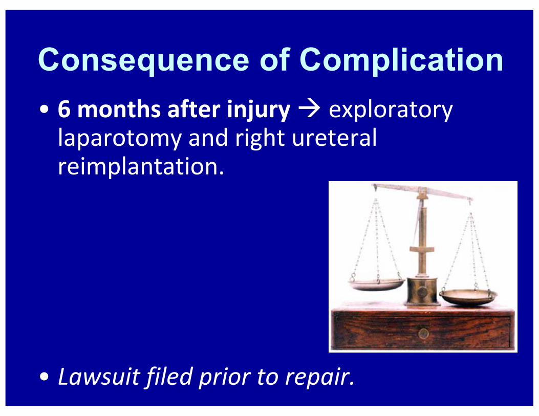
Consequence of Complication• 6monthsafterinjuryà exploratorylaparotomyandrightureteralreimplantation.
• Lawsuitfiledpriortorepair.

Expert Opinions:
• Plaintiff’sAllegations: “Itwasadeviationfromthestandardofcaretonothaverecognizedtheureterinjuryduringtheoriginalsurgery.”
• Defense: Thesurgerywasperformedappropriately.Knownandunavoidablecomplication.Noadditionalevaluationwasrequired.

Settle or go to Trial ?

•Trial: Defense Verdict

Case 3:Cervical Cancer
• 35 year old Gravida 0 accountant• Referred to OB/GYN by PCP for an ASCUS pap
(1st abnormal pap)• Repap à ASCUS, favor dysplasia (#2)• Repap à ASCUS (#3)• Repap à ASCUS (#4)• Repap à AGCUS (#5)• Repap à HSIL, AGCUS (#6) à Colposcopy

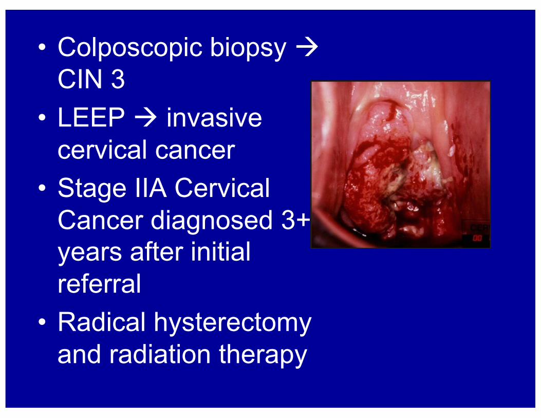
• Colposcopic biopsy àCIN 3
• LEEP à invasive cervical cancer
• Stage IIA Cervical Cancer diagnosed 3+ years after initial referral
• Radical hysterectomy and radiation therapy

• Plaintiff: Colposcopy should have occurred after 2nd abnormal pap and after all subsequent abnormal paps (4 missed opportunities for earlier diagnosis)
• Defense: Only repeating pap smears was acceptable as long as they did not worsen

Legal Issues• Regional variations in
acceptable triage protocols for abnormal cervical cytology?
• Conservative location of trial

Settle or go to Trial ?


•Trial: Defense Verdict

Case 4:Breast Cancer
• 35-year-old attorney with a family history of breast cancer. History of “fibrocystic breasts”
• Year 1: Dr. X documents “normal breast exam” on annual exam. No complaints
• Year 2: Dr. X documents “increased nodularity R > L”. No complaints
• Year 3: Dr. X documents “bilateral nodularity R > L”. No complaints

• Year 4: switches to Dr. Y for annual exam. No complaints. Dr. Y palpates a firm, mobile right breast mass
• Breast ultrasound and MXR à multiple calcifications highly suspicious for malignancy
• Core biopsies of the right breast àinvasive poorly differentiated ductal carcinoma
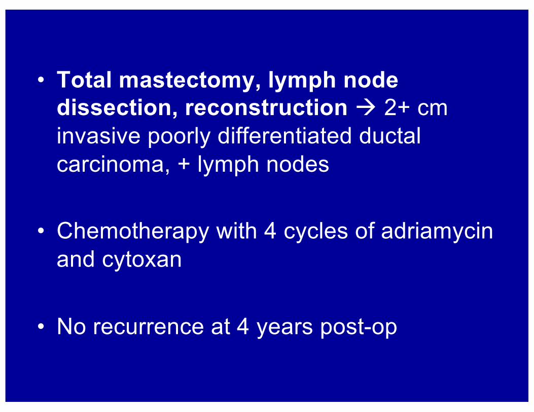
• Total mastectomy, lymph node dissection, reconstruction à 2+ cm invasive poorly differentiated ductal carcinoma, + lymph nodes
• Chemotherapy with 4 cycles of adriamycinand cytoxan
• No recurrence at 4 years post-op

Deposition Testimony
• Plaintiff stated that she did complain of breast pain and a breast mass to Dr. X
• Dr. X denied that she had complaints. He believed her breast exams were consistent with benign fibrocystic changes

• Plaintiff’s Allegation: Dr. X was negligent by not ordering breast imaging and referring to a breast surgeon when he palpated increased nodularity. This delayed diagnosis.
• Defense: Breast imaging and referral were not indicated in asymptomatic patient with known fibrocystic breasts

Legal Issue• Dr. X’s documentation of “increased nodularity R > L”and “bilateral nodularity R > L” when he had previously documented “normal breast exam.”

Settle or go to Trial ?

• Settled–$250K against the MD

Screening MammogramGuidelines

American Cancer Society2016 Guidelines
– Regular screening mammography starting at age 45 years (strong recommendation), however women should have the opportunity to begin annual screening between the ages of 40 and 44 years
– Age 45 to 54 years should be screened annually – Age 55 years and older should transition to biennial screening
or have the opportunity to continue screening annually– Women should continue screening mammography as long as
their overall health is good and they have a life expectancy of 10 years or longer
– Do not recommend clinical breast examination for breast cancer screening among average-risk women at any age
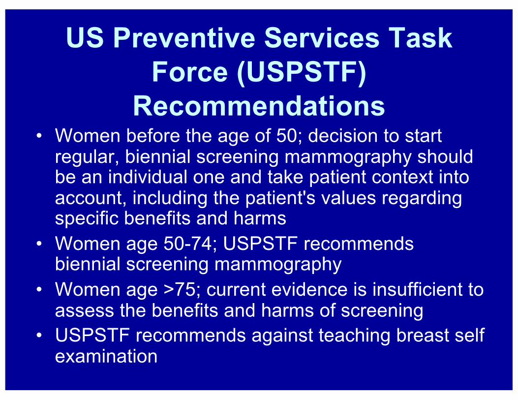
US Preventive Services Task Force (USPSTF)
Recommendations• Women before the age of 50; decision to start
regular, biennial screening mammography should be an individual one and take patient context into account, including the patient's values regarding specific benefits and harms
• Women age 50-74; USPSTF recommends biennial screening mammography
• Women age >75; current evidence is insufficient to assess the benefits and harms of screening
• USPSTF recommends against teaching breast self examination

American College of Radiology(ACR) Recommendations
• Recommend yearly mammograms starting at age 40
• Screening to continue until life expectancy is less than five to seven years, on the basis of age and co-morbidities

ACOG Guidelines • Recommend beginning annual
mammography at age 40 years • Women aged 29–39 years should
have a clinical breast exam every 1–3 years. Women aged 40 years and older should have one every year

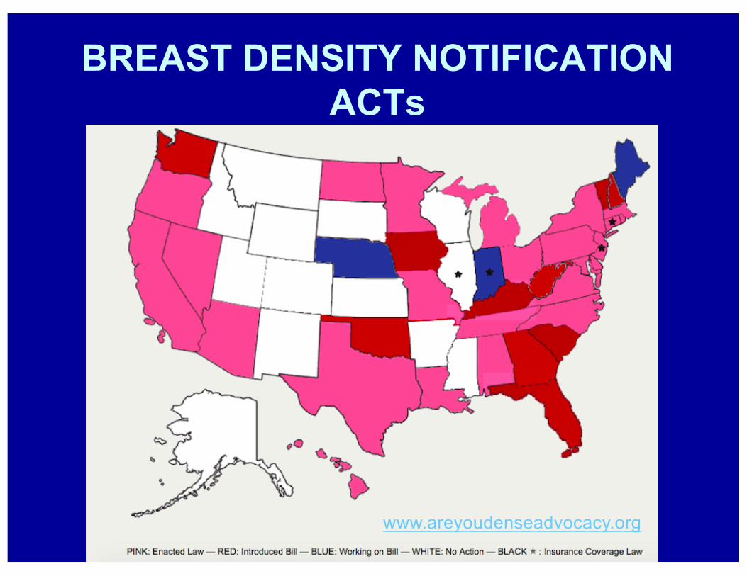
BREAST DENSITY NOTIFICATION ACTs
www.areyoudenseadvocacy.org
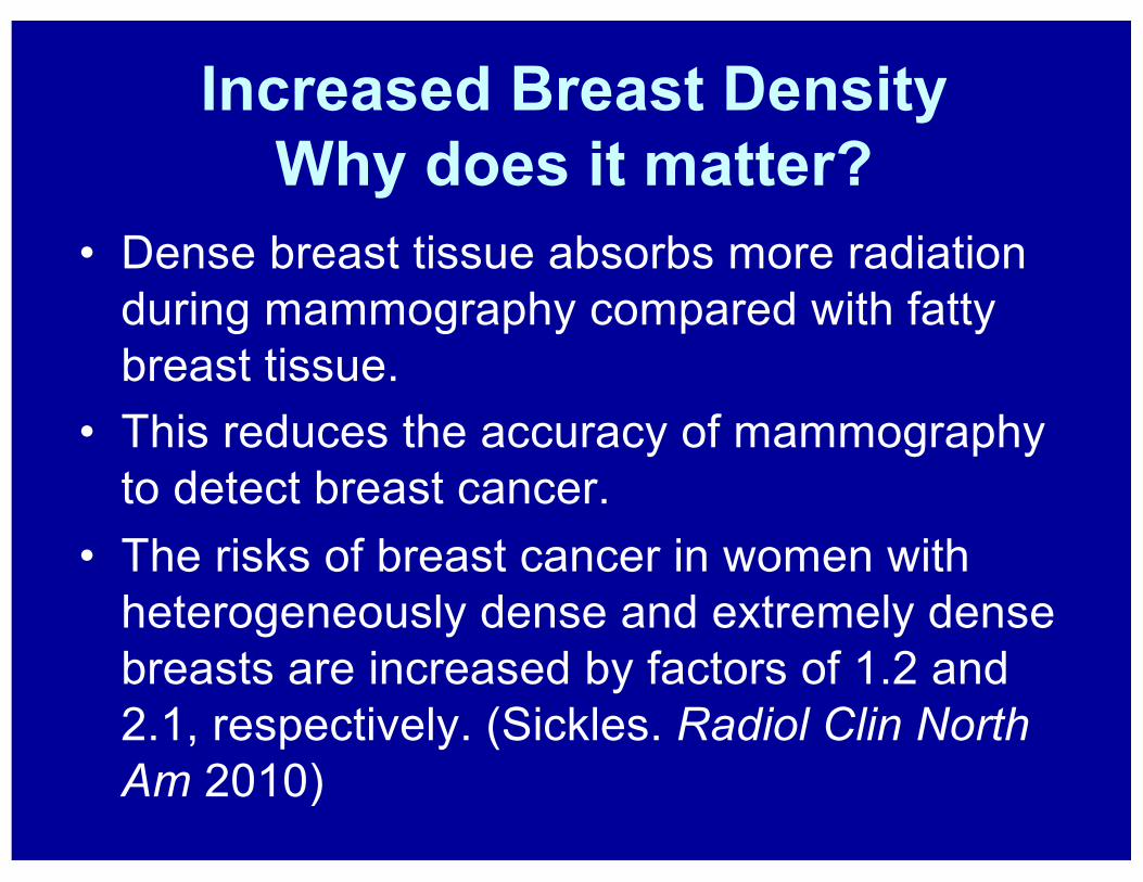
Increased Breast DensityWhy does it matter?
• Dense breast tissue absorbs more radiation during mammography compared with fatty breast tissue.
• This reduces the accuracy of mammography to detect breast cancer.
• The risks of breast cancer in women with heterogeneously dense and extremely dense breasts are increased by factors of 1.2 and 2.1, respectively. (Sickles. Radiol Clin North Am 2010)

BREAST DENSITY NOTIFICATION ACTs
www.areyoudenseadvocacy.org
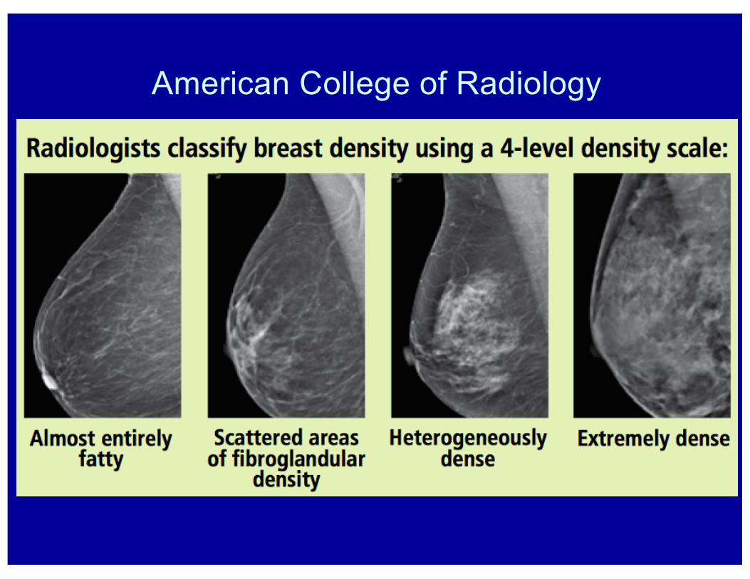
American College of Radiology

American College of Radiology
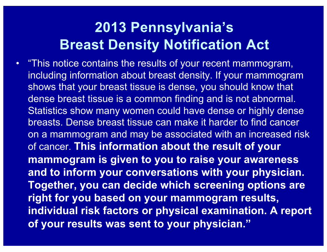
2013 Pennsylvania’sBreast Density Notification Act
• “This notice contains the results of your recent mammogram, including information about breast density. If your mammogram shows that your breast tissue is dense, you should know that dense breast tissue is a common finding and is not abnormal. Statistics show many women could have dense or highly dense breasts. Dense breast tissue can make it harder to find cancer on a mammogram and may be associated with an increased risk of cancer. This information about the result of your mammogram is given to you to raise your awareness and to inform your conversations with your physician. Together, you can decide which screening options are right for you based on your mammogram results, individual risk factors or physical examination. A report of your results was sent to your physician.”

DENSITY Case 1
• A 49-year-old asymptomatic woman with a recent normal check-up, including a breast exam, obtained a referral for a screening bilateral mammogram from her OB/GYN.
• The bilateral screening mammogram was performed.

• The patient later received a letter from the radiology center stating in bold at the top of the page “We wish to inform you that the results of your recent mammography examination are normal.”
• Below, within a descriptive paragraph, it was also stated “Your mammographic breast density is considered dense. Dense breast tissue is a common finding and is not abnormal. A report of your results was sent to your physician.”

• The OB/GYN received a mammography report from the radiology center stating that the study was classified as “BIRADS 1: Negative evaluation.” Listed under Impression were “1.) No mammographic evidence of malignancy in either breast.”and “2.) A bilateral mammogram is recommended in one year.”
• Within a descriptive paragraph was a statement “The breast tissue is heterogenously dense.”
• The OB/GYN was not provided with the letter that had been sent to the patient.

• The patient, believing that her “normal” mammogram required no additional action, did not contact the ordering physician for further discussion.
• No further action was taken by the OB/GYN.

• One year later, after newly appearing microcalcifications were noted on her next screening mammogram, a biopsy found invasive ductal breast carcinoma.
• She underwent a unilateral mastectomy, axillary node dissection, chemotherapy, and radiation treatment for a Stage II breast cancer.

• Lawsuit à OB/GYN negligently failed to comply with the state’s breast density notification act.
• The woman stated that she would have elected to undergo a breast MRI if offered to her due to her increased breast density.
• The Complaint alleged that a breast MRI would have led to earlier diagnosis and treatment of her breast cancer.
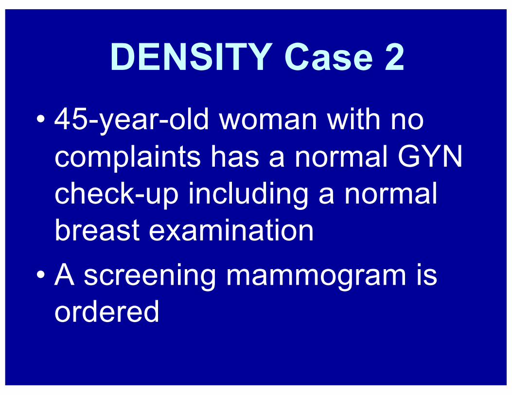
DENSITY Case 2• 45-year-old woman with no
complaints has a normal GYN check-up including a normal breast examination
• A screening mammogram is ordered

• The patient later received a letter from the radiology center stating in bold at the top of the page “We wish to inform you that the results of your recent mammography examination are normal.”
• Below, within a descriptive paragraph, it was also stated “Your mammographic breast density is considered dense. Dense breast tissue is a common finding and is not abnormal. A report of your results was sent to your physician.”

• The OB/GYN received a mammography report from the radiology center stating that the study was classified as “BIRADS 1: Negative evaluation.” Listed under Impression were “1.) No mammographic evidence of malignancy in either breast.”and “2.) A bilateral mammogram is recommended in one year.”
• Within a descriptive paragraph was a statement “The breast tissue is extremely dense.”
• The OB/GYN was not provided with the letter that had been sent to the patient.

• The patient, believing that her “normal”mammogram required no additional action, did not contact the ordering physician for further discussion.

• Her OB/GYN, aware of the Breast Density Notification Act, searches through the screening mammogram report, noting increased density.
• The OB/GYN calls her, notifies her of her increased density, and offers additional breast imaging.
• The patient, previously believing her screening mxr was completely normal, is confused.

• After 3 phone calls with her OB/GYN, the patient requests additional breast imaging.
• Her insurer declines coverage.• Out of pocket costs
– $2400 breast MRI– $250 breast ultrasound
• The patient cannot afford the additional testing
• Significant anxiety

ACOG 2015 Committee Opinion 625Management of Women With Dense
Breasts Diagnosed by Mammography • No studies have demonstrated earlier detection
or improved prognosis when additional breast imaging is obtained.
• ACOG advocates against recommending additional breast imaging in otherwise asymptomatic women with increased breast density.
• Physicians should comply with state laws requiring disclosure of increased mammographic breast density, many mandating offering additional breast imaging.

“… to inform your conversations with your physician. Together, you can decide which
screening options are right for you …”
• Which screening options are being referred to?–Breast ultrasound–Breast MRI–Breast tomosynthesis

Increased breast density on screening mammography:Laws leading to confusion amongst both patients and providers
Contemporary OB/GYN 2016
Jay Goldberg, MD, MSCP, Sara Mirghani, MD, Robin Metcalfe-Klaw, BS, Sarah Woodman, MD,
Debra Copit, MD, Christopher Scaven, DO, Arnold Cohen, MD, David Jaspan, DO
Einstein’s Departments of OB/GYN, Radiology, and Medicine
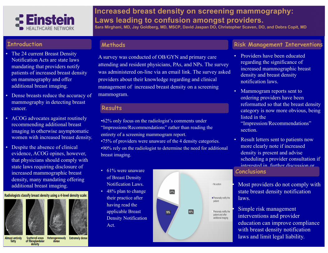
Results
Introduction
Discussion
• Providers have been educated regarding the significance of increased mammographic breast density and breast density notification laws.
• Mammogram reports sent to ordering providers have been reformatted so that the breast density category is now more obvious, being listed in the “Impression/Recommendations” section.
• Result letters sent to patients now more clearly note if increased density is present and advise scheduling a provider consultation if interested in further discussion or additional breast imaging.
Printed by
• The 24 current Breast Density Notification Acts are state laws mandating that providers notify patients of increased breast density on mammography and offer additional breast imaging.
• Dense breasts reduce the accuracy of mammography in detecting breast cancer.
• ACOG advocates against routinely recommending additional breast imaging in otherwise asymptomatic women with increased breast density.
• Despite the absence of clinical evidence, ACOG opines, however, that physicians should comply with state laws requiring disclosure of increased mammographic breast density, many mandating offering additional breast imaging.
Risk Management Interventions
• Most providers do not comply with state breast density notification laws.
• Simple risk management interventions and provider education can improve compliance with breast density notification laws and limit legal liability.
Increased breast density on screening mammography:Laws leading to confusion amongst providers.Sara Mirghani, MD, Jay Goldberg, MD, MSCP, David Jaspan DO, Christopher Scaven, DO, and Debra Copit, MD
•62% only focus on the radiologist’s comments under “Impressions/Recommendations” rather than reading the
entirety of a screening mammogram report.•75% of providers were unaware of the 4 density categories.
•90% rely on the radiologist to determine the need for additional breast imaging.
Conclusions
Methods
A survey was conducted of OB/GYN and primary care attending and resident physicians, PAs, and NPs. The survey was administered on-line via an email link. The survey asked providers about their knowledge regarding and clinical management of increased breast density on a screening mammogram.
• 61% were unaware
of Breast Density Notification Laws.
• 48% plan to change their practice after
having read the applicable Breast Density Notification
Act.
Results
Introduction
Discussion
� Providers have been educated regarding the significance of increased mammographic breast density and breast density notification laws.
� Mammogram reports sent to ordering providers have been reformatted so that the breast density category is now more obvious, being listed in the ³,PSUHVVLRQ�5HFRPPHQGDWLRQV´�section.
� Result letters sent to patients now more clearly note if increased density is present and advise scheduling a provider consultation if interested in further discussion or additional breast imaging.
Printed by
� The 24 current Breast Density Notification Acts are state laws mandating that providers notify patients of increased breast density on mammography and
additional breast imaging. � Dense breasts reduce the accuracy of
mammography in detecting breast cancer.
� ACOG advocates against routinely recommending additional breast imaging in otherwise asymptomatic women with increased breast density.
� Despite the absence of clinical evidence, ACOG opines, however, that physicians should comply with state laws requiring disclosure of increased mammographic breast density, many mandating offering additional breast imaging.
Risk Management Interventions
� Most providers do not comply with state breast density notification laws.
� Simple risk management interventions and provider education can improve compliance with breast density notification laws and limit legal liability.
Increased breast density on screening mammography: Laws leading to confusion amongst providers.
Sara Mirghani, MD, Jay Goldberg, MD, MSCP, David Jaspan DO, Christopher Scaven, DO, and Debra Copit, MD
� ����RQO\�IRFXV�RQ�WKH�UDGLRORJLVW¶V�FRPPHQWV�XQGHU�
³,PSUHVVLRQV�5HFRPPHQGDWLRQV´�UDWKHU�WKDQ�UHDGLQJ�WKH�HQWLUHW\�of a screening mammogram report.
� 75% of providers were unaware of the 4 density categories. � 90% rely on the radiologist to determine the need for additional
breast imaging.
Conclusions
60% 19%
21%
When Providers are notified of increased breast density on
mammogram:
No action
Personally notify thepatient
Personaly notifty thepatient and offeradditional imaging
Methods
A survey was conducted of OB/GYN and primary care attending and resident physicians, PAs, and NPs. The survey was administered on-line via an email link. The survey asked providers about their knowledge regarding and clinical management of increased breast density on a screening mammogram.
� 61% were unaware of Breast Density Notification Laws.
� 48% plan to change their practice after having read the applicable Breast Density Notification Act.

Provider Density Survey
• 79% unaware of the 4 density categories• 68% unaware of Breast Density
Notification Act• 64% focus on
“Impression/Recommendation” section of mammogram report
• 85% rely on radiology to determine if additional imaging is needed

Provider Density Survey
• When aware of increased density on a screening mammogram:–60% take no action–19% personally notify the woman–21% personally notify the woman
and discuss the option of additional breast imaging
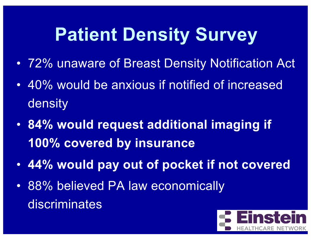
Patient Density Survey• 72% unaware of Breast Density Notification Act
• 40% would be anxious if notified of increased density
• 84% would request additional imaging if 100% covered by insurance
• 44% would pay out of pocket if not covered• 88% believed PA law economically
discriminates

Risk Management Interventions
• Providers education regarding the significance of increased breast density on mammography and the corresponding state law.
• Mammogram reports sent to ordering providers have been reformatted àbreast density category is now more obvious, being listed in the “Impression /Recommendations” section.
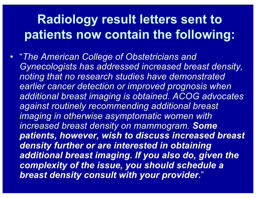
Radiology result letters sent to patients now contain the following:
• “The American College of Obstetricians and Gynecologists has addressed increased breast density, noting that no research studies have demonstrated earlier cancer detection or improved prognosis when additional breast imaging is obtained. ACOG advocates against routinely recommending additional breast imaging in otherwise asymptomatic women with increased breast density on mammogram. Some patients, however, wish to discuss increased breast density further or are interested in obtaining additional breast imaging. If you also do, given the complexity of the issue, you should schedule a breast density consult with your provider.”

Further Areas of Confusion• Is written notification from the radiologist or
health care provider legally acceptable or must a discussion regarding breast density occur?
• If a woman’s breast density is not reported as significantly changed from year to year, are providers legally required to repeat a discussion regarding increased mammographic density and offer additional imaging every time that a screening mammogram is performed?

Breast Density Notification Acts

Case:Brachial plexus injury following
shoulder dystocia
• 33-year-old gravida 1 with UANC• Normal diabetes screen, adequate
pelvimetry, and fundal height of 39 cm at 37 weeks documented
• Presents at 40 weeks with SROM• Vacuum delivery from +2/+3 station after
2+ hours of pushing• Shoulder dystocia

• MD’s deposition: McRobert’s positioning, suprapubic pressure, Wood’s screw, attempt to grasp posterior arm, attempt to fracture clavicle– only McRobert’s documented in chart
• 7 minutes• Male 3755 grams, 8 pounds 4 ounces• Apgars 7/9
• Baby diagnosed with a brachial plexus injury

• Plaintiff’s Allegations: –Only McRobert’s used–Excessive traction used–Episiotomy would have prevented injury

Plaintiff ExpertChairman of Johns Hopkins
• “The performance of the procto-episiotomy … would have led to the successful application of these maneuvers to allow for delivery of Ryan without stretch trauma to the brachial plexus.”
• “the failure to do that (episiotomy) led to … excessive traction which executed delivery.”

ACOG Practice Bulletin 40Shoulder dystocia
• Controversy exists as to whether episiotomy is necessary, because shoulder dystocia typically is not caused by obstructing soft tissue.

• Defense Expert: –McRobert’s positioning, suprapubic
pressure, Wood’s screw, attempt to grasp posterior arm, & attempt to fracture clavicle used (as per MD’s deposition not chart)
–No excessive traction used–Episiotomy would not have
prevented brachial plexus injury

Settle or go to Trial ?

• Trial: Plaintiff verdict $750K
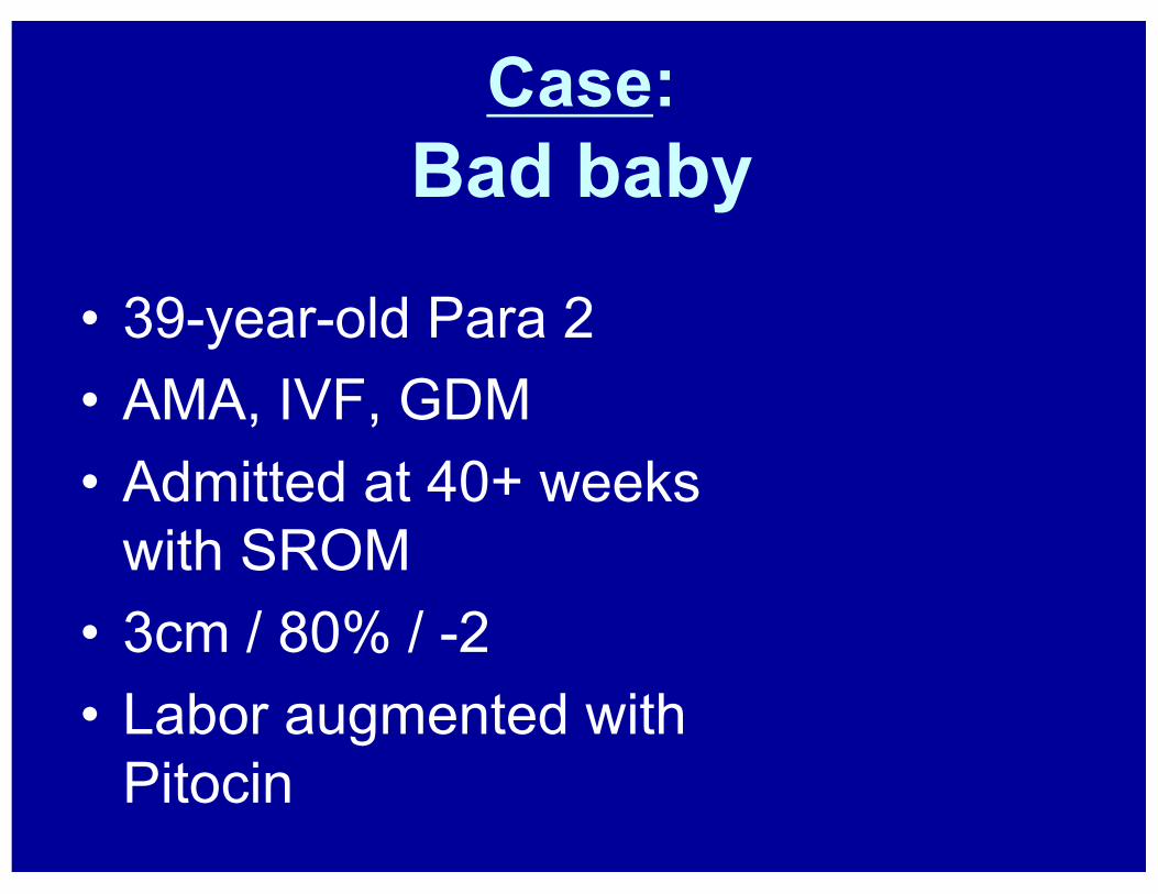
Case:Bad baby
• 39-year-old Para 2• AMA, IVF, GDM• Admitted at 40+ weeks
with SROM• 3cm / 80% / -2• Labor augmented with
Pitocin
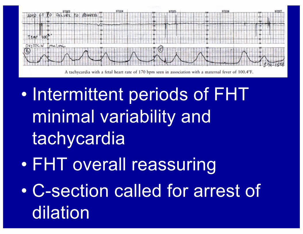
• Intermittent periods of FHT minimal variability and tachycardia
• FHT overall reassuring• C-section called for arrest of
dilation
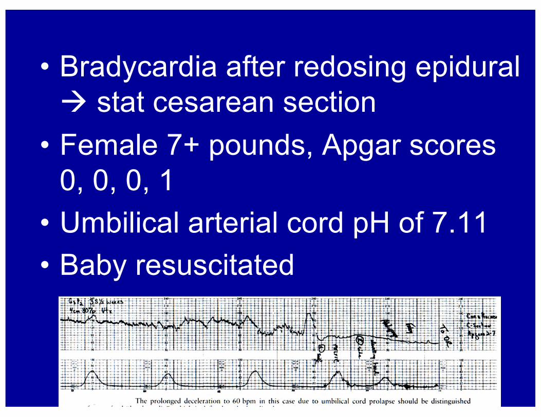
• Bradycardia after redosing epidural à stat cesarean section
• Female 7+ pounds, Apgar scores 0, 0, 0, 1
• Umbilical arterial cord pH of 7.11• Baby resuscitated

• Baby suffers from severe brain damage, cerebral palsy, vision impairment, developmental delays, feeding difficulty, and hypotonicity.
• Baby with possible dysmorphic features.

Apology LetterPA MCARE Act 2002
• Prior to outside review, without physician’s consent, hospital risk management sent patient a letter apologizing for the “medical error” that had occurred

• Plaintiff’s Allegations:– C-section should have been performed
earlier– L&D blood gas machine not properly
maintained with expired CLIA license
• Defense: – Fetal monitoring overall reassuring until
bradycardia– Blood gas machine accurate

Legal Issues• Expired CLIA license of L&D
blood gas machine• Apology letter stating that a
medical error had occurred• Sympathy for a severely
disabled child

Settle or go to Trial ?

• Settled–$12 Million against the hospital and MD

Case:Laparoscopic Bowel Injury
• 66-year-old with enlarging 5 cm ovarian complex mass
• CA-125 normal• Consented for
laparoscopy, possible x-lap

• Laparoscopic LSO and lysis of adhesions
• EBL 50 cc• Path → benign serous
cystadenoma• Patient discharged home

• POD #1 called with fever, decreased urination, nausea
• Sent immediately to ER• Acute abdomen• CT → dilated loops of bowel and
thickening of descending colon• X-lap, sigmoid colon resection, colostomy
– 3 mm defect in sigmoid identified by surgeon but not pathologist

•3 months later –colostomy reversed
•2 months later – repair of incisional hernia and lysis of adhesions
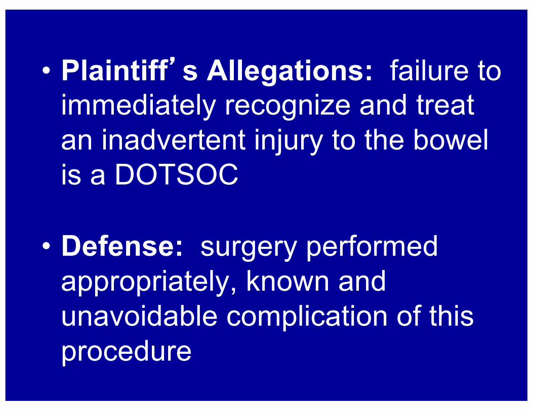
• Plaintiff’s Allegations: failure to immediately recognize and treat an inadvertent injury to the bowel is a DOTSOC
• Defense: surgery performed appropriately, known and unavoidable complication of this procedure
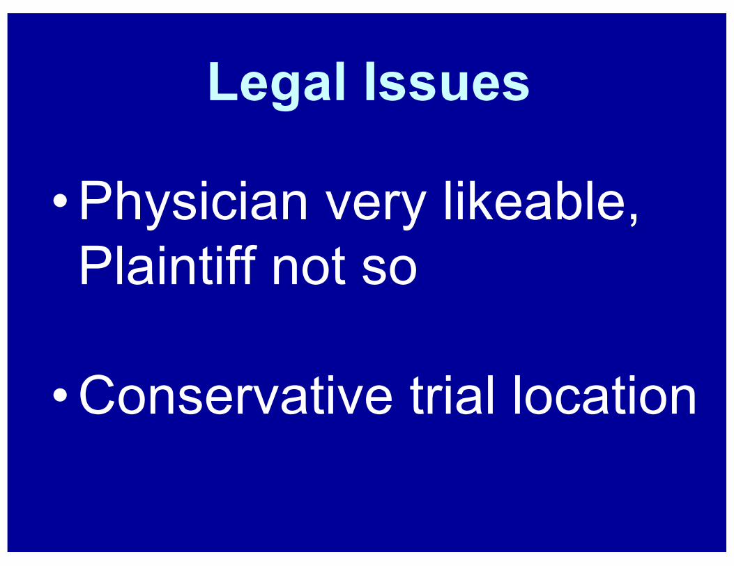
Legal Issues
• Physician very likeable, Plaintiff not so
• Conservative trial location

Settle or go to Trial ?

•Trial: Defense Verdict

THANKS FOR LISTENING!