obesity due to stress - steve gibson due to... · niet alleen het vet –en glucose metabolisme...
TRANSCRIPT

0
Rianne Kruit Examiner: Dr. Onno Meijer
3346668 Second Examiner: Dr. Eric Kalkhoven
Master Biology of Disease Department of Endocrinology
Utrecht University LUMC, Leiden
Obesity due to stress
A change in glucose and lipid metabolism due to altered cortisol levels

1
Content
Nederlandse samenvatting ..................................................................................................................... 3
Abstract ................................................................................................................................................... 4
List of Abbreviations ................................................................................................................................ 5
Introduction ............................................................................................................................................. 7
Obesity................................................................................................................................................. 7
Definition & Measurements ............................................................................................................ 7
Prevalence ....................................................................................................................................... 7
Health risks ...................................................................................................................................... 8
Obesity and Stress ............................................................................................................................... 9
Mechanisms of stress ............................................................................................................................ 10
Autonomic nervous system responses .............................................................................................. 10
The hypothalamic-pituitary-adrenal (HPA) axis ................................................................................ 11
The hypothalamic-pituitary-adrenal (HPA) axis & circadian rhythm ................................................ 11
Cortisol and its effects ....................................................................................................................... 11
The effects of cortisol & stress on the glucose metabolism ................................................................. 13
Gluconeogenesis ............................................................................................................................... 13
Glycogenolysis ................................................................................................................................... 13
Insulin resistance ............................................................................................................................... 14
11β-hydroxysteroid dehydrogenase type 1 ...................................................................................... 14
Stress and the lipid metabolism ............................................................................................................ 16
The normal lipid metabolism ............................................................................................................ 16
Lipolysis ............................................................................................................................................. 16
TNF-α, resistin and adiponectin ........................................................................................................ 17
De novo lipogenesis ........................................................................................................................... 18
Adipogenesis ..................................................................................................................................... 19
Stress and the central nervous system ................................................................................................. 20
Possible treatments for obesity induced by stress ............................................................................... 21
Mineralocorticoid & Glucocorticoid receptor antagonists ............................................................... 21
Inhibition of 11β-hydroxysteroid dehydrogenase type 1 .................................................................. 21
Neuropeptide Y ................................................................................................................................. 22
Other possibilities .............................................................................................................................. 22
Conclusion ............................................................................................................................................. 23
Acknowledgements ............................................................................................................................... 24

2
References ............................................................................................................................................. 25

3
Nederlandse samenvatting Tijdens deze literatuurstudie is er onderzoek gedaan naar stress geïnduceerde obesitas. Allereerst
zijn de gevaren en oorzaken van obesitas besproken. Vervolgens wordt het mechanisme van stress
uitgelegd met onder andere de HPA as, die verantwoordelijk is voor de secretie van het
glucocorticoïd cortisol. Cortisol kan binden aan twee verschillende receptoren en op die manier
meerdere processen beïnvloeden. In deze scriptie wordt de invloed van cortisol op 3 systemen
beschreven. Allereest het glucosemetabolisme. Daar heeft cortisol invloed op onder andere de
gluconeogenese, glycogenolyse en een speciaal enzym dat de inactieve vorm van cortisol omzet naar
de actieve vorm: 11β-hydroxysteriod dehydrogenase type 1. Cortisol heeft ook invloed op het
vetmetabolisme. Daar stimuleert het vooral de afbraak van vetweefsel (lipolyse), wat resulteert in
een grote hoeveelheid vrije vetzuren in het plasma. Dit heeft weer als gevolg dat andere processen
geremd of gestimuleerd worden. Niet alleen het vet –en glucose metabolisme worden door cortisol
beïnvloed, ook het centrale zenuwstelsel. Verschillende regio’s van het brein reageren op cortisol
door de morfologie van neuronen te veranderen, wat onder andere leidt tot “stress eten” en
geprikkeld zijn. Al deze effecten van cortisol kunnen leiden tot obesitas en een gevaar voor de mens
vormen. Gelukkig zijn er verschillende manieren waarop dit wellicht geremd kan worden en dat is het
onderwerp van het laatste hoofdstuk.

4
Abstract In this literature study different topics concerning stress-induced obesity are described. First of all
the dangers and causes of obesity are discussed. Next, the mechanism of stress is explained with
among others the HPA axis, which is responsible for the secretion of the glucocorticoid cortisol.
Cortisol is able to bind to two different receptors and thereby influence different processes. In this
thesis, the effects of cortisol on three systems are thoroughly described. One of these processes is
the glucose metabolism. Cortisol influences gluconeogenesis, glycogenolysis and a special enzyme
catalyzing the conversion of the inactive form of cortisol to the active form: 11β - hydroxysteroid
dehydrogenase type 1. Cortisol also has an effect on the lipid metabolism. Within this metabolism it
mainly stimulates the breakdown of adipose tissue (lipolysis), which results in a large amount of free
fatty acids in the plasma. This in turn has an effect on other processes which will be inhibited or
stimulated. Not only the lipid metabolism and glucose metabolism are affected by cortisol, also the
central nervous system. Different parts of the brain respond to cortisol by changing morphology of
neurons, which among others leads to "stress eating" and being agitated. All these effects of cortisol
can lead to obesity and form a danger for human. Fortunately, there are many ways in which this
might be treated and this is discussed in the last chapter of this study.

5
List of Abbreviations 11β-HSD1 11β-hydroxysteroid dehydrogenase type 1 11β-HSD2 11β-hydroxysteroid dehydrogenase type 2 ACTH Adrenocorticotropic hormone ANS Autonomic nervous system ATGL Adipose triglyceride lipase AVP Vasopressin BAT Brown adipose tissue BMI Body mass index cAMP cyclic AMP CHD Chronic heart disease CREB cAMP response element binding protein CRH Corticotropin-releasing hormone CRHR1 CRH1 receptor CRHR2 CRH2 receptor CVD Cardiovascular disease DNL De novo lipogenesis ECGC Epigallocatechin gallate Fbpase Fructose 1,6-bisphosphatase FFA Free fatty acids FoxOs Forkhead box class Os G6Pase Glucose 6 phosphatase G/G6P Glucose/glucose-6-phosphate GR Glucocorticoid receptor GRE Glucocorticoid response element HPA Hypothalamic-pituitary-adrenal HSL Hormone-sensitive lipase IKK IkappaB-kinase IRS-1 Insulin receptor substrate 1 IRSs Insulin-receptor substrates JNK c-jun N-terminal kinase LMO3 LIM domain only 3 LPL Lipoprotein lipase LSGRA Liver-selective glucocorticoid receptor antagonist MAG Monoacylglycerol MGL Monoglyceride lipase MR Mineralocorticoid receptor NEFAs Nonesterified fatty acids NPY Neuropeptide Y NPY2R Neuropeptide Y receptor p38MAPK p38 MAP kinase p70SK S6 kinase p70 PC Pyruvate carboxylase PEPCK Phosphoenolpyruvate carboxykinase PKA Protein kinase A PKC Protein kinase C POMC Pro-opiomelanocortin PVN Paraventricular nucleus (located in hypothalamus) ROS Reactive oxygen species SAM Sympatho-adrenomedullary SAPK Stress-activated protein kinase

6
SCN Suprachiasmatic nucleus sER smooth endoplasmatic reticulum SF Skinfold measurements T2DM Type 2 diabetes mellitus TAG Triacylglycerol WAT White adipose tissue WC Waist circumference WHR Waist-hip ratio WHO World Health Organization

7
Introduction
Obesity A spectrum of diseases is known to occur more frequently in people with obesity. Obesity is a serious
worldwide problem, nowadays affecting approximately 10% of the world’s population (World Health
Organization, www.who.int/gho/ncd/risk_factors/obesity_text/en/).
Definition & Measurements
Obesity is defined as an accumulation of adipose tissue that is of sufficient magnitude to impair life
(Robbins & Cotran, 8th edition). One method to measure obesity is the use of the body mass index
(BMI). The BMI of a person can be calculated by dividing the weight (kg) with the height in square
(m2) (Sweeting, 2007; Roche et al., 1981). The World Health Organisation (WHO) stated that a
median BMI for an adult population should be in the range of 21 - 23kg/m2 to achieve optimum
health. A BMI of 25 – 29,9kg/m2 increases risk for co-morbidities, and moderate to severe risk of co-
morbidities occur at a BMI of 30kg/m2 and higher (World Health Organization). An overview of BMI
classification is shown in table 1.
Table 1. Classification of BMI. Adapted from World Health Organization
Multiple authors have described disadvantages of the BMI method (Garn et al. 1986; Daniels et al.,
1997; Prentice and Jebb, 2001; Wells et al., 2002). One of the biggest disadvantages is that BMI
reflects both fat and fat-free components of weight and thereby does not directly measure adipose
tissue (Wells et al., 2002). Therefore other measurements are necessary.
These other measurements include complex methods and most of them are limited to research
settings (Sweeting, 2007). Some easier, anthropometric methods are skinfold measurements (SF),
waist circumference (WC) and waist-hip ratio (WHR) (Sweeting, 2007). These methods together with
BMI can diagnose a person to suffer from obesity or not.
Prevalence
In 2008, 35% of adults aged 20+ (worldwide) were overweight (BMI ≥ 25 kg/m2) (34% men and 35%
women). The worldwide prevalence of obesity has nearly doubled between 1980 and 2008. In 2008,
10% of men and 14% of women in the world were obese (BMI ≥30 kg/m2), compared with 5% for
men and 8% for women in 1980 (World Health Organization). In the Netherlands in 2011, 41% of the
population suffers from obesity, 10% of this percentage suffers from severe obesity (CBS,
www.cbs.nl/nl-NL/menu/themas/gezondheid-welzijn/publicaties/artikelen/archief/2012/2012-3651-
wm.htm).
The prevalence of overweight and obesity is highest in America (62% & 26%) and lowest in South
East Asia (14% & 3%). In all WHO regions women were more likely to be obese than men. The

8
prevalence of raised body mass index increases with income level of countries (World Health
Organization) suggesting that being wealthy results more likely in obesity compared to being poor.
Health risks
Some of the diseases thought to occur more frequently in people suffering from obesity are among
others metabolic syndrome, diabetes, cardiovascular disease and respiratory effects (Bray, 2004;
Kopelman, 2007).
The metabolic syndrome is a combination of metabolic risk factors that consist the following:
atherogenic dyslipidemia (elevated triglycerides, ≥150 mg/dl), elevated blood pressure (≥130/85 mm
Hg), elevated blood glucose (≥100 mg/dl), prothrombotic state and proinflammatory state (Grundy,
2004). It was found that many factors are increased in obese people plasma: Nonesterified fatty acids
(NEFAs), inflammatory cytokines, adiponectin, leptin and resistin. Al these factors are thought to
contribute to the development of the metabolic syndrome (Guerre-Millo, 2002) and explains why
obesity results in an increased risk for this disease.
Another health risk developing with obesity is diabetes type 2 (T2DM). About 90-95% of people with
diabetes have type 2 and in 80% of all people with diabetes type 2, obesity is the cause. When
diabetes type 2 is diagnosed, the pancreas is producing enough insulin, but the body does not
respond to it properly, also called insulin resistance (NIDDK, The National Institute of Diabetes and
Digestive and Kidney Diseases, http://diabetes.niddk.nih.gov/dm/pubs/overview/). Insulin resistance
leads to a decreased uptake of glucose in the muscle, reduced glycolysis and fatty acid oxidation in
the liver and an impairment to suppress hepatic gluconeogenesis. This in total results in an
accumulation of glucose in the blood, called hyperglycemia (Robbins & Cotran, 8th edition). Insulin
resistance can be reversed, for example by bilio-pancreatic diversion, causing lipid malabsorption
(Mingrone et al., 1997). Because of the insulin resistance the pancreas first tends to secrete even
more insulin to compensate. However, the body will still not react properly to it and this will finally
lead to β-cell dysfunction (production site of insulin) (Robbins & Cotran, 8th edition). This dysfunction
causes irreversibility of the insulin resistance and T2DM has occurred.
Obese persons have a higher prevalence of hypertension compared to lean persons. This is a strong
risk factor for cardiovascular disease (CVD) (Grundy, 2004; Chobanioan et al.,2003). Well-known
complications of hypertension are chronic heart disease (CHD), stroke, left ventricular hypertrophy,
heart failure, and chronic renal failure (Grundy, 2004; Ejerblad et al., 2006). Some of these
complications can also be caused by inflammation, which in turn also is a risk of obesity (Grundy,
2004).
Accumulation of fat tissue impairs ventilatory function by reducing volume and total lung capacity
(Chin et al., 1996; Poulain et al., 2006). Impaired ventilatory function is caused by the mechanical
effects of fat on the diaphragm and the chest wall (Ray et al., 1983; Poulain et al., 2006). Another
consequence of obesity is obstructive sleep apnea. Increased fat tissue deposition in the pharyngeal
region obstructs the upper airway and increases its collapsibility. This results in repetitive closures of
the airway during sleep (Resta et al., 2001). Obstructive sleep apnea is also associated with increased
mortality due to the high incidence of cardiovascular disorders reported in this condition (Wolk et al.,
2003).
9
Obesity and Stress Obesity can be caused by an inactive lifestyle, environmental factors (work schedules, oversized food
portions and lack of access to healthy food), genetic factors and family history (NIH, National
Institutes of Health, http://www.nhlbi.nih.gov/health/health-topics/topics/obe/causes). Many of
these factors can be counteracted by changing lifestyle. However, besides all listed causes, there may
be additional factors associated with the development of obesity, metabolic syndrome and disease.
One of these factors is stress and will be the main subject of this thesis.
All living organisms strive towards a dynamic equilibrium, called homeostasis. This involves chemical
and other processes to maintain the most optimal conditions for life. Homeostasis is continually
disrupted by environmental factors, external and internal stimuli. Physical and physiological events
causing the loss of optimal conditions are called stressors and can be experienced as having stress.
Stressors also trigger physiological and behavioural responses that are aimed to maintain
homeostasis. In response to stress, the brain can activate different systems (De Kloet et al., 2005).
This literature study is about those different systems activated by the brain and how this can lead to
obesity. Therefore the responses to stress will be further described. Next, this will be related to
changes in both glucose metabolism and lipid metabolism. Besides these peripheral effects of stress,
there also are some relevant effects within the central nervous system. For both aspects of
metabolism (glucose and lipid), mechanisms to prevent obesity due to stress, will be discussed.
Finally a summary of the literature will be given.

10
Mechanisms of stress When a situation is perceived as stressful, mainly two pathways are activated in order to maintain
homeostasis. The effects of both pathways will be explained with use of figure 2.
Autonomic nervous system responses The responses of the autonomic nervous system (ANS) are indicated in blue in figure 2. Exposure to
stressors results in activation of sympathetic neurons in the thoracolumbar (T1-L2) spinal cord. These
neurons in turn project to prevertebral or paravertebral ganglia which project to organs and cells of
the adrenal medulla. This sympathetic activation, also called the sympatho-adrenomedullary (SAM)
axis, represents the classic ‘fight or flight’ response which generally increases circulating levels of
adrenaline (from the adrenal medulla) and noradrenaline (from sympathetic nerves). Heart rate and
force of contraction, peripheral vasoconstriction, and energy mobilization are also increased by this
activation. Besides the sympathetic activation, the parasympathetic system can also be modulated
during stress. This is indicated in figure 2 by red dots. In the parasympathetic system, activation of
cranial nuclei results in changes in the cardiovascular system and activation of sacral nuclei
modulates changes in the abdominal viscera. Parasympathetic actions are generally opposite to
those of the sympathetic system (Ulrich-Lai & Herman, 2009).
Figure 2. The sympatho-adrenomedullary axis (left side) and the hypothalamic-pituitary-adrenal axis
(right side). The two main pathways activated by stress exposure to maintain homeostasis. Adapted from
Ulrich-Lai & Herman, 2009

11
The hypothalamic-pituitary-adrenal (HPA) axis The other pathway responding to stress is the hypothalamic-pituitary-adrenal (HPA) axis. This
pathway will be the main focus of this thesis and is depicted on the right side of figure 2. Exposure to
stressors activate the production of corticotropin-releasing hormone (CRH) and vasopressin (AVP) by
the parvocellular neurons located in the hypothalamic paraventricular nucleus (PVN). These neurons
secrete the peptides into the portal vessel system where they can bind the CRH1 receptor (CRHR1)
and activate the synthesis of pro-opiomelanocortin (POMC) in the anterior pituitary. POMC in turn
gets processed into among others adrenocorticotropic hormone (ACTH), opioid and melanocortin
peptides. CRH-induced release of ACTH stimulates the adrenal cortex to synthesize and secrete the
glucocorticoid cortisol. Cortisol acts as a negative feedback signal, inhibiting ACTH and CRH secretion
(Herman et al., 2003; De Kloet et al., 2005; Silverthorn, 5th edition).
The hypothalamic-pituitary-adrenal (HPA) axis & circadian rhythm The HPA axis is normally continuously active under the influence of the ANS. A basic characteristic of
the HPA axis is that unstressed animals have a circadian rhythm of ACTH and glucocorticoid
secretion. This rhythm is coordinated by outputs from the suprachiasmatic nucleus (SCN) of the
hypothalamus (Lightman & Conway-Campbell, 2010). The circadian rhythm appears to have an
underlying ultradian (hourly) activity of the HPA axis. The origin of ultradian rhythmicity in the HPA
axis is not known. Although there has been a general assumption that there must be a hypothalamic
pulse generator, there is little evidence to support this (Lightman & Conway-Campbell, 2010).
However, it is thought that simple feedforward and feedback interactions between the pituitary and
adrenal cortex can account for the glucocorticoid rhythms that have been observed experimentally
(Lightman & Conway-Campbell, 2010). Finally researchers conclude that both the circadian and
ultradian rhythms are crucial for optimal responsiveness of glucocorticoid-sensitive neural processes
(Lightman & Conway-Campbell, 2010). Stress is one of those neural processes where responsiveness
should be optimal.
Cortisol and its effects Cortisol belongs to a family called steroid hormones. This family can be further subdivided into three
classes: sex hormones, mineralocorticoids (named because of its effect on the minerals sodium and
potassium) and glucocorticoids (named because of its effect on plasma glucose concentrations).
Cortisol belongs to the glucocorticoids and is synthesized from cholesterol in the zona fasciculata of
the adrenal cortex which is the only zone containing the correct enzymes (21-hydroxylase and 17-α-
hydroxylase) for the synthesis (Silverthorn, 5th edition).
Approximately 75% of the cortisol in the circulation is bound to a plasma protein named transcortin
or corticosteroid binding globulin (CBG). Another 15% is bound to albumin, and the remaining 10% is
unbound or free. Free cortisol is biologically active. It is also the concentration of free cortisol that is
regulated. The half-life of cortisol in the circulation is 60-90 min and it is metabolized in the liver.
Most of the cortisol is reduced to dihydrocortisol and next to tetrahydrocortisol which is conjugated
to glucuronic acid. Some cortisol is converted to cortisone, which is an inactive glucocorticoid.
Cortisone is also reduced and conjugated to form tetrahydrocortisone glucuronide. The
tetrahydroglucuronide derivatives (like glucuronic acid) of cortisol and cortisone are water soluble
and are excreted in the urine (Boron & Boulpaep, 1st edition).

12
Cortisol can bind to both the mineralocorticoid receptor (MR) and the glucocorticoid receptor (GR).
However, the receptors have a different affinity for the hormone. The MR has a 10-fold higher
affinity for cortisol compared to the GR. Therefore the MR is active at both low and high
concentrations of the hormone and the GR becomes activated at only high concentrations of cortisol
(Funder, 1997; De Kloet et al., 2005). When cortisol binds the receptors (depending on the
concentration) they form dimers (either homo –or heterodimers) and migrate to the nucleus. There
they will interact with specific DNA sequences called glucocorticoid response elements (GRE), which
changes the transcription of multiple target genes. GR monomers can also interact with stress-
induced transcription factors (TFs) or other proteins to dampen their transcriptional activity (Stokes
et al., 2000; De Kloet et al., 2005). The described pathways are depicted in figure 3 below.
By changing the transcription of multiple target genes, cortisol has many metabolic effects, mainly
preventing hypoglycemia via stimulation of catabolic processes. Some of the processes cortisol
stimulates are: suppression of the immune system, creating a negative calcium balance (by adjusting
calcium absorption and secretion and promoting breakdown of calcified bone matrix), promoting
gluconeogenesis and lipolysis and the breakdown of skeletal muscle proteins. (Silverthorn, 5th
edition). The last two processes (gluconeogenesis and lipolysis) are part of the glucose and lipid
metabolism and will be extensively explained in the next chapters. The breakdown of skeletal muscle
proteins fits with lipolysis and gluconeogenesis because it serves the function of releasing energy
substrate for other processes.
Figure 3. Overview of the pathway after binding of cortisol to either the mineralocorticoid receptor or
glucocorticoid receptor. Adapted from De Kloet et al., 2005
13
The effects of cortisol & stress on the glucose metabolism In this chapter the influence of cortisol on the glucose metabolism will be described. Cortisol has
multiple effects on the glucose metabolism overall resulting in hyperglycemia.
Gluconeogenesis One of the processes cortisol has an effect on is gluconeogenesis. This is the opposite of glycolysis
and involves the synthesis of glucose from non-carbohydrate precursors. This process mainly takes
place in the liver (Nordlie and Foster, 1999). A first sign of cortisol having a counterregulatory effect
on hypoglycemia was discovered in 1989. During this experiment normal subjects got infused with
insulin. During the administration of several other compounds the glucose fluxes were continuously
monitored. One of the experimental groups received, despite the insulin, a blocker preventing the
levels of cortisol to increase. This group appeared to have a 22% decrease of hepatic glucose
production (gluconeogenesis) and a 15% increase in glucose utilization. From this could be concluded
that cortisol normally plays an important counterregulatory role during hypoglycemia by stimulating
glucose production and decreasing glucose utilization (De Feo et al., 1989). This outcome got
confirmed by other research groups (Rooney et al., 1993; Goldstein et al., 1993). Other research
groups were able to put these effects of cortisol in context with metabolic syndrome (Chrousos,
2000; Khani & Tayek, 2001; Anagnostis et al., 2009).
Cortisol is thought to stimulate gluconeogenesis by activation of key regulatory enzymes involved in
this process. These enzymes are: glucose 6 phosphatase (G6Pase), fructose 1,6-bisphosphatase
(Fbpase), pyruvate carboxylase (PC), and phosphoenolpyruvate carboxykinase (PEPCK). These
enzymes are activated by stimulation of intracellular pathways leading to activation of the key
transcription factors cAMP response element binding protein (CREB) and forkhead box class Os
(FoxOs) (Oh et al., 2013).
Due to reciprocal regulation of glycolysis and gluconeogenesis (Weber et al., 1967; Berg, 6th edition)
cortisol indirectly inhibits glycolysis and thereby also promotes hyperglycemia.
Glycogenolysis If cortisol has the function to counteract hypoglycaemia, not only a stimulation of gluconeogenesis
would be expected, but also an increase in glycogenolysis. This process includes the breakdown of
glycogen, mostly stored in the liver, to provide glucose 6-phosphate for further metabolism. The
hormones glucagon and epinephrine can stimulate glycogenolysis by binding to a G-protein coupled
receptor. This finally results in activation of the enzyme glycogen phosphorylase and stimulation of
glycogenolysis (Berg, 6th edition). In 1981 the effect of cortisol on glycogenolysis was discovered in
chicks. After injections of cortisol, glycogen content within the liver was decreased, suggesting an
increase in glycogenolysis (Eqana et al. 1981). This finding got confirmed by another group in the
salmon (Vijayan & Leatherland, 1989).
Nowadays it is thought that cortisol stimulates glycogenolysis by facilitating the epinephrine induced
activation of the enzyme glycogen phosphorylase (Kuo et al., 2013).

14
Insulin resistance Normally, the hormone insulin counteracts the hyperglycemic effects of cortisol. However, cortisol is
known to counteract insulin action and thereby creates an insulin resistance. Insulin resistance can
be quantified by numerous methods, usually involving measurement of the plasma insulin
concentration relative to plasma glucose concentration, or the amount of glucose infused to
maintain euglycaemia at a fixed insulin concentration (called glucose tolerance test)(Ferrannini and
Mari, 1998).
Normally insulin binds to the cell-surface insulin receptor (IR) which is a tyrosine kinase that
autophosphorylates and next phosphorylates the insulin receptor substrate (IRS). Tyrosine-
phosphorylated IRS associates with IR and activates downstream signaling pathways (Kuo et al.,
2013). Mice treated with cortisol show to have reduced levels of tyrosine-phosphorylated IR and
total IRS-1 proteins. The activity of two downstream signaling molecules: phosphoinositide-3-kinase
(PI3K) and Akt are also decreased. Moreover, the phosphorylation of serine 307 of IRS-1 is increased
after cortisol administration. This phosphorylation disrupts the association between IR and IRS-1 and
thereby reduces the insulin response (Giorgino et al., 1993; Kuo et al., 2013).
Insulin resistance may also reflect impaired insulin-dependent down-regulation of hepatic glucose
release and/or impaired insulin-mediated increase in peripheral glucose uptake (Andrews & Walker,
1999). Cortisol works via both ways. As already described, it promotes gluconeogenesis and thereby
impairs insulin-dependent down-regulation of hepatic glucose release. Other researchers found that
the cortisol-induced insulin resistance in man is due to the decrease in both hepatic and extrahepatic
sensitivity to insulin (Rizza et al., 1982).This decrease in insulin action can be explained by the
mechanism described above.
When insulin resistance occurs, due to elevated levels of cortisol, glycogenesis is no longer
stimulated and glucose will not be taken up by the liver to produce glycogen. This contributes to the
hyperglycemic effects of cortisol.
11β-hydroxysteroid dehydrogenase type 1 Hyperglycemia, due to increased gluconeogenesis and glycogenolysis, insulin resistance and “stress
eating” might result in obesity and the development of metabolic syndrome (Vicennati et al., 2009;
Gallagher et al., 2010). Obesity in turn also has an influence in cortisol metabolism. Within certain
tissues, active cortisol can be converted into the inactive cortisone and vice versa. For this conversion
two different enzymes are known to be important. The enzyme 11β-hydroxysteroid dehydrogenase
type 2 (11β-HSD2) converts cortisol into the inactive cortisone, which occurs mainly in the kidney.
Dysfunction of this enzyme results in hypertension (Edwards et al., 1988). The other enzyme 11β-
hydroxysteroid dehydrogenase type 1 (11β-HSD1) catalyzes the opposite conversion, from the
inactive cortisone to cortisol. This process mainly occurs in the liver and adipose tissue (Kotelevstev
et al., 1997). Several research groups have found that people with obesity have an increased activity
of 11β-HSD1 within the liver and adipose tissue and therefore an elevated level of active cortisol
(Stewart et al., 1999; Rask et al., 2001; Anagnostis et al., 2009). The increase in cortisol in turn
stimulates hyperglycemia and insulin resistance and a continuous positive feedback loop is created. A
scheme of this loop is visualised in figure 4.
15
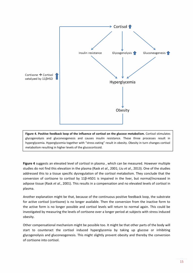
Figure 4 suggests an elevated level of cortisol in plasma , which can be measured. However multiple
studies do not find this elevation in the plasma (Rask et al., 2001; Liu et al., 2013). One of the studies
addressed this to a tissue specific dysregulation of the cortisol metabolism. They conclude that the
conversion of cortisone to cortisol by 11β-HSD1 is impaired in the liver, but normal/increased in
adipose tissue (Rask et al., 2001). This results in a compensation and no elevated levels of cortisol in
plasma.
Another explanation might be that, because of the continuous positive feedback loop, the substrate
for active cortisol (cortisone) is no longer available. Then the conversion from the inactive form to
the active form is no longer possible and cortisol levels will return to normal again. This could be
investigated by measuring the levels of cortisone over a longer period at subjects with stress induced
obesity.
Other compensational mechanism might be possible too. It might be that other parts of the body will
start to counteract the cortisol induced hyperglycemia by taking up glucose or inhibiting
glycogenolysis and gluconeogenesis. This might slightly prevent obesity and thereby the conversion
of cortisone into cortisol.
Figure 4. Positive feedback loop of the influence of cortisol on the glucoce metabolism. Cortisol stimulates
glycogenolysis and gluconeogenesis and causes insulin resistance. These three processes result in
hyperglycemia. Hyperglycemia together with “stress eating” result in obesity. Obesity in turn changes cortisol
metabolism resulting in higher levels of the glucocorticoid.

16
Stress and the lipid metabolism In this chapter the influence of cortisol on the lipid metabolism will be described. Before starting with
this the normal lipid metabolism will be explained.
The normal lipid metabolism The normal fat digestion and absorption is shown in figure 5. Large lipid droplets are emulsified by
bile salts secreted by the liver, resulting in smaller micelles. Pancreatic lipases enzymatically digest
triacylglycerol (TAG) into free fatty acids (FFAs) and monoacylglycerol (MAG). Next, FFAs and
monoglycerides diffuse across the apical membrane of the small intestine and move towards the
smooth endoplasmatic reticulum (sER) and recombine into triglycerides. The triglycerides join
cholesterol and proteins to form large droplets called chylomicrons. Chylomicrons leave the cell by
exocytosis and via the lymphatic system they enter into the cardiovascular system. Via this system
chylomicrons arrive at primarily adipose tissue and muscle tissue. There they bind to membrane –
bound lipoprotein lipase (LPL) and TAGs are once again degraded into FFA and MAG for transport
into the tissue. TAG is resynthesized inside the cell and stored (Silverthorn, 5th edition; Berg, 6th
edition).
Lipolysis TAGs can be catabolized into FFAs when energy for the body is needed. This biochemical catabolism
is also called lipolysis. Lipolysis occurs in all tissues, however most abundant in white adipose tissue
(WAT) and brown adipose tissue (BAT) (Lass et al., 2011). Three enzymes are implicated in the
hydrolysis of TAG molecules (shown in figure 6): adipose triglyceride lipase (ATGL) performs the first
and rate-limiting step hydrolyzing TAGs to generate diacylglycerols (DAGs) and FFAs (Zimmermann et
al., 2004). Hormone-sensitive lipase (HSL) is the rate-limiting enzyme for DAG catabolism (Osuga et
al., 2000; Haemmerle et al., 2002). Finally, monoglyceride lipase (MGL) cleaves MAG into glycerol
and FFAs (Karlsson et al., 1997).
Figure 5. Fat digestion and absorption. Large lipid droplets get emulsified to smaller micelles by bile salts.
Pancreatic lipases convert TAGs into FFA and monoglycerides, which enter the cell via diffusion. Triglycerides
are formed again in the sER and are packed, together with cholesterol and proteins, into chylomicrons.
Chylomicrons are released into the lymphatic system via exocytosis and finally get into the blood. Adapted
from ridge.icu.ac.jp/biobk/biobookdigest.

17
Cortisol is known to have a major influence on lipolysis. Short-term administration of cortisol in vivo
showed to promote adipose tissue lipolysis (Divertie et al., 1991). One of the causes might be that
this glucocorticoid creates a resistance to the inhibition of lipolysis by insulin (Dinneen et al., 1993).
Another group defined that this resistance probably appears due to a combination of increased LPL
activity and the increase of lipolysis itself (Samra et al., 1998). Increased LPL activity involves an
increased level of LPL mRNA, resulting in an increase in LPL synthesis, and additional
posttranslational regulation (Ottoson et al., 1994).
The stimulation of lipolysis results in an elevated concentration of FFAs in the plasma, which has
several consequences. The elevated circulating FFAs inhibit insulin-stimulated glucose uptake,
glycogen synthesis and glucose oxidation together with an increase in hepatic glucose output
(Bergman & Ader, 2000). Inhibition of insulin-stimulated glucose uptake is also called insulin
resistance and is found to be caused by protein kinase C (PKC) activation and oxidative stress-
activated signalling pathways. FFAs induce the activation of PKC which increases the phosphorylation
of serine 307 of IRS-1. This inhibits IRS-1 tyrosine phosphorylation by IkappaB-kinase (IKK) or c-jun N-
terminal kinase (JNK). The same study found that FFAs also activate several stress kinases: S6 kinase
p70 (p70SK), stress-activated protein kinase (SAPK) and p38 MAP kinase (p38MAPK). This suggests
the involvement of oxidative stress-activated signalling pathways (Ragheb et al., 2009).
TNF-α, resistin and adiponectin As already described in the paragraph discussing the effects of cortisol on the glucose metabolism,
insulin resistance results in hyperglycemia. Hyperglycemia together with “stress eating” results in
obesity, which in turn has other effects. One of the already described effects is the increased
turnover of cortisone into active cortisol catalysed by the enzyme 11β-HSD1. Other effects of obesity
are increases in tumour-necrosis factor-α (TNF-α) and resistin together with a decrease in
adiponectin (Saltiel & Kahn, 2001).
It was found that the expression of TNF-α is increased in fat of obese rodents and humans. This
induces phosphorylation of insulin receptor substrate 1 (IRS-1), resulting in reduced insulin receptor
activity and insulin resistance (Hotamisligil et al., 1996). Resistin is a peptide hormone secreted by
adipose tissue and was found to be elevated in obese mice. The use of anti-diabetic drugs and the
administration of anti-resistin antibody seemed to improve blood sugar and insulin action in mice
with diet-induced obesity, suggesting that elevated resistin levels contribute to insulin resistance
(Saltiel & Kahn, 2001). Another peptide derived from adipose tissue is adiponectin. Obese mice and
human were found to have a decreased expression of adiponectin mRNA. Treatment of mice with
this peptide decreased insulin resistance, the concentration of FFAs in plasma and the triglyceride
content of muscle and liver (Yamauchi et al., 2001). These findings suggest that increased TNF-α,
Figure 6. Lipolysis. Hydrolysis of TAG into FFAs and glycerol. Adapted from Lass et al., 2011

18
increased resistin and decreased adiponection, due to obesity, result in insulin resistance and
thereby close the positive feedback loop, because the insulin resistance results again in
hyperglycemia and obesity.
A summary of the effects described above is shown in the positive feedback loop of figure 7.
De novo lipogenesis Besides the big effect of cortisol on lipolysis, it also has an effect on de novo lipogenesis (DNL). This
process consists of the endogenous production of FFAs from dietary carbohydrates (Hellerstein,
1999). Glucocorticoids increase rates of hepatic DNL and thereby reduce the contribution from the
stored cytosolic TAG pool. This might result in a fatty liver (hepatic steatosis) and an increased export
of TAGs to adipose tissue (Dolinsky et al., 2004; Macfarlane et al., 2008). Normally FFAs would inhibit
DNL (Hillgartner et al., 1995), but the glucocorticoids probably overrule this negative feedback and
therefore stimulate an overall increase in DNL.
Figure 7. Positive feedback loop of the influence of cortisol on the lipid metabolism. Cortisol stimulates
lipolysis causing insulin resistance via an increase in circulating FFAs. This results in hyperglycemia and
finally obesity. Obesity changes cortisol metabolism resulting in higher levels of the glucocorticoid and
increases the levels of resistin and TNF-α, together with a decrease in adiponectin. These three changes
result in insulin resistance, closing the positive feedback loop.

19
Adipogenesis Another process glucocorticoids have influence on is adipogenesis, the development of mature
adipocytes. Cortisol stimulates the differentiation of pre-adipocytes into mature adipocytes
(Halvorsen et al., 2001) and thereby an increase in adipose tissue is observed. A mechanism for
adipgenesis stimulation is discovered by Lindroos et al. and shown in figure 8. Cortisol upregulates
the glucocorticoid-dependent gene LIM domain only 3 (LMO3), which also showed to have a tight
correlation with expression levels of 11β-HSD1 (meaning this level also increases). Next, LMO3
modulates adipocyte differentiation via PPARγ which in turn regulates a set of adipocyte specific
genes (Lindroos et al., 2013).
Figure 8. LMO3 is an important factor in cortisol-induced adipogenesis. Adapted from Lindroos et al., 2013

20
Stress and the central nervous system In the introduction is explained how stress induces the secretion of cortisol via the HPA axis. Besides
the effects of cortisol on different peripheral tissues, it has also effects on the central nervous system
itself. In this chapter those effects will be described.
Chronic stress has effects on the morphology and chemistry of different brain regions such as the
hippocampus, prefrontal cortex and amygdala (McEwen, 2008). Chronic stressors induce structural
remodelling of the hippocampus (synaptic plasticity) by suppressing neurogenesis (development of
neurons) and cell survival (Gould et al., 1997), and remodelling of dendrites (McKittrick et al., 2000).
It was found that remodelling of the hippocampus results in impairment of several hippocampal
dependent memory tasks (Coburn-Litvak et al., 2003). Chronic stress also has effect on the prefrontal
cortex and amygdala. In the prefrontal cortex it causes shortening of dendrites and in the amygdala it
stimulates dendritic growth of the neurons. The hyperactivity in the amygdala enhances fear-
conditioning and aggression (McEwen, 2008).
Acutely (within hours), cortisol directly inhibits the HPA axis, but chronically this glucocorticoid has
other effects on the brain. Chronically high concentrations of cortisol increase the expression of
corticotropin-releasing factor (CRF) mRNA in the central nucleus of the amygdala. This results in the
activation of a chronic stress-response network (Dallman et al., 2006). Besides, cortisol is thought to
increase the need to perform pleasurable activities (eating sugar and fat, using drugs or working out).
This might also result in eating comfort food. In rats, chronic stress and high cortisol levels result in
decreases body weight gain. On the other hand, in humans chronic stress induces either increased
“stress eating” and body weight gain or decreased food intake and body weight loss. It has been
found that patients with chronic stress, and as a result overeating, have decreased levels of
cerebrospinal CRF and therefore less HPA axis activity. From this might be concluded that people
might overeat in an attempt to inhibit the chronic stress-response network (Dallman, 2010).

21
Possible treatments for obesity induced by stress By thinking of possible treatments for obesity induced by stress it might be helpful to go through all
the processes contributing to obesitas. Inhibiting some of these processes and their pathways might
be a solution for the problem. In this chapter some of the possible solutions will be given.
Mineralocorticoid & Glucocorticoid receptor antagonists One of the first possibilities for treatment might be inhibiting/blocking the receptors cortisol acts on.
It would not be desirable to inhibit components of the HPA axis, because this axis is also responsible
for other processes (Buckley & Schatzberg, 2005; Karalis et al., 1997) and blocking them might
deregulate the body. In this paragraph a number of examples will be given.
The receptor antagonists might be used for are the MR and GR. Both possibilities have been tested.
To test the effect of blocking the MR a group used both obese mice and an adipocyte cell line. The
obese mice were treated for 3 weeks with the MR antagonist eplerenone. This treatment resulted in
a significantly reduced insulin resistance, suppresses macrophage infiltration and reactive oxygen
species (ROS) production in the adipose tissue and a correction of the mRNA levels of obesity-related
genes. The adipocytes were first treated with aldosterone and H2O2 to increase ROS and next
eplerenone was added to see if this antagonist reverses the amount of ROS. Indeed, eplerenone
seemed to decrease the levels of ROS again and also a dysregulation, of mRNAs of various genes
involved in ROS and and cytokines, was corrected (Hirata et al., 2009).
Inhibition of the GR was tested by using a recently developed mixed glucocorticoid receptor
agonist/antagonist named CORT 108297. For this experiment normal mice were put on a high-fat,
high-sugar diet and were extra given the antagonist (two different concentrations), mifepristone
(another antagonist known to reverse weight gain) or a vehicle. Compared to the mice administered
with the vehicle, the mice given mifepristone or CORT 108297 showed significantly less weight gain.
The mice receiving CORT 108297 also had significantly lower steady plasma glucose, compared to the
vehicle. This lowering in steady plasma glucose was not correlated with the reduction in weight gain
which suggests that the effect of CORT 108297 on insulin sensitivity might be independent of its
effect on weight gain (Asagami et al., 2011).
Another group used a GR antagonist specifically in the liver. A-348441 was the first liver-selective
glucocorticoid receptor antagonist (LSGRA) with anti-diabetic activity. This antagonist inhibits hepatic
genes upregulated by glucocorticoids. In insulin-resistant rats on high fat diet and fasted dogs, A-
348441 reduces hepatic glucose output and therefore might be a promising treatment (Jacobson et
al., 2005)
Inhibition of 11β-hydroxysteroid dehydrogenase type 1 When looking at the effects of cortisol on the glucose metabolism it is explained that the enzyme
11β-HSD1 converts the inactive form of cortisol (cortisone) into the active form and that the activity
of this enzyme is increased in obese people. Therefore it might be a good treatment to inhibit this
enzyme. One group investigated the effect of an 11β-HSD1 inhibitor on dogs. Administration of the
inhibitor resulted in decreased hepatic glucose production by reduction of glycogenolysis, an
increase of whole body glucose utilization and suppression of lipolysis (Winnick et al., 2013).
A different group tested an 11β-HSD1 inhibitor on rats with metabolic syndrome. In normal rats the
inhibitor decreased 11β-HSD1 activity in both adipose tissue and liver. In rats with metabolic

22
syndrome it decreased mean arterial pressure, glucose intolerance, insulin resistance,
hypertriglyceridemia, and plasma renin activity and thereby improved symptoms of metabolic
syndrome (Schnakenberg et al., 2013).
Both groups show that inhibiting 11β-HSD1 is a promising treatment for obesity induced by stress.
Very recently it was found that green tea, especially a specific compound, inhibits 11β-HSD1 activity.
This group found that the specific compound Epigallocatechin gallate (EGCG) inhibits 11β-HSD1
strongly by direct competition with substrate and/or cofactor binding (Hintzpeter et al., 2014).
Neuropeptide Y Something completely different is neuropeptide Y (NPY). Scientists stated this peptide as a switch
determining if stress results in body weight gain or not (Kuo et al., 2007). Stressors will induce the
release of NPY by sympathetic nerves. This results in an upregulation of both the peptide and its
receptors (NPY2R) which leads to the growth of abdominal fat. This growth is the result of a
stimulation of fat angiogenesis, macrophage infiltration and proliferation and differentiation of new
adipocytes. Pharmacological inhibition of NPY2R proved to be anti-angiogenic and anti-adipogenic
(Kuo et al., 2007). Therefore this also is a promising treatment for stress-induced obesity.
Other possibilities Besides the options described above, other possibilities are left. For example inhibiting/reducing the
increased lipolysis by blocking the activity of the involved lipases: ATGL, HSL and MGL. This might
finally result in a decreased level of FFAs and thereby improve insulin resistance.
Cytokines can also be targets of treatment. As already described above, the cytokine TNF-α is
increased in obese people. This causes insulin resistance by phosphorylation of IRS-1. A treatment
decreasing the level of TNF-α therefore might contribute to improvement of insulin sensitivity.
Another cytokine changed in obese people is adiponectin. This cytokine is decreased in obese people
and treatment with this cytokine in mice already showed to improve insulin sensitivity, the level of
FFAs in plasma and the triglyceride content in both muscle and liver (Yamauchi et al., 2001).
Reducing stress (for example by exercising), changing diet and regular exercise are also possibilities
and together with the clinical possibilities this might contribute to improvement of stress-induced
obesity.
Despite all the possible and promising treatments it has to be taken into account that inhibiting any
of the processes, cytokines etc. might also affect other processes within the body in a negative way.
Therefore all possibilities have to be tested thoroughly before even using them in clinical trials.
Another fact is that both the glucose and lipid metabolism consist of pathways closely linked to each
other. Both complete metabolisms are also closely linked. Inhibiting one pathway might affect
another in the other metabolism and no improvement or even worsening might occur. This also
illustrates that extensive research on all possibilities needs to occur. However, it can still be
concluded that there are many options to eventually counteract stress-induced obesity.

23
Conclusion From this literature study can be concluded that stress can play a very important role in the
development of obesity and even other diseases like metabolic syndrome and diabetes. In normal
situations the HPA axis already plays an important role, shown by the fact that there are circadian
oscillations of the glucocorticoid cortisol. Strong stimulation of this axis due to longer periods of
stress therefore results in dysregulation of several processes within the body. From the chapters
describing the effects of cortisol on the glucose –and lipid metabolism can be concluded that those
metabolisms are very tightly regulated and only a single stimulation or inhibition of a process can
result in major consequences. Stimulation of one process, for example gluconeogenesis, results in a
cascade of many other changes of processes and before you know the metabolisms are deregulated,
in this case resulting in the development of obesity. Another conclusion is that the complete
mechanism for cortisol inducing insulin resistance is not very well known, however parts of the
mechanism are discovered. It is said that cortisol causes insulin resistance by stimulating processes
counteracting insulin induced processes; however how this exactly happens is also not clear.
Modification of receptors is one of the mechanisms, but changing enzyme activity via certain
(unknown) pathways is also a possible mechanism involved. This accounts for both the glucose –and
lipid metabolism and therefore more research is needed to get a clear and total view on these exact
mechanisms. It is expected to be very difficult, because as already mentioned, both metabolisms are
so closely related that studying them, and especially different parts, is hard. Finally, because of all the
different processes cortisol has effects on, there also are different possible treatments for stress-
induced obesity. The amount of possibilities is promising; however the risk of inhibiting a process
also important for other pathways should be taken into account. The suggested treatments consist of
inhibiting processes not only involved in the described mechanisms, therefore partial inhibition might
be best. This suggests that many more experiments need to be performed before one of the
possibilities might be used for patients with stress-induced obesity.
Despite a lot of knowledge about stress-induced obesity via cortisol, still a lot about this disease is
unknown. However, for several years small parts and mechanisms are discovered. It will take a long
time and a lot of effort to explain and define all the underlying mechanisms, but every little discovery
counts and eventually we will be able to treat one of the worldwide problems and its risks: obesity.

24
Acknowledgements This is my final product of my master and I wish to thank various people for their contribution to this
thesis. First, I would like to thank Dr. Onno Meijer for giving me this very interesting subject and for
guiding me through the writing process. Your comments and suggestions were very helpful to bring
this thesis to a higher level. Dr. Eric Kalkohven also deserves some thanks for being my second
supervisor at the Utrecht University. Your tips and suggestions were also very helpful for me.

25
References Anagnostis, P., Athyros, V.G., Tziomalos, K., Karagiannis, A. and Mihailidis, D.P., 2009, The
Pathogenetic Role of Cortisol in the Metabolic Syndrome: A Hypothesis. The Journal of Clinical
Endocrinology and Metabolism 94: 2692-2701
Andrews, R.C. and Walker, B.R., 1999, Glucocorticoids and insulin resistance: old hormones, new
targets. Clinical Science 96: 513-523
Asagami, T., Belanoff, J.K., Azuma, J., Blasey, C.M., Clark, R.D. and Tsao, P.S., 2011, Selective
Glucocorticoid Receptor (GR-II) Antagonist Reduces Body Weight Gain in Mice. Journal of Nutrition
and Metabolism doi:10.1155/2011/235389
Berg, J.M., Tymoczko, J.L. and Stryer, L., 2007, Biochemistry 6th edition, W.H. Freeman and Company,
New York: chapter 16
Berg, J.M., Tymoczko, J.L. and Stryer, L., 2007, Biochemistry 6th edition, W.H. Freeman and Company,
New York: chapter 21
Berg, J.M., Tymoczko, J.L. and Stryer, L., 2007, Biochemistry 6th edition, W.H. Freeman and Company,
New York: chapter 22
Bergman, R.N. and Ader, M., 2000, Free fatty acids and pathogenesis of type 2 diabetes mellitus.
Trends in Endocrinology and Metabolism 11: 351-356
Boron & Boulpaep, 2003, Medical Physiology 1st edition, Saunders Elsevier: pp 1049-1065
Bray, G.A., 2004, Medical consequences of obesity. Journal of clinical endocrinology and metabolism
89: 2583-2584
Buckley, T.M. and Schatzberg, A.F., 2005, On the interactions of the Hypothalamic-Pituitary-Adrenal
(HPA) Axis and Sleep: Normal HPA axis Activity and Circadian Rhythm, Exemplary Sleep Disorders. The
Journal of Clinical Endocrinology & Metabolism 90: DOI:http://dx.doi.org/10.120/jc.2004-1056
CBS, Centraal Bureau voor de Statistiek, http://www.cbs.nl/nl-NL/menu/themas/gezondheid-
welzijn/publicaties/artikelen/archief/2012/2012-3651-wm.htm
Chin, D.J., Cotes, J.E. and Reed, J.W., 1996, Longitudinal effect of change in body mass on
measurements of ventilator capacity. Thorax 51: 699-704
Chobanian AV, Bakris GL, Black HR, Cushman WC, Green LA, Izzo Jr JL, Jones DW, Materson BJ, Oparil
S, Wright Jr JT, Roccella EJ; Joint National Committee on Prevention, Detection, Evaluation, and
Treatment of High Blood Pressure; National Heart, Lung, and Blood Institute; National High Blood
Pressure Education Program Coordinating Committee 2003 Seventh report of the Joint National
Committee on Prevention, Detection, Evaluation,and Treatment of High Blood Pressure.
Hypertension 42:1206–1252
Chrousos, G.P., 2000, The role of stress and the hypothalamic-pituitary-adrenal axis in the
pathogenesis of the metabolic syndrome: neuro-endocrine and target tissue-related causes.
International Journal of Obesity and Related Metabolis Disorders 24: S50-55

26
Coburn-Litvak, P.S., Pothakos, K., Tata, D.A., McCloskey, D.P. and Anderson, B.J., 2003, Chronic
administration of corticosterone impairs spatial reference memory before spatial wotking memory in
rats. Neurobiology of Learning & Memory 80: 11-23
Dallman, M.F., Pecoraro, N.C., la Fleur, S.E., Warne, J.P., Ginsberg, A.B., Akana, S.F., Laugero, K.C.,
Houshyar, H., Strack, A.M., Bhatnagar, S. and Bell, M.E., 2006, Glucocorticoids, chronic stress, and
obesity. Progress in Brain Research 153: 75-105
Dallman, M.F., 2010, Stress-induced obesity and the emotional nervous system. Trends in
Endocrinology & Metabolism 21: 159-165
Daniels, S.R., Khoury, P.R. and Morrison, J.A., 1997, The utility of body mass index as a measure for
body fatness of children and adolescents: differences by race and gender. Pediatrics 99: 804-807
De Feo, P., Perriello, G., Torlone, E., Ventura, M.M., Fanelli, C., Santeusanio, F., Brunetti, P., Gerich,
J.E. and Bolli, G.B., 1989, Contribution of cortisol to glucose counterregulation in humans. American
journal of Physiology 257: E35-42
De Kloet, E.R., Joëls, M. and Holsboer, F., 2005, Stress and the brain: From adaptation to disease.
Nature reviews Neuroscience 6: 463-475
Dinneen, S., Alzaid, A., Miles, J. and Rizza, R., 1993, Metabolic effects of the nocturnal rise in cortisol
on carbohydrate metabolism in normal humans. Journal of Clinical Investigation 92: 2283-2290
Divertie, G.D., Jensen, M.D. and Miles, J.M., 1991, Stimulation of lipolysis in humans by physiological
hypercortisolemia. Diabetes 40: 1228-1232
Dolinsky, V.W., Douglas, D.N., Lehner, R. and Vance, D.E., 2004, Regulation of the enzymes of hepatic
microsomal triacylglycerol lipolysis and re-esterification by the glucocorticoid dexamethasone.
Biochemical Journal 378: 967-974
Edwards, C.R.W., Stewart, P.M., Burt, D., Brett, L., McIntyre, M.A., Sutanto, W.S., De Kloet, E.R. and
Monder, C., 1988, Localisation of 11β-hydroxysteroid dehydrogenase- tissue specific protector of the
mineralocorticoid receptor. Lancet ii: 986-989
Ejerblad, E., Fored, C.M., Lindblad, P., Fryzek, J., McLaughlin, J.M. and Nyrén, O., 2006, Obesity and
risk for chronic renal failure. Journal of the American society of nephrology 17: 1695-1702
Eqana, M., Sancho, M.J. and Macarulla, J.M., 1981, An early effect of cortisol, previous to its
glycogenogenic action. Hormone and metabolic research 13: 609-611
Ferrannini, E. and Mari, A., 1998, How to measure insulin sensitivity. Journal of Hypertension 16: 895-
906
Funder, J.W., 1997, Glucocorticoid and mineralocorticoid receptors: biology and clinical relevance.
Annual review of medicine 48: 231-240
Gallagher, E.J., Leroith, D. and Karnieli, E., 2010, Insulin resistance in obesity as the underlying cause
for metabolic syndrome. The Mount Sinai journal of medicine 77: 511-523

27
Garn, S.M., Leonard, W.R. and Hawthorne, V.M., 1986, Three limitations of the body mass index. The
American Journal of Clinical Nutrition 44: 996-997
Giorgino, F., Almahfouz, A., Goodyear, L.J. and Smith, R.J., 1993, Glucocorticoid regulation of insulin
receptor and substrate IRS-1 tyrosine phosphorylation in rat skeletal muscle in vivo. Journal of Clinical
Investigation 91: 2020-2030
Goldstein, R.E., Wasserman, D.H., McGuinness, O.P., Lacy, D.B., Cherrington, A.D. and Abumrad,
N.N., 1993, Effects of chronic elevation in plasma cortisol on hepatic carbohydrate metabolism.
American journal of Physiology 264: E119-27
Gould, E., McEwen, B.S., Tanapat, P., Galea, L.A. and Fuchs, E., 1997, Neurogenesis in the dentate
gyrus of the adult tree shrew is regulated by psychosocial stress and NMDA receptor activation. The
Journal of Neuroscience 17: 2492-2498
Grundy, S.M., 2004, Obesity, metabolic syndrome and cardiovascular disease. Journal of clinical
endocrinology and metabolism 89: 2595-2600
Guerre-Millo, M., 2002, Adipose tissue hormones. Journal of endocrinological investigation 25: 855-
861
Haemmerle, G., Zimmermann, R., Hayn, M., Theussl, C., Waeg, G. and Wagner, E., 2002, Hormone
sensitive lipase deficiency in mice causes diglyceride accumulation in adipose tissue, muscle and
testis. Journal of Biological Chemistry 277: 4806-4815
Halvorsen, Y.D., Bond, A., Sen, A., Franklin, D.M., Lea-Currie, Y.R., Sujkowski, D., Ellis, P.N., Wilkinson,
W.O. and Gimble, J.M., 2001, Thiazolidinediones and glucocorticoids synergistically induce
differentiation of human adipose tissue stromal cells: biochemical, cellular, and molecular analysis.
Metabolism 50: 407-413
Hellerstein, M.K., 1999, De novo lipogenesis in humans: metabolic and regulatory aspects. European
Journal of Clinical Nutrition 53: S53-65
Herman, J.P., Figueiredo, H., Mueller, N.K., Ulrich-Lai, Y., Ostrander, M.M., Choi, D.C. and Cullinan,
W.E., 2003, Central mechanisms of stress integration: hierarchical circuitry controlling hypothalamo-
pituitary-adrenocortical responsiveness. Frontiers in Neuroendocrinology 24: 151-180
Hillgartner, F.B., Salati, L.M. and Goodridge, A.G., 1995, Physiological and molecular mechanisms
involved in nutritional regulation of fatty acid synthesis. Physiological Reviews 75: 47-76
Hintzpeter, J., Stapelfeld, C., Loerz, C., Martin, H. and Maser, E., 2014, Green Tea and One of Its
Constituents, Epigallocatechine-3-gallate, Are Potent Inhibitors of Human 11β-hydroxysteroid
Dehydrogenase Type 1. PLoS ONE 9: e84468. doi:10.1371/journal.pone.0084468
Hirata, A., Maeda, N., Hiuge, A., Hibuse, T., Fujita, K., Okada, T., Kihara, S., Funahashi, T. and
Shimomura, I., 2009, Blockade of mineralocorticoid receptor reverses adipocyte dysfunction and
insulin resistance in obese mice. Cardiovascular Research 84: 164-172
28
Hotamisligil, G.S., Peraldi, P., Budavari, A., Ellis, R., White, M.F. and Spiegelman, B.M., 1996, IRS-1-
mediated inhibition of insulin receptor tyrosine kinase activity in TNF-alpha- and obesity-induced
insulin resistance. Science 271: 665-668
Jacobson, P.B., von Geldern, T.W., Ohman, L., Osterland, M., Wang, J., Zinker, B., Wilcox, D., Nquyen,
P.T., Mika, A., Fung, S., Fey, T., Goos-Nilsson, A., Grynfarb, M., Barkhem, T., Marsh, K., Beno, D.W.,
Nqa-Nquyen, B., Kyrn, P.R., Link, J.T., Tu, N., Edgertin, D.S., Cherrington, A., Efendic, S., Lane, B.C. and
Opgenorth, T.J., 2005, Hepatic glucocorticoid receptor antagonism is sufficient to reduce elevated
hepatic glucose output and improve glucose control in animal models of type 2 diabetes. Journal of
pharmacology and experimental therapeutics 14: 191-200
Karalis, K., Muglia, L.J., Bae, D., Hilderbrand, H. and Majzoub, J.A., 1997, CRH and the immune
system. Journal of Neuroimmunology 72: 131-136
Karlsson, M., Contreras, J.A., Hellman, U., Tornqvist, H. and Holm, C., 1997, CDNA cloning, tissue
distribution, and identification of the catalytic triad of monoglyceride lipase. Evolutionary relationship
to esterases, lysophospholipases, and haloperoxidases. Journal of Biological Chemistry 272: 27218-
27223
Khani, S. and Tayek, J.A., 2001, Cortisol increases gluconeogenesis in humans: its role in the metabolic
syndrome. Clinical Science 101: 739-747
Kopelman, P., 2007, Health risks associated with overweight and obesity. Obesity reviews 8: 13-17
Kotelevtsev, Y.V., Holmes, M.C., Burchell, A., Houston, P.M., Scholl, D., Jamieson, P.M., Best, R.,
Brown, R.W., Edwards, C.R.W., Seckl, J.R. and Mullins, J.J., 1997, 11β-hydroxysteroid dehydrogenase
type 1 knockout mice show attenuated glucocorticoid inducible responses and resist hyperglycaemia
on obesity and stress. PNAS 94: 14924-14929
Kuo, L.E., Kitlinska, J.B., Tilan, J.U., Li, L., Baker, S.B., Johnson, M.D., Lee, E.W., Burnett, M.S., Fricke,
S.T., Kvetnansky, R., Herzog, H. and Zukowska, Z., 2007, Neuropeptide Y acts directly in the periphery
on fat tissue and mediates stress-induced obesity and metabolic syndrome. Nature Medicine 13: 803-
811
Kuo, T., Harris, C.A. and Wang, J., 2013, Metabolic functions of glucocorticoid receptor in skeletal
muscle. Molecular and Cellular Endocrinology 380: 79-88
Lass, A., Zimmermann, R., Oberer, M. and Zechner, R., 2011, Lipolysis- A highly regulated multi-
enzyme complex mediates the catabolism of cellular fat stores. Progress in Lipid Research 50: 14-27
Lightman, S.L. and Conway-Campbell, B.L., 2010, The crucial role of pulsatile activity of the HPA axis
for continuous dynamic equilibration. Nature reviews Neuroscience 11: 710-718
Lindroos, J., Husa, J., Mitterer, G., Haschemi, A., Rauscher, S., Haas, R., Gröger, M., Loewe, R.,
Kohrgruber, N., Schrögendorfer, K.F., Prager, G., Beck, H., Pospisilik, J.A., Zeyda, M., Stulnig, T.M.,
Patsch, W., Wagner, O., Esterbauer, H. and Bilban, M., 2013, Human but not mouse adipogenesis is
critically dependent on LMO3. Cell Metabolism 18: 62-74

29
Liu, S-Y., Han, L-S., Guo, J-Y., Zheng, Z-H., Li, H., Zhang, L., Zhang, X., He, Y-J., Gao, G-M., Liu, Z-S. and
Zeng, X-F., 2013, Lupus 22: 519-526
Macfarlane, D.P., Forbes, S. and Walker, B.R., 2008, Glucocorticoids and fatty acid metabolism in
humans: fuelling fat redistribution in the metabolic syndrome. Journal of Endocrinology 197: 189-204
McEwen, B.S., 2008, Central effects of stress hormones in health and disease: Understanding the
protective and damaging effects of stress and stress mediators. European Journal of Pharmacology
583: 174-185
McKittrick, C.R., Maqariños, A.M., Blanchard, D.C., Blanchard, R.J., McEwen, B.S. and Sakai, R.R.,
2000, Chronic social stress reduces dendritic arbors in CA3 of hippocampus and decreases binding to
serotonin transporter sites. Synapse 36: 85-94
Mingrone, G., DeGaetano, A., Greco, A.V., Capristo, E., Benedetti, G., Castagnetto, M. and Gasbarrini,
G., 1997, Reversability of insulin resistance in obese diabetic patients: role of plasma lipids.
Diabetologia 4-: 599-605
NIH, National Institutes of Health (http://www.nhlbi.nih.gov/health/health-
topics/topics/obe/causes)
NIDDK, A service of the National Institute of Diabetes and Digestive and Kidney Diseases, National
Institutes of Health (NIH)( http://diabetes.niddk.nih.gov/dm/pubs/overview/)
Nordlie, R.C. and Foster, J.D., 1999, Regulation of glucose production by the liver. Annual Reviews
Nutrition 19: 379-406
Oh, K-J., Han, H-S., Kim, M-J. and Koo, S-H., 2013, CREB and FoxO1: two transcription factors for the
regulation of hepatic gluconeogenesis. BMB Reports 46: 567-574
Osuga, J., Ishibashi, S., Oka, T., Yagyu, H., Tozawa, R. and Fujimoto, A., 2000, Targeted disruption of
hormone sensitive lipase results in male sterility and adipocyte hypertrophy, but not in obesity. PNAS
USA 97: 787-792
Ottosson, M., Vikman-Adolfsson, K., Enerbäck, S., Olivecrona, G. and Björntorp, P., 1994, The effects
of cortisol on the regulation of lipoprotein lipase activity in human adipose tissue. Journal of Clinical
Endocrinology and Metabolism 79: 820-825.
Poulain, M., Doucet, M., Major, G.C., Drapeau, V., Sériès, F., Boulet, L., Tremblay, A. and Maltais, F.,
2006, The effect of obesity on chronic respiratory diseases: pathophysiology and therapeutic
strategies. Canadian Medical Association Journal 174: 1293-1299
Prentice, A.M. and Jebb, S.A., 2001, Beyond body mass index. Obesity Reviews 2: 141-147
Ragheb, R., Shanab, G.M., Medhat, A.M., Seoudi, D.M., Adeli, K. and Fantus, I.G., 2009, Free fatty
acid-induced muscle insulin resistin and glucose uptake dysfunction: evidence for PKC activation and
oxidative stress-activated signalling pathways. Biochemical and Biophysical Research
Communications 389: 211-216

30
Rask, E., Olsson, T., Söderberg, S., Andrew, R., Livingstone, D.E., Johnson, O. and Walker, B.R., 2001,
Tissue-specific dysregulation of cortisol metabolism in human obesity. The journal of clinical
endocrinology & metabolism 86: 1418-1421
Ray, C.S., Sue, D.Y., Bray, G., Hansen, J.E. and Wasserman, K., 1983, Effects of obesity on respiratory
function. The American review of respiratory disease 128: 501-506
Resta, O., Foschino-Barnaro, M.P., Legari, G., Talamo, S., Bonfitto, P., Palumbo, A., Minenna, A.,
Giorgino, R. and De Pergola, G., 2001, Sleep-related breathing disorders, loud snoring and excessive
daytime sleepiness in obese subjects. International journal of obesity and related metabolic disorders
25: 669-675
Rizza, R.A., Mandarino, L.J., and Gerich, J.E., 1982, Cortisol-induced insulin resistance in man:
impaired suppression of glucose production and stimulation of glucose utilization due to a
postreceptor detect of insulin action. The Journal of Clinical Endocrinology and Metabolism 54: 131-
138
Robbins & Cotran, Kumar, V., Abbas, A.K., Fausto, N. and Aster, J.C., 2010, Pathologic Basis of
Disease 8th edition, Saunders Elsevier, Philadelphia: chapter 9
Robbins & Cotran, Kumar, V., Abbas, A.K., Fausto, N. and Aster, J.C., 2010, Pathologic Basis of
Disease 8th edition, Saunders Elsevier, Philadelphia: chapter 24
Roche, A.F., Siervogel, R.M., Chumlea, C. and Webb, P., 1981, Grading body fatness from limited
anthropometric data. The American Journal of Clinical Nutrition 34: 2831-2838
Rooney, D.P., Neeley, R.D., Cullen, C., Ennis, C.N, Sheridan, B., Atkinson, A.B., Trimble,E.R. and Bell,
P.M., 1993, The effect of cortisol on glucose/glucose-6-phosphate cycle activity and insulin action.
The Journal of Clinical Endocrinology and Metabolism 77: 1180-1183
Saltiel, A.R. and Kahn, C.R., 2001, Insulin signalling and the regulation of glucose and lipid
metabolism. Nature 414: 799-806
Samra, J.S., Clark, M.L., Humphreys, S.M., MacDonald, I.A., Bannister, P.A. and Frayn, K.N., 1998,
Effects of physiological hypercortisolemia on the regulation of lipolysis in subcutaneous adipose
tissue. Journal of Clinical Endocrinology and Metabolism 83: 626-631
Schnackenberg, C.G., Costell, M.H., Krosky, D.J., Cui, J., Wu, C.W., Hong, V.S., Harpel, M.R. Willette,
R.N. and Yue, T., 2013, Chronic inhibition of 11β-Hydroxysteroid Dehydrogenase Type 1 Activity
Decreases Hypertesion, Insulin Resistance, and Hypertriglyceridemia in Metabolic Syndrome. BioMed
Research International: http://dx.doi.org/10.1155/2013/427640
Silverthorn, D.U., Johnson, B.R., Ober, W.C., Garrison, C.W. and Silverthorn, A.C., 2010, Human
Physiology 5th edition, Pearson Education, San Francisco
Stewart, P.M., Boulton, A., Kumar, S., Clark, P.M.S. and Shackleton, C.H.L., 1999, Cortisol metabolism
in human obesity: impaired cortisone cortisol conversion in subjects with central adiposity. The
journal of clinical endocrinology & metabolism 84: 1022-1027

31
Stokes, J., Noble, J., Brett, L., Philips, C., Seckl, J.R., O’Brien, C. and Andrew, R., 2000, Distribution of
glucocorticoid and mineralocorticoid receptors and 11β-hydroxysteroid dehydrogenases in human
and rat ocular tissues. Investigative ophthalmology & visual science 41: 1629-1638
Sweeting, H.N., 2007, Measurement and Definitions of Obesity In Childhood and Adolescence: A field
guide for the uninitiated. Nutrition Journal 6: 32
Ulrich-Lai, Y.M. and Herman, J.P., 2009, Neural regulation of endocrine and autonomic stress
responses. Nature Reviews Neuroscience 10: 397-409
Vicennati, V., Pasqui, F., Cavazza, C., Paqotto, U. and Pasquali, R., 2009, Stress-related development
of obesity and cortisol in women. Obesity (Silver Spring) 17: 1678-1683
Vijayan, M.M. and Leatherland, J.F., 1989, Cortisol-induced changes in plasma glucose, protein, and
thyroid hormone levels, and liver glycogen content of coho salmon (Oncorhynchus kisutch Walbaum).
Canadian Journal of Zoology 67: 2746-2750
Weber, G., Lea, M.A., Hird Convery, H.J. and Stamm, N.B., 1967, Regulation of gluconeogenesis and
glycolysis: Studies of mechanisms controlling enzyme activity. Advances in Enzyme Regulation 5: 257-
298
Wells, J., Coward, W.A., Cole, T.J. and Davies, P., 2002, The contribution of fat and fat-free tissue to
body mass index in contemporary children and the reference child. International Journal of Obesity
26: 1323-1328
Winnick, J.J., Ramnanan, C.J., Saraswathi, V., Roop, J., Scott, M., Jacobson, P., Jung, P., Basu, R.,
Cherrington, A.D. and Edgerton, D.S., 2013, Effects of 11β-hydroxysteroid dehydrogenase- 1 inhibition
on hepatic glycogenolysis and gluconeogenesis. American Journal of Physiology, Endocrinology &
Metabolism 304: E747-E756
Wolk, R., Kara, T. and Somers, V.K., 2003, Sleep-disordered breathing and cardiovascular disease.
Circulation 108: 9-12
World Health Organization (www.who.int/gho/ncd/risk_factors/obesity_text/en/)
Yamauchi, T., Kamon, J., Waki, H., Terauchi, Y., Kubota, N., Hara, K., Mori, Y., Ide, T., Murakami, K.,
Tsuboyama-Kasaoka, N., Ezaki, O., Akanuma, Y., Gavrilova, O., Vinson, C., Reitman, M.L., Kaqechika,
H., Shudo, K., Yoda, M., Nakano, Y., Tobe, K., Naqai, R., Kimura, S., Tomita, M., Froquel, P. and
Kadowaki, T., 2001, The fat-derived hormone adiponectin reverses insulin resistance associated with
both lipoathrophy and obesity. Nature Medicine 7: 941-946.
Zimmermann, R., Strauss, J.G., Haemmerle, G., Schoiswohl, G., Birner-Gruenberger, R. and Riederer,
M., 2004, Fat mobilization in adipose tissue is promoted by adipose triglyceride lipase. Science 306:
1383-1386