obesity-associated breast cancer risk: a role for ... · obesity-associated breast cancer risk: a...
TRANSCRIPT

© The University of North Carolina at Chapel Hill
Obesity-associated breast cancer risk: a role for epigenetics? An examination of evidence
Obesity is prevalent (1 out of 3 people) in our society and is a documented risk factor for breast cancer as well as other diseases such as diabetes and heart disease. One type of breast cancer, basal-like breast cancer (BBC), is a very aggressive form of breast cancer that especially affects young and
African-American women; population studies have revealed that obesity is a contributing factor in at least half of BBC cases. This activity features the research of one scientist who is investigating the link between obesity and BBC to better understand the molecular mechanisms of obesity-related BBC in an effort to inform effective and targeted treatment of BBC. Students will examine data from a primary literature article that suggests a role for epigenetics in the onset of BBC. This activity also serves as review of cell communication (paracrine signaling, endocrine signaling and signal transduction pathways).
Curriculum Alignment Advanced Placement Biology Big Idea 2: Biological systems utilize free energy and molecular building blocks to grow, to reproduce and to maintain dynamic homeostasis. Enduring understanding 2.B: Growth, reproduction and dynamic homeostasis require that cells create and maintain
internal environments that are different from their external environments. Essential knowledge 2.B.1: Cell membranes are selectively permeable due to their structure.
Enduring understanding 2.D: Growth and dynamic homeostasis of a biological system are influenced by changes in the system’s environment.
Essential knowledge 2.D.1: All biological systems from cells to organisms to populations, communities and ecosystems are affected by complex biotic and abiotic interactions involving exchange of matter and free energy.
Essential knowledge 2.D.3: Biological systems are affected by disruptions to their dynamic homeostasis. Enduring understanding 2.E: Many biological processes involved in growth, reproduction, and dynamic homeostasis
include temporal regulation and coordination. Essential knowledge 2.E.1: Timing and coordination of specific events are necessary for the normal development of an
organism, and these events are regulated by a variety of mechanisms. Essential knowledge 2.E.2: Timing and coordination of physiological events are regulated by multiple mechanisms.
Big Idea 3: Living systems store, retrieve, transmit and respond to information essential to life processes. Enduring understanding 3.B: Expression of genetic information involves cellular and molecular mechanisms.
Essential knowledge 3.B.2: A variety of intercellular and intracellular signal transmissions mediate gene expression. Enduring understanding 3.D: Cells communicate by generating, transmitting and receiving chemical signals.
Essential knowledge 3.D.2: Cells communicate with each other through direct contact with other cells or from a distance via chemical signaling.
Essential knowledge 3.D.3: Signal transduction pathways link signal reception with cellular response.
Big Idea 4: Biological systems interact, and these systems and their interactions possess complex properties. Enduring understanding 4.C: Naturally occurring diversity among and between components within biological systems
affects interactions with the environment. Essential knowledge 4.C.2: Environmental factors influence the expression of
the genotype in an organism.
Next Generation Science Standards (NGSS) Scientific and Engineering Practices Asking questions and defining problems Analyzing and interpreting data Developing and using models Constructing explanations Obtaining, evaluating, and communicating information
Crosscutting Concepts Patterns Cause and effect: Mechanism and explanation Scale, proportion, and quantity Systems and system models Structure and Function Stability and change
Disciplinary Core Ideas in Life Science LS1: From Molecules to Organisms: Structures and Processes

© The University of North Carolina at Chapel Hill
LS3: Heredity: Inheritance and Variation of Traits
Learning Objectives Upon completion of this lesson students will be able to:
Distinguish between the following cell types in mammary tissue: epithelial, fibroblast, and adipocyte.
Describe the role of paracrine signaling in basal-like breast cancer (BBC).
Interpret data and describe the evidence for epigenetics in the formation of BBC tumors.
Background Information UNC-Chapel Hill scientist Liza Makowski, PhD, is studying the connection between obesity and breast cancer, specifically an aggressive form of breast cancer called basal-like breast cancer (BBC). BBC is an example of what is referred to as “triple negative breast cancer” meaning that BBC cells do not express the three most well-known protein receptors in breast cancer: estrogen receptor, progesterone receptor and human epidermal growth factor receptor 2. Most current breast cancer drugs target these receptors, hence BBC is very aggressive because there are no good targeted therapies. Identifying molecular mechanisms that lead to BBC will help identify novel targets for drug therapy. In the following activity, students learn about the research taking place to understand the link between obesity and breast cancer and more specifically to identify and understand the molecular mechanisms of obesity-related BBC in an effort to inform effective and targeted treatment of BBC among obese women. These researchers identified a specific protein pathway (secretion of hepatocyte growth factor or HGF) that was increased by obesity in a mouse model of BBC. HGF is named for being made by hepatocytes (or liver cells) but actually many different cell types make it. When cells were removed from the mouse, the cells from the obese mice still secreted more of this protein (HGF) when compared to cells taken from lean control mice. Therefore, the cells continued to behave as if “obese” even when removed from the obese environment in the mouse. This suggests that something has occurred to alter expression (secretion) of this protein that persists even after cells divide many times in culture. The DNA sequence inside these cells is not altered in response to obesity, but one way that obesity can change gene expression is through a phenomenon called “epigenetics”. Epigenetics literally means “on top of or in addition to genetics,” and is the study of changes in gene expression not accompanied by alterations to the DNA sequence. In parallel to the term genome, which defines the complete set of genetic information contained in the DNA of an organism, epigenome refers to the complete set of epigenetic pathways in an organism. Epigenetic modifications to DNA include DNA methylation and histone modification (methylation, acetylation, and others) and can exert profound influences on gene activity and these changes in gene expression can be inherited by daughter cells during mitosis. This activity will serve to introduce your students to the concept of epigenetics by revealing how one scientist discovered evidence that suggests a role for epigenetics in the onset of BBC. Once your students understand the implications of the research presented, that cells become “marked” as obese and act the same in culture (ex vivo) as they do in the organism (in vivo) - you can delve into the field of epigenetics by introducing them to the different types of epigenetic modifications: DNA methylation, histone modification (e.g., (methylation or acetylation) and microRNA (See Resources section for additional information).
Teacher Preparation Make copies of the student worksheet, one per student
Assemble colored pens or pencils for student use
© The University of North Carolina at Chapel Hill
Review teacher set of accompanying PowerPoint slides for projecting to class during activity
Students should be familiar with the following concepts prior to this activity: Cell differentiation Cell division, including abnormal cell division that leads to tumor formation Cell signaling (paracrine and endocrine) Gene expression Membrane receptor proteins Plasma membrane structure and function Signal transduction In vivo versus ex vivo
Activity Procedure 1. Introduce your students to the research taking place in Dr. Liza Makowski’s lab (see Teacher PPT slide 1 and also
http://makowskilab.web.unc.edu/). Tell them that Dr. Makowski is studying the connection between obesity and breast cancer, specifically an aggressive form of breast cancer called basal-like breast cancer (BBC). She is doing this by using two experimental tools: 1) the use of a mouse (murine) model that only develops BBC and 2) the use of an ex vivo cell culture model. You may choose to have your students read the abstract from the primary literature article that serves as the foundation for this activity (see Resources section for link to article and abstract).
2. Briefly summarize the findings from Dr. Makowski’s experiments with the mouse model – namely that the use of a specialized mouse model that only develops BBC revealed that obesity caused BBC tumors to form at a faster rate compared to lean mice, similar to human findings that obesity leads to BBC in young women (see Teacher PPT slide 2).
3. Next, tell students that the rest of this activity will be devoted to learning about how scientists used an ex vivo (primary) cell culture model (cells isolated from tissue and cultured in the lab) to gain insight into HOW obesity leads to the formation of BBC tumors.
4. Distribute one copy of the worksheet to each student. 5. Either working independently or with a partner, ask students to label a representational cross section of a
mammary duct (see Teacher PPT slides 3and 4). For this activity, students need to be able to identify the following cell types: epithelial cells, fat cells and fibroblasts; a short discussion about cell structure/function to distinguish between cell types can be useful. Note: Immune and other cells (such as endothelial) are also present in the stroma but are not shown in order to simplify schematic of the stroma.
6. Next, tell students that the fat cells and fibroblasts beneath the epithelial cells lining the lumen of milk ducts comprise what is referred to as the stroma of the mammary tissue. Milks ducts are where epithelial cells make milk and secrete during breast feeding. Then, introduce the soil/seed analogy of tumor formation offered by Dr. Makowski: “Our study was fairly unique in that we focused on the role that the surrounding tissue in the breast, known as the stroma, plays in breast cancer onset. Many scientists study the tumor alone, but the stroma “soil” where the cancer “seed” grows is important in helping that tumor grow.”
7. Thus, Dr. Makowski is studying the microenvironment (stroma) surrounding the BBC tumor (derived from epithelial cells) to determine how it influences the onset of BBC (see Teacher PPT slides 6and 7). You may choose to show this 16 minute TED talk by Mina Bissell on the role of the microenvironment in cancer: http://www.ted.com/talks/mina_bissell_experiments_that_point_to_a_new_understanding_of_cancer
8. Ask your students to identify possible research questions arising from their observations thus far: Possible questions the students generate may include:
What is the role of the stroma (surrounding tissue) in breast cancer onset?
How does the microenvironment surrounding a tumor differ between normal and obese females?
9. Next, tell students that researchers know that interactions between the cells of the stroma and epithelial cells in mammary tissue are important in the formation of breast cancer and that obesity has been observed to affect the mammary stroma. Ask students to characterize HOW these interactions between cells take place

© The University of North Carolina at Chapel Hill
based on what they already know about the structure and function of cells (cells communicate with each other through cell signaling (via paracrine or endocrine signaling) which requires protein receptors/signal transduction).
10. Tell students that one cell signal that has been identified to be important in BBC is a hepatocyte growth factor (HGF). Although named because it was found in the liver (hepatocytes are liver cells), HGF is made by many stromal cells and is important in normal development and functioning of the breast tissue. This molecule is made and secreted by fibroblasts and imparts a response in neighboring (local) epithelial cells via a receptor protein called c-Met (mesenchymal-epithelial transition factor (MET) receptor) which is an example of paracrine signaling. HGF can also be detected in blood (endocrine signaling). c-Met is a transmembrane receptor protein that is a tyrosine kinase – an enzyme that can transfer a phosphate group from ATP to another protein in a cell, essentially acting as an "on" or "off" switch in many cellular functions. c-Met activation leads to cell proliferation (tumor cell growth), angiogenesis (growth of blood vessels necessary for tumor to get bigger), and metastasis (ability of cells to move and spread across body). Many drug companies are looking at c-Met inhibitors for breast and other cancers.
11. Tell students that some researchers have already shown that obesity increases HGF in the blood of humans and mice, but Dr. Makowski is the first to link HGF to obesity and breast cancer.
12. Ask students to use colored pens or pencils to depict HGF secretion by fibroblasts and binding of HGF by c-Met receptor proteins on epithelial cells on Part II of their worksheet. Then review this example of paracrine signaling as a class by projecting Teacher PPT slides 8- 10.
13. Ask students, “given the results from the mouse model studies (obesity leads to BBC formation) and what you just learned about HGF, what do you think the researchers might have hypothesized about HGF leading to BBC in obese mice? The scientists “hypothesized that hepatocyte growth factor (HGF) plays a promoting role in BBC” thus obese mice will have increased levels of HGF in their mammary tissue and blood. Fibroblasts from obese mice would be expected have increased levels of HGF secretion compared to lean mice.
14. Next, describe the experimental design that Dr. Makowski and her team used to test the hypothesis that that hepatocyte growth factor (HGF) plays a promoting role in BBC using an ex vivo cell culture model (Teacher PPT slide 11). Tell students that researchers measured HGF secretion into the growth media from fibroblasts isolated from four different cell cultures (normal and cancer-associated (tumor –derived) fibroblast cells from mice that were either lean or obese) and from a BBC tumor cell line (epithethial cells) over a period of 24 hours. Researchers used an ELISA to measure HGF secretion, so you may opt to review that lab technique if it is relevant to your students. Remind students that the mice are genetically identical.
15. Next, direct your students’ attention to their worksheet that asks them to interpret the resulting data (Figures 6B and 6C) from the paper. Ask students to complete the worksheet either individually or in pairs, as they interpret the figures.
16. Project the figures (PPT slides 12 and 14) at front of the room and debrief each figure with the students (see Answer key) to ensure that they have drawn accurate conclusions about these data.
17. What is interesting about these data is that it suggests that a mechanism is in place that “marks” a fibroblast as being from an obese mouse and this “mark” persists even after the fibroblast is removed from the mouse! Furthermore, these “marks” are passed along to daughter cells during the many rounds of mitosis that may occur during cell culturing. These results suggest that in the case of BBC, secretion of HGF by fibroblasts from the obese state is controlled by epigenetic mechanisms and not by underlying mutations in DNA! This “story” is summarized in PPT slide 15.
18. This should lead into an introduction of epigenetics, which is the study of heritable changes in gene expression caused by mechanisms other than changes in a DNA sequence (mutation). You may want to introduce your students to the types of epigenetic modifications (e.g., DNA methylation/histone acetylation) that could affect gene expression (see PPT slide 16). This is a growing field of scientific research as scientists are learning that epigenetic mechanisms may underlie many diseases including certain types of cancer (see Background section and Resources section for more information).
19. These research findings, shared with your students, demonstrate the kind of evidence necessary to suggest that an epigenetic modification is responsible for a change in phenotype. And this evidence can inform additional avenues for research.
© The University of North Carolina at Chapel Hill
20. Share the conclusions from this research (stated below) with the class by reading aloud and/or have the students read the actual conclusion from the paper. “We discovered that a growth factor signaling pathway (HGF/cMet) was upregulated with obesity in the normal mammary gland stroma. HGF is a tumor promoting growth factor that is also linked to BBC development in humans. Using an ex vivo cell culture model, we showed that fibroblasts derived from obese mice secreted more HGF and that HGF signaling was central to tumor cell growth, suggesting that obesity induced permanent changes in the cells that persisted even when removed from the obese environment of the body and grown in cell culture. Taken together, our studies demonstrate for the first time that obesity drives BBC early onset and we discovered a link between the HGF/c-Met pathway and obesity-driven BBC.”
21. To conclude this activity, you and your students may wish to consider future directions of research. What questions arise among students after reading the conclusion? As a class discuss the possibilities for further avenues of research; possible questions researchers might pursue include:
How do fibroblasts “know” they are in an obese individual? How does obesity trigger epigenetic modification in fibroblasts?
How permanent are epigenetic marks arising from obesity?
How does weight loss affect HGF secretion by fibroblasts? Exercise?
What is/are the specific epigenetic mechanism(s) that triggers up-regulation of HGF secretion by fibroblasts in obese individuals?
o Other investigators studying liver cancer have obtained evidence for up-regulation of c-Met in BBC tumors via promoter demethylation. HGF is regulated by HGF activator and HGF activator inhibitor, which have also been shown to have epigenetic regulation.
How might fat cells and fibroblasts communicate within the stroma?
How important are components of the diet (such as high saturated fat which can cause inflammation) versus obesity itself?
Can drugs be designed to inhibit c-Met activation in obese individuals? o You may want to share with your students that drugs that target this pathway are already being tested in the
clinic for BBC and other cancers (See: https://clinicaltrials.gov/)
Can drugs be designed to inhibit HGF secretion by fibroblasts?
Related Activities DNA Wrap: Packaging Matters http://ie.unc.edu/dna-epigenetics/ In this introductory lesson, students learn about epigenetics and its role in gene expression. DNA Methylation & Cadmium Exposure in utero: An Epigenetic Analysis Activity for Students http://ie.unc.edu/dna-epigenetics/ In this activity, students learn about another scientist who is studying one type of epigenetic modification (DNA methylation) by identifying genes that demonstrate altered DNA methylation in response to exposure to one class of environmental contaminants, toxic metals, in utero. This activity can be conducted in tandem with this one to help students gain a sense of how scientists learn that epigenetics may be contributing to a disease phenotype and then how they can study specific epigenetic modifications and identify genes that become modified in response to environmental factors.
Resources Featured article: Sneha Sundaram, Alex J. Freemerman, Amy R. Johnson, J. Justin Milner, Kirk K. McNaughton, Joseph A. Galanko, Katharine M. Bendt, David B. Darr, Charles M. Perou, Melissa A. Troester, Liza Makowski. Role of HGF in obesity-associated tumorigenesis: C3(1)-TAg mice as a model for human basal-like breast cancer. Breast Cancer Res Treat. 2013 Dec;142(3):489-503. doi: 10.1007/s10549-013-2741-5. Epub 2013 Nov 12. http://link.springer.com/article/10.1007%2Fs10549-013-2741-5
© The University of North Carolina at Chapel Hill
An overview of the c-MET signaling pathway http://www.ncbi.nlm.nih.gov/pmc/articles/PMC3225017/ Evidence for up-regulation of c-Met in tumors via promoter demethylation
http://www.ncbi.nlm.nih.gov/pubmed/23723997
What is Triple Negative Breast Cancer? http://triplesteptowardthecure.org/understanding.php
Epigenetics: Background Reading Epigenetics. Genetic Science Learning Center, University of Utah http://learn.genetics.utah.edu/content/epigenetics/ Epigenetics. Looking beyond our DNA. Horizons in Bioscience. FASEB. 2014 www.faseb.org/Portals/2/PDFs/opa/2014/Epigenetics Horizons.pdf Epigenetics: The Science of Change. Environmental Health Perspectives (EHP). 2006. http://www.ncbi.nlm.nih.gov/pmc/articles/PMC1392256/ Uncertain Inheritance: Transgenerational effects of environmental exposures. EHP. 2013. http://ehp.niehs.nih.gov/wp-content/uploads/121/10/ehp.121-a298.pdf Scitable by Nature Education, Epigenetic Influences and Disease. http://www.nature.com/scitable/topicpage/Epigenetic-Influencesand-Disease-895 The Epigenetics Revolution: How Modern Biology Is Rewriting Our Understanding of Genetics, Disease and Inheritance Book by Nessa Carey How Epigenetics Works by How Stuff Works http://science.howstuffworks.com/environmental/life/genetic/epigenetics2.htm Epigenetic Influences and Disease http://www.nature.com/scitable/topicpage/epigenetic-influences-and-disease-895
Epigenetics: Multimedia
Epigenetics in NOVA Science (13 minute video narrated by Neil deGrasse Tyson http://www.pbs.org/wgbh/nova/body/epigenetics.html NOVA’s Ghost in your genes (DVD with companion website) http://www.pbs.org/wgbh/nova/genes/
Acknowledgements This work was funded by the National Institute of Environmental Health Sciences (P42ES005948; P30 ES010126). The author, Dana Haine, MS, would like to thank UNC researcher Liza Makowski, Ph.D, and members of her research team, Sneha Sundaram, Ph.D., and Luma Essaid for reviewing this lesson.
This lesson was thoughtfully reviewed and/or piloted by following biology teachers: Sherry Annee, Brebeuf Jesuit Preparatory School, Indianapolis, IN Deborah Hill, Norman High School, Norman, OK Donald R. Kirkpatrick, Marion High School, Marion, SC Jessica Mahoney, Edgewater High School, Orlando, FL

© The University of North Carolina at Chapel Hill
Obesity-associated breast cancer risk: a role for epigenetics? Student Worksheet Name:
Part I. Label the following cell types on the diagram below: epithelial cells, fat cells (adipocytes), fibroblasts.
Part II. Depict HGF secretion by fibroblasts and reception of HGF by c-Met receptor proteins on epithelial cells by generating your own diagram using colored pens or pencils.
Be sure to draw and label the following items: Epithelial cell(s) c-Met receptor(s) on epithelial cell fibroblast(s) hepatocyte growth factor (HGF) molecule(s)
Epithelial cell
The small black line in the diagram above indicates the area of the cross section shown on the diagram to the right. http://www.womenshealth.gov/
[Type a quote from the document or the
summary of an interesting point. You can
position the text box anywhere in the
document. Use the Drawing Tools tab to
change the formatting of the pull quote
text box.]
Stroma

© The University of North Carolina at Chapel Hill
Part III. Experimental Results 1. Below are the experimental results published in Figure 6B of the article Role of HGF in obesity-associated tumorigenesis: C3(1)-TAg mice as a model for human basal-like breast cancer. In this study, researchers measured HGF secretion from fibroblasts isolated from four different mouse models (normal and cancer associated fibroblast cells from mice that were lean or obese) and from BBC tumor cells over a period of 24 hours. Complete the right-hand column of table below as you interpret the figure and then answer the questions that follow.
Cell culture type
Diet
[HGF] (ng/ml) at time = 24 hours
BBC tumor cell line (epithelial cells) NA
Normal fibroblasts from lean mice Lean (10% fat diet)
Normal fibroblasts from obese mice Obese (60% fat diet)
Cancer-associated fibroblasts from lean mice Lean (10% fat diet)
Cancer-associated fibroblasts from obese mice Obese (60% fat diet)
a. Were results from the BBC tumor cell line (epithelial cells) expected? Why or why not?
b. For the fibroblast cultures, describe how HGF levels changed over time.
c. How did HGF levels compare between fibroblasts isolated from obese versus lean mice?
d. Which fibroblast cell culture resulted in the greatest level of HGF secretion?
e. Which fibroblast cell culture resulted in the lowest level of HGF secretion?
f. What conclusion(s) can you draw from these results?
Figure 6B. HGF was quantified in conditioned media at indicated time points from mono-cultures of BBC-like epithelial cells, as well as primary normal (NAF) and cancer associated fibroblasts (CAF) isolated from lean (10 % fat-fed mice) and obese (60 % fat -fed mice) for n = 3 experiments. *P = 0.0001 versus all other groups; + versus all other groups (P = 0.001); # versus 10 % NAF (P = 0.0001), 60 % NAF (P = 0.005), and 10 % CAF (P = 0.043).
BBC tumor cell line (epithelial cells) Normal fibroblasts (lean mice) Normal fibroblasts (obese mice) Cancer-associated fibroblasts (lean mice) Cancer-associated fibroblasts (obese mice)
HG
F co
nce
ntr
atio
n (
ng/
ml)

© The University of North Carolina at Chapel Hill
a. The next figure from the paper is a protein (Western blot) analysis showing relative levels of Phospho-c-Met (activated) and c-Met (HGF receptor) expression.
Figure 6C. Phospho-c-Met and total c-Met expression in BBC tumor cells was determined using Western immunoblot analysis after 15 min treatment with conditioned media (CM) from cancer-associated fibroblasts from obese mice or recombinant mouse (m) HGF (mHGF) (50 ng/ml). Loading control is non-specific band from c-Met immunoblot.
a. What is c-Met?
b. In the breast tissue, where is c-Met found?
c. What is Phospho-c-Met?
d. What do the results in Figure 6C (above) suggest?
e. Why was recombinant mouse (m) HGF used in this experiment?
3. Taken together, what conclusion(s) can you draw from the results shown in figures 6B and C?
4. Read the following excerpt from the paper’s conclusion and restate/summarize in your own words.
“HGF may be a potential mediator of tumor onset: expression is elevated by obesity in normal mammary glands and persists in isolated primary fibroblasts. Indeed, in vitro modeling of the normal and tumor microenvironment demonstrate that fibroblasts derived from tumors were an important regulator of proliferation… specifically through HGF/c-Met signaling….Our findings support a novel role for obesity-mediated HGF/c-Met signaling as modifiable risk factor for basal-like tumor onset, which has important public health implications with regard to BBC risk. Whether increased risk of breast cancer associated with obesity is reversible with weight loss is still an important area of uncertainty.”

© The University of North Carolina at Chapel Hill
Student Worksheet Name: Answer Key
Part I. Label the following cell types on the diagram below: epithelial cells, fat cells (adipocytes), fibroblasts.
fat (adipocyte)
cell
Fibroblast cell
Part II. Depict HGF secretion by fibroblasts and reception of HGF by c-Met receptor proteins on epithelial cells by generating your own diagram using colored pens or pencils.
Be sure to draw and label the following items: Epithelial cell(s) c-Met receptor(s) on epithelial cell fibroblast(s) hepatocyte growth factor (HGF) molecule(s)
Epithelial cell
The small black line in the diagram above indicates the area of the cross section shown on the diagram to the right. http://www.womenshealth.gov/
Epithelial cells
[Type a quote from the document or the
summary of an interesting point. You can
position the text box anywhere in the
document. Use the Drawing Tools tab to
change the formatting of the pull quote
text box.]
Stroma

© The University of North Carolina at Chapel Hill
Part III. Experimental Results 1. Below are the experimental results published in Figure 6B of the article Role of HGF in obesity-associated tumorigenesis: C3(1)-TAg mice as a model for human basal-like breast cancer. In this study, researchers measured HGF secretion from fibroblasts isolated from four different mouse models (normal and cancer associated fibroblast cells from mice that were lean or obese) and from BBC tumor cells over a period of 24 hours. Complete the right-hand column of table below as you interpret the figure and then answer the questions that follow.
Cell culture type
Diet
[HGF] (ng/ml) at time = 24 hours
BBC tumor cell line (epithelial cells) NA ~0.1
Normal fibroblasts from lean mice Lean (10% fat diet) ~2.8
Normal fibroblasts from obese mice Obese (60% fat diet) ~3.8
Cancer-associated fibroblasts from lean mice Lean (10% fat diet) ~4
Cancer-associated fibroblasts from obese mice Obese (60% fat diet) ~4.6
a. Were results from the BBC tumor cell line (epithelial cells) expected? Why or why not? These cells are not
fibroblasts and should not secrete HGF since it is a stromal derived molecule. As expected these cells did not secrete HGF
to any significant extent.
b. For the fibroblast cultures, describe how HGF levels changed over time. The levels increased dramatically
between 6 and 24 hours for all fibroblast cultures.
c. How did HGF levels compare between fibroblasts isolated from obese versus lean mice? HGF levels,
whether from normal or cancer-associated fibroblasts isolated from obese mice, were elevated compared to cells from lean mice.
d. Which fibroblast cell culture resulted in the greatest level of HGF secretion? Cancer-associated fibroblasts
from obese mice.
e. Which fibroblast cell culture resulted in the lowest level of HGF secretion? Normal fibroblasts from lean mice.
f. What conclusion(s) can you draw from these results? That fibroblasts secret HGF and this secretion is greatest in
cancer-associated fibroblasts from obese mice.
Figure 6B. HGF was quantified in conditioned media at indicated time points from mono-cultures of BBC-like epithelial cells, as well as primary normal (NAF) and cancer associated fibroblasts (CAF) isolated from lean (10 % fat-fed mice) and obese (60 % fat -fed mice) for n = 3 experiments. *P = 0.0001 versus all other groups; + versus all other groups (P = 0.001); # versus 10 % NAF (P = 0.0001), 60 % NAF (P = 0.005), and 10 % CAF (P = 0.043).
BBC tumor cell line (epithelial cells) Normal fibroblasts (lean mice) Normal fibroblasts (obese mice) Cancer-associated fibroblasts (lean mice) Cancer-associated fibroblasts (obese mice)
HG
F co
nce
ntr
atio
n (
ng/
ml)
© The University of North Carolina at Chapel Hill
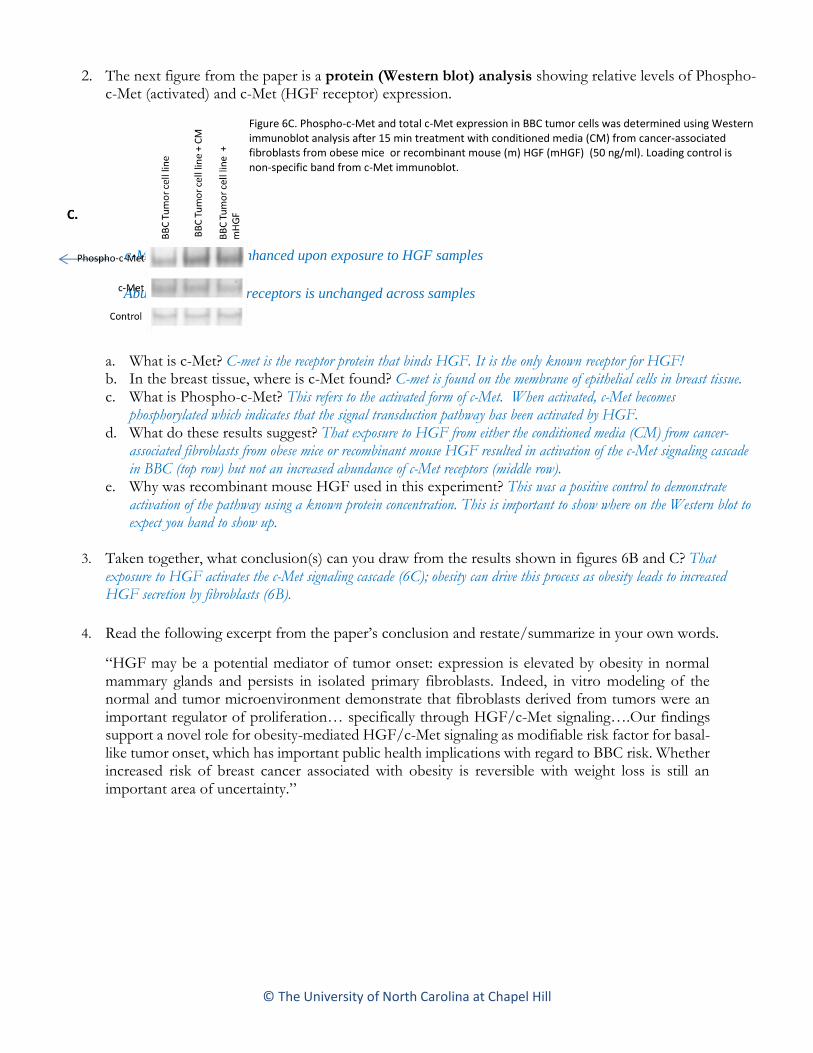
2. The next figure from the paper is a protein (Western blot) analysis showing relative levels of Phospho-c-Met (activated) and c-Met (HGF receptor) expression.
Figure 6C. Phospho-c-Met and total c-Met expression in BBC tumor cells was determined using Western immunoblot analysis after 15 min treatment with conditioned media (CM) from cancer-associated fibroblasts from obese mice or recombinant mouse (m) HGF (mHGF) (50 ng/ml). Loading control is non-specific band from c-Met immunoblot.
a. What is c-Met? C-met is the receptor protein that binds HGF. It is the only known receptor for HGF! b. In the breast tissue, where is c-Met found? C-met is found on the membrane of epithelial cells in breast tissue. c. What is Phospho-c-Met? This refers to the activated form of c-Met. When activated, c-Met becomes
phosphorylated which indicates that the signal transduction pathway has been activated by HGF. d. What do these results suggest? That exposure to HGF from either the conditioned media (CM) from cancer-
associated fibroblasts from obese mice or recombinant mouse HGF resulted in activation of the c-Met signaling cascade in BBC (top row) but not an increased abundance of c-Met receptors (middle row).
e. Why was recombinant mouse HGF used in this experiment? This was a positive control to demonstrate activation of the pathway using a known protein concentration. This is important to show where on the Western blot to expect you band to show up.
3. Taken together, what conclusion(s) can you draw from the results shown in figures 6B and C? That exposure to HGF activates the c-Met signaling cascade (6C); obesity can drive this process as obesity leads to increased HGF secretion by fibroblasts (6B).
4. Read the following excerpt from the paper’s conclusion and restate/summarize in your own words.
“HGF may be a potential mediator of tumor onset: expression is elevated by obesity in normal mammary glands and persists in isolated primary fibroblasts. Indeed, in vitro modeling of the normal and tumor microenvironment demonstrate that fibroblasts derived from tumors were an important regulator of proliferation… specifically through HGF/c-Met signaling….Our findings support a novel role for obesity-mediated HGF/c-Met signaling as modifiable risk factor for basal-like tumor onset, which has important public health implications with regard to BBC risk. Whether increased risk of breast cancer associated with obesity is reversible with weight loss is still an important area of uncertainty.”
Abundance of c-Met receptors is unchanged across samples
c-Met activation is enhanced upon exposure to HGF samples