nucleoredoxin promotes adipogenic … nucleoredoxin promotes adipogenic differentiation through...
TRANSCRIPT
1
Nucleoredoxin promotes adipogenic differentiation through regulation of Wnt/b-catenin
signaling
Young Jae Bahna,b, Kwang-Pyo Leeb, Seung-Min Leeb, Jeong Yi Choib, Yeon-Soo Seoa, and Ki-
Sun Kwonb,*
aDepartment of Biological Science, Korea Advanced Institute Science and Technology (KAIST),
Daejeon 305-701, Republic of Korea;
bAging Research Institute, Korea Research Institute of Bioscience and Biotechnology (KRIBB),
Daejeon 305-806, Republic of Korea
Running Title: Nucleoredoxin regulates adipogenesis.
*To whom the correspondence should be addressed: Ki-Sun Kwon, Aging Research Institute,
Korea Research Institute of Bioscience and Biotechnology (KRIBB), Daejeon 305-806,
Republic of Korea,Tel.: +82-42-860-4143, Fax: +82-42-879-8596, E-mail: [email protected].
by guest, on August 29, 2018
ww
w.jlr.org
Dow
nloaded from

2
ABSTRACT
Nucleoredoxin (NRX) is a member of the thioredoxin family of proteins that controls redox
homeostasis in cell. Redox homeostasis is a well-known regulator of cell differentiation into
various tissue types. We found that NRX expression levels were higher in white adipose tissue
of obese ob/ob mice and increased in the early adipogenic stage of 3T3-L1 preadipocyte
differentiation. Knockdown of NRX decreased differentiation of 3T3-L1 cells, whereas
overexpression increased differentiation. Adipose tissue-specific NRX (Adipo-NRX) transgenic
mice showed increases in adipocyte size as well as number compared to wild-type mice. We
further confirmed that the Wnt/β-catenin pathway was also involved in NRX-promoted
adipogenesis, consistent with a previous report showing NRX regulation of this pathway. Genes
involved in lipid metabolism were downregulated, whereas inflammatory genes, including those
encoding macrophage markers, were significantly upregulated, likely contributing to the obesity
in Adipo-NRX mice. Our results therefore suggest that NRX acts as a novel proadipogenic
factor and controls obesity in vivo.
Supplementary keywords: Nucleoredoxin, Wnt signaling, Adipogenesis, Obesity,
Inflammation
by guest, on August 29, 2018
ww
w.jlr.org
Dow
nloaded from

3
INTRODUCTION
Adipose tissues play important roles in the regulation of whole-body energy homeostasis (1).
Excess energy intake increases the number and size of fat cells (2). These obese adipose tissues
facilitate chronic low-grade inflammation and insulin resistance, which result in severe obesity
and diabetes (3, 4). Thus, understanding adipocyte development and adipogenesis could lay the
groundwork for the development of efficient therapeutic strategies for preventing and treating
metabolic disorders associated with obesity.
Adipogenesis is controlled by a balance of internal and external factors that either stimulate
or repress adipogenic differentiation. In the early phase of adipogenic differentiation, C/EBPβ
(CCAAT/enhancer-binding protein β) and C/EBPδ induce expression of C/EBPα and PPARγ
(peroxisome proliferator-activated receptor gamma), which are the principal adipogenic
transcription factors that control the early differentiation of preadipocytes into lipid-
accumulating fat cells (5, 6). In addition, PPARγ appears to suppress canonical Wnt signaling by
accelerating proteasome-dependent degradation of β-catenin. Conversely, β-catenin, a
transcriptional coactivator in the Wnt signaling pathway, blocks adipogenesis by repressing
PPARγ and C/EBPα (7, 8). A number of reports have suggested a relationship between Wnt
signaling and diabetes and adipogenesis (9, 10). Adipose tissue-specific expression of Wnt10b
reduces adiposity and improves insulin sensitivity in the ob/ob obesity model (11). Although the
extensive down-regulation of β-catenin expression and its anti-adipogenic effects during
adipogenic differentiation have been characterized, less is known about the factors that
modulate β-catenin activity during adipogenesis.
Wnt signaling is initiated upon binding of Wnt ligands to transmembrane Frizzled receptors.
In the canonical Wnt signaling pathway, Frizzled receptors transduce signals through
Dishevelled (Dvl) to inhibit glycogen synthase kinase 3 (GSK3β), resulting in
hypophosphorylation and subsequent stabilization of β-catenin. Following nuclear translocation,
active β-catenin binds to and coactivates members of the T-cell factor/lymphoid-enhancer factor
by guest, on August 29, 2018
ww
w.jlr.org
Dow
nloaded from

4
(TCF/LEF) family of transcription factors, leading to activation of target genes (12, 13).
Nucleoredoxin (NRX) is member of the thioredoxin (TRX) family of proteins. TRX family
proteins commonly possess a pair of redox-active, oxidation-sensitive cysteine residues in the
catalytic center that are directly involved in the reduction of disulfide bonds in target proteins
(14). Although NRX activity has been demonstrated in in vitro assays, whether the TRX-related
oxidoreductase activity of NRX plays a role in vivo is unknown (15). Previous studies have
shown that NRX is a multifunction protein that regulates target proteins through its direct
binding activity rather than its oxidoreductase activity (16, 17). Endogenous NRX protein is
predominantly localized in the cytosol of cells and its transcripts are widely expressed in all
adult tissues (18). Knockout of the NRX gene in mouse embryos is perinatally lethal; NRX-
knockout embryos (day 18.5) are smaller than their wild-type (WT) littermates and exhibit
craniofacial defects with short frontal regions (19). Interestingly, a genomic region around the
mouse NRX gene is involved in type 1 and type 2 diabetes (20). However, a role for NRX in
adipogenesis and obesity has not been reported.
Here, we investigated the role of NRX in preadipocyte differentiation and the obesity
phenotype using a 3T3-L1 preadipocyte differentiation system and adipose tissue-specific
transgenic mice. We show that NRX mediates adipogenesis by modulating β-catenin activity in
vitro and in vivo. Our findings suggest that NRX might act as a proadipogenic factor that is
involved in adipocyte differentiation and aspects of the obesity phenotype.
MATERIAL AND METHODS
Generation of adipose tissue-specific NRX transgenic mice
The LSL-NRX transgenic mice were obtained by microinjection and germ-line transmission
of the transgenic construct (Fig. S1A). The LSL-NRX mouse strain was backcrossed with the
C57BL/6 strain for more than eight generations to create a uniform genetic background.
Adipose tissue-specific NRX transgenic mice (Adipo-NRX) were produced by crossing LSL-
by guest, on August 29, 2018
ww
w.jlr.org
Dow
nloaded from

5
NRX mice with Adiponectin-Cre transgenic mice, generating mice with adipose tissue-specific
overexpression of hNRX. All animal experiments were performed according to protocols
approved by the Animal Care and Use Committee of the Korea Research Institute of Bioscience
and Biotechnology (KRIBB).
Potential founder mice were genotyped by polymerase chain reaction (PCR) analysis using
genomic DNA isolated from mouse tail clips. Primers used for the detection of the transgenic
product were 5′-TGC GAG ATT ACA CCA ACC TG-3’ and 5′-CCC ATA TGT CCT TCC GAG
TG-3’.
Isolation and culturing of primary adipocytes
Primary adipocytes were isolated from epididymal fat pads using the collagenase method, as
described previously (21). Briefly, freshly excised fat pads were minced and digested for 45
minutes to 1 h at 37°C in Krebs-Ringer bicarbonate (pH 7.4) containing 4% bovine serum
albumin and 1.5 mg/ml type I collagenase (Worthington, Lakewood, NJ). The digested tissue
was filtered through a 300-μm nylon mesh to remove undigested tissue and centrifuged at 500 ×
g for 5 minutes. The floating adipocyte fraction was removed, washed with buffer, and re-
centrifuged to isolate free adipocytes. The stromal-vascular pellet was resuspended in
erythrocyte lysis buffer (154 mM NH4Cl, 10 mM KHCO3, and 1 mM EDTA), filtered through a
45-μm nylon mesh to remove endothelial cells, and pelleted at 500 × g for 5 minutes. Enriched
cells were cultured in a growth medium composed of Dulbecco’s Modified Eagle Medium
(DMEM), 20% fetal bovine serum, 100 units/ml penicillin, and 100 mg/ml streptomycin
(Gibco-Invitrogen, Carlsbad, CA) at 37°C in a humidified 5% CO2 atmosphere.
Cell culture and adipogenic differentiation
The preadipocytes cell line 3T3-L1, derived from mouse embryo fibroblasts, was grown at
37°C in DMEM containing 10% heat-inactivated bovine calf serum (Gibco-invitrogen), 100
by guest, on August 29, 2018
ww
w.jlr.org
Dow
nloaded from

6
units/ml penicillin, and 100 mg/ml streptomycin in a humidified 5% CO2 atmosphere. 3T3-L1
cells were induced to differentiate into mature adipocytes according to the procedure of Student
et al.(22), with minor modifications. Briefly, 2 days after reaching confluence (day 0), cells
were placed in differentiation medium composed of DMEM, 10% fetal bovine serum and MDI,
a differentiation cocktail consisting of 0.5 mM IBMX, 1 μM dexamethasone and 10 μg/ml
insulin (Sigma, St. Louis, MO). The medium was replenished every other day.
Generation of stable 3T3-L1 cell lines
3T3-L1 cells stably expressing FLAG-tagged NRX were generated using a lentivirus-
mediated infection system. For expression of NRX, cDNA encoding FLAG-tagged NRX was
cloned into the multi-cloning site of the GFP-tagged pLentiM1.4 vector. Lentiviruses were
subsequently produced by transiently co-transfecting HEK293T cells with pLP1, pLP2, and
pVSV-G plasmid (Invitrogen, Carlsbad, CA) using Lipofectamine (Invitrogen). Forty-eight
hours after transfection, supernatants containing lentiviral particles were collected and used to
infect 3T3-L1 cells in the presence of 4 µg/ml polybrene. Infected cells were selected by
incubation with 2 µg/ml puromycin for 2–3 weeks, and used in experiments as indicated. NRX
cDNA was kindly provided by Dr. Tasuku Honjo (Kyoto University, Japan) (15). An NRX
mutant in which catalytic Cys205 and Cys208 residues were replaced with Ser (CS-NRX)
provided by Dr. Sung-Kyu Ju, was constructed using site-directed mutagenesis.
RNA interference
NRX knockdown in 3T3-L1 cells was accomplished using small hairpin RNA (shRNA)
against mouse NRX in pLKO.1-puro lentiviral vectors obtained from Sigma (clone ID
NM_022463.3-1358s1c1 and NM_022463.3-452s1c1), according to the manufacturer’s protocol.
Briefly, shRNA lentiviral particles were generated in HEK293T cells by transient transfection
with pLP1, pLP2, pVSV-G, and shRNA lentiviral vector or pLKO.1-scrambled (control) vector
by guest, on August 29, 2018
ww
w.jlr.org
Dow
nloaded from

7
(SHC002V; Sigma) using Lipofectamine. Forty-eight hours after transfection, supernatants
containing lentiviral particles were collected and used to infect 3T3-L1 cells in the presence of 4
µg/ml polybrene. Infected cells were selected by incubation with 2 µg/ml puromycin for 2–3
weeks, and used in experiments as indicated. A shRNA-resistant NRX incapable of shNRX
binding and subsequent NRX degradation was constructed by site-directed mutagenesis of the
target region of shNRX. The construct was obtained by standard methods using the primers 5’-
CTT TTG TGA ATG ACT TCT TGG CTG AAA AAC TC-3’ and 5’-TCA GGC TTG AGT TTT
TCAGCCAAG AAG TCA TT-3’. Underlined regions indicate the sites that were mutated
without changing amino acid residues.
RNA extraction and real-time PCR analysis
Total RNA was extracted using the Easy-Blue reagent (iNtRON Biotechnology, Korea),
according to the manufacturer’s instructions, and treated with RNase-free DNase I (Takara,
shiga, Japan) to remove contaminating genomic DNA. cDNA was then synthesized from total
RNA by reverse transcription (RT) using a DiaStar RT kit (Solgent, Korea). Quantitative RT-
PCR analysis was performed using the Step One Plus real-time PCR system (Applied
Biosystems, Waltham, MA) with the corresponding primers.
Immunoblotting and immunoprecipitation
Cells were lysed in a lysis buffer containing 20 mM HEPES (pH 7.2), 150 mM NaCl, 0.5%
Triton X-100, 0.1 mM Na3VO4, 1 mM NaF, 1 mM 4-(2-aminoethyl)-benzenesulfonyl fluoride
hydrochloride (AEBSF), and 5 mg/ml aprotinin (Sigma). Soluble proteins in cell lysates were
separated by sodium dodecyl sulfate-polyacrylamide gel electrophoresis (SDS-PAGE) and
analyzed by immunoblotting using anti-NRX (R&D Biosystems, Minneapolis, MN, AF5719),
anti-PPARγ (Cell Signaling Technology, Danvers, MA, #2430), anti-β-actin (Santa Cruz
Biotechnology, Santa Cruz, CA, SC-47778), anti-Dvl-1 (Santa Cruz Biotechnology, SC-8025),
by guest, on August 29, 2018
ww
w.jlr.org
Dow
nloaded from

8
anti-lamin A/C (Santa Cruz Biotechnology, SC-6215), anti-cyclin D1 (Cell Signaling
Technology, 2922), anti-β-catenin (Cell Signaling Technology, 9562), anti-α-tubulin (Millipore,
05-829), anti-AKT (Santa Cruz Biotechnology, SC-1618), anti-phospho-AKT (Cell Signaling
Technology, 9271L), and anti-FABP4 (Cell Signaling Technology, 2120) antibodies. WAT from
ob/ob mice was kindly provided by Dr. Chul-Ho Lee (KRIBB). For immunoprecipitation,
lysates were incubated with anti-FLAG agarose (Sigma) or anti-NRX antibody at 4°C for
several hours, after which the beads were washed three times with cell lysis buffer. The beads
were resuspended in 1X SDS-PAGE sample buffer and the eluted proteins were resolved by
SDS-PAGE.
In vitro gene transfer
For Amaxa nucleofections, pellets containing 0.5–1.5 × 10⁶ 3T3-L1 cells were carefully
resuspended in 100 µl of Nucleofector solution (Lonza, Allendale, NJ), mixed with 1–2 µg of
plasmids, and subjected to nucleofection using T-30 Amaxa protocols. The cells were then
gently transferred into a 6-well plate and cultured until analysis.
Luciferase assays and reagents.
Luciferase activity was assessed using the Luciferase Assay system (Promega, Madison, WI),
according to the manufacturer’s instructions. 3T3-L1 cells were transduced with TOPflash and
FOPflash exogenous reporter constructs together with pCMV-β-galactosidase. Luciferase
activity was normalized to β-galactosidase activity to adjust for transfection efficiency.
Oil-red O staining
Cultured cells were washed twice with phosphate-buffered saline (PBS) and fixed by
incubating with 3.7% paraformaldehyde for 15 minutes at room temperature. The cells were
then washed with distilled water and stained for 30 minutes with 0.3% filtered Oil-red O
by guest, on August 29, 2018
ww
w.jlr.org
Dow
nloaded from

9
solution in 60% isopropanol. The stained cells were washed twice with distilled water and
photomicrographed. Oil-red-O staining was then quantified as described previously (23).
Incorporated Oil-red O dye was extracted by adding absolute isopropanol to the stained cell-
culture dish and shaking the dish for 30 minutes. Triplicate samples were read at 510 nm using
an Ultrospec2000 Spectrophotometer (Pharmacia Biotech, Piscataway, NJ).
Hematoxylin and eosin staining
Adipose tissue, muscle, and liver samples were fixed for 12–16 h at room temperature in 10%
formalin (Sigma) and then embedded in paraffin. Five-micron sections cut at 50-μm intervals
were mounted on charged glass slides, deparaffinized in xylene, and stained with hematoxylin
and eosin (H&E). Adipocyte cross-sectional area was quantified for each adipocyte in each field
using NIH Image J software (http://rsb.info.nih.gov/ij/). To quantify cell number, adipocytes
were isolated from 100 mg of adipose tissue by using 2 mg/ml collagenase type II-S (Sigma)
digestion buffer and were counted using a cell counter (Logos Biosystems, Annandale, VA) (24).
Glucose and Insulin Tolerance Test
Glucose tolerance test (GTT) was performed with 8h fasted animals. After determination of
fasted blood glucose levels, each animal was injected intraperitoneally (i.p.) with 20% glucose
(1 g/kg). The insulin tolerance test (ITT) was performed, with an initial fasting for 4 h, and
subsequent i.p. injection of insulin (1U/kg). In all tests, tail blood glucose levels were measured
with glucometer (Roche Diagnostics, Mannheim, Germany) at the indicated times after injection.
Immunohistochemistry
Formalin-fixed, paraffin-embedded sections of 5 µm were mounted on charged glass slides,
deparaffinized in xylene, stained with hematoxylin, and processed for immunohistochemical
detection of F4/80 according to standard immunoperoxidase procedure using VECTASTAIN
by guest, on August 29, 2018
ww
w.jlr.org
Dow
nloaded from

10
Elite ABC kit (Vector Labs, Burlingame, CA) and anti-F4/80 antibody (Abcam, Cambridge,
UK).
Statistical analysis
Quantitative data are presented as means ± SD unless indicated otherwise. Differences
between means were evaluated using Student’s unpaired t test. A P-value < 0.05 was considered
statistically significant.
RESULTS
NRX levels increase in the early stage of 3T3-L1 preadipocyte differentiation.
We analyzed the expression of NRX during 3T3-L1 cells differentiation induced by a
differentiation cocktail composed of IBMX (3-isobutyl-1-methylxanthine), dexamethasone, and
insulin. NRX transiently increased in the early stages of adipocyte differentiation at both mRNA
and protein levels. Quantitative reverse transcription-polymerase chain reaction (qRT-PCR)
revealed that NRX mRNA reached a maximum 1 day after differentiation induction (Fig. 1A).
Protein levels of NRX were proportional to mRNA levels (Fig. 1B). We next determined
whether hyperglycemic conditions affected NRX levels in ob/ob mice. Interestingly, NRX
protein was upregulated in white adipose tissue (WAT) of ob/ob mice compared to those in WT
mice (Fig. 1C). These results suggest that increased expression of NRX may be associated with
adipogenesis and, ultimately, obesity.
NRX overexpression leads to adipocyte hypertrophy combined with hyperplasia in mouse
adipose tissue.
To further investigate the function of NRX in adipogenesis in vivo, we generated loxP-stop-
loxP-NRX transgenic mice (LSL-NRX), then crossed these mice with adiponectin-Cre mice to
generate adipose tissue-specific NRX transgenic mice (Adipo-NRX).To validate the specific
by guest, on August 29, 2018
ww
w.jlr.org
Dow
nloaded from

11
overexpression of NRX in adipose tissues, we analyzed NRX protein levels in several tissues
isolated from Adipo-NRX and WT mice. As expected, overexpression of NRX was observed
only in adipose tissues, including white and brown adipose tissue (Fig. S1B). To clarify activity
of the adiponectin promoter, we checked expression levels of adiponectin during adipogenesis.
We confirmed that the expression level of adiponectin was very low before induction of
differentiation, but were gradually increased after induction of differentiation (Fig. S1C), which
was consistent with previous report (25). We also observed that expression of adiponectin
promoter-driven NRX was induced at early differentiation stage in primary adipocytes isolated
from Adipo-NRX mice (Fig. S1D).
Although there was a slight increase in body weight in Adipo-NRX mice compared to WT
littermate controls, a comparison of organ weights revealed a notable increase in epididymal
WAT in Adipo-NRX mice (Fig. 2A). Epididymal and peri-renal fat masses were also
significantly larger in Adipo-NRX mice than in WT mice (Fig. 2B and C). There was no
difference in food intake between Adipo-NRX mice and WT littermate controls. An analysis of
adipocyte cross-sectional areas showed that epididymal fat of Adipo-NRX mice contained
hypertrophied adipocytes (Fig. 2D). A quantitative analysis revealed that adipose tissue
expansion of Adipo-NRX mice was caused by an increase of adipocyte numbers (Fig. 2E) as
well as an enlargement of adipocyte size, suggesting that hypertrophy was accompanied by
hyperplasia in adipose tissue of Adipo-NRX mice.
The plasma analysis revealed that blood glucose and insulin levels were higher in Adipo-
NRX mice than in WT mice (Table S1). We further assessed glucose homeostasis in WT and
Adipo-NRX mice via a glucose tolerance test and insulin tolerance test. Adipo-NRX mice
showed the tendency to glucose intolerance compared to WT mice (Fig. 2F), and exhibited
impaired insulin tolerance that was associated with reduced insulin sensitivity in WAT but not in
liver and skeletal muscle (Fig. 2G and H). Although the adipose tissue appeared lipid
accumulation, there was no significant change in lipid deposition in liver and skeletal muscle
by guest, on August 29, 2018
ww
w.jlr.org
Dow
nloaded from

12
(Fig. S2). These data demonstrate that adipocyte specific NRX overexpression increases lipid
accumulation in WAT, resulting in insulin resistance.
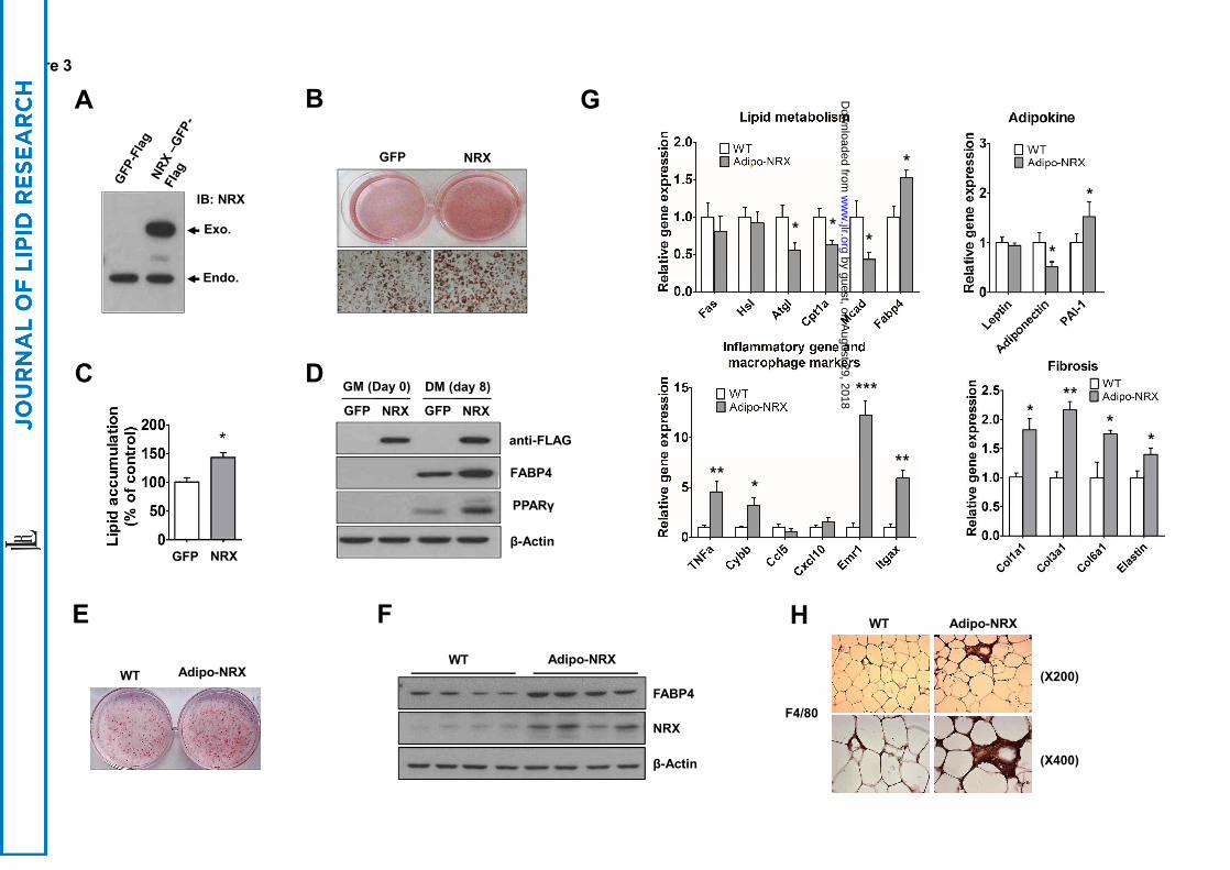
NRX overexpression leads to increases adipogenic differentiation.
To evaluate whether overexpression of NRX facilitates adipogenic differentiation in cell
culure system, we infected 3T3-L1 preadipocytes with a lentivirus expressing GFP-tagged
mouse NRX or a control lentivirus. Stable overexpression of NRX was monitored by
immunoblotting (Fig. 3A) and fluorescence imaging. 3T3-L1 preadipocytes stably
overexpressing NRX showed increased accumulation of lipid droplets following induction of
adipocyte differentiation compared to control cells (Fig. 3B and C). We confirmed that the
adipogenic markers, PPARγ and FABP4 (fatty acid binding protein 4, adipocyte), were also
upregulated in NRX-overexpressing cells compared with control cells (Fig. 3D). To confirm that
NRX overexpression positively regulates adipocyte differentiation, we next isolated and
cultured primary adipocytes from epididymal fat pad of Adipo-NRX and WT mice.
Differentiation of primary adipocytes was increased in Adipo-NRX mice compared with that of
WT mice (Fig. 3E). Consistent with these morphological observations, expression of the
adipogenic marker, FABP4, was also increased in primary adipocytes from Adipo-NRX mice
(Fig. 3F). These data strongly suggest that NRX enhances adipogenesis.
Decreased lipid catabolism and increased inflammation and fibrosis in WAT of Adipo-
NRX mice
We found that expression of enzymes involved in lipid catabolism, including Atgl (adipose
triglyceride lipase), Mcad (acyl-Coenzyme A dehydrogenase, medium chain), and Cpt1a
(carnitine palmitoyltransferase 1a) were downregulated, indicating that adipocytes of Adipo-
NRX mice lacked the ability to burn excess fat. These results suggest that increased fat mass in
Adipo-NRX mice is due to decreased lipolysis and fatty acid oxidation. Since adipokine
by guest, on August 29, 2018
ww
w.jlr.org
Dow
nloaded from

13
dysregulation is a hallmark of adipocyte impairment (26), we further measured adipokine
expression in WAT of Adipo-NRX mice. The mRNA levels of adiponectin were downregulated
and PAI-1 (plasminogen activator inhibitor type 1) was upregulated in WAT of Adipo-NRX
mice.
Obesity has been consistently associated with inflammation (4, 27). We examined mRNA
expression levels of several inflammatory genes in WAT of WT and Adipo-NRX mice. Among
the genes upregulated in Adipo-NRX mice were TNFa (tumor necrosis factor alpha) and
Cxcl10 (chemokine [C-X-C motif] ligand 10), which encode proinflammatory cytokines; Cybb
(cytochrome b-245, beta polypeptide), which is involved in phagocytosis; and Emr1 (EGF-like
module containing, mucin-like, hormone receptor-like 1) and Itgax (integrin alpha X), markers
of macrophage infiltration (Fig. 3G). Consistently, increased macrophage infiltration in WAT
was shown by immunostaining using a macrophage surface marker, F4/80 (Fig. 3H). Obesity is
associated with an overall increase in expression of several collagens that results in fibrotic state
(28, 29). Then, we tested mRNA levels of several fibrosis related genes, and found that Col1a1,
Col3a1, Col6a1, and Elastin were upregulated (Fig. 3G) in WAT of Adipo-NRX. These suggest
that adipocyte expansion is closely linked to inflammation and fibrosis in Adipo-NRX mice.
NRX knockdown attenuates adipogenic differentiation in 3T3-L1 preadipocytes.
To test whether endogenous NRX influences adipogenesis, we examined the effect of NRX
knockdown in 3T3-L1 preadipocytes. 3T3-L1 cells were infected with a lentivirus expressing a
small hairpin RNA (shRNA) targeting NRX (NRX shRNA) or non-targeting (scrambled)
shRNA. Depletion of endogenous NRX was confirmed by immunoblot analyses (Fig. 4A).
Knockdown of NRX attenuated differentiation of 3T3-L1 preadipocytes into mature adipocytes,
reducing accumulation of lipid droplets compared with control 3T3-L1 cells (Fig. 4B).Moreover,
expression levels of the adipocyte markers, PPARγ and FABP4, also were decreased in NRX-
depleted cells compared to control cells (Fig. 4C). Next, to exclude the possibility of off-target
by guest, on August 29, 2018
ww
w.jlr.org
Dow
nloaded from

14
effects of shRNA targeting the NRX coding sequence, we transfected NRX-knockdown 3T3-L1
cells with an expression plasmid for an shRNA-resistant NRX (resNRX) with a codon usage
different from that of the original plasmid. resNRX expression restored PPARγ and FABP4
expression levels (Fig. 4E) as well as accumulation of lipid droplets (Fig. 4D). These results
clearly demonstrate that NRX is involved in adipogenic differentiation.
NRX regulates adipogenesis through modulation of β-catenin signaling in 3T3-L1
preadipocytes.
The conserved Wnt/β-catenin signaling pathway mediates a variety of cellular responses,
from cell proliferation to cell-fate determination (30, 31). Dvl transduces signals from the Wnt
receptor, Frizzled, to downstream components through inhibition of GSK3β, leading to the
stabilization and nuclear translocation of β-catenin (12). It has been reported that NRX regulates
Wnt/β-catenin signaling by directly binding and inhibiting Dvl, and, thereby controls cell
proliferation (16). Wnt/β-catenin signaling is also a central negative regulator of adipogenesis
(7). Here, we found that NRX is involved in adipogenic differentiation, prompting us to
examine whether NRX regulates adipogenesis through Wnt/β-catenin regulation. First, we
tested the interaction between NRX and Dvl in adipocyte. Endogenous Dvl interacted with
ectopically expressed WT NRX, but not with a cysteine-mutant NRX defective in thiol reducing
activity, in 3T3-L1 preadipocytes (Fig. 5A). Moreover, unlike WT NRX, ectopically expressed
cysteine-mutant NRX did not increase 3T3-L1 differentiation (Fig. 5B). These results suggest
that NRX controls adipogenesis by interacting with Dvl through a thiol-based mechanism. We
further observed that endogenous NRX-Dvl binding was dissociated by Wnt3a treatment (Fig.
5C), suggesting that NRX controls adipogenesis via Wnt/β-catenin signaling. To test this
hypothesis, we determined whether NRX controls the nuclear localization of β-catenin in 3T3-
L1 cells and primary adipocytes isolated from Adipo-NRX mice. Nuclear β-catenin levels and
transcriptional activity were increased in NRX-knockdown cells, but were decreased in these
by guest, on August 29, 2018
ww
w.jlr.org
Dow
nloaded from
15
cells following restoration of NRX expression (Fig. 5D). Enhancing NRX expression by
increasing the amount of transfected plasmid induced a dose-dependent decrease in the levels of
nuclear β-catenin and its downstream effector cyclin D1 in Wnt3a-treated 3T3-L1 cells (Fig.
5E), resulting in recovery from Wnt3a-mediated inhibition of adipogenesis (Fig. 5F, third panel).
Consistent with the results obtained in 3T3-L1 preadipocytes (Fig. 5D and E), the expression of
β-catenin target genes was decreased in WAT of Adipo-NRX mice (Fig. 5G).
Next, we examined whether the interaction of NRX with Dvl plays a role in adipogenesis.
While single Dvl gene (Dvl-1, Dvl-2, or Dvl-3) knockdown showed no significant changes in
adipocyte differentiation compared with control group (Fig. S3), triple knockdown of all Dvl
genes enhanced differentiation of 3T3-L1 into mature adipocytes (Fig. 6A and B) accompanying
with an elevated expression of FABP4, an adipocyte differentiation marker, compared to control
(Fig. 6C). To test whether NRX role is dependent on Dvl, we checked the effect of NRX on
adipogenesis in the presence or absence of three Dvls. We found that the Dvls knockdown
suppressed reduced 3T3-L1 differentiation by NRX knockdown (Fig. 6D and E). NRX
knockdown reduced lipid accumulation by 31.2 %, while knockdown of Dvls together reduced
lipid accumulation only by 10.5 %. These results suggest that NRX regulates adipogenesis
through inhibition of Dvl-β-catenin axis in adipocyte differentiation.
DISCUSSION
Our study demonstrated that NRX mRNA and protein were increased in the early stages of
adipocyte differentiation and were also increased in WAT of ob/ob mice, a leptin-deficiency
model of obesity. Employing NRX-depleted and -overexpressing 3T3-L1 preadipocytes, we
showed that NRX is associated with adipogenic differentiation of 3T3-L1 cells. We found that
differentiation of primary adipocytes from Adipo-NRX mice was increased in vitro, and
epididymal and peri-renal fat mass were increased in Adipo-NRX mice in vivo. These
observations, taken together with our in vitro and in vivo adipogenesis data, thus uncover a
by guest, on August 29, 2018
ww
w.jlr.org
Dow
nloaded from

16
novel role for NRX as a proadipogenic factor.
Adipose tissue expansion occurs through an enlargement in adipocyte size (hypertrophy)
and/or an increase in adipocyte number (hyperplasia). We showed that Adipo-NRX mice
exhibited hyperplasia in adipocytes, which is linked to enhanced adipogenic differentiation (32,
33), as well as hypertrophy (Fig. 2D and E). Adipo-NRX mice showed decreased expression of
enzymes involved in lipolysis and fatty acid oxidation in WAT, while unchanged in lipogenic
enzymes compared to WT mice. Decreased triacylglycerol catabolism is associated with the
occurrence of prevalent metabolic diseases, such as obesity and type 2 diabetes (34-36).
Hypertrophic adipocyte in our model is likely caused by the decrease of triacylglycerol
catabolism rather than an increase in triacylglycerol synthesis. However, the mechanism by
which NRX regulates fatty acid catabolism remains to be determined.
Lipid accumulation increased exclusively in WAT of Adipo-NRX mice, with no change in
liver and skeletal muscle compared to WT mice (Fig. S2). In plasma analysis, Adipo-NRX mice
exhibited increased fasting insulin level accompanying with increased fasting glucose level,
suggesting that pancreas function was normal (Table S1). Adipo-NRX mice showed the
tendency to glucose intolerance in GTT analysis (Fig. 2F), and also showed mild insulin
resistance as shown in impaired insulin tolerance and reduced insulin sensitivity in WAT but not
in liver and skeletal muscle (Fig. 2G and H). Therefore, insulin resistance in our model is likely
caused by excessive fat deposition in WAT.
Infiltration of adipose tissue by inflammatory cells has been described as a common feature
of obesity (27). We also found increased expression of inflammatory gene and macrophage
markers as well as dysregulation of adipokines in Adipo-NRX mice (Fig. 3G). Many rodent
models of obesity are associated with chronic inflammation, and it is known that obesity-
induced insulin resistance is strongly correlated with expression of inflammatory markers (37,
38). However, the molecular links between obesity and inflammation have not been completely
elucidated. Adipo-NRX mice might be a potential model system for detailed mechanistic studies
by guest, on August 29, 2018
ww
w.jlr.org
Dow
nloaded from

17
of the relationship between obesity and inflammation.
Wnt signaling is an important regulator of fate decisions in mesenchymal cells (31). In
multipotent mesenchymal precursors in vitro, activation of Wnt/β-catenin signaling promotes
osteoblastogenesis, but inhibits adipogenesis (39). Given that NRX inhibits Wnt/β-catenin
signaling (16), it is possible that NRX reciprocally modulates adipogenic and osteogenic
differentiation. Activation of β-catenin in primary osteoblasts isolated from calvariae of NRX-/-
embryos results in enhanced osteoblastic differentiation (19). However, there have been no
reports of a role for NRX in adipogenesis. Here, we provide the first demonstration that NRX
positively regulates adipogenesis by inhibiting the Wnt/β-catenin pathway. Wnt10b is a likely
candidate for the endogenous Wnt that participates in adipogenesis. The expression level of
Wnt10b, which is highly expressed in preadipocytes, declined upon initiation of differentiation
(40), exhibiting a negative correlation with NRX expression (Fig. 1A).Wnt10b has been
reported to attenuate the development of obesity in ob/ob mice by stabilizing β-catenin and
subsequently inhibiting adipogenesis. Conversely, blocking Wnt signaling promotes
adipogenesis and obesity (11). Based on our data, it is likely that NRX is involved in obesity in
ob/ob mice through regulation of Wnt10b signaling.
Although current evidence suggests that β-catenin functions as a crucial regulator of
adipogenesis (41), how its expression and activity are regulated during adipogenic
differentiation, particularly the factors involved, remains largely unknown. During adipogenesis,
there is a considerable reduction in total β-catenin protein as well as a marked decrease in
nuclear β-catenin levels (8, 42).In addition to regulating β-catenin protein levels, we found that
NRX directly interacted with and inhibited the Wnt/β-catenin effector molecule Dvl during
adipogenesis. Both aspects of this negative regulation of Wnt/β-catenin signaling by NRX
enabled induction of the adipogenic transcription factor PPARγ, which, in turn, downregulated
β-catenin levels, ensuring complete terminal differentiation into mature adipocytes.
Our current findings show that the active site cysteine of NRX is required for proper
by guest, on August 29, 2018
ww
w.jlr.org
Dow
nloaded from

18
inhibition of the Wnt/β-catenin pathway. However, we do not yet know how redox controls the
inhibitory function of NRX in Wnt/β-catenin signaling, although we presume that thiol-disulfide
exchange of the active site cysteine residues leads to a conformational change in the NRX
molecule that modulates its inhibitory function. Further studies will be needed to confirm this.
In conclusion, our study provides the first evidence that NRX acts as a proadipogenic factor
by regulating Wnt signaling and is associated with phenotypic manifestations of obesity.
Therefore, we propose that modulating Wnt/β-catenin signaling by blocking NRX may
ultimately prove to be an effective therapeutic strategy for managing obesity and metabolic
disorders such as diabetes.
ACKNOWLEDGMENTS
We thank Dr. Dae-Sik Lim (KAIST) for kindly providing Adiponectin-Cre transgenic mice. We
are grateful to Dr. Tasuku Honjo (Kyoto University, Japan) for NRX cDNA. This study was
supported by grants from the Bio & Medical Technology Development Program (20110030133
and 2013M3A9B6076413, to K.-S.K.) of the National Research Foundation funded by the
Korea government (MSIP), and the KRIBB Research Initiative Program.
REFERENCES
1. Spiegelman, B. M., and J. S. Flier. 2001. Obesity and the regulation of energy balance.
Cell 104: 531-543.
2. Kopelman, P. G. 2000. Obesity as a medical problem. Nature 404: 635-643.
3. Wellen, K. E., and G. S. Hotamisligil. 2003. Obesity-induced inflammatory changes in
adipose tissue. The Journal of clinical investigation 112: 1785-1788.
4. Xu, H., G. T. Barnes, Q. Yang, G. Tan, D. Yang, C. J. Chou, J. Sole, A. Nichols, J. S.
by guest, on August 29, 2018
ww
w.jlr.org
Dow
nloaded from

19
Ross, L. A. Tartaglia, and H. Chen. 2003. Chronic inflammation in fat plays a crucial
role in the development of obesity-related insulin resistance. The Journal of clinical
investigation 112: 1821-1830.
5. Farmer, S. R. 2006. Transcriptional control of adipocyte formation. Cell metabolism 4:
263-273.
6. Rosen, E. D., and B. M. Spiegelman. 2000. Molecular regulation of adipogenesis.
Annual review of cell and developmental biology 16: 145-171.
7. Ross, S. E., N. Hemati, K. A. Longo, C. N. Bennett, P. C. Lucas, R. L. Erickson, and O.
A. MacDougald. 2000. Inhibition of adipogenesis by Wnt signaling. Science 289: 950-
953.
8. Moldes, M., Y. Zuo, R. F. Morrison, D. Silva, B. H. Park, J. Liu, and S. R. Farmer. 2003.
Peroxisome-proliferator-activated receptor gamma suppresses Wnt/beta-catenin
signalling during adipogenesis. The Biochemical journal 376: 607-613.
9. Grant, S. F., G. Thorleifsson, I. Reynisdottir, R. Benediktsson, A. Manolescu, J. Sainz,
A. Helgason, H. Stefansson, V. Emilsson, A. Helgadottir, U. Styrkarsdottir, K. P.
Magnusson, G. B. Walters, E. Palsdottir, T. Jonsdottir, T. Gudmundsdottir, A. Gylfason,
J. Saemundsdottir, R. L. Wilensky, M. P. Reilly, D. J. Rader, Y. Bagger, C. Christiansen,
V. Gudnason, G. Sigurdsson, U. Thorsteinsdottir, J. R. Gulcher, A. Kong, and K.
Stefansson. 2006. Variant of transcription factor 7-like 2 (TCF7L2) gene confers risk of
type 2 diabetes. Nature genetics 38: 320-323.
10. Longo, K. A., W. S. Wright, S. Kang, I. Gerin, S. H. Chiang, P. C. Lucas, M. R. Opp,
and O. A. MacDougald. 2004. Wnt10b inhibits development of white and brown
adipose tissues. The Journal of biological chemistry 279: 35503-35509.
11. Wright, W. S., K. A. Longo, V. W. Dolinsky, I. Gerin, S. Kang, C. N. Bennett, S. H.
Chiang, T. C. Prestwich, C. Gress, C. F. Burant, V. S. Susulic, and O. A. MacDougald.
2007. Wnt10b inhibits obesity in ob/ob and agouti mice. Diabetes 56: 295-303.
by guest, on August 29, 2018
ww
w.jlr.org
Dow
nloaded from

20
12. Behrens, J., B. A. Jerchow, M. Wurtele, J. Grimm, C. Asbrand, R. Wirtz, M. Kuhl, D.
Wedlich, and W. Birchmeier. 1998. Functional interaction of an axin homolog,
conductin, with beta-catenin, APC, and GSK3beta. Science 280: 596-599.
13. Nelson, W. J., and R. Nusse. 2004. Convergence of Wnt, beta-catenin, and cadherin
pathways. Science 303: 1483-1487.
14. Lillig, C. H., and A. Holmgren. 2007. Thioredoxin and related molecules--from biology
to health and disease. Antioxidants & redox signaling 9: 25-47.
15. Kurooka, H., K. Kato, S. Minoguchi, Y. Takahashi, J. Ikeda, S. Habu, N. Osawa, A. M.
Buchberg, K. Moriwaki, H. Shisa, and T. Honjo. 1997. Cloning and characterization of
the nucleoredoxin gene that encodes a novel nuclear protein related to thioredoxin.
Genomics 39: 331-339.
16. Funato, Y., T. Michiue, M. Asashima, and H. Miki. 2006. The thioredoxin-related
redox-regulating protein nucleoredoxin inhibits Wnt-beta-catenin signalling through
dishevelled. Nature cell biology 8: 501-508.
17. Hayashi, T., Y. Funato, T. Terabayashi, A. Morinaka, R. Sakamoto, H. Ichise, H. Fukuda,
N. Yoshida, and H. Miki. 2010. Nucleoredoxin negatively regulates Toll-like receptor 4
signaling via recruitment of flightless-I to myeloid differentiation primary response
gene (88). The Journal of biological chemistry 285: 18586-18593.
18. Funato, Y., and H. Miki. 2007. Nucleoredoxin, a novel thioredoxin family member
involved in cell growth and differentiation. Antioxidants & redox signaling 9: 1035-
1057.
19. Funato, Y., T. Terabayashi, R. Sakamoto, D. Okuzaki, H. Ichise, H. Nojima, N. Yoshida,
and H. Miki. 2010. Nucleoredoxin sustains Wnt/beta-catenin signaling by retaining a
pool of inactive dishevelled protein. Current biology : CB 20: 1945-1952.
20. Babaya, N., H. Ikegami, T. Fujisawa, K. Nojima, M. Itoi-Babaya, K. Inoue, T. Ohno, M.
Shibata, and T. Ogihara. 2005. Susceptibility to streptozotocin-induced diabetes is
by guest, on August 29, 2018
ww
w.jlr.org
Dow
nloaded from

21
mapped to mouse chromosome 11. Biochemical and biophysical research
communications 328: 158-164.
21. Soukas, A., N. D. Socci, B. D. Saatkamp, S. Novelli, and J. M. Friedman. 2001. Distinct
transcriptional profiles of adipogenesis in vivo and in vitro. The Journal of biological
chemistry 276: 34167-34174.
22. Student, A. K., R. Y. Hsu, and M. D. Lane. 1980. Induction of fatty acid synthetase
synthesis in differentiating 3T3-L1 preadipocytes. The Journal of biological chemistry
255: 4745-4750.
23. Ramirez-Zacarias, J. L., F. Castro-Munozledo, and W. Kuri-Harcuch. 1992.
Quantitation of adipose conversion and triglycerides by staining intracytoplasmic lipids
with Oil red O. Histochemistry 97: 493-497.
24. Carnevalli, L. S., K. Masuda, F. Frigerio, O. Le Bacquer, S. H. Um, V. Gandin, I.
Topisirovic, N. Sonenberg, G. Thomas, and S. C. Kozma. 2010. S6K1 plays a critical
role in early adipocyte differentiation. Developmental cell 18: 763-774.
25. Diez, J. J., and P. Iglesias. 2003. The role of the novel adipocyte-derived hormone
adiponectin in human disease. European journal of endocrinology / European
Federation of Endocrine Societies 148: 293-300.
26. Huh, J. Y., Y. Kim, J. Jeong, J. Park, I. Kim, K. H. Huh, Y. S. Kim, H. A. Woo, S. G.
Rhee, K. J. Lee, and H. Ha. 2012. Peroxiredoxin 3 is a key molecule regulating
adipocyte oxidative stress, mitochondrial biogenesis, and adipokine expression.
Antioxidants & redox signaling 16: 229-243.
27. Weisberg, S. P., D. McCann, M. Desai, M. Rosenbaum, R. L. Leibel, and A. W. Ferrante,
Jr. 2003. Obesity is associated with macrophage accumulation in adipose tissue. The
Journal of clinical investigation 112: 1796-1808.
28. Halberg, N., T. Khan, M. E. Trujillo, I. Wernstedt-Asterholm, A. D. Attie, S. Sherwani,
Z. V. Wang, S. Landskroner-Eiger, S. Dineen, U. J. Magalang, R. A. Brekken, and P. E.
by guest, on August 29, 2018
ww
w.jlr.org
Dow
nloaded from

22
Scherer. 2009. Hypoxia-inducible factor 1alpha induces fibrosis and insulin resistance
in white adipose tissue. Molecular and cellular biology 29: 4467-4483.
29. Wang, Q. A., P. E. Scherer, and R. K. Gupta. 2014. Improved methodologies for the
study of adipose biology: insights gained and opportunities ahead. Journal of lipid
research 55: 605-624.
30. Clevers, H. 2006. Wnt/beta-catenin signaling in development and disease. Cell 127:
469-480.
31. Teo, R., F. Mohrlen, G. Plickert, W. A. Muller, and U. Frank. 2006. An evolutionary
conserved role of Wnt signaling in stem cell fate decision. Developmental biology 289:
91-99.
32. Wang, Q. A., C. Tao, R. K. Gupta, and P. E. Scherer. 2013. Tracking adipogenesis
during white adipose tissue development, expansion and regeneration. Nature medicine
19: 1338-1344.
33. Sun, K., C. M. Kusminski, and P. E. Scherer. 2011. Adipose tissue remodeling and
obesity. The Journal of clinical investigation 121: 2094-2101.
34. Langin, D., A. Dicker, G. Tavernier, J. Hoffstedt, A. Mairal, M. Ryden, E. Arner, A.
Sicard, C. M. Jenkins, N. Viguerie, V. van Harmelen, R. W. Gross, C. Holm, and P.
Arner. 2005. Adipocyte lipases and defect of lipolysis in human obesity. Diabetes 54:
3190-3197.
35. Zechner, R., P. C. Kienesberger, G. Haemmerle, R. Zimmermann, and A. Lass. 2009.
Adipose triglyceride lipase and the lipolytic catabolism of cellular fat stores. Journal of
lipid research 50: 3-21.
36. Jocken, J. W., D. Langin, E. Smit, W. H. Saris, C. Valle, G. B. Hul, C. Holm, P. Arner,
and E. E. Blaak. 2007. Adipose triglyceride lipase and hormone-sensitive lipase protein
expression is decreased in the obese insulin-resistant state. The Journal of clinical
endocrinology and metabolism 92: 2292-2299.
by guest, on August 29, 2018
ww
w.jlr.org
Dow
nloaded from

23
37. Blackburn, P., J. P. Despres, B. Lamarche, A. Tremblay, J. Bergeron, I. Lemieux, and C.
Couillard. 2006. Postprandial variations of plasma inflammatory markers in
abdominally obese men. Obesity 14: 1747-1754.
38. Aron-Wisnewsky, J., J. Tordjman, C. Poitou, F. Darakhshan, D. Hugol, A. Basdevant, A.
Aissat, M. Guerre-Millo, and K. Clement. 2009. Human adipose tissue macrophages:
m1 and m2 cell surface markers in subcutaneous and omental depots and after weight
loss. The Journal of clinical endocrinology and metabolism 94: 4619-4623.
39. Cawthorn, W. P., A. J. Bree, Y. Yao, B. Du, N. Hemati, G. Martinez-Santibanez, and O.
A. MacDougald. 2012. Wnt6, Wnt10a and Wnt10b inhibit adipogenesis and stimulate
osteoblastogenesis through a beta-catenin-dependent mechanism. Bone 50: 477-489.
40. Bennett, C. N., S. E. Ross, K. A. Longo, L. Bajnok, N. Hemati, K. W. Johnson, S. D.
Harrison, and O. A. MacDougald. 2002. Regulation of Wnt signaling during
adipogenesis. The Journal of biological chemistry 277: 30998-31004.
41. Kennell, J. A., and O. A. MacDougald. 2005. Wnt signaling inhibits adipogenesis
through beta-catenin-dependent and -independent mechanisms. The Journal of
biological chemistry 280: 24004-24010.
42. Hou, J. C., S. Shigematsu, H. C. Crawford, P. Z. Anastasiadis, and J. E. Pessin. 2006.
Dual regulation of Rho and Rac by p120 catenin controls adipocyte plasma membrane
trafficking. The Journal of biological chemistry 281: 23307-23312.
FOOTNOTES
Abbreviations: Fabp4, fatty acid binding protein 4; C/EBPα, CCAAT/enhancer-binding protein
α; PPARγ, peroxisome proliferator-activated receptor gamma; TNFa, tumor necrosis factor
alpha; Cxcl10, chemokine [C-X-C motif] ligand 10); Cybb, cytochrome b-245, beta polypeptide;
Emr1, EGF-like module containing, mucin-like, hormone receptor-like 1; Itgax, integrin alpha
by guest, on August 29, 2018
ww
w.jlr.org
Dow
nloaded from

24
X; Atgl, adipose triglyceride lipase; Mcad, acyl-Coenzyme A dehydrogenase, medium chain;
Cpt1a, carnitine palmitoyltransferase 1a; PAI-1, plasminogen activator inhibitor type 1; FAS,
fatty acid synthase; HSL, hormone sensitive lipase; Ccl5, Chemokine (C-C motif) ligand 5;
MMP-7, matrix metalloproteinase-7; NRX, nucleoredoxin; Dvl, Dishevelled; Adipo-NRX mice,
adipose tissue-specific NRX transgenic mice; LSL-NRX mice, loxP-stop-loxP-NRX transgenic
mice.
FIGURE LEGENDS
Fig. 1. NRX expression during adipogenic differentiation of 3T3-L1 preadipocytes and in
WAT of obese mice. (A-B) 3T3-L1 cells were induced to differentiate into adipocytes by
treatment with MDI medium. A) Expression of NRX was analyzed using real-time quantitative
RT-PCR. Total RNA was extracted at the indicated days of differentiation. β-Actin was used as
an internal control. B) NRX expression was analyzed by Western blotting at the indicated days
of differentiation. PPARγ and FABP4 were used as adipocyte differentiation markers. β-Actin
was used as a loading control. C) Western blot analysis of NRX expression in visceral WAT of
WT and ob/ob mice. Data are presented as mean fold-changes ± SD (*P<0.05, **P<0.01).
Fig. 2. Adipo-NRX transgenic mice have larger amounts of adipose tissue compared to WT
mice. A) Body weights and organ weights of 6-month-old Adipo-NRX and WT mice were
determined (n=6). B, C) Representative pictures of epididymal and peri-renal fat dissected from
fat pads of Adipo-NRX and WT mice. D) H&E staining of WAT from epididymal fat pads of
Adipose-NRX and WT mice (left). Quantitative analysis of cell numbers/mm2 in sections of
WAT (right) E) Total cell number of adipocytes per fat fad isolated from WT and Adipo-NRX
mice (n=3). F) Glucose tolerance test and G) Insulin tolerance test in WT and Adipo-NRX mice.
Total area under curve (AUC) of each graph was measured (insets). Fasted mice were injected
by guest, on August 29, 2018
ww
w.jlr.org
Dow
nloaded from

25
with glucose (1g/kg i.p.) or insulin (1U/kg i.p.). Blood glucose levels (mg/dL) were determined
at the indicated time points. Values are means ±SD of 6-8 mice. H) Western blotting analysis of
phospho-AKT (AKT-S473 phosphorylation) in indicated tissues isolated from WT and Adipo-
NRX mice (n=3) after i.p. injection of insulin (10U/kg). The symbol “ ̶ ” or “+” means without
or with insulin stimulation, respectively. Data are presented as mean percentages ± SD (*P<0.05,
**P <0.01).
Fig. 3. Overexpression of NRX promotes adipogenic differentiation of 3T3-L1
preadipocytes and primary adipocytes. 3T3-L1 cells were infected with lentiviruses
expressing NRX (pLenti-NRX-GFP-flag) or control (pLenti-GFP-flag), selected with
puromycin, and induced to differentiate using MDI medium. A) Ectopic expression of NRX was
monitored by Western blot analysis with anti-NRX antibody. B) After 8 days of differentiation,
the degree of lipid accumulation was visualized by staining cells with Oil red-O. C) Staining in
cells was quantified using a dye-extraction solution (n=4). D) Whole-cell lysates were extracted
on day 0 or day 8 for Western blot analysis of NRX, PPARγ, FABP4, and β-actin. E) Primary
white adipocytes isolated from Adipo-NRX and WT mice were cultured in MDI medium
containing rosiglitazone to induce differentiation into adipocytes. After 10 days of
differentiation, cells were stained with Oil red-O dye. F) Whole-cell lysates were extracted from
differentiated white adipocytes on day 10 for Western blot analysis of NRX, FABP4, and β-actin
(n=4). G) Relative mRNA abundance was examined in WAT from WT and Adipo-NRX mice by
real-time quantitative RT-PCR (n=3). Data are presented as mean fold-changes ± SD (*P<0.05,
**P<0.01, ***P<0.001). H) Frozen sections of WAT isolated from WT and Adipo-NRX were
immunostained with antibody againstF4/80, a macrophage surface marker. Images were taken at
magnification X200 (upper panels) and X400 (lower panels).
Fig. 4. NRX shRNA attenuates adipogenic differentiation in 3T3-L1 preadipocytes. A)
by guest, on August 29, 2018
ww
w.jlr.org
Dow
nloaded from
26
NRX protein levels in NRX-knockdown and control, non-target (NT) 3T3-L1 cells were
monitored by Western blot analysis with anti-NRX antibody. B) NRX-knockdown and control
3T3-L1 cells were differentiated for 8 days in MDI medium and then stained with Oil red-O dye.
C) After induction of differentiation, whole-cell lysates were prepared from non-target and
NRX-knockdown cells on the indicate days for Western blot analysis of NRX, PPARγ, FABP4,
and β-actin. β-Actin was used as a loading control. D) Rescue experiments were performed by
transfecting NRX-knockdown 3T3-L1 cells with plasmids encoding shRNA-resistant NRX
(resNRX). Eight days after differentiation, cells were stained with Oil red-O dye. E) Analysis of
protein levels during adipogenic differentiation of NRX-knockdown and NRX-rescued cells.
Whole-cell lysates were extracted on day 8 for Western blot analysis of NRX, PPARγ, FABP4,
and β-actin.
Fig. 5. NRX regulates adipogenesis via modulation of β-catenin in 3T3-L1 cells. (A-B) WT
NRX interacts with Dvl and increases 3T3-L1 differentiation. A) 3T3-L1 cells were transfected
with vectors expressing FLAG-tagged WT or mutant (Mut) NRX, lysed and
immunoprecipitated with anti-FLAG antibody, and analyzed by immunoblotting with anti-Dvl
antibody. B) After 8 days of incubation in differentiation media, cells were stained with Oil red-
O dye. C) 3T3-L1 cell lysates were immunoprecipitated using anti-NRX antibodies after
treatment with 200 ng/ml Wnt3a followed by immunoblotting, as indicated. (D-F) NRX inhibits
β-catenin activity and promotes recovery from Wnt3a-mediated inhibition of adipogenesis. D)
Reporter gene (TOPflash) expression assays in NRX-knockdown 3T3-L1 cells after
cotransfection of increasing amounts of resNRX. Nuclear (N) and cytosolic (C) fractions were
analyzed by immunoblotting. Lamin A/C was used as a control for the nuclear fraction. E)
3T3-L1 cells transfected with vectors expressing FLAG-tagged NRX were treated with Wnt3a.
Cytosolic and nuclear proteins were isolated for Western blot analysis of FLAG-NRX, β-catenin,
lamin A/C, and cyclin D1. F) 3T3-L1 cells overexpressing NRX were induced to differentiate
by guest, on August 29, 2018
ww
w.jlr.org
Dow
nloaded from
27
into adipocytes with Wnt3a for the first 2 days. After 8 days of differentiation, cells were stained
with Oil red-O dye. Whole-cell lysates were prepared on day 8 for Western blot analysis of
NRX, FABP4, and β-actin. G) β-Catenin target genes were examined in WAT from WT and
Adipo-NRX mice by real-time quantitative RT-PCR (n=3). Data are presented as mean fold-
changes ± SD (*P<0.05, **P<0.01)
Fig. 6. Dvls Knockdown suppresses reduced adipogenesis in NRX-depleted 3T3-L1 cells. A)
After transfection with three siRNAs targeting Dvl-1, 2 and 3 together siRNAs (Dvls siRNA) in
3T3-L1, relative mRNA levels of Dvls were analyzed using real-time quantitative RT-PCR
compared to control siRNA transfected cells. B) Representative images of lipid accumulation of
control or Dvls siRNA transfected cells were taken by Oil red O staining at 8 days post
adipogenic differentiation. C) Relative mRNA levels of FABP4 were analyzed using real-time
quantitative RT-PCR at 4 (left) or 8 (right) day post adipogenic differentiation. D)
Representative Oil red-O stained images of control or NRX-depleted 3T3-L1 cells transfected
with indicated siRNAs. At 8day post adipogenic differentiation, cells were stained with Oil red-
O dye. E) Oil red-O Stained cells were quantified using a dye-extraction solution (n=3).
by guest, on August 29, 2018
ww
w.jlr.org
Dow
nloaded from
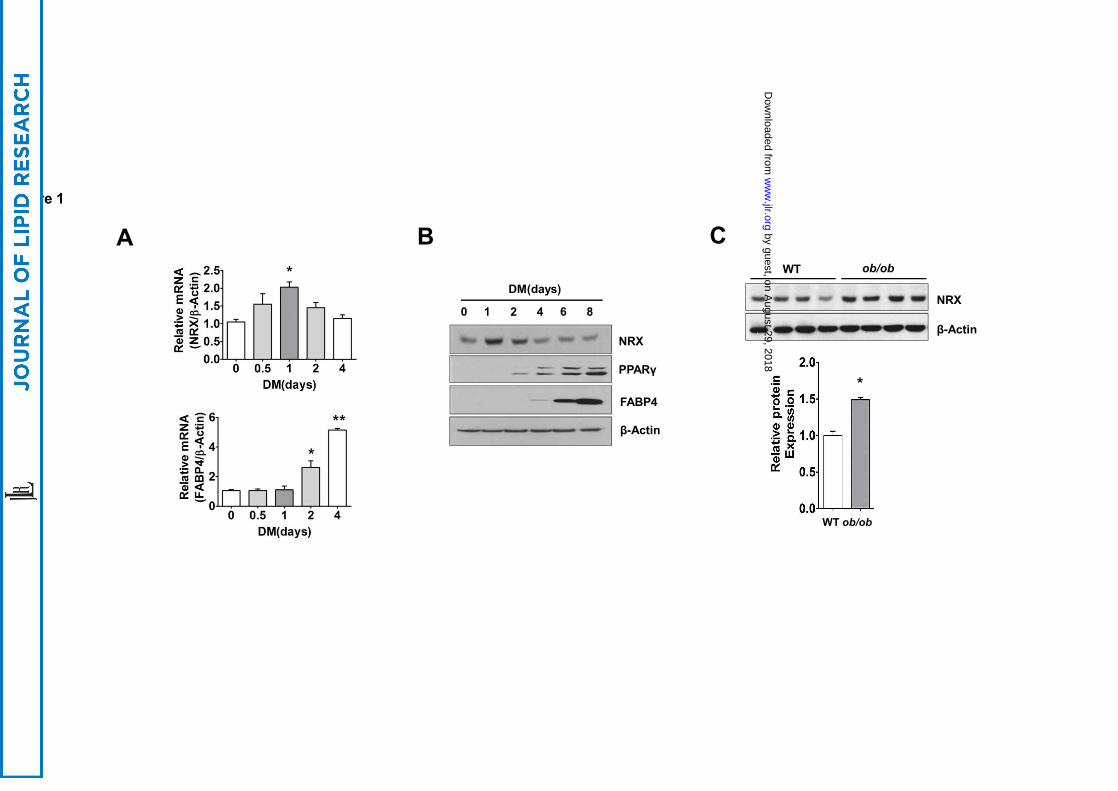
Figure 1
Bob/obWT
β-Actin
NRX
C
DM(days)
PPARγ
NRX
β-Actin
0 1 2 4 6 8
FABP4
A
*
WT ob/ob
*
*
**
by guest, on August 29, 2018
ww
w.jlr.org
Dow
nloaded from

G
WT
Adip
o-N
RX
Figure 2
AW
T
Adip
o-N
RX
WT
Adip
o-N
RX
WT
Adip
o-N
RX
WT
Adip
o-N
RX
WT
Adip
o-N
RX
WT Adipo-NRX
F
WT Adipo-NRX
Epididymal fat
Adipo-NRXWT
Peri-renal fat
D E
H
B C
Sk. Muscle
+ + +
WT Adipo-NRX
WAT
Insulin
Liverp-AKT
AKT
p-AKT
AKT
+ + +
p-AKT
AKT
** * *
*
Adip
o-N
RX
WT
***
by guest, on August 29, 2018
ww
w.jlr.org
Dow
nloaded from
NRX
anti-FLAG
FABP4
β-Actin
GFP
PPARγ
NRXGFP
GM (Day 0) DM (day 8)
GFP NRX
IB: NRX
Endo.
Exo.
GFP NRX
A
E
C
WT Adipo-NRX
D
F
BFigure 3
FABP4
NRX
β-Actin
WT Adipo-NRX
G
* **
*
*
*
*
***
****
H WT Adipo-NRX
F4/80
(X400)
(X200)
***
**
by guest, on August 29, 2018
ww
w.jlr.org
Dow
nloaded from

Figure 4
β-actin
NRX
NT shNRXNT shNRX
NT
FABP4
shNRX
PPARγ
NRX
β-Actin
DM(days)0 1 2 4 6 80 1 2 4 6 8
A B
C
NT shNRX shNRX +resNRX
PPARγ
β-Actin
FABP4
NT shNRX
GM (Day 0) DM (day 8)
NRX
shNRX+ resNRX NT shNRX shNRX+
resNRX
D
E by guest, on A
ugust 29, 2018w
ww
.jlr.orgD
ownloaded from

Figure 5
A
IP:anti-FLAG
FLAG-WT NRX
FLAG-Mut NRX+
+
WCL
NRX
Dvl-1
anti-FLAG
Dvl-1
--
--
B
0 15 30 (min)
WCL
IP: NRX
NRX
Dvl-1
Wnt3a
NRX
Dvl-1
C
NT shNRX
Flag-NRX Nuc.β-catenin
NRX
Lamin A/C
CyclinD1
+ ++--
N
C
D
F
GFP NRX
+ Wnt3a(mock) + Wnt3a
Nuc.β-catenin
anti-FLAG
FLAG-NRX
Lamin A/C
+ Wnt3a
CyclinD1N
C
+ ++- +++
E
G
NRX
β-Actin
FABP4
Wnt3a+ +
GFP
-NRX
Control WT NRX Mut NRX
1 2 3 4 1 2 3 4
*
*
**
*
*
β-catenin target genesTopFlash
by guest, on August 29, 2018
ww
w.jlr.org
Dow
nloaded from

Figure 6
A B
C
shNRX +Dvls siRNA
D
contr. siRNA
DvlssiRNA
NT +Dvls siRNA
E
shNRX +Contr. siRNA
NT +Contr. siRNA
contr. siRNA Dvls siRNA
by guest, on August 29, 2018
ww
w.jlr.org
Dow
nloaded from