ntp technical report on toxicity studies of · ntp technical report on toxicity studies of ....
TRANSCRIPT

National Toxicology Program Toxicity Report Series
Number 15
NTP Technical Report on Toxicity Studies of
t-Butyl Perbenzoate (CAS NUMBER: 614-45-9)
Administered by Gavage to F344/N Rats and B6C3F1 Mice
H.B. Matthews, PhD, Study Scientist National Toxicology Program
Post Office Box 12233 Research Triangle Park, NC 27709
NIH Publication No. 92-3134 July 1992
United States Department of Health and Human Services Public Health Service
National Institutes of Health

2 t-BUTYL PERBENZOATE, NTP TOXICITY REPORT NUMBER 15
CONTRIBUTORS
The NTP report on the toxicity studies of t-butyl perbenzoate is based primarily on 14-day and 13-week studies performed over the period between March, 1985, and July, 1988, at Battelle Memorial Laboratories, Columbus, OH; and on disposition and stability studies performed at Research Triangle Institute, Research Triangle Park, NC.
National Toxicology Program Research Triangle Institute Evaluated experiment, interpreted results, Conducted disposition studies and reported findings
Robert Jeffcoat, PhDH.B. Matthews, PhD Principal Investigator
Study Scientist Monroe Wall, PhDJohn Bucher, PhD Co-Study Director
Leo T. Burka, PhD Thorir Bjornsson, PhD PharmacokineticsMichael Elwell, DVM, PhD
Donald Feldman, DVMJoel Mahler, DVM Study Veterinarian
Morrow Thompson, DVM, PhD Timothy Tippin, BAErrol Zeiger, PhD Chemist
Coordinated Report Preparation
Experimental Pathology Laboratories, Inc.Jane Lambert, BS Provided pathology quality assurance
Diane Overstreet, BS Kristine Witt, MS John Peckham, DVM
Oak Ridge Associated Universities
NTP Pathology Working Group Analytical Sciences, Inc.Evaluated slides and prepared pathology report Provided statistical analysis
Katharina Heider, DVM Richard Morris, MSChairperson CIBA-GEIGY Steven Seilkop, MS
Michael Elwell, DVM, PhD Janet Teague, MS National Toxicology Program
Jeffrey Everitt, DVM CIIT
Joel Leininger, DVM, PhD National Toxicology Program
Margarita McDonald, DVM, PhD National Toxicology Program
John Peckham, DVM Experimental Pathology Laboratories, Inc.
Battelle Columbus Laboratories Principal contributors
Arthur C. Peters, DVM Principal Investigator
M. Hejtmancik, PhD Study Director, Toxicologist
Ming Chang, PhD Chemistry
P. Jepsen, DVM, MS Study Veterinarian
L. Mezza, DVM, MS Pathology

3 t-BUTYL PERBENZOATE, NTP TOXICITY REPORT NUMBER 15
CONTENTS
CONTRIBUTORS.......................................................................................................................2
TABLE OF CONTENTS................................................................................................................3
ABSTRACT ..............................................................................................................................5
PEER REVIEW PANEL................................................................................................................7
PEER REVIEW SUMMARY..........................................................................................................8
INTRODUCTION........................................................................................................................9
MATERIALS AND METHODS .....................................................................................................11
Procurement and Characterization of t-Butyl Perbenzoate ................................................11 Animals ..........................................................................................................................11 Stability Studies .............................................................................................................12 Disposition Studies ........................................................................................................12 14-Day Study Design.......................................................................................................13 13-Week Study Design ....................................................................................................13 Statistical Methods.........................................................................................................14 Quality Assurance...........................................................................................................14
RESULTS ..............................................................................................................................16
Stability and Disposition Studies ....................................................................................16 14-Day Toxicity Studies in F344/N Rats...........................................................................19 13-Week Toxicity Studies in F344/N Rats ........................................................................20 14-Day Toxicity Studies in B6C3F1 Mice...........................................................................25 13-Week Toxicity Studies in B6C3F1 Mice ........................................................................27 Genetic Toxicology ..........................................................................................................29
DISCUSSION ..........................................................................................................................30
REFERENCES .........................................................................................................................33
TABLES
Table 1 Experimental Design and Materials and Methods in the 14-Day and 13-Week Gavage Studies of t-Butyl Perbenzoate............................................14
Table 2 Stability of t-Butyl Perbenzoate on Isolated Rat Skin...........................................17
Table 3 Cumulative Excretion of 14C after Dermal Application of [14C]-t-Butyl Perbenzoate to F344/N Rats ...........................................................17
Table 4 Cumulative Excretion of Total 14C after Intravenous Administration of 3.7 mg/kg of [14C]-t-Butyl Perbenzoate to Male F344/N Rats............................18
Table 5 Concentration of t-Butyl Perbenzoate-derived 14C in Tissues 24 Hours after Intravenous or Dermal Administration of [14C]-t-Butyl Perbenzoate .............18
Table 6 Survival and Weight Gain of F344/N Rats in the 14-Day Gavage Studies of t-Butyl Perbenzoate, t-Butanol, and Benzoic Acid.............................................19

able 7 Survival and Weight Gain of F344/N Rats in the 13-Week Gavage Studies of t-Butyl Perbenzoate ........................................................................................20
able 8 Histopathologic Lesions in F344/N Rats in the 13-Week Gavage Studies of t-Butyl Perbenzoate ........................................................................................22
able 9 Survival and Weight Gain of B6C3F1 Mice in the 14-Day Gavage Studies of t-Butyl Perbenzoate, t-Butanol, and Benzoic Acid.............................................25
able 10 Histopathologic Lesions in B6C3F1 Mice in the 14-Day Gavage Studies
of t-Butyl Perbenzoate......................................................................................................................26
able 11 Survival and Weight Gain of B6C3F1 Mice in the 13-Week Gavage Studies of t-Butyl Perbenzoate ........................................................................................27
able 12 Histopathologic Lesions in B6C3F1 Mice in the 13-Week Gavage Studies
of t-Butyl Perbenzoate......................................................................................................................29
IGURE
igure 1 Body Weights of F344/N Rats Exposed to t-Butyl Perbenzoate by Gavage for 13 Weeks ......................................................................................21
igure 2 Body Weights of B6C3F1 Mice Exposed to t-Butyl Perbenzoate by Gavage for 13 Weeks ......................................................................................28
LATES ..........................................................................................................................23
PPENDICES
ppendix A Organ Weights and Organ-Weight-to-Body-Weight Ratios in the 13-Week Dosed Feed Studies of t-Butyl Perbenzoate ...................... A-1
ppendix B Genetic Toxicity Studies of t-Butyl Perbenzoate........................................ B-1
T
T
T
T
T
T
F
F
F
P
A
A
A
4 t-BUTYL PERBENZOATE, NTP TOXICITY REPORT NUMBER 15

5 t-BUTYL PERBENZOATE, NTP TOXICITY REPORT NUMBER 15
t-BUTYL PERBENZOATE
C
O
OO C
CH3
CH3
CH3
Molecular Formula: C11H14O3
CAS Number: 614-45-9
Molecular Weight: 194.25
Synonyms: Benzenecarboperoxoic acid; 1,1-dimethylester; Esperox 10; Trigonox C; t-BP.
ABSTRACT
t-Butyl perbenzoate (t-BP) is a relatively stable, lipid-soluble, organic peroxide widely used in the
polymer industry. Studies were designed to determine the stability of t-BP in various biological
media, its dermal absorption and distribution in intact animals, and the toxicity of t-BP when
administered orally to both sexes of rats and mice for 14 days or 13 weeks. In genetic toxicity
studies, t-BP was found to be mutagenic in Salmonella typhimurium strains TA100, TA1537, and
TA98, with and without metabolic activation. t-BP-induced sister-chromatid exchange and
chromosomal aberrations in Chinese hamster ovary cells in vitro but did not induce formation of
micronuclei in peripheral blood in mice in the 13-week studies.
Stability studies indicated t-BP was sufficiently stable in dose formulations to permit
administration by gavage, intravenous injection, or dermally. However, t-BP degraded rapidly in
blood, stomach contents, and liver homogenates, or in the presence of glutathione. Initial
degradation products of t-BP are benzoic acid and t-butanol. Studies of t-BP disposition
determined that approximately 16% of dermal doses administered to rats was absorbed and
rapidly eliminated without tissue accumulation. Similarly, t-BP given intravenously was rapidly
degraded and eliminated, primarily in urine, with no apparent accumulation in any tissue.
Because dermal absorption was considered insufficient to administer a toxic dose, studies of t-
BP toxicity were performed using gavage administration.
Results of 14-day toxicity studies with 5 animals of each sex of rats and mice indicated that t-
BP, adminstered by gavage in corn oil in doses ranging from 70 to 1112 mg/kg, produced no
marked signs of systemic toxicity. Toxicity in mice, attributable to t-BP, was limited largely to
increased stomach weights in males and females receiving the highest doses. This toxicity was

6 t-BUTYL PERBENZOATE, NTP TOXICITY REPORT NUMBER 15
characterized by forestomach epithelial hyperplasia, ulceration, and acute inflammation.
Equimolar doses of the degradation products of t-BP (t-butanol and benzoic acid) also were
administered in the 14-day studies to determine if t-BP toxicity could be attributed to the parent
compound or products of its chemical degradation and/or metabolism. Results of these studies
indicated that equimolar doses of t-butanol were not toxic in either sex or species. Some
systemic toxicity of benzoic acid was observed in both sexes of mice, but not rats, receiving the
highest dose (642 mg/kg). Toxicity was evidenced by the poor condition of dosed animals and in
several deaths during the first week of the study. No lesions were observed microscopically, and
it is speculated that this toxicity may have been due to acidosis.
In the 13-week studies, t-BP was administered by gavage in water to 10 rats and 10 mice of each
sex, at doses up to 500 mg/kg. The doses resulted in depressed body-weight gains in the
highest dose groups and in dose-dependent increases in forestomach weights. Hyperplasia of
the forestomach mucosa was observed in most groups of dosed rats and increased in severity
with dose. Hyperplasia was characterized by increased cellularity and basophilia of the
squamous epithelium with variable degrees of hyperkeratosis. t-BP toxicity observed in mice
was limited to increased forestomach weight in most dose groups and to less dramatic increases
in glandular stomach weight in mice receiving the highest doses. Forestomach toxicity was
characterized by dose-dependent increases in hyperplasia of the squamous epithelium in all
mice except those in the low dose group.
Based on the results presented in this report, it is concluded that the no-observed-adverse-
effect-level (NOAEL) for t-BP to induce forestomach lesions in rats and mice is approximately 30
mg/kg. Systemic toxicity was not observed in either species with oral doses as high as 1112
mg/kg.
7 t-BUTYL PERBENZOATE, NTP TOXICITY REPORT NUMBER 15
PEER REVIEW
Peer Review Panel
The members of the Peer Review Panel who evaluated the draft report on the toxicity studies on t-butyl perbenzoate on March 11-12, 1991, are listed below. Panel members serve as independent scientists, not as representatives of any institution, company, or governmental agency. In this capacity, panel members act to determine if the design and conditions of the NTP studies were appropriate and to ensure that the toxicity study report presents the experimental results and conclusions fully and clearly.
National Toxicology Program’s Board of Scientific Counselors Technical Reports Review Subcommittee
Paul T. Bailey, PhD Mobil Oil Corporation Toxicology Division Princeton, NJ
Daniel S. Longnecker, MD, Chair Department of Pathology Dartmouth Medical School Hanover, NH
Ad Hoc Subcommittee Panel of Experts
Louis S. Beliczky, MS, MPH Department of Industrial Hygiene United Rubber Workers Intl. Union 87 South High Street Akron, OH
Gary P. Carlson, PhD Department of Pharmacology and Toxicology Purdue University West Lafayette, IN
Harold Davis, DVM, PhD School of Aerospace Medicine Brooks Air Force Base, TX
Robert H. Garman, DVM Consultants in Veterinary Pathology Murrysville, PA
Jay I. Goodman, PhD Department of Pharmacology and Toxicology Michigan State University East Lansing, MI
Ellen K. Silbergeld, PhD University of Maryland Medical School Baltimore, MD
David W. Hayden, DVM, PhD Department of Veterinary Pathobiology College of Veterinary Medicine University of Minnesota St. Paul, MN
Curtis D. Klaassen, PhD Department of Pharmacology and Toxicology University of Kansas Medical Center Kansas City, KS
Barbara McKnight, PhD Department of Biostatistics University of Washington Seattle, WA
Lauren Zeise, PhD California Department of Health Services Berkeley, CA

8 t-BUTYL PERBENZOATE, NTP TOXICITY REPORT NUMBER 15
Summary of Peer Review Comments
Dr. H.B. Matthews, NIEHS, NTP Staff Scientist, introduced the short-term toxicity studies of t-
butyl perbenzoate (t-BP) by reviewing the uses of t-BP and the rationale for the study, findings
from chemical disposition studies, experimental design, and results.
Dr. Hayden, a principal reviewer, said this was a well-documented and clearly written report
indicating that t-BP had little or no toxicity to rodents other than changes in the stomach. He
cautioned that on a long-term study there might be problems with stomach ulcers or perforating
ulcers leading to excessive mortality.
Dr. Bailey, a second principal reviewer, also thought this was a well-performed study and a well-
written report. He commented on an apparent contradiction in the draft report, that while the
text states that t-BP is not “normally inhaled,” it also cites reports of workers exposed to the
chemical by inhaling t-BP vapors.
Dr. Davis asked whether the forestomach lesions were focused primarily around the limiting
ridge as in some recent studies. Dr. M. Elwell, NIEHS, said that at the higher doses lesions
were seen over large areas of the forestomach.
Dr. Carlson noted the disposition studies indicated significant amounts of radiolabel were found
in skin taken from sites other than that used for dermal administration. Dr. Matthews
indicated that this was a frequent finding in dermal disposition studies performed with volatile
chemicals, and that it was not an artifact.
Following a short discussion of editorial and other comments, Dr. Longnecker indicated that the
panel would accept the report, with the indicated changes.

9 t-BUTYL PERBENZOATE, NTP TOXICITY REPORT NUMBER 15
INTRODUCTION
t-Butyl perbenzoate (t-BP) is a relatively stable organic peroxide used almost exclusively as a
free radical-initiator in the polymer industry. It is one of the catalysts most commonly used to
promote polymerization of unsaturated resins such as styrene and vinyl chloride. It also is used
in curing unsaturated polyester resins, in polymerizing various monomers, and in crosslinking of
polymers. Annual U.S. production is estimated to be 3 to 4 million pounds; another 1 to 2
million pounds are imported (USITC, 1988). An estimated 7,000 to 8,000 workers are exposed to
t-BP in the work place; depending upon the job involved, worker exposure may be dermal, or,
less commonly, by inhalation or ingestion (Free-radical initiators, 1983-84; Plunkett, 1976).
Like most peroxides, t-BP is very reactive; risks associated with its use are primarily those
related to explosion and fire. Direct contact with concentrated solutions of peroxides can result
in skin and eye irritation, and in chemical burns. Angina, acute respiratory disease, and
pneumonia have been reported among workers engaged in production and use of t-BP (Emergency
Response Guidebook, 1987; Mohan, 1982). t-BP is not sufficiently stable to persist in the
environment, and potential exposure of the general public is negligible (Radding, 1977).
t-BP was nominated for study by the National Cancer Institute because of its large-volume use,
the potential for worker exposure during its manufacture and use, and as a representative
organoperoxide, since it is one of the more stable members of this highly unstable class of
chemicals. It also is more lipid-soluble than most organoperoxides and thus more likely to
cross cell membranes to reach subcellular target sites. The toxicity of organoperoxides is of
interest because, in addition to being commonly used industrial intermediates, these
compounds generate free radicals. It is believed that free radicals generated intracellularly, as a
result of chemical metabolism, may account for both the acute and chronic toxicity of many
drugs and chemicals. However, free radicals are generated intracellularly as a result of
metabolism of the respective chemicals by mixed-function oxidases or by other enzymatic
activity (Kehrer, 1988). Little has been done to characterize the fate and toxicity of external
sources of free radicals such as t-BP and related organoperoxides, other than to describe the
acute effects resulting from contact with high concentrations.
Chronic studies with organoperoxides have been limited. Perbenzoate was shown to act as a
promoter of carcinogenicity in mice initiated with other chemicals (Bock, 1975); in another study,
benzoyl peroxide administered topically to mice and subcutaneously to mice and rats did not
produce an increased incidence of tumors (Van Duuren, 1963; Sharratt, 1964). Thus, it was of
interest to characterize both the fate of an organoperoxide in biological systems and intact
animals, and the toxicity resulting from repeated exposure to a range of doses. The following
report describes such studies, using t-BP as a model to determine the fate of an organoperoxide
in rats, and the toxicity induced in 14-day and 13-week exposures in rats and mice. In addition,
the toxicities of the degradation products of t-BP, which are t-butanol and benzoic acid, were
evaluated in 14-day studies for comparative purposes. The gavage route was chosen to
administer t-BP because the compound is unstable in food and is not normally inhaled, and
because its absorption from skin was considered insufficient to permit administration of a toxic
dose.

10 t-BUTYL PERBENZOATE, NTP TOXICITY REPORT NUMBER 15

t-BUTYL PERBENZOATE, NTP TOXICITY REPORT NUMBER 15 11
MATERIALS AND METHODS
Procurement and Characterization of t-Butyl Perbenzoate
t-BP used in these toxicity studies was manufactured by Penwalt Corporation (Lucidol Division, Buffalo, NY); the chemical was identified by NMR, infrared, and ultraviolet spectroscopy. Cumulative data derived from iodometric titration, elemental analysis, HPLC, and thin layer chromatography indicated a purity of > 98.8%. The bulk chemical was stored at room temperature, protected from light. Quantitative reanalyses were performed within a month prior to the initiation and completion of the 13-week studies; no degradation of the material was evident.
For the stability and disposition studies, [14C]-t-butyl perbenzoate (Lot # 830107) was prepared by Pathfinder Laboratories, Inc. (St. Louis, MO). Labeled in the ring, the [14C]-t-BP had a specific activity of 10 mCi/mmol. Unlabeled t-butyl perbenzoate (Lot # 32120J) was procured from Aldrich Chemical Co. (Milwaukee, WI). The purities of the unlabeled and labeled material were determined using 2 HPLC systems with a Waters Associates liquid chromatograph (Waters Chromatography, Milford, MA) equipped with 2 model 6000A pumps, a model 660 solvent programmer, a model U6K injector, and a model 440 ultraviolet detector operated at 254 nm. The flow rate was 2 ml/min. The first HPLC system that was used to determine the purity incorporated a linear solvent gradient beginning with CH3CN:0.04M NH4OAc; pH 6.5 (50:50) and ending with CH3CN:0.04M NH4OAc; pH 6.5 (95:5) in 10 minutes; the system used a Whatman
Partisil® 10/ODS-3 column (Whatman, Inc., Clifton, NJ). Unlabeled t-BP was pure by HPLC analysis; radiochemical purity of the [14C]-t-BP was 94 - 97%. The second HPLC system employed a linear gradient using a Lichrosorb® diol column (E. Merck, Rahway, NJ) with a mobile phase of heptane for 5 minutes and then heptane to heptane/n-propanol (95:5) in 5 minutes. The [14C]-t-BP was 97% radiochemically pure; the unlabeled compound was essentially 100% pure.
Animals
F344/N rats and B6C3F1 mice used in the 14-day and 13-week studies were produced under
strict barrier conditions at Simonsen Laboratories, Inc. (Gilroy, CA). Animals were progeny of defined, microflora-associated parents that were transferred from isolators to barrier-maintained rooms. Rats and mice were shipped to the study laboratory at 4 to 5 weeks of age, quarantined there for 11 days, and placed on study at approximately 6 weeks of age. Blood samples were collected and the sera analyzed for viral titers from 5 animals per sex and species at study start and termination in the 13-week studies. Data from 5 viral screens performed in rats and 12 viral screens performed in mice showed that there were no positive antibody titers (Boorman et al., 1986; Rao et al., 1989). For additional details on study design, see Table 1.
Adult male F344/N rats used in the disposition studies were purchased from Charles River Breeders (Kingston, NY). The rats were examined for diseases and abnormalities upon arrival and quarantined for 2 weeks before being used in a study. Animals were fed Certified Purina Rat Chow and water, ad libitum, for the duration of the studies. Food and water were withheld 15 hours prior to dose administration. Animals were transferred to glass metabolism chambers the day prior to being used in an experiment.

12 t-BUTYL PERBENZOATE, NTP TOXICITY REPORT NUMBER 15
Stability Studies
Stability studies were performed with 14C-labeled t-BP in corn oil at concentrations of 3 and 30
mg/ml, and in various biological media at concentrations of 1.1, 0.11, or 0.011 mg/ml. Biological
media studied included 40 mg/ml BSA in buffered saline; Sorensen's buffer, 0.067M (pH 7.4); rat
serum; whole blood; 20% stomach contents in Sorensen's buffer (pH 4.1); HEPES buffer (pH 7.4),
with and without 5 mM glutathione; and liver microsomes and soluble fractions, with and
without 5 mM glutathione. [14C]-t-BP solutions in biological media were incubated at 37°C.
Aliquots of 1 ml were withdrawn at 0, 5, 15, 30, and 60 minutes and extracted twice with ether.
The pH of the sample was adjusted to 2, and the sample was extracted twice with ether.
Radioactivity in the extracts was determined by scintillation spectrometry. The samples then
were concentrated and reconstituted to 100-300 µl in ethanol and analyzed by HPLC.
t-BP/corn oil solutions were held at room temperature for for 0, 1, 2, or 24 hours. Aliquots of 10
µl were diluted with 60 µl of hexane and analyzed by HPLC. Initial conditions for the analysis
were 100% hexane for 10 minutes followed by a 5-minute gradient of 100% hexane to 70:30 of
hexane:hexane/ethanol (95:5). Analyses were performed using a Lichrosorb® diol column with a
flow rate of 2 ml/min. Incubations (15 min) of 1.1 mg t-BP in 1 ml of liver microsomes, prepared
from rats, were performed to analyze t-butanol content. GC/FID analysis was performed by
direct injection of the incubation mixture onto an 80/100 Carbopak® column (Supelco,
Bellefont, PA) at 120°C; nitrogen was used as the carrier gas, at a flow rate of 20 ml/min.
The stability of t-BP on isolated rat skin was assessed by direct application of t-BP solutions
containing 0.1, 1, and 10 mg t-BP/20 µl ether, and 1.6, 2.0, and 2.3 x 10⁶ DPM/20 µl ether,
respectively. Skin was obtained from rats anesthetized with Ketamine®/Xylazine® by
intraperitoneal injection. Their backs and sides were shaved; the animals were killed; and
portions of the shaved skin were dissected into 2 cm2 sections. Skin sections were placed in
Petri dishes containing moist paper towels kept at 37°C, and 20 µl of each t-BP solution
described above was placed on a 1 cm2 area on each of 2 pieces of skin. The skin sections were
kept at 37°C in a covered glass container with a small piece of moist paper towel. Skin samples
receiving each of the 3 concentrations of t-BP were withdrawn and analyzed after 1 hour and
after 24 hours. Each skin piece was rinsed with 5 ml of ether and 2 ml of ethanol, then
sonicated in 20 ml of ether for 5 minutes. An aliquot of the extract was analyzed by HPLC using a Partisil® 10 ODS-3 column developed with a 20-minute linear gradient from 0.01M NH4OAc
(pH 4) to CH3CN:0.01M NH4OAc, pH 4 (98:2).
Disposition Studies
Distribution and excretion studies were performed following intravenous and dermal dose
administration. Intravenous doses were administered in a tail vein and consisted of a dose volume
of 1 µl/gram body weight Sorensen's buffer containing 4% rat serum albumin (w/v) and t-BP to obtain
a target concentration level of 4 mg/kg. Dermal doses were prepared from 14C-labeled and unlabeled
material dissolved in ether to give target application doses of 0.38, 3.4, and 39 mg/kg t-BP. Dermal
dosing solutions of 20 µl were applied to a 1 cm2 shaved area on the backs of anesthetized animals.
The area was secured using a 4 x 4 cm2 square of adhesive backed foam with a 3 cm2 square hole cut

t-BUTYL PERBENZOATE, NTP TOXICITY REPORT NUMBER 15 13
from the center, placed around the dosed area, and secured with Superglue® (Loctite Corp.,
Cleveland, OH). A piece of hard-backed waxed paper was placed over the hole and secured with
adhesive tape and Superglue®. Elastic adhesive bandage (Elastoplast®, Beirsdorf, Inc., South
Norwalk, CT) then was placed over the entire area and glued around the edges to the skin.
Three animals receiving dermal applications at each dose level were placed in glass metabolism
cages. Urine and feces were collected separately. Urine was collected at 2, 4, 6, 8, and 24 hours, and feces at 8 and 24 hours. Volatile organics and expired CO2 were collected by drawing
air from the metabolism cage at 200 - 500 ml/min, through an ethanol trap at 0°C, and through a
series of 2 traps each containing 400 ml of 1N NaOH. Blood was collected by cardiac puncture at
the end of the experiment (esterase activity was inhibited by addition of 12 mM physostigmine).
A portion of each blood sample was separated into plasma and packed RBCs by centrifugation.
Breath trap solutions were stored at room temperature. Blood was stored in the dark at 4°C
until analyzed. All remaining samples were stored in the dark at -20°C.
All animals were killed by an overdose of Ketamine®/Xylazine® adminstered intravenously.
Samples of all major tissues plus possible target tissues were taken for analysis of t-BP-derived 14C. Plasma and urine analyses were performed to determine total radioactivity. Duplicate
aliquots of plasma (0.1-0.2 ml) and urine (0.5 ml) were added to 10 ml of Scintiverse E® (Fisher
Chemical Co., Pittsburgh, PA). Water or methanol was added as needed to obtain homogenous
samples for scintillation counting. Feces and large tissues were homogenized in water. Entire
small tissues and aliquots of the homogenates and blood were combusted in a Packard Model
306 oxidizer (Packard Instrument Co., Downers Grove, IL). Combusted samples were stored
overnight in the dark before scintillation counting.
14-Day Study Design
Since t-BP degrades on contact with most biological media, it was of interest to determine if any toxicity observed on administration of t-BP was due to the parent compound or the degradation products. Studies were designed to evaluate the toxicity of equimolar doses of the parent compound and of its degradation products, t-butanol and benzoic acid. Doses chosen for t-BP were 70, 140, 278, 556, and 1112 mg/kg for both sexes of rats and mice. The high dose represents approximately one-fourth the oral LD50 of this compound for rats and one-half the LD50 for mice.
Equimolar doses of t-butanol were 30, 60, 120, 242, and 484 mg/kg; equimolar doses of benzoic acid were 40, 80, 160, 321, and 642 mg/kg. All chemicals were administered in corn oil, 5 ml/kg, to groups of 5 animals in daily doses, 5 days per week. Details of clinical and pathology examinations are outlined in Table 1. Organs weighed at the end of the studies included thymus, heart, lung, esophagus, stomach, liver, kidney, brain, urinary bladder, and testis.
13-Week Study Design
Groups of 10 rats and 10 mice of each sex were given t-BP by gavage in deionized water, 5 ml/kg, at levels of 0, 30, 60, 125, 250, and 500 mg/kg body weight. Systemic toxicity observed in administration of benzoic acid, and the lack of comparable systemic toxicity induced by t-BP in 14-day studies, led to speculation that benzoic acid, insoluble in corn oil, was absorbed directly from the stomach as a bolus, while t-BP was absorbed more slowly from the small intestine as the corn oil was digested. To more closely mimic any possible human exposure expected to

14 t-BUTYL PERBENZOATE, NTP TOXICITY REPORT NUMBER 15
result in absorption of t-BP and its degradation products from the stomach, doses in the 13-week studies were administered in water. The selected high dose represents the highest dose of t-BP that can be prepared as a homogeneous suspension in water.
Details of clinical examinations and pathology performed are outlined in Table 1. Animals surviving to the end of the study were killed with CO2. Complete necropsies were performed on
all animals; organs and tissues were examined for gross lesions. Tissues were preserved in 10% neutral buffered formalin and routinely processed for preparation of histologic sections for microscopic examination. Tissues for microscopic evaluation were trimmed to a maximum of 3 mm. Following dehydration and embedding, tissues were sectioned at approximately 5 microns, stained with hematoxylin and eosin, and examined microscopically. The specific tissues examined are listed in Table 1. Organs weighed at the end of the study include brain, forestomach, glandular stomach, spleen, right kidney, testis, thymus, liver, heart, and lung.
Upon completion of the histologic evaluation by the laboratory pathologist, slides, paraffin blocks, and residual wet tissues were sent to the NTP Archives for inventory, slide/block match, and wet tissue audit. Slides, individual animal data records, and pathology tables were sent to an independent pathology laboratory for quality assessment; the results were reviewed and evaluated by NTP’s Pathology Working Group (PWG). The final diagnoses represent a consensus of contractor pathologists and the PWG. Details of these review procedures have been described by Maronpot and Boorman (1982) and Boorman et al. (1985). At the end of the 13-week study, blood smears were prepared from mice for erythrocyte micronuclei determinations. Blood smears were prepared (unstained) and fixed in 100% methanol.
TABLE 1 Experimental Design and Materials and Methods in the 14-Day and 13-Week Gavage Studies of t-Butyl Perbenzoate
Study Laboratory Battelle Columbus Laboratories Columbus, OH
Study Dates March, 1985 -- July, 1988
Strain and Species F344/N rats; B6C3F1 mice
Animal Source Simonsen Laboratories, Inc., Gilroy, CA
Chemical Source Penwalt Corporation, Lucidol Division, Buffalo, NY
Size of Study Groups 14-Day Studies: 5 males and 5 females of each species per dose group. Rats and mice were housed 5 per cage.
13-Week Studies: 10 males and 10 females of each species per dose group. Rats were housed 5 per cage; mice were housed individually.
Doses 14-Day Studies: 0, 70, 140, 278, 556, 1112 mg t-butyl perbenzoate; 30, 60, 120, 242, 484 mg t-butanol; or 40, 80, 160, 321, 642 mg benzoic acid per kg body weight in corn oil, by gavage.
13-Week Studies: 0, 30, 60, 125, 250, 500 mg t-butyl perbenzoate per kg body weight in deionized water by gavage.
Method of Animal Distribution Animals randomized and assigned to identification numbering system.
study groups using a consecutive
Diet NIH 07 pelleted feed and water, ad libitum
Animal Room Environment Temp: 68-75°F; relative humidity: 35-65%; fluorescent light 12 h/d; 12-15 room air changes/h.

t-BUTYL PERBENZOATE, NTP TOXICITY REPORT NUMBER 15 15
TABLE 1 Experimental Design and Materials and Methods in the 14-Day and 13-Week Gavage Studies of t-Butyl Perbenzoate (continued)
Time Held Before Study 14-Day and 13-Week Studies: Rats: 11 d; Mice: 11 d
Age When Placed on Study 14-Day and 13-Week Studies: 6 wks
Duration of Dosing 14-Day Studies: 1 x d for 5 d/wk for total of 12 doses over 16 days. 13-Week Studies: 1 x d for 5 d/wk with 2 consecutive doses prior to necropsy;
last dose within 24 hours of necropsy.
Age When Killed 14-Day Studies: 8 wks 13-Week Studies: 19 wks
Type and Frequency of Observation 14-Day Studies: Observed 2 x d for mortality/moribundity; 1 x wk for clinical signs of toxicity; weighed initially, on day 8, and at necropsy.
13-Week Studies: Observed 2 x d for mortality/moribundity; 1 x wk for clinical signs of toxicity; weighed initially, weekly, and at necropsy.
Necropsy and Histologic Examinations Necropsy performed on all animals; the following tissues were examined microscopically:
14-Day Studies -- for mice, complete examination of controls and high dose animals; stomach, esophagus, urinary bladder, and right kidney examined at all lower doses; for rats, complete examination of controls and 3 highest doses of benzoic acid, and highest dose groups for t-BP and t-butanol; stomach, esophagus, urinary bladder, and right kidney examined in all other dose groups.
13-week studies -- for mice and rats, complete examination of all controls and high dose animals; forestomach and gross lesions examined at lower dose levels. Complete histopathologic examination included the following tissues: gross lesions and tissue masses (regional lymph nodes), blood smear, mandibular and mesenteric lymph node, salivary gland, sternebrae, femur, or vertebrae (including marrow), thyroid, parathyroids, liver, gall bladder (mice), heart, esophagus, stomach (glandular and forestomach), brain (frontal cortex, basal ganglia, pariteal cortex and thalamus, cerebellum and pons), thymus, pancreas, trachea, small intestine (duodenum, jejunum, ileum), large intestine (cecum, colon, and rectum), prostate, testes/epididymus, uterus, ovaries, preputial and clitoral glands, lungs and mainstem bronchi, nasal cavity and turbinates, spleen, kidneys, adrenals, urinary bladder, pituitary, spinal cord and sciatic nerve (if neurologic symptoms present), eyes (if grossly abnormal), mammary gland (to include surface skin).
Statistical Methods
The significance of differences between dosed and control group means was assessed using multiple comparison procedures designed to protect against false positive inferences. Either Dunn's test or Williams' modification of Shirley's multiple comparisons procedure was applied based on the occurrence of a dose-related response in the data (Dunn, 1964; Shirley, 1977; and Williams, 1986). Shirley's test is designed to detect treatment-related differences when the response to treatment consistently increases or decreases as the dose level increases. Dunn's test is appropriate if the departure from monotonicity is severe. If the p value from Jonckheere's test (Hollander and Wolfe, 1973) for a dose-related trend was greater than or equal to 0.10, Dunn's test was used rather than Shirley's test. The outlier test of Dixon and Massey (1951) was employed to detect extreme values. Details of further statistical methods are given in table footnotes.
Quality Assurance
The t-BP studies were performed in compliance with FDA Good Laboratory Practices regulations (21 CFR 58). The Quality Assurance Unit of Battelle Columbus Laboratories performed audits and inspections of protocols, procedures, data, and reports throughout the course of the studies. The NTP monitored operations of the Quality Assurance Unit.

16 t-BUTYL PERBENZOATE, NTP TOXICITY REPORT NUMBER 15
RESULTS
Stability and Disposition Studies
t-BP containing a 14C label in the benzoate portion of the molecule was used in all studies of
the fate and stability of this compound. Through HPLC analysis, t-BP was determined to be
stable for 24 hours in corn oil at room temperature, and 97% stable for 1 hour at room
temperature in a solution of 4% rat serum albumin in Sorensen's buffer, pH 7.4. This stability
was sufficient to permit preparation and administration of dose solutions for oral gavage in corn
oil, and i.v. administration in buffered albumin. These preparations were used in the respective
in vivo studies. t-BP was stable in HEPES buffer, pH 7.4, for up to an hour at 37°C; the addition
of glutathione to the HEPES buffer, however, resulted in concentration and time dependent
degradation. t-BP solutions of 0.011, 0.11, and 1.1 mg/ml in HEPES buffer, pH 7.4, containing 5
mM glutathione, were degraded by 22, 18, and 10% in 15 minutes, respectively.
In vitro studies established that t-BP was not stable in rat blood at 37°C. At all concentrations
studied (0.011 to 16 mg/ml), more than 50% of the t-BP degraded within 15 minutes after
addition to blood. t-BP appeared to be even less stable in experiments with human blood than
in those with rat blood. Half-lives of 4 mg/ml in rat and human blood were estimated to be 10.4
and 4.0 minutes, respectively. Degradation or metabolism of t-BP in microsomal or soluble
enzyme preparations from rat liver was extremely rapid; less than 1% of concentrations of 0.011,
0.11 or 1.1 mg/ml could be recovered as parent compound from incubations with either fraction
after 15 minutes. Benzoic acid and t-butanol made up 93% of the major degradation and/or
metabolic products. t-BP was stable in Sorensen's buffer, pH 4.1, for 1 hour, but degraded in a
20% suspension of stomach contents in this buffer in a concentration-dependent fashion. t-BP
concentrations of 1.1, 0.11, and 0.011 mg/ml degraded by 0, 31, and 74%, respectively, in 1 hour
at 37°C in a suspension of stomach contents.
Human exposure to t-BP is anticipated to be primarily by the dermal route because of its
instability in the environment and pattern of use. t-BP’s stability on skin and its distribution
following dermal administration were determined in the rat. Tissue distribution of t-BP following
i.v. administration was determined, for comparative purposes, to simulate 100% absorption of
intact chemical.
The stability of t-BP on isolated skin at 37°C is shown in Table 2. After 1 hour, both the fraction
of t-BP that could be removed from the skin by rinsing, and the proportion of this material
remaining as parent compound, were greater at 10 mg/cm2 than at lower concentrations. I t
appeared that t-BP was quite stable on skin for 1 hour at a concentration of 10 mg/cm2, but
degraded by approximately 34% and 48% at 1.0 and 0.1 mg/cm2, respectively. Absorption or
binding of t-BP increased as the doses decreased and accounted for over 30% of the lower dose
within 1 hour. At 24 hours, the amount of t-BP bound or absorbed to skin accounted for
approximately 70% of all doses; the amount of parent compound detected in the rinse was
minimal. The major decomposition product of t-BP detected in the rinse or extract of all skin
samples in this study was benzoic acid.

t-BUTYL PERBENZOATE, NTP TOXICITY REPORT NUMBER 15 17
TABLE 2 Stability of t-Butyl Perbenzoate on Isolated Rat Skina
Amount of t-BP/cm2
(mg)b Time Exposed
to Skin (hr) % Total Rinsed
from Skinc % Rinse as t-BPd
% Total in Skin after rinsinge
% Total (Rinse + Skin)
Recovered
10.0 1.0 0.1
10.0 1.0 0.1
1 1 1
24 24 24
106f 73 72
8 7 7
100 66 52
4 0 0
2 10 31 73 77 70
107 83
100 81 83 76
a Shaved skin isolated from backs of adult male rats and held in humid chamber at 37°C. b [14C]-t-BP applied in 20 ml ether to 1 cm2 isolated rat skin. c Skin was extracted sequentially first with ethanol, then ether. d Determined by HPLC analysis. e Determined by digestion and scintillation counting. f Data represent an average of 2 determinations.
In intact animals, the degree of absorption of a dermal dose of t-BP apparently was not affected by the size of the dose administered in the range studied (Table 3). These data indicate that approximately 13% to 14% of the administered radioactivity was absorbed from skin and eliminated in urine within the first 24 hours after dosing. Elimination in feces and expired breath (data not shown) was minimal; combined, they never accounted for more than 1% of the radioactivity administered. When a dose of 3.7 mg/kg t-BP was administered i.v. to simulate 100% absorption, most of the dose was excreted in urine within 8 hours (Table 4). Excretion in feces was minimal, and less than 0.1% of the dose was eliminated in breath (data not shown).
TABLE 3 Cumulative Excretion of 14C after Dermal Application of [14C]-t-Butyl Perbenzoate to F344/N Ratsa
Dose Applied 0.377 3.37 38.96 (mg/kg) Urine Feces Urine Feces Urine Feces
Time (hr) 2 0.67 ± 0.58b 1.01 ± 1.10 0.16 ± 0.14 4 3.76 ± 0.42 2.23 ± 1.35 0.34 ± 0.42 6 4.81 ± 1.03 4.48 ± 1.72 2.10 ± 1.84 8 5.29 ± 1.84 0.00 ± 0.00 5.61 ± 3.29 0.30 ± 0.27 2.87 ± 1.32 0.17 ± 0.24
24 13.10 ± 2.7 0.59 ± 0.46 12.70 ± 5.8 0.38 ± 0.30 14.30 ± 1.31 0.39 ± 0.45
a [14c]-t-BP applied in 20 ml ethyl ether to 1 cm2 of shaved back area of anesthetized rats. Application site was protected from grooming by a nonocclusive cover.
b Percent Dose ± Standard Deviation
An estimate of dermal absorption based on a comparison of data in Tables 3 and 4, assuming that elimination is not altered by the route of administration, indicated that approximately 16% of each of the dermal doses was absorbed. The nature of the material absorbed (that is, parent or degradation products of t-BP) could not be determined due to the instability of t-BP on skin and in blood.
The distribution of t-BP-derived radioactivity following i.v. or dermal administration is shown in Table 5. As could be inferred from data presented in Tables 3 and 4, the levels of radioactivity remaining in tissues 24 hours after administration were low. Further, radioactivity retained in tissues 24 hours after dosing was relatively evenly distributed throughout the tissues; in most

18 t-BUTYL PERBENZOATE, NTP TOXICITY REPORT NUMBER 15
instances, tissue/blood ratios did not vary from unity by a factor of more than 2 or 3. Skin was an exception in the dermal studies, in that the concentration of t-BP-derived radioactivity in skin was usually at least 10 times higher than that in blood. This was not seen with i.v. administration and probably represents some cross contamination from the dose site in the dermal studies. Concentrations in the intestines were high, but these data were too variable to permit speculation as to the significance of this observation. Radioactivity remaining in the tissues at the dose site in the dermal studies was quite high. Retention of radioactivity in these tissues is consistent with the binding of t-BP and/or its degradation products to skin observed in the in vitro studies described in Table 2. It is interesting to note that retention at the dose site is proportional to the dose administered.
TABLE 4 Cumulative Excretion of Total 14C after Intravenous Administration of 3.7 mg/kg of [14C]-t-Butyl Perbenzoate to Male F344/N Ratsa
Time (hr) Urine Feces Total
2 4 6 8
24 48 72
56.1 ± 22.2 76.6 ± 7.2 78.5 ± 6.8 81.4 ± 6.4 85.0 ± 4.3 87.2 ± 0.0 89.4 ± 3.9
1.0 ± 1.1 1.1 ± 1.2 1.2 ± 1.2
56.1 ± 22.2 76.6 ± 7.2 78.5 ± 6.8 81.4 ± 6.4 86.0 ± 4.4 88.3 ± 1.2 90.6 ± 4.1
a I.V. administration into a tail vein in a solution of 1% ethanol in Sorensen's buffer, pH 7.4, containing 40 mg rat serum albumin/ml. b Percent Dose ± Standard Deviation
TABLE 5 Concentration of t-Butyl Perbenzoate-Derived 14C in Tissues 24 Hours after Intravenousa or Dermalb Administration of [14C]-t-BP
ng - eq/g c
DOSE (mg/kg) 3.9 (i.v.) 0.377 (Dermal) 3.37 (Dermal) 39.0 (Dermal)
Blood 52 ± 4 1.0 ± 0.13 10.9 ± 0.6 141 ± 18 Skin 50 ± 28 14.0 ± 2 120 ± 40 860 ± 290 Stomach 13 ± 8 2.9 ± 2.3 49 ± 36 210 ± 50 Liver 32 ± 3 2.3 ± 1.5 32 ± 17 140 ± 8 Lung 35 ± 6 1.1 ± 0.4 10 ± 6.5 86 ± 13 Heart 22 ± 6 0.94 ± 0.32 3.9 ± 2.5 52 ± 4 Kidneys 35 ± 17 4.5 ± 0.9 33 ± 12 540 ± 132 Adipose 18 ± 7 1.4 ± 0.4 4.8 ± 1.8 720 ± 770 Adrenals 37 ± 4.6 0.9 ± 0.05 5.0 ± 2.9 99 ± 21 Small Intestines, full 536 ± 783 8.9 ± 12 29 ± 42 400 ± 430 Small Intestines, clean 47 ± 22 1.8 ± 1.0 63 ± 42 880 ± 2,100 Large Intestines, full 69 ± 17 6.6 ± 1.8 59 ± 60 1,800 ± 2,500 Large Intestines, clean 22 ± 1 1.1 ± 0.1 6.5 ± 4.0 4,200 ± 7,100 Cecum 79 ± 34 7.3 ± 3.2 41 ± 44 730 ± 320 Muscle 8 ± 5.2 0.48 ± 0.08 3.1 ± 3.4 380 ± 180 Brain 11 ± 3 0.16 ± 0.01 1.3 ± 0.4 120 ± 10 Spleen 17 ± 2 0.50 ± 0.01 4.0 ± 1.1 41 ± 6
Site of Dose Muscle under Dose Site 1.1 ± 0.3 4.3 ± 3.1 110 ± 50 Subcutaneous Fat Under Dose Site 6.3 ± 3.4 20 ± 9 390 ± 170 Brown Fat Under Dose Site 16 ± 8 51 ± 14 1,700 ± 1,700 Dermis Under Dose Site 4,500 ± 4,300 5,400 ± 7,300 64,000 ± 170,000 Dose Site 1,900 ± 700 21,000 ± 13,000 240,000 ± 59,000
a I.V. dose administered as described in Table 4. b Dermal dose applied as described in Table 3. c All data represent average (±SD) ng t-BP equivalents per gram tissue obtained from at least 3 animals.

t-BUTYL PERBENZOATE, NTP TOXICITY REPORT NUMBER 15 19
14-Day Toxicity Studies in F344/N Rats
No deaths occurred among control rats, or among rats that received t-BP or t-butanol. Two male
rats receiving benzoic acid died during the study (Table 6). One receiving 321 mg/kg was killed in
a
TABLE 6 Survival and Weight Gain of F344/N Rats in the 14-Day Gavage Studies of t-Butyl Perbenzoate, t-Butanol, and Benzoic Acid
Dose Level (mg/kg) Survivala
Mean Body Weight (grams) Initial Final Changeb
Final Weight Relative to Controls(%)c
MALE t-Butyl Perbenzoate 1112 5/5
556 5/5 278 5/5 140 5/5
70 5/5
102 100 102 103 104
153 165 169 173 173
51 65 67 70 69
100 108 110 113 113
t-Butanol 484 242 120
60 30
5/5 5/5 5/5 5/5 5/5
100 105 101 100
99
168 176 170 169 170
68 71 69 69 71
110 115 111 110 111
Benzoic Acid 642 321 160
80 40
5/5 4/5 4/5 5/5 5/5
102 102 100 104 102
167 166 161 175 169
65 64 61 71 67
109 108 105 114 110
Vehicle Control 0 5/5 101 153 52
FEMALE t-Butyl Perbenzoate 1112 5/5
556 5/5 278 5/5 140 5/5
70 5/5
84 86 84 84 87
119 132 126 124 126
35 46 42 40 39
96 106 102 100 102
t-Butanol 484 242 120
60 30
5/5 5/5 5/5 5/5 5/5
84 84 83 83 81
123 125 129 119 121
39 41 46 36 40
99 101 104
96 98
Benzoic Acid 642 321 160
80 40
5/5 5/5 5/5 5/5 5/5
84 85 85 86 84
116 122 122 128 124
32 37 37 42 40
94 98 98
103 100
Vehicle Control 0 5/5 85 124 39
a Number surviving at 14 days/number of animals per dose group. b Mean weight change of the animals in each dose group. c (Dosed group mean/Control group mean) x 100.

20 t-BUTYL PERBENZOATE, NTP TOXICITY REPORT NUMBER 15
moribund condition on day 6; the other, in the 160 mg/kg group, died on day 13. Food
consumption by dosed males and females was comparable to or slightly higher than that of the
control groups (data not shown). Initial and final mean body weights for rats are shown in Table
6. Body-weight gains of all treated male rats, except those in the high dose t-BP group, were
greater than those of the control group. However, the low weight gain of the control group of
male rats may account for these findings. Female rats receiving the highest dose of t-BP or
benzoic acid gained less weight than controls, but the difference was not statistically significant.
During the in-life portion of this study, 1 male rat receiving 321 mg/kg of benzoic acid was
observed to have labored respiration and was lethargic, leading to its moribund sacrifice. No
treatment-related gross lesions were observed in rats at the end of the studies. At study
termination, liver weights of female rats receiving 556 and 278 mg/kg t-BP were higher by as
much as 20%, than those of controls; both these groups, and females in the high dose group
(1112 mg/kg), had higher mean liver-to-body-weight ratios. Thymus weights and thymus-to-
body-weight ratios of female rats in the highest dose t-BP group were about 20% lower than
controls. Stomach was considered a possible target tissue for t-BP administrated by gavage, but
the only increases in absolute and relative stomach weight (about 25%) were seen with male
rats receiving the highest dose. Other variations in organ weights observed in male and female
rats appeared neither remarkable nor dose-related. Histopathological examination revealed no
lesions that were considered related to administration of t-BP, t-butanol, or benzoic acid.
13-Week Toxicity Studies in F344/N Rats
All treated and control male rats survived to the end of the study. One female in the 250 mg/kg
group died during week 5 (Table 7); a control in the female study was removed because it was
missexed. Food consumption was similar in all groups of the treated and control animals except
TABLE 7 Survival and Weight Gain of F344/N Rats in the 13-Week Gavage Studies of t-Butyl Perbenzoate
Dose Concentration Mean Body Weight (grams) Final Weight Relative (mg/kg) Survivala Initial Final Changeb to Controls (%)c
MALE 0 10/10 118 356 238
30 10/10 120 369 249 103 60 10/10 117 358 241 101
125 10/10 117 359 242 101 250 10/10 120 357 237 100 500 10/10 117 336 219 94
FEMALE 0 9/10 97 203 106
30 10/10 98 198 100 98 60 10/10 98 195 97 96
125 10/10 98 196 98 96 250 9/10 99 201 102 99 500 10/10 97 186 89 92
a Number surviving at 13 weeks/number of animals per dose group. b Mean weight change of the animals in each dose group. c (Dosed group mean/Control group mean) x 100.

t-BUTYL PERBENZOATE, NTP TOXICITY REPORT NUMBER 15 21
Figure 1 Body Weights of F344/N Rats Exposed to t-Butyl Perbenzoate by Gavage for 13-Weeks

22 t-BUTYL PERBENZOATE, NTP TOXICITY REPORT NUMBER 15
in high dose female rats, whose food consumption was about 7% less than controls. Mean body
weights are shown in Figure 1 and in Table 7. Body-weight gains of male and female rats in the
highest dose groups were depressed after about week 7.
A variety of clinical observations were noted in both male and female rats during the course of
the study, but none were attributed to administration of t-BP. Similarly, at necropsy, no
apparent chemical-related gross lesions were observed in either sex of rats. Forestomach
weights were increased in male rats receiving the 250 and 500 mg/kg doses and in female rats
receiving 60 mg/kg and higher doses (Appendix A, Table A1). Weights of the glandular stomachs
also were increased in both males and females, but the increases were largely restricted to high
dose animals and the percent increase was smaller than observed in the forestomach. Other
changes in organ weights included slightly decreased spleen weights in males and females
receiving the high dose, and increased kidney weights in female rats receiving 250 mg/kg
(Appendix A, Table A1).
Epithelial hyperplasia and inflammation were observed in the forestomach of dosed rats (Table
8). Dose-related increases in the incidence and severity of squamous epithelial hyperplasia
were seen in male and female rats. Within the hyperplastic epithelium there was increased
mitotic activity of the basal cell layer, rete peg-like downgrowths of hyperplastic cells, and
variable hyperkeratosis, which appeared to increase in severity with the degree of hyperplasia
present (Plates 1-3). Inflammatory cell infiltration also was evident in the forestomach of rats
in the higher dose groups (Table 8). These inflammatory changes included leukocytic exocytosis
with neutrophil aggregates within the hyperkeratotic layer, as well as within intraepithelial
clefts and vesicles (Plate 4); congestion of subepithelial capillaries, perivascular edema, and
microhemorrhages were components of inflammation in some rats.
TABLE 8 Histopathologic Lesions in F344/N Rats in the 13-Week Gavage Studies of t-Butyl Perbenzoate
0 t-BP (mg/kg)
3 0 6 0 1 2 5 2 5 0 5 0 0
MALE Forestomach
Epithelial hyperplasia 0/10 2/10 (1.0)a 4/10 (1.0) 5/10 (1.0) 10/10 (1.4) 10/10 (1.7) Inflammation 0/10 0/10 0/10 0/10 5/10 (1.2) 8/10 (1.0)
FEMALE Forestomach
Epithelial hyperplasia 0/9 0/10 2/10 (1.0) 5/10 (1.0) 9/10 (1.2) 10/10 (1.8) Inflammation 0/9 0/10 0/10 1/10 (1.0) 2/10 (1.0) 3/10 (1.3)
a Average severity score based on a scale of 1 to 4: 1 = minimal, 2 = mild, 3 = moderate, 4 = marked. Scores are averages based on the number of animals with lesions.


24 t-BUTYL PERBENZOATE, NTP TOXICITY REPORT NUMBER 15

t-BUTYL PERBENZOATE, NTP TOXICITY REPORT NUMBER 15 25
14-Day Toxicity Studies in B6C3F1 Mice
No male mice receiving t-BP or t-butanol died during the study. However, 3 males receiving the
highest dose of benzoic acid died during week 1 (Table 9). One female mouse receiving the highest
TABLE 9 Survival and Weight Gain of B6C3F1 Mice in the 14-Day Gavage Studies of t-Butyl Perbenzoate, t-Butanol, and Benzoic Acid
Dose Level Mean Body Weight (grams) Final Weight Relative (mg/kg) Survival a Initial Final Changeb to Controls (%)c
MALE t-Butyl Perbenzoate 1112 5/5 21 25 4 100
556 5/5 22 25 3 100 278 5/5 21 25 4 100 140 5/5 21 26 5 104
70 5/5 22 25 3 100
t-Butanol 484 5/5 21 24 3 96 242 5/5 21 25 4 100 120 5/5 21 25 4 100
60 5/5 21 25 4 100 30 5/5 21 24 3 96
Benzoic Acid 642 2/5 21 25 4 100 321 5/5 21 24 3 96 160 5/5 21 24 3 96
80 5/5 23 24 2 96 40 5/5 21 24 3 96
Vehicle Control 0 5/5 22 25 3
FEMALE t-Butyl Perbenzoate 1112 4/5 18 21 3 100
556 5/5 18 21 3 100 278 5/5 18 21 3 100 140 5/5 18 21 3 100
70 5/5 18 21 3 100
t-Butanol 484 4/5 18 21 3 100 242 5/5 17 21 4 100 120 5/5 18 21 3 100
60 5/5 18 21 3 100 30 5/5 18 21 3 100
Benzoic Acid 642 4/5 18 20 2 95 321 5/5 18 21 3 100 160 5/5 18 21 3 100
80 5/5 18 21 3 100 40 5/5 18 21 3 100
Vehicle Control 0 5/5 17 21 4
a Number surviving at 14 days/number of animals per dose group. b Mean weight change of the animals in each dose group.
(Dosed group mean/Control group mean) x 100. c
26 t-BUTYL PERBENZOATE, NTP TOXICITY REPORT NUMBER 15
dose of t-BP was killed in moribund condition on study day 3; another female, receiving the
highest dose of t-butanol, died on day 4; and a female receiving the highest dose of benzoic acid
was killed in moribund condition midway through the study (Table 9). Food consumption was
somewhat lower in groups of mice of both sexes receiving t-butanol and benzoic acid, but
differences were not significant in either sex compared to controls. Weight gains of groups of
treated male and female mice were generally similar to those of controls (Table 9).
Mice dosed with t-BP or t-butanol showed no clinical signs considered to be related to
administration of the chemical. However, male and female mice receiving the 642 mg/kg dose of
benzoic acid exhibited rough hair coats, labored breathing, hunched posture, salivation, and
enlarged abdomen. As indicated above, 3 of 5 male mice in this dose group died during week 1.
The only lesion observed at necropsy that was considered possibly treatment-related was a
single pigmented focus in the stomach of a female mouse receiving the highest dose of t-BP.
Similarly, little or no effect of administration of these 3 chemicals was observed on most
absolute or relative organ weights. However, stomach weights were increased by as much as 2-
fold, in a dose-dependent manner, in the 3 highest dose groups of male mice and in the 2
highest dose groups of female mice. Increases in stomach weights also were observed in mice
receiving t-butanol, but these were not dose-related; no increases in stomach weights were
observed in mice receiving benzoic acid.
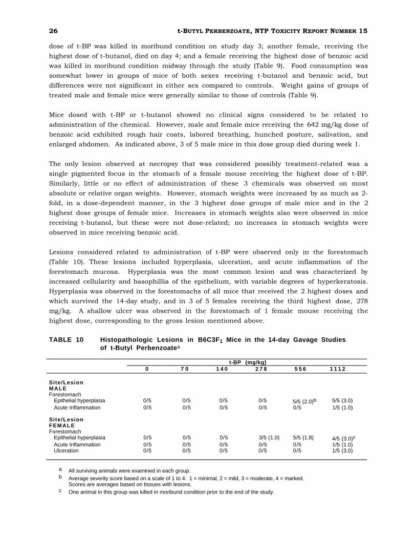
Lesions considered related to administration of t-BP were observed only in the forestomach
(Table 10). These lesions included hyperplasia, ulceration, and acute inflammation of the
forestomach mucosa. Hyperplasia was the most common lesion and was characterized by
increased cellularity and basophillia of the epithelium, with variable degrees of hyperkeratosis.
Hyperplasia was observed in the forestomachs of all mice that received the 2 highest doses and
which survived the 14-day study, and in 3 of 5 females receiving the third highest dose, 278
mg/kg. A shallow ulcer was observed in the forestomach of 1 female mouse receiving the
highest dose, corresponding to the gross lesion mentioned above.
TABLE 10 Histopathologic Lesions in B6C3F1 Mice in the 14-day Gavage Studies of t-Butyl Perbenzoatea
0 7 0 t-BP (mg/kg)
1 4 0 2 7 8 5 5 6 1 1 1 2
Site/Lesion MALE Forestomach
Epithelial hyperplasia Acute Inflammation
0/5 0/5
0/5 0/5
0/5 0/5
0/5 0/5
5/5 (2.0)b
0/5 5/5 (3.0) 1/5 (1.0)
Site/Lesion FEMALE Forestomach
Epithelial hyperplasia Acute Inflammation Ulceration
0/5 0/5 0/5
0/5 0/5 0/5
0/5 0/5 0/5
3/5 (1.0) 0/5 0/5
5/5 (1.8) 0/5 0/5
4/5 (3.0)c
1/5 (1.0) 1/5 (3.0)
a All surviving animals were examined in each group. b Average severity score based on a scale of 1 to 4: 1 = minimal, 2 = mild, 3 = moderate, 4 = marked.
Scores are averages based on tissues with lesions. c One animal in this group was killed in moribund condition prior to the end of the study.

t-BUTYL PERBENZOATE, NTP TOXICITY REPORT NUMBER 15 27
13-Week Toxicity Studies in B6C3F1 Mice
Two mice died during the study. One control male died on day 4 of apparent gavage error, and a
female in the 250 mg/kg dose group died on day 3. Mean diet consumption and body-weight
gains by all dosed groups of mice were similar to those of the respective control groups (Figure 2
and Table 11). Clinical observations during the course of the study and gross observations at
necropsy revealed few signs of toxicity or macroscopic lesions related to t-BP administration.
TABLE 11 Survival and Weight Gain of B6C3F1 Mice in the 13-Week Gavage Studies of t-Butyl Perbenzoate
Dose Concentration Mean Body Weight (grams) Final Weight Relative (mg/kg) Survivala Initial Final Changeb to Controls (%)c
MALE 0 9/10 23 34 11
30 10/10 23 35 12 103 60 10/10 23 34 11 100
125 10/10 22 34 12 100 250 10/10 23 34 11 100 500 10/10 23 33 10 97
FEMALE 0 10/10 19 31 11
30 10/10 20 29 9 93 60 10/10 19 30 11 97
125 10/10 20 30 10 97 250 9/10 19 28 9 90 500 10/10 21 30 9 97
a Number surviving at 13 weeks/number of animals per dose group. b Mean weight change of the animals in each dose group. c (Dosed group mean/Control group mean) x 100.
Evidence of t-BP toxicity in mice was limited to increased stomach weights and lesions in the
stomachs of dosed animals. Forestomach weights of both sexes receiving 250 and 500 mg/kg
were increased by 50% or more (Appendix A, Table A2). Additionally, glandular stomach weights
of female mice in the 500 mg/kg group, and the glandular stomach-to-body-weight ratios of
female mice in the 250 mg/kg group were significantly increased compared to controls. Other
changes in organ weights and organ-to-body-weight ratios were not considered related to
chemical toxicity. Histopathological examination of the forestomachs revealed compound-
related hyperplasia of the stratified squamous epithelium in male and female mice. In males,
all dose groups were affected, although hyperplasia in a single animal in the 30 mg/kg group was
minimal and not clearly compound-related. In females, the lesion was seen in the 60 mg/kg and
higher dose groups (Table 12). This lesion increased in both frequency and severity as the dose
increased. Hyperplasia was characterized by increased cellularity and basophilia of the
squamous epithelium, with hyperkeratosis that also appeared to increase with dose.

28 t-BUTYL PERBENZOATE, NTP TOXICITY REPORT NUMBER 15
Figure 2 Body Weights of B6C3F1 Mice Exposed to t-Butyl Perbenzoate by Gavage for 13-Weeks

t-BUTYL PERBENZOATE, NTP TOXICITY REPORT NUMBER 15 29
TABLE 12 Histopathologic Lesions in B6C3F1 Mice in the 13-Week Gavage Studies of t-Butyl Perbenzoate
t-BP (mg/kg) 0 3 0 6 0 1 2 5 2 5 0 5 0 0
MALE Forestomach
Epithelial Hyperplasia 0/10 1/10 (1.0)a 4/10 (1.0) 10/10 (1.0) 10/10(1.0) 10/10 (2.0)
FEMALE Forestomach
Epithelial Hyperplasia 0/10 0/10 5/10 (1.0) 10/10 (1.1) 10/10 (1.1) 10/10 (1.7)
a Average severity score based on a scale of 1 to 4: 1 = minimal, 2 = mild, 3 = moderate, 4 = marked. Scores are averages based on the number of animals with lesions.
Genetic Toxicology
t-BP (0.300-333 µg/plate) was mutagenic in Salmonella typhimurium strains TA100, TA1537, and
TA98 when tested in a preincubation protocol with and without Aroclor 1254-induced male
Sprague-Dawley rat or Syrian hamster liver S9; no mutagenic response was observed in strain
TA1535 with or without S9 (Mortelmans et al., 1986; Appendix B, Table B1). In cytogenetic tests
with Chinese hamster ovary (CHO) cells, t-BP induced sister-chromatid exchanges (SCE) within
a concentration range of 1.0 to 5.0 µg/ml in the absence of S9 activation; no induction of SCE
was observed in the presence of Aroclor 1254-induced male Sprague-Dawley rat liver S9
(Appendix B, Table B2). t-BP was an effective inducer of chromosomal aberrations in CHO cells
with and without S9 (Appendix B, Table B3). Peripheral blood samples from the 13-week study
animals were examined for presence of micronucleated polychromatic and normochromatic
erythrocytes; no induction of micronuclei was observed in any of the dose groups (Appendix B,
Table B4).

30 t-BUTYL PERBENZOATE, NTP TOXICITY REPORT NUMBER 15
DISCUSSION
Organoperoxides are a relatively large class of chemicals to which a segment of the population is
exposed in the work place. They are difficult to study because of their inherent instability. Few
studies have been designed to characterize their toxicity, and such knowledge is limited largely
to anecdotal reports of human exposure and cursory studies of acute toxicity. These reports
have confirmed that organoperoxides induce chemical burns at the point of contact when
administered in neat or concentrated form (Radding, 1977), but little is known regarding toxicity
induced by repeated exposure. The present study was designed to examine the fate and toxicity
of a representative of this important chemical class. t-BP was selected for the study because it
is one of the more commonly used organoperoxides, is sufficiently stable to permit dose
preparation and administration, and is sufficiently lipid-soluble to cross cell membranes and
reach potential subcellular target sites.
Preliminary stability studies of t-BP confirmed that the compound is sufficiently stable to permit
dose preparation and administration. However, its relatively rapid degradation in the presence
of glutathione, one of numerous reducing agents found in biological systems, indicates that it
should have a short half-life in intact animals. This assumption was confirmed by in vitro
studies that demonstrated that relatively high concentrations of t-BP had half-lives of 4.0 and
10.4 minutes in human and rat blood, respectively. Enzymatic or chemical degradation in the
presence of liver fractions was even more rapid, demonstrating that any t-BP absorbed into the
body would be expected to have a very short half-life. Benzoic acid and t-butanol constituted at
least 93% of the products of enzymatic and/or chemical degradation in in vitro systems. The
stability of t-BP in a suspension of stomach contents was concentration dependent but was
thought to be sufficient to permit some absorption of the parent molecule into stomach tissue.
t-BP was relatively stable on isolated skin; t-BP-derived radioactivity was absorbed into or bound
to skin with continued contact (Table 2). Dermal absorption of t-BP-derived radioactivity was
confirmed by the observation that when placed on the skin of living animals, approximately 16%
of the radiolabel from a wide range of t-BP doses was absorbed and excreted in urine in 24
hours. Due to the rapid chemical and/or enzymatic degradation of t-BP in biological systems, it
was not possible to determine if the radiolabel absorbed from skin represented parent compound
or products of t-BP degradation. In any case, only traces of t-BP-derived radioactivity appeared
to be retained in tissues following dermal or i.v. administration.
t-BP-derived material excreted in urine was not identified because preliminary studies showed a
quantitative yield of t-butanol and benzoic acid on degradation and/or metabolism. Because the
radiolabel was in the benzoic acid portion of the molecule, it was assumed that material
excreted in urine represented metabolites of benzoic acid. Results of previous studies
conducted in this laboratory indicated that both rats and mice metabolize more than 90 percent
of the benzoic acid (derived from benzyl acetate) to hippuric acid, and excrete it in urine (Abdo et
al., 1985). Further, in the previous study, metabolism and elimination of benzoic acid was
observed to be linear with dose, with no evidence of saturation at doses up to an equivalent of
approximately 380 mg/kg benzoic acid in the rat and over 700 mg/kg in the mouse.

t-BUTYL PERBENZOATE, NTP TOXICITY REPORT NUMBER 15 31
Corn oil was used as a vehicle for gavage administration of t-BP and for molar equivalent doses
of benzoic acid and t-butanol in the 14-day studies. t-BP and t-butanol were soluble in corn oil;
benzoic acid was administered as a suspension. Signs of toxicity observed in rats were largely
limited to increased stomach weights in males receiving the highest dose of t-BP. Food
consumption and weight gain were little affected in mice receiving any dose of t-BP or t-butanol;
there was little effect of t-butanol administration on any parameter measured. Significant
organ-weight effects observed in rats and mice receiving t-BP were limited to increased stomach
weights in both sexes receiving the higher doses. Histologic lesions induced by t-BP were
limited to the forestomach and were characterized by hyperplasia, ulceration, and acute
inflammation.
Mice appeared to be more sensitive to toxic effects of benzoic acid than rats; both sexes showed
symptoms of intoxication. Toxicity appeared to be dose-dependent, and 3 of 5 male mice and 1
of 5 female mice in the highest-dose groups (642 mg/kg) did not survive the 14-day treatment.
Weight gain in female mice receiving benzoic acid was significantly depressed in a dose-
dependent fashion. Benzoic acid may have been more toxic than t-BP because it was
administered as a suspension in corn oil. In this form, it may have rapidly partitioned from the
oil into the stomach to result in a bolus dose. Animals that died following benzoic acid
administration did not have notable lesions; they may have died from acidosis resulting from the
rapid absorption of this acid, but this was not confirmed. On the other hand, since t-BP is quite
soluble in corn oil, it would be expected to partition from the oil into the gastrointestinal tract
more slowly as the oil was digested in the small intestine.
In the 13-week studies, t-BP was administered as a suspension in water, to more closely
simulate possible human exposure which might result from ingestion. Any material ingested
would be rapidly absorbed through the stomach rather than slowly absorbed from the intestines
as with gavage in corn oil. Results of these studies indicate that toxicity resulting from t-BP
administration was minimal in both rats and mice, In rats, there was a slight depression in
food consumption; body weight gains of both sexes in the highest dose group were significantly
depressed after week 7. No clinical effects were observed which could be attributed to t-BP
administration. Variations in organ weights were largely restricted to increased stomach
weights in both male and female rats. Both the glandular stomach and forestomachs were
affected, but the effect on the forestomach was much greater (Table 8). Dose-dependent
forestomach hyperplasia was observed in all dosed male rats and in all females except those
receiving the lowest dose.
Mice receiving t-BP for 13 weeks exhibited few symptoms of intoxication. Survival was good, and
no deaths were attributed to t-BP administration. Neither food consumption nor body-weight
gains were affected significantly; no clinical toxicity or gross lesions observed at necropsy could
be attributed to chemical administration. Evidence of t-BP toxicity was limited to increased
forestomach weight and dose-related hyperplasia in both sexes (Table 10). Glandular stomach
weights were increased slightly, but no histopathological lesions were observed.
In summary, results of this study indicate that oral gavage administration of t-BP at doses up to
500 mg/kg produced little or no toxicity past the point of initial contact, the stomach. Toxicity
observed in the stomach, primarily the forestomach, was due probably to the inherent reactivity

32 t-BUTYL PERBENZOATE, NTP TOXICITY REPORT NUMBER 15
of t-BP to release free radicals which in turn reacted with the cell membranes of the stomach
mucosa. However, the reactivity of t-BP also accounts for its very short half-life in biological
systems. Its reaction with stomach contents, stomach tissue, and blood probably prevented t-
BP from reaching the systemic circulation and thus accounted for its lack of systemic toxicity.
The degradation products of t-BP, which are t-butanol and benzoic acid, are likely to be absorbed
into the systemic circulation, but they are relatively nontoxic. Both t-butanol and benzoic acid
have been the subjects of other studies conducted by the National Toxicology Program and other
organizations; they do not appear to be carcinogens or to produce other chronic toxicity.
Based on the observations described in this report, it is concluded that toxicity resulting from
human exposure to t-BP would most likely be limited to the site of contact. On contact with
human tissues, t-BP would be expected to cause local irritation of the skin and nasobronchial
epithelium such as has been described in the literature (Radding, 1977). It is not likely that t-
BP would be ingested in sufficient quantities to cause appreciable gastric toxicity; if ingested, t-
BP would not be expected to gain access to the systemic circulation of humans to result in
toxicity such as has been described for free radicals generated intracellularly (Kehrer, 1988).
t-BUTYL PERBENZOATE, NTP TOXICITY REPORT NUMBER 15 33
REFERENCES
Abdo, K.M., Huff, J.E., Haseman, J.K., Boorman, G.A., Eustis, S.L., Matthews, H.B., Burka, L.T., Prejean, J.D., and Thompson, R.B. (1985) Benzyl acetate carcinogenicity, metabolism, and disposition in Fisher 344 rats and B6C3F1 mice. Toxicology 37, 159-170.
Bock, F.G., Myers, H.K., and Fox, H.W. (1975) Carcinogenic activity of peroxy compounds. J. Nat. Can. Inst. 55, 1359-1361.
Boorman, G.A., Hickman, R.L., Davis, G.W., Rhode, L.S., White, N.W., Griffin, T.A., Mayo, J., and Hamm, T.E., Jr. (1986) Serological titers to murine viruses in 90-day and 2-year studies, in T.E. Hamm, Jr. (ed.), Complications of Viral and Mycoplasmal Infections in Rodents to Toxicology Research and Testing. New York: Hemisphere, pp. 11-23.
Boorman, G.A., Montgomery, C.A., Jr., Eustis, S.L., Wolfe, M.J., McConnell, E.E., and Hardisty, J. (1985) Quality assurance in pathology for rodent carcinogenicity studies, in H. Milman and E. Weisburger (eds.), Handbook of Carcinogen Testing. Park Ridge, NH: Noyes Publications, pp. 345-357.
Dixon, W., and Massey, F. (1951) Introduction to Statistical Analysis. New York: McGraw-Hill, pp. 145-147.
Dunn, O.J. (1964) Multiple comparisons using rank sums. Technometrics 6, 241-252.
Emergency Response Guidebook (1987) DOT Publication 5800.4. Washington, D.C.: U.S. Government Printing Office, G-48.
Free-radical initiators (1983-1984), in Modern Plastics Encyclopedia, Vol. 60, #12A. New York: McGraw-Hill, Inc., p. 624.
Galloway, S., Armstrong, M., Reuben, C., Colman, S., Brown, B., Cannon, C., Bloom, A., Nakamura, F., Ahmed, M., Duk, S., Rimpo, J., Margolin, B., Resnick, M., Anderson, B., and Zeiger, E. (1987) Chromosome aberration and sister-chromatid exchanges in Chinese hamster ovary cells: Evaluations of 108 chemicals. Environ. Molec. Mutagen. 10 (Suppl l0), 1-176.
Galloway, S., Bloom, A., Resnick, M., Margolin, B., Nakamura, F., Archer, P., and Zeiger, E. (1985) Development of a standard protocol for in vitro cytogenetic testing with CHO cells: Comparison of results for 22 compounds in two laboratories. Environ. Mutagen. 7, 1-52.
Hollander, M., and Wolfe, D. A. (1973) Nonparametric Statistical Methods. New York: John Wiley & Sons, pp. 120-123.
issues in toxicology: Free-radical Pharmacol. 95, 349-362.
Kehrer, J.P., Mossman, B.T., Sevanimechanisms
an, A., Trush, in M.A
chemical ., and Sm
pathogenith, M.T. (1988)
esis. ContemToxicol.
porary Appl.
MacGregor, J.T., Wehr, C.M., and Langlois, R.G. (1983) A simple fluorescent staining procedure for micronuclei and RNA in erythrocytes using Hoechst 33258 and pyronin Y. Mutat. Res. 120, 269-275.
Maronpot, R.R., and Boorman, G.A. (1982) Interpretation of rodent hepatocellular proliferative alterations and hepatocellular tumors in chemical safety assessment. Toxicol. Pathol. 10, 71-80.
Mohan, V.K. (1982) Hazard evaluation of organic peroxides. J. Hazard. Mater. 5, 197-220.
Morrison, D.F. (1976) Multivariate Statistical Methods. New York: McGraw Hill, Inc., pp. 170-179.

34 t-BUTYL PERBENZOATE, NTP TOXICITY REPORT NUMBER 15
Morrissey, R.E., Schwetz, B.A., Lamb, J.C., IV, Ross, M.C., Teague, J.L., and Morris, R.W. (1988) Evaluation of rodent sperm, vaginal cytology, and reproductive organ weight data from National Toxicology Program thirteen-week studies. Fundam. Appl. Toxicol. 11, 343-358.
Mortelmans, K., Haworth, S., Lawlor, T., Speck, W., Tainer, B., and Zeiger, E. (1986) Salmonella mutagenicity tests. II. Results from the testing of 270 chemicals. Environ. Mutagen. 8 (Suppl 7), 1-119.
Plunkett, E.R. (1976) Handbook of Industrial Toxicology. New York: Chemical Publishing Company, Inc., pp. 308-309.
Radding, S.B. (1977) Review of the Environmental Fate of Selected Chemicals, EPA-560/5-77-003. Washington D.C.: U.S. Environmental Protection Agency, Office of Toxic Substances, pp. 63-67.
Rao, G.N., Haseman, J.K., and Edmondson, J. (1989) Influence of viral infections on body weight, survival, and tumor prevalence in Fischer 344/NCr rats on two-year studies. Lab. Animal Sci. 39, 389-393.
Rao, G.N., Piegorsch, W.W., Crawford, D.D., Edmondson, J., and Haseman, J.K. (1989) Influence of viral infections on body weight, survival, and tumor prevalence of B6C3F1 (C57BL/6N X C3H/HeN) Mice in Carcinogenicity Studies. Fundam. Appl. Toxicol. 13, 156-164.
Sharratt, M., Frazer, A.C., and Forbes, O.C. (1964) Study of the biological effects of benzoyl peroxide. Fd. Cosmet. Toxicol. 2, 527-538.
Shirley, E. (1977) A non-parametric equivalent of Williams’ test for contrasting increasing dose levels of a treatment. Biometrics 33, 386-389.
U.S. International Trade Commission (USITC) (1988) Synthetic Organic Chemicals, U.S. Production and Sales. Washington, D.C.: U.S. Government Printing Office, USITC Publication 2219.
Van Duuren, B.L. (1963) Carcinogenicity of epoxides, lactones, and peroxy compounds. J. Nat. Can. Inst. 31(1), 41-55.
Williams, D.A. (1986) A note on Shirley's nonparametric test for comparing several dose levels with a zero control. Biometrics 42, 183-186.
Zeiger, E., Anderson, B., Haworth, S., Lawlor, T., and Mortelmans, K. (1988) Salmonella mutagenicity tests: IV. Results from the testing of 300 chemicals. Environ. Molec. Mutagen. 11 (Suppl. 12), 1-158.

B-1 t-BUTYL PERBENZOATE, NTP TOXICITY REPORT NUMBER 15
APPENDIX B
Genetic Toxicology Table B1 Mutagenicity of t-Butyl Perbenzoate in Salmonella typhimurium .....................B-2
Table B2 Induction of Sister-Chromatid Exchanges in Chinese Hamster Ovary Cells by t-Butyl Perbenzoate ......................................................................B-4
Table B3 Induction of Chromosomal Aberrations in Chinese Hamster Ovary Cells by t-Butyl Perbenzoate ......................................................................B-6
Table B4 Frequency of Micronuclei in Peripheral Blood Erythrocytes of Mice Exposed to t-Butyl Perbenzoate for 13 Weeks ..............................................B-7

B-2 t-BUTYL PERBENZOATE, NTP TOXICITY REPORT NUMBER 15
Table B1 Mutagenicity of t-Butyl Perbenzoate in Salmonella typhimurium a
D O S E REVERTANTS/PLATEb
mg/Plate (-) S9 10% Hamster S9 10% Rat S9 T A 1 0 0
0.0 0.3 1.0 3.3
10.0 16.0 33.0 66.7
100.0 167.0 333.0
Trial summary
Positive controlc
105 ± 6.7
96 ± 5.9 93 ± 5.4 92 ± 2.5
Toxic
Toxic
Negative
1486 ± 26.4
88 ± 1.2 76 ± 1.5 79 ± 0.7 89 ± 8.2
110 ± 3.8
126 ± 5.0
Weakly Positive
738 ± 23.6
91 ± 5.9 85 ± 4.3 104 ± 1.2 118 ± 0.9 129 ± 7.5
Toxic
Weakly Positive
1616 ± 45.9
166 ± 4.5 182 ± 6.7
153 ± 5.7 190 ± 5.0 147 ± 7.9
178 ± 7.1 287 ± 8.8 232 ± 18.7
268 ± 8.2 208 ± 18.5 136 ± 21.4
Toxic
Weakly Positive
Weakly Positive
2642 ± 45.8 2045 ± 109.9
100 ± 12.4
147 ± 4.6 127 ± 7.3 146 ± 4.4 227 ± 14.0 269 ± 4.4
Positive
1248 ± 54.5
142± 2.8 145± 10.2
129 ± 2.3 159 ± 10.7 145 ± 7.2
147 ± 5.5 287 ± 19.0 213 ± 14.3
266 ± 1.9 149 ± 4.6 Toxic
Toxic
Equivocal Weakly Positive
2645 ± 18.4 2393 ± 65.2
141 ± 3.5
161 ± 7.2 165 ± 12.0 182 ± 4.7 246 ± 24.6 288 ± 8.1
Positive
1060 ± 71.2
T A 1 5 3 5 0.0 1.0 3.3
10.0 33.0
100.0 333.0
Trial summary
Positive controlc
T A 1 5 3 7
10 ± 1.7 4 ± 1.2 20 ± 1.2 15 ± 1.5 11 ± 1.7 11 ± 1.3 13 ± 1.9 4 ± 0.3 17 ± 2.8 11 ± 2.6 5 ± 0.6 20 ± 3.7 16 ± 2.0 8 ± 0.6 13 ± 3.2
9 ± 1.5 4 ± 0.9 22 ± 2.4 14 ± 0.9 10 ± 1.3 14 ± 3.5 9 ± 2.3 6 ± 0.9 19 ± 5.0 12 ± 1.0 17 ± 2.9 9 ± 0.7
13 ± 3.2 6 ± 1.5 12 ± 2.0 12 ± 0.3 12 ± 3.3 11 ± 1.5 Toxic Toxic Toxic
Negative Negative Negative Negative Negative Negative
1152 ± 144.3 643 ± 43.4 229 ± 10.1 278 ± 10.4 200 ± 24.1 283 ± 27.7
0.0 1.0 3.3
10.0 16.0 33.0 66.7
100.0 167.0 333.0
Trial summary
Positive controlc
9 ± 1.2 10 ± 0.6 10 ± 1.5
9 ± 2.4
12 ± 2.1
23 ± 2.1
Equivocal
161 ± 11.8
9± 2.0
11± 2.3 17± 0.9 21± 1.0 Toxic Toxic
Weakly Positive
223 ± 101.7
3± 0.0 3± 0.9 3± 0.9 6± 1.0
10± 2.7
16 ± 3.1
Weakly Positive
62 ± 11.6
16 ± 2.9
15 ± 2.6 13 ± 3.4
56 ± 0.9 65 ± 10.4 46 ± 10.2
Toxic
Equivocal
521 ± 27.1
21 ± 0.6
25 ± 1.8 36 ± 0.9 30 ± 0.6 47 ± 7.0 Toxic
Positive
216 ± 33.1
13 ± 0.9
20 ± 3.1 19 ± 0.0 15 ± 2.6
64 ± 5.2
Positive
233 ± 14.4
15 ± 0.0
18 ± 3.5 15 ± 2.3
65 ± 7.8
14 ± 2.2
Toxic
Equivocal
483 ± 1.5
23 ± 1.0
22 ± 0.3 24 ± 2.7 48 ± 13.7 59 ± 12.3 Toxic
Positive
380 ± 120.7
6 ± 0.9
7 ± 1.5 14 ± 1.5 41 ± 5.3 26 ± 6.6 Toxic
Positive
337 ± 12.8

B-3 t-BUTYL PERBENZOATE, NTP TOXICITY REPORT NUMBER 15
Table B1 Mutagenicity of t-Butyl Perbenzoate in Salmonella typhimuriuma (continued)
D O S E REVERTANTS/PLATEb
mg/Plate (-) S9 10% Hamster S9 10% Rat S9 T A 9 8
0.0 1.0 3.3
10.0 16.0 33.0 66.7
100.0 333.0
Trial summary
Positive controlc
22 ± 2.3 20 ± 3.8 27 ± 2.3 28 ± 2.4
20 ± 4.3
28 ± 2.0
Negative
195 ± 27.9
11 ± 0.3 14 ± 0.6 17 ± 2.0 16 ± 0.9
25 ± 4.1
34 ± 5.2
Positive
188 ± 11.6
15± 2.6 17± 2.2 18 ± 2.6 21± 3.5
26 ± 0.6
36± 4.0
Positive
171 ± 9.0
37 ± 2.8 40 ± 0.6
38 ± 1.5 44 ± 3.4 44 ± 2.6
60 ± 2.6 65 ± 6.8 76 ± 6.4
55 ± 7.6 35 ± 5.2 Toxic
Toxic
Weakly Positive
Weakly Positive
1611 ± 75.1 1305 ± 219.9
29 ± 3.8
54 ± 5.0 41 ± 2.3 45 ± 6.4 71 ± 6.8 75 ± 11.8
Positive
595 ± 49.4
29 ± 1.5 44 ± 0.9
32 ± 3.2 43 ± 3.5 41 ± 2.9 50 ± 2.1 56 ± 2.9 72 ± 0.6 77 ± 5.7
23 ± 3.8 33 ± 2.0 Toxic
Toxic
Positive Weakly Positive
1747 ± 26.4 2223 ± 214.3
31 ± 1.5
53 ± 5.5
57 ± 7.0 87 ± 5.5 76 ± 4.8
Positive
508 ± 34.4
a Study performed at Case Western Reserve University. The detailed protocol and these data are presented in Mortelmans et al. (1986). Cells and study compound or solvent (dimethylsulfoxide) were incubated in the absence of exogenous metabolic activation (-S9) or with Aroclor 1254-induced S9 from male Syrian hamster liver or male Sprague Dawley rat liver. High dose was limited by toxicity or solubility, but did not exceed 10 mg/plate; 0 mg/plate dose is the solvent control.
b Revertants are presented as mean ± the standard error from 3 plates. c Positive control; 2-aminoanthracene was used on all strains in the presence of S9. In the absence of metabolic activation, 4
nitro-o-phenylenediamine was tested on TA98, sodium azide was tested on TA100 and TA1535, and 9-aminoacridine was tested on TA1537.
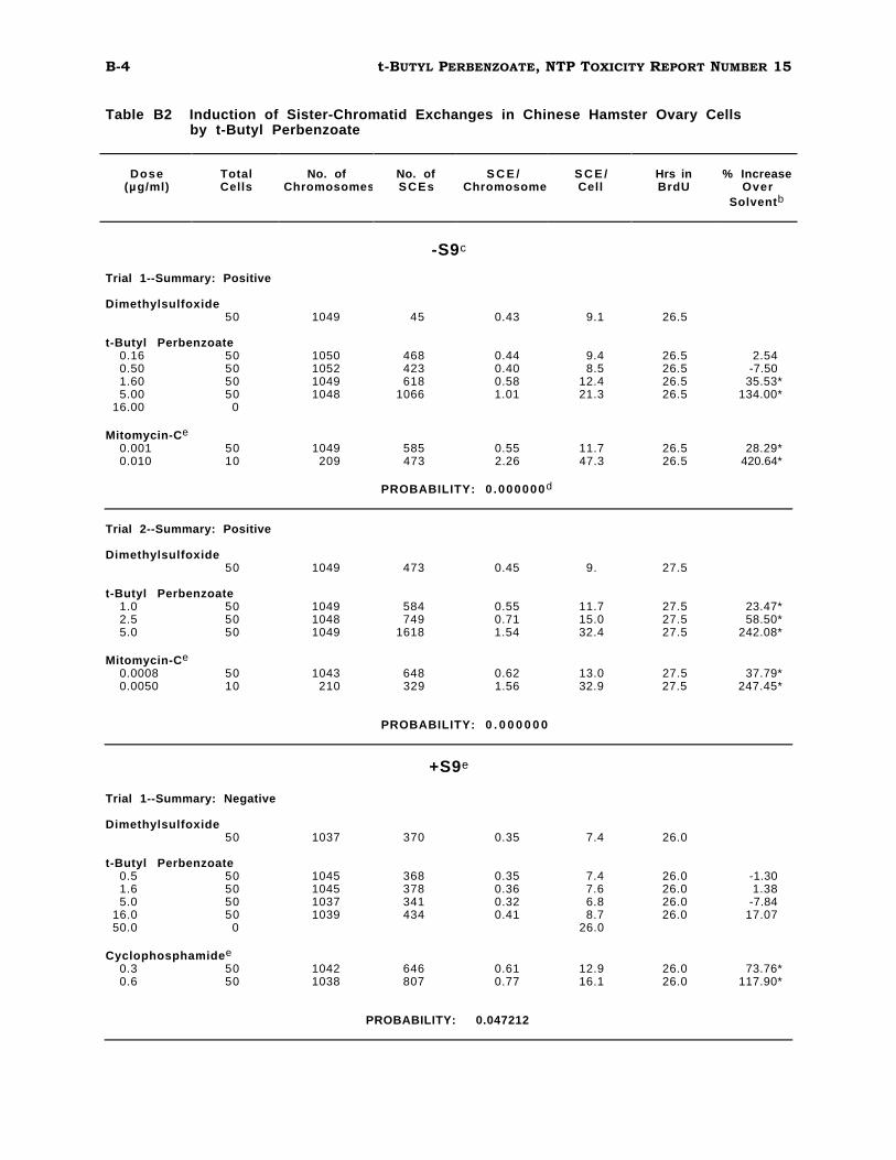
B-4 t-BUTYL PERBENZOATE, NTP TOXICITY REPORT NUMBER 15
Table B2 Induction of Sister-Chromatid Exchanges in Chinese Hamster Ovary Cells by t-Butyl Perbenzoate
Dose (µg/ml)
Total Cells
No. of Chromosomes
No. of SCEs
S C E / Chromosome
S C E / Cell
Hrs in BrdU
% Increase Over
Solventb
-S9c
Trial 1--Summary: Positive
Dimethylsulfoxide 50 1049 45 0.43 9.1 26.5
t-Butyl Perbenzoate 0.16 50 0.50 50 1.60 50 5.00 50
16.00 0
1050 1052 1049 1048
468 423 618
1066
0.44 0.40 0.58 1.01
9.4 8.5
12.4 21.3
26.5 26.5 26.5 26.5
2.54 -7.50
35.53* 134.00*
Mitomycin-Ce
0.001 0.010
50 10
1049 209
585 473
0.55 2.26
11.7 47.3
26.5 26.5
28.29* 420.64*
PROBABILITY: 0 .000000d
Trial 2--Summary: Positive
Dimethylsulfoxide 50 1049 473 0.45 9. 27.5
t-Butyl Perbenzoate 1.0 50 2.5 50 5.0 50
1049 1048 1049
584 749
1618
0.55 0.71 1.54
11.7 15.0 32.4
27.5 27.5 27.5
23.47* 58.50*
242.08*
Mitomycin-Ce
0.0008 0.0050
50 10
1043 210
648 329
0.62 1.56
13.0 32.9
27.5 27.5
37.79* 247.45*
PROBABILITY: 0 . 0 0 0 0 0 0
+S9e
Trial 1--Summary: Negative
Dimethylsulfoxide 50 1037 370 0.35 7.4 26.0
t-Butyl Perbenzoate 0.5 50 1.6 50 5.0 50
16.0 50 50.0 0
1045 1045 1037 1039
368 378 341 434
0.35 0.36 0.32 0.41
7.4 7.6 6.8 8.7
26.0
26.0 26.0 26.0 26.0
-1.30 1.38
-7.84 17.07
Cyclophosphamidee
0.3 50 0.6 50
1042 1038
646 807
0.61 0.77
12.9 16.1
26.0 26.0
73.76* 117.90*
PROBABILITY: 0.047212

B-5
c
t-BUTYL PERBENZOATE, NTP TOXICITY REPORT NUMBER 15
Table B2 Induction of Sister-Chromatid Exchanges in Chinese Hamster Ovary Cells by t-Butyl Perbenzoate (continued)
Dose Total No. of No. of S C E / S C E / Hrs in % Increase (µg/ml) Cells Chromosomes SCEs Chromosome Cell BrdU Over
Solventb
+S9e
Trial 2--Summary: Negative
Dimethylsulfoxide 50 1046 413 0.39 8.3 26.5
t-Butyl Perbenzoate 1.0 50 1051 400 0.38 8.0 26.5 -3.61 5.0 50 1050 429 0.40 8.6 26.5 3.48
10.0 50 1048 439 0.41 8.8 26.5 6.09
Cyclophosphamidee
0.3 50 1048 549 0.52 11.0 26.5 32.68*
PROBABILITY: 0 . 1 2 3 1 9 2
a Study performed at Environmental Health Research & Testing. SCE = sister chromatid exchange; BrdU = bromodeoxyuridine. A detailed description of the SCE protocol is presented by Galloway et al. (1985, 1987). Briefly, Chinese hamster ovary cells were incubated with study compound or solvent (dimethylsulfoxide) as described in (c) and (d) below, and cultured for sufficient time to reach second metaphase division. Cells were then collected by mitotic shake-off, fixed, air-dried, and stained.
b SCE's/chromosome of culture exposed to study chemical relative to those of culture exposed to solvent. In the absence of S9, cells were incubated with study compound or solvent for 2 h at 37°C. Then BrdU was added and incubation was continued for 24 h. Cells were washed, fresh medium containing BrdU and colcemid was added, and incubation was continued for 2-3 h.
d Trend test. e In the presence of S9, cells were incubated with study compound or solvent for 2 h at 37°C. The cells were then washed, and
medium containing BrdU was added. Cells were incubated for a further 26 h, with colcemid present for the final 2-3 h. S9 was from the livers of Aroclor 1254-induced male Sprague Dawley rats.
* Positive response.

B-6 t-BUTYL PERBENZOATE, NTP TOXICITY REPORT NUMBER 15
Table B3 Induction of Chromosomal Aberrations in Chinese Hamster Ovary Cells by t-Butyl Perbenzoatea
-S9b +S9c
Dose (µg/ml)
Total Cells
No. of Abs
Abs/Cell % Cells w/ Abs
Trial 1
Dose (µg/ml)
Total Cells
No. of Abs Abs/Cell
% Cells w/ Abs
Harvest time: 12.5 hours Harvest time: 13.0 hours
Dimethylsulfoxide
100 1 0.01 1.0 100 1 0.01 1.0
t-Butyl perbenzoate
5.0 100 9 10.0 100 42
0.09 9.0* 0.42 31.0*
10.0 100 6 16.0 100 9 30.0 100 15 50.0 100 22
100.0 34 26
0.06 5.0 0.09 9.0* 0.15 14.0* 0.22 20.0* 0.76 26.0*
Summary: Positive Summary: Positive
Mitomycin-Cd Cyclophosphamided
.25 100 33
.00 50 32 0.33 27.0 0.64 50.0
15.0 100 45 0.4 38.0*
-S9b
Trial 2
Harvest time: 17.5 hoursd
Dimethylsulfoxide
100 3 0.03 3.0
t-Butyl perbenzoate
5.0 100 26 0.26 23.0* 10.0 100 38 0.38 32.0*
Summary: Positive
Mitomycin-Cd
0.125 100 26 0.26 21.0* 0.250 50 19 0.38 22.0*
(a) Study performed at Environmental Health Research & Testing. Abs = aberrations. A detailed presentation of the technique for detecting chromosomal aberrations is found in Galloway et al. (1985, 1987). Briefly, Chinese hamster ovary cells were incubated with study compound or solvent (dimethylsulfoxide) as indicated in (b) and (d). Cells were arrested in first metaphase by addition of colcemid and harvested by mitotic shake off, fixed, and stained in 6% Giemsa.
(b) In the absence of S9, cells were incubated with study compound or solvent for 8-10 h at 37° C. Cells were then washed and fresh medium containing colcemid was added for an additional 2-3 h followed by harvest.
(c) In the presence of S9, cells were incubated with study compound or solvent for 2 h at 37°C. Cells were then washed, medium was added, and incubation was continued for 8-10 h. Colcemid was added for the last 2-3 h of incubation before harvest. S9 was from the livers of Aroclor 1254-induced male Sprague Dawley rats.
(d) Because of significant chemical-induced cell cycle delay, incubation time prior to addition of colcemid was lengthened to provide sufficient metaphases at harvest.
* Positive response.

B-7 t-BUTYL PERBENZOATE, NTP TOXICITY REPORT NUMBER 15
Table B4 Frequency of Micronuclei in Peripheral Blood Erythrocytes of Mice Exposed to t-Butyl Perbenzoate for 13 Weeksa
MICRONUCLEATED CELLS/1000 CELLSb
NUMBER OF DOSE (mg/kg) P C E N C E MICE
Male mice
0 2.53 ± 0.53 1.63 ± 0.10 9 30 2.20 ± 0.36 1.46 ± 0.09 10 60 2.20 ± 0.47 1.39 ± 0.12 10
125 2.17 ± 0.49 1.54 ± 0.15 10 250 1.76 ± 0.37 1.78 ± 0.12 10 500 .48 ± 0.37 1.75 ± 0.19 10
P = 0.9653 P = 0.2297Trend testc
Female mice
0 2.12 ± 0.45 1.12 ± 0.14 10 30 1.91 ± 0.35 1.22 ± 0.14 10 60 1.51 ± 0.50 1.06 ± 0.15 10
125 0.83 ± 0.26 1.11 ± 0.09 10 250 0.93 ± 0.32 0.98 ± 0.06 9 500 1.15 ± 0.37 1.28 ± 0.1 10
P = 0.9770 P = 0.4995Trend testc
(a) Smears were prepared from peripheral blood samples obtained by cardiac puncture of dosed and control animals at the time of terminal kill. Slides were stained with Hoechst 333258/pyronin Y (MacGregor et al., 1983). At least 10000 NCE and 2000 PCE from each animal were scored for micronuclei. No significant elevation in the frequency of micronucleated erythrocytes was observed in either male or female mice chronically exposed to t-butyl perbenzoate.
(b) Mean ± standard error of the mean. (c) Cochran-Armitage linear regression of proportions for PCE's or linear contrasts from Analysis of Variance for NCE's.