ntp technical report on toxicity studies of … institutes of... · national toxicology program...
TRANSCRIPT
National Toxicology Program Toxicity Reports Series
Number 16
NTP Technical Report on Toxicity Studies of
Glyphosate (CAS No. 1071-83-6)
Administered in Dosed Feed
to F344/N Rats and B6C3F1 Mice
Po C. Chan, PhD, and Joel F. Mahler, DVM, Study Scientists
National Toxicology Program Post Office Box 12233
Research Triangle Park, NC 27709
NIH Publication 92-3135 July 1992
United States Department of Health and Human Services Public Health Service
National Institutes of Health

2 GLYPHOSATE, NTP TOXICITY REPORT NUMBER 16
CONTRIBUTORS
The NTP report on the toxicity studies of glyphosate is based on disposition studies conducted at
the College of Pharmacy, University of Arizona, Tucson, AZ, in November, 1987; 13-week studies performed between May and September, 1988, at Southern Research Institute, Birmingham, AL;
and 14-day studies performed in 1990 at the National Institute of Environmental Health Sciences, Research Triangle Park, NC.
National Toxicology Program Evaluated experiment, interpreted results, and reported findings
Po C. Chan, PhD Joel F. Mahler, DVM
Study Scientists John R. Bucher, PhD
Leo T. Burka, PhD Rajendra S. Chhabra, PhD
Michael P. Dieter, PhD Michael R. Elwell, DVM, PhD H.B. Matthews, PhD
Morrow B. Thompson, DVM, PhD Errol Zeiger, PhD
Coordinated report preparation
Jane M. Lambert, BS
Diane Overstreet, BS Kristine Witt, MS
Oak Ridge Associated Universities
NTP Pathology Review Evaluated slides and prepared pathology report
Sondra Grumbein, DVM, PhD, Pathology Associates, Inc.
Michael R. Elwell, DVM, PhD National Toxicology Program
Southern Research Institute Principal contributors
J.D. Prejean, PhD Principal Investigator
H. Giles, DVM A.G. Manus, DVM, L.M. Thigpen, DVM
R. Thompson, DVM
University of Arizona, College of Pharmacy Principal contributors, disposition studies
I. Glenn Sipes, PhD Christy Duerson, MS Experimental Pathology Laboratories, Inc. Provided pathology quality assurance
Jerry F. Hardisty, DVM
Environmental Health Research and Testing, Inc. Provided sperm morphology and reproductive toxicology evaluation
Dushant K. Gulati, PhD Teresa Cocanougher, BA Susan Russell, BA
Analytical Sciences, Inc. Provided statistical analysis
Steven Seilkop, MS Janet Teague, MS

National Toxicology Program Toxicity Reports Series
Number 16
NTP Technical Report on Toxicity Studies of
Glyphosate (CAS No. 1071-83-6)
Administered in Dosed Feed
to F344/N Rats and B6C3F1 Mice
Po C. Chan, PhD, and Joel F. Mahler, DVM, Study Scientists
National Toxicology Program Post Office Box 12233
Research Triangle Park, NC 27709
NIH Publication 92-3135 July 1992
United States Department of Health and Human Services Public Health Service
National Institutes of Health
2 GLYPHOSATE, NTP TOXICITY REPORT NUMBER 16
CONTRIBUTORS
The NTP report on the toxicity studies of glyphosate is based on disposition studies conducted at
the College of Pharmacy, University of Arizona, Tucson, AZ, in November, 1987; 13-week studies performed between May and September, 1988, at Southern Research Institute, Birmingham, AL;
and 14-day studies performed in 1990 at the National Institute of Environmental Health Sciences, Research Triangle Park, NC.
National Toxicology Program Evaluated experiment, interpreted results, and reported findings
Po C. Chan, PhD Joel F. Mahler, DVM
Study Scientists John R. Bucher, PhD
Leo T. Burka, PhD Rajendra S. Chhabra, PhD
Michael P. Dieter, PhD Michael R. Elwell, DVM, PhD H.B. Matthews, PhD
Morrow B. Thompson, DVM, PhD Errol Zeiger, PhD
Coordinated report preparation
Jane M. Lambert, BS
Diane Overstreet, BS Kristine Witt, MS
Oak Ridge Associated Universities
NTP Pathology Review Evaluated slides and prepared pathology report
Sondra Grumbein, DVM, PhD, Pathology Associates, Inc.
Michael R. Elwell, DVM, PhD National Toxicology Program
Southern Research Institute Principal contributors
J.D. Prejean, PhD Principal Investigator
H. Giles, DVM A.G. Manus, DVM, L.M. Thigpen, DVM
R. Thompson, DVM
University of Arizona, College of Pharmacy Principal contributors, disposition studies
I. Glenn Sipes, PhD Christy Duerson, MS Experimental Pathology Laboratories, Inc. Provided pathology quality assurance
Jerry F. Hardisty, DVM
Environmental Health Research and Testing, Inc. Provided sperm morphology and reproductive toxicology evaluation
Dushant K. Gulati, PhD Teresa Cocanougher, BA Susan Russell, BA
Analytical Sciences, Inc. Provided statistical analysis
Steven Seilkop, MS Janet Teague, MS
GLYPHOSATE, NTP TOXICITY REPORT NUMBER 16 3
CONTENTS
CONTRIBUTORS .........................................................................................................................................2 TABLE OF CONTENTS ................................................................................................................................3 ABSTRACT .................................................................................................................................................5 PEER REVIEW PANEL ...............................................................................................................................7 SUMMARY OF PEER REVIEW COMMENTS ................................................................................................8 INTRODUCTION ..........................................................................................................................................9 MATERIALS AND METHODS .................................................................................................................... 11
Procurement and Characterization of Glyphosate ............................................................................. 11
Disposition Studies........................................................................................................................... 11
13-Week Study Design ..................................................................................................................... 12
Reproductive Toxicity ....................................................................................................................... 13
Study of the Mechanism of Induction of Salivary Gland Lesions by
Glyphosate................................................................................................................................ 15
Genetic Toxicity Studies ................................................................................................................... 16
Statistical Methods ........................................................................................................................... 16
Quality Assurance ............................................................................................................................ 17 RESULTS .................................................................................................................................................. 18
Disposition Studies........................................................................................................................... 18
13-Week Studies in F344/N Rats ..................................................................................................... 19
13-Week Studies in B6C3F1 Mice ..................................................................................................... 23
Mechanism of Induction of Salivary Gland Lesions........................................................................... 28
Genetic toxicology............................................................................................................................. 33
DISCUSSION ............................................................................................................................................ 34 REFERENCES ........................................................................................................................................... 37
TABLES
Table 1 Experimental Design and Materials and Methods in the 13-Week Studies of Glyphosate.......................................................................... 14
Table 2 Treatment Groups in the Study to Determine the Mechanism of Induction of Salivary Gland Lesions by Glyphosate.................................................. 15
Table 3 Cumulative Percentage of Oral or I.V. Dose of Glyphosate Eliminated in Urine and Feces ..................................................................................... 18 Table 4 Percentage of Dose in Tissues Following Oral Administration of Glyphosate at 5.6 mg/kg.......................................................................................... 18 Table 5 Survival, Weight Gain, and Feed Consumption of F344/N Rats in the 13-Week Dosed Feed Study of Glyphosate ......................................................... 19
4 GLYPHOSATE, NTP TOXICITY REPORT NUMBER 16
Table 6 Incidence and Severity of Cytoplasmic Alteration of the Parotid and Submandibular Salivary Glands (Combined) in F344/N Rats in the 13-Week Dosed Feed Study of Glyphosate ......................................................... 22 Table 7 Survival, Weight Gain, and Feed Consumption of B6C3F1 Mice
in the 13-Week Dosed Feed Study of Glyphosate ......................................................... 23 Table 8 Incidence and Severity of Cytoplasmic Alteration of the Parotid Salivary Gland in B6C3F1 Mice in the 13-Week Dosed Feed Study of Glyphosate ..................... 23
Table 9 Feed Consumption and Weight Gain of F344/N Rats in the 14-Day Mechanism Study of Glyphosate ........................................................... 28 Table 10 Salivary Gland Weights of F344/N Rats in the 14-Day Mechanism Study of Glyphosate ........................................................... 29 Table 11 Incidence and Severity of Cytoplasmic Alteration of the Salivary Glands of F344/N Rats in the 14-Day Mechanism Study of Glyphosate................................... 29
FIGURES
Figure 1 Blood Levels of 14C-Glyphosate Following Oral Administration
of 14C-Glyphosate at 5.6 or 56 mg/kg ......................................................................... 20
Figure 2 Level of Radioactivity in Blood after a Single I.V. Dose of 5.6 mg/kg Glyphosate ............................................................................................. 20
Figure 3 Body Weights of F344/N Rats Exposed to Glyphosate by Dosed Feeding for 13 Weeks.................................................................................... 21 Figure 4 Body Weights of B6C3F1 Mice Exposed to Glyphosate
by Dosed Feeding for 13 Weeks.................................................................................... 27
PLATES Plates 1-2 .................................................................................................................................... 24 Plates 3-5 .................................................................................................................................... 30 APPENDICES Appendix A Organ Weights and Organ-to-Body-Weight Ratios...................................................... A-1 Appendix B Hematology and Clinical Chemistry Results............................................................... B-1 Appendix C Reproductive Tissue Evaluations and Estrous Cycle Length ...................................... C-1 Appendix D Genetic Toxicology ..................................................................................................... D-1
GLYPHOSATE, NTP TOXICITY REPORT NUMBER 16 5
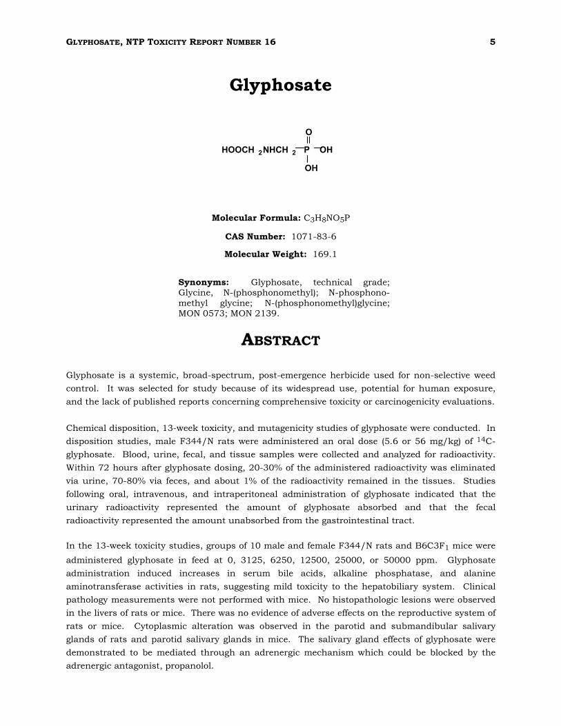
Glyphosate
HOOCH 2NHCH 2 P OH
OH
O
Molecular Formula: C3H8NO5P
CAS Number: 1071-83-6
Molecular Weight: 169.1
Synonyms: Glyphosate, technical grade; Glycine, N-(phosphonomethyl); N-phosphono-
methyl glycine; N-(phosphonomethyl)glycine; MON 0573; MON 2139.
ABSTRACT
Glyphosate is a systemic, broad-spectrum, post-emergence herbicide used for non-selective weed
control. It was selected for study because of its widespread use, potential for human exposure,
and the lack of published reports concerning comprehensive toxicity or carcinogenicity evaluations.
Chemical disposition, 13-week toxicity, and mutagenicity studies of glyphosate were conducted. In
disposition studies, male F344/N rats were administered an oral dose (5.6 or 56 mg/kg) of 14C-
glyphosate. Blood, urine, fecal, and tissue samples were collected and analyzed for radioactivity.
Within 72 hours after glyphosate dosing, 20-30% of the administered radioactivity was eliminated
via urine, 70-80% via feces, and about 1% of the radioactivity remained in the tissues. Studies
following oral, intravenous, and intraperitoneal administration of glyphosate indicated that the
urinary radioactivity represented the amount of glyphosate absorbed and that the fecal
radioactivity represented the amount unabsorbed from the gastrointestinal tract.
In the 13-week toxicity studies, groups of 10 male and female F344/N rats and B6C3F1 mice were
administered glyphosate in feed at 0, 3125, 6250, 12500, 25000, or 50000 ppm. Glyphosate
administration induced increases in serum bile acids, alkaline phosphatase, and alanine
aminotransferase activities in rats, suggesting mild toxicity to the hepatobiliary system. Clinical
pathology measurements were not performed with mice. No histopathologic lesions were observed
in the livers of rats or mice. There was no evidence of adverse effects on the reproductive system of
rats or mice. Cytoplasmic alteration was observed in the parotid and submandibular salivary
glands of rats and parotid salivary glands in mice. The salivary gland effects of glyphosate were
demonstrated to be mediated through an adrenergic mechanism which could be blocked by the
adrenergic antagonist, propanolol.
6 GLYPHOSATE, NTP TOXICITY REPORT NUMBER 16
Glyphosate was not mutagenic in Salmonella, and did not induce micronuclei in mice. The no-
observed-adverse-effect level (NOAEL) for the salivary gland lesions was 3125 ppm in the diet for
mice. A NOAEL could not be determined from the rat study.
GLYPHOSATE, NTP TOXICITY REPORT NUMBER 16 7
PEER REVIEW
Peer Review Panel
The members of the Peer Review Panel who evaluated the draft report on the toxicity studies on
glyphosate on July 10, 1991, are listed below. Panel members serve as independent scientists, not
as representatives of any institution, company, or governmental agency. In this capacity, panel
members act to determine that the design and conditions of the NTP studies were appropriate and
to ensure that the toxicity study report presents the experimental results and conclusions fully and
clearly.
National Toxicology Program’s Board of Scientific Counselors Technical Reports Review Subcommittee Paul T. Bailey, PhD Mobil Oil Corporation Toxicology Division Princeton, NJ
Louis S. Beliczky, MS, MPH Director of Industrial Hygiene
Department of Industrial Hygiene
United Rubber Workers Intl. Union
87 South High Street
Akron, OH
Gary P. Carlson, PhD Department of Pharmacology and Toxicology
Purdue University
West Lafayette, IN
Harold Davis, DVM, PhD School of Aerospace Medicine
Brooks Air Force Base, TX
Robert H. Garman, DVM Consultants in Veterinary Pathology
Murrysville, PA
Jay I. Goodman, PhD Department of Pharmacology and Toxicology Michigan State University East Lansing, MI
*Unable to attend
David W. Hayden, DVM, PhD Department of Veterinary Pathobiology
College of Veterinary Medicine
University of Minnesota
St. Paul, MN
Daniel S. Longnecker, MD, Chair Department of Pathology Dartmouth Medical School Hanover, NH
Curtis D. Klaassen, PhD Department of Pharmacology and Toxicology
University of Kansas Medical Center
Kansas City, KS
Barbara McKnight, PhD Department of Biostatistics
University of Washington
Seattle, WA
*Ellen K. Silbergeld, PhD University of Maryland Medical School
Baltimore, MD
Lauren Zeise, PhD California Department of Health Services
Berkeley, CA
8 GLYPHOSATE, NTP TOXICITY REPORT NUMBER 16
Summary of Peer Review Comments
On July 9 and 10, 1991, the Technical Reports Review Subcommittee of the Board of Scientific
Counselors for the National Toxicology Program met in Research Triangle Park, NC, to review the
draft technical report on toxicity studies of glyphosate.
Dr. Po Chan, NIEHS, introduced the short-term toxicity studies of glyphosate by reviewing the uses
and rationale for the study, findings from chemical disposition studies, experimental design, and
results.
Dr. Garman, a principal reviewer, said that the report was thoroughly prepared and detailed, and
that it did an excellent job reviewing the background for the study and the available literature on
glyphosate. He added that the isoproterenol/propranolol study included in the report is quite
interesting and helps establish the mechanism for salivary gland alteration.
Dr. Garman said that certain details of the salivary gland alteration study should be clarified,
namely, which type of glandular acinus within the submandibular salivary gland was most affected
by glyphosate, and whether, in Table 11, only the parotid salivary gland was assayed in measuring
the severity of changes brought on by glyphosate treatment. Dr. J. Mahler, NIEHS, said the
severity grades were based on the parotid glands only.
Dr. Goodman, another principal reviewer, said the report was well-written. He suggested that the
the lack of of any reproductive toxicity attributable to glyphosate treatment was an important
finding and should be included in the abstract of the report.
After further discussion of editorial matters, Dr. Longnecker accepted the report on behalf of the
panel.
GLYPHOSATE, NTP TOXICITY REPORT NUMBER 16 9
INTRODUCTION
Glyphosate is a nonvolatile white solid with a melting point of 200°C and a negligible vapor
pressure. It is soluble to 1.2% in water at 25°C but is not soluble in organic solvents (Beste, 1983).
Glyphosate has been available commercially since 1974. It is marketed as Roundup® (comprised
of the isopropylamine salt of glyphosate (41.0%) and inert ingredients, including surfactants), and
as Rodeo® (which contains the isopropylamine salt of glyphosate (53.5%) and inert ingredients).
The surfactant in Roundup® facilitates foliage absorption. Roundup® is used as a nonselective,
systemic, broad-spectrum, post-emergence herbicide for managing vegetation in agriculture and
forestry; Rodeo® is used for aquatic weed control. Information on production volume, sales, and
the identity of the "inert ingredients" is proprietary.
The mechanism of phytotoxic action of glyphosate is inhibition of the 5-enolpyruvylshikimate-3-
phosphate synthase (EC 2.5.1.19) activity, thus blocking aromatic amino acid synthesis (Amrhein
et al., 1980, 1981). The resulting reduction in protein synthesis causes cessation of growth and,
eventually, cellular disruption and death. Glyphosate has nonspecific, metal-chelating properties
(Glass, 1984); it inhibits enzymes which require transitional metal cations for activity such as the
3-deoxy-2-oxo-D-arabino-heptulosonate-7-phosphate synthase and 5-dehydroquinate synthase
(Ghassemi et al., 1982; Hoagland and Duke, 1982). Glyphosate's effectiveness as a phytotoxin is
due in part to its low molecular weight and high water solubility, which aid its rapid absorption
and translocation by plant tissues; it is not metabolized to any significant degree in plant tissues
(Ghassemi et al., 1982).
Glyphosate is strongly adsorbed to soils and is not readily leached. The mobility of glyphosate in
the soil is affected by soil type, phosphate level, and pH. Adsorption of glyphosate is higher in soils
containing clay and organic matter than in sandy loam soils, but lower in high-phosphate or high-
pH soils. It is susceptible to degradation, possibly by microbial co-metabolism (Sprankle et al.,
1975), and thus relatively nonpersistent in soils. Information provided by Monsanto to the U.S.
Environmental Protection Agency reportedly showed the half-life of glyphosate in soil normally was
less than 60 days (U.S. EPA, 1979). The half-life was 17 to 19 weeks in sandy soil and 3 weeks in
silt loam (Ghassemi et al., 1982). Newton et al. (1984) reported the half-lives of glyphosate in a
forest-brush field ecosystem in Oregon after aerial spray were 10.4 to 26.6 days in the foliage and
litter, 40.2 days for exposed soil, and 29.2 days for litter-covered soil. However, Stark (1983)
reported that residues still may be found in the soil for 2 years or longer. The major degradation
product of glyphosate in soil is (aminomethyl)phosphonic acid. Minor metabolites include N-
methylaminomethylphosphonic acid, glycine, N-dimethylaminomethylphosphonic acid, and
hydroxy-methylphosphonic acid (Ghassemi et al., 1982; Rueppel et al., 1977; Sprankle et al.,
1975).
No information is available on the absorption of glyphosate after oral administration to mammals.
Wester et al. (1991) reported poor absorption of glyphosate, as Roundup®, after dermal application
to rhesus monkeys. It has been reported that glyphosate does not bioaccumulate in living cells
(Ghassemi et al., 1982) because of its high water solubility and the absence of any active processes
which concentrate or conserve glyphosate.
10 GLYPHOSATE, NTP TOXICITY REPORT NUMBER 16
Fifty-six cases of unspecified toxicities associated with exposure to Roundup® were reported in
Japan between June, 1984, and March, 1986 (Sawada et al., 1988). Analyses showed that the
surfactants used in the formulation, rather than glyphosate per se, were the main cause of toxicity.
A similar conclusion was reached by Folmar et al. (1979) in evaluating the toxicity of a technical-
grade glyphosate (MON 0573), the isopropylamine salt of glyphosate (MON 0139), Roundup® (MON
2139), and the Roundup® surfactant (MON 0818) in aquatic species. Wan et al. (1989) confirmed
that the surfactant MON 0818 is a more potent toxicant to salmonids than glyphosate, MON 8709,
or Roundup®. The authors further demonstrated that the toxicity of glyphosate to salmonids is
affected by the hardness and pH of the water; glyphosate is more toxic to juvenile salmonids in soft
water than in hard water (Wan et al., 1989).
The acute lethal oral dose (LD50) of glyphosate without surfactants is 4873 mg/kg for rats and
1568 mg/kg for mice (Bababunmi et al., 1978); the acute lethal dose by intraperitoneal injection is
235 mg/kg for rats and 130 mg/kg for mice (Olorunsogo and Bababunmi, 1980). Glyphosate
administered intragastrically to rats at 1 mMol/kg daily for 2 weeks had no effect on kidney and
intestinal drug-metabolizing enzymes, including aryl hydrocarbon hydroxylase, ethoxycoumarin-O-
deethylase, epoxide hydrolase, or UDP-glucuronosyltransferase (with 4-nitrophenol or 4-
methylumbelliferone as the aglycone) (Ahotupa et al., 1983). In rats administered glyphosate
intragastrically at 500 mg/kg (3 mMol/kg) for 4 days followed by 300 mg/kg (1.8 mMol/kg) for 10
days, there were significant decreases in the activities of hepatic cytochrome c reductase,
cytochrome P-450 mediated diphenyloxazole hydroxylase, ethoxycoumarin O-deethylase and mono-
oxygenase, and the intestinal aryl hydrocarbon hydroxylase (Hietanen et al., 1983). Uncoupling of
oxidative phosphorylation was observed in isolated rat liver mitochondria incubated with
glyphosate in vitro (Bababunmi et al., 1979). It has been postulated that uncoupling of
mitochondrial oxidative phosphorylation may play a major role in glyphosate intoxication
(Olorunsogo et al., 1979). Support of the hypothesis was provided by studies demonstrating
inhibition of the energy-linked nicotinamide nucleotide transhydrogenase reaction in intact
mitochondria isolated from the livers of rats 5 hours following intraperitoneal dosing with 15
mg/kg or more glyphosate. In these studies, glyphosate probably exerts its toxic effect first by
uncoupling oxidative phosphorylation, which in turn interferes with the energy-requiring
transhydrogenase reaction in the cell (Olorunsogo 1982a, 1982b).
Glyphosate was nominated for study by the California Regional Water Quality Control Board-North
Coast Region, State of California, because it was found in water runoff in areas of glyphosate use.
The NTP selected glyphosate for toxicity evaluation because of widespread use, its potential for
human exposure, and the lack of published reports concerning comprehensive toxicity or
carcinogenicity evaluations. The NTP studies included genetic toxicity studies, disposition studies
in F344/N rats, and 13-week dosed feed toxicity studies in F344/N rats and B6C3F1 mice. A 14-
day study with male F344/N rats also was conducted to investigate a possible adrenergic
mechanism in the pathogenesis of a salivary gland change, as noted in the 13-week studies.
Copies of proprietary reports of toxicity studies performed by Monsanto Corporation were made
available to the NTP for use in designing its glyphosate studies.
GLYPHOSATE, NTP TOXICITY REPORT NUMBER 16 11
MATERIALS AND METHODS
Procurement and Characterization of Glyphosate
The glyphosate used in all studies was obtained from Monsanto Agricultural Products (St. Louis,
MO). Samples of glyphosate were analyzed at Midwest Research Institute and found to be
approximately 99% pure. The infrared, ultraviolet/visible, and nuclear magnetic resonance spectra
were consistent with the structure of glyphosate and available literature references. Elemental
analysis results for carbon, hydrogen, nitrogen, and phosphorous agreed with theoretical values.
Karl Fischer titrimetry indicated 0.18 ± 0.04% water. Titration of the acidic functional groups with
tetrabutylammonium hydroxide indicated a purity of 98.6 ± 0.4%. Analysis by thin-layer
chromatography indicated a major spot and 2 trace impurities. Analyses indicated glyphosate,
when mixed with feed and stored in at room temperature in the dark, was stable for at least 3
weeks.
The 14C-glyphosate [N-(phosphono-14C-methyl)-glycine, 1.97 mCi/mM, radiochemical purity 99%]
and Roundup® used in the disposition studies also were obtained from Monsanto.
Disposition Studies
Male F344/N rats (170-280 g, purchased from Harlan-Sprague-Dawley (Indianapolis, IN), were
fasted overnight before dosing. Between 8 a.m. and 10 a.m., each rat received a single gavage dose
of 14C-glyphosate in deionized, distilled water, at levels of either 5.6 or 56 mg/kg body weight. The
rats were housed individually in metabolic cages and fed Wayne Lab Blox rat chow and deionized
water ad libitum.
Urine and feces were collected for 72 hours, at 24-hour intervals. One hundred Ol of urine was
mixed with 20 ml of Betaphase scintillation cocktail and analyzed for 14C using a Beckman LS
2800 liquid scintillation counter (Beckman Instruments, Inc., Fullerton CA). Feces were weighed
and mixed in 15 ml of 0.5 M NaOH for 24 hours before homogenization. Aliquots of fecal
homogenate were oxidized in a United Technologies Packard Model 306 oxidizer (Packard
Instrument Co., Downers Grove, IL), then analyzed for 14C with the Beckman LS 2800.
At termination, aliquots of brain, heart, lung, liver, kidney, spleen, testes, muscle, skin, fat, small
and large intestine, stomach, and blood were collected. The samples were weighed, oxidized, and
analyzed for 14C as described above; the contents of the small and large intestines and the
stomach were analyzed separately for radioactivity. The resulting values were combined and added
to the last fecal time point.
Groups of rats were given a single dose of 5.6 mg glyphosate/kg intravenously via the tail vein
(dose volume 1.0 ml/kg), intraperitoneally, or orally to study the elimination of glyphosate following
various routes of administration. Urine and feces were collected and analyzed for radioactivity over
a 24-hour period.
12 GLYPHOSATE, NTP TOXICITY REPORT NUMBER 16
Additional groups of rats were pretreated with Roundup® at 0.5 or 10 ppm in drinking water. for
16 days to determine the effect of the surfactants and inert ingredients on glyphosate absorption.
The rats received a single oral dose of [14C]-glyphosate (5.6 mg/kg), either on day one, prior to
treatment with Roundup®, or on day 16 of treatment.
Blood samples were obtained by cardiac puncture from rats given the oral doses of glyphosate at
5.6 or 56 mg/kg, to determine the effect of dose on the absorption of glyphosate from the
gastrointestinal tract. The samples were analyzed for radioactivity according to previously
described procedures.
13-Week Study Design
Groups of 10 male and 10 female F344/N rats and B6C3F1 mice were given glyphosate in feed at
dietary concentrations of 0 ppm (0%), 3125 ppm (0.3125%), 6250 ppm (0.625%), 12500 ppm
(1.25%), 25000 ppm (2.5%), or 50000 ppm (5.0%). Ten additional rats/sex were included at each
dietary level for evaluation of hematologic and clinical pathology parameters. Male and female
F344/N rats and B6C3F1 mice used in this study were produced under strict barrier conditions at
Simonsen Laboratories (Gilroy, CA). The animals were progeny of defined microflora-associated
parents that were transferred from isolators to barrier-maintained rooms. Rats and mice were
shipped to the study laboratory at 31 and 38 days of age, quarantined at the study laboratory for
12 and 11 days, and placed on study at 43 days and 49 days of age, respectively. Blood samples
were collected and the sera analyzed for viral titers from 5 animals per sex and species at study
start and at termination in the 13-week studies. Data from 5 viral screens performed in rats and
12 viral screens performed in mice showed that there were no positive antibody titers (Boorman et
al., 1986; Rao et al, 1989, 1989a). Additional details concerning study design and performance are
listed in Table 1.
Animals surviving to the end of the studies were killed with carbon dioxide. The heart, right
kidney, liver, lung, right testis, and thymus were weighed. Sperm morphology and vaginal cytology
evaluations were performed at the end of the study and during the preceding 2 weeks on rats and
mice from the untreated controls and 3 highest dose groups (0, 12500, 25000, and 50000 ppm).
Blood smears were prepared from mice for determination of micronuclei in erythrocytes.
A necropsy was performed on all animals. Organs and tissues were examined for gross lesions
(Table 1). Tissues were preserved in 10% neutral buffered formalin. Following dehydration and
embedding, tissues were sectioned at approximately 5 OM, stained with hematoxylin and eosin,
then examined microscopically. A complete histopathologic evaluation was conducted on all
animals in the untreated control group and the highest dose group (50000 ppm). The single
identified target organ, the salivary gland, was examined in all dosed groups. Tissues examined for
rats and mice of both sexes are listed in Table 1.
Upon completion of the histologic evaluation of the 13-week study by the laboratory pathologist,
the slides, paraffin blocks, and residual wet tissues were sent to the NTP Archives for inventory,
slide/block match, and wet tissue audit. The slides, individual animal data records, and pathology
tables were sent to an independent pathology laboratory where quality assessment was performed;
the results were reviewed and evaluated through an NTP Pathology Review. The final diagnoses
represent a consensus of contractor and review pathologists.
GLYPHOSATE, NTP TOXICITY REPORT NUMBER 16 13
For clinical pathology studies, male and female rats were anesthetized with a mixture of carbon
dioxide and oxygen (70%:30%), and blood samples were collected from the retroorbital sinus using
heparinized microcapillary tubes. Samples for determination of hematologic and biochemical
variables were collected from additional study animals on study days 5 and 21, and from the
regular study animals at 13 weeks. Blood samples for hematologic analyses (approximately 0.5 ml)
were collected in plastic tubes coated with potassium EDTA (Microvette CB 1000, Sarstedt,
Numbrecht, Germany) and held at room temperature. Samples for biochemical analyses
(approximately 0.75 ml) were collected in plastic tubes containing serum separator gel (Microtainer
serum separator tube, Becton Dickinson, Rutherford, NJ). These samples were allowed to clot for
30 minutes at room temperature. At the end of this period, samples were centrifuged at 5000 g for
10 minutes and serum was removed for biochemical analyses.
Automated hematologic analyses were performed with an Ortho ELT-8 hematology system (Ortho
Diagnostics Systems, Inc., Westwood, NJ). The following variables were measured: erythrocyte,
leukocyte, and platelet counts; mean corpuscular volume (MCV), mean corpuscular hemoglobin
concentration (MCHC), and mean corpuscular hemoglobin (MCH); hematocrit (HCT); and
hemoglobin concentration (HGB). Leukocyte differentials were determined by microscopic
evaluation of Wright-stained blood smears. Reticulocytes were stained by mixing equal volumes of
blood with new methylene blue stain. Relative numbers of reticulocytes, determined by
microscopic examination of approximately 1000 erythrocytes, were converted to absolute counts
based on the total erythrocyte count.
Analyses of biochemical variables in serum were performed using a Roche Cobas Fara chemistry
system (Roche Diagnostics Systems, Nutley, NJ). For the following variables, reagent kits and
applications developed by the manufacturer were used: alanine aminotransferase (ALT), total
protein, albumin, urea nitrogen (UN), creatinine, creatine kinase (CK), and alkaline phosphatase
(AP). For determinations of sorbitol dehydrogenase (SDH) and total bile acids, reagent kits were
obtained from Sigma Chemical Company (St. Louis, MO) and applications were developed in-house
for the chemistry analyzer.
Reproductive Toxicity
In screening for potential reproductive toxicity, the caudal, epididymal, and testicular weights,
sperm motility, sperm count per gram caudal tissue, and testicular spermatid head count were
evaluated at necropsy. Vaginal cytology was evaluated on animals during the 2 weeks just
preceding necropsy, using procedures outlined by Morrissey et al. (1988). For the 12 days prior to
sacrifice, females were subject to vaginal lavage with saline. The aspirated cells were air-dried onto
slides, stained with Toluidine Blue O, and cover slipped. The relative preponderance of leukocytes,
nucleated epithelial cells, and large squamous epithelial cells were used to identify the stages of the
estrual cycle.
Sperm motility was evaluated at necropsy as follows: The left epididymis was removed and quickly
weighed; the cauda epididymis was removed at the junction of the vas deferens and the corpus
14 0 GLYPHOSATE, NTP TOXICITY REPORT NUMBER 16
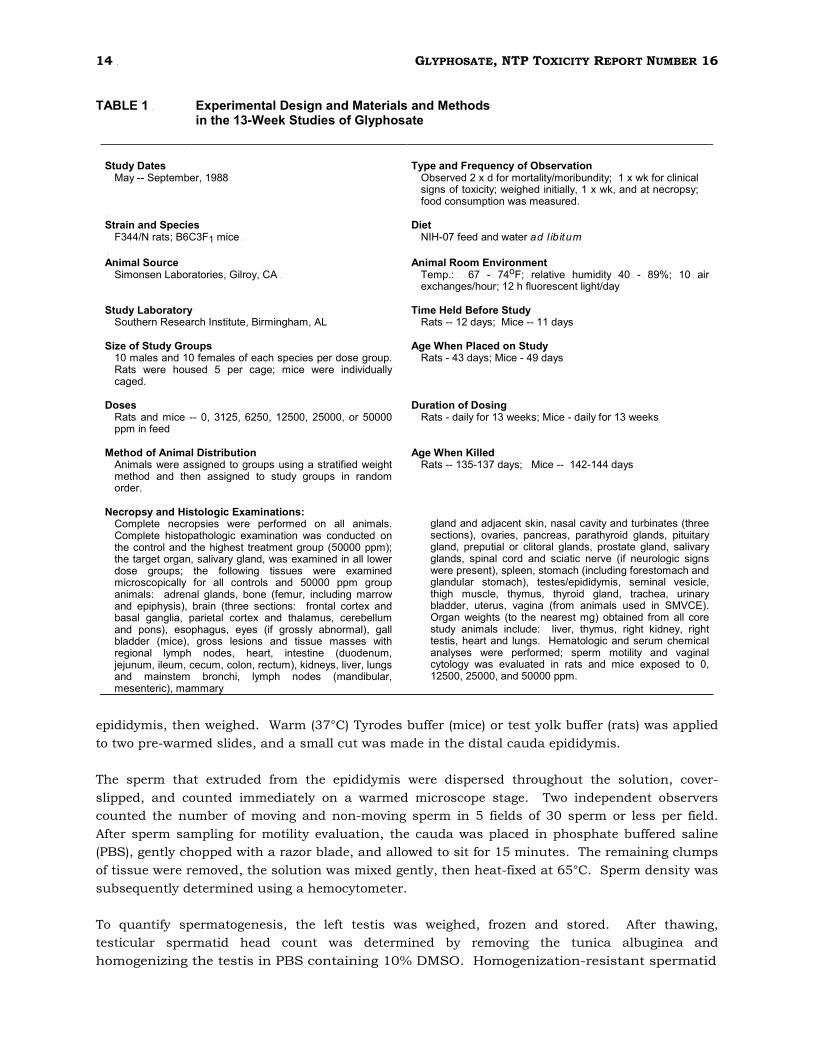
TABLE 1 k Experimental Design and Materials and Methods
in the 13-Week Studies of Glyphosate
-
Study Dates - Type and Frequency of Observation May -- September, 1988 k Observed 2 x d for mortality/moribundity; 1 x wk for clinical
signs kof ktoxicity; kweighed kinitially, k1 kx kwk, kand kat knecropsy; food consumption was measured.
k Strain and Species - Diet F344/N rats; B6C3F1 mice k NIH-07 feed and water a d l ib it u m
k Animal Source - Animal Room Environment Simonsen Laboratories, Gilroy, CA k Temp.: k k 67 k - k 74oF; k relative k humidity k 40 k - k 89%; k 10 k air
exchanges/hour; 12 h fluorescent light/day
Study Laboratory Time Held Before Study Southern Research Institute, Birmingham, AL Rats -- 12 days; Mice -- 11 days
Size of Study Groups Age When Placed on Study 10 males and 10 females of each species per dose group. Rats were housed 5 per cage; mice were individually caged.
Rats - 43 days; Mice - 49 days
Doses Duration of Dosing Rats and mice -- 0, 3125, 6250, 12500, 25000, or 50000 ppm in feed
Rats - daily for 13 weeks; Mice - daily for 13 weeks
Method of Animal Distribution Age When Killed Animals were assigned to groups using a stratified weight method and then assigned to study groups in random order.
Rats -- 135-137 days; Mice -- 142-144 days
Necropsy and Histologic Examinations: Complete necropsies were performed on all animals. Complete histopathologic examination was conducted on the control and the highest treatment group (50000 ppm); the target organ, salivary gland, was examined in all lower dose groups; the following tissues were examined microscopically for all controls and 50000 ppm group animals: adrenal glands, bone (femur, including marrow and epiphysis), brain (three sections: frontal cortex and basal ganglia, parietal cortex and thalamus, cerebellum and pons), esophagus, eyes (if grossly abnormal), gall bladder (mice), gross lesions and tissue masses with regional lymph nodes, heart, intestine (duodenum, jejunum, ileum, cecum, colon, rectum), kidneys, liver, lungs and mainstem bronchi, lymph nodes (mandibular, mesenteric), mammary
gland and adjacent skin, nasal cavity and turbinates (three sections), ovaries, pancreas, parathyroid glands, pituitary gland, preputial or clitoral glands, prostate gland, salivary glands, spinal cord and sciatic nerve (if neurologic signs were present), spleen, stomach (including forestomach and glandular stomach), testes/epididymis, seminal vesicle, thigh muscle, thymus, thyroid gland, trachea, urinary bladder, uterus, vagina (from animals used in SMVCE). Organ weights (to the nearest mg) obtained from all core study animals include: liver, thymus, right kidney, right testis, heart and lungs. Hematologic and serum chemical analyses were performed; sperm motility and vaginal cytology was evaluated in rats and mice exposed to 0, 12500, 25000, and 50000 ppm.
epididymis, then weighed. Warm (37°C) Tyrodes buffer (mice) or test yolk buffer (rats) was applied
to two pre-warmed slides, and a small cut was made in the distal cauda epididymis.
The sperm that extruded from the epididymis were dispersed throughout the solution, cover-
slipped, and counted immediately on a warmed microscope stage. Two independent observers
counted the number of moving and non-moving sperm in 5 fields of 30 sperm or less per field.
After sperm sampling for motility evaluation, the cauda was placed in phosphate buffered saline
(PBS), gently chopped with a razor blade, and allowed to sit for 15 minutes. The remaining clumps
of tissue were removed, the solution was mixed gently, then heat-fixed at 65°C. Sperm density was
subsequently determined using a hemocytometer.
To quantify spermatogenesis, the left testis was weighed, frozen and stored. After thawing,
testicular spermatid head count was determined by removing the tunica albuginea and
homogenizing the testis in PBS containing 10% DMSO. Homogenization-resistant spermatid
GLYPHOSATE, NTP TOXICITY REPORT NUMBER 16 15
nuclei were enumerated using a hemocytometer; the data were expressed as spermatid heads per
total testis, and per gram of testis.
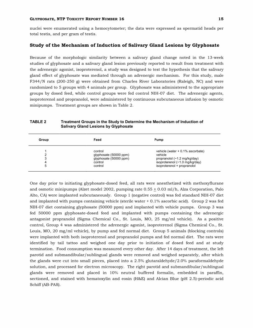
Study of the Mechanism of Induction of Salivary Gland Lesions by Glyphosate
Because of the morphologic similarity between a salivary gland change noted in the 13-week
studies of glyphosate and a salivary gland lesion previously reported to result from treatment with
the adrenergic agonist, isoproterenol, a study was designed to test the hypothesis that the salivary
gland effect of glyphosate was mediated through an adrenergic mechanism. For this study, male
F344/N rats (200-250 g) were obtained from Charles River Laboratories (Raleigh, NC) and were
randomized to 5 groups with 4 animals per group. Glyphosate was administered to the appropriate
groups by dosed feed, while control groups were fed control NIH-07 diet. The adrenergic agents,
isoproterenol and propranolol, were administered by continuous subcutaneous infusion by osmotic
minipumps. Treatment groups are shown in Table 2.
TABLE 2 Treatment Groups in the Study to Determine the Mechanism of Induction of
Salivary Gland Lesions by Glyphosate
Group
Feed
Pump
1
control
vehicle (water + 0.1% ascorbate)
2 glyphosate (50000 ppm) vehicle 3 glyphosate (50000 ppm) propranolol (~1.2 mg/kg/day) 4 control isoproterenol (~1.0 mg/kg/day) 5 control isoproterenol + propranolol
One day prior to initiating glyphosate-dosed feed, all rats were anesthetized with methoxyflurane
and osmotic minipumps (Alzet model 2002, pumping rate 0.55 + 0.03 ml/h, Alza Corporation, Palo
Alto, CA) were implanted subcutaneously. Group 1 (negative control) was fed standard NIH-07 diet
and implanted with pumps containing vehicle (sterile water + 0.1% ascorbic acid). Group 2 was fed
NIH-07 diet containing glyphosate (50000 ppm) and implanted with vehicle pumps. Group 3 was
fed 50000 ppm glyphosate-dosed feed and implanted with pumps containing the adrenergic
antagonist propranolol (Sigma Chemical Co., St. Louis, MO, 25 mg/ml vehicle). As a positive
control, Group 4 was administered the adrenergic agonist, isoproterenol (Sigma Chemical Co., St.
Louis, MO, 20 mg/ml vehicle), by pump and fed normal diet. Group 5 animals (blocking controls)
were implanted with both isoproterenol and propranolol pumps and fed normal diet. The rats were
identified by tail tattoo and weighed one day prior to initiation of dosed feed and at study
termination. Food consumption was measured every other day. After 14 days of treatment, the left
parotid and submandibular/sublingual glands were removed and weighed separately, after which
the glands were cut into small pieces, placed into a 2.5% glutaraldehyde/2.0% paraformaldehyde
solution, and processed for electron microscopy. The right parotid and submandibular/sublingual
glands were removed and placed in 10% neutral buffered formalin, embedded in paraffin,
sectioned, and stained with hematoxylin and eosin (H&E) and Alcian Blue (pH 2.5)-periodic acid
Schiff (AB-PAS).
16 GLYPHOSATE, NTP TOXICITY REPORT NUMBER 16
Genetic Toxicity Studies
Mutagenicity Studies
Mutagenicity studies of glyphosate in Salmonella typhimurium were conducted as described in
Zeiger et al. (1988). Glyphosate was tested for genotoxicity in S. typhimurium strains TA100,
TA1535, TA97, and TA98 using the plate-incorporation assay in both the absence or presence of
Aroclor 1254-induced S9 from male Syrian hamster liver or male Spraque-Dawley rat liver.
Glyphosate was dissolved in distilled water and tested at doses up to 10,000 Og/plate. A positive
response is defined in this assay as a reproducible, dose-related increase in histidine-independent
(revertant) colonies in any one strain/activation combination. An equivocal response is defined as
an increase in revertants which was not dose-related, not reproducible, or of insufficient magnitude
to support a determination of mutagenicity. A negative response is obtained when no increase in
revertant colonies is observed following chemical treatment.
Mouse Peripheral Blood Micronucleus Test
At the termination of the 13-week study, blood smears were prepared from peripheral blood
samples obtained by cardiac puncture of dosed and control mice. The slides were stained with
Hoechst 33258/pyronin Y (MacGregor et al., 1983). Ten thousand normochromatic erythrocytes
from each animal were scored for micronuclei.
Statistical Methods
Analysis of Continuous Variables
Two approaches were employed to assess the significance of pairwise comparisons between dosed
and control groups in the analysis of continuous variables. Organ and body weight data, which are
approximately normally distributed, were analyzed using the parametric multiple comparisons
procedures of Williams (1971, 1972, 1986 ) and Dunnett (1955). Clinical pathology and hema-
tology data, which typically have skewed distributions, were analyzed using the nonparametric
multiple comparisons methods of Shirley (1977) and Dunn (1964). Jonckheere's test (Jonckheere,
1954) was used to assess the significance of dose-response trends and to determine whether a
trend-sensitive test (Williams, Shirley) was more appropriate for pairwise comparisons than a test
capable of detecting departures from monotonic dose-response (Dunnett, Dunn). If the P-value
from Jonckheere's test was greater than or equal to 0.10, Dunn's or Dunnett's test was used rather
than Shirley's or Williams' test.
The outlier test of Dixon and Massey (1951) was employed to detect extreme values. No value
selected by the outlier test was eliminated unless it was at least twice the next largest value or at
most half of the next smallest value.
GLYPHOSATE, NTP TOXICITY REPORT NUMBER 16 17
Analysis of Vaginal Cytology Data
Since the data are proportions (the proportion of the observation period that an animal was in a
given estrous state), an arcsine transformation was used to bring the data into closer conformance
with normality assumptions. Treatment effects were investigated by applying a multivariate
analysis of variance (Morrison, 1976) to the transformed data to test for the simultaneous equality
of measurements across dose levels.
Analysis of Micronuclei Data
Statistical analyses for micronuclei were completed using linear trend tests on polychromatic
erythrocytes data and log-transformed data for normochromatic erythrocytes, and analysis of
variance on ranks (ANOVA) for percentage polychromatic cells among total erythrocytes. The
frequency of micronuclei in the dosed groups was compared with the frequency determined for the
concurrent untreated control animals using the Student t-test.
Quality Assurance
The 13-week toxicity studies of glyphosate were performed in compliance with FDA Good
Laboratory Practices regulations (21 CFR 58). The Quality Assurance Unit of Southern Research
Institute performed audits and inspections of protocols, procedures, data, and reports throughout
the course of the studies. The operations of the Quality Assurance Unit were monitored by the
NTP.
18 GLYPHOSATE, NTP TOXICITY REPORT NUMBER 16
RESULTS
Disposition Studies
More than 90% of the radioactivity from either a 5.6 or 56 mg/kg oral dose of [14C]-glyphosate was
eliminated within 72 hours. Approximately 50% was eliminated in the feces in the first 24 hours;
urinary elimination of radioactivity was essentially complete by 12 hours. The apparent decrease
in cumulative percentage eliminated in urine after the 5.6 mg/kg oral dose probably is due to
interindividual variation, and variances (from 10 to 3) in the number of animals per time point. In
contrast, following an intravenous dose of [14C]-glyphosate at 5.6 mg/kg, 90% of radioactivity was
eliminated in urine in the first 6 hours (Table 3).
TABLE 3 Cumulative Percentage of Oral or I.V. Dose of Glyphosate
Eliminated in Urine and Fecesa
Oral 5.6 mg/kg Oral 56 mg/kg I.V. 5.6 mg/kg
Time (Hours) Urine Feces Urine Feces Urine Feces
6 10 ± 5 7 ± 11 90 ± 7 0.3 ± 0.2
12 31 ± 10 28 ± 10 95 ± 9 0.5 ± 0.5 24 26 ± 14 55 ± 13 28 ± 10 47 ± 12 98 ± 11 3 ± 2 48 18 ± 2 71 ± 8 33 ± 12 57 ± 15 72 19 ± 2 74 ± 5 34 ± 12 58 ± 15
a N = 3-10
TABLE 4 Percentage of Dose in Tissues Following Oral Administration
of Glyphosate at 5.6 mg/kga
Time (h)
Tissue 3b 6b 12b 24c 96c
Small Intestine 7.72 ± 1.74 10.20 ± 5.49 4.12 ± 2.25 0.48 ± 0.51 0.03 ± 0.01 Large Intestine 1.21 ± 1.07 0.51 ± 0.01 0.46 ± 0.28 0.17 ± 017 0.01 ± 0.00 Liver 0.10 ± 0.00 0.07 ± 0.04 0.11 ± 0.01 0.14 ± 0.08 0.05 ± 0.05 Kidney 0.36 ± 0.19 0.48 ± 0.42 0.31 ± 0.06 0.10 ± 0.07 ND Skin 0.70 ± 0.45 0.18 ± 0.25 0.21 ± 0.12 NDd ND
Blood 0.28 ± 0.01 0.18 ± 0.06 0.31 ± 0.10 0.03 ± 0.06 ND Tissue Total 12.00 ± 0.33 11.67 ± 6.29 5.54 ± 2.35 0.89 ± 0.84 0.10 ± 0.06
a Data represented as percent of dose administered ± standard deviation. b N = 2 rats. c N = 3 rats. d ND notes that the values were not determined as the amount of radioactivity in the samples was below the level of accurate
analytical measurement (<100 dpm).
The tissue distribution of radioactivity from a single oral 5.6 mg/kg dose of [14C]-glyphosate is
presented in Table 4. At time points up to 24 hours, most of the radioactivity was found in the
gastrointestinal tract; only 1% remained in the tissues at 24 hours.
In animals given a 56 mg/kg oral dose, the peak blood level of radioactivity occurred later than in
those given a 5.6 mg/kg oral dose (1 hour vs. 2 hours); the peak blood concentration was more
than 30 times higher following the 56 mg/kg oral dose (Figure 1). Radioactivity rapidly declined in
GLYPHOSATE, NTP TOXICITY REPORT NUMBER 16 19
blood following a 5.6 mg/kg i.v. dose (Figure 2). The blood radioactivity vs. time plot fits a 2-
compartment model with an alpha (distribution) phase of about 0.5 hour and a beta (elimination)
phase of 13 hours.
Rats were exposed to Roundup® (the isopropylamine salt of glyphosate and added surfactants) in
drinking water at concentrations of 0.5 to 100,000 ppm for 9 to 16 days. No differences were
observed in the elimination of an oral dose of 5.6 mg/kg [14C]-glyphosate following any of these
exposures, as compared with the elimination of a similar dose 1 day prior to beginning
administration of Roundup® (data not shown).
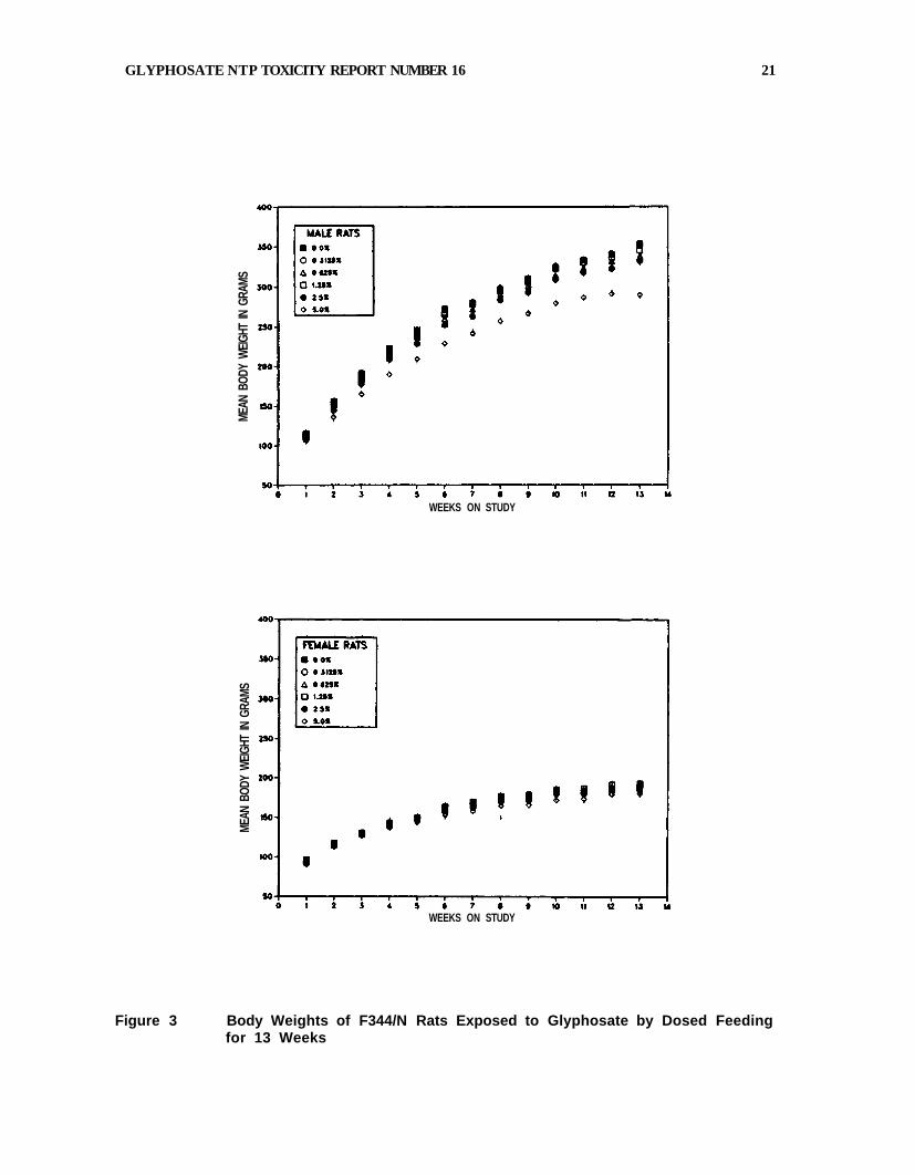
13-Week Studies in F344/N Rats
All animals survived until the end of the study. Diarrhea was observed in the 50000 ppm groups
of both sexes for the first 50 days, though not thereafter. In males, reduced weight gains were
observed in the 25000 and 50000 ppm groups. The final mean body weight of the 50000 ppm
group was approximately 18% less than that of controls (Table 5 and Figure 3). In females, there
was only a marginal effect on body weight gain, with the high dose group 5% lighter than controls
at the end of the study (Figure 3). In male rats, small increases in relative organ weights were
observed for liver, kidney, and testicle; a decrease in relative weight was observed in the thymus
(Appendix A, Table A1). In females, changes in organ weights were minor and could not be related
definitely to treatment. There were no treatment-related effects on food consumption throughout
the study. The mean, time-weighted chemical consumption for each group, based on food intake,
is given in Table 5.
TABLE 5 Survival, Weight Gain, and Feed Consumption of F344/N Rats in the 13-Week Dosed Feed Study of Glyphosate
Dose (ppm) Mean Body Weight (grams) Final Weight Relative Average Feed Glyphosate
In Feed Survivala Initial Final Changeb to Controls (%)c Consumptiond Consumede
MALE
0 10/10 115 353 238 17 0 3125 10/10 111 352 241 100 17 205 6250 10/10 111 338 227 96 17 410
12500 10/10 113 345 232 98 17 811 25000 10/10 108 332 224 94 17 1678 50000 10/10 112 290 178 82 15 3393
FEMALE
0 10/10 95 191 96 11 0 3125 10/10 92 190 98 100 11 213 6250 10/10 94 194 100 102 11 421
12500 10/10 96 193 97 101 11 844 25000 10/10 92 186 94 97 11 1690 50000 10/10 95 181 86 95 10 3393
a Number of animals surviving at 13 weeks/number/dose group. b Mean weight change of the animals in each dose group. c (Dosed group mean/Control group mean) x 100. d Average food consumption in gm/animal/day. e Estimated, mean, time-weighted chemical consumption in mg/kg/day.
20 GLYPHOSATE, ΝΤΡ TOXICITY REPORT NUMBER 16
Figure 1 Blood Levels of 14 C-Glyphosate Following Oral Administration of14C-Glyphosate at 5.6 or 56 mg/kg (% dose ± standard deviation)
Figure 2 Levels of Radioactivity in Blood after a Single i.v. Doseof 5.6 mg/kg Glyphosate (2 rats per time point, results averaged).
56 mg/kg
5.6 mg/kg
GLYPHOSATE NTP TOXICITY REPORT NUMBER 16 21
MEAN
BOD
Y W
EIGHT
IN G
RAMS
WEEKS ON STUDY
MEAN
BOD
Y W
EIGH
T IN
GRA
MS
WEEKS ON STUDY
Figure 3 Body Weights of F344/N Rats Exposed to Glyphosate by Dosed Feedingfor 13 Weeks
22 GLYPHOSATE, NTP TOXICITY REPORT NUMBER 16
Chemically-related changes in hematological parameters observed in male rats at 13 weeks
included mild increases in hematocrit and RBC at 12500, 25000, and 50000 ppm, hemoglobin at
25000 and 50000 ppm, and platelets at 50000 ppm. In female rats, minimal but significant
increases occurred in lymphocyte and platelet counts, WBC, MCH, and MCV. Treatment-related
alterations in clinical chemistry parameters included increases in alkaline phosphatase in males
and in females at all time points, alanine aminotransferase activity in males and females at all time
points except 90 days, total bile acids at days 23 and 90 in males and at day 23 in females, total
protein in females at all time points, and sporadic increases in urea nitrogen and albumin
(Appendix B).
In reproductive studies, male rats experienced a significant decrease (20%) in sperm counts in the
25000 and 50000 ppm groups. Left caudal, epididymal and testicular weights, epididymal sperm
motility, total spermatid heads/testes, and total spermatid heads/g caudal tissue were not
different from those of controls (Appendix C, Table C1). Female rats had a longer estrous cycle
length (5.4 days vs. 4.9 days) in the 50000 ppm group compared to controls (Appendix C, Table
C1).
At necropsy, no gross lesions were observed that were considered possibly related to glyphosate
administration. Morphologic changes attributed to glyphosate were observed microscopically in the
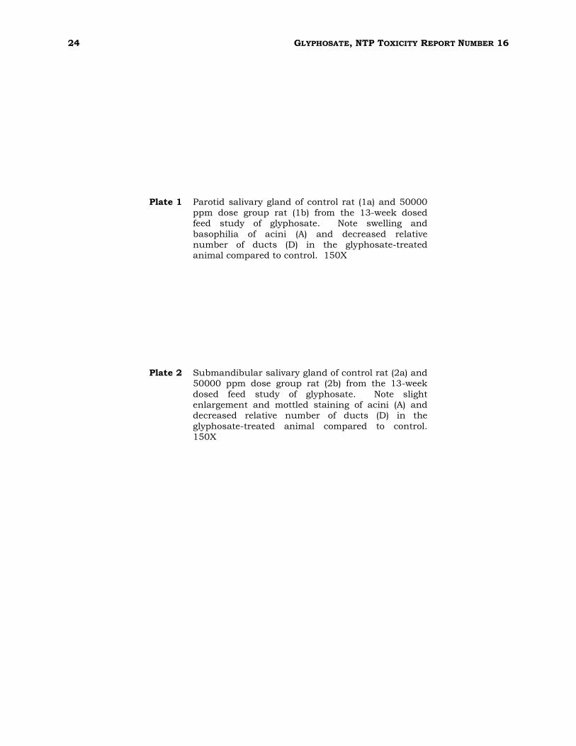
parotid and submandibular salivary glands of male and female rats. Salivary gland lesions were
diagnosed as "cytoplasmic alteration" and consisted of basophilic change and hypertrophy of acinar
cells. These changes were more evident in the parotid gland in which the normal granular,
eosinophilic staining cytoplasm of the acinar epithelial cells was replaced by basophilic and finely
vacuolated cytoplasm (Plate 1). This effect varied in distribution from multifocal in less severe
cases, imparting a mottled tinctorial staining appearance to the gland, to diffuse involvement in
higher dose animals. In addition, acinar cells appeared swollen, resulting in enlargement of
secretory acini and a relative reduction in the number of secretory ducts seen. Nuclei of affected
acinar cells were hyperchromatic. In the submandibular salivary gland, similar cytoplasmic
tinctorial changes and hypertrophic effects were observed (Plate 2). The sublingual gland was not
detectably altered.
A no-effect level for cytoplasmic alteration of the parotid and submandibular salivary glands in this
study was not reached. One control female rat had a small basophilic focus in the parotid gland
which was typical of the spontaneous lesion occasionally seen in rats. Table 6 presents incidence
and severity data of glyphosate-induced cytoplasmic alteration of the salivary glands from the 13-
week dosed feed study in rats. No other lesions in rats appeared related to glyphosate
administration.
TABLE 6 Incidence and Severity of Cytoplasmic Alteration of the Parotid and Submandibular
Salivary Glands (combined) in F344/N Rats in the 13-Week Dosed Feed Study of Glyphosate
Dose (ppm) 0 3125 6250 12500 25000 50000
MALES 0/10 6/10 (1.0)* 10/10 (1.0) 10/10 (1.8) 10/10 (2.7) 10/10 (2.9)
FEMALES
0/10 8/10 (1.0) 10/10 (1.0) 10/10 (2.1) 10/10 (2.4) 10/10 (3.0)
* Average severity score based on a scale of 1=minimal, 2=mild, 3=moderate, 4=marked.
GLYPHOSATE, NTP TOXICITY REPORT NUMBER 16 23
13-Week Studies in B6C3F1 Mice
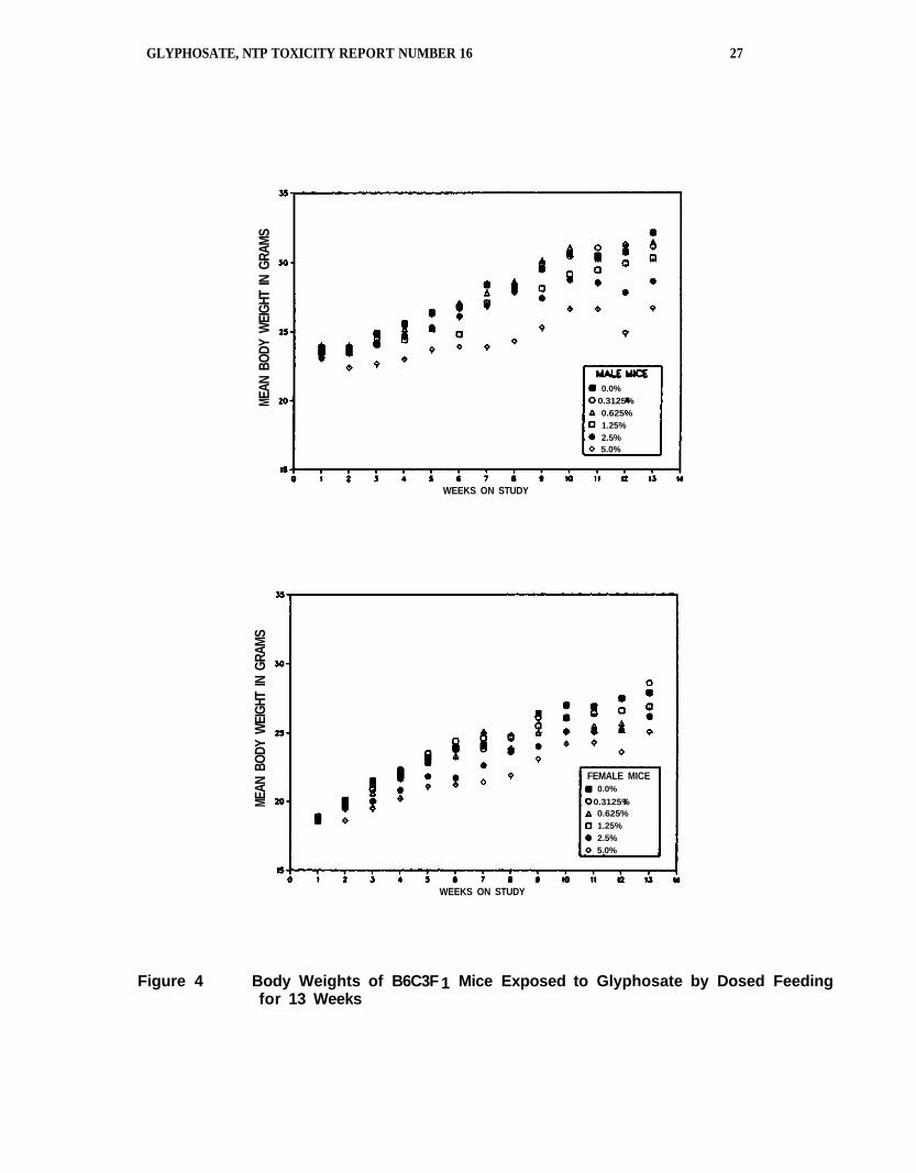
Body weight gains were depressed in the 2 highest dose groups of both sexes (Table 7 and Figure
4). There were 2 early deaths in the study: An untreated female was accidentally killed, and a
high dose female died from undetermined causes (Table 7). Increases in relative organ weights
were observed in the heart, kidney, liver, lung, thymus, and testis of male mice (Appendix A, Table
A2). There were no differences in food consumption between the dosed and control groups.
TABLE 7 Survival, Weight Gain, and Feed Consumption of B6C3F1 Mice
in the 13-Week Dosed Feed Study of Glyphosate
Dose (ppm) Mean Body Weight (grams) Final Weight Relative Average Feed Glyphosate
in Feed Survivala Initial Final Changeb to Controls(%)c Consumptiond Consumede
MALE
0 10/10 23.5 32.1 8.6 4.6 0 3125 10/10 23.2 31.1 7.9 97 4.5 507 6250 10/10 23.4 31.5 8.1 98 4.7 1065
12500 10/10 23.2 30.3 7.1 94 4.9 2273 25000 10/10 23.0 28.6 5.6 89 5.1 4776 50000 10/10 23.5 26.7 3.2 83 5.3 10780
FEMALE
0 9/10 18.9 27.9 9.0 5.4 0 3125 10/10 18.4 28.6 10.2 103 5.8 753 6250 10/10 18.2 26.2 8.0 94 5.3 1411
12500 10/10 18.8 26.9 8.1 96 5.2 2707 25000 10/10 18.5 26.2 7.7 94 5.3 5846 50000 9/10 18.5 25.1 6.6 90 5.2 11977
a Number of animals surviving at 13 weeks/number in dose group. b Mean weight change of the animals in each dose group. c (Dosed group mean/Control group mean) x 100. d Average food consumption in gm/animal/day. e Estimated, mean, time-weighted chemical consumption in mg/kg/day.
A "dark" salivary gland in a high-dose male was the only significant gross finding at necropsy. No
effects were observed on sperm motility or estrual cycle length. Treatment-related microscopic
changes were limited to the parotid salivary gland; the changes consisted of a diffuse increase in
basophilia of the acinar cells, diagnosed as "cytoplasmic alteration." In more severely affected
glands, the cells and acini also appeared enlarged with an associated relative reduction in the
number of ducts. Submandibular and sublingual glands were not detectably altered. The inci-
dence and severity of cytoplasmic alteration of the parotid salivary gland was dose-related (Table 8).
TABLE 8 Incidence and Severity of Cytoplasmic Alteration of the Parotid Salivary Gland in
B6C3F1 Mice in the 13-Week Glyphosate-Dosed Feed Study
Dose (ppm) 0 3125 6250 12,500 25,000 50,000
MALES 0/10 0/10 5/10 (1.0)* 9/10 (1.6) 10/10 (2.8) 10/10 (4.0)
FEMALES
0/10 0/10 2/10 (1.0) 9/10 (1.3) 10/10 (2.1) 10/10 (3.1)
* Average severity score based on a scale of 1=minimal, 2=mild, 3=moderate, 4=marked.
24 GLYPHOSATE, NTP TOXICITY REPORT NUMBER 16
Plate 1 Parotid salivary gland of control rat (1a) and 50000
ppm dose group rat (1b) from the 13-week dosed feed study of glyphosate. Note swelling and
basophilia of acini (A) and decreased relative number of ducts (D) in the glyphosate-treated animal compared to control. 150X
Plate 2 Submandibular salivary gland of control rat (2a) and 50000 ppm dose group rat (2b) from the 13-week
dosed feed study of glyphosate. Note slight enlargement and mottled staining of acini (A) and decreased relative number of ducts (D) in the
glyphosate-treated animal compared to control. 150X
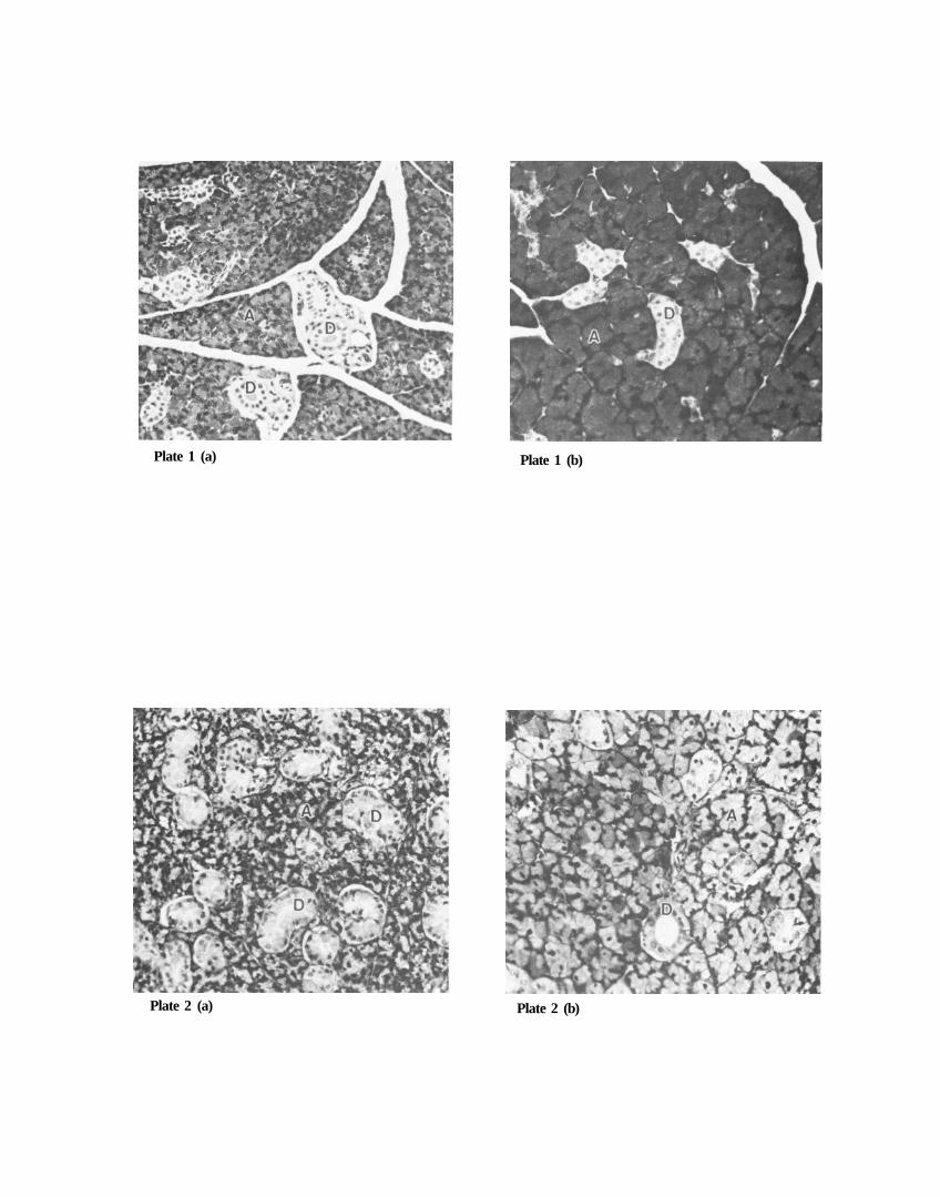
Plate 1 (a) Plate 1 (b)
Plate 2 (a) Plate 2 (b)

26 GLYP HOSATE, NTP TOXICITY REP ORT NUMB ER 16
GLYPHOSATE, NTP TOXICITY REPORT NUMBER 16 27
Figure 4 Body Weights of B6C3F1 Mice Exposed to Glyphosate by Dosed Feedingfor 13 Weeks
MEAN
BOD
Y W
EIGH
T IN
GRA
MS
MEAN
BOD
Y W
EIGH
T IN
GRA
MS
WEEKS ON STUDY
WEEKS ON STUDY
FEMALE MICE0.0%
0.3125%0.625%1.25%2.5%5.0%
0.0%0.3125%0.625%1.25%2.5%5.0%
28 GLYPHOSATE, NTP TOXICITY REPORT NUMBER 16
Mechanism of Induction of Salivary Gland Lesion
Cytoplasmic alteration of salivary gland acinar cells induced by glyphosate in the 13-week studies
was similar morphologically to the reported effect of chronic treatment with the adrenergic
mediator isoproterenol. To test the hypothesis that the salivary gland effect of glyphosate is
mediated through an adrenergic mechanism, a special study was designed in which rats were
concurrently administered glyphosate by dosed feed and/or adrenergic agents by subcutaneous
minipump infusion.
All rats survived to the end of the 14-day study; the implanted minipumps were well-tolerated.
Rats receiving isoproterenol were hypoactive and had increased respiratory rates on day 1 following
pump implantation, but were normal by the following day. Feces of rats receiving glyphosate-dosed
feed were observed to be slightly softer in consistency and wetter than normal in appearance by
study day 7; perianal fecal staining was also evident in several of these animals. Average food
consumption and body weight gains are presented by group in Table 9. It is apparent that there
was no food avoidance in those groups receiving the glyphosate-dosed feed; there was a significant
decrease in body weight gains in those groups, however.
TABLE 9 Feed Consumption and Weight Gain of F344/N Rats
in the 14-Day Mechanism Study of Glyphosate
Treatment Group (diet/pump)
Food Consumption (gm/rat/day)
Weight Gain (gm)
1 (control diet/vehicle) 14.4 16.0 ± 2.9 2 (glyphosate/vehicle)* 17.6 6.3 ± 2.0 3 (glyphosate/propranolol)* 20.4 6.0 ± 2.4 4 (control diet / isoproterenol) 14.9 16.7 ± 1.6 5 (control diet/isoproterenol + propranolol) 15.0 17.5 ± 8.0
* Glyphosate was given in the diet at a concentration of 50000 ppm.
Parotid and submandibular/sublingual salivary gland weight data are shown in Table 10. Both
isoproterenol, the adrenergic agonist given by subcutaneous infusion, and glyphosate, in dosed
feed, induced significant enlargement of these glands, glyphosate having much greater effect than
isoproterenol. The parotid was the much more affected of the two glands. The adrenergic
antagonist, propranolol, inhibited the effect of both isoproterenol and glyphosate on salivary gland
weights. In the parotid, there was approximately a 50% increase in gland weight following
isoproterenol administration, an effect blocked completely blocked by concurrent administration of
propranolol. Glyphosate induced an almost 3-fold increase in parotid weight, an effect significantly
inhibited, though not completely, by propranolol. These trends were paralleled by smaller changes
in submandibular/sublingual gland weights.
Microscopically, both isoproterenol and glyphosate given in the 14-day study induced lesions in the
parotid gland similar to those seen in the 13-week study. These lesions consisted of cytoplasmic
basophilic change, fine vacuolation, and swelling of acinar cells, diagnosed collectively as
cytoplasmic alteration. A distinct gradation in the severity of these lesions was possible based on
the extent of involvement and degree of tinctorial alteration and cell enlargement present.
GLYPHOSATE, NTP TOXICITY REPORT NUMBER 16 29
TABLE 10 Salivary Gland Weights of F344/N Rats
in the 14-day Mechanism Study of Glyphosate
Parotid
Submandibular/Sublingual
Group (diet/pump) Absolute (mg) Relative* Absolute (mg) Relative*
1 (control diet/vehicle) 126.2 ± 16.4 0.50 ± 0.08 209.7 ± 14.8 0.83 ± 0.04 2 (glyphosate/vehicle) 354.0 ± 37.5 1.47 ± 0.12 375.0 ± 26.3 1.56 ± 0.07 3 (glyphosate/propranolol) 245.0 ± 10.4 1.06 ± 0.06 261.0 ± 6.4 1.13 ± 0.04 4 (control diet / isoproterenol) 194.2 ± 15.6 0.76 ± 0.06 259.7 ± 10.6 1.03 ± 0.03 5 (control diet/isoproterenol + propranolol) 137.2 ± 19.1 0.55 ± 0.07 225.5 ± 7.8 0.91 ± 0.05
* mg/g body weight
Glyphosate-treated animals were most severely affected; glands from all these animals were
characterized by diffuse, intense basophilic change of acinar cells with clearly evident acinar
enlargement, resulting in a relative reduction in the number of ducts present. Concurrently, the
cytoplasm of affected cells was finely vacuolated, and nuclei were hyperchromatic and displaced
more basally by increased cytoplasmic volume. In serial sections stained with Alcian Blue/periodic
acid Schiff (AB/PAS), areas of cytoplasmic alteration were seen to be associated with loss of PAS
positive staining of secretory granules. Animals receiving the adrenergic antagonist, propranolol,
subcutaneously and concurrently with glyphosate-dosed feed were clearly protected from the more
severe lesions. All animals dosed with isoproterenol were likewise affected with cytoplasmic
alteration of salivary acinar cells; basophilic tinctorial change in these animals was multifocal to
diffuse, and hypertrophy was less prominent than in the glyphosate group. Propranolol resulted in
only modest protection from isoproterenol effects based on histomorphology. The incidence and
average severity of cytoplasmic alteration of the parotid gland is shown in Table 11.
Cytoplasmic alteration of the submandibular gland was detectable by light microscopy only in the
glyphosate-dosed animals (Table 11). The lesion consisted primarily of cellular and acinar swelling
with a relative reduction in the number of duct profiles per field. Tinctorial change was less of a
component of the submandibular lesion than in the parotid, with most acinar cells being slightly
more pale staining than controls, with scattered individual cells or acini being more basophilic,
imparting a mottled staining pattern to the tissue. AB-PAS reactivity was essentially unchanged in
affected glands.
TABLE 11 Incidence and Severity of Cytoplasmic Alteration of the Salivary Glands of F344/N
Rats in the 14-Day Mechanism Study of Glyphosate
Group (Feed/Pump)
Parotid
Submandibular
Sublingual
1 (control diet/vehicle) 1/4 (1.0) * 0/4 0/3 2 (glyphosate/vehicle) 4/4 (4.0) 4/4 0/4 3 (glyphosate/propranolol) 3/4 (1.5) 4/4 0/2 4 (control diet / isoproterenol) 4/4 (2.7) 0/4 0/1 5 (control diet/isoproterenol +
propranolol)
4/4 (2.0) 0/4 0/4
* Average severity grades for parotid gland lesions in affected animals, based on the following scale:
1=Focal change; 2=Multifocal, confluent change; 3=Diffuse change; 4=Diffuse change with intense basophilia and marked acinar swelling.
30 GLYPHOSATE, NTP TOXICITY REPORT NUMBER 16
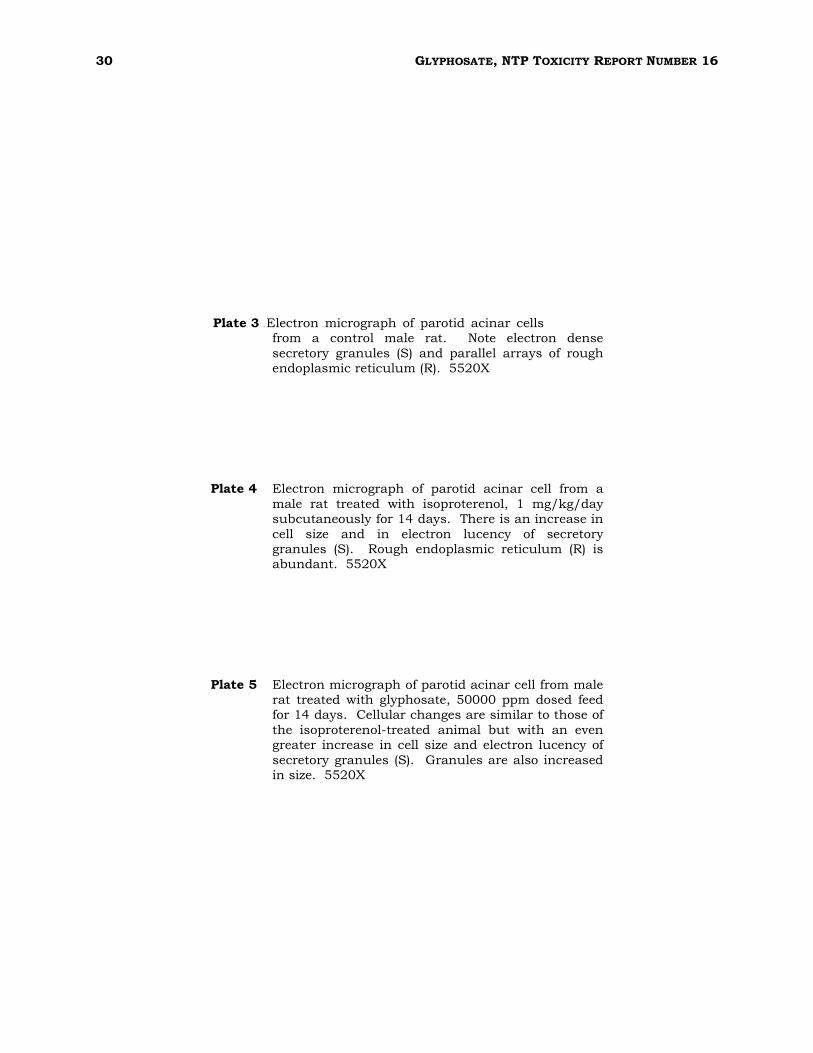
Plate 3 0Electron vmicrograph vof vparotid vacinar vcells from a control male rat. Note electron dense
secretory granules (S) and parallel arrays of rough endoplasmic reticulum (R). 5520X
Plate 4 Electron micrograph of parotid acinar cell from a
male rat treated with isoproterenol, 1 mg/kg/day subcutaneously for 14 days. There is an increase in
cell size and in electron lucency of secretory granules (S). Rough endoplasmic reticulum (R) is abundant. 5520X
Plate 5 Electron micrograph of parotid acinar cell from male rat treated with glyphosate, 50000 ppm dosed feed for 14 days. Cellular changes are similar to those of
the isoproterenol-treated animal but with an even greater increase in cell size and electron lucency of
secretory granules (S). Granules are also increased in size. 5520X
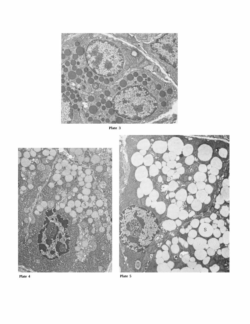
Plate 4 Plate 5
Plate 3

32 GLYPHOSATE, NTP TOXICITY REPORT NUMBER 16
GLYPHOSATE, NTP TOXICITY REPORT NUMBER 16 33
The lesions of the submandibular gland were more subtle than those in the parotid; differences in
the severity of the cytoplasmic alteration in this gland were not appreciable by light microscopy.
There was no definite, inhibitory effect of propanolol on the incidence of the glyphosate-induced
change detected histologically in the submandibular gland. No microscopic change was observed
in this gland in rats treated with isoproterenol. No changes in morphology or Alcian blue-periodic
acid Schiff reactivity were seen in the sublingual glands examined from any groups.
Parotid and submandibular acinar cells from control, isoproterenol-treated, and glyphosate-treated
animals were examined ultrastructurally. Parotid acinar cells of the control animals were of typical
appearance, with basally oriented nuclei surrounded by rough endoplasmic reticulum (Plate 3).
Electron dense secretory granules were concentrated in the apical cytoplasm. In contrast,
secretory granules from the isoproterenol-treated animals were electron lucent in affected cells
(Plate 4). Also, these cells obviously were enlarged, as evident from the increased cell area when
compared to control cells at equivalent magnification; the number of secretory granules and
volume of rough endoplasmic reticulum seemed to be increased concurrently. Similar changes,
though of greater magnitude, were seen in parotid acinar cells from the glyphosate-dosed rats
(Plate 5). There was a further progression in the lucency of the secretory granules, and the
granules were noticeably enlarged and coalescent. Abundant rough endoplasmic reticulum
surrounded the granules and nuclei, and the overall cell area was increased.
Ultrastructurally, control submandibular acini contained both mucous- and serous-type cells.
Mucous cells were more prominent due to their larger size, central location within the acinus, and
the large number of confluent, electron-lucent mucous granules. Serous cells were small and
peripherally located in the acinus, and the electron-dense granules were few in number and
relatively inconspicuous. Both cell types were dark-staining and contained abundant rough
endoplasmic reticulum. In submandibular acini from the isoproterenol-treated animals, cells
appeared swollen due to an increase in the number of granules; granules were heterogeneously
stained, most with finely granular contents and others with granular stippling surrounding a more
electron-dense core. Submandibular cells and acini from the glyphosate-exposed animals were
markedly enlarged due to cytoplasmic engorgement with secretory granules, mostly of the lucent
type, with some more heterogenous as seen in the isoproterenol animals. In these cells, granules
were not limited to apical areas as in the controls but diffusely present throughout the cytoplasm.
It could not be determined if the serous or mucous glandular acini were selectively affected by
glyphosate.
Genetic Toxicology
Glyphosate (0-10000 Og/plate) did not induce gene mutations in Salmonella typhimurium strains
TA100, TA1535, TA97, or TA98 when tested in a preincubation protocol in the presence and the
absence of Aroclor 1254-induced male Sprague-Dawley rat or Syrian hamster liver S9 (Appendix D,
Table D1). Peripheral blood normochromatic erythrocytes from male and female mice were
analyzed at the termination of the 13-week feed study for frequency of micronuclei; no increase in
micronuclei was observed in either males or females at any dietary concentration of glyphosate
(Appendix D, Table D2).
34 GLYPHOSATE, NTP TOXICITY REPORT NUMBER 16
DISCUSSION
Disposition studies showed that after a dose of glyphosate at either 5.6 or 56 mg/kg, over 70% of
the administered dose was eliminated within 24 hours. Tissue distribution data indicate most of
the radioactivity was in the gastrointestinal tract following oral administration, indicating the
compound may not be completely absorbed. Comparison of the pattern of elimination following i.v.
and oral administration of [14C]-glyphosate also supports the conclusion that the compound is
incompletely absorbed. Radioactivity is eliminated primarily in feces after oral administration and
primarily in urine following i.v. administration. If the usual assumption is made that i.v.
administration represents the fate of a completely absorbed dose, then about 30% of the 5.6 mg/kg
oral dose of glyphosate was absorbed; there is some evidence that a relatively higher percentage of
the 56 mg/kg dose was absorbed. The 10-fold increase in dose resulted in a 30-fold increase in
peak blood concentration. There also was a trend toward a higher percentage of the 56 mg/kg
dose being eliminated in urine, but the differences were not statistically significant. Perhaps there
is some interaction between glyphosate and the stomach/intestinal contents that binds a relatively
larger percentage of the low dose, making it less available for absorption.
In the 13-week studies, glyphosate did not affect survival of F344/N rats or B6C3F1 mice. Body
weight gains were depressed in rats and mice at the 2 highest dose levels; weight gain depression
was more severe in males than in females. Kubena et al. (1981) reported that body weight gains
were reduced (about 50%) in male and female chicks fed a diet containing 6080 ppm of the
isopropylamine salt of glyphosate for 21 days, beginning at 1 day of age; the calcium and
magnesium content of the tibiotarsus bone was increased compared to controls. There were no
differences in body weights in chicks fed a dose of 608 ppm or lower. In the Kubena study (which
did not mention feed palatability) and in our 13-week study, the possibility of reduced food intake
in the high dose groups cannot be ruled out; more food tends to be spilled when it is not palatable,
and our food consumption measurements did not account for scattered feed. Poor palatability of
feed containing high concentrations of glyphosate is suggested by the finding that rats drank less
water containing Roundup® at 10000 ppm or higher. Another possibility is that the higher
concentrations of glyphosate in feed result in poor absorption of dietary components from the GI
tract. However, if uncoupling of oxidative phosphorylation, as proposed by Olorunsogo et al. (1979)
and Bababunmi et al. (1979), is occurring as a result of glyphosate ingestion, then a reduction in
weight gain for a given amount of food consumed would be expected.
Hematologic effects in rats dosed with glyphosate were unremarkable and generally consistent with
mild dehydration (increases in RBC counts, hematocrit, and hemoglobin concentrations). This
conclusion also is supported by the mild increases that occurred at various time points in serum
concentrations of urea nitrogen, total protein and albumin. Mild but significant increases in
concentrations of TBA and in activities of serum alanine aminotransferase and alkaline
phosphatase at multiple time points in male and female rats are consistent with an hepatobiliary
effect. These findings likely reflect hepatocellular leakage or perhaps single cell necrosis (ALT) and
cholestasis (TBA and ALP). Increases in absolute and relative liver weights in female rats also were
suggestive of an effect of glyphosate on the liver, and support the clinical pathology findings.
However, the lack of histopathologic evidence for a treatment-related effect on the liver indicates
the mild nature of the hepatotoxicity. Vainio et al. (1983) reported an absence of peroxisome
GLYPHOSATE, NTP TOXICITY REPORT NUMBER 16 35
proliferation or hypolipidemia in male Wistar rats given Roundup® daily by gavage at 300 mg/kg, 5
times a week for 2 weeks; these daily doses were more than 10-fold lower than those achieved in
the highest dose groups in the current study.
Measures of sperm density, or the number of sperm/g caudal epididymal tissue, were reduced
somewhat in male rats in the 2 highest dose groups; other spermatozoal measurements were not
different from controls in rats or mice. There was a slight lengthening of the estrous cycle in high
dose female rats, but the biologic significance of these findings, if any, is not known.
It is noteworthy that the U.S. Environmental Protection Agency, after reviewing an unpublished 2-
year carcinogenicity study of glyphosate in CD-1 mice, announced that there was "an equivocal
carcinogenic response, possibly causing a slight increase in the incidence of renal tubular
adenomas in male mice at the highest dose tested (30000 ppm)." A carcinogenicity study in rats
has yet to be reviewed (Anonymous, 1991). In the present study, however, the salivary gland was
identified as the sole target organ for glyphosate toxicity in both rats and mice. The lesion was
diagnosed as cytoplasmic alteration of the acinar epithelial cells, consisting of increased basophilic
staining and vacuolation of cytoplasm, and enlargement of cells and acini. This lesion was limited
to the parotid gland in mice but affected both parotid and submandibular glands in rats; the
sublingual gland was not affected. Salivary gland lesions are relatively uncommon in toxicity
studies; however, both spontaneous and chemically-induced changes of a similar nature to those
seen in the glyphosate study have been described. So-called "basophilic hypertrophic foci"
occasionally may be seen as a spontaneous lesion in the parotid gland of rats and mice (Chiu and
Chen, 1986); however, these are infrequent and focal in nature. More extensive and diffuse
basophilic and hypertrophic change has been described in subchronic studies with some
chemicals, such as doxylamine (Jackson and Blackwell, 1988) and methapyrilene (Jackson and
Sheldon, 1984). By far, the most extensive and detailed studies of these changes in salivary glands
have been done with sympathomimetic agents -- for example, the adrenergic agonist, isoproterenol,
which induces striking morphologic changes in salivary glands (Schneyer, 1962; Fukuda, 1968).
As with glyphosate’s effects on the salivary glands, isoproterenol affects the parotid and
submandibular glands but not the sublingual. This is due to the fact that, in the rat, the acini of
the parotid and submandibular are richly supplied with adrenergic fibers, while the sublingual
gland is devoid of adrenergic innervation (Nordenfelt, 1967). Because glyphosate and isoproterenol
are similar in both the morphologic effects induced in the salivary glands and the gland specificity
of those effects, it was hypothesized that glyphosate-related lesions were mediated through an
adrenergic mechanism. A study was designed to test this hypothesis.
Two weeks' exposure to glyphosate by dosed feed resulted in marked increases in parotid and
submandibular salivary gland weights. This effect on salivary gland weights is similar to that of
isoproterenol, both as described in the literature (Schneyer, 1962) and as seen in the positive
control group of this study. Increased salivary gland weights were associated histologically with
cytoplasmic alteration of acinar cells. This effect was more marked in the parotid than in the
submandibular gland. In the parotid, the cytoplasmic change induced by both glyphosate and
isoproterenol was associated with a loss of the normal PAS-positive reactivity of the secretory
granules, indicating either a loss of the granules or a change in their chemical composition. The
sublingual gland was not affected histologically by either glyphosate or isoproterenol,
demonstrating target specificity of glyphosate- and isoproterenol-associated lesions to those
salivary glands which are innervated by adrenergic fibers (Nordenfelt, 1967).
36 GLYPHOSATE, NTP TOXICITY REPORT NUMBER 16
The effect of adrenoreceptor stimulation on parotid acinar cells has been described by
ultrastructural and morphometric criteria to be increases in cell size, primarily due to increases in
the number and size of secretory granules, as well as changes in the staining of these granules
from electron dense to lucent, interpreted to represent a mucoid transformation of the cell
(Schneyer, 1962; Henriksson, 1982; Carlsoo et al., 1984). These findings are identical to those
found upon electron microscopic examination of parotid cells from animals treated with both
glyphosate and isoproterenol in this study, the effects varying only in degree between the
chemicals. Ultrastructural effects in the submandibular gland were similar between these
compounds, though of a less well-defined nature, These effects consisted of cell enlargement due to
accumulation of lucent or heterogenous staining mucoid type granules, although it was not clear
whether the serous or mucous cells of the acinus were being affected. This study led to the
conclusion that the salivary gland effect is mediated through an adrenergic mechanism, as
evidenced by (1) inhibition of the glyphosate-induced effect by the adrenergic antagonist,
propranolol; (2) the similarity between the effects of glyphosate and the adrenergic agonist,
isoproterenol; and (3) the specificity of those effects for salivary glands with adrenergic innervation.
The biologic significance of this finding is unknown. In addition to basophilic and hypertrophic
morphologic changes of acinar cells, treatment with isoproterenol has been associated with
increased cell proliferation in the parotid gland (Schneyer et al., 1967). This suggests that if
glyphosate is acting through an adrenergic pathway, it may likewise induce hyperplasia in this
gland, possibly predisposing it to neoplastic change; however, this is not considered likely, since
spontaneous basophilic, hypertrophic foci of the parotid, as well as of the pancreas (an
anatomically similar tissue) are not considered to be preneoplastic lesions. Moreover, there was no
increased incidence in rats of salivary gland tumors in a 2-year study of methapyrilene (personal
communication, Dr. I. Hirono, Fujita Gakuen Health University, Japan, May 17, 1991), a chemical
which induced similar salivary gland lesions as glyphosate in subchronic studies.
The results of the Salmonella typhimurium assays and micronuclei tests showed no evidence that
glyphosate is genotoxic. A similar conclusion was drawn by Li and Long (1988) after evaluating
glyphosate in a battery of genotoxicity assays including Salmonella typhimurium reversion, E. coli
WP-2 reversion, CHO/HGPRT gene mutation, hepatocyte/DNA repair, and in vivo rat bone marrow
cytogenetics. Moriya et al. (1983) also reported negative findings in Salmonella (TA100, TA98,
TA1535, TA1537, and TA1538) and E. coli (WP2 hcr) assays.
In summary, these studies demonstrated that glyphosate was incompletely absorbed from the
gastrointestinal tract and excreted in the urine after oral administration. The unabsorbed portion
of the dose was excreted in feces. There was no evidence of genetic or reproductive toxicity of
glyphosate. At doses of 25000 and 50000 ppm in the feed, glyphosate reduced body weight gain,
caused cytoplasmic alteration and hypertrophy of salivary gland acinar cells, and elevated serum
bile acids, alkaline phosphatase, and alanine aminotransferase activities, although there was no
histopathologic evidence of liver injury. The effects on salivary glands appeared to be
adrenergically mediated and could be counteracted by the adrenergic antagonist propranolol. The
no-observed-adverse effect level (NOAEL) for the salivary gland lesion was 3125 ppm in the feed for
mice, but the lesion was observed at all dose levels studied in rats.
GLYPHOSATE, NTP TOXICITY REPORT NUMBER 16 37
REFERENCES Ahotupa, M., Hietanen, E., Mantyla, E., and Vainio, H. (1983) Enhancement of extrahepatic drug metabolism by phenoxyacid herbicides and clofibrate, in Rydstrjom, J., Montehus, J., and Bengtsson,
M. (eds.), Extrahepatic Drug Metabolism and Chemical Carcinogenesis. Amsterdam: Elsevier Science Publishers, pp. 245-6.
Amrhein, N., Deus, B., Gehrke, P., Hollander, H., Schab, J., Schulz, A., Steinrucken, H.C. (1981) Interference of glyphosate with the shikimate pathway. Proc. Plant Growth Regul. Soc. Am., 8, 99-106. Amrhein, N., Schab, J., Steinrucken, H.C. (1980) The mode of action of the herbicide glyphosate.
Naturwissensch 67, 356-357. Anonymous (1991) Glyphosate tolerances proposed. Pesticide & Toxic Chemical News, February 13.
Bababunmi, E.A., Olorunsogo, O. O., and Bassir, O. (1978) Toxicology of glyphosate in rats and mice.
Toxicol. Appl. Pharmacol., 45, 319-320. Bababunmi, E.A., Olorunsogo, O.O., and Bassir, O. (1979) The uncoupling effect of N-
(phosphonomethyl) glycine on isolated rat liver mitochondria. Biochem. Pharmacol. 28, 925-927.
Beste, C.E. (1983) Herbicide Handbook of the Weed Science Society of America, 5th Edition. Weed Science Society of America, pp. 258-263.
Boorman, G.A., Hickman, R.L., Davis, G.W., Rhode, L.S., White, N.W., Griffin, T.A., Mayo, J., and Hamm, T.E., Jr. (1986) Serological titers to murine viruses in 90-day and 2-year studies, in Hamm, T.
E., Jr., (ed.), Complications of Viral and Mycoplasmal Infections in Rodents to Toxicology Research and Testing, New York: Hemisphere, pp 11-23.
Carlsoo, B., Danielsson, A., Henriksson, R., Jonsson, G., and Sunstrom, S. (1984) Inhomo-geneities in glycoprotein cytochemistry of secretory granules in rat parotid acinar cells after selective beta-1
adrenoceptor stimulation. Archs. Oral. Biol. 29(12), 953-958. Chiu, T. and Chen, H.C. (1986) Spontaneous basophilic hypertrophic foci of the parotid glands of rats
and mice. Vet. Path. 23, 606-609.
Dixon, W., and Massey, F. (1951) Introduction to Statistical Analysis. New York: McGraw Hill, pp. 145-147.
Dunn, O.J. (1964) Multiple comparisons using rank sums. Technometrics 6, 241-252.
Dunnett, W. (1955) A multiple comparison procedure for comparing several treatments with a control. J. Am. Stat. Assoc. 50, 1095-1121.
Folmar, L.C., Sanders, H.O., and Julin, A.M. (1979) Toxicity of the herbicide glyphosate and several of its formulations to fish and aquatic invertebrates. Arch. Environm. Contam. Toxicol. 8, 269-278. Fukuda, M. (1968) The influence of isoprenaline and propranolol on the submaxillary gland of the rat. Jap. J. Pharmac. 18, 185-199. Ghassemi, M., Quinlivan, S., and Dellarco, M. (1982) Environmental effects of new herbicides for
vegetation control in forestry. Environ. Int. 7, 389-402. Glass, R.L. (1984) Metal complex formation by glyphosate. J. Agric. Food Chem. 32, 1249-1253. Henriksson, R. (1982) Beta-1 and beta-2 adrenoceptor agonists have different effects on rat parotid
acinar cells. Am. J. Physiol. 242(5), G481.
38 GLYPHOSATE, NTP TOXICITY REPORT NUMBER 16
Hietanen, E., Linnainmaa, K., Vainio, H. (1983) Effects of phenoxyherbicides and glyphosate on the
hepatic and intestinal biotransformation activities in the rat. Acta. Pharmacol. Toxicol. 53, 103-112.
Hoagland, R.E. and Duke, S.O. (1982) Biochemical effects of glyphosate [N-(phosphono-methyl)glycine]. ACS Symp. Ser. 181, 175-205.
Jackson, C.D., and Blackwell, B. (1988) Subchronic studies of doxylamine in Fischer 344 rats. Fundam. Appl. Toxicol. 10, 243-253. Jackson, C.D. and Sheldon, W. (1984) 90-day subchronic report on methapyrilene (neutral drinking water) in Fischer 344 rats, NCTR Technical Report for Experiment No. 382. Jefferson, AR: National
Center for Toxicological Research.
Jonckheere, A.R. (1954) A distribution-free k-sample test against ordered alternatives. Biometrika 41, 133-145.
Kubena, L.F., Smalley, H.E., and Farr, F.M. (1981) Influence of Glyphosate [N-(phosphonomethyl) glycine] on performance and selected parameters in broilers. Poultry Sci. 60, 132-136. Li, A.P., and Long, T.J. (1988) An evaluation of the genotoxic potential of glyphosate. Fundam. Appl. Toxicol. 10, 537-546. MacGregor, J.T., Wehr, C.M., and Langlois, R.G. (1983) A simple fluorescent staining procedure for
micronuclei and RNA in erythrocytes using Hoechst 33258 and pyronin Y. Mutat. Res. 120, 269-275. Moriya, M., Ohta, T., Watanabe, K., Miyazawa, T., Kato, K., and Shirasu, Y. (1983) Further
mutagenicity studies on pesticides in bacterial reversion systems. Mutat. Res. 116, 185-216.
Morrison, D. F. (1976) Multivariate Statistical Methods. New York: McGraw Hill, pp. 170-179. Morrissey, R.E., Schwetz, B.A., Lamb, J.G., IV, Ross, M.C., Teague, J.L., and Morris, R.W. (1988)
Evaluation of rodent sperm, vaginal cytology, and reproductive organ weight data from National Toxicology Program thirteen-week studies. Fundam. Appl. Toxicol. 11, 343-358. Newton, M., Howard, K.M., Kelpsas, B.R., Danhaus, R., Lottman, M., and Dubelman, S. (1984) Fate of glyphosate in an Oregon forest ecosystem. J. Agric. Food Chem. 32, 1144-1151. Nordenfelt, I. (1967) Metabolism of transmitter substances in salivary glands, in Schneyer, L.H., and
Schneyer, C.A., (eds.), Secretory Mechanisms of Salivary Glands. New York: Academic Press, pp. 142-154.
Olorunsogo, O.O. (1982a) Defective nicotinamide nucleotide transhydrogenase reaction in hepatic mitochondria of N-(phosphonmeklthyl)-glycine treated rats. Biochem. Pharmacol. 31, 2191-2192. Olorunsogo, O.O. (1982b) Inhibition of energy-dependent transhydrogenase reaction by N-(phosphonomethyl) glycine in isolated rat liver mitochondria. Toxicol. Lett. 10, 91-95. Olorunsogo, O.O. and Bababunmi, E.A. (1980) Interference of herbicides with mitochondrial oxidative
phosphorylation: the N-(phosphonomethyl)glycine model. Toxicol. Lett. (Spec. Issue 1), 148. Olorunsogo, O.O., Bababunmi, E.A., and Bassir, O. (1979) Effects of glyphosate on rat liver
mitochondria in vivo. Bull. Environm. Contam. Toxicol. 22, 357-364.
Rao, G.N., Haseman, J.K., and Edmonson, J. (1989) Influence of viral infections on body weight, survival, and tumor prevalence in Fischer 344/NCr rats on two-year studies. Lab. Animal Sci. 39, 389-393.
Rao, G.N., Piegorsch, W.W., Crawford, D.D., Edmondson, J., and Haseman, J.K. (1989a) Influence of
viral infections on body weight, survival, and tumor prevalence of B6C3F1 (C57BL/6N X C3H/Hen)
mice in carcinogenicity studies. Fund. Appl. Toxicol. 13, 156-164.
GLYPHOSATE, NTP TOXICITY REPORT NUMBER 16 39
Rueppel, M., Brightwell, B.B., Schaefer, J., and Marvel, J.T. (1977) Metabolism and degradation of
glyphosate in soil and water. J. Agric. Food Chem. 25, 517-528.
Sawada, Y., Nagai, Y., Ueyama, M., and Yamamoto, I. (1988) Probable toxicity of surface-active agent in commercial herbicide containing glyphosate. The Lancet 1, 299.
Schneyer, C.A. (1962) Salivary gland changes after isoproterenol-induced enlargement. Am J. Physiol. 203, 232-236. Schneyer, C.A., Finley, W.H., and Finley, S.C. (1967) Increased chromosome number of rat parotid cells after isoproterenol. Proc. Soc. Exp. Biol. Med. 125, 722. Shirley, E. (1977) A non-parametric equivalent of William's test for contrasting increasing dose levels
of a treatment. Biometrics 33, 386-389. Sprankle, P., Meggitt, W.F., and Penner, D. (1975) Absorption, mobility, and microbial degradation of
glyphosate in the soil. Weed Science 23, 229-234.
Stark, J. (1983) Persistence of herbicides in forest soils. Weeds and Weed Control, 24, 275-286. U.S. Environmental Protection Agency (U.S. EPA) (1979) Information provided based on review of
glyphosate registration files (majority of the data originally supplied by Monsanto Company). Washington, D.C.
Vainio, H., Linnainmaa, K., Kahonen, M., Nickels, J., Hietanen, E., Marniemi, J., and Peltonen, P. (1983) Hypolipidemia and peroxisome proliferation induced by phenoxyacetic acid herbicides in rats.
Biochem. Pharmacol. 32, 2775-2779.
Wan, M.T., Watts, R.G., and Moul, D.J. (1989) Effects of different dilution water types on the acute toxicity to juvenile Pacific salmonids and rainbow trout of glyphosate and its formulated products. Bull. Environ. Contam. Toxicol. 43, 378-385. Wester, R.C., Melendres, J., Sarason, R., McMaster, J., and Maibach, H.I. (1991) Glyphosate skin
binding, absorption, residual tissue distribution, and skin decontamination. Fundam. Appl. Toxicol. 16, 725-732.
Williams, D.A. (1971) A test for differences between treatment means when several dose levels are compared with a zero dose control. Biometrics 27, 103-117. Williams, D.A. (1972) The comparison of several dose levels with a zero dose control. Biometrics 28, 519-531.
Williams, D.A. (1986) A note on Shirley's nonparametric test for comparing several dose levels with a
zero control. Biometrics 42, 183-186. Zeiger, E., Anderson, B., Haworth, S., Lawlor, T., and Mortelmans, K. (1988) Salmonella mutagenicity
tests. IV. Results from the testing of 300 chemicals. Environ. Molec. Mutagen. 11 (Suppl 12), 1-158.
GLYPHOSATE, N T P TOXICITY REPORT NUMBER 16 A - 1
APPENDIX A
Organ Weights andOrgan^tfeight-to-Body-Weight Ratios
Table A1 Organ Weights and Organ-Weight-to-Body-Weight Ratios for F344/N Ratsin the 13-Week Feed Study of Glyphosate A-2
Table A2 Organ Weights and Organ-Weight-to-Body-Weight Ratios for B6C3F1 Micein the 13-Week Feed Study of Glyphosate A-3
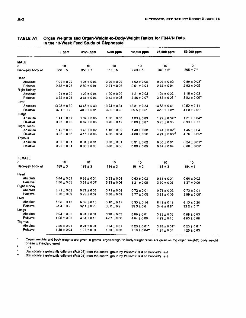
A-2 GLYPHOSATE, NTP TOXICITY REPORT NUMBER 16
TABLE A1 Organ Weights and Organ-Weight-to-Body-Weight Ratios for F344/N Ratsin the 13-Week Feed Study of Glyphosate1
MALEη
Necropsy body wt
HeartAbsoluteRelative
Right KidneyAbsoluteRelative
LiverAbsoluteRelative
LungsAbsoluteRelative
Right TestisAbsoluteRelative
ThymusAbsoluteRelative
FEMALEη
Necropsy body wt
HeartAbsoluteRelative
Right KidneyAbsoluteRelative
LiverAbsoluteRelative
LungsAbsoluteRelative
ThymusAbsoluteRelative
0 ppm
10358 ±5
1 02 ± 0 022 83 ± 0 03
1 21 ± 0 023 38 ± 0 06
13 28 ± 0 3237 1 ± 1 0
1 41 ± 0 023 95 ± 0 08
1 42 ± 0 033 95 ± 0 05
0 33 ± 0 010 92 ± 0 04
10189 ±3
0 64 ± 0 013 36 ± 0 05
0 71 ± 0 023 73 ± 0 09
5 93 ± 0 1331 4 ± 0 7
0 94 ± 0 024 95 ± 0 08
0 26 ± 0 011 35 ± 0 04
3125 ppm
10358 ± 7
1 01 ± 0 032 82 ± 0 04
1 29 ± 0 043 61 ± 0 06
14 45 ± 0 4940 3 ± 0 9*
1 32 ± 0 033 69 ± 0 08
1 48 ± 0 024 15 ± 0 06
031 ±0010 86 ± 0 03
10189 ±3
0 63 ± 0 013 31 ± 0 07
0 71 ± 0 023 73 ± 0 09
6 07 ± 0 1032 1 ± 0 7
0 91 ± 0 04481 ±016
0 24 ± 0 011 27 ± 0 04
6250 ppm
10351 ±5
0 96 ± 0 022 74 ± 0 03
1 20 ± 0 023 42 ± 0 05
13 74 ± 0 3139 2 ± 0 8*
1 30 ± 0 053 70 ± 0 12
1 40 ± 0 024 00 ± 0 04
0 30 ± 0 010 86 ± 0 05
10194 ±3
0 63 ± 0 013 23 ± 0 06
0 71 ± 0 023 66 ± 0 09
6 40 ±0 1733 0 ± 0 9
0 90 ± 0 024 67 ± 0 08
0 24 ± 0 011 23 ± 0 03
12,500 ppm
10350 ±5
1 02 ± 0 022 91 ± 0 04
1 21 ± 0 033 46 ± 0 07
13 81 ± 0 3439 5 ± 0 6*
1 33 ± 0 033 80 ± 0 07
1 40 ± 0 084 00 ± 0 20
0 31 ± 0 020 88 ± 0 05
10191 ± 2
0 63 ± 0 023 31 ± 0 08
0 72 ± 0 013 77 ± 0 05
6 35 ± 0 1433 3 ± 0 6
0 89 ± 0 014 64 ± 0 06
0 23 ± 0 01*1 18 ± 0 04**
25,000 ppm
10340 ± 5*
0 96 ± 0 022 83 ± 0 04
1 24 ± 0 023 65 ± 0 06**
14 58 ± 0 4142 8 ± 1 0**
1 27 ± 0 04**3 73 ± 0 08
1 44 ± 0 022
4 24 ± 0 06*2
0 30 ± 0 010 87 ± 0 04
10185 ±3
061 ±0013 30 ± 0 05
0 71 ± 0 023 81 ± 0 06
6 42 ± 0 1834 6 ± 0 6*
0 93 ± 0 034 99 ± 0 10
023±001*1 25 ± 0 05
50,000 ppm
10305 ± 7**
0 89 ± 0 03**2 92 ± 0 05
1 16 ± 0 033 82 ± 0 06**
12 52 ± 0 4141 0±06**
1 21 ± 0 04**3 99 ± 0 11
1 45 ± 0 044 76 ± 0 05**
0 24 ± 0 01**0 80 ± 0 03*
10184 ±5
0 60 ± 0 023 27 ± 0 09
0 73 ± 0 013 99 ± 0 09*
6 10 ± 0 2033 2 ± 0 7*
0 88 ± 0 034 80 ± 0 08
023±001*1 25 ± 0 03
Organ weights and body weights are given in grams, organ-weight-to-body-weight ratios are given as mg organ weight/g body weight(mean ± standard error)n=9
Statistically significantly different (P<0 05) from the control group by Williams' test or Dunnett's testStatistically significantly different (P<0 01) from the control group by Williams' test or Dunnett's test
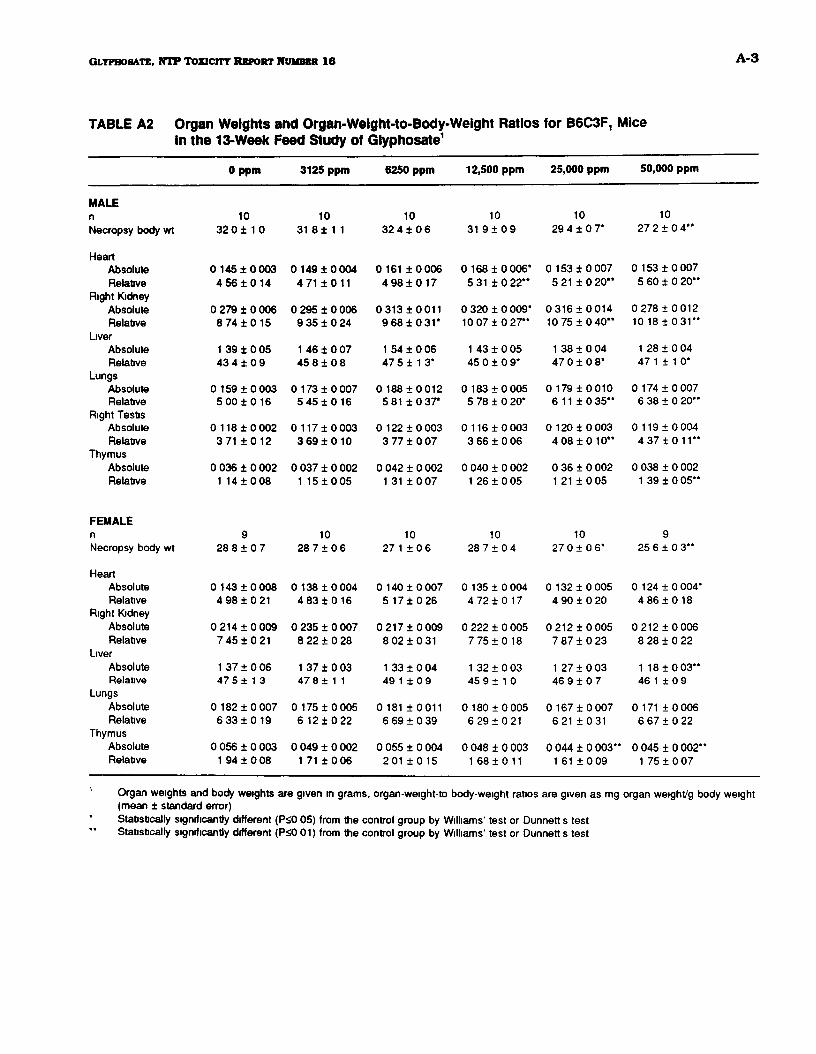
QLYPBOBATB, N T P TOZICITT REPORT NUMBER I β A-3
TABLE A2 Organ Weights and Organ-Weight-to-Body-Weight Ratios for B6C3F, MiceIn the 13-Week Feed Study of Glyphosate1
MALEη
Necropsy body wt
HeartAbsoluteRelative
Right KidneyAbsoluteRelative
LiverAbsoluteRelative
LungsAbsoluteRelative
Right TestisAbsoluteRelative
ThymusAbsoluteRelative
FEMALEη
Necropsy body wt
HeartAbsoluteRelative
Right KidneyAbsoluteRelative
LiverAbsoluteRelative
LungsAbsoluteRelative
ThymusAbsoluteRelative
Oppm
1032 0 ± 1 0
0 145 ± 0 0034 56 ± 0 14
0 279 ± 0 0068 74 ± 0 15
1 39 ± 0 0543 4 ± 0 9
0 159 ± 0 0035 00 ± 0 16
0 118 ± 0 0023 71 ± 0 12
0 036 ± 0 0021 14 ± 0 08
928 8 ± 0 7
0 143 ± 0 0084 98 ± 0 21
0 214 ± 0 0097 45 ± 0 21
1 37 ± 0 0647 5 ± 1 3
0 182 ±0 0076 33 ± 0 19
0 056 ± 0 0031 94 ± 0 08
3125 ppm
1031 8± 1 1
0 149 ±00044 71 ±011
0 295 ± 0 0069 35 ± 0 24
1 46 ± 0 0745 8 ± 0 8
0 173 ±0 0075 45 ±0 16
0 117 ±0 0033 69 ±0 10
0 037 ± 0 0021 15 ±0 05
1028 7 ± 0 6
0 138 ± 0 0044 83 ± 0 16
0 235 ± 0 0078 22 ± 0 28
1 37 ± 0 0347 8 ± 1 1
0 175 ± 0 0056 12±022
0 049 ± 0 0021 71 ± 0 06
6250 ppm
1032 4 ± 0 6
0 161 ± 0 0064 98 ± 0 17
0313±00119 68 ±0 31*
1 54 ± 0 0647 5 ± 1 3*
0 188 ±0 0125 81 ±0 37*
0 122 ±0 0033 77 ± 0 07
0 042 ± 0 0021 31 ±0 07
1027 1 ± 0 6
0 140 ±0 0075 17 ± 0 26
0 217 ±0 0098 02 ±0 31
1 33 ± 0 0449 1 ± 0 9
0 181 ±00116 69 ± 0 39
0 055 ± 0 004201 ±015
12,500 ppm
1031 9 ± 0 9
0 168 ± 0 006*5 31 ±0 22"
0 320 ± 0 009*10 07 ± 0 27"
1 43 ± 0 0545 0 ± 0 9*
0 183 ±0 0055 78 ± 0 20*
0 116 ± 0 0033 66 ± 0 06
0 040 ± 0 0021 26 ± 0 05
1028 7 ± 0 4
0 135 ±0 0044 72 ± 0 17
0 222 ± 0 0057 75 ±0 18
1 32 ± 0 0345 9 ± 1 0
0 180 10 0056 291021
0 048 ± 0 0031 68 ± 0 11
25,000 ppm
1029 4 ± 0 7*
0 153 ± 0 0075 21 ±0 20"
0316±001410 75 ± 0 40"
1 38 ± 0 0447 0 ± 0 8*
0 179±00106 11± 0 35"
0 120 ± 0 0034 08 ± 0 10"
0 36 ± 0 0021 21 ± 0 05
1027 0 ±06*
0 132 ±0 0054 90 ± 0 20
0 212 ±0 0057 87 ± 0 23
1 27 ± 0 0346 9 ± 0 7
0 16710 007621 ±031
0 044 1 0 003"1 61 10 09
50,000 ppm
1027 2 1 0 4 "
0 153 1 0 0075 60 1 0 20"
0 278 1 0 01210 18 ± 0 3 1 "
1 28 1 0 0447 1 1 1 0*
0 174 1 0 0076 38 1 0 20"
0 119 1 0 0044 3 7 1 0 1 1 "
0 038 1 0 0021 39 1 0 05"
925 6 1 0 3**
0 124 1 0 004*4 8 6 1 0 1 8
0 2 1 2 1 0 0 0 68 28 1 0 22
1 1 8 1 0 0 3 "46 1 1 0 9
0 171 1 0 0066 67 1 0 22
0 045 1 0 002**1 75 1 0 07
Organ weights and body weights are given in grams, organ-weight-to body-weight ratios are given as mg organ weight/g body weight(mean i standard error)Statistically significantly different (P<0 05) from the control group by Williams' test or Dunnett s testStatistically significantly different (P<0 01) from the control group by Williams' test or Dunnett s test

GLYPHOSATE, NTP TOXICITY REPORT NUMBER Iβ Β-1
APPENDIX Β
Hematology and Clinical Chemistry
Table Bl Hematology Data for F344/N Rats in the 13-Week Feed Study of Glyphosate B-2
Table B2 Clinical Chemistry Data for F344/N Rats in the 13-Week Feed Studyof Glyphosate B-5
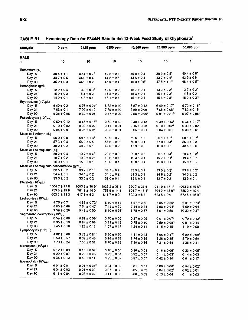
B-2 GLYPHO8ATE, N T P TOXICITY REPORT NUMBER 1 6
TABLE B1 Hematology Data for F344/N Rats in the 13-Week Feed Study of Glyphosate1
Analysis
MALEη
Hematocrit (%)Day 5Day 21Day 90
Hemoglobin (g/dL)Day 5Day 21Day 90
Erythrocytes (10β/μΙ_)Day 5Day 21Day 90
Reticulocytes (10%d-)Day 5Day 21Day 90
Mean cell volume (fL)Day 5Day 21Day 90
Mean cell hemoglobinDay 5Day 21Day 90
Mean cell hemoglobinDay 5Day 21Day 90
Platelets (103/μΙ_)
0 ppm
10
38 4 ± 1 143 7 ± 0 645 2 ± 0 3
1 2 9 ± 0 41 5 0 ± 0 21 4 9 1 0 1
6 4 0 1 0 2 17 63 1 0 119 36 1 0 06
0 6 2 1 0 1 20 1 0 1 0 020 041001
60 0 1 0 95 7 3 1 0 448 2 1 0 2
(pg)20 2 1 0 41 9 7 1 0 21 5 9 1 0 1
concentration33 5 1 0 234 4 1 0 133 0 1 0 2
Day 5 1004 7 ± 17 8Day 21Day 90
Leukocytes (103/μΙ_)Day 5Day 21Day 90
753 8 1 19 9603 3 1 13 3
4 75 1 0 716 80 1 0 689 59 1 0 29
Segmented neutrophils (103/μί)Day 5Day 21Day 90
Lymphocytes (10%iL)Day 5Day 21Day 90
Monocytes (103/μί)Day 5Day 21Day 90
Eosinophils (ΙΟ^μί)Day 5Day 21Day 90
0 59 1 0 090 9 5 1 0 1 01 4 5 1 0 18
4 02 1 0 685 59 1 0 577 70 1 0 24
0 12 1 0 030 22 1 0 070 3 4 1 0 1 0
001 1 0 0 10 04 1 0 020 1 3 1 0 04
3125 ppm
10
39 4 1 0 Τ44 9 1 0 444 9 1 0 2
13 3 1 0 32
1 5 4 1 0 21 4 8 1 0 1
6 76 1 0 242
7 9 9 1 0 1 09 32 1 0 05
0 4 6 1 0 162
0 09 1 0 020 051001
58 6 1 1 32
56 3 1 0 548 2 1 0 1
19 7 1 0 4 2
19 2 1 0 2 *1 5 9 1 0 1
(g/dL)33 7 1 0 12
34 1 1 0 233 0 1 0 2
1003 9 1 38 92
761 1 1 14 06 1 7 8 1 6 2
4 66 1 0 722
7 54 1 0 479 42 1 0 50
0 69 1 0 092
0 94 1 0 061 2 6 1 0 13
3 7 8 1 0 612
6 32 1 0 437 55 1 0 38
0 1 8 1 0 042
0 26 1 0 060 5 2 1 0 1 4
001 1 0 0 1 2
0 05 1 0 020 08 1 0 02
6250 ppm
10
40 2 1 0 344 2 1 0 545 9 1 0 4
1 3 6 1 0 21 5 2 1 0 215 1 1 0 1
6 7 2 1 0 1 07 7 9 1 0 1 09 47 1 0 09
0 5 2 1 0 1 30 11 1 0 0 10 05 + 001
59 9 1 0 756 8 1 0 248 5 1 0 2
20 2 1 0 21 9 6 1 0 11 6 0 1 0 1
33 7 1 0 234 5 1 0 233 0 1 0 1
1029 2 1 3 8 8758 9 1 14 1611 7 1 9 2
6 101 0 587 13 1 0 708 1 0 1 0 36*
0 70 1 0 09091 1 0 131 0 7 1 0 1 7
5 20 1 0 505 96 1 0 566 70 1 0 32
0 16 1 0 040 22 1 0 040 22 1 0 07
0 04 1 0 020 07 1 0 030 11 1 0 03
12,500 ppm
10
40 8 1 0 444 8 1 0 446 0 1 0 5*
1 3 7 1 0 11 5 3 1 0 115 1 1 0 1
6 8 7 1 0 1 27 89 1 0 099 58 1 0 09*
0 4 0 1 0 1 30 1 6 1 0 030 0 5 1 0 0 1
59 6 1 1 056 8 1 0 44 7 9 1 0 2
20 0 1 0 31 9 4 1 0 11 5 8 1 0 1
33 5 1 0 134 3 1 0 132 8 1 0 1
990 7 1 28 4801 7 1 16 4*592 3 1 8 8
5 8 7 1 0 5 27 84 1 0 748 78 1 0 37
0 87 1 0 060 7 5 1 0 1 01 24 1 0 11
4 81 1 0 486 74 1 0 657 1 0 1 0 35
0 16 1 0 030 32 1 0 070 37 1 0 07
001 1 0 0 10 05 1 0 020 08 1 0 03
25,000 ppm
10
38 9 1 0 43
43 7 1 0 42
4 7 8 1 1 1**
1 3 0 1 0 2 3
15 1 1 0 2 2
15 6 1 0 3 *
6 4 9 1 0 173
7 68 1 0 082
9 91 1 0 21**
0 4 9 1 0 143
0 1 0 1 0 022
0 0 4 1 0 0 1
60 1 1 1 2s
57 0 1 0 42
48 3 1 0 2
20 1 1 0 43
1 9 7 1 0 12
1 5 8 1 0 1
33 5 1 0 23
34 6 1 0 22
32 7 1 0 2
1051 0 1 17 13
794 2 1 15 9*2
624 5 1 9 9
3 85 1 0 553
5 98 1 0 942
8 91 1 0 59
0 61 1 0 073
0 59 1 0 08*2
1 151015
3 08 1 0 A73
5 26 1 0 852
7 21 1 0 54
0 16 1 0 063
0 11 1 0 032
0 4 2 1 0 10
0 00 1 0 003
0 04 1 0 022
0 1 3 1 0 04
50,000 ppm
10
40 4 1 0 63
42 9 1 0 848 4 1 0 5**
1 3 7 1 0 2 3
1 4 8 1 0 315 9 1 0 2 * *
6 7 2 1 0 163
7 6 2 1 0 1 59 97 1 0 08**
0 6 4 1 0 173
0 09 1 0 020 0 3 1 0 0 1
60 1 1 0 73
56 3 1 0 348 5 1 0 3
20 4 1 0 33
1 9 4 1 0 11 5 9 1 0 1
33 9 1 0 23
34 5 1 0 23 2 9 1 0 1
1093 3 1 19 8*3
756 0 1 19 4672 5 1 15 9**
6 91 1 0 743
6 69 ± 0 6410 30 1 0 47
0 7 0 1 0 123
081 1 0 141 19 1 0 09
6 00 1 0 683
5 70 1 0 548 3 8 1 0 4 1
0 23 1 0 053
0 14 1 0 03061 1 0 17
0 04 1 0 023
0 0 2 1 0 0 10 11 1 0 03
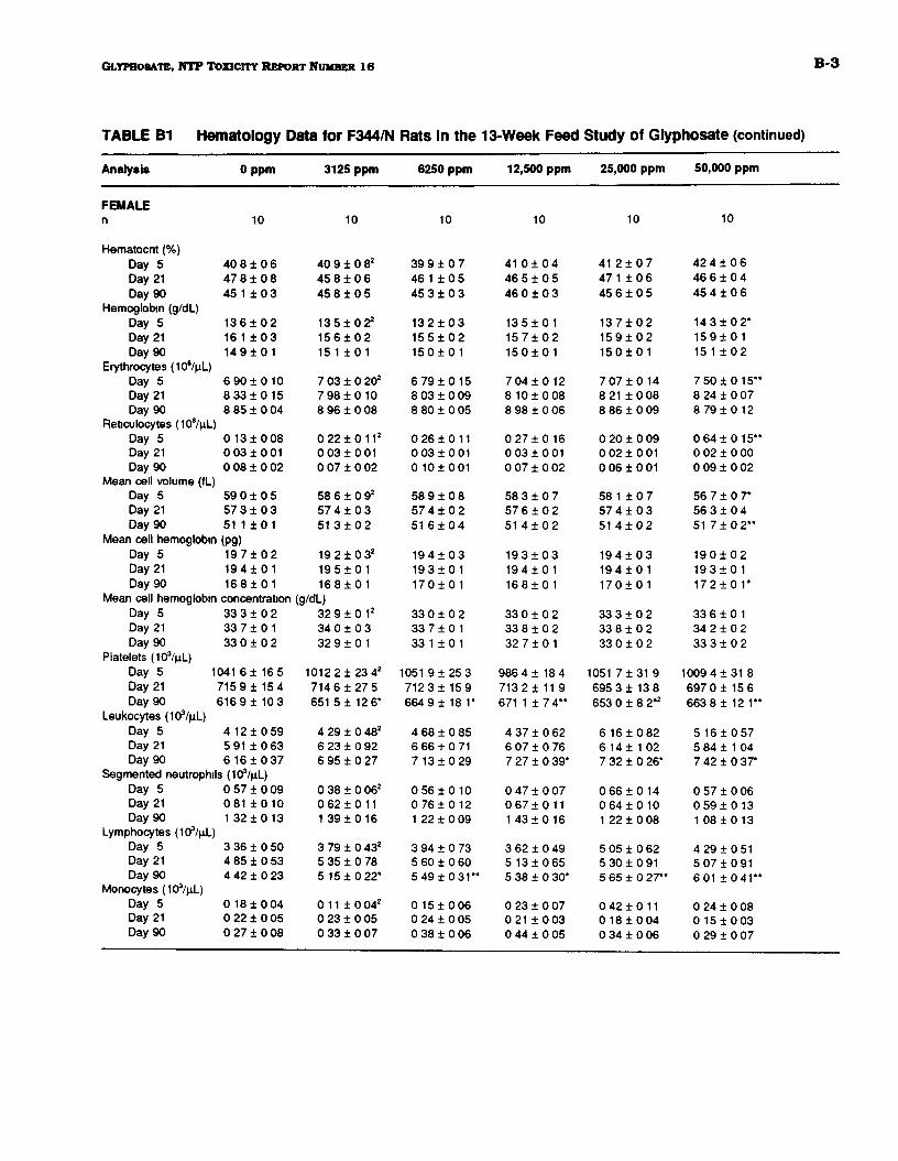
GLYPHOSATE. NTP TOXICITY REPORT NUMBER Iβ B-3
TABLE B1 Hematology Data for F344/N Rats In the 13-Week Feed Study of Glyphosate (continued)
Analysis
FEMALEη
Hematocnt (%)Day 5Day 21Day 90
Hemoglobin (g/dL)Day 5Day 21Day 90
Erythrocytes (10β/μΙ_)Day 5Day 21Day 90
Reticulocytes (106/μί)Day 5Day 21Day 90
Mean cell volume (fL)Day 5Day 21Day 90
Mean cell hemoglobinDay 5Day 21Day 90
Mean cell hemoglobinDay 5Day 21Day 90
Platelets (103/μί.)
0 ppm
10
4 0 8 1 0 647 8 ± 0 845 1 ± 0 3
1 3 6 ± 0 2161 ± 0 31 4 9 ± 0 1
6 90 ± 0 108 33 ± 0 158 85 ± 0 04
0 13 ± 0 080 03 ± 0 010 08 ± 0 02
59 0 ± 0 557 3 ± 0 351 1 ± 0 1
(pg)1 9 7 ± 0 21 9 4 ± 0 11 6 8 ± 0 1
concentration33 3 ± 0 233 7 ± 0 133 0 ± 0 2
Day 5 1041 6 ± 16 5Day 21Day 90
Leukocytes (103/μί)Day 5Day 21Day 90
7159± 1546169± 103
4 12 ± 0 595 91 ± 0 636 16 ± 0 37
Segmented neutrophils (10%iL)Day 5Day 21Day 90
Lymphocytes (ΙΟ^μί)Day 5Day 21Day 90
Monocytes (103/μί)Day 5Day 21Day 90
0 57 ± 0 09081 ± 0 1 01 32 ± 0 13
3 36 ± 0 504 85 ± 0 534 42 ± 0 23
0 18 ± 0 040 22 ± 0 050 27 ± 0 08
3125 ppm
10
40 9 ± 0 82
45 8 ± 0 645 8 ± 0 5
1 3 5 ± 0 2 2
1 5 6 ± 0 215 1 ± 0 1
7 03 ± 0 202
7 98 ± 0 108 96 ± 0 08
0 2 2 ± 0 112
0 03 ± 0 010 07 ± 0 02
58 6 ± 0 92
57 4 ± 0 351 3 ± 0 2
19 2 ± 0 3 2
19 5 ± 0 11 6 8 ± 0 1
(g/dL)3 2 9 ± 0 12
34 0 ± 0 332 9 ± 0 1
1012 2 ± 23 42
714 6 ±27 5651 5 ± 12 6*
4 29 ± 0 482
6 23 ± 0 926 95 ± 0 27
0 38 ± 0 062
0 62 ± 0 111 39 ± 0 16
3 79 ± 0 432
5 35 ± 0 785 15 ± 0 22*
0 11 ± 0 042
0 23 ± 0 050 33 ± 0 07
6250 ppm
10
39 9 ± 0 746 1 ± 0 545 3 ± 0 3
1 3 2 ± 0 31 5 5 ± 0 21 5 0 ± 0 1
6 79 ± 0 158 03 ± 0 098 80 ± 0 05
0 26 ± 0 110 03 ± 0 010 10±001
58 9 ± 0 857 4 ± 0 251 6 ± 0 4
1 9 4 ± 0 3193±0 11 7 0 ± 0 1
33 0 ± 0 233 7 ± 0 133 1 ± 0 1
1051 9± 25 37123± 159664 9 ± 18 1*
4 68 ± 0 856 66 ± 0 717 13 ± 0 29
0 56 ± 0 100 76 ± 0 121 22 ± 0 09
3 94 ± 0 735 60 ± 0 605 49 ± 0 3 1 "
0 15 ± 0 060 24 ± 0 050 38 ± 0 06
12,500 ppm
10
41 0 ± 0 446 5 ± 0 546 0 ± 0 3
135±0 11 5 7 ± 0 21 5 0 ± 0 1
7 04 ± 0 128 10 ± 0 088 98 ± 0 06
0 27 ± 0 160 03 ± 0 010 07 ± 0 02
58 3 ± 0 757 6 ± 0 251 4 ± 0 2
1 9 3 ± 0 31 9 4 ± 0 1168 ± 0 1
33 0 ± 0 233 8 ± 0 232 7 ± 0 1
986 4 ± 18 47132± 11 9671 1 ±74**
4 37 ± 0 626 07 ± 0 767 27 ± 0 39*
0 47 ± 0 070 67 ± 0 111 43 ± 0 16
3 62 ± 0 49513±0655 38 ± 0 30*
0 23 ± 0 070 21 ± 0 030 44 ± 0 05
25,000 ppm
10
41 2 ± 0 747 1 ± 0 645 6 ± 0 5
1 3 7 ± 0 21 5 9 ± 0 21 5 0 ± 0 1
7 07 ± 0 148 21 ± 0 088 86 ± 0 09
0 20 ± 0 090 02 ± 0 010 06 ± 0 01
58 1 ± 0 757 4 ± 0 351 4 ± 0 2
1 9 4 ± 0 319 4 ± 0 11 7 0 ± 0 1
33 3 ± 0 233 8 ± 0 233 0 ± 0 2
1051 7 ±31 9695 3 ± 13 8653 0 ± 8 2*2
6 16±0826 14 ± 1 027 32 ± 0 26*
0 66 ± 0 140 64±0 101 22 ± 0 08
5 05 ± 0 625 30 ± 0 915 65 ± 0 27**
0 42 ± 0 110 18 ± 0 040 34 ± 0 06
50,000 ppm
10
42 4 ± 0 646 6 ± 0 445 4 ± 0 6
143±02*1 5 9 ± 0 1151 ± 0 2
7 50 ± 0 15**8 24 ± 0 078 79 ± 0 12
0 64 ± 0 15**0 02 ± 0 000 09 ± 0 02
56 7 ± 0 7*56 3 ± 0 451 7 ± 0 2**
1 9 0 ± 0 21 9 3 ± 0 11 7 2 ± 0 1 *
33 6 ± 0 134 2 ± 0 233 3 ± 0 2
1009 4 ±31 8697 0± 15 6663 8 ± 12 1**
5 16 ± 0 575 84 ± 1 047 42 ± 0 37*
0 57 ± 0 060 59 ± 0 131 08 ± 0 13
4 29 ± 0 515 07 ± 0 91601 ±041**
0 24 ± 0 080 15 ± 0 030 29 ± 0 07
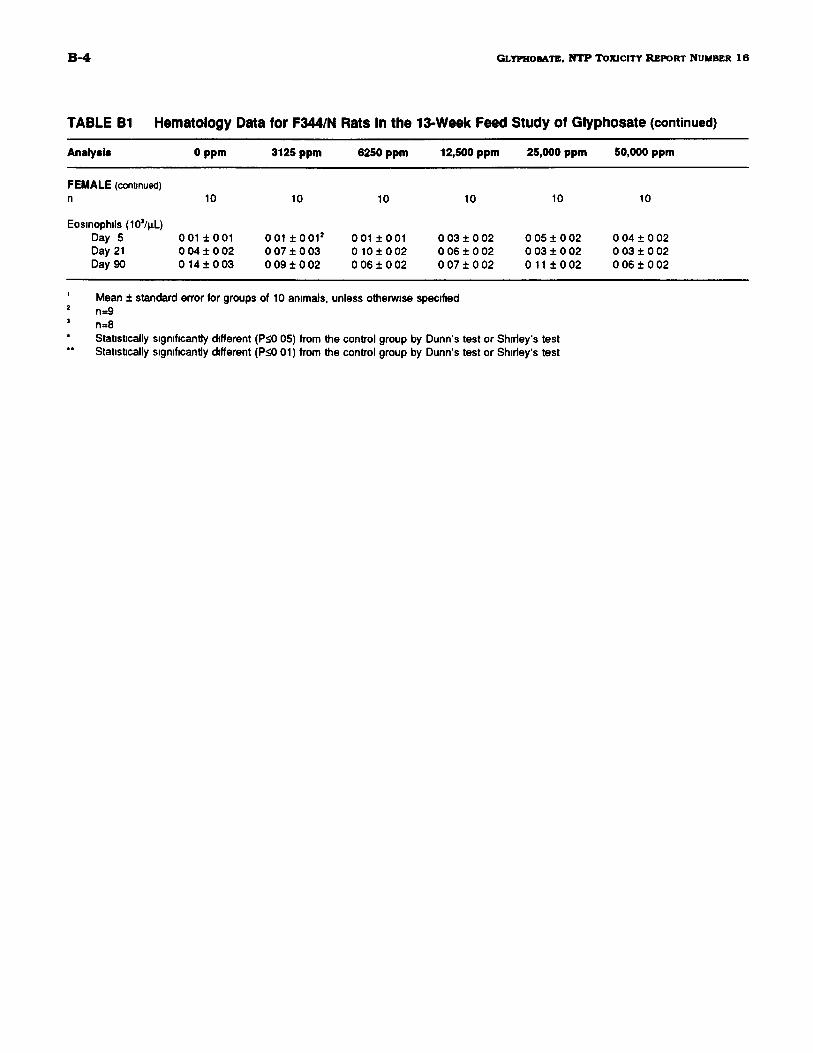
B-4 GLYPHOSATE, NTP TOXICITY REPORT NUMBER Iβ
TABLE B1 Hematology Data for F344/N Rats In the 13-Week Feed Study of Glyphosate (continued)
Analysis 0 ppm 3125 ppm 6250 ppm 12,500 ppm 25,000 ppm 50,000 ppm
FEMALE (continued)10
Eosinophils (103/μί.)Day 5 0 01 ± 0 01Day 21 0 04 ± 0 02Day 90 0 1 4 1 0 03
10
001 ± 0 0 1 2
0 07 ± 0 030 0 9 ± 0 0 2
10
001 ±0010 10 ± 0 020 06 ± 0 02
10
0 03 ± 0 020 06 ± 0 020 07 ± 0 02
10
0 05 ± 0 020 03 ± 0 020 11 ± 0 02
10
0 04 ± 0 020 03 ± 0 020 06 ± 0 02
Mean ± standard error for groups of 10 animals, unless otherwise specifiedn=9n=8Statistically significantly different (P<0 05) from the control group by Dunn's test or Shirley's testStatistically significantly different (P<0 01) from the control group by Dunn's test or Shirley's test
GLYPHO8ATE, N T P TOXICITY REPORT NUMBER I β B-5
TABLE B2 Clinical Chemistry Data for F344/N Rats In the 13-Week Feed Study of Giyphosate1
Analysis
MALEη
Urea nitrogen (mg/dLDay 5Day 21Day 90
Creatinine (mg/dL)Day 5Day 21Day 90
Total protein (g/dL)Day 5Day 21Day 90
Albumin (g/dL)Day 5Day 21Day 90
Alkaline phosphataseDay 5Day 21Day 90
0 ppm
10
)20 0 ± 0 720 3 ± 0 622 1 ± 0 5
0 55 ± 0 030 58 ± 0 010 56 ± 0 02
6 1 ± 0 16 3 ± 0 16 9 ± 0 0
3 6 ± 0 13 8 ± 0 13 7 ± 0 1
(IU/L)764 ± 20528 ± 11289 ± 7
Alanine aminotransferase (IU/L)Day 5Day 21Day 90
50 ± 244 ± 246 ± 2
Creatine phosphokinase (IU/L)Day 5Day 21Day 90
544 ± 87488 ± 56247 ± 49
Sorbitol dehydrogenase (IU/L)Day 5Day 21Day 90
Bile acids (μπιοΙ/L)Day 5Day 21Day 90
FEMALEη
Urea nitrogen (mg/dL)Day 5Day 21Day 90
Creatinine (mg/dL)Day 5Day 21Day 90
Total protein (g/dL)Day 5Day 21Day 90
6 ± 05 ± 0 2
10± 1
30 60 ± 3 5619 33± 2 012
12 40 ± 1 10
10
22 1 ± 0 6196± 1 022 4 ± 0 7
0 55 ± 0 030 58 ± 0 020 60 ± 0 02
615 ± 0 116 46 ± 0 096 93 ± 0 1 2
3125 ppm
10
1 9 9 ± 0 920 2 ± 0 722 8 ± 0 6
0 54 ± 0 020 58 ± 0 020 59 ± 0 02
6 0 ± 0 16 3 ± 0 16 9 ± 0 1
3 6 ± 0 13 8 ± 0 13 8 ± 0 1
798 ± 25543 ± 13283 ± 9
53 ± 246 ± 153 ± 2"
714 ± 173477 ±47219 ±78
7± 16 ± 08 ± 1
23 80 ± 2 6319 70 ± 2 1015 30 ± 2 66
10
20 5 ± 0 7183± 1 023 2 ± 0 7
0 54 ± 0 010 58 ± 0 020 63 ± 0 02
601 ±0106 1 7 ± 0 1 07 01 ± 0 06
6250 ppm
10
21 1 ± 0 71 9 4 ± 0 824 3 ± 0 7*
0 55 ± 0 030 54 ± 0 040 57 ± 0 01
6 0 ± 0 16 2 ± 0 17 0 ± 0 1
3 5 ± 0 13 8 ± 0 03 9 ± 0 Γ
816 ± 19557 ± 15293 ± 8
57 ± r*49 ± r52 ±2*
587 ± 92637±168226 ± 40
7 ± 06± 18 ± 0
30 20 ± 4 5622 60 ± 2 7711 30 ± 0 63
10
20 7 ± 0 620 1 ± 1 122 8 ± 0 5
0 47 ± 0 050 58 ± 0 020 63 ± 0 02
5 97 ± 0 056 20 ± 0 06*6 76 ± 0 08
12,500 ppm
10
1 9 9 ± 0 420 9 ± 0 622 2 ± 0 5
0 52 ± 0 020 58 ± 0 020 53 ± 0 02
6 0 ± 0 16 2 ± 0 16 8 ± 0 1
3 6 ± 0 13 7 ± 0 03 8 ± 0 1
876 ± 28**601 ± 16"299 ± 4
67 ± 3 "50 ± 1*57 ± 2"
615 ± 120577 ±75169 ± 13
7± 16 ± 19 ± 0
30 90 ± 3 5226 80 ± 2 26*1300± 1 15
10
20 9 ± 0 719 1 ± 0 724 5 ± 1 0*
0 52 ± 0 030 55 ± 0 020 62 ± 0 02
5 97 ± 0 086 20 ± 0 096 86 ± 0 07
25,000 ppm
10
20 0 ± 0 82
20 2 ± 0 Τ23 5 ± 0 6
0 49 ± 0 032
0 55 ± 0 032
0 55 ± 0 02
6 0 ± 0 12
6 3 ± 0 1 2
6 9 ± 0 1
3 6 ± 0 02
3 7 ± 0 1 2
3 8 ± 0 1
888±21**2
639 ± 12**2
306 ± 6
62 ± 4**2
51 ± 1**2
65 ± 6**
587 ± 1252
523 ± 592
242 ± 35
6 ± 0 2
5 ± 0 2
9 ± 1
35 56 ± 4 Θ72
31 0 0 ± 3 58**2
15 40 ± 1 19
10
20 9 ± 1 0194± 1024 2 ± 0 7*
0 54 ± 0 020 55 ± 0 020 61 ± 0 03
5 92 ± 0 07617±007*6 71 ±0 11
50,000 ppm
10
1 9 7 ± 0 620 3 ± 0 625 2 ± 0 8**
0 52 ± 0 020 54 ± 0 010 53 ± 0 02
6 1 ± 0 16 2 ± 0 171 ± 0 1
3 7 ± 0 13 8 ± 0 14 0 + 0 1 "
928 ± 30##
627 ± 11·*253 ± 16
65 ± 3**47 ±2*53 ± 2**
911 ± 139*531 ± 29242 ± 25
6 ± 05 ± 08 ± 1
38 00 ± 3 6332 80 ± 4 35**20 90 ± 1 62**
10
19 0 ±05**1 7 6 ± 0 524 2 ± 1 2
0 50 ± 0 020 57 ± 0 032
0 64 ± 0 02
5 85 ± 0 06*6 06 ± 0 13**6 49 ± 0 12*

B-β GLYPHOSATE, N T P TOXICITY REPORT NUMBER 1 6
TABLE B2 Clinical Chemistry Data for F344/N Rats In the 13-Week Feed Study of Glyphosate(continued)
Analysis 0 ppm 3125 ppm 6250 ppm 12,500 ppm 25,000 ppm 50,000 ppm
FEMALE (continued)10
Albumin (g/dL)Day 5 3 57 ± 0 07Day 21 4 00 ± 0 09Day 90 4 31 ± 0 08
Alkaline phosphatase (IU/L)Day 5 615 ± 17Day 21 374 ±17Day 90 231 ± 10
Alanine aminotransferase (IU/L)Day 5 45 ± 3Day 21 33 ± 1Day 90 44 ± 3
Creatine phosphokinase (IU/L)Day 5 559 ± 79Day 21 434 ± 50Day 90 304 ± 29
Sorbitol dehydrogenase (IU/L)Day 5 5 ± 0Day 21 5 ± 0Day 90 4 ± 0
Bile acids (μπιοΙ/L)Day 5 24 00 ± 3 67Day 21 12 90 ± 2 12Day 90 25 20 ± 3 09
10 10 10 10 10
3 62±3 87±4 33±
653 ±406 ±224 ±
51 ±38 ±42 ±
599 ±414 ±247 ±
5 ±5 ±4 ±
28 30±1590±24 20±
0 070 070 08
18102
2*1 "2
557237
000
1642 793 00
3 49 ± 0 043 98 ± 0 064 09 ± 0 10
669 ± 21432 ± 13*240 ± 8
49 ±2*42 ± 2 "44 ± 1
586 ± 74656 ± 225237 ± 35
5 ± 05 ± 05 ± 0
27 10 ± 2 2919 20 ± 4 0230 50 ± 5 66
3 49 ± 0 083 79 ± 0 084 26 ± 0 07
679 ± 20*432 ± 1Γ248 ± 10
53 ± 2 "41 ± Γ*46 ± 2
744 ± 114345 ± 57277 ±45
5 ± 05 ± 05 ± 0
25 70 ± 4 7019 50 ± 2 7116 60 ± 1 73
3 47±3 92±4 1 3 ±
731 ±464 ±291 ±
55 ±44 ±54 ±
663 ±421 ±289 ±
5 ±5 ±4 ±
29 30±22 40±27 60±
0 050 070 07*
2 Γ *14**6**
2··1··2··
1097224
000
4 043 43*6 09
3 44 ± 0 053 79 ± 0 083 99 ± 0 09*
771 ± 13**501 ± 15**319± 11**
62 ± 4**47 ± 1**54 ± 2**
724 ± 53294 ± 45*277 ±32
5 ± 06 ± 14 ± 0
33 89 ± 3 962
26 00 ± 3 97*40 00 ± 4 67
Mean ± standard error for groups of 10 animals, unless otherwise specifiedn=9Statistically significantly different (P<0 05) from the control group by Dunn's test or Shirley's testStatistically significantly different (P<0 01) from the control group by Dunn's test or Shirley's test
GLYPHO8ATE, NTP TOXICITY REPORT NUMBER I β C - 1
APPENDIX C
Reproductive Tissue Evaluationsand Estrous Cycle Characterization
Table CI Summaiy of Reproductive Tissue Evaluations and Estrous Cycle Lengthin F344/N Rats in the 13-Week Feed Study of Glyphosate C-2
Table C2 Summaiy of Reproductive Tissue Evaluations and Estrous Cycle Lengthin B6C3F! Mice in the 13-Week Feed Study of Glyphosate C-3
C-2 GLYPHO8ATE, Ν Τ Ρ TOXICITY REPORT NUMBER 1 6
TABLE C1 Summary of Reproductive Tissue Evaluations and Estrous Cycle LengthIn F344/N Rats In the 13-Week Feed Study of Glyphosate
StudyParameters1 0 ppm 12,500 ppm 25,000 ppm 50,000 ppm
MALE
Weights (g)Necropsy body weight 385 ± 5 350 ± 5 340 ± 5* 305 ± 7 "Left epididymal tail 0.170 ±0.004 0.168 ±0.006 0.167 ±0.004 0.179 ±0.006Left testis 1.54 ± 0.03 1.52 ± 0.05 1.56 ± 0.03 1.56 ± 0.02Left epididymis 0.448 ± 0.007 0.437 ± 0.016 0.440 ± 0.004 0.452 ± 0.007
Spermatozoal measurementsConcentration (10e) 610 ± 36 561 ± 23 485 ± 2 3 " 486 ± 2 3 "Motility (%) 91 ± 1 92 ± 1 92 ± 2 91 ± 1Spermatid count (mean/104 mL suspension) 70.15 ± 3.00 65.33 ± 5.49 67.23 ± 2.05 69.00 ± 1.71Spermatid heads (107/testis) 14.03 ± 0.60 13.07 ±1.10 13.45 ± 0.41 14.06 ± 0.36.Spermatid heads (107/g testis) 9.10 ±0.35 8.48 ± 0.64 8.63 ± 0.30 9.04 ± 0.20
FEMALE
Estrous cycle length (days) 4.90 ±0.10 5.00 ± 0.07 4.90 ±0.10 5.40 ±0.21*
Data presented as mean ± standard error; n=10. Differences from the control group for testicular, epididymal, and epididymal tailweights are not significant by Dunnett's test; spermatozoal measurements were not significant by Dunn's test or Shirley's test.Statistically significantly different (P<0.05) from the control group by Williams' test or Shirley's test.Statistically significantly different (P<0.01) from the control group by Williams' test or Shirley's test.
GLYPHO8ATE, Ν Τ Ρ TOXICITY REPORT NUMBER I β C-3
TABLE C2 Summary of Reproductive Tissue Evaluations and Estrous Cycle LengthIn B6C3F, Mice In the 13-Week Feed Study of Glyphosate
StudyParameters1 0 ppm 12,500 ppm 25,000 ppm 50,000 ppm
MALE
Weights (g)Necropsy body weight 3 2 0 ± 1 0 31 9 ± 0 9 29 4 ± 0 Τ 27 2 ± 0 4 "Left epididymal tail 0 015 ± 0 001 0 014 ± 0 001 0 014 ± 0 001 0 014 ± 0 001Left testis 0 110 ± 0 0 0 2 0 111 ± 0 0 0 3 0 111 ± 0 0 0 2 0 110 ± 0 0 0 3Left epididymis 0 044 ± 0 001 0 043 ± 0 002 0 044 ± 0 001 0 042 ± 0 001
Spermatozoal measurementsConcentration (10e) 1162 ± 44 1370 ± 130 1189 ± 60 1308 ± 97Motility (%) 91 ± 1 91 ± 1 92 ± 1 89 ± 1Spermatid count (mean/104 mL suspension) 67 20 ± 2 30 63 18 ± 3 06 61 93 ± 1 92 65 40 ± 2 89Spermatid heads (107/testis) 2 15 ± 0 07 2 02 ± 0 10 1 98 ± 0 06 2 09 ± 0 09Spermatid heads (107/g testis) 19 61 ± 0 92 18 17 ± 0 71 17 87 ± 0 60 18 99 ± 0 73
FEMALE
Estrous cycle length (days) 4 06 ± 0 062 4 00 ± 0 00 4 00 ± 0 00 4 00 ± 0 002
Data presented as mean ± standard error, n=10 except where noted Differences from the control group for testicular, epididymal,and epididymal tail weights are not significant by Dunnett's test, spermatozoal measurements were not significant by Dunn's testEstrous cycle length was not significant by Dunn's testn=9Statistically significantly different (P<0 05) from the control group by Williams' testStatistically significantly different (p 0 01) from the control group by Williams' test

QLTPHO8ATB, N T P TOMCITT REPORT NUMBER 1 6
APPENDIX D
Genetic Toxicology
Table Dl Mutagenicity of Glyphosate in SaLmonella typhimurium D-2
Table D2 Frequency of Micronuclei in Mouse Peripheral Blood Erythrocytesin the 13-Week Feed Study of Glyphosate D-3
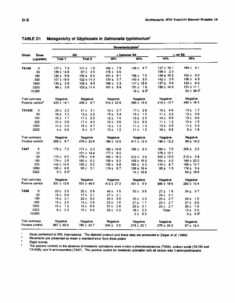
D-2 GLYPHOSATE, N T P TOXICITY REPORT NUMBER 1 6
TABLE D1 Mutagenicity of Glyphosate in Salmonella typhlmurlum1
Strain Dose^g/plate)
TA100 033
100333
10003333
10,000
Trial summaryPositive control4
TA1535 033
100333
10003333
Trial summaryPositive control
TA97 01033
100333
10003333
Trial summaryPositive control
TA98 033
100333
10003333
10,000
Trial summaryPositive control
Trial 1
127±120 ±124 ±107 ±133 ±99 ±
7 31489 9
1053 83 8
Negative422 ±
20 ±18±15±11 ±11 ±4±
14 1
2 21 21 70 90 30 9
Negative269 ±
172 ±
173 ±174±142 ±100 ±
0 ±
8 7
7 2
6 03 5
1696 800 3
Negative331 ±
23 ±16±15±15±15±8 ±
136
2 50 92 12 01 22 3
Negative667 ± 85 0
-S9Trial 2
111 ±97 ±
134 ±122 ±109 ±103 ±
1 55 56 0
11 39 3
11 4
Negative433 ±
21 ±13±17±17±15±9 ±
6 7
3 12 22 04 00 70 7
Negative479 ±
171 ±151 ±178 ±190 ±136 ±93 ±
32 9
2 31446 95 23 23 1
Negative501 ±
25 ±17±23 ±14±15±15±
48 5
0 92 13 22 90 60 9
Negative785 ± 24 7
IRevertants/plate2
+ hamster S910%
162 ±176 ±151 ±133 ±106±101 ±
7 96 68 13 72 36 8
Negative514 ±
14±15±12±10±12±10±
22 9
0 73 41 03 61 21 2
Negative196 ±
180 ±177 ±168 ±159 ±135 ±116 ±
125
1689 2
1839 29 88 7
Negative412 ±
40 ±27 ±34 ±22 ±31 ±25 ±
27 3
1 52 13 51 52 90 3
Negative346 ± 3 5
30%
148±
156 ±142 ±117 ±131 ±18±
8 7
7 53 3
1561 966s
Negative590 ±
17±15±13±12±11 ±11 ±
159
3 81 22 00 31 31 0
Negative411 ±
159 ±
212 ±159 ±162 ±139 ±74 ±
123
8 3
3 81036 3
164159
Negative451 ±
32 ±
25 ±27 ±25 ±19±2 ±
5 0
3 5
2 31 73 12 20 3
Negative274 ± 32 1
+10%
147 ± 16 1158 ± 2 3148 ± 102142 ± 3 5137 ± 3 0150 ± 140
Negative419 ± 137
18± 4611 ± 2214± 0911 ± 1212± 2510± 06
Negative146 ± 12 2
190 ± 78179± 10 1203 ± 12 5154 ± 4 2119 ± 8 786 ± 15
Negative385 ± 18 6
27 ± 1824 ± 2 125 ± 2 723 ± 2 723 ± 2 7
Toxic
Negative275 ± 24 2
rat S930%
169 ± 4 1
154 ± 90136 ± 4 3133 ± 85131 ± 11 152 ± 38 53
Negative490 ± 16 5
13± 1 712± 0 913± 3 010± 1 511 ± 0 39 ± 1 8
Negative89 ± 14 2
205 ± 2 5
212 ± 2 8160 ± 20 0186 ± 14 7174 ± 5 463 ± 19 5
Negative283 ± 13 4
24 ± 3 7
26 ± 1527 ± 2 920 ± 1513± 2 30 ± 00 3
Negative87± 124
Study performed at SRI, International The detailed protocol and these data are presented in Zeiger et al (1988)Revertants are presented as mean ± standard error from three platesSlight toxicityThe positive controls in the absence of metabolic activations were 4-nitro-o-phenylenediamine (TA98), sodium azide (TA100 andTA1535), and 9-aminoacndine (TA97) The positive control for metabolic activation with all strains was 2-aminoanthracene
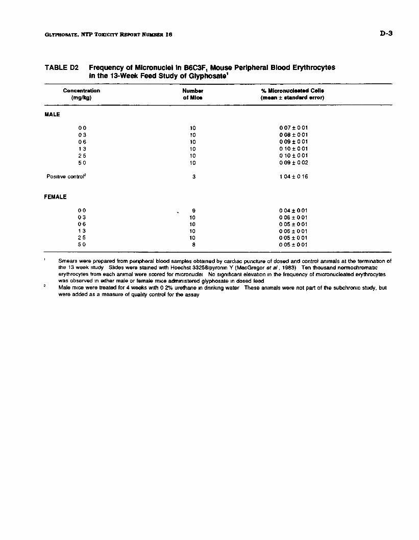
GLYPHO8ATB, Ν Τ Ρ TOXICITY REPORT NUMBER I β D-3
TABLE D2 Frequency of Micronuclel In B6C3F1 Mouse Peripheral Blood ErythrocytesIn the 13-Week Feed Study of Glyphosate1
Concentration Number % Micronucleated Cells(mg/kg) of Mice (mean ± standard error)
MALE
0 0 10 00710010 3 10 0 08 ± 0 010 6 10 0 09100113 10 010100125 10 01010015 0 10 0 091002
Positive control2 3 1 04 1 0 16
FEMALE
00 ^ 9 0 0 4 1 0 010 3 10 0 0 6 1 0 0 10 6 10 0 0 5 1 0 0 113 10 0 0 6 1 0 0 12 5 10 0 0 5 1 0 0 15 0 8 0 0 5 1 0 0 1
1 Smears were prepared from peripheral blood samples obtained by cardiac puncture of dosed and control animals at the termination ofthe 13 week study Slides were stained with Hoechst 33258/pyronin Υ (MacGregor et al, 1983) Ten thousand normochromalerythrocytes from each animal were scored for micronuclei No significant elevation in the frequency of micronucleated erythrocyteswas observed in either male or female mice administered glyphosate in dosed feed
2 Male mice were treated for 4 weeks with 0 2% urethane in dnnking water These animals were not part of the subchronic study, butwere added as a measure of quality control for the assay