notch-1 up-regulation and signaling following macrophage ... · notch-1 up-regulation and signaling...
TRANSCRIPT

of July 2, 2018.This information is current as Activity
Antigen-Presenting Capacity and Cytotoxic Gene Expression Patterns Known to Affectfollowing Macrophage Activation Modulates Notch-1 Up-Regulation and Signaling
Díaz-GuerraJuan C. Gómez, Jorge Laborda and María José M.Ruiz-Hidalgo, Victoriano Baladrón, José J. García-Ramírez, Eva Monsalve, Miguel A. Pérez, Antonio Rubio, María José
http://www.jimmunol.org/content/176/9/5362doi: 10.4049/jimmunol.176.9.5362
2006; 176:5362-5373; ;J Immunol
Referenceshttp://www.jimmunol.org/content/176/9/5362.full#ref-list-1
, 41 of which you can access for free at: cites 69 articlesThis article
average*
4 weeks from acceptance to publicationFast Publication! •
Every submission reviewed by practicing scientistsNo Triage! •
from submission to initial decisionRapid Reviews! 30 days* •
Submit online. ?The JIWhy
Subscriptionhttp://jimmunol.org/subscription
is online at: The Journal of ImmunologyInformation about subscribing to
Permissionshttp://www.aai.org/About/Publications/JI/copyright.htmlSubmit copyright permission requests at:
Email Alertshttp://jimmunol.org/alertsReceive free email-alerts when new articles cite this article. Sign up at:
Print ISSN: 0022-1767 Online ISSN: 1550-6606. Immunologists All rights reserved.Copyright © 2006 by The American Association of1451 Rockville Pike, Suite 650, Rockville, MD 20852The American Association of Immunologists, Inc.,
is published twice each month byThe Journal of Immunology
by guest on July 2, 2018http://w
ww
.jimm
unol.org/D
ownloaded from
by guest on July 2, 2018
http://ww
w.jim
munol.org/
Dow
nloaded from

Notch-1 Up-Regulation and Signaling following MacrophageActivation Modulates Gene Expression Patterns Known toAffect Antigen-Presenting Capacity and Cytotoxic Activity1
Eva Monsalve,* Miguel A. Perez,* Antonio Rubio,† Marıa Jose Ruiz-Hidalgo,*Victoriano Baladron,* Jose J. Garcıa-Ramırez,* Juan C. Gomez,† Jorge Laborda,* andMarıa Jose M. Dıaz-Guerra2*
Notch signaling has been extensively implicated in cell-fate determination along the development of the immune system. However,a role for Notch signaling in fully differentiated immune cells has not been clearly defined. We have analyzed the expression ofNotch protein family members during macrophage activation. Resting macrophages express Notch-1, -2, and -4, as well as theNotch ligands Jagged-1 and -2. After treatment with LPS and/or IFN-�, we observed a p38 MAPK-dependent increase in Notch-1and Jagged-1 mRNA and protein levels. To study the role of Notch signaling in macrophage activation, we forced the transientexpression of truncated, active intracellular Notch-1 (Notch-IC) proteins in Raw 264.7 cells and analyzed their effects on theactivity of transcription factors involved in macrophage activation. Notch-IC increased STAT-1-dependent transcription. Fur-thermore, Raw 264.7 Notch-IC stable transfectants increased STAT1-dependent transcription in response to IFN-�, leading tohigher expression of IFN regulatory factor-1, suppressor of cytokine signaling-1, ICAM-1, and MHC class II proteins. This effectwas independent from an increase of STAT1 Tyr or Ser phosphorylation. However, inducible NO synthase expression and NOproduction decreased under the same conditions. Our results show that Notch up-regulation and subsequent signaling followingmacrophage activation modulate gene expression patterns known to affect the function of mature macrophages. The Journal ofImmunology, 2006, 176: 5362–5373.
M acrophages are cells essential for the immune re-sponse. After their precursors are produced in the bonemarrow, they are transported in the blood to the dif-
ferent tissues in the organism, where they can differentiate to ma-ture macrophages and be activated by pathogens and proinflam-matory cytokines (1, 2).
Macrophages, and other phagocytic cells, such as neutrophilsand dendritic cells, can discriminate between pathogens and selfthrough the signals triggered by specific TLRs present at the cellmembrane. TLRs recognize different pathogens’ components,called pathogen-associated molecular patterns. LPS, an integralelement present at the outer membrane of Gram-negative bacteria,is recognized by the TLR4, thus initiating a complex signalingpathway finally leading to the activation of several transcriptionfactors that control macrophage activation, such as NF-�B, IFNregulatory factor (IRF)3-3, and AP-1, among others (3, 4).
In addition to pathogen-associated molecular patterns, macro-phages can also become fully functional after the binding ofIFN-�, a cytokine released by activated T lymphocytes and NKcells, to its specific membrane receptor, IFN-�R (5–7). Followingthis interaction, activated IFN-�R binds and activates the JAK 1and 2 that, in turn, trigger the phosphorylation of STAT1. Phos-phorylated STAT1 dimerizes and translocates to the nucleus,where it binds to the IFN-� activation promoter site (GAS) toinitiate or suppress transcription of IFN-�-regulated genes (5).Some of these genes, like IRF-1, are transcription factors able todrive regulation of a second wave of gene expression. Althoughbiochemical and genetic studies show that STAT1 plays a criticalrole in IFN-�-dependent signaling, recent work has revealed thatadditional signals are required for the full range of responses elic-ited by IFN-� (7).
Notch proteins comprise a family of epidermal growth factor-liketransmembrane receptors that control different aspects of tissue de-velopment and homeostasis (8, 9). Four distinct Notch genes havebeen identified in mammals, Notch-1, -2, -3, and -4 (9). Notch ligandsare also epidermal growth factor-like transmembrane proteins. Fivedifferent Notch ligands have been characterized in mammals,Jagged-1 and -2, and Delta-1, -3, and -4 (8). After effective ligandinteraction, Notch receptors undergo at least two proteolytic cleavagesteps that release the intracellular Notch (Notch-IC) receptor regionfrom the membrane. Notch-IC then translocates to the nucleus, whereit associates with the transcription factor CSL (CBF1, Suppressor ofHairless, LAG-1/RBP-J�). In the absence of Notch-IC, CSL binds tocorepressors, thus inhibiting transcription. Notch-IC displaces the
*Facultad de Medicina, Centro Regional de Investigaciones Biomedicas (CRIB), Al-bacete, Spain; and †Complejo Hospitalario Universitario de Albacete, Albacete, Spain
Received for publication September 7, 2005. Accepted for publication February14, 2006.
The costs of publication of this article were defrayed in part by the payment of pagecharges. This article must therefore be hereby marked advertisement in accordancewith 18 U.S.C. Section 1734 solely to indicate this fact.1 This work was supported by Grant 03/0766 from Ministerio de Sanidad y Consumoand Grant 02012-00 from Consejerıa de Sanidad de la Junta de Comunidades deCastilla-La Mancha, Spain.2 Address correspondence and reprint requests to Dr. Marıa Jose M. Dıaz-Guerra,Centro Regional de Investigaciones Biomedicas, Facultad de Medicina, Avenida deAlmansa No. 14, 02006 Albacete, Spain. E-mail address: [email protected] Abbreviations used in this paper: IRF, IFN regulatory factor; GAS, IFN-� activationpromoter site; Notch-IC, intracellular Notch; iNOS, inducible NO synthase; RAM,
RBP-J�-associated molecule; PEST, proline-glutamate-serine-threonine-rich; SOCS,suppressor of cytokine signaling.
The Journal of Immunology
Copyright © 2006 by The American Association of Immunologists, Inc. 0022-1767/06/$02.00
by guest on July 2, 2018http://w
ww
.jimm
unol.org/D
ownloaded from

corepressors, recruits coactivators, and activates CSL-dependent tran-scription. Some target genes of this process have been characterized,including the basic helix-loop-helix transcription factors belonging tothe HES family, including Hes-1 and Hes-5, that are expressed bycells of the hemopoietic lineage (10). Although some CSL-indepen-dent Notch signaling can occur, its mechanism is not well character-ized (11). Notch signaling is modulated by extracellular, cytoplasmic,and nuclear proteins (8). Several enzymes, like Fringe, glycosylatespecific extracellular Notch sites, modifying ligand-mediated signal-ing (12, 13).
Notch signaling is highly involved in lymphopoiesis and, to alesser extent, in myeloid differentiation and generation of embry-onic hemopoietic stem cells (10, 14, 15). Notch signaling in my-eloid cells has been associated with maintenance of undifferenti-ated phenotypes (16, 17), but recently it has been implicated inmonocyte differentiation to dendritic cells (18).
Although Notch signaling has been extensively implicated incell-fate selection through the development of the immune system,less attention has been paid to the function of Notch receptors indifferent mature immune cells expressing high levels of those pro-teins. In this study, we show that different members of the Notchfamily are expressed in macrophages, and some of them, likeNotch-1 and Jagged-1, are up-regulated in the course of macro-phage activation. In addition, transfection of constitutively activeNotch-IC proteins in Raw 264.7 cells induced elevated responsesto IFN-� through increased STAT1-dependent transcription, lead-ing to higher expression of ICAM-1 and class II MHC proteins. Incontrast, after activation with LPS or IFN-�, these transfectantcells displayed lower expression of inducible NO synthase (iNOS)and decreased NO production. We show in this study for the firsttime that Notch expression is modulated in the course of macro-phage activation, and that Notch signaling can regulate specificgene expression patterns induced by different activation signalsthat may modulate the function of mature macrophages.
Materials and MethodsChemicals
Reagents were purchased from Sigma-Aldrich, Boehringer Mannheim, orMerck. Abs were purchased from Santa Cruz Biotechnology or Sigma-Aldrich. LPS from Salmonella typhimurium, kinase inhibitors, and cyto-kines were purchased from Sigma-Aldrich, Calbiochem, and Roche, re-spectively. Serum and culture medium were acquired from BioWhittaker.Electrophoresis equipment and reagents were acquired from Bio-Rad.
Cell culture
Raw 264.7 and J774 cells were subcultured at a density of 6–8 � 104/cm2
in RPMI 1640 supplemented with 2 mM glutamine, 10% FCS, and anti-biotics (50 mg/ml each of penicillin, streptomycin, and gentamicin). After2 days in culture, the medium was replaced by RPMI 1640 containing 1%FCS, and then cells were used in the next 24 h. 293T cells were culturedin DMEM supplemented with glutamine, 10% FCS, and antibiotics, asdescribed previously. Elicited peritoneal macrophages were obtained frommale mice 4 days after i.p. inoculation of 1 ml of sterile 10% (weight/volume)thioglycollate broth as described previously (19). Cells were seeded at a den-sity of 105/cm2 in RPMI 1640 supplemented with 10% heat-inactivated FCSand 50 �g/ml gentamicin, penicillin, and streptomycin.
Plasmids
pNIC-2 plasmid encodes for an intracellular active form of human Notch-1protein (aa 1759–2556) containing the RAM (RBP-J�-associated mole-cule) domain, the six cdc10/ankyrin repeats, a homopolymer repeat of glu-tamine (OPA) domain, three nuclear localization signal sequences, and aproline-glutamate-serine-threonine-rich (PEST) domain, linked to a mycepitope fused to the C-terminal region. pNIC-1 plasmid encodes for a hu-man Notch-1-IC protein (aa 1759–2237), lacking the OPA and PEST do-mains. Both plasmids were derived from the pLNCX2 retroviral vector(BD Biosciences), engineered by us to contain a myc tag epitope. The myctag epitope was generated by hybridization of two complementary oligo-nucleotides (5�-TCG ACT CAA TGC AGA AGC TGA TCT CAG AGG
AGG ACC TCT AAT-3� and 5�-CGA TTA GAG GTC CTC CTC TGAGAT CAG CTT CTG CAT TGA G-3�), which were then cloned at the SalIand ClaI restriction sites of pLNCX2. Human intracellular Notch-1 cDNAswere obtained by RT-PCR amplification of total RNA from human HepG2cells, by using Pfu DNA Polymerase (Stratagene). The insert to be clonedinto pNIC-2 was amplified with the following primers: 5�-CCG GAA GCTTGC ACC ATG GCA CGC AAG CGC CGG CGG CAG-3� and 5�-GCGACG TCG ACC TTG AAG GCC TCC GAA T-3�; and the insert to becloned into pNIC-1 was amplified with the following primers: 5�-CCGGAA GCT TGC CAC CAT GGC ACG CAA GCG CCG GCG GCA G-3�and 5�-GCG ACG TCG ACG AGG GGC ACG GAC GGA GAC T-3�.Both amplified fragments were cloned at the HindIII and SalI restrictionsites of the pLNCX2-myc vector. All vectors were confirmed by restrictionanalysis and sequencing. CBF1-Luc, NF-�B-Luc, AP-1-Luc, and m67-Lucplasmids, used to detect Notch-, NF-�B-, AP-1-, or STAT-dependent tran-scription activities, respectively, have been described previously (20, 21). Allplasmids were purified using EndoFree Qiagen columns.
Cell transfection
Subconfluent Raw 264.7 cells were seeded in 24-well or 6-cm plates andtransfected on the following day with 600 ng or 2.5 �g of plasmid DNAsby using Fugene 6 reagent (Roche), according to the manufacturer’s rec-ommendations (20). HEK 293T cells were transfected with Superfect re-agent (Qiagen), following the manufacturer’s recommendations. Theamount of transfected DNA was kept constant by addition of appropriatedamounts of the parental empty vectors. �-galactosidase or GFP expressionvectors were used as internal controls for transfection efficiency. Luciferaseand �-galactosidase activities were measured by using a commercial kit(Promega). Raw 264.7 stable transfectants were obtained by transfection ofpLNCX2, or pLNCX2-Notch-IC plasmids with Fugene 6 reagent. Twenty-four hours later, cells were released by trypsin treatment and diluted 1/10with fresh medium. After the cells were attached to the plates, transfectedcells were selected by resistance to neomycin (350 �g/ml G-418 (Sigma-Aldrich) in complete culture medium). Cells resistant to the drug werepooled and screened for Neo and human Notch-IC expression. Selectedcells were always maintained with medium containing G-418.
Analysis of gene expression by RT-PCR
Total RNA (2–4 � 106 cells) was extracted following the TRIzol method(Sigma-Aldrich) and treated with DNase to eliminate any potential DNAcontamination. For RT-PCR, cDNA was generated from 2 �g of total RNAby using the cDNA synthesis kit RevertAid HMinus First Strand (Fermen-tas). Semiquantitative RT-PCR was performed as described previously(22). PCR assays were run for 25–40 cycles, depending on the gene to bestudied, to obtain a PCR yield proportional to the amount of initial cDNAtemplate. Amplification of the housekeeping gene P0 was used as a qualityand loading control (23). The forward and reverse primers, respectively,used for PCR amplification are as follows: Notch-1 (5�-CTG TGT GGATGA GGG AGA TAA-3� and 5�-GGC ATA GAC AGC GGT AGA AA-3�); Notch-2 (5�-CAG CCG GTC TCC GTG TAA AAA CAA AG-3� and5�-GCG AAG AGT GGA GGT GCA GTT G-3�); Notch-3 (5�-ACA CTGGGA GTT CTC TGT-3� and 5�-GTC TGC TGG CAT GGG ATA-3�);Notch-4 (5�-CCT CCA GCC TCC AGC CAG TG-3� and 5�-TGT TTGTCC AGT TCG GGT GTT TTG-3�); Jagged-1 (5�-CCT CCA GCC TCCAGC CAG TG-3� and 5�-TGT TTG TCC AGT TCG GGT GTT TTG-3�);Jagged-2 (5�-AAG GAC ATA CTC TAC CAG TGC-3� and 5�-ACG TCCTGG TAC TTC TGA CG-3�); Hes-1 (5�-CGA AAA TGC CAG CTG ATATAA TGG-3� and 5�-GCA GTG GCC TGA GGC TCT CAG TTC-3�);Hes-5 (5�-GAG AAA AAC CGA CTG CGG AAG-3� and 5�-TGT AGTCCT GGT GCA GGC TCT T-3�); Delta-1 (5�-CCA CGG AAG CTT AGCGGT ACC ATG GGC CGT CGG AGC-3� and 5�-GCC GCG TCG ACATCT TAC ACC TCA CTC GCT ATA ACA-3�); Presenilin-1 (5�-TGGTGT GGT CGG GAT GAT TGC C-3� and 5�-GTC TCC TCC TCG GGCTTG CTC T-3�); Kuzbanian (5�-AGG AGC CCG GGC ACA TCC AGAG-3� and 5�-AGG AGC CCG GGC ACA TCC AGA G-3�); Lunatic Fringe(5�-GGA TCC ACC GCC CGG GGT CGC T-3� and 5�-GAG GGG TACACC CAG CAG AGC C-3�); Manic Fringe (5�-GGC CGC CCA GCTTCC GGA GCA GG-3� and 5�-GGC CAG CTG AGC AGC GCC AGGA-3�); Radical Fringe (5�-CAG CAG AGC GCG TTC GGC TGC C-3� and5�-GGA CCT GTG GCT GGG CTG GGA A-3�); and P0 (5�-GCA CTTTCG CTT TCT GGA GGG TGT C-3� and 5�-TGA CTT GGT TGC TTTGGC GGG ATT AG-3�).
Quantitative PCR was performed with SYBR Green PCR mix fromApplied Biosystems. Quantification of gene expression was performed us-ing P0 expression as a control. The forward and reverse primers used,respectively, are as follows: P0 (5�-AAG CGC GTC CTG GCA TTGTCT-3� and 5�-CCG CAG GGG CAG CAG TGG T-3�); Notch-1 (5�-GCT
5363The Journal of Immunology
by guest on July 2, 2018http://w
ww
.jimm
unol.org/D
ownloaded from
GAG CAT GTA CCC GAG C-3� and 5�-ATG ACG CTT GAA GAC CACGTT-3�); and Jagged-1 (5�-AGA AGT CAG AGT TCA GAG GCGTCC-3� and 5�-AGT AGA AGG CTG TCA CCA AGC AAC-3�).
Preparation of cell extracts and Western blot analysis
Adherent macrophages (1–3 � 106 cells) were washed twice with ice-coldPBS, scraped off the dishes, and collected by centrifugation. Cell pelletswere homogenized with 200 �l of lysis buffer (20 mM Tris-HCl (pH 7.8),0.4 M NaCl, 5 mM MgCl2, 10 mM KCl, 0.5 mM EGTA, 1 mM PMSF, 0.5mM DTT, 10 �M leupeptin, 10 mM sodium fluoride, and 1 mM sodiumvanadate). After 10 min at 4°C, Nonidet P-40 was added to reach a 0.5%final concentration. The tubes were gently vortexed for 15 s. Nuclei werecollected by centrifugation at 8,000 � g for 15 min, and the supernatantswere stored at �80°C (cytosolic extracts). To obtain nuclear protein ex-tracts, the pellets were resuspended in 50 �l of lysis buffer supplementedwith 20% glycerol and 0.4 M KCl, and gently shaken for 30 min at 4°C,followed by centrifugation at 13,000 � g for 15 min. To obtain total ex-tracts, cell pellets were homogenized with 200 �l of lysis buffer supple-mented with 1% Nonidet P-40, and then gently shaken for 30 min at 4°C.Protein concentration of the extracts was determined by using the Bio-Raddetergent-compatible protein reagent. Depending on the experiment, a vol-ume containing from 80 to 120 �g of total cell protein extract from eachsample was boiled in denaturing buffer (250 mM Tris-HCl (pH 6.8), 2%SDS, 10% glycerol, and 2% 2-ME) and subjected to 10% polyacrylamideSDS-PAGE. The gels were transferred to Hybond-C extra (Amersham Bio-sciences) nitrocellulose membranes, which were then processed accordingto the recommendations provided by the Ab suppliers. Several Abs wereused for the Western blot assays: anti-Notch-1 (C-20) intracellular region;anti-Notch 1 (H-131) extracellular region; anti-Notch 2 (M-20); anti-Jagged 1 (H-66); anti-Jagged 2 (H-143); anti-IRF-1 (M-20); anti-suppressor of cytokine signaling (SOCS)-1 (H-93); anti-NOS 2 (H-174);anti-Stat1 p84/p91 (C-136); anti-phospho-Tyr701-Stat-1; anti-Stat-3 (F-2);anti-phospho-Tyr-Stat 3 (B-7); anti-Myc (9E10); anti-�-actin; anti-GFP;and anti-Hes1 (H-20) were all purchased from Santa Cruz Biotechnology.Anti-phospho-Ser727-STAT-1 was acquired from Sigma-Aldrich. Proteinswere detected by the ECL technique (Amersham Pharmacia Biotech). Ineach assay, several film exposure times were used to avoid film saturation.
EMSAs
EMSAs were performed as described previously (19). Briefly, binding re-actions were prepared with 5–20 �g of nuclear extracts and 50,000 cpm32P-labeled probe in the presence of 2 �g of poly(dI-dC) in a final volumeof 15 �l of 1� binding buffer (10 mM HEPES, (pH 7.9), 100 mM KCl, 5mM MgCl, 0.12 mM EDTA, and 1 mM DTT). After 15 min of incubationat 4°C, the DNA-protein complexes were separated on native 6% poly-acrylamide gels in 0.5% Tris-borate-EDTA buffer. A double-stranded oli-gonucleotide probe corresponding to the high-affinity SIE m67 site (5�-GTC GAC ATT TCC CGT AAA TCG-3�) was 5�-end labeled using T4polynucleotide kinase (Fermentas) and used as described previously (21).
Determination of NO synthesis
NO release was spectrophotometrically determined by measuring the ac-cumulation of nitrite in the medium 24 h after cell activation, as describedpreviously (24). Nitrite concentrations were calculated from a standardcurve derived from the reaction of NaNO2 in the assay. Results were ex-pressed as the amount of nitrite released per milligram of cell protein.
Confocal microscopy
Raw 264.7 cells were grown on coverslips and activated with both LPS andIFN-� for 24 h. After washing the covers twice with ice-cold PBS, cellswere fixed for 2 min with methanol at �20°C, blocked with 3% BSA for30 min at room temperature, and incubated for 1 h with specific primaryAbs. Biotin anti-mouse CD54 (ICAM-1) and biotin anti-mouse I-Ak werepurchased from BD Pharmingen. Anti-Notch 1 extracellular region (H-131), anti-Jagged-1, and anti-NOS 2 (H-174) were obtained as indicatedabove. Ab dilutions used ranged from 1/500 to 1/1000. After washing thecoverslips twice with PBS, the cells were incubated for 1 h with secondaryAbs (diluted 1/1000) marked with Alexa 488 (BD Pharmingen). The cellswere visualized using a MRC-1024 confocal microscope (Bio-Rad).
Flow cytometric analysis
Raw 264.7 cells were stimulated for 24 h with LPS and/or IFN-�, washedtwice with PBS, collected, incubated with biotin anti-mouse-CD54(ICAM-1, 1/500 dilution) or biotin anti-mouse I-Ak (1/200 dilution) for 1 hat 4°C, washed twice with PBS, and stained with streptavidin-PE for 30min. Following incubations with the Abs, cells were washed and analyzed
with a FACSVantage (BD Biosciences). Data were processed using theCellQuest program (BD Biosciences). Abs were purchased from BDPharmingen.
Data analysis
The number of experiments performed and analyzed is indicated in thecorresponding figure legends. Statistical differences ( p � 0.05) betweenmean values were determined by one-way analysis of the variance, fol-lowed by Student’s t test. In experiments using x-ray films (Hyperfilm),different exposure times were used to avoid signal saturation.
ResultsMacrophages express different molecules involved in the Notchsignaling pathway
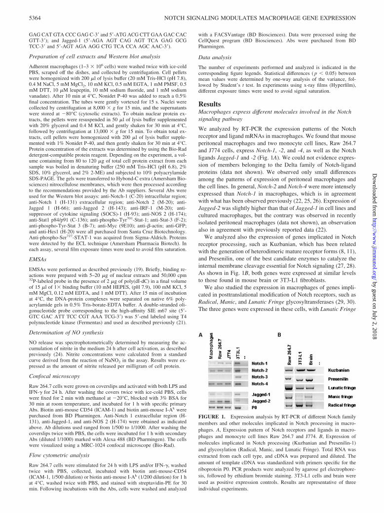
We analyzed by RT-PCR the expression patterns of the Notchreceptor and ligand mRNAs in macrophages. We found that mouseperitoneal macrophages and two monocyte cell lines, Raw 264.7and J774 cells, express Notch-1, -2, and -4, as well as the Notchligands Jagged-1 and -2 (Fig. 1A). We could not evidence expres-sion of members belonging to the Delta family of Notch-ligandproteins (data not shown). We observed only small differencesamong the patterns of expression of peritoneal macrophages andthe cell lines. In general, Notch-2 and Notch-4 were more intenselyexpressed than Notch-1 in macrophages, which is in agreementwith what has been observed previously (22, 25, 26). Expression ofJagged-2 was slightly higher than that of Jagged-1 in cell lines andcultured macrophages, but the contrary was observed in recentlyisolated peritoneal macrophages (data not shown), an observationalso in agreement with previously reported data (22).
We analyzed also the expression of genes implicated in Notchreceptor processing, such as Kuzbanian, which has been relatedwith the generation of heterodimeric mature receptor forms (8, 11),and Presenilin, one of the best candidate enzymes to catalyze theinternal membrane cleavage essential for Notch signaling (27, 28).As shown in Fig. 1B, both genes were expressed at similar levelsto those found in mouse brain or 3T3-L1 fibroblasts.
We also studied the expression in macrophages of genes impli-cated in posttranslational modification of Notch receptors, such asRadical, Manic, and Lunatic Fringe glycosyltransferases (29, 30).The three genes were expressed in these cells, with Lunatic Fringe
FIGURE 1. Expression analysis by RT-PCR of different Notch familymembers and other molecules implicated in Notch processing in macro-phages. A, Expression pattern of Notch receptors and ligands in macro-phages and monocyte cell lines Raw 264.7 and J774. B, Expression ofmolecules implicated in Notch processing (Kuzbanian and Presenilin-1)and glycosylation (Radical, Manic, and Lunatic Fringe). Total RNA wasextracted from each cell type, and cDNA was prepared and diluted. Theamount of template cDNA was standardized with primers specific for theriboprotein P0. PCR products were analyzed by agarose gel electrophore-sis, followed by ethidium bromide staining. 3T3-L1 cells and brain wereused as positive expression controls. Results are representative of threeindividual experiments.
5364 NOTCH SIGNALING MODULATES MACROPHAGE GENE EXPRESSION
by guest on July 2, 2018http://w
ww
.jimm
unol.org/D
ownloaded from

being the most represented (Fig. 1B). These results indicate thatdifferent genes, implicated in Notch processing and modification,are expressed in macrophages.
LPS and IFN-� up-regulate Notch-1 and Jagged-1 expression inmacrophages
As shown in Fig. 2, A and B, the treatment of macrophages withbacterial LPS (100 ng/ml) increased the level of Notch-1 (�3-foldincrease) and Jagged-1 (�4-fold increase) mRNAs, but not those ofNotch-2, Jagged-2, or Delta mRNAs (data not shown). Simultaneoustreatment of macrophages with LPS and IFN-� (10 U/ml) induced anincrease of �10-fold in Notch-1 expression. However, Jagged-1 in-duction remained similar to that obtained with LPS treatment alone.As a control for macrophage activation by LPS and IFN-�, we de-termined NOS expression and NO production (data not shown).
Notch-1 and Jagged-1 up-regulation was a rapid process, whichreached maximal mRNA expression levels �2 h after macrophagestimulation (Fig. 2B). This expression pattern is similar to thatshown by other genes implicated in macrophage activation, such as
NOS or cyclooxygenase-2 (31). Notch-1 and Jagged-1 protein up-regulation after macrophage activation could be evidenced also byWestern blot or by immunocytochemistry assays (Fig. 2, C and D).Confocal microscopy studies showed that Notch-1 and Jagged-1proteins were mainly present at the plasma membrane, althoughsome protein was intracellular, probably located at the endoplas-mic reticulum. NOS, used as a positive control for macrophageactivation, was mainly detected in the cytoplasm (Fig. 2D).
To study the effects of proinflammatory cytokines in Notch-1 andJagged-1 gene expression, cultured peritoneal macrophages or Raw264.7 cells were stimulated with TNF-�, IL-1, or IL-6, and Notch-1and Jagged-1 protein levels were determined by Western blot. Max-imal Notch-1 induction was obtained after stimulation with both LPSand IFN-�. Much smaller induction was observed after treatment withIL-1 or IL-6, alone, or in combination with IFN-� (data not shown).Treatment with TNF-� did not induce Notch-1 expression, althoughit was able to induce NOS or I�B-� expression (data not shown).Maximal expression of Jagged-1 (�5-fold increase) was obtained bytreatment with LPS or IL-6 alone. As observed for LPS, IFN-� did
FIGURE 2. Different members ofthe Notch family are up-regulated aftermacrophage activation with LPS andIFN-�. Raw 264.7 cells were triggeredwith LPS (50 ng/ml) and/or IFN-� (20U/ml) for different times. A, mRNAexpression was evaluated at 6 h bysemiquantitative RT-PCR analysis. B,Time-course analyses for Notch-1 andJagged-1 expression were also per-formed using real-time quantitativeRT-PCR (mean � SD; n � 4). C, Theamount of Notch-1, Jagged-1, and iNOSproteins was evaluated by Western blotin 120 �g of total cell protein extracts18 h after macrophage activation. Re-sults show the mean of three indepen-dent experiments and a representativeblot. D, Immunolocalization of Notch-1,Jagged-1, and iNOS proteins in macro-phages cultured on glass slides (see Ma-terials and Methods) activated for 18 h.Cells were fixed and the protein detectedby binding of specific Abs that were re-vealed with Alexa 488-labeled anti-rabbit or anti-goat Ig.
Table I. Effect of different protein kinase inhibitors in the up-regulation of Notch-1 and Jagged-1 mRNA and protein after macrophage activation byLPS and IFN-�a
Jagged-1 Notch-1
mRNA Protein mRNA Protein
100 100 100 100PD98059 (20 �M) 80 � 15 102 � 12 65 � 15 93 � 11SB203580 (10 �M) 20 � 7* 55 � 8* 5 � 3* 50 � 7*Wortmanin (10 �M) 110 � 20 114 � 17 97 � 16 120 � 13
a Cells were pretreated with the kinase inhibitors and after 1 h, stimulated with 50 ng/ml LPS and 20 U/ml IFN�. mRNA and protein levels were evaluated after 6 and 18 h,respectively. The image shows a representative Western blot. The table shows the mean � SD of four experiments.
*, p � 0.05 relative to the control conditions in the absence of inhibitors.
5365The Journal of Immunology
by guest on July 2, 2018http://w
ww
.jimm
unol.org/D
ownloaded from

not increase further Jagged-1 induction by IL-6 (data not shown).These results indicate that the intracellular signals implicated inNotch-1 and Jagged-1 induction are not identical, although some ofthem are probably shared.
We did not observe changes in the expression of the differentgenes implicated in Notch processing, such as Kuzbanian and Pre-senilin, or those implicated in Notch glycosylation, such as Manic,Lunatic, or Radical Fringe, in macrophages activated by proin-flammatory signals.
p38 MAPK is implicated in Notch-1 and Jagged-1 induction byproinflammatory signals
We used different protein kinase inhibitors to characterize the sig-naling pathways implicated in the induction of Notch-1 andJagged-1 expression following macrophage activation. As shownin Table I, only SB203580, a p38 MAPK inhibitor, but neither
PD98059, a MEK-1 inhibitor upstream of p42/44 MAPKs, norWortmanin, a phosphoinositide 3-OH kinase inhibitor, blockedNotch-1 and Jagged-1 induction. In addition, inhibition of prosta-glandin synthesis by the cyclooxygenase inhibitor 5,5-dimethyl-3-(3-fluorophenyl)-4-(4-methylsulfonyl)phenyl-2(5H)-furan one didnot modify Notch-1 and Jagged-1 expression, revealing that pros-taglandins are not implicated in the regulation of these genes aftermacrophage activation (data not shown).
Constitutively active Notch-IC proteins modify the activity ofAP-1- and STAT1/3-dependent reporter genes in activatedmacrophages
What is the biological meaning of Notch-1 up-regulation in theprocess of macrophage activation? In the last years, some contro-versial data have been published describing interactions betweenthe Notch-IC proteins and different transcription factors, some of
FIGURE 3. Constitutively active Notch-IC modifies the activity of AP-1 and STAT1/3 reporter genes in activated macrophages. Two expression vectorsencoding for active forms of human Notch-1 were constructed. One of them (pNIC-2, aa 1759–2556) contains the RAM domain, the ankyrin repeats, theglutamine-rich region, and the PEST domain linked to a myc epitope. The second one (pNIC-1, aa 1759–2232) lacks the glutamine-rich region and the PESTdomain. A, The structure of the entire Notch-1 receptor and the two truncated IC proteins used in this work. B, Expression of active Notch-IC proteins wasassessed in HEK 293T cells by Western blot after transient transfection. C, Activity of truncated proteins was checked by transient transfection with aCBF1-dependent reporter gene (CBF-Luc) in Raw 264.7 cells stimulated (u) or not (�) with LPS (50 ng/ml) and IFN-� (50 U/ml). D, Vectors encodingfor the intracellular active Notch-1 proteins were transiently transfected with a GFP expression vector into Raw 264.7 cells. GFP� cells were separated ina sorter flow cytometer, and the level of Hes-1 mRNAs was analyzed by RT-PCR. 3T3-L1 cells were used as a positive control for Hes-1 expression. E,Intracellularly active Notch-1 expression vectors and a Hes-1 expression vector were transiently cotransfected in Raw 264.7 cells with NF-�B-Luc,AP-1-Luc, or m67-Luc plasmids, and a �-galactosidase expression vector. Luciferase activity was measured 24 h after activation with LPS and IFN-�, andresults were normalized by using the data from �-galactosidase activity. Results represent the mean � SD of four experiments. The � indicates a statisticallysignificant difference in reporter gene activity (p � 0.05), relative to that of the respective control in the absence of Notch-1 proteins.
5366 NOTCH SIGNALING MODULATES MACROPHAGE GENE EXPRESSION
by guest on July 2, 2018http://w
ww
.jimm
unol.org/D
ownloaded from
which are involved in macrophage activation, such as NF-�B andAP-1 (3). To study whether these interactions could be happeningin macrophages, we decided to analyze the effects of Notch sig-naling in NF-�B, AP-1, and STAT-1 activation after macrophagetriggering with LPS and IFN-�. Because Notch activation impliesits transmembrane proteolysis and the release and translocation tothe nucleus of its intracellular active region to activate expressionof target genes, we studied the relationship between typical in-flammatory signaling pathways and Notch-1 activation in Raw264.7 cells, using the expression vectors pNIC-1 and pNIC-2 (seeMaterials and Methods). pNIC-1 and pNIC-2 encode for activeforms of human Notch-1 intracellular protein, from aas 1759 to2237 and 1759 to 2556, respectively. Unlike pNIC-2, pNIC-1 lacksthe glutamine-rich region and the PEST domain (Fig. 3A). Expres-sion of these active Notch-1 polypeptides was assessed by Westernblot after transient transfection of HEK 293T cells (Fig. 3B).
Expression of both intracellular forms of human Notch-1 pro-teins in Raw 264.7 cells induced a strong activation of a CBF1-dependent reporter gene (CBF1-Luc) in control or activated mac-rophages, demonstrating that these Notch-1-truncated proteinswere active, and that the glutamine-rich region and the PEST do-main were not essential for Notch-1-induced CBF1 activation inthese cells (Fig. 3C). CBF1 activation was greater in control thanin activated macrophages. This could be the result of a competitionfor coactivators between Notch and NF-�B, as it has been de-scribed in 32D cells activated with TNF-� (32, 33). Interaction ofNotch-IC with the CBF1 complex in the nucleus leads to dissoci-ation of corepressors from the CBF1 protein enabling it to up-regulate downstream Notch-1 target genes, such as Hes-1 (10–12).As shown in Fig. 3D, transient expression of the active pNIC-1protein induced Hes-1 expression in activated macrophages. Fig.3E shows that, after macrophage triggering with LPS and IFN-�,expression of both Notch-IC proteins did not modify significantlythe activity of a NF-�B-dependent reporter gene (NF-�B-Luc).However, both proteins drastically inhibited the activity of an AP-1-dependent reporter gene (AP-1-Luc) and strongly increased theactivity of a STAT1/3-dependent reporter gene (m67-Luc). It isnoteworthy that despite Notch-IC activity being lower in activatedmacrophages than in control cells, it is nonetheless sufficient tomodify the activity of AP-1 and STAT1/3 proteins.
Because Notch-1 functional requirements for CBF1-, AP-1-,and STAT-dependent transcription seem similar, and transcriptionof Hes-1 is dependent upon CBF1 activation, we examinedwhether the protein Hes-1 was also able to modulate AP-1- andSTAT-dependent reporter gene activity. As shown in Fig. 3E, theexpression of Hes-1 did not modify NF-�B-reporter gene activity,but inhibited AP1 activity and increased STAT-reporter gene ex-pression, similarly to that observed with Notch-IC proteins. Theseresults suggest that the protein Hes-1 mediates the cross-talk be-tween Notch and AP-1/STAT signaling pathways after macro-phage triggering with LPS and IFN-�.
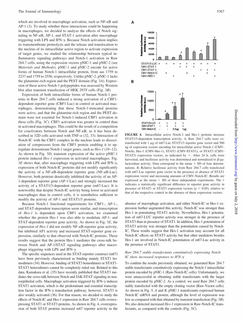
The specific sequences used in the STAT-reporter construct (m67)have been previously characterized as binding mainly STAT1 ho-modimers (34). However, binding of STAT3 homodimers or STAT1-STAT3 heterodimers cannot be completely ruled out. Related to thisdata, Kamakura et al. (35) have recently published that STAT3 me-diates the cross-talk between Notch and JAK-STAT signaling in neu-roepithelial cells. Macrophage activation triggered by IFN-� inducesSTAT1 activation, which is the predominant and essential transcrip-tion factor in the IFN-� transduction pathway; however, STAT3 isalso weakly activated (36). For that reason, we decided to study theeffects of Notch-IC and Hes-1 expression in Raw 264.7 cells overex-pressing STAT1 or STAT3 proteins. As shown in Fig. 4, overexpres-sion of both STAT proteins increased m67 reporter activity in the
absence of macrophage activation, and either Notch-IC or Hes-1 ex-pression further augmented this activity. Notch-IC was stronger thanHes-1 in potentiating STAT1 activity. Nevertheless, Hes-1 potentia-tion of m67-LUC reporter activity was stronger in the presence ofSTAT3 than in presence of STAT1. In addition, Hes-1 potentiation ofSTAT3 activity was stronger than the potentiation caused by Notch-IC. These results suggest that Hes-1 activation may account for allNotch-IC effects on STAT3 activity but that other mediators besidesHes-1 are involved in Notch-IC potentiation of m67-Luc activity inthe presence of STAT1.
Raw 264.7 stable transfectants constitutively expressing Notch-IC show increased responses to IFN-�
To confirm the results previously obtained, we generated Raw 264.7stable transfectants constitutively expressing the Notch-1 intracellularprotein encoded by pNIC-1 (Raw-Notch-IC cells). Unfortunately, wewere unsuccessful in obtaining stable transfectants with the largerprotein encoded by pNIC-2. As a control, we used Raw 264.7 cellsstably transfected with the empty cloning vector (Raw-Vector cells).As shown in Fig. 5, A and B, pNIC-1 transfectants expressed humanNotch-IC mRNA and protein, although the level of expression waslow as compared with that obtained by transient transfection (Fig. 3B).We also detected increased Hes-1 expression in Raw-Notch-IC trans-fectants, as compared with the controls (Fig. 5C).
FIGURE 4. Intracellular active Notch-1 and Hes-1 proteins increaseSTAT1/3-dependent transcription activity. A, Raw 264.7 cells were co-transfected with 1 �g of m67-Luc STAT1/3 reporter gene vector and 500ng of expression vectors encoding for intracellular active Notch-1 (CMV-Notch), Hes-1 (CMV-Hes-1), STAT1 (CMV-STAT1), or STAT3 (CMV-STAT3) expression vectors, as indicated by �. After 24 h, cells wereharvested, and luciferase activity was determined and normalized to �-ga-lactosidase activity. Data correspond to the mean � SD of four determi-nations. B, Relative luciferase activity from Raw 264.7 cells transfectedwith m67-Luc reporter gene vector in the presence or absence of STAT1expression vector and increasing amounts of CMV-Notch-IC. Results areexpressed as the mean � SD of three independent experiments. The �
indicates a statistically significant difference in reporter gene activity inpresence of STAT1 or STAT3 expression vectors (p � 0.05), relative tothat of the respective control in the absence of these expression vectors.
5367The Journal of Immunology
by guest on July 2, 2018http://w
ww
.jimm
unol.org/D
ownloaded from

CBF1-, NF-�B-, AP-1-, or STAT-dependent reporter genes weretransiently transfected in Raw-Vector or Raw-Notch-IC cells. As ex-pected, CBF1-dependent reporter activity increased (�4- to 6-fold) inpNIC-1 stable transfectants (Fig. 5D). In contrast, NF-�B-dependentreporter activity was not significantly modified by Notch-IC expres-sion. However, AP1- and STAT-dependent reporter activities weresignificantly affected. Whereas AP1 activity decreased, STAT activityincreased in cells expressing the Notch-1 intracellular domain. Theseresults confirm the results obtained previously, and further suggestthat endogenous activation of the Notch signaling pathway can mod-ulate gene expression triggered by macrophage activation.
Because STAT activation is a key event in the IFN-� signalingpathway, we analyzed the expression of well-characterized IFN targetgenes in our stable transfectants. We studied the expression of bothIRF-1, a transcription factor involved in type I and II IFNs signalcascades, and SOCS-1, a protein involved in the feedback inhibitionof IFNs signaling pathways. Both genes present GAS elements intheir promoter regions, and studies with STAT1�/� mice showed thattheir expression is mediated by STAT1 and not by STAT3 (6, 7). Asshown in Fig. 6A, both proteins were more intensely induced by treat-ment with LPS and IFN-� in Raw-Notch-IC cells than in control cells.We also analyzed two surface proteins involved in macrophage func-tion that are up-regulated after IFN-� treatment: the adhesion proteinICAM-1 and class II MHC molecules, implicated in Ag presentation(5). Fig. 6, B and C, show the mean of three experiments in whichsurface expression of these proteins was evaluated by flow cytometricanalysis. Induction of both ICAM-1 and class II MHC molecules wassignificantly higher in transfectants expressing the Notch-IC proteinthan in control cells.
Increased STAT1 phosphorylation, nuclear translocation, orDNA binding are not responsible for the elevated IFN-�response in Raw-Notch-IC stable transfectants
Biochemical and genetic studies have shown that STAT1 plays acritical role in IFN-�-dependent signaling. STAT1 and, to a lesser
extent, STAT3 are rapidly Tyr-phosphorylated and activated afterIFN-� triggering. Recently, it has been described that the Notcheffectors Hes-1 and Hes-5 facilitate STAT3 phosphorylation andactivation (35). We analyzed STAT1 and STAT3 phosphorylationin our Raw 264.7 stable transfectants after their activation withLPS and IFN-�. As shown in Fig. 7A, we found no differences inSTAT1, and only a small increase in STAT3 Tyr phosphorylationin these transfectants; however, an increase in the levels of IRF-1was detected. We analyzed also STAT1 nuclear translocation us-ing cytosolic and nuclear extracts; no differences in STAT1 trans-location were detected (data not shown). Binding of STAT1 tohigh-affinity SIE m67 site was similar in extracts from control cellsor from Notch-IC Raw 264.7 stable transfectants (Fig. 7B).
Because the levels of Notch-IC and Hes-1 proteins were lowerin stable than in transient transfectants, we decided to study theNotch-1 and Hes-1 implication in STAT phosphorylation by usingtransient transfectants of HEK 293T cells, which generally showabundant mRNA and protein expression levels of the transfectedgenes. HEK 293T cells were cotransfected with GFP, STAT1,Notch-IC, or Hes-1, or cotransfected with GFP, STAT3, Notch-IC,or Hes-1 expression vectors, and the efficiency of transfection wasdetermined by examining GFP expression by fluorescence micros-copy. As shown in Fig. 7C (upper panel), we observed increasedSTAT3 phosphorylation in Hes-1 and, to a lesser extent, inNotch-IC HEK 293T STAT3 cotransfectants (probably due to themuch lesser expression of Hes-1, undetectable in our experiments),but STAT1 phosphorylation levels remained the same in STAT1cotransfectants. Nevertheless, overexpression of STAT1 andNotch-IC or Hes-1 increased IRF-1 expression to levels similar tothose obtained after treatment with IFN-� (Fig. 7C, lower panel).STAT1 overexpression increased the basal level of STAT1 Tyrphosphorylation (Fig. 7C, right lower panel), probably due to thebasal activity of JAKs (21). This activation resulted in a weakincrease of IRF-1 expression that was further promoted byNotch-IC and Hes-1. In contrast, STAT3 overexpression did not
FIGURE 5. Raw 264.7 stable trans-fectants constitutively expressingNotch-IC present increased CBF1 ac-tivity and modified AP-1- and STAT1/3-dependent reporter gene expressionafter activation with LPS and IFN-�.Raw 264.7 pNIC1 stable transfectantsexpress human Notch mRNA (A) andprotein (B) and show increased expres-sion of the protein Hes-1 (C). D, StableRaw 264.7-transfected cells, Raw-vec-tor (empty vector), or Raw-Notch-IC(pNIC1 expression vector) were tran-siently transfected with CBF-Luc, NF-�B-Luc, AP-1-Luc, or m67-Luc plas-mids, and a �-galactosidase expressionvector. Luciferase activity in controland LPS plus IFN-�-activated cellswas measured after 24 h and normal-ized with �-galactosidase activity. Re-sults are expressed as the mean � SDof three individual experiments. The �
indicates a statistically significant dif-ference in reporter gene activity (p �0.05), relative to that of the respectivecontrol in the absence of Notch-1proteins.
5368 NOTCH SIGNALING MODULATES MACROPHAGE GENE EXPRESSION
by guest on July 2, 2018http://w
ww
.jimm
unol.org/D
ownloaded from

show the same pattern of IRF-1 induction (data not shown). Mul-tiple studies in the last years have shown that STAT1 phosphor-ylation at serine 727 is essential for maximal transcription of targetgenes (5, 37). For that reason, we studied whether Ser727 phos-phorylation could be responsible for the increased transactivationcapacity of STAT1 induced by Notch-IC in Raw 264.7 stabletransfectants. We could not observe differences in STAT1 Ser727
phosphorylation between control transfectants and cells expressingNotch-IC (Fig. 7D).
All of these results suggest that Notch-IC and Hes-1 proteinspotentiate the transactivation capacity of STAT1 without increas-ing its level of Tyr phosphorylation, nuclear translocation, or DNAbinding. Our results also confirm that Notch-IC and Hes-1 increaseSTAT3 phosphorylation in transiently transfected HEK 293T cellsand in the Raw 264.7 stable transfectants.
iNOS expression is inhibited in Raw-Notch-IC stabletransfectants
We monitored iNOS expression and NO production in stable trans-fectants expressing or not expressing active Notch-IC proteins. Be-cause STAT1 and IRF-1, together with NF-�B, are the major tran-scription factors implicated in iNOS gene expression (38), weexpected to find an increase in iNOS expression. Nevertheless, wewere surprised to find weaker iNOS expression and NO productionin Raw-Notch-IC transfectants than in control Raw-Vector trans-fectants (Fig. 8, A–C). This lower iNOS expression resulted fromdiminished transcriptional activity of the iNOS promoter (Fig. 8D).Further studies are needed to dissect the mechanism by whichNotch-IC appears to down-regulate iNOS expression, but our re-sults suggest that macrophage activation, gene expression, andproduction of macrophage cytotoxic substances are modified byNotch-1 activation.
DiscussionIn the last years, multiple lines of evidence have implicated Notchsignaling in lymphoid (39, 40) and myeloid differentiation (16).Nevertheless, less attention has been focused on the role of Notchproteins in mature, fully differentiated cells, despite Notch recep-tors and ligands being often highly expressed by these cells (10).In this study, we show that macrophages express Notch-1, -2, and-4, as well as the ligands Jagged-1 and -2. These results agree withthose from different authors who have reported the expression ofthese genes and proteins by monocytes and macrophages (25, 26,41). Our results indicate that macrophages also express differentproteins implicated in Notch posttranslational modification, pro-cessing and signaling, such as Kuzbanian, Presenilin, and Fringe.We also show in this study that macrophage activation by LPSspecifically increases the expression of Notch-1 and Jagged-1 (Fig.2). Moreover, IFN-�, the most potent activator of macrophages,synergizes with LPS to up-regulate the expression of Notch-1, butnot that of Jagged-1, Jagged-2, or Notch-2. In addition, the ex-pression patterns of Notch-1, Jagged-1, and iNOS genes duringmacrophage activation are similar (Fig. 2).
Bacterial lipopolysaccharides are recognized by the macroph-age’s Toll-4 membrane receptors. Signal transduction throughToll-4 receptors implicates the activation of multiple pathways,including NF-�B activation, p42/44 MAPKs, stress-activated pro-tein kinases, JNK and p38 kinase, and phosphoinositide 3-OH ki-nase (3). Our results implicate the p38 signaling pathway in theinduction of Notch-1 and Jagged-1 expression by LPS. Previouswork has implicated NF-�B proteins in Jagged-1 induction (42).Recently, Amsen et al. (43) have shown that LPS inducesJagged-1 expression via a Myd88-independent pathway. Li et al.(44) found that Notch-1 expression is dependent on NF-�B activityin pre-B cell lines. Very recently, Adler et al. (45) have shown that,in primary CD4� T cells, Notch-1 expression is induced after spe-cific peptide-Ag stimulation, for which NF-�B is also a key ele-ment (46, 47). Notch-1 induction and activation have also been de-scribed after oncogenic Ras signaling through a p38-mediatedpathway (48). Our results, together with those mentioned above, dem-onstrate that Notch proteins can be up-regulated in different immunecells, including macrophages, and that activation of p38 MAPK andNF-�B seems to be an essential element in this induction.
FIGURE 6. Raw 264.7 stable Notch-IC transfectants present increasedresponse to IFN-�. A, Raw 264.7 stable Notch-IC transfectants were stim-ulated with LPS (50 ng/ml) and IFN-� (50 U/ml) for the indicated times.Total cell extracts were obtained after 1 or 2 h of incubation, and 80 �g ofproteins were analyzed by Western blot to determine SOCS-1 and IRF-1expression. Expression of �-actin was used as a loading control. The blotshown is representative of three individual experiments. ICAM-1 (B) andtype II MHC protein expression (C) was evaluated by flow cytometry 24 hafter macrophage activation. Cells were first labeled with biotin anti-mouseCD54 (ICAM-1) or biotin anti-mouse I-Ak, followed by streptavidin-PEincubation. Results are expressed as the mean � SD of three experiments.The � indicates a statistically significant difference in protein expression(p � 0.05), relative to that of the respective control.
5369The Journal of Immunology
by guest on July 2, 2018http://w
ww
.jimm
unol.org/D
ownloaded from

Given that high levels of Notch-1 receptors are present in acti-vated macrophages after triggering with LPS and IFN-�, we haveinvestigated the potential role of Notch-1 signaling in macrophagebiology. Truncated Notch molecules lacking the transmembraneand extracellular regions (Notch-IC) behave as constitutively ac-tive Notch receptors and have been used extensively to study thefunction of Notch in multiple systems (49–51). Using this strategy,we have shown that Notch-IC interferes with the activity of dif-ferent transcription factors implicated in macrophage activation,such as AP-1 or STAT1/3, without significantly affecting others,such as NF-�B (Figs. 3E and 5). Different authors have exploredthe potential interaction between Notch and NF-�B signaling path-ways. Our results agree with those reported by Bresnick and co-workers (52), who did not find any interaction between Notch andNF-�B signaling pathways. Nevertheless, Guan and coworkers(53, 54) have reported NF-�B inhibition by interaction of its p50subunit with Notch-IC. In addition, Oakley et al. (55) have shownthat the basal expression level of the NF-�B inhibitor, I�B�, iscontrolled by the mammalian transcriptional repressor RBP-J(CBF1) and its activator Notch1.
In any case, because Notch signaling may be required for basalsynthesis of NF-�B components (51, 56), interplay between Notchand NF-�B seems to be a complex process, probably depending onseveral factors, including cell type. An inhibitory cross-talk be-tween Notch and NF-�B pathways has also been described byEspinosa et al. (32, 57), who observed that these proteins competedfor nuclear corepressors, such as N-CoR. When searching for thereasons that could explain the discrepancies among all theseworks, we observed that NF-�B inhibition appears to occur whenthe Notch-IC-truncated proteins lack part of the RAM domain, butnot when they correspond to the entire Notch region normallyliberated after ligand activation. In support of these data, we foundNF-�B inhibition when using a vector expressing an Notch-ICprotein starting at aa 1771, but not starting at aa 1759 (data notshown). Notch-IC proteins starting at aa 1771 are indeed poorlyactive when a CBF1-reporter gene is used (57). Although we can-not completely rule out an inhibition of NF-�B by Notch signaling,according to our data, if it existed at all, it would be small.
If NF-�B activity is not strongly affected by Notch signaling, thesame does not hold true for other transcription factors involved in
FIGURE 7. Increased STAT1 phosphorylation is not responsible for the elevated IFN-� response in Raw-Notch-IC. A, Raw 264.7 stable transfectantswere stimulated with LPS (50 ng/ml) and IFN-� (50 U/ml) for the indicated times. After stimulation, total extracts were obtained, and 80 �g of proteinswere analyzed by Western blot to determine STAT1/3 Tyr phosphorylation. Blots were also revealed with anti-STAT1 or anti-STAT3 Abs to evaluate gelloading. B, EMSA with the high-affinity SIE m67 probe and nuclear extracts prepared at different times after triggering macrophage activation. Results showa representative experiment of three. C (upper panel), Hes-1 and Notch-IC overexpression induces STAT3, but not STAT1 phosphorylation, in HEK 293Tcells transiently transfected with CMV-STAT1 or CMV-STAT3. Transfection efficiency was assessed by determining GFP fluorescence 24 h aftertransfection, and total protein extracts were subjected to immunoblot analysis with Abs to phospho-Tyr701-STAT1, phospho-Tyr705-STAT-3, STAT1,STAT3, Hes-1, and myc (Notch-IC). Lower panel, Overexpression of STAT1 and Hes-1 or STAT1 and Notch-IC induced IRF-1 expression in HEK 293Tcells. Cells were transiently transfected, and 24 h later, cells were stimulated or not with 50 U/ml human IFN-� for 2 h. Then, total extracts were obtainedand analyzed by Western blot. Endogenous human Hes-1 protein is not detected by this Ab, but its expression was assessed by RT-PCR (data not shown).On the right panel, an overexposed blot shows that overexpression of STAT1 is sufficient to increase basal STAT1 Tyr phosphorylation levels. D, STAT1Ser phosphorylation was also evaluated in Raw 264.7 stable transfectants by using a specific anti-phospho-Ser727-STAT1 Ab. In all of the cases, blotscorrespond to representative experiments of a minimum of three individual assays.
5370 NOTCH SIGNALING MODULATES MACROPHAGE GENE EXPRESSION
by guest on July 2, 2018http://w
ww
.jimm
unol.org/D
ownloaded from

macrophage activation. We have observed an intense inhibition ofAP-1-dependent reporter activity in the presence of Notch-IC (Fig.5). AP-1 is a transcription factor involved in macrophage activa-tion that modulates the expression of cytokines and enzymes con-trolling matrix remodeling (58). Although its role appears to beless important than that of NF-�B, AP-1 inhibition could haverepercussions on macrophage gene expression. Inhibition of AP-1activity by Notch-IC has been recently described by other inves-tigators, and it has been assigned to the Notch-IC RAM domainand its ability to bind CBF1 (52, 59). The mechanism responsiblefor the inhibition of AP-1-dependent transcription is still unknown,but it does not involve AP-1 binding changes or c-Jun amino-terminal modifications (59). Indeed, we report in this study that, atleast in part, Notch inhibition of AP-1 activity could be explainedby the action of Hes-1 (Fig. 3E). Further work is needed to fullyunderstand the molecular mechanism of this inhibition, but twomodels have been proposed to explain it: either a disruption ofAP-1 complex assembly, or an impaired coactivator use by theAP-1 nucleoprotein complex (52, 59). Our data support the ideathat either AP-1 complex disruption or impaired AP-1 coactivatorrecruitment are mediated by Hes-1, but not by Notch-IC directly.
Interestingly, contrary to what was seen with AP-1, we observedthat Notch-IC increases the activity of a STAT1 reporter genepreviously used to evaluate STAT1 activity (21). Although bio-chemical and genetic studies show that STAT1 plays a critical rolein IFN-�-dependent signaling (5), recent work reveals that addi-tional signals are required for the full range of responses caused byIFN-� (6, 7). STAT3 activation has been observed also after mac-
rophage activation (36). Recently, cross-talking between STAT3and Notch has been described in neuroepithelial cells. In thesecells, the Notch effector proteins Hes-1 and Hes-5 associate withJAK2 and STAT3 to promote STAT3 phosphorylation and acti-vation (35). We have not observed differences in STAT1 phos-phorylation in the presence or absence of Notch-IC. In addition,we have not detected differences between control and Notch-ICstable transfectants in STAT1 nuclear translocation or DNA bind-ing. Moreover, phosphorylation of STAT1 at Serine 727, shown tobe essential to reach its maximal transactivation potential (5), wasnot affected by the expression of Notch-IC. However, an increasein STAT3 phosphorylation was detected in Raw 264.7 cells over-expressing Notch-IC. Our studies in HEK 293T cells confirmedthat Hes-1 and, to a lesser extent, Notch-IC expression increasedSTAT3 phosphorylation levels, as described previously (35), butleft STAT1 phosphorylation levels unaffected (Fig. 7). Although aNotch-IC-dependent increase of STAT1 phosphorylation, translo-cation or binding did not seem to occur in Raw 264.7 Notch-ICstable transfectants, following activation of these cells with LPSand IFN-� we observed a clear increase in transcription of STAT1-dependent genes, such as IRF-1, SOCS-1, ICAM-1, and class IIMHC proteins (Fig. 6), typically induced by STAT1 activity. Ex-pression of IRF-1, SOCS-1, and ICAM-1 appears to be strictlydependent on STAT1 activity, because macrophages fromSTAT1�/� mice triggered with IFN-�, despite showing increasedphosphorylation and activation of STAT3 (36), cannot expressthese genes (6, 7). For that reason, it appears that increased acti-vation of STAT3 by Notch/Hes-1 cannot compensate for the lack
FIGURE 8. iNOS expression is diminished in activated Raw-Notch-IC cells. A, Raw 264.7 stable transfectants were stimulated with LPS (50 ng/ml) andIFN-� (50 U/ml) for the indicated times. Total extracts were obtained, and 80 �g of protein were analyzed by Western blot to determine iNOS expression.Blots were also revealed with anti-�-actin to evaluate gel loading. The blot shown here corresponds to a representative experiment of three different assays.B, Immunolocalization of ICAM-1 and iNOS in Raw 264.7 Notch-IC stable transfectants stimulated with LPS and IFN-� as described above for 18 h. C,Nitrite accumulation in the culture medium 18 h after macrophage activation. D, Stable transfectant cells, Raw-vector or Raw-Notch-IC were transientlytransfected with p-iNOS-Luc and a �-galactosidase expression vector and stimulated with LPS and IFN-�. Luciferase activity was measured 24 h later andnormalized by �-galactosidase activity. Results represent the mean � SD of three independent experiments.
5371The Journal of Immunology
by guest on July 2, 2018http://w
ww
.jimm
unol.org/D
ownloaded from
of STAT1 activity. This result suggests that the increased STAT1transcription activity observed in Raw 264.7 cells stably trans-fected with Notch-IC expression constructs and activated by LPSand IFN-� is not caused by the increase in STAT3 phosphorylationthat we observe. Thus, this led us to consider three potential ex-planations, which were not mutually exclusive, for these data.First, it is still possible that small undetectable changes in STAT1phosphorylation, which could explain the increased STAT1 tran-scription activity observed, may occur in the presence of Notch-IC.Indeed, after triggering HEK 293T cells with IFN-�, we observeda discrete increase in STAT-1 phosphorylation in the presence ofHes-1, but not in the presence of Notch-IC (Fig. 7C), suggestingthat intense Notch activity through Hes-1 may lead to an increasein STAT1 phosphorylation. Second, it is equally possible that, inthe presence of Notch-IC, STAT3 could potentiate STAT1 tran-scriptional activity independently of changes in STAT1 phosphor-ylation levels. However, transient transfection of HEK 293T cellswith STAT3 and Hes-1 or Notch-IC expression vectors does notresult in changes in IRF-1 expression, making this second possi-bility unlikely. Finally, Hes-1, Notch, or some other Notch path-way downstream proteins could affect STAT1 activity by provid-ing coactivators, or by competing for and displacing nuclearcorepressors, as has been described previously (8, 9). Further workis needed to fully elucidate the molecular mechanism of this po-tentiation. However, our work adds further evidence to the increas-ing number of works showing the existence of different mecha-nisms able to modify, promote, or inhibit IFN-�/STAT signals(60). Notch signaling can be one of them.
We evaluated iNOS expression and NO production as a controlto check for adequate macrophage activation. We expected to finda similar increase of iNOS expression after IFN-� stimulation oftransfected or control cells, because iNOS is a gene up-regulatedby IFN-� (1). Surprisingly, Raw 264.7 Notch-IC transfectantsshowed a lower capacity to increase iNOS expression and NOproduction than control cells (Fig. 8). iNOS transcription dependson the activation of multiple transcription factors. Its promoterregion contains binding sites for at least two NF-�B complexes,AP-1, two GAS elements binding STAT1 homodimers, and a com-plex IFN-stimulated regulatory element binding IRF-1 (61). Al-though specific mutation of the different binding elements willpermit to dissect what sites are involved, we can already concludethat transcriptional inhibition of iNOS must not be directly due toSTAT1 or IRF-1 proteins, because expression of other STAT1-dependent genes, including IRF-1, are increased in macrophagesor Raw 264.7 Notch-IC transfectants. It is possible that AP-1 in-hibition could account, at least partially, for the lower iNOS ex-pression observed. Moreover, two studies have shown inhibition ofNF-�B-dependent transcription after STAT3 activation. Yu andKone (62) reported iNOS inhibition by direct interaction of STAT3with p50 and p65, without affecting NF-�B binding to DNA. Morerecently, Hoentjen et al. (63) have shown that STAT3 inhibits cRelrecruitment to the IL-12p40 promoter. Indeed, genetic deletion hasshown that STAT3 has a pivotal role in controlling innate immuneresponses, and it is a key element in signal transduction by inhib-itory cytokines, like IL-10 (64). Considering these data, it is rea-sonable to speculate that Notch-dependent increase of STAT3 ac-tivation and AP-1 inhibition could be responsible for the lowerincrease of iNOS expression in Notch-IC-transfected cells.
What is the role that Notch-1 plays in macrophage immunefunction? In view of our results, the Notch-1 receptor signalingpathway can cross-talk with different routes induced during mac-rophage activation, modulating specific gene expression patternsthat could affect macrophage function. In this context, Notch ac-tivation seems to drive macrophages toward a more prominent role
as APCs by increasing ICAM-1 and MHC type II protein expres-sion, and by limiting their cytotoxic capacity through diminishingNOS expression. Notch activation has been found to modulate thefunction and fate of other immune cells. For instance, Notch ac-tivation appears to play a role in dendritic cell differentiation andmaturation (65, 66). Recent reports have implicated Notch signal-ing in the interaction between APCs and T cells. It has been re-ported that Notch signaling augments T cell responsiveness byincreasing the expression of the high-affinity IL-2R (45), and thatit is required for TCR-mediated proliferation and production ofIFN-� in peripheral T cells (67). In addition, T cell stimulation byAPC engineered to overexpress Jagged-1 can induce Ag-specificregulatory T cells (68, 69). Finally, expression of different Notchligands on APC can instruct CD4 T cells to differentiate into theTh1 or Th2 lineages (43).
The results presented in this study invite us to consider the pos-sibility that Notch signaling could also be involved in the modu-lation of macrophage function depending on the immunologicalenvironment in which these cells may work. For example, macro-phages fighting in an infectious focus would not find the appro-priated ligands to activate Notch, a situation that would favor theircytotoxic activity. On the contrary, macrophages that have mi-grated to lymphoid organs to activate T cells could receive appro-priate Notch-activating signals potentiating their Ag-presenting ca-pability and limiting their cytotoxic effects. We are currentlyworking to confirm this hypothesis.
AcknowledgmentsWe thank Santiago Vernia, Marta Casado, Ma Luisa Nueda, Samuel Riv-ero, Angela Ballesteros, Amparo Ruvira, and Beatriz Solana for their in-valuable technical support. We also thank Dr. Lisardo Bosca (Centro Na-cional de Investigaciones Cardiovasculares (CNIC), Madrid, Spain) forNF-�B and AP-1 reporter plasmids; Dr. Joaquın Jordan (Facultad de Me-dicina, Albacete, Spain) for the m67-Luc construct; and Dr. Alberto Al-varez (CNIC, Madrid, Spain) for confocal microscopy technical supportand flow cytometric analysis. We also thank Paloma Martın and LisardoBorca for critical reading of the manuscript and helpful comments.
DisclosuresThe authors have no financial conflict of interest.
References1. MacMicking, J., Q. W. Xie, and C. Nathan. 1997. Nitric oxide and macrophage
function. Annu. Rev. Immunol. 15: 323–350.2. Xaus, J., M. Comalada, A. F. Valledor, M. Cardo, C. Herrero, C. Soler,
J. Lloberas, and A. Celada. 2001. Molecular mechanisms involved in macrophagesurvival, proliferation, activation or apoptosis. Immunobiology 204: 543–550.
3. Takeda, K., T. Kaisho, and S. Akira. 2003. Toll-like receptors. Annu. Rev. Im-munol. 21: 335–376.
4. Akira, S., and K. Takeda. 2004. Functions of Toll-like receptors: lessons fromKO mice. C. R. Biol. 327: 581–589.
5. Schroder, K., P. J. Hertzog, T. Ravasi, and D. A. Hume. 2004. Interferon-�: anoverview of signals, mechanisms and functions. J. Leukocyte Biol. 75: 163–189.
6. Gil, M. P., E. Bohn, A. K. O’Guin, C. V. Ramana, B. Levine, G. R. Stark,H. W. Virgin, and R. D. Schreiber. 2001. Biologic consequences of Stat1-inde-pendent IFN signaling. Proc. Natl. Acad. Sci. USA 98: 6680–6685.
7. Ramana, C. V., M. P. Gil, R. D. Schreiber, and G. R. Stark. 2002. Stat1-depen-dent and -independent pathways in IFN-�-dependent signaling. Trends Immunol.23: 96–101.
8. Hansson, E. M., U. Lendahl, and G. Chapman. 2004. Notch signaling in devel-opment and disease. Semin. Cancer Biol. 14: 320–328.
9. Mumm, J. S., and R. Kopan. 2000. Notch signaling: from the outside in. Dev.Biol. 228: 151–165.
10. Ohishi, K., N. Katayama, H. Shiku, B. Varnum-Finney, and I. D. Bernstein. 2003.Notch signalling in hematopoiesis. Semin. Cell Dev. Biol. 14: 143–150.
11. Zlobin, A., M. Jang, and L. Miele. 2000. Toward the rational design of cell fatemodifiers: notch signaling as a target for novel biopharmaceuticals. Curr. Pharm.Biotechnol. 1: 83–106.
12. Haines, N., and K. D. Irvine. 2003. Glycosylation regulates Notch signalling. Nat.Rev. Mol. Cell Biol. 4: 786–797.
13. Okajima, T., A. Xu, and K. D. Irvine. 2003. Modulation of notch-ligand bindingby protein O-fucosyltransferase 1 and fringe. J. Biol. Chem. 278: 42340–42345.
14. Milner, L. A., and A. Bigas. 1999. Notch as a mediator of cell fate determinationin hematopoiesis: evidence and speculation. Blood 93: 2431–2448.
5372 NOTCH SIGNALING MODULATES MACROPHAGE GENE EXPRESSION
by guest on July 2, 2018http://w
ww
.jimm
unol.org/D
ownloaded from

15. Kojika, S., and J. D. Griffin. 2001. Notch receptors and hematopoiesis. Exp.Hematol. 29: 1041–1052.
16. Schroeder, T., and U. Just. 2000. Notch signalling via RBP-J promotes myeloiddifferentiation. EMBO J. 19: 2558–2568.
17. Schroeder, T., H. Kohlhof, N. Rieber, and U. Just. 2003. Notch signaling inducesmultilineage myeloid differentiation and up-regulates PU.1 expression. J. Immu-nol. 170: 5538–5548.
18. Ohishi, K., B. Varnum-Finney, R. E. Serda, C. Anasetti, and I. D. Bernstein.2001. The Notch ligand, Delta-1, inhibits the differentiation of monocytes intomacrophages but permits their differentiation into dendritic cells. Blood 98:1402–1407.
19. Castrillo, A., M. J. Diaz-Guerra, S. Hortelano, P. Martin-Sanz, and L. Bosca.2000. Inhibition of I�B kinase and I�B phosphorylation by 15-deoxy-�12,14-prostaglandin J2 in activated murine macrophages. Mol. Cell. Biol. 20:1692–1698.
20. Castrillo, A., P. G. Traves, P. Martin-Sanz, S. Parkinson, P. J. Parker, andL. Bosca. 2003. Potentiation of protein kinase C � activity by 15-deoxy-�12,14-prostaglandin J2 induces an imbalance between mitogen-activated protein kinasesand NF-�B that promotes apoptosis in macrophages. Mol. Cell. Biol. 23:1196–1208.
21. Sironi, J. J., and T. Ouchi. 2004. STAT1-induced apoptosis is mediated bycaspases 2, 3, and 7. J. Biol. Chem. 279: 4066–4074.
22. Yamaguchi, E., S. Chiba, K. Kumano, A. Kunisato, T. Takahashi, T. Takahashi,and H. Hirai. 2002. Expression of Notch ligands, Jagged1, 2 and Delta1 in an-tigen presenting cells in mice. Immunol. Lett. 81: 59–64.
23. Laborda, J. 1991. 36B4 cDNA used as an estradiol-independent mRNA controlis the cDNA for human acidic ribosomal phosphoprotein PO. Nucleic Acids Res.19: 3998.
24. Diaz-Guerra, M. J., A. Castrillo, P. Martin-Sanz, and L. Bosca. 1999. Negativeregulation by phosphatidylinositol 3-kinase of inducible nitric oxide synthaseexpression in macrophages. J. Immunol. 162: 6184–6190.
25. Nomaguchi, K., S. Suzu, M. Yamada, H. Hayasawa, and K. Motoyoshi. 2001.Expression of Jagged1 gene in macrophages and its regulation by hematopoieticgrowth factors. Exp. Hematol. 29: 850–855.
26. Jonsson, J. I., Z. Xiang, M. Pettersson, M. Lardelli, and G. Nilsson. 2001. Distinctand regulated expression of Notch receptors in hematopoietic lineages and duringmyeloid differentiation. Eur. J. Immunol. 31: 3240–3247.
27. Saxena, M. T., E. H. Schroeter, J. S. Mumm, and R. Kopan. 2001. Murine notchhomologs (N1–4) undergo presenilin-dependent proteolysis. J. Biol. Chem. 276:40268–40273.
28. Schroeter, E. H., J. A. Kisslinger, and R. Kopan. 1998. Notch-1 signalling re-quires ligand-induced proteolytic release of intracellular domain. Nature 393:382–386.
29. Okajima, T., A. Xu, and K. D. Irvine. 2003. Modulation of notch-ligand bindingby protein O-fucosyltransferase 1 and fringe. J. Biol. Chem. 278: 42340–42345.
30. Okajima, T., and K. D. Irvine. 2002. Regulation of notch signaling by O-linkedfucose. Cell 111: 893–904.
31. Terenzi, F., M. J. Diaz-Guerra, M. Casado, S. Hortelano, S. Leoni, and L. Bosca.1995. Bacterial lipopeptides induce nitric oxide synthase and promote apoptosisthrough nitric oxide-independent pathways in rat macrophages. J. Biol. Chem.270: 6017–6021.
32. Espinosa, L., J. Ingles-Esteve, A. Robert-Moreno, and A. Bigas. 2003. I�B� andp65 regulate the cytoplasmic shuttling of nuclear corepressors: cross-talk betweenNotch and NF�B pathways. Mol. Biol. Cell 14: 491–502.
33. Aguilera, C., R. Hoya-Arias, G. Haegeman, L. Espinosa, and A. Bigas. 2004.Recruitment of I�B� to the hes1 promoter is associated with transcriptional re-pression. Proc. Natl. Acad. Sci. USA 101: 16537–16542.
34. Cassatella, M. A., S. Gasperini, C. Bovolenta, F. Calzetti, M. Vollebregt,P. Scapini, M. Marchi, R. Suzuki, A. Suzuki, and A. Yoshimura. 1999. Interleu-kin-10 (IL-10) selectively enhances CIS3/SOCS3 mRNA expression in humanneutrophils: evidence for an IL-10-induced pathway that is independent of STATprotein activation. Blood 94: 2880–2889.
35. Kamakura, S., K. Oishi, T. Yoshimatsu, M. Nakafuku, N. Masuyama, andY. Gotoh. 2004. Hes binding to STAT3 mediates crosstalk between Notch andJAK-STAT signalling. Nat. Cell Biol. 6: 547–554.
36. Qing, Y., and G. R. Stark. 2004. Alternative activation of STAT1 and STAT3 inresponse to interferon-�. J. Biol. Chem. 279: 41679–41685.
37. Kovarik, P., M. Mangold, K. Ramsauer, H. Heidari, R. Steinborn, A. Zotter,D. E. Levy, M. Muller, and T. Decker. 2001. Specificity of signaling by STAT1depends on SH2 and C-terminal domains that regulate Ser727 phosphorylation,differentially affecting specific target gene expression. EMBO J. 20: 91–100.
38. Lowenstein, C. J., E. W. Alley, P. Raval, A. M. Snowman, S. H. Snyder,S. W. Russell, and W. J. Murphy. 1993. Macrophage nitric-oxide synthase gene:two upstream regions mediate induction by interferon-� and lipopolysaccharide.Proc. Natl. Acad. Sci. USA 90: 9730–9734.
39. Allman, D., J. A. Punt, D. J. Izon, J. C. Aster, and W. S. Pear. 2002. An invitationto T and more: notch signaling in lymphopoiesis. Cell 109(Suppl): S1–S11.
40. Izon, D. J., J. A. Punt, and W. S. Pear. 2002. Deciphering the role of Notchsignaling in lymphopoiesis. Curr. Opin. Immunol. 14: 192–199.
41. Amsen, D., J. M. Blander, G. R. Lee, K. Tanigaki, T. Honjo, and R. A. Flavell.2004. Instruction of distinct CD4 T helper cell fates by different notch ligands onantigen-presenting cells. Cell 117: 515–526.
42. Bash, J., W. X. Zong, S. Banga, A. Rivera, D. W. Ballard, Y. Ron, andC. Gelinas. 1999. Rel/NF-�B can trigger the Notch signaling pathway by induc-ing the expression of Jagged1, a ligand for Notch receptors. EMBO J. 18:2803–2811.
43. Amsen, D., J. M. Blander, G. R. Lee, K. Tanigaki, T. Honjo, and R. A. Flavell.2004. Instruction of distinct CD4 T helper cell fates by different notch ligands onantigen-presenting cells. Cell 117: 515–526.
44. Li, J., G. W. Peet, D. Balzarano, X. Li, P. Massa, R. W. Barton, and K. B. Marcu.2001. Novel NEMO/I�B kinase and NF-�B target genes at the pre-B to immatureB cell transition. J. Biol. Chem. 276: 18579–18590.
45. Adler, S. H., E. Chiffoleau, L. Xu, N. M. Dalton, J. M. Burg, A. D. Wells,M. S. Wolfe, L. A. Turka, and W. S. Pear. 2003. Notch signaling augments T cellresponsiveness by enhancing CD25 expression. J. Immunol. 171: 2896–2903.
46. Weil, R., and A. Israel. 2004. T-cell-receptor- and B-cell-receptor-mediated ac-tivation of NF-�B in lymphocytes. Curr. Opin. Immunol. 16: 374–381.
47. Thome, M., and J. Tschopp. 2003. TCR-induced NF-�B activation: a crucial rolefor Carma1, Bcl10 and MALT1. Trends Immunol. 24: 419–424.
48. Weijzen, S., P. Rizzo, M. Braid, R. Vaishnav, S. M. Jonkheer, A. Zlobin,B. A. Osborne, S. Gottipati, J. C. Aster, W. C. Hahn, et al. 2002. Activation ofNotch-1 signaling maintains the neoplastic phenotype in human Ras-transformedcells. Nat. Med. 8: 979–986.
49. Morimura, T., R. Goitsuka, Y. Zhang, I. Saito, M. Reth, and D. Kitamura. 2000.Cell cycle arrest and apoptosis induced by Notch1 in B cells. J. Biol. Chem. 275:36523–36531.
50. Qi, R., H. An, Y. Yu, M. Zhang, S. Liu, H. Xu, Z. Guo, T. Cheng, and X. Cao.2003. Notch1 signaling inhibits growth of human hepatocellular carcinomathrough induction of cell cycle arrest and apoptosis. Cancer Res. 63: 8323–8329.
51. Cheng, P., A. Zlobin, V. Volgina, S. Gottipati, B. Osborne, E. J. Simel, L. Miele,and D. I. Gabrilovich. 2001. Notch-1 regulates NF-�B activity in hemopoieticprogenitor cells. J. Immunol. 167: 4458–4467.
52. Chu, J., S. Jeffries, J. E. Norton, A. J. Capobianco, and E. H. Bresnick. 2002.Repression of activator protein-1-mediated transcriptional activation by theNotch-1 intracellular domain. J. Biol. Chem. 277: 7587–7597.
53. Guan, E., J. Wang, J. Laborda, M. Norcross, P. A. Baeuerle, and T. Hoffman.1996. T cell leukemia-associated human Notch/translocation-associated Notchhomologue has I�B-like activity and physically interacts with nuclear factor-�Bproteins in T cells. J. Exp. Med. 183: 2025–2032.
54. Wang, J., L. Shelly, L. Miele, R. Boykins, M. A. Norcross, and E. Guan. 2001.Human Notch-1 inhibits NF-�B activity in the nucleus through a direct interac-tion involving a novel domain. J. Immunol. 167: 289–295.
55. Oakley, F., J. Mann, R. G. Ruddell, J. Pickford, G. Weinmaster, and D. A. Mann.2003. Basal expression of I�B� is controlled by the mammalian transcriptional re-pressor RBP-J (CBF1) and its activator Notch1. J. Biol. Chem. 278: 24359–24370.
56. Bellavia, D., A. F. Campese, E. Alesse, A. Vacca, M. P. Felli, A. Balestri,A. Stoppacciaro, C. Tiveron, L. Tatangelo, M. Giovarelli, et al. 2000. Constitu-tive activation of NF-�B and T-cell leukemia/lymphoma in Notch3 transgenicmice. EMBO J. 19: 3337–3348.
57. Espinosa, L., S. Santos, J. Ingles-Esteve, P. Munoz-Canoves, and A. Bigas. 2002.p65-NF�B synergizes with Notch to activate transcription by triggering cyto-plasmic translocation of the nuclear receptor corepressor N-CoR. J. Cell Sci. 115:1295–1303.
58. Shaulian, E., and M. Karin. AP-1 as a regulator of cell life and death. 2002. Nat.Cell Biol. 4: E131–E136.
59. Chu, J., and E. H. Bresnick. 2004. Evidence that C promoter-binding factor 1binding is required for Notch-1-mediated repression of activator protein-1.J. Biol. Chem. 279: 12337–12345.
60. Park, E. J., K. A. Ji, S. B. Jeon, W. H. Choi, I. O. Han, H. J. You, J. H. Kim,I. Jou, and E. H. Joe. 2004. Rac1 contributes to maximal activation of STAT1 andSTAT3 in IFN-�-stimulated rat astrocytes. J. Immunol. 173: 5697–5703.
61. Xie, Q. W., R. Whisnant, and C. Nathan. 1993. Promoter of the mouse geneencoding calcium-independent nitric oxide synthase confers inducibility by in-terferon � and bacterial lipopolysaccharide. J. Exp. Med. 177: 1779–1784.
62. Yu, Z., and B. C. Kone. 2004. The STAT3 DNA-binding domain mediates in-teraction with NF-�B p65 and inducible nitric oxide synthase transrepression inmesangial cells. J. Am. Soc. Nephrol. 15: 585–591.
63. Hoentjen, F., R. B. Sartor, M. Ozaki, and C. Jobin. 2005. STAT3 regulates NF-�Brecruitment to the IL-12p40 promoter in dendritic cells. Blood 105: 689–696.
64. Riley, J. K., K. Takeda, S. Akira, and R. D. Schreiber. 1999. Interleukin-10receptor signaling through the JAK-STAT pathway: requirement for two distinctreceptor-derived signals for anti-inflammatory action. J. Biol. Chem. 274:16513–16521.
65. Weijzen, S., M. P. Velders, A. G. Elmishad, P. E. Bacon, J. R. Panella,B. J. Nickoloff, L. Miele, and W. M. Kast. 2002. The Notch ligand Jagged-1 isable to induce maturation of monocyte-derived human dendritic cells. J. Immu-nol. 169: 4273–4278.
66. Mizutani, K., T. Matsubayashi, S. Iwase, T. S. Doi, K. Kasai, M. Yazaki,Y. Wada, T. Takahashi, and Y. Obata. 2000. Murine Delta homologue, mDelta1,expressed on feeder cells controls cellular differentiation. Cell Struct. Funct. 25:21–31.
67. Palaga, T., L. Miele, T. E. Golde, and B. A. Osborne. 2003. TCR-mediated Notchsignaling regulates proliferation and IFN-� production in peripheral T cells.J. Immunol. 171: 3019–3024.
68. Yvon, E. S., S. Vigouroux, R. F. Rousseau, E. Biagi, P. Amrolia, G. Dotti,H. J. Wagner, and M. K. Brenner. 2003. Overexpression of the Notch ligand,Jagged-1, induces alloantigen-specific human regulatory T cells. Blood 102:3815–3821.
69. Vigouroux, S., E. Yvon, H. J. Wagner, E. Biagi, G. Dotti, U. Sili, C. Lira,C. M. Rooney, and M. K. Brenner. 2003. Induction of antigen-specific regulatoryT cells following overexpression of a Notch ligand by human B lymphocytes.J. Virol. 77: 10872–10880.
5373The Journal of Immunology
by guest on July 2, 2018http://w
ww
.jimm
unol.org/D
ownloaded from