noninvasive neurostimulation of left ventral motor cortex
TRANSCRIPT
Contents lists available at ScienceDirect
Brain and Language
journal homepage: www.elsevier.com/locate/b&l
Noninvasive neurostimulation of left ventral motor cortex enhancessensorimotor adaptation in speech production
Terri L. Scotta,b, Laura Haenchena, Ayoub Daliria,c, Julia Chartoveb, Frank H. Guenthera,d,Tyler K. Perrachionea,⁎
a Department of Speech, Language, & Hearing Sciences, Boston University, Boston, MA 02215, United StatesbGraduate Program for Neuroscience, Boston University, Boston, MA 02215, United Statesc College of Health Solutions, Arizona State University, Tempe, AZ 85287, United StatesdDepartment of Biomedical Engineering, Boston University, Boston, MA 02215, United States
A R T I C L E I N F O
Keywords:Sensorimotor adaptationSpeech productiontDCSPremotor cortexAuditory feedback controlAuditory feedback perturbationFormant perturbationMotor learning
A B S T R A C T
Sensorimotor adaptation—enduring changes to motor commands due to sensory feedback—allows speakers tomatch their articulations to intended speech acoustics. How the brain integrates auditory feedback to modifyspeech motor commands and what limits the degree of these modifications remain unknown. Here, we in-vestigated the role of speech motor cortex in modifying stored speech motor plans. In a within-subjects design,participants underwent separate sessions of sham and anodal transcranial direct current stimulation (tDCS) overspeech motor cortex while speaking and receiving altered auditory feedback of the first formant. Anodal tDCSincreased the rate of sensorimotor adaptation for feedback perturbation. Computational modeling of our resultsusing the Directions Into Velocities of Articulators (DIVA) framework of speech production suggested that tDCSprimarily affected behavior by increasing the feedforward learning rate. This study demonstrates how focalnoninvasive neurostimulation can enhance the integration of auditory feedback into speech motor plans.
1. Introduction
The brain maintains fast and precise motor actions by adaptinglearned motor commands to changing conditions. When there is asustained mismatch between intended motor events and sensory feed-back, the motor system exhibits sensorimotor adaptation: feedback-basedmotor learning that accumulates over longer timescales to change es-tablished motor plans. Sensorimotor adaptation serves a particularlyimportant role during speech production, where acoustics of contrastivespeech categories depend on minuscule articulation differences, yetspeech intelligibility must be preserved during vocal tract ontogeny(Redford, 2019). In order to execute the specialized motor actions re-quired for accurate speech under varying conditions, the speech motorsystem must be able to incorporate information from sensory feedbackinto established feedforward motor commands (Guenther, 2016). Thisability to use sensory feedback to alter feedforward commands accountsfor how consistent speech execution is maintained under physicalchanges to the vocal tract, such as in typical development and aging,and injury.
Sensorimotor adaptation has been demonstrated experimentally forseveral different auditory characteristics of speech using feedback
perturbations (Houde & Jordan, 1998; Jones & Munhall, 2000; Purcell& Munhall, 2006; Shiller, Sato, Gracco, & Baum, 2009; Villacorta,Perkell, & Guenther, 2007). Speakers’ compensatory response typicallyadjusts their speech productions to oppose the perceived acoustic per-turbation; however, the cortical mechanisms that support the integra-tion of auditory feedback with motor planning are unknown. Speechmotor control models, such as the Directions Into Velocities of Articu-lators (DIVA) model (Golfinopoulos, Tourville, & Guenther, 2010;Guenther, 1994, 1995; Guenther, Ghosh, & Tourville, 2006), posit thatmotor programs for common phoneme sequences are represented in leftventral premotor cortex (vPMC) and serve as templates against whichto compare sensory feedback during speech production. Mismatch be-tween these learned motor representations and auditory feedback isthought to be transformed into compensatory gestures in ventral motorcortex (vMC; Tourville & Guenther, 2011). Correlational support forthis model comes from neuroimaging studies in which neural activationin these regions is found during speech production (Basilakos, Smith,Fillmore, Fridriksson, & Fedorenko, 2018; Ghosh, Tourville, &Guenther, 2008; Tourville, Reilly, & Guenther, 2008) and is propor-tional to speakers’ compensation for unexpected, intermittent, auditoryfeedback perturbations (Behroozmand et al., 2015; Niziolek &
https://doi.org/10.1016/j.bandl.2020.104840Received 29 June 2019; Received in revised form 27 May 2020; Accepted 15 July 2020
⁎ Corresponding author at: Department of Speech, Language, and Hearing Sciences, Boston University, 635 Commonwealth Ave., Boston, MA 02215, United States.E-mail address: [email protected] (T.K. Perrachione).
Brain and Language 209 (2020) 104840
0093-934X/ © 2020 Elsevier Inc. All rights reserved.
T
Guenther, 2013).The first aim of our study was to determine the effect of noninvasive
neurostimulation applied to left ventral sensorimotor cortex on sen-sorimotor adaptation to auditory perturbation of speech. Participantsunderwent an established speech sensorimotor adaptation protocolwith perturbed auditory feedback while we measured the magnitudeand rate of sensorimotor adaptation reflected by changing speechacoustics. To modulate neural function of left ventral sensorimotorcortex during the task, participants simultaneously received tran-scranial direct current stimulation (tDCS)—a noninvasive neuro-stimulation technique in which a low current is applied over the scalpvia electrodes to induce small changes to the electric field in underlyingcortex. The polarity of current flow is believed to determine the effect ofstimulation on cortical function, with anodal stimulation increasingneural excitability and cathodal stimulation decreasing excitability(Dayan, Censor, Buch, Sandrini, & Cohen, 2013; Filmer, Dux, &Mattingley, 2014; Nitsche & Paulus, 2000). Additionally, tDCS is be-lieved to modulate cortical plasticity, as its neuromodulatory effects canbe measured for some time after stimulation has ceased (Nitsche &Paulus, 2000; Nitsche et al., 2007; Rroji, van Kuyck, Nuttin, &Wenderoth, 2015). In the language domain, tDCS has been demon-strated to facilitate word naming (Fertonani, Rosini, Cotelli, Rossini, &Miniussi, 2010; Malyutina & Den Ouden, 2015) and production ofdifficult phoneme sequences (Buchwald et al., 2019), amongst otherlanguage tasks (reviewed in Monti et al., 2013).
We found that anodal tDCS of left sensorimotor cortex was indeedassociated with increase in the rate of sensorimotor adaptation tospeech. However, because multiple neural mechanisms may be affectedby tDCS, the second aim of this study was to ascertain, in mechanisticterms, how tDCS may have affected cortical function for speech motoradaptation using computational simulations of the DIVA model. Weidentified several candidate neurocomputational variables that couldhypothetically be altered by anodal tDCS in our experiment. First, byincreasing cortical excitation under tDCS, increased sensitivity to au-ditory errors could elicit a greater compensatory response associatedwith within-trial auditory feedback control-based mechanisms.Alternatively, tDCS could act to modulate trial-to-trial adaptation(plasticity) and increase learning-based anticipatory corrections.Ultimately, computational modeling favored an effect of tDCS on therate of trial-to-trial adaptation of motor programs, as well as a smalldecrease in sensitivity to somatosensory feedback that normally op-poses compensation to perturbed auditory feedback (Katseff, Houde, &Johnson, 2011; Lametti, Nasir, & Ostry, 2012; Nasir & Ostry, 2009).These results expand our understanding of the neurobiological bases ofspeech adaptation. Furthermore, these results demonstrate the ex-planatory power of combining neurostimulation with computationalmodeling to make inferences about cortical function.
2. Materials and methods
2.1. Participants
Right-handed, native speakers of American English free fromspeech, language, or hearing deficits completed this study (N = 18; 4male, 14 female; age 18–28 years, M = 20.4 ± 2.1). Because not allspeakers adapt to auditory perturbation (Lametti et al., 2012; Purcell &Munhall, 2006), we recruited a total of 37 participants (10 male, 27female; age 18–28 years, M = 21.3 ± 2.2) to complete an initialscreening session that did not involve tDCS. To be included in the tDCSportion of the study, we required each participant to demonstratesensorimotor adaptation such that auditory perturbation resulted insignificantly lower first formant (F1) frequencies relative to his or herown baseline productions (see §2.2.1 below). We used a two-sample,one-sided Kolmogorov-Smirnov test to test whether each participantexhibited significant (p < 0.001) adaptation during the second half ofthe perturbation phase of the experiment relative to the baseline trials.
Twenty-three recruited participants met our inclusion criterion (5 male,18 female; 18–28 years, M = 21.08 ± 2.4), but five of these did notcomplete one or both tDCS sessions and were withdrawn from thestudy. Of the 14 participants who did not meet our inclusion criteria forsensorimotor adaptation, two exhibited “following” responses (Burnett,Freedland, Larson, & Hain, 1998), as determined by another Kolmo-gorov-Smirnov test to determine whether F1 frequencies measuredduring the second half of the perturbation phase were significantlyhigher than at baseline. Screening session data for all recruited parti-cipants is given in supplemental Fig. S1. Participants provided written
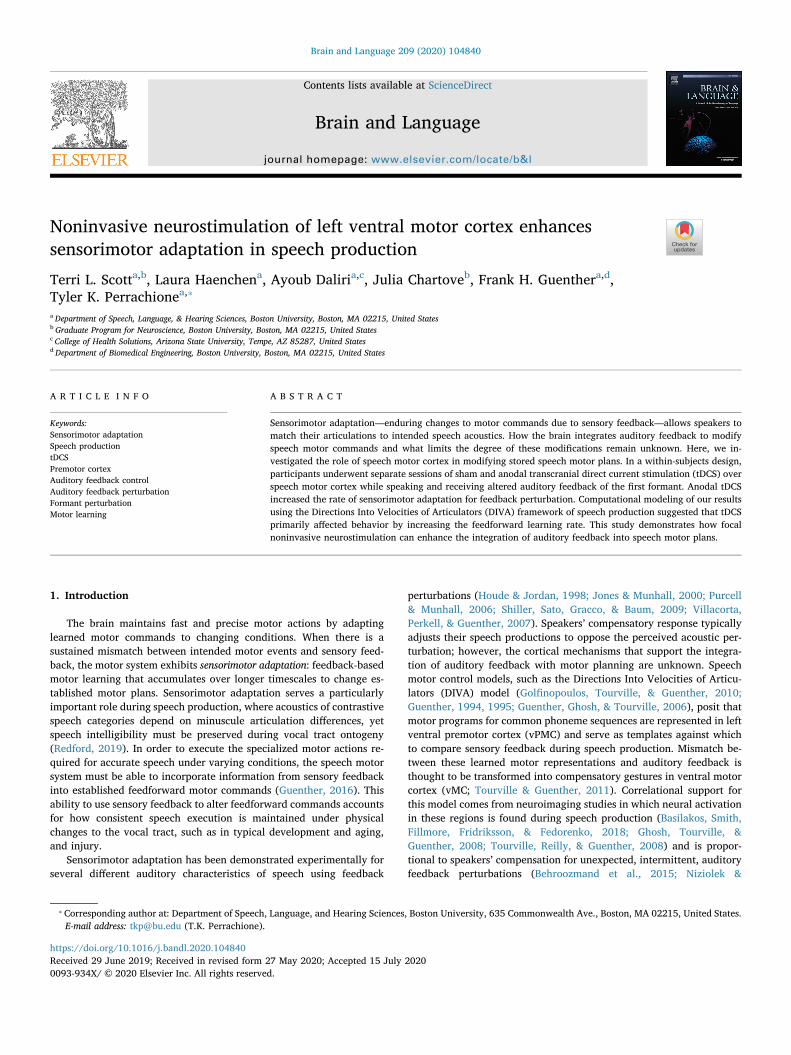
Fig. 1. Paradigm design and tDCS stimulation. (A) During their first visit to thelab, participants underwent an initial session of the experiment without tDCS toconfirm they adapted to auditory perturbations. Over two subsequent visits,they completed the tDCS sessions, with order counterbalanced across partici-pants. (B) Schematic of the equipment setup and behavioral paradigm. (C) Thebehavioral paradigm was the same on each visit. Participants' baseline speechacoustics was measured without perturbation; then F1 perturbation was in-creased to +15% during the ramp phase, held at +30% during the shift phase,and presented again without perturbation during the return phase. (D) Locationof anodes (FC5, C5; red points) and cathodes (AF7, FC1, C1, P5; blue points) inthe 10–10 electrode system (left) and with their location overlaid on the cor-tical surface (middle). Also shown are the locations of the articulator maps(tongue and jaw; yellow points) and speech sound maps (green points) from theDIVA model (Table D.1 in Guenther, 2016, p391). Estimated cortical surfacefield intensity from this stimulation montage is shown at right.
T.L. Scott, et al. Brain and Language 209 (2020) 104840
2
informed consent, approved and overseen by the Institutional ReviewBoard at Boston University, and were paid for their participation.
2.2. Experimental design
Participants completed three sessions in which they underwent thesame behavioral paradigm (Fig. 1A). Each session was separated fromthe previous by at least 7 days to reduce the potential for carry-over oflearning across sessions. In an initial session without tDCS, we con-firmed that participants adapted to auditory feedback perturbation oftheir speech. Participants were then assigned to receive either anodal orsham stimulation during their second session and the other during theirthird session. Nine participants completed each order of stimulation.Although participants were told before each tDCS session that theywould either be receiving active stimulation or be in a control condition(sham), participants were blind not only to which condition they werein on each visit, but also to the fact that they would be in both condi-tions on counterbalanced visits.
2.2.1. Behavioral paradigmEach session was conducted in a sound-attenuated chamber.
Stimulus delivery, recording, and real-time resynthesis for auditoryperturbation were controlled via the Audapter software (Cai, Boucek,Ghosh, Guenther, & Perkell, 2008) implemented in MATLAB vR2014b(The Mathworks, Natick, MA). Participants’ speech was transducedusing a Shure MX153 earset microphone, Behringer Ultragain Pro two-channel microphone amplifier, and Roland Quad Capture sound card.Auditory stimulation was delivered via the same sound card, an ArtHeadAmp6 Pro headphone amplifier, and Etymotic ER-3C insert ear-phones.
Participants were prompted by the Audapter software to say thewords “bed,” “dead,” and “head,” in a pseudorandom order. Theparadigm began with a brief training phase, in which participants re-ceived feedback to insure they were producing the words at a sufficientloudness (72–88 dB SPL) and duration (400–600 ms); trials in thetraining phase were repeated until productions of suitable intensity andduration were achieved. Participants continued to receive feedbackabout the intensity and duration of their speech during the experiment,but trials were not repeated. The Audapter software performed real-time analysis, replay, resynthesis, and recording of participants' speechacoustics (F1 and F2) (Fig. 1B).
The behavioral paradigm consisted of four phases (Fig. 1C). Thebaseline phase consisted of 57 trials in which participants spoke thetarget words and heard their own, unperturbed speech as auditoryfeedback. Next, during the ramp phase, real-time perturbation of par-ticipants’ F1 was introduced at + 15% for 3 trials; the ramp phase wasincluded to reduce participants’ conscious detection of the auditoryperturbation, but was kept brief to allow us to observe continuedlearning during the subsequent shift phase. During the perturbationphase, which lasted for 60 trials, participants heard as auditory feed-back a real-time perturbation of their own speech in which F1 wasincreased by 30%. Finally, during the 60 trials of the return phase,participants again heard their own, unperturbed speech as auditoryfeedback. Auditory feedback was presented at 5 dB SPL above theparticipant’s own productions.
2.2.2. tDCS stimulationNeurostimulation was controlled and delivered using a Soterix MxN
high-definition (HD) tDCS system. HD-tDCS was used both because itoffers more focused stimulation and avoids strong effects of equal andopposite current density in brain areas outside of the region of interest.Stimulating electrodes (2 mA) were placed at FC5 and C5, and returnelectrodes were placed at AF7, FC1, C1, and P5, in a roughly center-surround configuration (Datta et al., 2009; Kuo et al., 2013). Thismontage was selected to optimize field intensity and current flow overleft vPMC and vMC (Fig. 1D), as determined by simulation using the
HD-Explore software (Soterix Medical Inc.; Datta et al., 2009; Huanget al., 2017). These areas were targeted in this study because they arethe theoretical location of the speech sound maps and articulator mapsfor feedforward control of speech production (Guenther, 2016;Tourville & Guenther, 2011).
After insuring the resistance of each channel was < 10 kΩ, anodalstimulation began with a 30-s linear ramp from 0 to 2 mA, with tonic2 mA stimulation continuing for the remainder of the session(~20 min). The procedure for sham stimulation was the same, but afterthe 30-s ramp to 2 mA, stimulation was linearly decreased over 30 sback to 0 mA, where it remained throughout the behavioral paradigm.This procedure effectively blinded participants to whether they werereceiving anodal or sham stimulation during the behavioral task, whichwas begun 60 s after the onset of stimulation (see §3.1, below).Stimulation began before the training phase and ended after the lasttrial of the return phase for a mean duration of 17 min 13 s (range:16 min 36 s − 18 min 8 s). The mean durations of the different phasesof the experiment (excluding the screening session) are as follows:training, 1 min 46 s; baseline, 4 min 54 s; ramp, 15 s; full perturbation,5 min 9 s; and return, 5 min 3 s.
2.3. Statistical analysis
Speech acoustics (mean F1 and F2 frequencies) were obtained fromeach participant on each trial in each condition using Audapter. Vowelformant frequencies were isolated by analyzing 60% of the word’sduration beginning 10% after the onset of voicing. Outlier trials, inwhich F1 deviated by more than two standard deviations from the meanvalue in the respective session and phase, were excluded from theanalysis (< 5% of total trials). In a repeated-measures analysis of var-iance (ANOVA), the number of F1 outliers did not differ as function ofstimulation (no-tDCS, anodal, or sham; F2,34 = 0.29, p = 0.75), phase(baseline, perturbation, return; F2,34 = 0.16, p = 0.85), or their in-teraction (F4,68 = 1.12, p = 0.36). Participants' F1 and F2 measure-ments were then normalized (proportionally) to the mean F1 and F2values obtained during the baseline phase of each session. To controlfor errors in production and automated formant tracking errors, spec-trograms of all trials were visually inspected using the Praat software(Boersma, 2001), and F1 and F2 were measured manually and com-pared to the program’s measured values to insure accurate formantmeasures on each trial.
Speech acoustics data were analyzed in R using linear mixed-effectsmodels implemented in the package lme4 (Bates, Mächler, Bolker, &Walker, 2014). The models’ fixed-effect terms included categoricalfactors for stimulation (anodal vs. sham) and session (2 vs. 3), the mean-centered continuous factor time (trial), and the stimulation × time, sti-mulation × session, session × time, and stimulation × time × sessioninteractions. The models’ random effects terms included by-participantintercepts, by-participant slopes for the fixed factors stimulation, time,and session and by-item intercepts for each word. Statistical compar-isons of model terms were determined via application of deviation-coded contrasts to the model matrix. Significance of main effects andinteractions was determined by adopting a significance criterion ofα = 0.05, with p-values for model terms based on the Satterthwaiteapproximation of the degrees of freedom obtained from the packagelmerTest (Kuznetsova, Brockhoff, & Christensen, 2017).
Additional analysis using nonlinear (exponential) models was con-ducted; however, while the group average perturbation curve is ex-ponential (Fig. 2A), in many cases individual participants' adaptationduring the perturbation phase was not well described by an exponentialfunction (e.g., when participants exhibited no adaptation during theperturbation phase in some condition; see Fig. 3B for individual data).For participants and stimulation conditions where adaptation was evi-dent, linear and exponential adaptation models did not differ in fit.Correspondingly, we chose to model these results using linear mixedeffects models because of their power and precision. Further, we chose
T.L. Scott, et al. Brain and Language 209 (2020) 104840
3
ab initio to employ models with maximal fixed and random effectsstructures, (Barr, Levy, Scheepers, & Tily, 2013), as the purpose of thesemodels was confirmatory hypothesis testing rather than model selection(Meteyard & Davies, 2020). For analyses with only a single value perparticipant per factor level, data were analyzed using repeated-mea-sures ANOVA in the package ez (Lawrence, 2013).
3. Results
3.1. Somatic and psychological experiences related to tDCS
After each tDCS session, participants completed a questionnairedetailing the presence and severity of any symptoms or side effects they
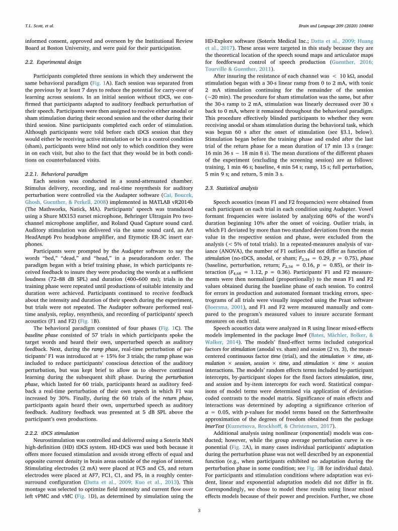
Fig. 2. Speech adaptation under perturba-tion during anodal tDCS vs. sham. (A) Inresponse to a perceived increase in F1 fre-quency during the auditory feedback per-turbation phase of each session, participantscompensated by lowering the F1 frequencyof their own speech productions. The rate ofadaptation under anodal tDCS (orange) wassignificantly enhanced relative to shamtDCS (purple). Blocks represent averagesacross 3 trials (consisting of 1 trial for eachof the three presented words) calculatedwithin participants. Shaded regions indicatestandard error of the mean across partici-pants for each block. Vertical lines indicatethe onset/offset of each phase, with the twolines before the perturbation phase in-dicating the brief ramp phase. (B) Theaverage magnitude of compensatory re-sponses scaled with respect to the full per-turbation (+30% of baseline) during thelatter half of the perturbation phase of theanodal (orange) and sham tDCS sessions(purple). Error bars represent the standarderror of the mean across participants. (C)We measured speakers’ F2 values to test ifsensorimotor adaptation under tDCS wasconfined to F1. We did not observe sys-tematic changes to F2 under anodal tDCS(orange) or sham tDCS (purple) during theperturbation. Shaded regions indicate stan-dard error of the mean across participantsfor each block. (D) The average magnitudeof F2 relative to baseline and scaled by thesame percent factor as F1 in (B) during thelatter half of the perturbation phase of theanodal (orange) and sham tDCS sessions(purple). Error bars represent the standarderror of the mean across participants.
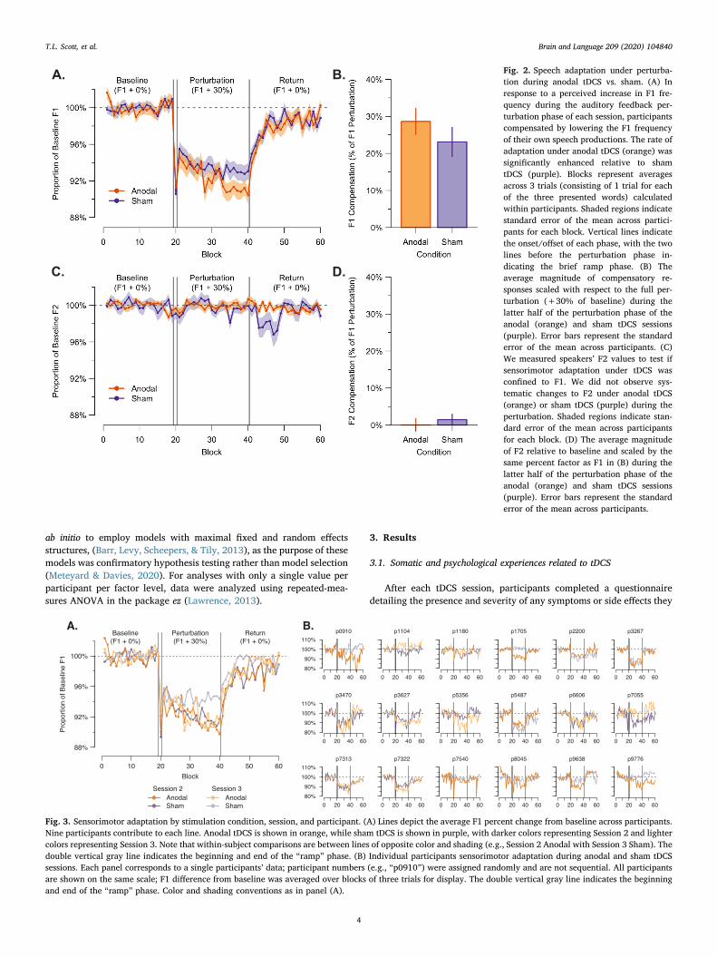
Fig. 3. Sensorimotor adaptation by stimulation condition, session, and participant. (A) Lines depict the average F1 percent change from baseline across participants.Nine participants contribute to each line. Anodal tDCS is shown in orange, while sham tDCS is shown in purple, with darker colors representing Session 2 and lightercolors representing Session 3. Note that within-subject comparisons are between lines of opposite color and shading (e.g., Session 2 Anodal with Session 3 Sham). Thedouble vertical gray line indicates the beginning and end of the “ramp” phase. (B) Individual participants sensorimotor adaptation during anodal and sham tDCSsessions. Each panel corresponds to a single participants’ data; participant numbers (e.g., “p0910”) were assigned randomly and are not sequential. All participantsare shown on the same scale; F1 difference from baseline was averaged over blocks of three trials for display. The double vertical gray line indicates the beginningand end of the “ramp” phase. Color and shading conventions as in panel (A).
T.L. Scott, et al. Brain and Language 209 (2020) 104840
4

experienced, as well as whether they believed these effects to be relatedto the administration of tDCS (Brunoni et al., 2011). Participants filledout identical forms after both sessions, as they were not told whetherthey had received anodal or sham stimulation in each session. Nearly allparticipants reported mild to moderate tingling sensations in both an-odal and sham sessions. Less frequently, participants reported experi-encing pain or burning on their scalp. The prevalence or intensity ofthese sensations did not differ between the anodal and sham conditions,suggesting that participants were effectively blinded to whether theywere receiving active or control stimulation (Fig. S2). We did not in-quire directly as to whether participants thought they had receivedsham or anodal stimulation. Several participants also reported feelingsleepy or distracted, but unlike their somatic experiences, participantsrarely attributed their state of arousal to tDCS.
3.2. Primary outcome measures: Adaptation and recovery
3.2.1. Speech adaptation during perturbationWe determined whether participants' motor adaptation to auditory
F1 perturbation during speech production was affected by tDCS in alinear mixed-effects model of trial-by-trial F1 adaptation magnitude (%of mean baseline F1) during the perturbation phases of the anodal andsham tDCS conditions. A corresponding model was run on F2 acousticsas a control, as auditory feedback of F2 was not perturbed. We omittedthe “ramp” phase of the perturbation from these analyses and focusedonly on the trials for which the feedback perturbation remained con-stant.
We observed a significant main effect of time, such that adaptation(the lowering of F1) increased over the perturbation period(β = −0.0005, s.e. = 0.0001, t = −4.65, p = 0.0003) in all condi-tions. Importantly, there was a significant stimulation × time interactionsuch that participants showed greater adaptation with time under an-odal stimulation than sham (Fig. 2A; β = 0.0002, s.e. = 0.0001,t = 2.99, p = 0.003). We also observed a significant time × sessioninteraction such that participants showed a greater rate of adaptationduring session 2 vs. session 3 (Fig. 3A; β = −0.0001, s.e. = 0.0001,t = −2.40, p = 0.02). We did not observe a main effect of stimulation(β = 0.0061, s.e. = 0.0058, t = 1.06, p = 0.31) when considering theentire perturbation period. No significant effect of session, no stimula-tion × session interaction, and no three-way interaction was observed(see Table S1). Speakers’ F1 during the latter half of the perturbationphase under anodal stimulation was 91.4%±4.5% that of the baseline,whereas under sham stimulation it was 93.1 ± 5.0% of baseline,corresponding to compensation of 28.7% and 23.0% of the full auditoryperturbation, respectively (Fig. 2B). Individual participant data forsham and anodal tDCS sessions are given in supplemental Fig. 3B.
The corresponding model of F2 showed no effects of stimulation(Fig. 2C; β = −0.0007, s.e. = 0.0027, t = −0.26, p = 0.80), time(β = 1.6 × 10⁻⁵, s.e. = 0.0001, t = 0.22, p = 0.83), or session(Fig. 3A; β = 0.0041, s.e. = 0.0027, t = 1.53, p = 0.15) on this un-perturbed feature, and no significant interactions (Table S2). Mean F2values during the latter half of the perturbation phase were100.0%±2.2% of baseline during anodal tDCS and 99.6%±2.0%during sham (Fig. 2D).
3.2.2. Speech recovery following perturbationWe also analyzed whether participants' motor recovery after re-
moval of auditory perturbation of F1 was affected by tDCS using alinear mixed-effects model of trial-by-trial F1 difference from baselineduring the return phases of the anodal and sham tDCS conditions. Thismodel included the same fixed and random factors described in §3.2.1and modeled trials beginning with the last trial in which the auditoryperturbation was presented through the end of the session. The timefactor was centered on its mean value.
We observed a significant effect of time as participants’ F1 valuesgradually returned to baseline (Fig. 2A; β = 0.0006, s.e. = 0.0001,
t = 5.82, p ≪ 0.0001). We did not observe significant effects of sti-mulation or session, nor any significant interactions between factors(Table S3). We ran the corresponding model on the participants’ F2values during the return phase and found significant interactions be-tween stimulation and time (Fig. 2C; β = 0.0002, s.e. = 0.0001,t = 3.65, p = 0.0002) and stimulation and session (β = −0.0086,s.e. = 0.0027, t = −3.13, p = 0.006). (These results appear to havebeen driven primarily by one participant who exhibited substantialinconsistency in their F2 productions during their sham session;whereas most participants showed no effect of stimulation, perturba-tion, or its withdrawal on F2 productions.)
3.3. Secondary outcomes and control measures
3.3.1. Consistency in individual differences in compensation acrossconditions
We investigated whether the average magnitude of adaptationduring the latter half of the perturbation phase (where productiontargets were expected to most approach stability) was consistent withinparticipants across the stimulation conditions using Spearman’s rankcorrelation. Participants' F1 adaptation in the initial session withouttDCS was not significantly correlated with their adaptation during shamstimulation (r = 0.41, p = 0.09) or during anodal stimulation(r = 0.39, p = 0.11). The magnitude of adaptation was also not sig-nificantly correlated between sham and anodal stimulation conditions(r = 0.42, p = 0.09).
Additionally, we investigated whether participants’ magnitude ofadaptation was correlated across sessions regardless of stimulationcondition. F1 adaptation was significantly correlated between thescreening session and the second session (r= 0.55, p= 0.02), while thecorrelation between screening and the third session was not significant(r = 0.45, p = 0.06). We ran a post-hoc analysis to better understandwhether these two last correlations were significantly different fromeach other due to the fact that they fell on either side of our significancecriterion, and found that they were not (Pearson and Filon’s z = 0.48,p = 0.31, implemented in the R package cocor; Diedenhofen & Musch,2015).
3.3.2. Speech production variability under tDCSWe also investigated whether the coefficient of variation (s/x̄ ; a
measure of instability obtained from speech variability across in-dividual trials) for participants' F1 differed as a function of stimulation(anodal, sham stimulation) during each phase of the experiment(baseline, perturbation, return). We limited analysis to the latter half ofthe perturbation and return phases to avoid biased coefficients of var-iation resulting from effects related to the initial administration andcessation of auditory perturbation. In a repeated-measures ANOVA ofthe coefficient of variation of F1 with within-subject factors of stimu-lation and phase, we found a significant effect of stimulation(F1,17 = 5.17, p = 0.04, η2G = 0.05) such that the coefficient of var-iation tended to be greater under anodal than sham stimulation, noeffect of phase (F2,34 = 1.55, p = 0.23, η2G = 0.02), and no stimula-tion × phase interaction (F2,34 = 0.52, p = 0.60, η2G = 0.01). Thisanalysis was repeated for participants’ coefficient of variation of F2 inwhich we found no significant effects of stimulation (F1,17 = 1.42,p= 0.25, η2G = 0.01), phase (F2,34 = 0.59, p= 0.56, η2G = 7.3 × 10⁻⁴),and no stimulation × phase interaction (F2,34 = 0.32, p = 0.72,η2G = 5.6 × 10⁻⁴).
3.3.3. Speech production baseline under tDCSTo determine whether the application of tDCS had an effect on
speech production acoustics independent of the perturbation manip-ulation, we performed a series of linear-mixed effects models testingwhether speakers' F1 frequency during the baseline phase was affectedby session (1, 2, or 3) and stimulation (no-tDCS, anodal, or sham).
We first tested a linear mixed-effects model including a categorical
T.L. Scott, et al. Brain and Language 209 (2020) 104840
5

fixed factor for all levels of session, random slopes and intercepts byparticipant, and random item intercepts. (The stimulation factor was notincluded in this model because, for all participants, the first session didnot involve any tDCS, meaning these levels of the two factors wereperfectly colinear, and a model including both together would be rankdeficient.) We found a significant difference in baseline F1 productionsbetween the two earlier sessions (Session 2 – Session 1; β = −10.67,s.e. = 4.20, t = −2.54, p = 0.02), whereas we did not find any dif-ference in the latter two sessions (Session 3 – Session 2; β = −4.12,s.e. = 8.28, t = −0.50, p = 0.63). F1 values across participants werehighest during the first visit (701 ± 95 Hz), lower during the secondvisit (690 ± 97 Hz), and lowest during the third visit (686 ± 97 Hz).The direction of this learning effect (lowered F1 values across sessions)is consistent with long-term retention of adaptation for the auditoryperturbation (raised F1 values) in this study.
Additionally, we tested whether the order of stimulation sessionsaffected baseline F1 frequency; e.g., if receiving anodal stimulationduring Session 2 was associated with a greater change in F1 baseline atSession 3. In an ANOVA on a second model including only the secondand third session baselines, with categorical fixed factors includingsession (2 vs. 3), stimulation (anodal vs. sham), and their interaction, andrandom factors including by-participant intercepts and by-participantslopes for the effect of stimulation, we found no effect of session(F1,16 = 0.22, p = 0.65), stimulation (F1,16 = 0.0047, p = 0.84) and nostimulation by session interaction (F1,16 = 0.0013, p = 0.97), suggestingthe tDCS manipulation did not affect learning across sessions.
3.3.4. Amount of compensation to initial perturbation trialThe primary outcome measures indicated that anodal tDCS had an
effect on the rate of adaptation during the perturbation phase; however,the prior literature distinguishes compensatory (or reflexive) responsesto unexpected perturbation from adaptive responses to ongoing sen-sorimotor mismatch that involves modifications to feedforward com-mands (Burnett et al., 1998; Guenther, 2016). We therefore also in-vestigated whether reflexive response to the initial application ofperturbed auditory feedback on the first trial of the ramp phase differedbetween conditions. In a repeated measures ANOVA of F1 compensa-tion on the first perturbation trial, with stimulation (anodal vs. sham) asthe within-subjects factor, we found no effect of tDCS on the responsemagnitude to initial perturbation (F1,17 = 0.04, p = 0.84, η2G = 0.002;anodal: 94.0%±8.3%; sham: 94.5%±5.3%). In a correspondinganalysis of session (1 vs. 2 vs. 3) we observed a trend for the magnitudeof the reflexive response on the first perturbation trial to decrease as afunction of experience with the task (Session 1: 91.8%±7.3% ofbaseline; Session 2: 92.9%±7.9%; Session 3: 95.7%±5.6%); how-ever, this trend was not statistically significant (F1,17 = 3.62, p = 0.07,η2G = 0.18). Finally, reflexive response magnitude to the initial per-turbation trial was not correlated with speakers' overall adaptationmagnitude, either during the initial visit (r= −0.19, p = 0.44), anodaltDCS (r = 0.11, p = 0.67), or sham tDCS (r = 0.22, p = 0.36).
4. Computational modeling
4.1. Model description
Several distinct motor control mechanisms can contribute to com-pensatory responses during motor adaptation under sensory perturba-tions (Scott, 2004; Shadmehr, Smith, & Krakauer, 2010). To investigatewhich aspects of motor learning and performance were responsible forchanges in adaptive responses under neurostimulation, we performedcomputer simulations using SimpleDIVA (Kearney et al., 2020)—asimplified version of the DIVA model (Guenther et al., 2006; Guenther,2016) that characterizes the neural computations involved in speechmotor control. The SimpleDIVA model is designed to capture the ag-gregate contributions of DIVA model’s auditory feedback control, so-matosensory feedback control, and feedforward control subsystems to
speech acoustics, without needing to model each system’s variouscomponents in detail. Further, rather than modeling the configurationand trajectory of the various vocal tract articulators in detail, Simple-DIVA abstracts motor control to the realized acoustic output, here F1frequency. In this implementation, SimpleDIVA accounts for trial-by-trial changes in speech acoustics by estimating the aggregate con-tributions of these three subsystems to speech acoustics during sen-sorimotor adaptation experiments.
The first mechanism that contributes to compensatory responses isthe auditory feedback control subsystem of the speech motor controller.This subsystem translates production errors detected via the auditorysystem into corrective movements, with a latency of approximately100–200 ms from error/perturbation onset to the start of the correctivemovement. We will refer to this within-trial component of the com-pensatory response as the reflexive response, borrowing terminologyfrom Larson and colleagues (Burnett et al., 1998; Hain et al., 2000),while noting that this “reflex” involves processing in the cerebral cortex(Tourville et al., 2008). The term auditory feedback control gain (αA) willbe used to describe the size of this response relative to the size of theauditory error. A gain of 1 would indicate that the auditory feedbackcontrol system is completely counteracting the perturbation, but inactuality the auditory feedback control gain appears to be muchsmaller, with prior studies indicating a compensatory response that istypically less than 25% of the size of the perturbation (e.g., Burnettet al., 1998; Chen, Lui, Xu, & Larson, 2007; Tourville et al., 2008;Niziolek & Guenther, 2013). Compensatory responses to auditory per-turbations have the effect of generating somatosensory feedback that nolonger matches the motor system’s expectations (somatosensory target)for the speech gesture. This will invoke somatosensory feedback controlmechanisms that tend to counteract the compensatory response. Thesize of the somatosensory feedback controller’s opposition to the com-pensatory response will depend on the somatosensory feedback controlgain (αS).
If the perturbation is sustained over many productions of the samesound, a second mechanism is invoked by the motor system to coun-teract the perturbation: trial-to-trial adaptation of the feedforwardcommand, or stored “motor program.” We will refer to this as theadaptive response, the size of which is modulated by the feedforwardcommand learning rate (λFF). Thus, in the terminology used here, thecompensatory response to a sustained perturbation is composed of areflexive response and an adaptive response.
The following equations used in the current simulations capture thekey aspects of the DIVA model in a simplified form (Kearney et al.,2020) that involves only three free parameters (αA, αS, and λFF),thereby eliminating redundancies in the set of fitting parameters thatwould otherwise obfuscate the neural mechanisms underlying com-pensation since such redundancies can lead to multiple parameter va-lues that produce equivalent fits to the data. Eq. (1) defines the value ofF1 produced by the subject on a given trial (indexed by n) as:
= +F1 (n) F1 (n) ΔF1 (n)produced FF FB (1)
In words, the F1 value produced on a trial is a combination of afeedforward command (F1FF) and a sensory feedback-based correction(ΔF1FB) that is initiated if/when the auditory and somatosensoryfeedback controllers detect production errors on the current trial. At thestart of each simulation, F1FF is initialized to the average F1 measuredduring the baseline phase of the experiment across participants. Eq. (2)defines the feedback-based correction as:
= × − + × −α αΔF1 (n) (F1 F1 (n)) (F1 F1 (n))FB A AT perceived S ST FF (2)
where F1AT and F1ST are the F1 values specified by previously learnedauditory and somatosensory targets, respectively, for the vowel;F1perceived is the value of F1 heard by the subject (including the per-turbation, when one is applied) before feedback control mechanismskick in on that trial (i.e., F1perceived = F1FF(n) + perturbation size); andαA and αS are the gains of the auditory and somatosensory feedback
T.L. Scott, et al. Brain and Language 209 (2020) 104840
6

control subsystems, respectively. In the simulations, F1AT and F1ST areset to the average F1 of the baseline phase, corresponding to the as-sumption that the auditory and somatosensory targets will not changesubstantially over the course of the experiment. Eq. (3) describes theprocedure for updating the feedforward command from trial to trial:
+ = + ×λF1 (n 1) F1 (n) ΔF1 (n)FF FF FF FB (3)
where λFF is a learning rate parameter for the feedforward command. Inwords, the feedforward command for the next trial is updated by addingsome fraction (characterized by λFF) of the feedback-based correctivecommand for the current trial.
To fit the model to the data from the sham and anodal stimulationconditions, a particle swarm optimization procedure was used to findoptimized values of the three free parameters of the model (αA, αS, andλFF) to fit the mean data for each block in each condition. The para-meter estimates resulting from this procedure were highly robust toinitial conditions, indicative of reaching the global minimum of the rootmean square error (RMSE) measure.
Additionally, we examined the fit to our data of an alternative state-space model previously used to estimate and quantify learning andsensitivity to errors during motor learning (Galea, Mallia, Rothwell, &Diedrichsen, 2015; Huberdeau, Krakauer, & Haith, 2015; Smith,Ghazizadeh, & Shadmehr, 2006; Thoroughman & Shadmehr, 2000).This model yielded qualitatively similar results, which are included inthe Supplemental Materials (Figure S3).
4.2. DIVA model fits
The DIVA model fits to the two experimental conditions are pro-vided in Fig. 4A. In both cases, the model fit falls within the standarderror of the sample mean for all blocks except the ramp block (block 20)and immediately after cessation of auditory feedback perturbation(sham: fit normalized RMSE = 0.01; Pearson’s r = 0.93; anodal: fitnormalized RMSE = 0.01; r = 0.95).
Table 1 compares the model parameter values for the two stimu-lation conditions. Whereas the values for the auditory feedback controlgain, αA, are nearly the same for the two conditions (αA = 0.172 duringsham, αA = 0.174 during anodal stimulation, an increase of 1%), thesomatosensory feedback control gain, αS, decreased by 18% fromαS = 0.372 in the sham condition to 0.304 in the anodal stimulationcondition, and the value of the trial-to-trial feedforward commandlearning rate λFF increased by 63% in the anodal stimulation condition(λFF = 0.194) compared to the sham condition (λFF = 0.119). This is
also shown graphically in Fig. 4B. (To illustrate how the values of thefree-parameters of this model affect the slope and magnitude of sen-sorimotor adaptation, three series of simulations in which only oneparameter varies at a time are visualized in Fig. S4.)
5. Discussion
The results of this study extend our understanding of the mechan-isms through which speakers learn to adjust their feedforward motorplans in response to perturbed sensory feedback during speech pro-duction. When applying noninvasive neurostimulation over left ventralsensorimotor cortex, we observed an increased rate of adaptation re-sponses to perturbed auditory feedback. Moreover, we found that thiseffect was specific to F1—the perturbed feature—and did not generalizeto F2, indicating a task-specific effect rather than a global modulationof motor control processes.
The rate of increasing adaptive responses potentially depends onboth the gain of the auditory feedback control subsystem for speech,which is responsible for within-trial reflexive responses to perceivedauditory errors, and the rate of learning of feedforward commands,which is responsible for trial-to-trial increases in the anticipatorycomponent of the compensatory response. Because the mechanisms bywhich tDCS affects cortical activity are uncertain and may be specific toa study’s particular task (see Bortoletto, Pellicciari, Rodella, & Miniussi,2015), theoretically either error sensitivity, cortical plasticity, or bothcould have been modulated during anodal stimulation. We thereforeutilized computational simulations using a simplified version of an es-tablished model of speech motor control, the DIVA model (Kearneyet al., 2020), to decompose the adaptation responses into distinct,mechanistically precise components. Specifically, we extracted
Fig. 4. SimpleDIVA model fits to behavioral data. (A) Solid lines depict the best-fit models identified by SimpleDIVA model simulations for both anodal tDCS (orange)and sham tDCS (purple). The shaded regions indicate the standard errors around the mean for the behavioral data, shown here for comparison with the models. (B)The percent change of our free parameter estimates is shown for anodal stimulation with respect to sham stimulation. The auditory gain factor (αA) is shown in green,somatosensory gain factor (αS) in blue, and the learning coefficient (λFF) is in red.
Table 1SimpleDIVA model best fit parameter estimates. Best fit parameter values formodel simulations of the sham and anodal tDCS conditions. No differencesbetween the two conditions was found for auditory feedback control gain (αA),while somatosensory feedback control gain (αS) decreased during anodal tDCSand feedforward learning/adaptation rate (λFF) increased under anodal tDCSrelative to sham stimulation.
Parameter Sham tDCS Estimate Anodal tDCS Estimate Difference
αA 0.172 0.174 +1.15%αS 0.372 0.304 −18.28%λFF 0.119 0.194 +63.03%
T.L. Scott, et al. Brain and Language 209 (2020) 104840
7

estimates of three key parameters characterizing the main controlsubsystems of the speech motor controller—the auditory feedbackcontrol gain, the somatosensory feedback control gain, and the feed-forward command learning/adaptation rate—under anodal tDCS andsham stimulation. These simulations indicated that stimulation resultedin an increase in the feedforward learning rate, whereas the auditoryfeedback control gain was essentially unaffected by the perturbation.Thus, the performance gains resulting from stimulation were pre-sumably due to increased adaptation of the feedforward commands forsubsequent productions rather than increased within-trial reflexive re-sponses by the auditory feedback controller.
Furthermore, best-fit models also included a small, unanticipateddecrease in the gain of the somatosensory feedback control subsystem.While a change in somatosensory feedback control gain may initially besurprising given the auditory perturbation used in this study, a changein this parameter makes sense when its role is considered in context ofthe feedback control system in aggregate: Under normal circumstances,the somatosensory control subsystem counteracts compensatory adjust-ments to auditory perturbations, because these adjustments have theeffect of producing somatosensory feedback that mismatches the so-matosensory target for a given articulation. Teleologically, decreasingthe gain of the somatosensory feedback control subsystem reduces thiscounteraction, allowing for more complete motor adaptation to theauditory perturbation, as seen in the larger magnitude of adaptationunder anodal tDCS (Fig. 2B). Mechanistically, however, the source ofthe change in the somatosensory feedback control gain is less certain.This change may reflect the relatively limited spatial resolution of tDCS,in that our electrode montage also likely resulted in stimulating currentto left ventral somatosensory cortical areas in postcentral gyrus(Fig. 1D)—including tissue comprising somatosensory state, target, anderror maps (Guenther, 2016)—in addition to speech motor controlareas in left vPMC and vMC. Given the spatial proximity of motor andsomatosensory cortex, identifying the causal mechanisms that affect theintegration of somatosensory information will require stimulation ap-proaches with greater spatial specificity. For instance, noninvasivetechniques such as TMS, and invasive techniques such as corticalcooling, have been used to make finer-grained functional dissociationsbetween adjacent perisylvian neuroanatomy (e.g., Pulvermüller et al.,2006; Long et al., 2016), and applying computational modeling to thebehavioral changes measured under more targeted stimulation mayoffer insight into mechanistic changes to sensory feedback gain control.
Previous work on sensorimotor adaptation during auditory feedbackperturbation indicated that individual differences in participants’ au-ditory acuity, or ability to detect feedback errors, explained a sig-nificant portion of the variance in the degree of adaptation measuredacross subjects (Ghosh et al., 2010; Villacorta et al., 2007). Here, weshow that it is not auditory error detection that increases under sti-mulation of ventral sensorimotor cortex, but rather the motor adapta-tion rate. This is not to say that auditory acuity does not play a role inadaptation, but that the areas we stimulated do not appear to mediateauditory acuity or error detection; instead, these areas must support, atsome level, updating of stored motor programs for speech sounds,consistent with the DIVA model. The DIVA model also predicts that, incontrast with left vPMC, right vPMC is responsible for transformingauditory and somatosensory error signals into corrective motor com-mands. We therefore hypothesize that if the right vPMC were stimu-lated using anodal tDCS, we would see modulation of auditory andsomatosensory feedback control gains but not the feedforward com-mand adaptation rate. Testing these predictions must be the goal offuture work that compares differences in reflexive vs. adaptive re-sponses under right- vs. left-hemisphere stimulation.
It is important to note that while the results of this study are con-sistent with a model in which left sensorimotor cortex plays a causalrole in sensorimotor adaptation, the evidence presented here is by itselfnot sufficient to establish the unique causal involvement of this region,as we did not test for effects of anodal stimulation on some other
putatively unrelated control region. It may be the case that anodalstimulation to any region of brain increases the rate learning. However,while both empirical (Huang et al., 2017) and modelling (Datta, Zhou,Su, Parra, & Bikson, 2013) work on the physiological effects of targetedtranscranial electrical stimulation suggest that these can be quite focaldepending on local field strength, further work remains necessary toestablish the causal contribution of this or other cortical areas in sen-sorimotor adaptation.
Further support for the view that anodal tDCS of left sensorimotorcortex affects learning rate and not error detection is the lack of dif-ferences between anodal and sham stimulation on compensation mag-nitude during the first perturbation trial. If anodal stimulation en-hanced error detection during compensation, we might have observedthose differences earlier in the perturbation phase of the session, duringa period in which compensation should be dominated by reflexive re-sponses. A previous study employing anodal tDCS over either motorcortex or cerebellum during visuomotor adaptation by Galea, Vazquez,Pasricha, Orban de Xivry, and Celnik (2011) showed early effects oncompensation when stimulation was applied to cerebellum, but sig-nificant after-effects when stimulation was applied to motor cortex,providing further evidence that motor cortex supports adaptive re-sponses. Lametti, Smith, Freidin, and Watkins (2017) also reporteddistinct roles for motor cortex and cerebellum in a similar auditoryfeedback perturbation study where tDCS was applied to either brainregion. In order to better understand the extent of the dissociationbetween the neural mechanisms supporting adaptive and reflexive re-sponses, future studies are needed to directly compare the effects oftDCS on sensorimotor adaptation to those during unexpected/randomfeedback perturbations, in which adaptive, but not reflexive, responsesshould be reduced. Furthermore, studies directly comparing reflexiveand adaptive processes can be used to inform modifications to thesimplified DIVA model to better capture participants’ behavior duringtransitional periods during the present paradigm, given that our modelwas least successful estimating behavior during the ramp and beginningof the return phase.
We observed a significant effect of anodal tDCS on F1 trial-to-trialvariability compared to sham. Because we did not find any interactionbetween stimulation condition and phase of each session, we mightconclude that anodal tDCS caused increased variability that was notrelated to the increased feedforward learning rate. However, the factthat we did not see a corresponding difference in F2 productionvariability suggests instead that the effects on F1 are related to learning,and perhaps the differences with phase of the experiment are too smallto observe at our current power. We performed post-hoc pairedStudent’s t-tests on the degree of variability during anodal and shamtDCS for each phase and observed that, whereas the baseline phaseshowed little difference between stimulation conditions (two-tailed;t17 = 0.57, p = 0.58, Cohen’s d = 0.20), the perturbation and returnphases showed differences that trended in the direction of highervariability during anodal tDCS than sham (perturbation: t17 = 2.03,p = 0.06, d = 0.54; return: t17 = 1.92, p = 0.07, d = 0.67). Before wecan draw conclusions about how variability and learning might be re-lated in this paradigm, we may need to better understand speech motorvariability under tDCS without auditory feedback perturbations, whichis as of yet, unstudied.
In addition to the sensorimotor adaptation we observed during theperturbation phase of each session, we also recorded a significantdownward shift in baseline F1 after the first session, despite requiring aminimum of 7 days between visits to the lab. Previous work from Healdand Nusbaum (2015) found remarkable day-to-day consistency in theacoustics of speakers’ vowel productions and so our observation is no-table, especially given that the perturbation periods lasted only ap-proximately five minutes per session. We did not find any interactionbetween session number and stimulation condition; therefore, we donot have evidence that increased adaptation during anodal tDCS hadany long-term effects on speech production beyond repeating the
T.L. Scott, et al. Brain and Language 209 (2020) 104840
8
behavioral task. This raises the possibility that behavioral interventionswith repeated sensory feedback perturbation may be useful in trainingor retraining target outputs in speech motor learning, such as in thecase of second language learning, vocal accent or gender modification,or speech motor recovery following pathology. However, we also noteda reduced rate of adaptation in the third session compared to thesecond. While this session-by-time effect did not interact with stimu-lation manipulation, it indicates that participants’ susceptibility to theperturbation manipulation itself may differ when undergoing repeatedtreatments. Whether this is due to the accumulation of changes ob-served in baseline speech production targets measured at the thirdsession, decreased sensitivity to auditory perturbations, or some otherfactor remains a question for future research. Indeed, before we canassess the applied or clinical utility of such paradigms, future work willneed to assess individual consistency in rate and magnitude of speechmotor adaptation over multiple sessions, without involving tDCS. Thiswill also provide important information about how we might control forsession-to-session learning in within-subjects experimental designsmore completely.
Approximately two thirds of participants who completed thescreening session of our study showed significant adaptation during theauditory feedback perturbation. We chose to focus the stimulation as-pect of the study on those who showed adaptation because variability inthis behavior has been documented (e.g., Purcell & Munhall, 2006) buthas yet to be successfully explained. There are several hypotheses as towhy certain people do not adapt, such as inability to perceive theperturbation due to poor auditory acuity (Ghosh et al., 2010) or astronger adherence to somatosensory speech targets than auditory ones.It may be useful to perform future studies with these participants todetermine their auditory acuity and to see how their behavior is af-fected by tDCS. Given the results of our model simulations, it is possiblethat anodal tDCS to left ventral sensorimotor cortex could cause non-adapting participants to depend less on their somatosensory feedbackand therefore increase the magnitude of their adaptation responses.However, even within our adapting participants, we observed in-dividual differences in behavior. Given that some individuals adaptmore than others, we tested for correlations in adaptation magnitudeacross conditions and sessions. We found a significant correlation be-tween sessions 1 and 2, and not between 1 and 3; however, the nu-merical difference between these two correlations was small, and so wedo not feel we have sufficient evidence to say whether or not individualdifferences in overall adaptation magnitude are demonstrated by ourresults. Though most of our participants showed increased adaptationduring anodal stimulation relative to the sham condition, few showedeither no effect of tDCS or an opposite pattern of behavior. Some ofthese differences may be attributed to high variability in speech pro-duction across trials and sessions, but we cannot rule out the possibilitythat tDCS affects some people differently (e.g., Schall et al., 2015). As arelatively new technology, more work is needed to better understandsources of behavioral variability under tDCS.
In summary, participants showed increased sensorimotor adapta-tion under anodal tDCS to left ventral sensorimotor cortex during per-turbed auditory feedback, demonstrating the ability of noninvasivebrain stimulation to enhance how speakers learn to integrate sensoryfeedforward and feedback speech motor commands to modify storedmotor programs for speech. Through computational modeling, we wereable to verify the effects of anodal tDCS on sensorimotor learning andgain insights into the cortical mechanisms that limit adaptation to on-going perceived auditory errors. The results of this study further ourknowledge of the cortical mechanisms supporting the speech motorsystem’s ability to adapt in response to altered sensory feedback.Additionally, these findings have implications for understanding how toeffectively deploy tDCS as both a research instrument and a therapeutictechnique in treatment of speech motor control issues involving ab-normal feedback-based adaptation, such as stuttering (Cai et al., 2012;Chesters, Möttönen, & Watkins, 2018; Daliri, Weiland, Cai, Guenther, &
Chang, 2017), aphasia (Behroozmand et al., 2018), and Parkinson’sdisease (Abur et al., 2018).
6. Statement of significance
Accurate speech production requires adjusting speech motor com-mands via sensory feedback. To compensate for sensorimotor mis-match, speakers update feedforward motor commands to produce in-tended speech acoustics. Applying tDCS over left ventral motor cortexwhile speaking accelerates adaptation to perturbed auditory feedback,an effect derived primarily by enhancing the feedforward learning ratefor sensorimotor integration compared to sham stimulation.
Acknowledgments
We thank Jason Tourville, Alfonso Nieto-Castañón, Shanqing Cai,Sara Dougherty, Jennifer Golditch, Elly Hu, Cecilia Cheng, EmilyThurston, and Ja Young Choi. This research was supported by NIDCD ofthe NIH under award numbers R03DC014045 to TP, and R01DC002852to FG. TLS was supported by T90DA032484. The content is solely theresponsibility of the authors and does not necessarily represent theofficial views of the National Institutes of Health.
Appendix A. Supplementary material
Supplementary data to this article can be found online at https://doi.org/10.1016/j.bandl.2020.104840.
References
Abur, D. A., Lester-Smith, R. A., Daliri, A., Lupiani, A. A., Guenther, F. H., & Stepp, C. E.(2018). Sensorimotor adaptation of voice fundamental frequency in Parkinson’sdisease. PLoS ONE, 13, Article e0191839.
Barr, D. J., Levy, R., Scheepers, C., & Tily, H. J. (2013). Random effects structure forconfirmation hypothesis testing: Keep it maximal. Journal of Memory and Language,68, 255–278.
Basilakos, A., Smith, K. G., Fillmore, P., Fridriksson, J., & Fedorenko, E. (2018).Functional characterization of the human speech articulation network. CerebralCortex, 28, 1816–1830.
Bates, D., Mächler, M., Bolker, B., & Walker, S. (2014) Fitting linear mixed-effects modelsusing lme4. arXiv preprint arXiv:1406.5823.
Behroozmand, R., Phillip, L., Johari, K., Bonilha, L., Rorden, C., Hickok, G., & Fridriksson,J. (2018). Sensorimotor impairment of speech auditory feedback processing inaphasia. Neuroimage, 165, 102–111.
Behroozmand, R., Shebek, R., Hansen, D. R., Hiroyuki, O., Robin, D. A., Howard, M. A.,III, & Greenlee, J. D. W. (2015). Sensory-motor networks involved in speech pro-duction and motor control: An fMRI study. Neuroimage, 109, 418–428.
Boersma, P. (2001). Praat, a system for doing phonetics by computer. Glot International, 5,341–345.
Bortoletto, M., Pellicciari, M. C., Rodella, C., & Miniussi, C. (2015). The interaction withtask-induced activity is more important than polarization: A tDCS study. BrainStimulation, 8, 269–276.
Brunoni, A. R., Amadera, J., Berbel, B., Volz, M. S., Rizzerio, B. G., & Frengi, F. (2011). Asystematic review on reporting and assessment of adverse effects associated withtranscranial direct current stimulation. International Journal ofNeuropsychopharmacology, 14, 1133–1145.
Buchwald, A., Calhoun, H., Rimikis, S., Lowe, M. S., Wellner, R., & Edwards, D. J. (2019).Using tDCS to facilitate motor learning in speech production: The role of timing.Cortex, 111, 274–285.
Burnett, T. A., Freedland, M. B., Larson, C. R., & Hain, T. C. (1998). Voice f0 responses tomanipulations in pitch feedback. Journal of the Acoustical Society of America, 103,3153–3161.
Cai, S., Boucek, M., Ghosh, S. S., Guenther, F. H., & Perkell, J. S. (2008). A system foronline dynamic perturbation of formant trajectories and results from perturbations of theMandarin triphthong /iau/. Presented at the eighth international seminar on speech pro-duction.
Cai, S., Beal, D. S., Ghosh, S. S., Tiede, M. K., Guenther, F. H., & Perkell, J. S. (2012).Weak responses to auditory feedback perturbation during articulation in persons whostutter: Evidence for abnormal auditory-motor transformation. PLoS ONE, 7, Articlee41830.
Chen, S. H., Lui, H., Xu, Y., & Larson, C. R. (2007). Voice F0 responses to pitch-shiftedvoice feedback during English speech. Journal of the Acoustical Society of America,121, 1157–1163.
Chesters, J., Möttönen, R., & Watkins, K. E. (2018). Transcranial direct current stimula-tion over left inferior frontal cortex improves speech fluency in adults who stutter.Brain, 141, 1161–1171.
T.L. Scott, et al. Brain and Language 209 (2020) 104840
9

Daliri, A., Weiland, E. A., Cai, S., Guenther, F. H., & Chang, S. E. (2017). Auditory-motoradaptation is reduced in adults who stutter but not in children who stutter.Developmental Science, 21, Article e12521.
Datta, A., Bansal, V., Diaz, J., Patel, J., Reato, D., & Bikson, M. (2009). Gyri-pricise headmodel of transcranial direct current stimulation: Improved spatial focality using aring electrode versus conventional rectangular pad. Brain Stimulation, 2, 201–207.
Datta, A., Zhou, X., Su, Y., Parra, L. C., & Bikson, M. (2013). Validation of finite elementmodel of transcranial electrical stimulation using scalp potentials: Implications forclinical dose. Journal of Neural Engineering, 10, Article 036018.
Dayan, E., Censor, N., Buch, E. R., Sandrini, M., & Cohen, L. G. (2013). Noninvasive brainstimulation: From physiology to network dynamics and back. Nature Neuroscience, 16,838–844.
Diedenhofen, B., & Musch, J. (2015). cocor: A comprehensive solution for the statisticalcomparison of correlations. PLoS ONE, 10(4), Article e0121945.
Fertonani, A., Rosini, S., Cotelli, M., Rossini, P. M., & Miniussi, C. (2010). Naming fa-cilitation induced by transcranial direct current stimulation. Behavioural BrainResearch, 208, 311–318.
Filmer, H. L., Dux, P. E., & Mattingley, J. B. (2014). Applications of transcranial directcurrent stimulation for understanding brain function. Trends in Neurosciences, 37,742–753.
Galea, J. M., Mallia, E., Rothwell, J., & Diedrichsen, J. (2015). The dissociable effects ofpunishment and reward on motor learning. Nature Neuroscience, 18, 597–602.
Galea, J. M., Vazquez, A., Pasricha, N., Orban de Xivry, J. J., & Celnik, P. (2011).Dissociating the roles of the cerebellum and motor cortex during adaptive learning:The motor cortex retains what the cerebellum learns. Cerebral Cortex, 21, 1761–1770.
Ghosh, S. S., Tourville, J. A., & Guenther, F. H. (2008). A neuroimaging study of premotorlateralization and cerebellar involvement in the production of phonemes and sylla-bles. Journal of Speech, Language, and Hearing Research, 51, 1183–1202.
Ghosh, S. S., Matthies, M. L., Maas, E., Hanson, A., Tiede, M., Ménard, L., ... Perkell, J. S.(2010). An investigation of the relation between sibilant production and somato-sensory and auditory acuity. Journal of the Acoustical Society of America, 128,3079–3087.
Golfinopoulos, E., Tourville, J. A., & Guenther, F. H. (2010). The intergration of large-scale neural network modeling and functional brain imaging in speech motor control.Neuroimage, 52, 862–874.
Guenther, F. H. (1994). A neural network model of speech acquisition and motorequivalent speech production. Biological Cybernetics, 72, 43–53.
Guenther, F. H. (1995). Speech sound acquisition, coarticulation, and rate effects in aneural network model of speech production. Psychological Review, 102, 694–1621.
Guenther, F. H. (2016) Auditory feedback control. In: Neural control of speech (pp.153–176). Cambridge, MA: The MIT Press.
Guenther, F. H., Ghosh, S. S., & Tourville, J. A. (2006). Neural modeling and imaging ofthe cortical interactions underlying syllable production. Brain and Language, 96,280–301.
Hain, T. C., Burnett, T. A., Kiran, S., Larson, C. R., Singh, S., & Kenney, M. K. (2000).Instructing subjects to make a voluntary response reveals the presence of two com-ponents to the audio-vocal reflex. Experimental Brain Research, 130, 133–141.
Heald, S. L. M., & Nusbaum, H. C. (2015). Variability in vowel production within andbetween days. PLoS ONE, 10(9), Article e0136791. https://doi.org/10.1371/journal.pone.0136791.
Houde, J. F., & Jordan, M. I. (1998). Sensorimotor adaptation in speech production.Science, 279, 1213–1216.
Huang, Y., Liu, A. A., Lafon, B., Friedman, D., Dayan, M., Wang, X., ... Parra, L. C. (2017).Measurements and models of electric fields in the in vivo human brain during tran-scranial electric stimulation. eLife, 6, Article e18834.
Huberdeau, D. M., Krakauer, J. W., & Haith, A. M. (2015). Dual-process decomposition inhuman sensorimotor adaptation. Current Opinion in Neurobiology, 33, 71–77.
Jones, J. A., & Munhall, K. G. (2000). Perceptual calibration of F0 production: Evidencefrom feedback perturbation. Journal of the Acoustical Society of America, 108,1246–1251.
Katseff, S., Houde, J., & Johnson, K. (2011). Partial compensation for altered auditoryfeedback: A tradeoff with somatosensory feedback? Language and Speech, 55,295–308.
Kearney, E., Nieto-Castañón, A., Weerathunge, H. R., Falsini, R., Daliri, A., Abur, D., ...Guenther, F. (2020). A simple 3-parameter model for examining adaptation in speechand voice production. Frontiers in Psychology. https://doi.org/10.3389/fpsyg.2019.02995.
Kuo, H. I., Bikson, M., Datta, A., Minhas, P., Paulus, W., Kuo, M. F., & Nitsche, M. A.(2013). Comparing cortical plasticity induced by conventional and high-definition 4× 1 ring tDCS: A neurophysiological study. Brain Stimulation, 6, 644–648.
Kuznetsova, A., Brockhoff, P. B., & Christensen, R. H. B. (2017). lmerTest package: Testsin linear mixed effects models. Journal of Statistical Software, 82(13).
Lametti, D. R., Nasir, S. M., & Ostry, D. J. (2012). Sensory preference in speech productionrevealed by simultaneous alteration of auditory and somatosensory feedback. Journalof Neuroscience, 32, 9351–9358.
Lametti, D. R., Smith, H. J., Freidin, P. F., & Watkins, K. E. (2017). Cortico-cerebellarnetworks drive sensorimotor learning in speech. Journal of Cognitive Neuroscience, 30,540–551.
Lawrence, M. A. (2013) ez: Easy Analysis and Visualization of Factorial Experiments. Rpackage version 4.3.
Long, M. A., Katlowitz, K. A., Svirsky, M. A., Clary, R. C., McAllister, T., Majaj, N., ...Greenlee, J. D. W. (2016). Functional segregation of cortical regions underlyingspeech timing and articulation. Neuron, 89, 1187–1193.
Malyutina, S., & Den Ouden, D. B. (2015). High-definition tDCS of noun and verb retrievalin naming and lexical decision. NeuroRegulation, 2, 111–125.
Meteyard, L., & Davies, R. A. I. (2020). Best practice guidance for linear mixed-effectsmodels in psychological science. Journal of Memory and Language, 112, Article104092.
Monti, A., Ferrucci, R., Fumagalli, M., Mameli, F., Cogiamanian, F., Ardolino, G., & Priori,A. (2013). Transcranial direct current stimulation (tDCS) and language. Journal ofNeurology, Neurosurgery and Psychiatry, 84, 832–842.
Nasir, S. M., & Ostry, D. J. (2009). Auditory plasticity and speech motor learning.Proceedings of the National Academy of Sciences, 106, 20470–20475.
Nitsche, M. A., & Paulus, W. (2000). Excitability changes induced in the human motorcortex by weak transcranial direct current stimulation. Journal of Physiology, 527,633–639.
Nitsche, M. A., Roth, A., Min-Fang, K., Fischer, A. J., Liebetanz, D., Lang, N., ... Paulus, W.(2007). Timing dependent modulation of associative plasticity by general networkexcitability in the human motor cortex. Journal of Neuroscience, 27, 3807–3812.
Niziolek, C. A., & Guenther, F. H. (2013). Vowel category boundaries enhance corticaland behavioral responses to speech feedback. Journal of Neuroscience, 33,12090–12098.
Pulvermüller, F., Huss, M., Kherif, F., del Prado Martin, F. M., Hauk, O., & Shtyrov, Y.(2006). Motor cortex maps articulatory features of speech sounds. Proceedings of theNational academy of Sciences of the United States of America, 103, 7865–7870.
Purcell, D. W., & Munhall, K. G. (2006). Adaptive control of vowel formant frequency:Evidence from real-time formant manipulation. Journal of the Acoustical Society ofAmerica, 120, 966–977.
Redford, M. A. (2019). Speech production from a developmental perspective. Journal ofSpeech, Language, and Hearing Research, 62, 2946–2962.
Rroji, O., van Kuyck, K., Nuttin, B., & Wenderoth, N. (2015). Anodal tDCS over the pri-mary motor cortex facilitates long-term memory formation reflecting use-dependentplasticity. PLoS ONE, 10, Article e0127270.
Schall, N. K., Krause, V., Lange, K., Banissy, M. J., Williamson, V. J., & Pollock, B. (2015).Pitch memory in nonmusicians and musicians: Revealing functional differences usingtranscranial direct current stimulation. Cerebral Cortex, 25, 2774–2782.
Scott, S. H. (2004). Optimal feedback control and the neural basis of volitional motorcontrol. Nature Reviews Neuroscience, 5, 532–546.
Shadmehr, R., Smith, M. A., & Krakauer, J. W. (2010). Error correction, sensory predic-tion, and adaptation in motor control. Annual Review of Neuroscience, 33, 89–108.
Shiller, D. M., Sato, M., Gracco, V. L., & Baum, S. R. (2009). Perceptual recalibration ofspeech sounds following speech motor learning. Journal of the Acoustical Society ofAmerica, 125, 1103–1113.
Smith, M. A., Ghazizadeh, A., & Shadmehr, R. (2006). Interacting adaptive processes withdifferent timescales underlie short-term motor learning. PLoS Biology, 4, Article e179.
Thoroughman, K. A., & Shadmehr, R. (2000). Learning of action through adaptive com-bination of motor primatives. Nature, 407, 742–747.
Tourville, J. A., Reilly, K. J., & Guenther, F. H. (2008). Neural mechanisms underlyingauditory feedback control of speech. Neuroimage, 39, 1429–1443.
Tourville, J. A., & Guenther, F. H. (2011). The DIVA model: A neural theory of speechacquisition and production. Language and Cognitive Processes, 26, 952–981.
Villacorta, V. A., Perkell, J. S., & Guenther, F. H. (2007). Sensorimotor adaptation tofeedback perturbations of vowel acoustics and its relation to perception. Journal of theAcoustical Society of America, 122, 2306–2319.
T.L. Scott, et al. Brain and Language 209 (2020) 104840
10