non-eua cdc zika mac-elisa instructions · the zika mac-elisa is intended for use by trained...
TRANSCRIPT
Zika MAC-ELISA
Centers for Disease Control and Prevention
This document has been modified from the Emergency Use
Authorization (US labs) to include materials used by non-US labs.
US labs should consult the official EUA protocol provided by the
Laboratory Response Network
Instructions for Use
January 26, 2018 2
Table of Contents
Introduction ...................................................................................................................................... 3
Specimens ........................................................................................................................................ 5
Equipment and Consumables ............................................................................................................ 6
Formulations .................................................................................................................................... 8
Quality Control ............................................................................................................................... 10
Testing Algorithm ........................................................................................................................... 12
Zika MAC-ELISA Assay ..................................................................................................................... 13
Interpreting Test Results ................................................................................................................. 17
Assay Limitations ............................................................................................................................ 19
Performance Characteristics ............................................................................................................ 21
Contact ........................................................................................................................................... 26
References ...................................................................................................................................... 26
January 26, 2018 3
Introduction
PURPOSE
This document describes the use of an IgM antibody capture enzyme-linked immunosorbent assay
(MAC-ELISA) for the presumptive detection of antibodies to Zika virus in persons meeting Centers for
Disease Control and Prevention (CDC) clinical and/or epidemiological criteria for Zika virus testing.
This test is only intended for use as described in the CDC Zika diagnostic testing guidance and under
the Food and Drug Administration’s (FDA) Emergency Use Authorization (EUA). Please refer to the CDC
website for current laboratory guidance: http://www.cdc.gov/zika/state-labs/index.html. International
labs are not governed by these restrictions.
INTENDED USE
The CDC Zika MAC-ELISA is intended for the qualitative detection of Zika virus IgM antibodies in human
sera or cerebrospinal fluid (CSF) that is submitted alongside a patient-matched serum specimen,
collected from individuals meeting CDC Zika virus clinical criteria (e.g., a history of clinical signs and
symptoms associated with Zika virus infection) and/or CDC Zika virus epidemiological criteria (e.g.,
recent history of travel to geographic regions during a period of active Zika virus transmissions at the
time of travel, or other epidemiologic criteria for which Zika virus testing may be indicated as part of a
public health response). The assay is intended for use in qualified laboratories designated by the CDC,
as a part of a multi-test algorithm.
Assay results are for the presumptive identification of IgM antibodies to Zika virus. Positive and
equivocal results are not definitive for diagnosis of Zika virus infection. False positive results are
possible in patients with a history of infection with other flaviviruses. Confirmation of the presence of
anti-Zika IgM antibodies in equivocal or presumptive positive specimens requires additional testing by
CDC, or by qualified laboratories designated by CDC in consultation with CDC, using the CDC-issued
algorithm. Laboratories are required to report positive results to the appropriate public health
authorities. Within the United States and its territories, equivocal and presumptive positive results
must be reported to CDC by qualified laboratories designated by CDC.
January 26, 2018 4
Results of this test cannot be used as the sole basis of patient management decisions and must be
combined with clinical observations, patient history, epidemiological information, and other laboratory
evidences. Zika IgM levels over the course of illness are not well characterized. IgM levels are variable,
but generally are positive starting near day four post onset of symptoms and continuing for 12 or more
weeks following initial infection.
Negative results do not preclude the possibility of Zika virus infection, past or present. Negative results
may be seen in specimens collected before day four post onset of symptoms or after the window of
detectable IgM closes.
The Zika MAC-ELISA is intended for use by trained laboratory personnel who are proficient in
performing and interpreting immunoassays in qualified laboratories designated by the CDC. The Zika
MAC-ELISA is only for use under the FDA’s EUA for US labs. Those outside the US are not governed by
the restrictions of the EUA and this protocol may be used as guidance.
PROTOCOL USE LIMITATIONS
The MAC-ELISA assay described here has not been extensively tested with clinical specimens.
Modifications of these assays (i.e., use of platforms or chemistries other than those described) is not
permitted. These assays should not be further distributed without the explicit consent of the CDC.
ASSAY PRINCIPLE
Assays that detect viral specific immunoglobulin M (IgM) are advantageous because they detect
antibodies produced during the first few days after onset of clinical symptoms in a primary infection,
obviating the need for convalescent-phase specimens in many cases. IgM capture is the optimum
approach to IgM detection because it is simple, sensitive, and applicable to serum and cerebrospinal
fluid (CSF) samples from a variety of animal species (e.g. human, equine, avian).
IgM antibody capture enzyme-linked immunosorbent assay (MAC-ELISA) provides a useful alternative
to immunofluorescence for documentation of a serologic response. ELISA is less subjective than
immunofluorescence and large numbers of samples can be processed. Anti-IgM (the capture antibody)
is coated on 96-well plates. This is followed sequentially by adding the patient's serum, then known
non-infectious viral antigen. The presence of antigen is detected by using enzyme-conjugated anti-viral
January 26, 2018 5
antibody. A colorimetric result is generated by the interaction of the enzyme and a chromogenic
substrate. This colorimetric change is detected by a spectrophotometer (ELISA reader).
Specimens
ACCEPTABLE SPECIMENS
Acute and convalescent human serum
NOTE: Serum should be collected in a serum separator tube. Tube should be centrifuged and
serum decanted prior to shipment to avoid hemolysis.
Cerebrospinal fluid (CSF) specimens
CSF may only be tested when submitted alongside a patient-matched serum specimen
SPECIMEN HANDLING AND STORAGE
Store all diagnostic specimens at 2-8° C prior to testing, and ≤ -20° C after all anticipated testing has
been completed. Avoid repeated freeze-thaw cycles.
Patient samples should be heat inactivated for 30 minutes in a 56° C water bath. If a possibility exists
that chikungunya virus may be in the sample, inactivation should be extended to 2 hours.
SAFETY/PRECAUTIONS
It is recommended that laboratories perform a risk assessment when conducting new tests and safety
precautions should be based on the laboratory’s risk assessment. If infection with chikungunya virus
may be possible, then laboratorians should recognize that chikungunya virus produces high levels of
viremia and serum from suspected chikungunya virus cases should be treated as potentially infectious
even for serological procedures. Please review CDC guidance for state and local public health
laboratories: http://www.cdc.gov/zika/state-labs/index.html. See the Biosafety in Microbiological and
Biomedical Laboratories (BMBL) for additional biosafety information about these viruses and
laboratory biosafety practices.
January 26, 2018 6
This procedure should be performed under laboratory safety conditions that take into consideration
the potential infectious nature of the serum specimens involved. At a minimum, following heat
inactivation, it is recommended that these procedures be performed using BSL-2 facilities and BSL-3
practices. To ensure safety of laboratory personnel, perform all sample manipulations within a Class II
(or higher) biological safety cabinet (BSC).
DISCLAIMER: Names of vendors or manufacturers are provided as examples of suitable product
sources. Use of trade names is for identification purposes only and does not constitute endorsement
by CDC or the Department of Health and Human Services.
Equipment and Consumables
MATERIALS PROVIDED BY CDC
NOTE: These materials will be provided by CDC, Ft. Collins, CO. To request these reagents, please email
[email protected]. Labs under PAHO should contact their national reference labs or PAHO for
assistance.
Normal Antigen: Lyophilized normal Vero E6 antigen or normal COS-1 antigen.
Zika Antigen: Lyophilized Zika Vero E6 antigen (inactivated) prepared for use in Zika IgM
ELISA or non-infectious Zika antigen produced in COS-1.
Flavivirus IgM positive control: Chimeric monoclonal antibody specific for Flavivirus;
lyophilized.
Detecting antibody conjugate: Horseradish peroxidase conjugated monoclonal
antibody
6B6C-1. Available from CDC by special arrangement. Commercial sources are:
Hennessy Research, catalog #DC153-100 (for use specifically with Vero E6 antigen) or
InBios International,
o Item 500510: 6B6C-1/HRP Conjugate (Undiluted), 50 uL
o Item 500510D: 6B6C-1/HRP Conjugate (1/100 Diluted from Stock), 1mL
Positive and negative assay controls should be run concurrently with all test samples.

January 26, 2018 7
MATERIALS REQUIRED BUT NOT PROVIDED
NOTE: for materials requiring dilution/titration, see Formulations below.
Goat anti-human IgM (Kirkegaard and Perry Laboratories, catalog #01-10-03)
Deionized water
Hydrochloric acid (to adjust pH of coating buffer)
Sodium carbonate (Na2CO3); (available from multiple commercial sources, e.g. Sigma, Thermo
Fisher, etc.)
Sodium bicarbonate (NaHCO3); (available from multiple commercial sources, e.g. Sigma,
Thermo Fisher, etc.)
Phosphate buffered saline (PBS); (available from multiple commercial sources, e.g. Sigma,
Thermo Fisher, etc.)
Tween 20 (available from multiple commercial sources, e.g. Sigma, Thermo Fisher, etc.)
Nonfat dry milk (available from multiple commercial sources, e.g. Sigma, Thermo Fisher, etc.)
Immulon II HB flat-bottomed 96 well plates, Dynatech Technologies catalog #3455 (available
from multiple commercial sources, e.g. Sigma, Thermo Fisher, etc.)
NOTE: This is the only 96 well plate approved for this assay.
Enhanced K-Blue TMB substrate (3,3', 5, 5' tetramethylbenzidine base; Neogen Corp, catalog #
308175
Normal human sera—tested negative for Zika virus antibodies
TMB Stop Solution (KPL) OR 1N H2SO4 (available from multiple commercial sources, e.g. Sigma,
Thermo Fisher)
EQUIPMENT AND CONSUMABLES
Microplate washer
Microplate reader with 450 nm filter
Biosafety cabinet (BSC)
Incubator set at 37° C
January 26, 2018 8
Single and multi-channel pipettors (100 µL and/or 200 µL Single Channel, 100 µL and/or 200 µL
12 channel)
Pipet tips for listed pipettors
Reagent reservoirs
Timer
Reagent mixing bottles; sterile 1L glass bottles; Gibco or alternate vendor
Microfuge tubes to dilute patient serum; purchase sterile or autoclave and cool before use;
Corning or alternate vendor
Weigh boats for measuring dry chemical components, chemical resistant
Permanent marker
Formulations
NOTE: Dilutions given are a starting point for titration. Laboratories must determine the optimum
dilution for their individual laboratory. See additional information in Assay Standardization on page 14.
Coating buffer: Carbonate/bicarbonate buffer, pH 9.6
1.59 g Na2CO3 + 2.93 g NaHCO3 diluted in 1L water.
Wash buffer: Phosphate buffered saline (PBS); 0.05% Tween 20, pH 7.2.
PBS is available in powdered form from multiple commercial sources
Blocking buffer: PBS/ 5% milk/ 0.5% Tween 20
Stop solution: 1 N H2SO4
Detecting antibody conjugate: CDC conjugate can be diluted up to 1:5000 in blocking buffer
Flavivirus IgM positive control: Flavivirus IgM positive control diluted up to 1:3000 in wash
buffer
Zika Vero E6 antigen: diluted up to 1:160 in wash buffer; Zika COS-1 antigen up to 1:800 in wash
buffer
Normal Vero E6 antigen: diluted up to 1:160 in wash buffer; Normal COS-1 antigen up to 1:800
in wash buffer
Goat anti-human IgM: diluted 1:2000 in coating buffer (titration may be required)
Patient serum: diluted 1:400 in wash buffer (no titration required)
January 26, 2018 9
Negative control: Normal human sera diluted to 1:400 (no titration required)
o New lots of normal human sera should be tested using this protocol as if they were
experimental. If the OD on viral antigen is NOT 2X greater than the OD on normal
antigen, it may be presumed to be negative.
January 26, 2018 10
Quality Control
GENERAL CONSIDERATIONS
Personnel must be familiar with the protocol and instruments used.
Wear clean, previously unworn, disposable gowns and new, powder-free gloves during assay
reagent setup and handling. Change gloves whenever you suspect they may be contaminated.
Store all reagents at appropriate temperatures (see product inserts). Do not use reagents
beyond their expiration dates.
Keep reagent tubes capped as much as possible.
Use aerosol barrier (filter) pipette tips only.
Empty all trash daily.
ASSAY CONTROLS
Assay controls should be run concurrently with all test samples.
Antibody controls:
Positive control: Flavivirus IgM Positive Control
Negative control: normal human serum.
Background Determination:
Specimen reacted with Normal Vero E6 antigen (to measure background signal generated by
the specimen).
January 26, 2018 11
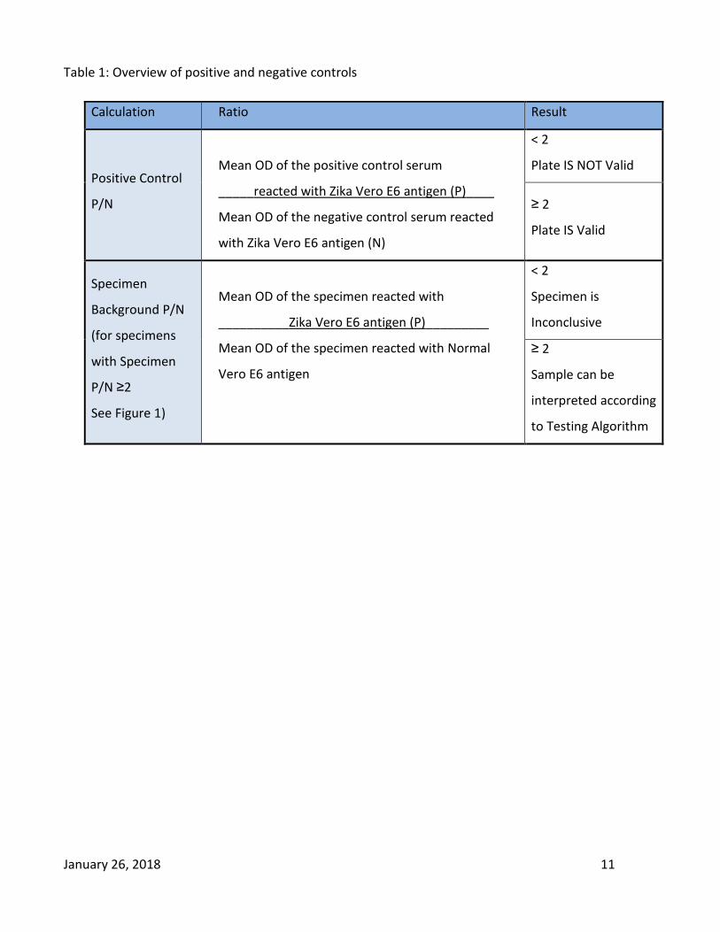
Table 1: Overview of positive and negative controls
Calculation Ratio Result
Positive Control
P/N
Mean OD of the positive control serum
_____reacted with Zika Vero E6 antigen (P)____
Mean OD of the negative control serum reacted
with Zika Vero E6 antigen (N)
< 2
Plate IS NOT Valid
≥ 2
Plate IS Valid
Specimen
Background P/N
(for specimens
with Specimen
P/N ≥2
See Figure 1)
Mean OD of the specimen reacted with
__________Zika Vero E6 antigen (P)_________
Mean OD of the specimen reacted with Normal
Vero E6 antigen
< 2
Specimen is
Inconclusive
≥ 2
Sample can be
interpreted according
to Testing Algorithm
January 26, 2018 12
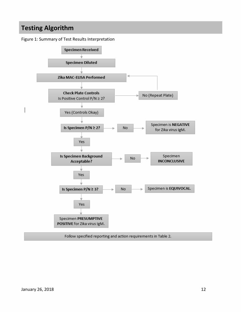
Testing Algorithm
Figure 1: Summary of Test Results Interpretation
January 26, 2018 13
Zika MAC-ELISA Assay
NOTES REGARDING THE ELISA PROCEDURE:
Plates can be coated and kept at 2-8o C for up to a week. (See Step 2: Coating the Plates,
below).
Undiluted control sera can be stored at 2-8o C for up to 2 weeks.
Reconstituted, undiluted viral and Normal Vero E6 antigens can be stored at ≤ -20o C for an
undefined period of time.
Test and control sera can be diluted to the working dilutions and refrigerated one day prior to
use.
Antigens and conjugate must be diluted to the working dilutions immediately prior to use.
NOTE: THE FOLLOWING PROCEDURE INCLUDES INFORMATION ON QUALITY CONTROL AND
INTERPRETATION. EACH SERUM SPECIMEN IS TESTED IN TRIPLICATE ON BOTH VIRAL AND NORMAL
VERO E6 ANTIGENS. EIGHT (8) TEST SPECIMENS CAN BE ANALYZED PER PLATE. DUE TO LIMITED
VOLUME, CSF SPECIMENS ARE USUALLY TESTED ONLY SINGLY.
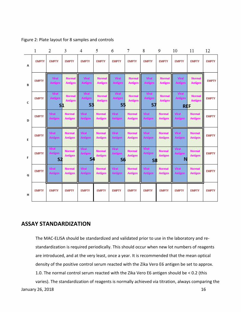
1. PREPARING THE PLATE:
Determine the number of ELISA plates needed. Using a fine-tipped permanent marker, number
and label the 96-well plates. Identify the location of each clinical specimen (S1-S8) by using a
corresponding template (see Fig. 2). To keep timing of reagent addition consistent, process
plates in the order that they are numbered during all steps of the procedure. Plates should be
kept in an enclosed, humidified environment during all incubation times with the exception of
the coating step. A large Ziploc-type bag containing a moist paper towel works well for this
purpose.
2. COATING THE PLATES:
Dilute goat anti-human IgM 1:2000 in coating buffer, pH 9.6.
Coat the inner 60 wells of the 96 well plate with 75 µL per well of diluted goat anti-
human IgM. Leave outer rows/columns empty (see Fig. 2).
Incubate at 2-8°C overnight. Plates should remain at 2-8°C until needed for testing, up
to one week.
January 26, 2018 14
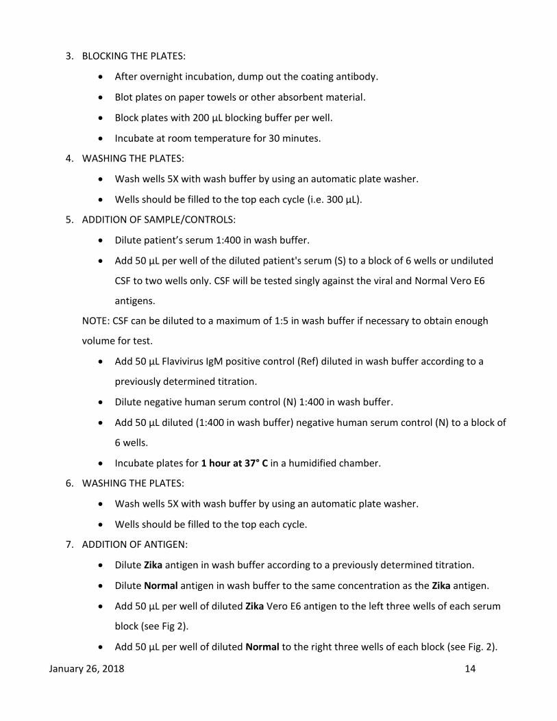
3. BLOCKING THE PLATES:
After overnight incubation, dump out the coating antibody.
Blot plates on paper towels or other absorbent material.
Block plates with 200 µL blocking buffer per well.
Incubate at room temperature for 30 minutes.
4. WASHING THE PLATES:
Wash wells 5X with wash buffer by using an automatic plate washer.
Wells should be filled to the top each cycle (i.e. 300 µL).
5. ADDITION OF SAMPLE/CONTROLS:
Dilute patient’s serum 1:400 in wash buffer.
Add 50 µL per well of the diluted patient's serum (S) to a block of 6 wells or undiluted
CSF to two wells only. CSF will be tested singly against the viral and Normal Vero E6
antigens.
NOTE: CSF can be diluted to a maximum of 1:5 in wash buffer if necessary to obtain enough
volume for test.
Add 50 µL Flavivirus IgM positive control (Ref) diluted in wash buffer according to a
previously determined titration.
Dilute negative human serum control (N) 1:400 in wash buffer.
Add 50 µL diluted (1:400 in wash buffer) negative human serum control (N) to a block of
6 wells.
Incubate plates for 1 hour at 37° C in a humidified chamber.
6. WASHING THE PLATES:
Wash wells 5X with wash buffer by using an automatic plate washer.
Wells should be filled to the top each cycle.
7. ADDITION OF ANTIGEN:
Dilute Zika antigen in wash buffer according to a previously determined titration.
Dilute Normal antigen in wash buffer to the same concentration as the Zika antigen.
Add 50 µL per well of diluted Zika Vero E6 antigen to the left three wells of each serum
block (see Fig 2).
Add 50 µL per well of diluted Normal to the right three wells of each block (see Fig. 2).
January 26, 2018 15
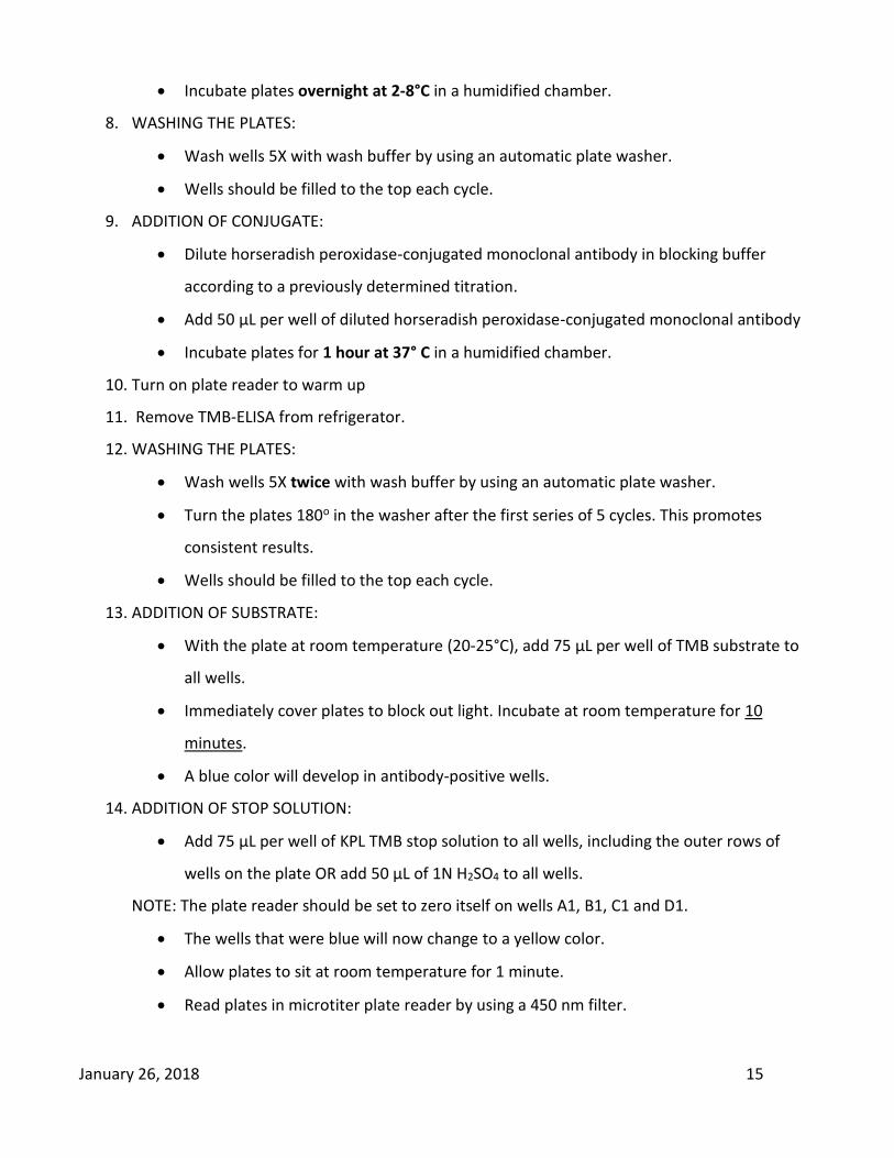
Incubate plates overnight at 2-8°C in a humidified chamber.
8. WASHING THE PLATES:
Wash wells 5X with wash buffer by using an automatic plate washer.
Wells should be filled to the top each cycle.
9. ADDITION OF CONJUGATE:
Dilute horseradish peroxidase-conjugated monoclonal antibody in blocking buffer
according to a previously determined titration.
Add 50 µL per well of diluted horseradish peroxidase-conjugated monoclonal antibody
Incubate plates for 1 hour at 37° C in a humidified chamber.
10. Turn on plate reader to warm up
11. Remove TMB-ELISA from refrigerator.
12. WASHING THE PLATES:
Wash wells 5X twice with wash buffer by using an automatic plate washer.
Turn the plates 180o in the washer after the first series of 5 cycles. This promotes
consistent results.
Wells should be filled to the top each cycle.
13. ADDITION OF SUBSTRATE:
With the plate at room temperature (20-25°C), add 75 µL per well of TMB substrate to
all wells.
Immediately cover plates to block out light. Incubate at room temperature for 10
minutes.
A blue color will develop in antibody-positive wells.
14. ADDITION OF STOP SOLUTION:
Add 75 µL per well of KPL TMB stop solution to all wells, including the outer rows of
wells on the plate OR add 50 µL of 1N H2SO4 to all wells.
NOTE: The plate reader should be set to zero itself on wells A1, B1, C1 and D1.
The wells that were blue will now change to a yellow color.
Allow plates to sit at room temperature for 1 minute.
Read plates in microtiter plate reader by using a 450 nm filter.
January 26, 2018 16
Figure 2: Plate layout for 8 samples and controls
ASSAY STANDARDIZATION
The MAC-ELISA should be standardized and validated prior to use in the laboratory and re-
standardization is required periodically. This should occur when new lot numbers of reagents
are introduced, and at the very least, once a year. It is recommended that the mean optical
density of the positive control serum reacted with the Zika Vero E6 antigen be set to approx.
1.0. The normal control serum reacted with the Zika Vero E6 antigen should be < 0.2 (this
varies). The standardization of reagents is normally achieved via titration, always comparing the
January 26, 2018 17
optical densities of the reagents when reacted on viral and Normal Vero E6 antigen.
Standardization and re-standardization may be confirmed by testing verification panels.
Interpreting Test Results
TEST VALIDITY DETERMINATION
Before the results can be calculated for each clinical specimen, the test must be determined to be
valid. For a test to be valid, the following ratio must be greater than or equal to 2.0. This is the P/N of
the positive control.
Mean OD of the Flavivirus IgM Positive Control reacted with Zika Vero E6 antigen (P)
Mean OD of the negative control reacted with Zika Vero E6 antigen (N)
Test validity must be determined for each plate. Results for clinical specimens may only be determined
if the test is valid. If the test is not valid, then that plate must be repeated. If the P/N for the positive
control still fails after a repeat, then one or more of the reagent or test parameters was likely in error,
and troubleshooting should be performed.
DETERMINATION OF SPECIMEN P/N
To determine whether the clinical specimens (S1-S8) contain IgM to the Zika virus (which would
indicate recent infections with that virus) the following must be calculated:
Mean OD of the test specimen reacted with Zika Vero E6 antigen (P)
Mean OD of the normal human serum reacted with Zika Vero E6 antigen (N)
This is the P/N of the test specimen.
All specimens for which Specimen P/N is < 2, report as negative. No further analysis is required. See
Table 2 below.
January 26, 2018 18
SPECIMEN BACKGROUND EVALUATION
For each specimen with a Specimen P/N ≥ 2, determine whether non-specific background is being
generated.
The value of P (mean of the test specimen reacted with Zika Vero E6 antigen) for the test
specimen must be greater than or equal to twice (2X) the mean OD of the test specimen
reacted with Normal Vero E6 antigen. If this requirement is not met, non-specific background is
being generated, and the result MUST be reported as inconclusive. Inconclusive specimens
should be retested. If repeat testing also yields inconclusive results, forward specimen for
further analysis and/or request collection of additional serum for analysis. If requirement is
met, proceed with specimen result interpretation.
ANALYSIS OF POSITIVE AND EQUIVOCAL RESULTS
All test specimen P/N values greater than or equal to 3.0 should be reported as presumptive IgM-
positive (see table below), as long as they meet the requirements listed above. In the event that an
early acute CSF or serum is negative by this test, a convalescent serum specimen must be requested
and tested before that patient is reported as negative for serological evidence of recent viral infection.
Without testing of a convalescent specimen, a negative result may reflect testing of an acute-phase
specimen obtained before antibody has risen to detectable levels.
P/N values that lie between 2.0 and 3.0 should be considered equivocal. Further tests should be
performed to determine the status of these specimens (see Table 2 below).
It should be stressed that the P/N value for a specimen at the screening dilution of 1:400 is not an
indication of absolute antibody concentration, i.e., the P/N value is not quantitative.
Table 2: Zika MAC-ELISA Results Interpretation
Test Specimen
P/N Interpretation Report Action
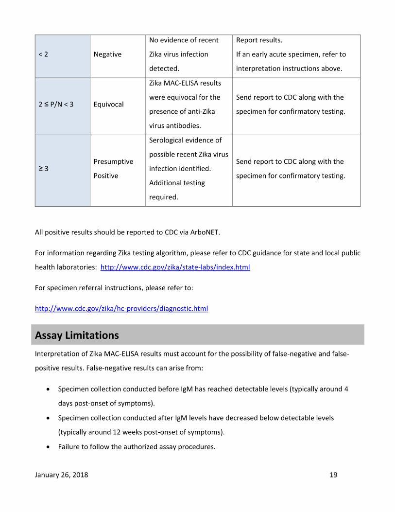
January 26, 2018 19
< 2 Negative
No evidence of recent
Zika virus infection
detected.
Report results.
If an early acute specimen, refer to
interpretation instructions above.
2 ≤ P/N < 3 Equivocal
Zika MAC-ELISA results
were equivocal for the
presence of anti-Zika
virus antibodies.
Send report to CDC along with the
specimen for confirmatory testing.
≥ 3 Presumptive
Positive
Serological evidence of
possible recent Zika virus
infection identified.
Additional testing
required.
Send report to CDC along with the
specimen for confirmatory testing.
All positive results should be reported to CDC via ArboNET.
For information regarding Zika testing algorithm, please refer to CDC guidance for state and local public
health laboratories: http://www.cdc.gov/zika/state-labs/index.html
For specimen referral instructions, please refer to:
http://www.cdc.gov/zika/hc-providers/diagnostic.html
Assay Limitations
Interpretation of Zika MAC-ELISA results must account for the possibility of false-negative and false-
positive results. False-negative results can arise from:
Specimen collection conducted before IgM has reached detectable levels (typically around 4
days post-onset of symptoms).
Specimen collection conducted after IgM levels have decreased below detectable levels
(typically around 12 weeks post-onset of symptoms).
Failure to follow the authorized assay procedures.
January 26, 2018 20
The most common cause of false-positive results is cross reactivity with IgM specific for other
flaviviruses such as dengue virus. Only limited evaluation of cross-reactivity with flaviviruses or
arboviruses has been conducted. No evaluation of cross-reactivity with Rheumatoid Factor has been
conducted. Clinical data indicate cross-reactivity with anti-dengue virus antibodies is likely. Follow-up
testing is necessary to rule-out a false-positive result. Confirmation of the presence of anti-Zika IgM
requires testing by CDC or a CDC-designated laboratory. The gold-standard method for confirmation of
the presence of anti-Zika antibodies is the plaque reduction neutralization test (PRNT).
All Zika testing must be conducted following the CDC-issued Zika laboratory guidance and testing
algorithms: http://www.cdc.gov/zika/state-labs/index.html.
Negative results do not preclude infection with Zika virus and should not be used as the sole basis of a
patient treatment/management decision. All results should be interpreted by a trained professional in
conjunction with review of the patient’s history and clinical signs and symptoms.
This assay is for in vitro diagnostic use under FDA Emergency Use Authorization only and is limited to
qualified laboratories designated by CDC International labs are not governed by the restrictions of the
FDA EUA.
All specimens should be handled as if infectious. Proper biosafety precautions, including personal
protective equipment, must be used when handling specimen materials.
Proper collection, storage and transport of specimens are essential for correct results.
Performance has only been established with the specimen types listed in the Intended Use. Other
specimen types are not acceptable for use with this assay.
It should be noted that as of April 2016, any patient whose sample yields positive PRNT results (i.e.,
>=10) for both Zika virus and for dengue virus, is now classified as having a “flavivirus” infection. The
virus of infection is only specified if one result is positive and the other negative. The former 4-fold
difference to identify the infection has been shown to be incorrect in a number of instances and has
now been abandoned for Zika diagnosis only.
January 26, 2018 21
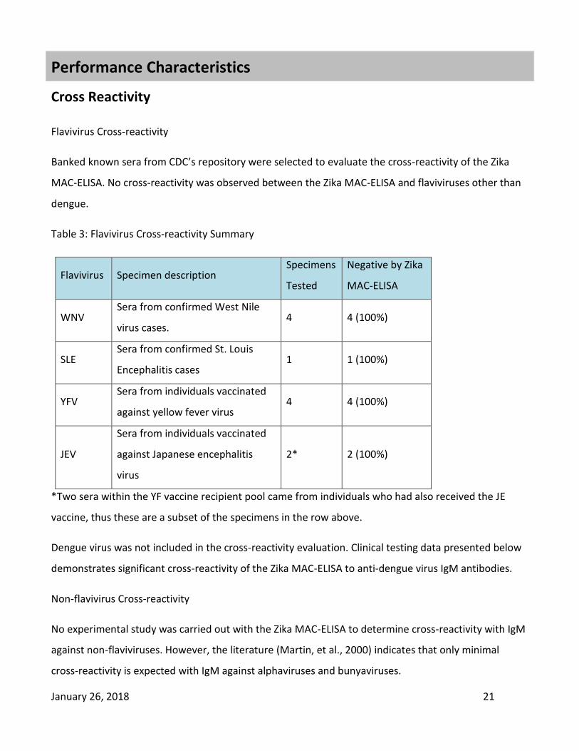
Performance Characteristics
Cross Reactivity
Flavivirus Cross-reactivity
Banked known sera from CDC’s repository were selected to evaluate the cross-reactivity of the Zika
MAC-ELISA. No cross-reactivity was observed between the Zika MAC-ELISA and flaviviruses other than
dengue.
Table 3: Flavivirus Cross-reactivity Summary
Flavivirus Specimen description Specimens
Tested
Negative by Zika
MAC-ELISA
WNV Sera from confirmed West Nile
virus cases. 4 4 (100%)
SLE Sera from confirmed St. Louis
Encephalitis cases 1 1 (100%)
YFV Sera from individuals vaccinated
against yellow fever virus 4 4 (100%)
JEV
Sera from individuals vaccinated
against Japanese encephalitis
virus
2* 2 (100%)
*Two sera within the YF vaccine recipient pool came from individuals who had also received the JE
vaccine, thus these are a subset of the specimens in the row above.
Dengue virus was not included in the cross-reactivity evaluation. Clinical testing data presented below
demonstrates significant cross-reactivity of the Zika MAC-ELISA to anti-dengue virus IgM antibodies.
Non-flavivirus Cross-reactivity
No experimental study was carried out with the Zika MAC-ELISA to determine cross-reactivity with IgM
against non-flaviviruses. However, the literature (Martin, et al., 2000) indicates that only minimal
cross-reactivity is expected with IgM against alphaviruses and bunyaviruses.
January 26, 2018 22
Arboviruses were originally delineated into three groups based on significant serological differences as
characterized with early, crude serological techniques. These delineations remain: Group A viruses are
now alphaviruses; Group B are now flaviviruses; Group C are now bunyaviruses. As serological methods
have evolved, the serological distinctions that originally defined the groups mean that cross reactivity
between groups in immunoassays is not expected.
Clinical Performance
Performance with U. S. Specimens Submitted to CDC, Ft. Collins, 2015 to Present
In the period from January 2015 to February 13, 2016, 167 sera and 2 CSF specimens were tested by
both the CDC Zika MAC-ELISA and by the CDC Zika PRNT assay.
Summary of Clinical Performance with Sera
Of the 167 sera test records, a subset were paired specimens from serial bleeds. From serial bleeds,
only the first IgM positive or equivocal bleed were included. If both serial bleeds were negative, only
the first one was included. The resulting data set used in this analysis is 161 sera. A summary of results
for these sera is presented in Table 4. Forty-four of these testing records indicated they are from
pregnant women. A summary of data for the subset of sera from pregnant women is presented in
Table 5.
January 26, 2018 23
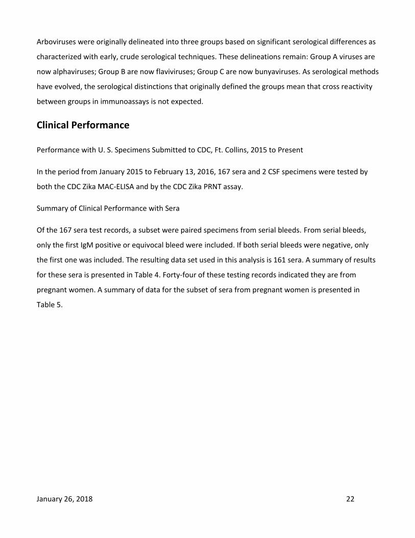
Table 4: Data for Sera Submitted to CDC Ft. Collins for Testing 2015-present
PRNT Results
Zika flavivirus dengue negative
Zika
MAC-
ELISA
positive 45 16 23 9
equivocal 1 0 9 13
negative 0 0 6 39
Positive percent agreement (PRNT definitive Zika positives only): 45/46 = 97.8% (95% CI: 88.7% -
99.6%)
Negative percent agreement: 45/99 = 45.5% (95% CI: 36.0% - 55.3%)
Table 5: Data for Sera from Pregnant Women Submitted to CDC Ft. Collins for Testing 2015-present
PRNT Results
Zika flavivirus dengue negative
Zika
MAC-
ELISA
positive 3 2 1 3
equivocal 0 0 2 8
negative 0 0 1 24
Positive percent agreement (PRNT definitive Zika positives only): 3/3 = 100% (95% CI: 43.9% – 100%)
Negative percent agreement: 25/39 = 64.1% (95% CI: 48.4% - 77.3%)
Summary of CSF Data
Both CSF specimens tested by CDC yielded positive results for Zika virus infection by both Zika MAC-
ELISA and by PRNT. The CSF results agreed with the paired serum testing.
Evaluation of Performance with Primary and Secondary Zika Infections, Yap State, Micronesia, 2007
The CDC Zika MAC-ELISA was included in a battery of CDC MAC-ELISA and PRNT flavivirus
immunological methods for the evaluation of paired serum specimens from 11 Zika virus cases
identified in a Zika outbreak in Yap State, Micronesia in 2007 (Lanciotti, et al., 2008). At the time of this
outbreak, the Zika MAC-ELISA employed sucrose-acetone extracted suckling mouse brain antigen. Four

January 26, 2018 24
of the 11 cases are primary flavivirus infections, while seven are probable secondary flavivirus
infections.
Of the paired sera collected and evaluated in the publication, all but one patient has at least one serum
specimen within our claimed window of ≥ 4 days post onset of symptoms and < 12 weeks post onset of
symptoms. For each of the remaining 10 cases, the earliest collected serum specimen within our
claimed window is included in our analysis.
Zika MAC-ELISA results for these specimens are compared to their Plaque Reduction Neutralization
Test (PRNT) results, the gold standard for flavivirus immunological testing.
Table 6: Summary of Zika MAC-ELISA and Flavivirus PRNT Results for earliest within-window specimens
for primary and secondary Zika infections in Yap State, Micronesia, 2007
Case Specimen Days Post
Onset
Zika
MAC-ELISA
PRNT
(7 flaviviruses)
Primary
Infections
822 822a 5 23.2 Zika
830 830b 21 16.3 Zika
849 849b 18 18.2 Zika
862 862a 6 25.4 Zika
Probable
Secondary
Infections
817 817b 19 8.1 flavivirus
833 833b 19 3.1 Zika
844 844b 16 12.7 dengue
955 955b 14 10.9 flavivirus
968 No specimens within claimed window
839 839b 20 17.2 Zika
847 847a 5 0.94 yellow fever
PRNT Results (7 flaviviruses)
Zika flavivirus dengue yellow fever
Positive 6 2 1 0

January 26, 2018 25
Zika
MAC-
ELISA
Negative 0 0 0 1
January 26, 2018 26
Primary infections:
All 4 cases identified as primary infections yielded positive Zika MAC-ELISA results for their initial
within-window serum specimen. These 4 specimens also yielded positive results by PRNT for Zika virus
infection.
Secondary Infections (probable):
Of the six cases with within-window serum specimens, five yielded positive results for the earliest
within-window serum specimens by Zika MAC-ELISA. Two of these (833b and 839b) yielded clearly
positive results for Zika virus infection by PRNT. Two other specimens (817b and 955b) yielded greater
PRNT results for Zika virus than for any of the other flaviviruses tested. However, these results were
not 4-fold higher than all other results, thus were interpreted a Flavivirus positive by PRNT. The
remaining specimen yielded positive results for Zika MAC-ELISA and 4-fold higher PRNT results for
Dengue than for Zika, the only specimen for which the PRNT did not agree with the Zika MAC-ELISA.
Specimen 847a, a day 5 specimen, was negative by Zika MAC-ELISA and was positive for Yellow Fever
Virus by PRNT. No neutralization effect was observed for Zika by PRNT. Thus the MAC-ELISA and PRNT
results for this specimen are in agreement.
Contact
Questions or comments about this procedure may be directed to the [email protected]
References
Johnson, AJ, Martin, DA, Karabatsos, N and Roehrig, JT. Detection of anti-arboviral immunoglobulin G
by using a monoclonal antibody-based capture enzyme-linked immunosorbent assay. J. Clinical
Microbiology, 38:1827-1831, 2000.
Koneman EW, Allen SD, Janda WM, Schreckenberger PC, and Winn Jr. WC , (Eds). Diagnosis of
Infections caused by Viruses, Chlamydia, Rickettsia, Diagnostic Microbiology, 4th Edition, JB Lippicott
Co: 956-1074, 1992.

January 26, 2018 27
Lanciotti, RS, O.L. Kosoy, J.J. Laven, J.O. Velez, A.J. Lambert, A.J. Johnson, S.M. Stanfield, and M.R.
Duffy. Genetic and Serologic Properties of Zika Virus Associated with an Epidemic, Yap State,
Micronesia, 2007. Emerg Infect Dis. 2008 (Aug); 14(8): 1232-1239.
Monath, TP, Nystrom, RR, Bailey, RE, Calisher, CH, and Muth, DJ:
Immunoglobulin M antibody capture enzyme-linked immunosorbent assay for diagnosis of St. Louis
encephalitis. Journal of Clinical Microbiology 20:784-790, 1984.
Martin, DA., Muth, DA., Brown, T., Karabatsos, N., and Roehrig, JT.
Standardization of immunoglobulin M capture enzyme-linked immunosorbent assays (MAC-ELISA) for
routine diagnosis of arboviral infections. Journal of Clinical Microbiology 38:1823-1826, 2000.
Tsai, TH: Arboviruses, In Rose NR, Marcario EC, Fahey JL, Friedman H, and Penn GM, (Eds):
Manual of Clinical Laboratory Immunology, 4th Edition, American Society for Microbiology: 606-618,
1976.