non convulsive status epilepticus clinical features, diagnosis
TRANSCRIPT

Dr Mohammad A.S Kamil
Consultant neurologist
Neurosciences hospital


DEFINITIONS A condition of ongoing or intermittent clinical
epileptic activity (without convulsions) for at least 30 minutes with EEG evidence of seizures.
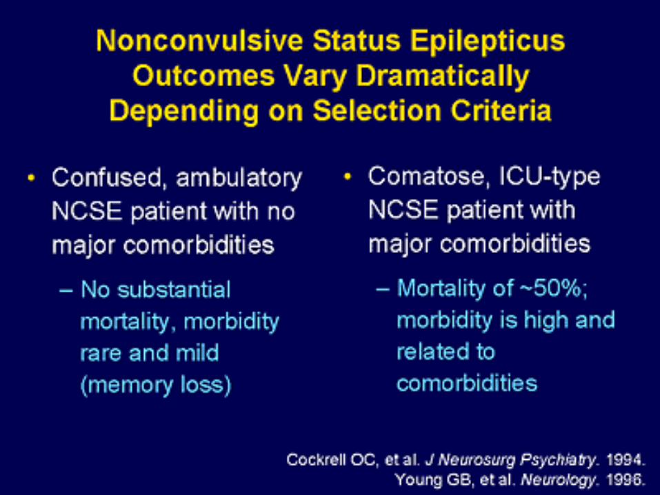
Non convulsive SE is believed to occur in about 8% of all comatose patients without evidence of significant motor signs and persists in 14% of patients following generalized convulsive status epilepticus.


Non convulsive SE is likely under recognized and under diagnosed.

Classification of nonconvulsivestatus epilepticus (NCSE)

NCSE occurring in the neonatal and infantile epilepsy syndromes
West syndrome
Ohtahara syndrome
Severe myoclonic encephalopathy of infancy (SMEI; Dravet syndrome)
NCSE in other forms of neonatal or infantile epilepsy

NCSE occurring only in childhood
NCSE in Early-onset benign childhood occipital epilepsy (Panayiotopoulos syndrome)
NCSE in other forms of childhood epileptic encephalopathies, syndromes and etiologies, e.g., Ring chromosome X andother karyotype abnormalities, Angelman syndrome, Rett syndrome, myoclonic-astaticepilepsy, other childhood myoclonic encephalopathies
Electrical status epilepticus in slow wave sleep (ESES)
Landau-Kleffner syndrome

NCSE occurring in both childhood and adult life
With epileptic encephalopathy
NCSE in the Lennox-Gastautsyndrome
i. Atypical absence status epilepticus
ii. Tonic–status epilepticus
Other forms of NCSE in patients with learning disability
or disturbed cerebral development (cryptogenic or
symptomatic)
Without epileptic encephalopathy
Typical absence status epilepticus in idiopathic generalized epilepsy
Complex partial status epilepticus:
i. Limbic ii. Nonlimbic
NCSE in the postictal phase of tonic–clonic seizures
Subtle Status epilepticus (myoclonic SE occurring in the late stage of convulsive
SE)
Aura continua (with: i. sensory, ii. special sensory, iii. autonomic, iv. cognitive
symptoms)

NCSE occurring in late adult life
De novo absence status epilepticus of late onset

Boundary syndromes : cases in which it is not clear to what
extent the continuous epileptiform electrographic abnormalities are contributing to the clinical impairment.
Some cases of epileptic encephalopathy.
Some cases of coma due to acute brain injury with epileptiform EEG changes.
Some cases of epileptic behavioral disturbance or psychosis.
Some cases of drug induced or metabolic confusional state with epileptiform EEG changes.

47 year-old man with generalized tonic-clonic seizures and absences since age 17. Valproic acid (VPA) and primidone never completely controlled his seizures. He developed severe hyperammonemic encephalopathy and had to be switched to levetiracetam (LEV), lamotrigine (LTG), and topiramate. He then experienced several episodes of AS where he was walking around, but was confused. He could speak and responded to questions, but mimicked Ganser’s syndromes in that his most answers were “near- correct” (October 17 instead of November 17, for example). The EEG showed almost permanent primary generalized (poly-)spike-wave discharges with short bouts of normal background activity (A). Absence status did not stop after i/v-administration of 8 mg of lorazepam (LZP), but the background activity became flattened and beta activity was increased (B). The subsequent i/v-administration of 1 mg of midazolam (MDL)(C) completely abolished the epileptic activity within 90sec.(D).


84 year-old otherwise healthy woman who was found slightly confused in her apartment. A CT scan and the CSF were completely normal. Within 24 hours, she became comatose. The EEG showed diffuse, irregular, sharp-contured, high-amplitude theta- and delta activity, intermingled with multifocal sharp waves (A). This activity did not change upon eye opening (B). The i/v-administration of 0.5 mg MDL (C) markedly reduced the epileptic activity and led to an accelerated, more regular background activity within 90 seconds (D). The patient opened her eyes and briefly talked. Extensive work-up did not reveal another cause than BZD intake for insomnia and an involuntary stop of this medication a few days before admission because of medication run-out.


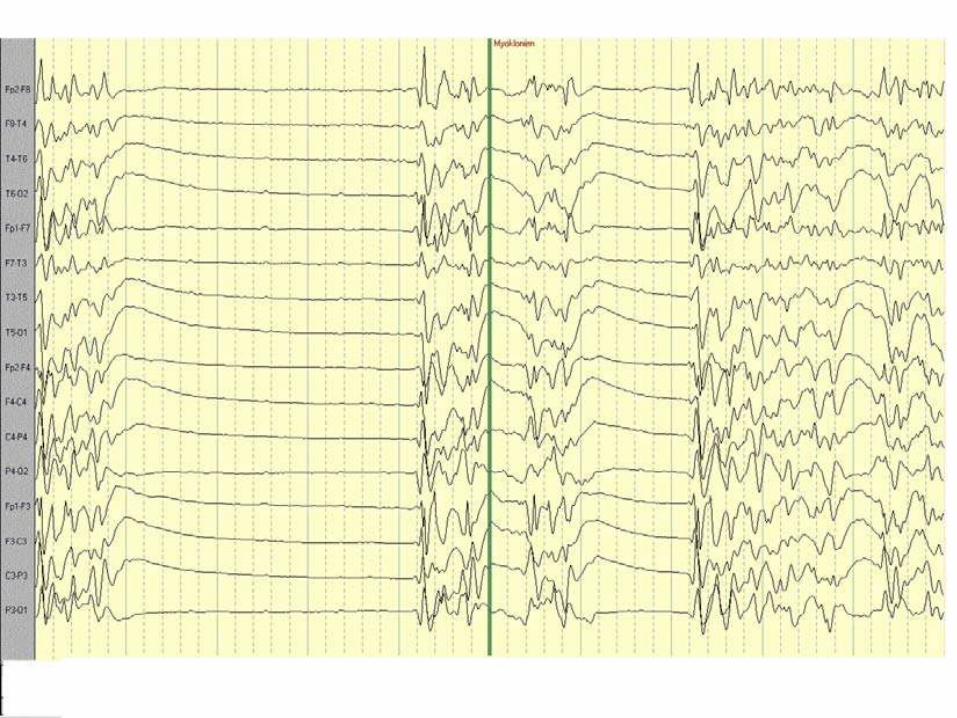
53 year-old woman with acute respiratory exhaustion after left ventricular decompensationand subsequent pulseless electric activity. Successful outdoor reanimation after an estimated time of hypoxia of 35 minutes. She was treated by hypothermia for 24 h. EEG after rewarming without sedative drugs showed a spontaneous burst-suppression pattern with spikeslow-and sharp-slow-waves with clinical myocloni. She remained deeply comatose and somatosensory evoked potentials 48h later showed absence of cortical responses.

81 year-old patient with sepsis caused by E. coli, prosthetic hip infection and multiple retroperitoneal abscesses wastreated with rifampicine and cefepime; two days later, acute renal failure occurred and the patient was comatose despite immediatedialysis.
The EEG (A) showed periodic triphasic waves (TPW) (*left box) with fronto-occipital shift (**); additionally, multifocal epileptic discharges (***, boxes in the middle and at the right) were observed in both paracentral regions and over the right temporal region. Intravenous administration of 1 mg of LZP (B) led to complete abolition of both the TPW and the epileptic discharges.


(A) Normal electroencephalogram from a 6-month-old child. (B) Absence status epilepticus in a 6-year-old child who presented with unresponsiveness with subtle twitching of the corner of the mouth. The electroencephalogram shows continuous rhythmic generalized spike-and-wave discharges with frontal predominance.


Treatment of absence SE There is no evidence that absence status induces neuronal damage, and thus
aggressive treatment is not warranted. Treatment can either be intravenous or oral. Absence status epilepticus is often precipitated by the prescription of
inappropriate antiepileptic drugs in idiopathic generalised epilepsy (e.g. carbamazepine).
Absence status epilepticus responds rapidly to intravenous benzodiazepines, and these are so effective that the response is diagnostic.
Lorazepam at 0.05-0.1 mg/kg is the benzodiazepine of choice. The effect may only be transient and a longer acting AED may need to be given. If intravenous treatment is required, but either benzodiazepines are ineffective
or contraindicated then intravenous valproate (20-40 mg/kg) can be given. In cases of primary generalised epilepsy treatment should be continued with a suitable AED.
If a precipitating factor can be identified in lateonset de novo cases, then longterm therapy is not usually indicated.

Complex partial status epilepticus How aggressively complex partial status epilepticus needs to be
treated depends upon: the prognosis of the condition; and if treatment improves the prognosis.
As in all epilepsies the prognosis relates partly to the prognosis of the underlying aetiology and any concomitant medical conditions.
At present, early recognition of the condition and treatment with oral or rectal benzodiazepines is recommended; oral clobazamhas proven to be an effective treatment .
In patients who have repetitive attacks of complex partial status epilepticus, oral clobazam (10-20 mg/day) over a period of 2-3 days given early at home can usually abort the status epilepticus, and such strategies should be discussed with patient and carers.

Atypical absence status epilepticus This condition is usually poorly responsive to
intravenous benzodiazepines, which should, in any case, be given cautiously, as they can induce tonic status epilepticus in these patients.
Oral rather than intravenous treatment is usually more appropriate, and the drugs of choice are valproate, lamotrigine, topiramate, clonazepam and clobazam.
Sedating medication, carbamazepine and vigabatrin have been reported to worsen atypical absences.

Nonconvulsivestatus epilepticus in coma Electrographic status epilepticus in coma is not uncommon and is seen
in up to 8% of patients in coma with no clinical evidence of seizure activity.
The diagnosis is often debatable as in many instances burst suppression patterns, periodic discharges and encephalopathictriphasic patterns have been proposed to represent electrographic status epilepticus, while these mostly indicate underlying widespread cortical damage or dysfunction.
This condition should be treated aggressively with deep anaesthesiaand concomitant AEDs.
The association of electrographic status epilepticus with subtle motor activity often follows hypoxic brain activity and has a poor prognosis, but aggressive therapy with benzodiazepines, phenytoin and increased anaesthesia is perhaps justified, since the little evidence available indicates that such treatment improves prognosis.