nipple sparing mastectomy for a giant phyllodes tumor; a
TRANSCRIPT
International Journal of Surgery Case Reports 88 (2021) 106470
Available online 12 October 20212210-2612/© 2021 The Authors. Published by Elsevier Ltd on behalf of IJS Publishing Group Ltd. This is an open access article under the CC BY-NC-ND license(http://creativecommons.org/licenses/by-nc-nd/4.0/).
Case report
Nipple sparing mastectomy for a giant phyllodes tumor; a case report
Akiko Okamoto a,*, Tadahiro Goto b, Makoto Omori c, Masaru Miyashita a
a Department of Breast Surgery, Konan Medical Center, 1-5-16 Kamokogahara, Higashinada-Ku, Kobe City, Hyogo 658-0064, Japan b Department of Surgery, Division of Hepato-Biliary-Pancreas Surgery, Kobe University Hospital, 7-5-1 Kusunoki-Cho, Chuo-Ku, Kobe City, Hyogo 650-0017, Japan c Department of Plastic Surgery, Yodogawa Christian Hospital, 1-7-50 Kunijima, Higashiyodogawa-Ku, Osaka City, Osaka 533-0024, Japan
A R T I C L E I N F O
Keywords: Phyllodes tumor Oncoplastic surgery Primary reconstruction of the breast
A B S T R A C T
Introduction: Oncoplastic surgery has come into the limelight in the surgical treatment of breast cancer. In this report, we will introduce our challenge to apply oncoplastic surgery to a benign neoplasm like phyllodes tumor (PT). Presentation of case: A 45-year-old female visited our hospital complaining of a rapidly growing lump on her left breast. She already had experienced lumpectomy twice on the same breast. Her left breast was occupied by a 14 × 10 cm mass with another small 1.7 × 1.6 cm nodule considered as a daughter lesion. Core needle biopsy suggested that it was a benign PT. We conducted nipple sparing mastectomy (NSM) and immediate recon-struction of the breast by latissimus dorsi muscle flap. During 7-years follow up, she has no recurrence and is satisfied with the reconstructed breast. Discussion: There are some reports that performed conventional or radical mastectomy with immediate breast or chest wall reconstruction for giant PT. Reports about NSM with breast reconstruction for PT are rare, there are 5 including ours. All the cases accomplished long term recurrent free survival. All except ours were reconstructed by implants. Implant reconstruction is technically easier, but recently, malignant lymphoma after putting breast implant is concerned. Another merit of autologous tissue reconstruction is that they change naturally as age like contralateral breast so that it can achieve better long-term cosmetic result. Conclusion: NSM with autologous tissue reconstruction is a good option for PT treatment even though it is not malignant.
1. Introduction
Oncoplastic surgery has been a featured field in surgical treatment of breast cancer. The aim is to achieve both curability of cancer and cosmetic satisfaction of the patients.
Although most of phyllodes tumors (PT) are histologically catego-rized into benign or borderline-malignant, their local recurrence rate is high. And they often grow into large tumors. These characteristics of PT eventually make it challenging for surgeons to compensate for the large volume defect and deformity of the breast after surgery. We suggest we can apply oncoplastic surgery to a benign tumor like PT, other than malignant tumor like breast cancer.
We herein report a case of a huge PT with repeated recurrence that received nipple sparing mastectomy (NSM) with primary breast recon-struction using latissimus dorsi muscle (LD) flap. The work has been reported in line with the SCARE criteria [1].
2. Case presentation
2.1. Patient information
A 45-year-old woman came to our hospital complaining of a rapidly growing lump on her left breast. She noticed it 5 month before visiting us. She had already received lumpectomy twice because of tumors on the same breast. She received her first lumpectomy 7 years ago and the second one 5 years ago, both in China. She had no detailed information about the surgeries or pathological results, but as far as she knew, neither of the tumors was malignant.
She has no other relevant personal or family medical history.
2.2. Physical examination
A 8.5 × 8.0 cm lobulated mass was palpable on the center of her left breast. It had good morbidity and occupied almost all the left breast. The
* Corresponding author. E-mail address: [email protected] (A. Okamoto).
Contents lists available at ScienceDirect
International Journal of Surgery Case Reports
journal homepage: www.elsevier.com/locate/ijscr
https://doi.org/10.1016/j.ijscr.2021.106470 Received 20 August 2021; Received in revised form 29 September 2021; Accepted 3 October 2021

International Journal of Surgery Case Reports 88 (2021) 106470
2
left breast looked larger than the right because of the tumor (Fig. 1-a).
2.3. Diagnostic assessment
Mammography showed a well circumscribed lobulated large mass on the left breast, suspected of being a benign tumor.
Ultrasound study demonstrated a 14 × 10cm well circumscribed lobulated hypoechoic mass on the central part of the left breast. There was another small nodule of 1.7 × 1.6 cm on the upper-inner quadrant of the left breast.
MRI showed a 10x9cm lobulated, well enhanced mass at the center of the left breast, and there was a 1.0 cm nodule on the upper-inner side from the main tumor. Neither of them had invasion to the skin or the nipple (Fig. 2). The time-intensity curve of the main tumor expressed rapid-persistent pattern which was characteristic of benign breast neoplasms.
In case of a malignant PT, we conducted CT scanning to search for any metastatic lesion. There were no findings indicating either axillary lymph node metastasis or any other distant metastasis.
Core needle biopsy (CNB) of the main tumor contained abundant fibrous stroma that was consistent with PT. Since there was no cellular atypia of epithelial cells and little cell division, it was categorized as benign.
2.4. Surgical procedure
The patient was positioned supine, under general anesthesia, as we first conducted NSM of the left breast. The surgical scars of the
lumpectomies before and the scar of CNB were removed including them in the skin incision (Fig. 1-b, -c). We detached the tumor from the skin, keeping the skin about 1 cm thickness to prevent burn caused by electric knife and skin necrosis after reconstruction. There was enough distance between the small daughter nodule and the nipple so that we could easily detach the nipple from it. Some part of the larger mass had strong adhesion to the fascia of the major pectoral muscle; we removed the fascia together with the tumor (Fig. 1-d). We could keep the major pectoral muscle without damage.
After NSM, the patient was positioned on the right lateral position. The plastic surgeons started breast reconstruction. They designed an 18 × 7cm oblique spindle flap on the left lateral back area under the scapula. Using the breast side of the flap as the axis of rotation, they rotated the flap to the front of the chest. They carefully remained the feeding artery of the flap from thoracodorsal artery. They made the flap denuded, removing whole epidermis. They adjusted the shape of the flap, making it similar to the contralateral side of the breast.
The total operating time was 438 min, Blood loss was 500 ml; we did not do blood transfusion. The resected specimen weighed 530 g.
2.5. Pathological result
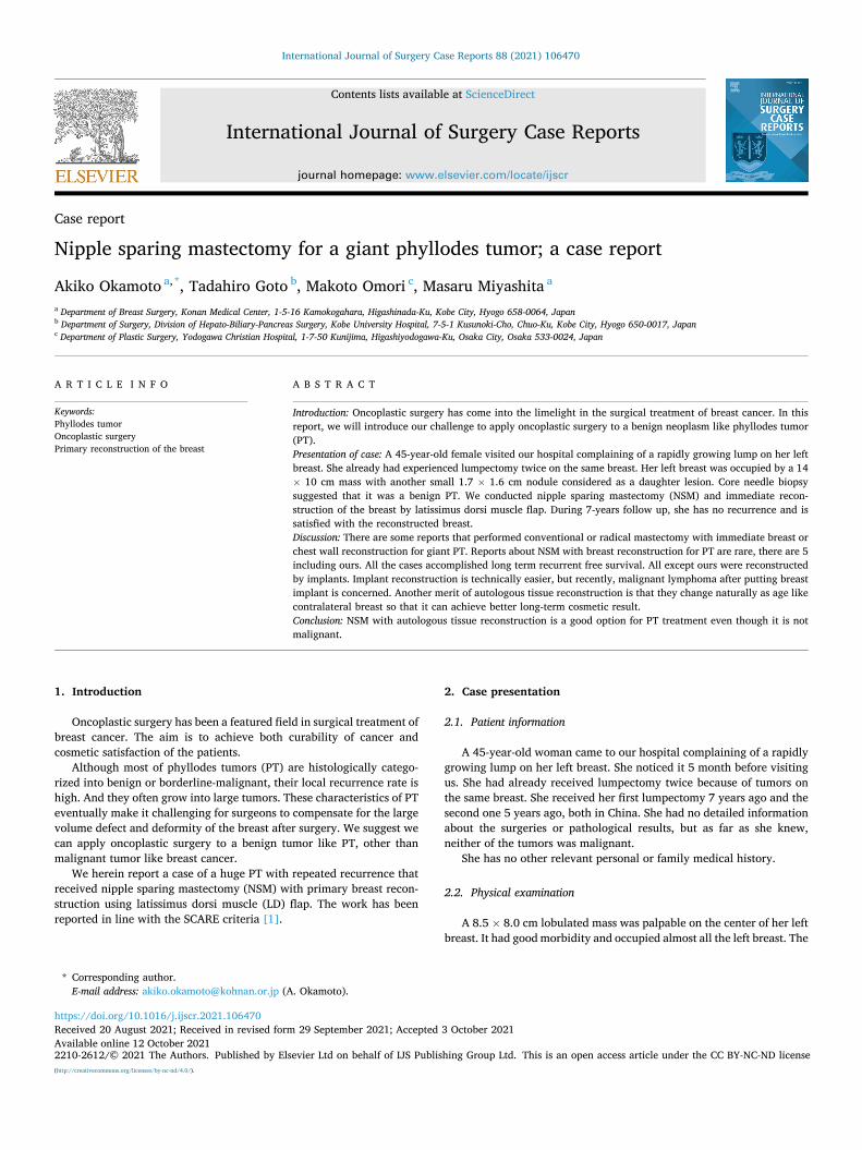
There was a 10 × 10cm well- circumscribed tumor on the central part of the left breast and a 1.0 × 1.0 cm tumor on upper-inner side of the main tumor. The two tumors had no direct connection, contained abundant fibrous stroma, and leaf-like constructions made of stroma and epithelial cells, compatible with PT. In some of the main tumor, stromal cells had a little cellular atypia; it was categorized as borderline-
a b
dc
Fig. 1. (a) Appearance before the surgery. The left breast looked much larger than the right because of the tumor. (b) Appearance before the surgery. Black arrows; the surgical scars of the past lumpectomies. White arrow; the scar of CNB. (c) Resected specimen. The scars of the past surgeries and CNB(black and white arrows) were included in the skin incision. (d) Right before the mass removal.
A. Okamoto et al.

International Journal of Surgery Case Reports 88 (2021) 106470
3
malignant (Fig. 3a,b). As the small nodule had no cellular atypia, it was categorized as benign.
2.6. Outcome
The patient went through no complications after the surgery and was discharged from the hospital on post-operative day 16. She had annual surveillance by physical examination, mammography and ultrasound. It is now 7 years since she received the surgery, she has no recurrence and is satisfied with the shape of her reconstructed breast (Fig. 4a,b).
3. Discussion
Phyllodes tumor (PT) is a rare fibroepithelial tumor of the breast, accounting for less than 1% of all breast neoplasms [2]. It is classified into benign, borderline, and malignant; the occurrence is reported as benign (60–75%), borderline (13–26%) and malignant (10–20%) [3].
Even for malignant PT, we do not yet have good results with chemotherapy or radiation therapy, therefore, surgery is the key treat-ment. But how much surgical margin is necessary for PT resection is still controversial [4–7]. The NCCN guidelines recommend 1 cm margin. Recently, however, some studies reported that among patients with close(<1 mm) or positive margins, there was no significant difference in disease recurrence. It may not always necessary to remove a PT with so much margin. According to the systematic review and meta-analysis including 9234 cases performed by Lu et al., local recurrence rate of PT reported 8–10% for benign, 10–13% for borderline, and 15–20% for malignant. Surgical procedures, such as breast-conserving surgery versus mastectomy, and positive versus negative surgical margins, were significantly associated with an increased local recurrence risk only in malignant PT [8]. They suggested that for benign or borderline PTs, positive margin is acceptable.
However, we believe keeping some margin is better than close or positive margin. One of the reasons is that the needle biopsy specimen before surgery is often too little to make precise diagnosis whether the PT is benign or not. According to the recent pathological review about PT [9], they pointed out that there is heterogeneity in PT tumor, and that it may contain foci of benign, borderline, and malignant within the same tumor. They recommend careful histological examination of the whole
specimen. In case the PT turns out to be malignant against pre-surgical diagnosis, it is better to keep some resected margin. Another reason is that in cases that have experienced repeated recurrence like our case, we suspect that the tumor has stronger potential to recur than a sporadic PT. While the tumor repeats recurrence, it may attain some malignant characters. Tan EY et al., researched 37 recurrent PT cases [10]. They reported 7(19%) of the cases developed malignant recurrence from initially benign or borderline PT. We should be more cautious about recurrent cases.
In our case, as there were two tumors, one of which was over 10 cm large, we planned that we should remove the whole breast tissue to obtain enough surgical margin. The MRI showed some distance between the tumors and nipple or between the tumors and skin. We considered we could conserve the nipple and most of the skin of the left breast. We decided to perform NSM rather than conventional mastectomy. Nipple is a characteristic spot on the chest wall regardless of sex. There is a sig-nificant difference on the first look impression between with and without the nipple. Although plastic surgeon can reconstruct the nipple later, it takes some time until patients can receive nipple reconstruction. We believe it is less stressful for patients to have their nipple conserved. Concerning the safety of conserving the nipple, in 2020, Sun et al. re-ported a rare case of PT recurrence on the nipple one year after breast conserving surgery. According to their review, their case was the first and the only one reported about PT occurred on the nipple [11]. As PT quite rarely happens on the nipple, it is considerably safe to conserve the nipple for PT surgery.
There are some reports about PT to whom they performed conven-tional mastectomy or radical mastectomy with reconstruction of the breast or chest wall [12–14], but the reports about NSM with immediate reconstruction for PT are few. As far as we could find during the past 20 years, there are 5 case reports including ours [15–18]. All the cases had no recurrence during the follow-up period. All other cases except ours were reconstructed by implants (Table 1). As there were only few cases, we could not find any statistically significant difference in patients' factors, but the tumor size seems to be a little larger in our case than other cases reconstructed by implants. Although the reason we chose LD flap was not the tumor size, we infer that the surgeons of other cases may have chosen implant reconstruction because they considered it was appropriate to apply a less stressful surgery to a medium size of benign
Fig. 2. MRI findings. There were two tumors. Non-enhancing internal septations were found inside the larger tumor.
A. Okamoto et al.
International Journal of Surgery Case Reports 88 (2021) 106470
4
tumor. Implant reconstruction is technically easier than autologous tissue reconstruction. The surgical time is shorter and the physical stress to the patients is less than autologous tissue reconstruction because they don't need to have additional scar on the graft site. If the patient come to receive surgery again because of recurrence, the implant removal is also easier than autologous tissue flap.
However, there are some reasons why we believe autologous tissue reconstruction is better than implant for this case. It comes to be a sig-nificant problem that textured breast implant has a risk of causing a special type of malignant lymphoma called breast-implant-associated anaplastic large-cell lymphoma (BIA-ALCL). In December 2018,
French government first ordered the company to recall the silicon implant, since July 2019 the US government and other countries also ordered to recall. The risk of BIA-ALCL is low, it is estimated between 1/ 2832 and 1/30,000. Most of the BIA-ALCL cases were diagnosed as early stage of malignant lymphoma, and the main treatment was the removal of the implant and lymphoma lesion. But there are some cases that were diagnosed as over Stage2 and had to receive chemotherapy and radia-tion therapy. 36 deaths are reported world-wide until August 2020. Thus, early detection of BIA-ALCL is so important that patients should receive regular medical checkup after implant reconstruction. While surgical stress and the risk of acute surgical complication are higher in
a
b
Fig. 3. Pathological findings of the main tumor. (a) ×10. There are abundant fibrous stroma, and leaf-like constructions. (b) ×20. The stromal cells had a little cellular atypia, it was categorized as intermediate-malignant PT.
A. Okamoto et al.

International Journal of Surgery Case Reports 88 (2021) 106470
5
autologous reconstruction, we can manage them more easily and less harmfully than BIA-ALCL. After the flap successfully got engrafted to the body, patients do not have to receive frequent medical check-up. Therefore, looking in the long term, the stress for patients is less in autologous reconstruction than in implant reconstruction. Especially when treating a benign neoplasm like our case, we believe we must avoid the risk of a malignant disease which can cause death or necessity for chemotherapy.
Another merit of autologous reconstruction is that the graft changes similarly to contralateral breast according to age. In our case, as she got older, the reconstructed left breast dangled down naturally in the same way as the right (Fig. 4a,b). If we performed implant reconstruction, we could not see this natural falling-down according to age.
There are several methods of autologous tissue breast reconstruction.
a
b
Fig. 4. (a) 2 weeks after the surgery. (b) 7 years after the surgery.
Table 1 Reported cases of phyllodes tumor that received nipple-sparing mastectomy with immediate breast reconstruction.
Author CS FE LG ME Ours (Reference
number) 2011 [15]
2014 [16]
2015 [17]
2020 [18]
2021
Age 22 51 19 28 45 Tumor size (cm) 6.5 5.0 5.3 8.0 10.0 Reconstruction IMPa IMP IMP IMP LDb
Follow-up (month) 41 12 12 28 84 Recurrence No No No No No
a IMP; implant. b LD; latissimus dorsi muscle flap.
A. Okamoto et al.

International Journal of Surgery Case Reports 88 (2021) 106470
6
For example, Latissimus dorsi muscle(LD) flap, transverse rectus abdominus myocutaneous (TRAM) flap, and deep inferior epigastric perforator (DIEP) flap are frequently performed. As LD is close to the breast, it is the simplest way among the three, we can keep good blood flow into the flap without vessel anastomosis. In TRAM flap, sometimes we do not need vessel anastomosis, but sometimes the blood flow be-comes unstable compressed by the skin tunnel from the abdomen to the chest, we have to add vessel anastomosis. As DIEP flap is a free flap composed of skin and fat tissue of the abdomen without muscle, it is very important to maintain the blood flow by performing vessel anastomosis under microscope. DIEP flap requires higher techniques and longer surgical time than the other flaps. Although LD is the simplest way, we cannot compensate for a large volume. If we need a large volume, we should choose abdominal tissue flap as TRAM or DIEP. In our case, the size of the contralateral side of her breast was moderate. It is around 15 cm diameter and 4.5 cm height at the spine position. We could attain enough volume by LD flap.
It is stressful for a woman to lose her whole breast even because of life-threatening disease like breast cancer, it would be more unbearable because of less malignant neoplasms like PT. Thus, we believe onco-plastic surgery is as important for benign tumor as for breast cancer. Although our patient had already experienced recurrence twice, after this surgery, she was able to live without recurrence for over 7 years. We believe this surgical procedure achieved both curability and good quality of life as a woman.
4. Conclusion
Caring for cosmetic result is rather important for benign breast tumor like PT than cancer because patients can expect long life after surgery. In the quality of their long life after surgery, appearance takes an important part. NSM with autologous tissue reconstruction is a good option for this purpose.
Abbreviations
PT Phyllodes tumor MRI Magnetic resonance imaging CT computed tomography CNB Core needle biopsy NSM nipple-sparing mastectomy LD latissimus dorsi muscle flap. TRAM Transverse rectus abdominus myocutaneous flap DIEP deep inferior epigastric perforator flap NCCN National comprehensive cancer network
Ethics approval and consent to participate
This article does not contain any studies with human participants or animals performed by any of the authors.
Funding
None.
CRediT authorship contribution statement
A.O. collected the data and wrote the manuscript. T.G. helped collecting the data and writing the manuscript. M.O. is the plastic surgeon who conducted the breast reconstruction,
and gave the authors much instruction about breast reconstruction. M.M. is the attending surgeon and keeps annual observation of the
patient after surgery.
Guarantor
None.
Consent for publication
Written informed consent was obtained from the patient for publi-cation of this case report and accompanying images on 28th July 2021. A copy of the written consent is available for review by the Editor-in- Chief of this journal on request.
Availability of data and materials
Not applicable.
Provenance and peer review
Not commissioned, externally peer-reviewed.
Declaration of competing interest
All of the authors declare that they have no competing interests.
Acknowledgements
We are deeply grateful to Dr. Yonson Ku, president of Konan Medical Center, former professor of the department of surgery of Kobe Univer-sity, for giving us insightful advice and improving our manuscript.
References
[1] R.A. Agha, T. Franchi, C. Sohrabi, G. Mathew, for the SCARE Group, The SCARE 2020 guideline: updating consensus Surgical CAse REport (SCARE) guidelines, Int. J. Surg. 84 (2020) 226–230.
[2] G. Spitaleri, A. Toesca, E. Botteri, et al., Breast phyllodes tumor: a review of literature and a single center retrospective series analysis, Crit. Rev. Oncol. Hematol. 88 (2) (2013 Nov) 427–436, https://doi.org/10.1016/j. critrevonc.2013.06.005. Epub 2013 Jul 17 PMID: 23871531.
[3] S.R. Lakhani, I.O. Ellis, S.J. Schnitt, in: WHO Classification of Tumours of the Breast, Fourth edition, IARC WHO, World Heal. Organ. Press, 2012, pp. 143–147.
[4] R. Tremblay-LeMay, J.C. Hogue, L. Provencher, et al., How wide should margins be for phyllodes tumors of the breast? Breast J. 23 (3) (2017 May) 315–322, https:// doi.org/10.1111/tbj.12727. Epub 2016 Nov 30 PMID: 27901301.
[5] A. Moutte, N. Chopin, C. Faure, et al., Surgical management of benign and borderline phyllodes tumors of the breast, Breast J. 22 (5) (2016 Sep) 547–552, https://doi.org/10.1111/tbj.12623. Epub 2016 Jun 6 PMID: 27265474.
[6] T.A. Moo, H. Alabdulkareem, A. Tam, et al., Association between recurrence and re-excision for close and positive margins versus observation in patients with benign phyllodes tumors, Ann. Surg. Oncol. 24 (10) (2017 Oct) 3088–3092, https://doi.org/10.1245/s10434-017-5955-7. Epub 2017 Aug 1 PMID: 28766221.
[7] S. Ogunbiyi, A. Perry, K. Jakate, et al., Phyllodes tumour of the breast and margins: how much is enough, Can. J. Surg. 62 (1) (2019) E19–E21, https://doi.org/ 10.1503/cjs.005718.
[8] Y. Lu, Y. Chen, L. Zhu, et al., Local recurrence of benign, borderline, and malignant phyllodes tumors of the breast: a systematic review and meta-analysis, Ann. Surg. Oncol. 26 (2019) 1263–1275.
[9] Y. Zhang, C.G. Kleer, Phyllodes tumor of the breast: histopathologic features, differential diagnosis, and molecular/genetic updates, Arch. Pathol. Lab. Med. 140 (7) (2016 Jul) 665–671, https://doi.org/10.5858/arpa.2016-0042-RA. PMID: 27362571.
[10] E.Y. Tan, P.H. Tan, W.S. Yong, et al., Recurrent phyllodes tumours of the breast: pathological features and clinical implications, ANZ J. Surg. 76 (6) (2006 Jun) 476–480. PMID: 16768772.
[11] D. Sun, L. Tang, H. Xing, et al., Recurrent borderline phyllodes tumor in nipple: a rare case report and review of the literature, Gland Surg. 9 (2) (2020 Apr) 452–458. PMID: 32420273; PMCID: PMC7225488.
[12] A. Banno, A. Shimada, K. Aga, et al., Total mastectomy and chest reconstruction for a rapidly progressing giant phyllodes tumor with skin necrosis: a case report, Surg. Case Rep. 1 (82) (2015 Sep 14), https://doi.org/10.1186/s40792-015-0082-9. PMID: 26389024; PMCID: PMC4569663.
[13] A. Rajesh, M. Forooq, Resection and reconstruction following recurrent malignant phyllodes- case report and review of literature, Ann. Med. Surg. 16 (2017) 14–18.
[14] C.L. Fang, C.H. Hsu, C.W. Tu, The reconstruction choice for giant phyllodes tumor of breast: bi-pedicled deep inferior epigastric perforator flap, Aesth. Plast. Surg. 41 (2017) 768–772.
A. Okamoto et al.

International Journal of Surgery Case Reports 88 (2021) 106470
7
[15] S.A. Crenshaw, M.D. Roller, J.K. Chapman, Immediate breast reconstruction with a saline implant and AlloDerm, following removal of a phyllodes tumor, World J. Surg. Oncol. 21 (9) (2011 Mar) 34. PMID: 21418652; PMCID: PMC3070675.
[16] G.T. Farias-Eisner, K. Small, A. Swistel, U. Ozerdem, M. Talmor, Immediate implant breast reconstruction with acellular dermal matrix for treatment of a large recurrent malignant phyllodes tumor, Aesthet. Plast. Surg. 38 (2014) 373–378, https://doi.org/10.1007/s00266-014-0283-9.
[17] G. Libondi, M. Solinas, E.M. Martella, L. Cattelani, Nipple sparing mastectomy with immediate silicone implant reconstruction for malignant phyllodes tumor in a 19- year-old girl, Eur. Rev. Med. Pharmacol. Sci. 19 (2015) 4498–4500.
[18] E. Morioka, M. Noguchi, M. Noguchi, et al., A case of recurrent malignant phyllodes tumor undergoing nipple-sparing mastectomy with immediate breast reconstruction, Surg Case Rep. 6 (1) (2020 Nov 25) 297. PMID: 33237380; PMCID: PMC7688876.
A. Okamoto et al.