newhelicalbindingdomainmediatesaglycosyltransferase ... · fap1. c-terminal dgt1 (cgt) is predicted...
TRANSCRIPT
New Helical Binding Domain Mediates a GlycosyltransferaseActivity of a Bifunctional Protein*□S
Received for publication, April 12, 2016, and in revised form, August 17, 2016 Published, JBC Papers in Press, August 17, 2016, DOI 10.1074/jbc.M116.731695
Hua Zhang‡, Meixian Zhou‡, Tiandi Yang§, Stuart M. Haslam§, Anne Dell§, and Hui Wu‡1
From the ‡Departments of Pediatric Dentistry and Microbiology, Schools of Dentistry and Medicine, University of Alabamaat Birmingham, Birmingham, Alabama 35294 and the §Department of Life Sciences, Imperial College London,London, SW7 2AZ, United Kingdom
Serine-rich repeat glycoproteins (SRRPs) conserved in strep-tococci and staphylococci are important for bacterial coloniza-tion and pathogenesis. Fap1, a well studied SRRP is a major sur-face constituent of Streptococcus parasanguinis and is requiredfor bacterial adhesion and biofilm formation. Biogenesis of Fap1is a multistep process that involves both glycosylation and secre-tion. A series of glycosyltransferases catalyze sequential glyco-sylation of Fap1. We have identified a unique hybrid proteindGT1 (dual glycosyltransferase 1) that contains two distinctdomains. N-terminal DUF1792 is a novel GT-D-type glycosyl-transferase, transferring Glc residues to Glc-GlcNAc-modifiedFap1. C-terminal dGT1 (CgT) is predicted to possess a typicalGT-A-type glycosyltransferase, however, the activity remainsunknown. In this study, we determine that CgT is a distinctglycosyltransferase, transferring GlcNAc residues to Glc-Glc-GlcNAc-modified Fap1. A 2.4-Å x-ray crystal structure revealsthat CgT has a unique binding domain consisting of three �helices in addition to a typical GT-A-type glycosyltransferasedomain. The helical domain is crucial for the oligomerization ofCgT. Structural and biochemical studies revealed that the helixdomain is required for the protein-protein interaction and cru-cial for the glycosyltransferase activity of CgT in vitro and invivo. As the helix domain presents a novel structural fold, weconclude that CgT represents a new member of GT-A-type gly-cosyltransferases.
Protein glycosylation is one of the most common post-trans-lational modifications. There are two major types of modifica-tions resulting in N-linked and O-linked glycosylation, respec-tively. Both are involved in the regulation of various biologicalprocesses (1), and have been implicated in human health anddiseases (2).
Serine-rich repeat proteins (SRRPs)2 are O-linked glycopro-teins. They belong to a growing family of bacterial adhesins
(3–7). Fimbriae associate protein (Fap1) of Streptococcus para-sanguinis is the first SRRP identified, and is required for biofilmformation (8 –10). Fap1-like proteins are highly conserved inother oral streptococci (11–13) and widespread in pathogenicstreptococci and staphylococci (4, 5). For instance, Srr-2 ofStreptococcus agalactiae is associated with bacterial virulence(5), Hsa of Streptococcus gordonii and SraP of Staphylococcusaureus contribute to the pathogenesis of infective endocarditis(4, 14). PsrP of Staphylococcus pneumoniae mediates frequencyof invasive pneumococcal disease (15). Thus Fap1-like SRRPsrepresent a new family of adhesins that are important in bacte-rial virulence, but little is known about their biogenesis.
Glycosylation of Fap1 is crucial for bacterial adhesion (16),and biofilm formation (17) and is required for assembly of thefimbriae-like surface structure in S. parasanguinis (18). An11-gene cluster flanking the fap1 gene locus is essential for Fap1glycosylation and biogenesis (17). Several glycosyltransferasegenes including gly, gtf3, dgT1, and galT2 are found locatedupstream of fap1, and genes coding for accessory secretion pro-teins such as SecY2, Gap1, Gap2, Gap3, SecA2, and glycosyl-transferase Gtf1, and its chaperone Gtf2 are located down-stream of fap1. SecY2 and SecA2 encode accessory Secproteins. SecY2 is essential for the biogenesis of mature Fap1(19), and SecA2 is important for export of mature Fap1 to thecell wall (20, 21). The glycosylation-associated proteins (Gap1,Gap2, and Gap3) modulate Fap1 secretion and biogenesis (18,22, 23). A glycosyltransferase enzyme complex, consisting ofGtf1 and its chaperone Gtf2, catalyzes the first step of Fap1glycosylation, transferring GlcNAc residues to the Fap1 poly-peptide (17, 24). Gtf3 mediates the second step by transferringGlc to Gtf1–2-modified Fap1 (25, 26). Our previous studieshave determined that N-terminal dGT1 (DUF1792) transfersglucose residues to Gtf1, -2, and -3-modified Fap1 (27, 28). Thesubsequent step of Fap1 glycosylation is still unknown.
dGT1 of S. parasanguinis contains a conserved new glycosyl-transferase (DUF1792) at the N terminus and a putative glyco-syltransferase at the C terminus (27). In this study, we deter-mined the crystal structure of C-terminal dGT1, and found thatthe C-terminal dGT1 is a GlcNAc transferase, which possessesa glycosyltransferase activity that is distinct from the N-termi-nal activity, transferring GlcNAc to the Fap1 substrate modifiedfirst by the N-terminal DUF1792 glycosyltransferase. The 2.4-Åx-ray crystal structure reveals a new helix domain that is crucialfor both oligomerization and enzymatic activity of CgT.Together, we determine that dGT1 is a bifunctional protein
* This work was supported by National Institutes of Health NIDCR GrantR01DE17954 (to H. W.) and F33DE022215 (to H. W.), Biotechnology andBiological Sciences Research Council Grant BB/K016164/1, Core Supportfor Collaborative Research (to A. D. and S. M. H.), and a Wellcome TrustSenior Investigator Award (to A. D.). The authors declare no conflicts ofinterest with the contents of this article. The content is solely the respon-sibility of the authors and does not necessarily represent the official viewsof the National Institutes of Health.Author’s Choice—Final version free via Creative Commons CC-BY license.
□S This article contains supplemental Fig. S1.1 To whom correspondence should be addressed: 1919 7th Ave. South, Bir-
mingham, AL 35294. E-mail: [email protected] The abbreviations used are: SRRP, serine-rich repeat protein; ITC, isothermal
titration calorimetry; PDB, Protein Data Bank.
THE JOURNAL OF BIOLOGICAL CHEMISTRY VOL. 291, NO. 42, pp. 22106 –22117, October 14, 2016Author’s Choice © 2016 by The American Society for Biochemistry and Molecular Biology, Inc. Published in the U.S.A.
crossmark
22106 JOURNAL OF BIOLOGICAL CHEMISTRY VOLUME 291 • NUMBER 42 • OCTOBER 14, 2016
that catalyzes successive steps of glycosylation of Fap1 in vitroand in vivo in S. parasanguinis.
Results
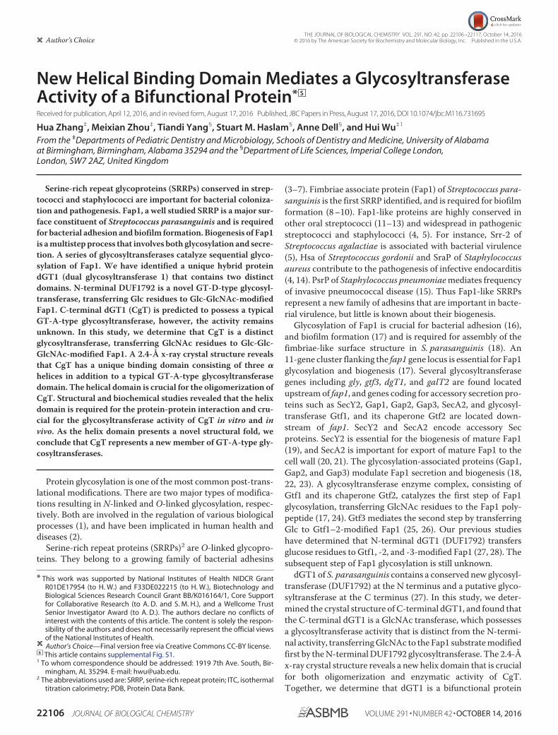
CgT Plays an Important Role in the Fap1 Glycosylation—dGT1 contains an N-terminal unknown function domain(DUF1792) and C-terminal putative glycosyltransferasedomain (CgT). Our studies have shown that DUF1792 is a newtype of glycosyltransferases, transferring glucose residues toGlc-GlcNAc-Fap1, and that the C terminus did not have thesame activity in vitro (27, 28). However, DUF1792 alone failedto complement the dGT1 mutant in S. parasanguinis (27), sug-gesting there is an unknown activity of CgT. To determine theCgT activity, we established an in vivo glycosylation system inEscherichia coli, using a small recombinant Fap1 (rFap1R1) as asubstrate (27). In the system, rFap1R1 was co-expressed withN-terminal DUF1792, C-terminal dGT1 (CgT), or the full-length dGT1 in addition to three glycosyltransferases, Gtf1,Gtf2, and Gtf3. The presence for DUF1792 super-shifted therecombinant Fap1 modified by Gtf123 (Fig. 1, lane 2 versus 1).Introduction of the C-terminal domain alone did not modifythe recombinant Fap1 glycosylated by Gtf123 (Fig. 1, lane 3).When only the full-length dGT1 (Fig. 1, lane 4) was used therecombinant Fap1 was further super-shifted to two highermolecular weight positions, indicating that the glycosylation ofrFap1RI by DUF1792 is a prerequisite for the subsequent mod-ification by CgT. These data suggest that CgT catalyzes thefourth step of the Fap1 glycosylation.
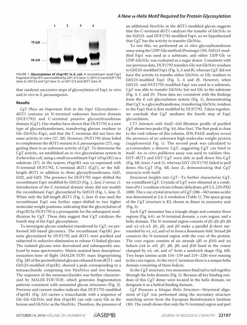
To investigate glycan residue(s) transferred by CgT, we per-formed MS-based glycomics. The recombinant Fap1R1 pro-teins glycosylated by DUF1792 and dGT1 were purified andsubjected to reductive elimination to release O-linked glycans.The isolated glycans were derivatized and subsequently ana-lyzed by mass spectrometry. Matrix-assisted laser desorption/ionization-time of flight (MALDI-TOF) mass fingerprinting(Fig. 2B) of the permethylated glycans released from dGT1- andGtf123-modified rFap1R1, showed a peak corresponding to atetrasaccharide comprising two HexNAcs and two hexoses.The sequence of the monosaccharides was further character-ized by MALDI-TOF/TOF, which generates fragmentationpatterns consistent with annotated glycan structures (Fig. 2).Previous and current studies indicate that DUF1792-modifiedrFap1R1 (Fig. 2A) carries a trisaccharide with a sequence ofGlc-Glc-GlcNAc and that rFap1R1 can only carry Glc as thehexose and GlcNAc as the HexNAc. Therefore, the presence of
an additional HexNAc in the dGT1-modified glycan suggeststhat the C-terminal dGT1 catalyzes the transfer of GlcNAc tothe Gtf123- and DUF1792-modified Fap1, so we hypothesizedthat CgT has the activity to transfer GlcNAc.
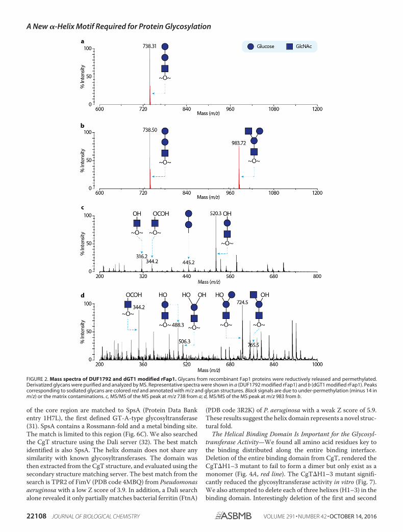
To test this, we performed an in vitro glycosyltransferaseassay using the UDP-Glo method (Promega) (29). Gtf123-mod-ified Fap1 was used as a substrate, and either UDP-Glc orUDP-GlcNAc was evaluated as a sugar donor. Consistent withour previous data, DUF1792 transfers Glc not GlcNAc residuesto Gtf123-modified Fap1 (Fig. 3, A and B), whereas CgT did nothave the activity to transfer either GlcNAc or Glc residues toGtf123-modified Fap1 (Fig. 3, A and B). However, whenGtf123- and DUF1792-modified Fap1 was used as a substrate,CgT was able to transfer GlcNAc but not Glc to the substrate(Fig. 3, C and D). These data are consistent with the findingsfrom the E. coli glycosylation system (Fig. 1), demonstratingthat CgT is a glycosyltransferase, transferring GlcNAc residuesto the Fap1 that is first modified by DUF1792. Taken together,we conclude that CgT mediates the fourth step of Fap1glycosylation.
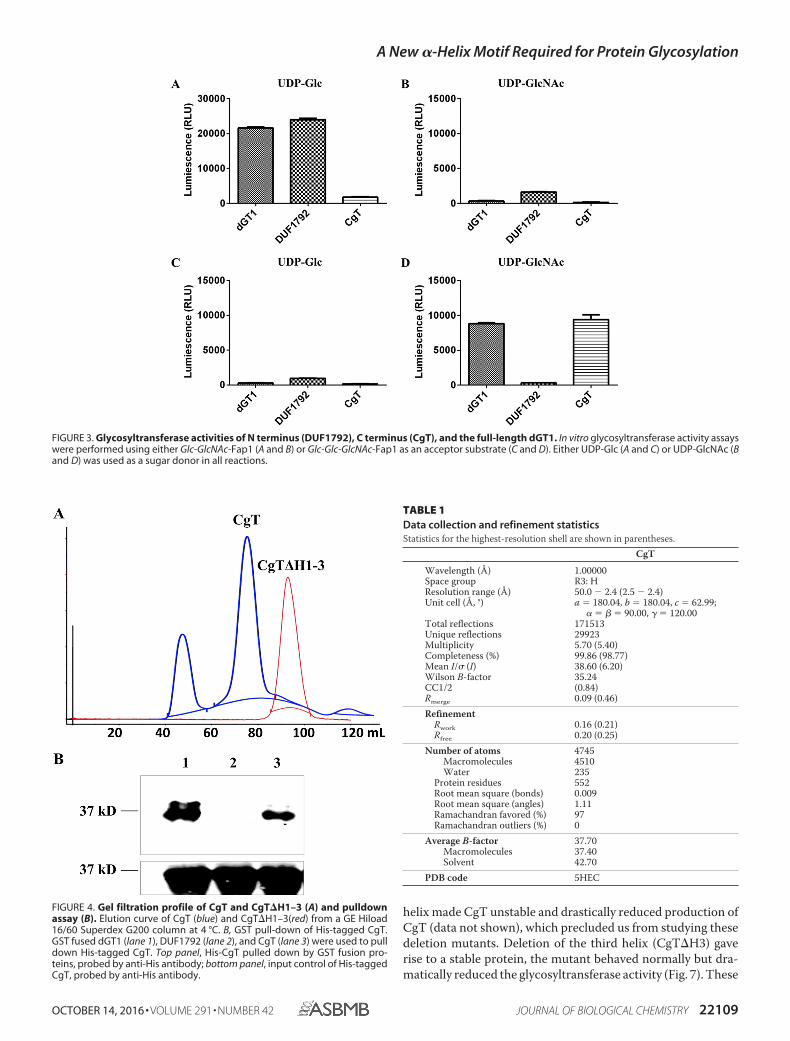
CT1 Interacts with Itself—Gel filtration profile of purifiedCgT shows two peaks (Fig. 4A, blue line). The first peak is closeto the void volume of this column, SDS-PAGE analysis revealthe presence of an unknown high molecular weight protein(supplemental Fig. 1). The second peak was calculated toaccommodate a dimeric CgT, suggesting CgT can bind toitself. An in vitro pulldown assay was used to validate this.GST-dGT1 and GST-CgT were able to pull down His-CgT(Fig. 4B, lanes 1 and 3), whereas GST-DUF1792 failed to pulldown His-CgT (Fig. 4B, lane 2), demonstrating that CgTinteracts with itself.
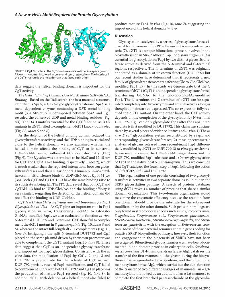
Structural Insights into CgT—To further characterize CgT,we crystallized CgT. Crystals of CgT were obtained at a condi-tion of 0.1 M sodium citrate tribasic dehydrate, pH 5.5, 22% PEG1000. The x-ray crystal structure of CgT (286 –582 amino acids)was determined at 2.4-Å resolution (Table 1). The space groupof the CgT structure is R3, shown in dimer in symmetry unit(Fig. 5).
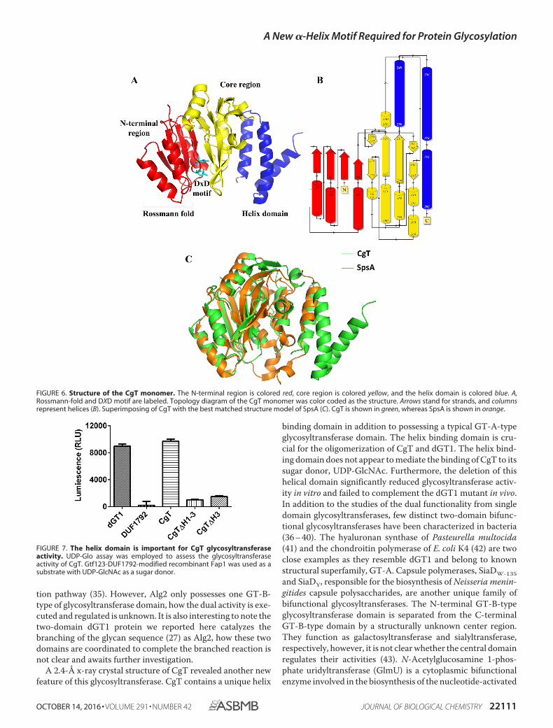
Each CgT monomer has a triangle shape and contains threeregions (Fig. 6A): an N-terminal domain, a core region, and ahelix domain. The N-terminal region consists of �1-�2-�3-�4and �1-�2-�3. �1, �2, and �3 make a parallel �-sheet sur-rounded by �1, �2, and �3 to form a Rossmann-fold. Strand �4connects the N-terminal region with the core of the protein.The core region consists of six strands (�5 to �10) and sixhelixes (�4 to �9). �7, �8, �9, and �10 lined in the centerclamped by �5, �6, and �7 form a sandwich shape (Fig. 6B).Two loops (amino acids 154 –159 and 219 –228) were missingin the core region. At the very C terminus there is a unique helixdomain consisting of three helices.
In the CgT structure, two monomers bind tail to tail togetherthrough the helix domain (Fig. 5). Because all key binding resi-dues of this CgT dimer were located in the helix domain, wedesignate it as a helical binding domain.
CgT Possesses a Unique Helix Structure—Structural align-ment of CgT was performed using the secondary structurematching server from the European Bioinformatics Institute(30). The result shows that only the N-terminal region and part
FIGURE 1. Glycosylation of rFap1R1 in E. coli. A recombinant small Fap1fragment (rFap1R1) was modified by Gtf1/2/3 (lane 1), Gtf1/2/3 and DUF1792(lane 2), Gtf123 and CgT (lane 3), or Gtf1/2/3 and dGT1 (lane 4).
A New �-Helix Motif Required for Protein Glycosylation
OCTOBER 14, 2016 • VOLUME 291 • NUMBER 42 JOURNAL OF BIOLOGICAL CHEMISTRY 22107
of the core region are matched to SpsA (Protein Data Bankentry 1H7L), the first defined GT-A-type glycosyltransferase(31). SpsA contains a Rossmann-fold and a metal binding site.The match is limited to this region (Fig. 6C). We also searchedthe CgT structure using the Dali server (32). The best matchidentified is also SpsA. The helix domain does not share anysimilarity with known glycosyltransferases. The domain wasthen extracted from the CgT structure, and evaluated using thesecondary structure matching server. The best match from thesearch is TPR2 of FimV (PDB code 4MBQ) from Pseudomonasaeruginosa with a low Z score of 3.9. In addition, a Dali searchalone revealed it only partially matches bacterial ferritin (FtnA)
(PDB code 3R2K) of P. aeruginosa with a weak Z score of 5.9.These results suggest the helix domain represents a novel struc-tural fold.
The Helical Binding Domain Is Important for the Glycosyl-transferase Activity—We found all amino acid residues key tothe binding distributed along the entire binding interface.Deletion of the entire binding domain from CgT, rendered theCgT�H1–3 mutant to fail to form a dimer but only exist as amonomer (Fig. 4A, red line). The CgT�H1–3 mutant signifi-cantly reduced the glycosyltransferase activity in vitro (Fig. 7).We also attempted to delete each of three helixes (H1–3) in thebinding domain. Interestingly deletion of the first and second
FIGURE 2. Mass spectra of DUF1792 and dGT1 modified rFap1. Glycans from recombinant Fap1 proteins were reductively released and permethylated.Derivatized glycans were purified and analyzed by MS. Representative spectra were shown in a (DUF1792 modified rFap1) and b (dGT1 modified rFap1). Peakscorresponding to sodiated glycans are colored red and annotated with m/z and glycan structures. Black signals are due to under-permethylation (minus 14 inm/z) or the matrix contaminations. c, MS/MS of the MS peak at m/z 738 from a; d, MS/MS of the MS peak at m/z 983 from b.
A New �-Helix Motif Required for Protein Glycosylation
22108 JOURNAL OF BIOLOGICAL CHEMISTRY VOLUME 291 • NUMBER 42 • OCTOBER 14, 2016
helix made CgT unstable and drastically reduced production ofCgT (data not shown), which precluded us from studying thesedeletion mutants. Deletion of the third helix (CgT�H3) gaverise to a stable protein, the mutant behaved normally but dra-matically reduced the glycosyltransferase activity (Fig. 7). These
FIGURE 3. Glycosyltransferase activities of N terminus (DUF1792), C terminus (CgT), and the full-length dGT1. In vitro glycosyltransferase activity assayswere performed using either Glc-GlcNAc-Fap1 (A and B) or Glc-Glc-GlcNAc-Fap1 as an acceptor substrate (C and D). Either UDP-Glc (A and C) or UDP-GlcNAc (Band D) was used as a sugar donor in all reactions.
FIGURE 4. Gel filtration profile of CgT and CgT�H1–3 (A) and pulldownassay (B). Elution curve of CgT (blue) and CgT�H1–3(red) from a GE Hiload16/60 Superdex G200 column at 4 °C. B, GST pull-down of His-tagged CgT.GST fused dGT1 (lane 1), DUF1792 (lane 2), and CgT (lane 3) were used to pulldown His-tagged CgT. Top panel, His-CgT pulled down by GST fusion pro-teins, probed by anti-His antibody; bottom panel, input control of His-taggedCgT, probed by anti-His antibody.
TABLE 1Data collection and refinement statisticsStatistics for the highest-resolution shell are shown in parentheses.
CgT
Wavelength (Å) 1.00000Space group R3: HResolution range (Å) 50.0 � 2.4 (2.5 � 2.4)Unit cell (Å, °) a � 180.04, b � 180.04, c � 62.99;
� � � � 90.00, � � 120.00Total reflections 171513Unique reflections 29923Multiplicity 5.70 (5.40)Completeness (%) 99.86 (98.77)Mean I/� (I) 38.60 (6.20)Wilson B-factor 35.24CC1/2 (0.84)Rmerge 0.09 (0.46)Refinement
Rwork 0.16 (0.21)Rfree 0.20 (0.25)
Number of atoms 4745Macromolecules 4510Water 235
Protein residues 552Root mean square (bonds) 0.009Root mean square (angles) 1.11Ramachandran favored (%) 97Ramachandran outliers (%) 0
Average B-factor 37.70Macromolecules 37.40Solvent 42.70
PDB code 5HEC
A New �-Helix Motif Required for Protein Glycosylation
OCTOBER 14, 2016 • VOLUME 291 • NUMBER 42 JOURNAL OF BIOLOGICAL CHEMISTRY 22109
data suggest the helical binding domain is important for theCgT activity.
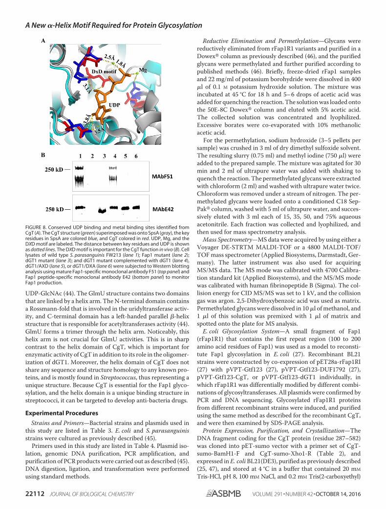
The Helical Binding Domain Does Not Mediate UDP-GlcNAcBinding—Based on the Dali search, the best matched structureidentified is SpsA, a GT-A-type glycosyltransferase. SpsA is ametal-dependent enzyme, containing a DXD metal bindingmotif (33). Structure superimposed between SpsA and CgTrevealed the conserved UDP and metal binding residues (Fig.8A). The DXD motif is essential for the CgT function, as DXDmutants in dGT1 failed to complement dGT1 knock-out in vivo(Fig. 8B, lanes 5 and 6).
As the deletion of the helical binding domain reduced theglycosyltransferase activity, and the UDP binding is crucial andclose to the helical domain, we also examined whether thehelical domain affects the binding of CgT to its substrateUDP-GlcNAc using isothermal titration calorimetry (ITC)(Fig. 9). The Kd value was determined to be 10.67 and 12.15 mM
for CgT and CgT�H1–3 binding, respectively (Table 2), whichis much weaker than the reported interactions between glyco-syltransferases and their sugar donors. Human �1,4-N-acteyl-hexosaminyltransferase binds to UDP-GlcNAc at Kd of 61 �M
(34). Both CgT and CgT�H1–3 have the same binding ratio toits substrate as being 1:1. The ITC data reveal that both CgT andCgT�H1–3 bind to UDP-GlcNAc, and the binding affinity isvery similar, suggesting the deletion of the helical domain didnot affect the binding to UDP-GlcNAc.
CgT Is a Distinct Glycosyltranferase and Important for Fap1Glycosylation in Vivo—As CgT plays an important role in Fap1glycosylation in vitro, transferring GlcNAc to Glc-Glc-GlcNAc-modified Fap1, we also evaluated its function in vivo.N-terminal DUF1792 and C-terminal CgT alone fail to comple-ment the dGT1 mutant in S. parasanguinis (Fig. 10, lanes 5 and6), whereas the intact full-length dGT1 complements (Fig. 10,lane 4). Intriguingly the split N-terminal DUF1792 and CgTplaced on the same plasmid driven by their own promoter wasable to complement the dGT1 mutant (Fig. 10, lane 8). Thesedata suggest that CgT is an independent glycosyltransferaseand important for Fap1 glycosylation. Consistent with the invitro data, the modification of Fap1 by Gtf1, -2, and -3 andDUF1792 is prerequisite for the activity of CgT in vivo.DUF1792 partially rescued Fap1 modification, and CgT failedto complement. Only with both DUF1792 and CgT in place wasthe production of mature Fap1 rescued (Fig. 10, lane 8). Inaddition, dGT1 with deletion of a helical motif also failed to
produce mature Fap1 in vivo (Fig. 10, lane 7), suggesting theimportance of the helical domain in vivo.
Discussion
Glycosylation catalyzed by a series of glycosyltransferases iscrucial for biogenesis of SRRP adhesins in Gram-positive bac-teria (7). dGT1 is a unique bifunctional protein involved in thebiosynthesis of an SRRP adhesin Fap1 of S. parasanguinis. It isessential for glycosylation of Fap1 by two distinct glycosyltrans-ferase activities derived from the N-terminal and C-terminalregions, respectively. The N terminus of dGT1 was originallyannotated as a domain of unknown function (DUF1792) butour recent studies have determined that it represents a newfamily of glycosyltransferases transferring Glc to Glc-GlcNAc-modified Fap1 (27). In this study we demonstrate that the Cterminus of dGT1 (CgT) is an independent glycosyltransferase,transferring GlcNAc to the Glc-Glc-GlcNAc-modifiedFap1. The N terminus and C terminus of dGT1 can be sepa-rated completely into two enzymes and are still active as long asthe split domains are co-expressed. The co-expression fully res-cued the dGT1 mutant. On the other hand, the CgT activitydepends on the completion of the glycosylation by N-terminalDUF1792. CgT can only glycosylate Fap1 after the Fap1 inter-mediate is first modified by DUF1792. This claim was substan-tiated by several pieces of evidence in vitro and in vivo. 1) The invivo E. coli glycosylation system reconstituted by rFap1 andcorresponding glycosyltransferases; 2) mass spectrometricanalysis of glycans released from recombinant Fap1 differen-tially modified by dGT1 or DUF1792; 3) in vitro glycosyltrans-ferase reactions using the UDP-GlcNAc sugar donor and theDUF1792-modified Fap1 substrate; and 4) in vivo glycosylationof Fap1 in the native host S. parasanguinis. Thus we concludethat CgT catalyzes the fourth step of Fap1 following the actionof Gtf1/Gtf2, Gtf3, and DUF1792.
The organization of one protein consisting of two glycosyl-transferase activities in two separate domains is unique in theSRRP glycosylation pathway. A search of protein databasesusing dGT1 reveals a number of proteins that share a similardomain organization. This two-domain organization wouldmaximize the enzymatic efficiency because the reaction fromone domain should provide the substrate for the subsequentmodification by the other domain. Such protein homologs areonly found in streptococcal species such as Streptococcus iniae,S. agalactiae, Streptococcus suis, Streptococcus plurextorum,Streptococcus lutetiensis, Streptococcus hyovaginalis, and Strep-tococcus gallolyticus with the exception of Enterococcus ceco-rum. Most of those bacterial genomes contain genes coding forputative SRRP biosynthetic pathways, however, their functionand engagement in the biogenesis of SRRPs have not beeninvestigated. Bifunctional glycosyltransferases have been docu-mented in one-domain proteins in eukaryotic cells. Saccharo-myces cerevisiae �1,4-mannosyl-transferase Alg1 catalyzes thetransfer of the first mannose to the glycan during the biosyn-thesis of asparagine-linked glycoproteins, and the bifunctionalmannosyltransferase Alg2, which carries out the dual functionof the transfer of two different linkages of mannoses, an �1,3-mannosylation followed by an addition of an �1,6-mannose tocomplete the first branched pentasaccharide of the glycosyla-
FIGURE 5. CgT Structure. The CgT structure exists in dimer in a space group ofR3; each monomer is colored in green and cyan, respectively. The interface inthe CgT structure is the helix domain that faced each other.
A New �-Helix Motif Required for Protein Glycosylation
22110 JOURNAL OF BIOLOGICAL CHEMISTRY VOLUME 291 • NUMBER 42 • OCTOBER 14, 2016
tion pathway (35). However, Alg2 only possesses one GT-B-type of glycosyltransferase domain, how the dual activity is exe-cuted and regulated is unknown. It is also interesting to note thetwo-domain dGT1 protein we reported here catalyzes thebranching of the glycan sequence (27) as Alg2, how these twodomains are coordinated to complete the branched reaction isnot clear and awaits further investigation.
A 2.4-Å x-ray crystal structure of CgT revealed another newfeature of this glycosyltransferase. CgT contains a unique helix
binding domain in addition to possessing a typical GT-A-typeglycosyltransferase domain. The helix binding domain is cru-cial for the oligomerization of CgT and dGT1. The helix bind-ing domain does not appear to mediate the binding of CgT to itssugar donor, UDP-GlcNAc. Furthermore, the deletion of thishelical domain significantly reduced glycosyltransferase activ-ity in vitro and failed to complement the dGT1 mutant in vivo.In addition to the studies of the dual functionality from singledomain glycosyltransferases, few distinct two-domain bifunc-tional glycosyltransferases have been characterized in bacteria(36 – 40). The hyaluronan synthase of Pasteurella multocida(41) and the chondroitin polymerase of E. coli K4 (42) are twoclose examples as they resemble dGT1 and belong to knownstructural superfamily, GT-A. Capsule polymerases, SiaDW-135and SiaDY, responsible for the biosynthesis of Neisseria menin-gitides capsule polysaccharides, are another unique family ofbifunctional glycosyltransferases. The N-terminal GT-B-typeglycosyltransferase domain is separated from the C-terminalGT-B-type domain by a structurally unknown center region.They function as galactosyltransferase and sialyltransferase,respectively, however, it is not clear whether the central domainregulates their activities (43). N-Acetylglucosamine 1-phos-phate uridyltransferase (GlmU) is a cytoplasmic bifunctionalenzyme involved in the biosynthesis of the nucleotide-activated
FIGURE 6. Structure of the CgT monomer. The N-terminal region is colored red, core region is colored yellow, and the helix domain is colored blue. A,Rossmann-fold and DXD motif are labeled. Topology diagram of the CgT monomer was color coded as the structure. Arrows stand for strands, and columnsrepresent helices (B). Superimposing of CgT with the best matched structure model of SpsA (C). CgT is shown in green, whereas SpsA is shown in orange.
FIGURE 7. The helix domain is important for CgT glycosyltransferaseactivity. UDP-Glo assay was employed to assess the glycosyltransferaseactivity of CgT. Gtf123-DUF1792-modified recombinant Fap1 was used as asubstrate with UDP-GlcNAc as a sugar donor.
A New �-Helix Motif Required for Protein Glycosylation
OCTOBER 14, 2016 • VOLUME 291 • NUMBER 42 JOURNAL OF BIOLOGICAL CHEMISTRY 22111
UDP-GlcNAc (44). The GlmU structure contains two domainsthat are linked by a helix arm. The N-terminal domain containsa Rossmann-fold that is involved in the uridyltransferase activ-ity, and C-terminal domain has a left-handed parallel �-helixstructure that is responsible for acetyltransferases activity (44).GlmU forms a trimer through the helix arm. Noticeably, thishelix arm is not crucial for GlmU activities. This is in sharpcontrast to the helix domain of CgT, which is important forenzymatic activity of CgT in addition to its role in the oligomer-ization of dGT1. Moreover, the helix domain of CgT does notshare any sequence and structure homology to any known pro-teins, and is mostly found in Streptococcus, thus representing aunique structure. Because CgT is essential for the Fap1 glyco-sylation, and the helix domain is a unique binding structure instreptococci, it can be targeted to develop anti-bacteria drugs.
Experimental Procedures
Strains and Primers—Bacterial strains and plasmids used inthis study are listed in Table 3. E. coli and S. parasanguinisstrains were cultured as previously described (45).
Primers used in this study are listed in Table 4. Plasmid iso-lation, genomic DNA purification, PCR amplification, andpurification of PCR products were carried out as described (45).DNA digestion, ligation, and transformation were performedusing standard methods.
Reductive Elimination and Permethylation—Glycans werereductively eliminated from rFap1R1 variants and purified in aDowex� column as previously described (46), and the purifiedglycans were permethylated and further purified according topublished methods (46). Briefly, freeze-dried rFap1 samplesand 22 mg/ml of potassium borohydride were dissolved in 400�l of 0.1 M potassium hydroxide solution. The mixture wasincubated at 45 °C for 18 h and 5– 6 drops of acetic acid wasadded for quenching the reaction. The solution was loaded ontothe 50E-8C Dowex� column and eluted with 5% acetic acid.The collected solution was concentrated and lyophilized.Excessive borates were co-evaporated with 10% methanolicacetic acid.
For the permethylation, sodium hydroxide (3–5 pellets persample) was crushed in 3 ml of dry dimethyl sulfoxide solvent.The resulting slurry (0.75 ml) and methyl iodine (750 �l) wereadded to the prepared sample. The mixture was agitated for 30min and 2 ml of ultrapure water was added with shaking toquench the reaction. The permethylated glycans were extractedwith chloroform (2 ml) and washed with ultrapure water twice.Chloroform was removed under a stream of nitrogen. The per-methylated glycans were loaded onto a conditioned C18 Sep-Pak� column, washed with 5 ml of ultrapure water, and succes-sively eluted with 3 ml each of 15, 35, 50, and 75% aqueousacetonitrile. Each fraction was collected and lyophilized, andthen used for mass spectrometry analysis.
Mass Spectrometry—MS data were acquired by using either aVoyager DE-STRTM MALDI-TOF or a 4800 MALDI-TOF/TOF mass spectrometer (Applied Biosystems, Darmstadt, Ger-many). The latter instrument was also used for acquiringMS/MS data. The MS mode was calibrated with 4700 Calibra-tion standard kit (Applied Biosystems), and the MS/MS modewas calibrated with human fibrinopeptide B (Sigma). The col-lision energy for CID MS/MS was set to 1 kV, and the collisiongas was argon. 2,5-Dihydroxybenzoic acid was used as matrix.Permethylated glycans were dissolved in 10 �l of methanol, and1 �l of this solution was premixed with 1 �l of matrix andspotted onto the plate for MS analysis.
E. coli Glycosylation System—A small fragment of Fap1(rFap1R1) that contains the first repeat region (100 to 200amino acid residues of Fap1) was used as a model to reconsti-tute Fap1 glycosylation in E. coli (27). Recombinant BL21strains were constructed by co-expression of pET28a-rFap1RI(27) with pVPT-Gtf123 (27), pVPT-Gtf123-DUF1792 (27),pVPT-Gtf123-CgT, or pVPT-Gtf123-dGT1 individually, inwhich rFap1R1 was differentially modified by different combi-nations of glycosyltransferases. All plasmids were confirmed byPCR and DNA sequencing. Glycosylated rFap1R1 proteinsfrom different recombinant strains were induced, and purifiedusing the same method as described for the recombinant CgT,and were then examined by SDS-PAGE analysis.
Protein Expression, Purification, and Crystallization—TheDNA fragment coding for the CgT protein (residue 287–582)was cloned into pET-sumo vector with a primer set of CgT-sumo-BamH1-F and CgT-sumo-Xho1-R (Table 2), andexpressed in E. coli BL21(DE3), purified as previously described(25, 47), and stored at 4 °C in a buffer that contained 20 mM
Tris-HCl, pH 8, 100 mM NaCl, and 0.2 mM Tris(2-carboxyethyl)
FIGURE 8. Conserved UDP binding and metal binding sites identified fromCgT (A). The CgT structure (green) superimposed was onto SpsA (gray), the keyresidues in SpsA are colored blue, and CgT colored in red. UDP, Mg, and theDXD motif are labeled. The distance between key residues and UDP is shownas dotted lines. The DXD motif is important for the CgT function in vivo (B). Celllysates of wild type S. parasanguinis FW213 (lane 1); Fap1 mutant (lane 2);dGT1 mutant (lane 3); and dGT1 mutant complemented with dGT1 (lane 4),dGT1/AXD (lane 5), or dGT1/DXA (lane 6) were subjected to Western blottinganalysis using mature Fap1-specific monoclonal antibody F51 (top panel) andFap1 peptide-specific monoclonal antibody E42 (bottom panel) to monitorFap1 production.
A New �-Helix Motif Required for Protein Glycosylation
22112 JOURNAL OF BIOLOGICAL CHEMISTRY VOLUME 291 • NUMBER 42 • OCTOBER 14, 2016

phosphine. Crystallization screens were conducted using thesitting drop vapor diffusion method with a Phoenix crystalliza-tion robot on a 96-well Intelli-plate (Art Robbins Instrument).Designed crystallizations were set up manually using thevapor diffusion hanging drop method with 24-well plates.The drops were set up at a 1:1 ratio of protein to motherliquor and incubated at 20 °C. Crystals grown in a well with
0.1 M sodium citrate tribasic dehydrate, pH 5.5, and 22%PEG1000 of IndexHT (Hampton Research) were used todetermine the CgT structure.
X-ray Data Collection and Processing—Diffraction datasetsfor CgT (2.4 Å) were collected at the Argonne National Labo-ratory on a beam station ID22 at 100 K. Paratone-N was used asa cryoprotectant. The datasets were processed by HKL2000(48).
Molecular Replacement and Refinement—Phases for CgTwere calculated using the structure of a glycosyltransferasefrom Bacteroides fragilis (PDB code 3BCV) as a starting modelby the molecular replacement method using PHENIX softwaresuite (49). Further refinement and model building were per-formed using the PHENIX software suite and COOT (49, 50).The final CgT structure was obtained through several refine-ment cycles, and TLS was used for the final refinement.
UDP-Glo Assay—UDP-GloTM assay (Promega) was used todetermine in vitro glycosyltransferase activity of CgT. His-tagged recombinant Fap1 (amino acid, 100 –200) proteins dif-ferentially modified by different glycosyltransferases were puri-fied and used as acceptors. Purified dGT1, DUF1792, and CgTwere used as enzymes, and either UDP-Glc or UDP-GlcNAc asa sugar donor. A corresponding acceptor, an enzyme, and asugar donor were reconstituted in the same reaction buffer (20
FIGURE 9. Isothermal titration calorimetric graphs of CgT and CgT�H1–3 titrated with UDP-GlcNAc. Binding of UDP-GlcNAc to CgT or CgT�H1–3 wasassessed as follows. The reaction cells contained either CgT (A) or CgT�H1–3 at 1 mM (B) and the syringe was supplied with UDP-GlcNAc at 15 mM. Data obtainedfrom 20 injections of 2-�l aliquots of UDP-GlcNAc at 3-min intervals are shown in the top graphs. The lower plots show the integrated binding isotherm with theexperimental points (f) and best fit.
TABLE 2ITC thermodynamic parameters�H, �S, number of binding sites (n), and the binding constant (Kd) for donor binding with proteins in solution at 20 °C.
Cell Ligand Number of sites Kd �H �S
n mM cal/mol cal/mol/degA CgT UDP-GlcNAc 1.10 � 0.02 10.67 � 1.33 �4358.5 � 65.5 3.80 � 0.47B CgT�H1–3 UDP-GlcNAc 0.88 � 0.01 12.15 � 1.35 �2148 � 63 11.45 � 0.45
FIGURE 10. CgT and the helix domain are required for dGT1 function invivo. Wild type S. parasanguinis FW213 (lane 1), Fap1 mutant (lane 2), dGT1mutant (lane 3) and its complement variants, dGT1 complemented with thefull-length dGT1 (lane 4), DUF1792 (lane 5), CgT (lane 6) with dGT1 without thehelix domain (lane 7), and with both DUF1792 and CgT (lane 8) were examinedfor the production of Fap1 by Western blotting analysis using Fap1 peptide-specific antibody E42 and mature Fap1-specific antibody F51.
A New �-Helix Motif Required for Protein Glycosylation
OCTOBER 14, 2016 • VOLUME 291 • NUMBER 42 JOURNAL OF BIOLOGICAL CHEMISTRY 22113

mM Tris, pH 8.0, 100 mM NaCl, 0.5 mM MnCl), in a total volumeof 5 �l and incubated for 2 h. 5 �l of UDP-Glo reagent wereadded into each mixture to react for 1 h. Luminescence wasdetermined to monitor glycosyltransferase activity.
Isothermal Titration Calorimetry—Thermodynamics ofbinding of enzyme (either CgT or CgT�H1–3) and ligand
UDP-GlcNAc were characterized using MicroCal Auto-iTC200 at 25 °C. Enzymes were dialyzed extensively against 20mM Tris-HCl buffer, pH 8.0, containing 100 mM NaCl and 20mM MgCl. Ligand UDP-GlcNAc was dissolved in the samebuffer. 400 �l of enzyme solutions (CgT or CgT�H1–3 at 1 mM)were titrated with 20 injections of the ligand (2 �l of
TABLE 3Strains and plasmids used in this study
Strains or plasmids Relevant properties Source
StrainsE. coli Top10 Host for propagation of the recombinant plasmids InvitrogenE. coli BL21-Gold (DE3) pET system hos strain InvitrogenS. parasanguinis FW213 Wild typeS. parasanguinis FW213 Fap1 deletion mutant Wild type; Fap1 knockout; Fap1::aphA3;Kanr 8S. parasanguinis FW213 dGT1 deletion mutant Wild type; dGT1 knockout; dGT1::aphA3;Kanr 27
Strains used to produce recombinant proteinsCgT pET-sumo: CgT transformed into BL21 In this studyHis-CgT pET-28b: CgT transformed into BL21 In this studyCgT�H1–3 pET-sumo: CgT�H1–3 transformed into BL21 In this studyCgT-DH1 pET-sumo: CgT-DH1 transformed into BL21 In this studyCgT-DH2 pET-sumo: CgT-DH2 transformed into BL21 In this studyCgT-DH3 pET-sumo: CgT-DH3 transformed into BL21 In this studyHis-sFap1-GlcNAc-Glc pET-28b: sFap1 and pvpt-Gtf123 co-transformed into BL21 27His-sFap1-GlcNAc-Glc-Glc pET-28b: sFap1 and pvpt-Gtf123-DUF1792 co-transformed into BL21 In this studyHis-sFap1-GlcNAc-Glc(-Glc)-GlcNAc pET-28b: sFap1 and pvpt-Gtf123-T1 co-transformed into BL21 In this studyGST-DUF1792 pGEx-5x-1: DUF1792 transformed into Top10 27GST-dGT1 pGEx-5x-1: dGT1 transformed into Top10 27GST-CgT pGEx-5x-1: CgT transformed into Top10 In this study
Plasmidspvpt-hsv-his E. coli-streptococci shuttle vector; Ermr 45pET-sumo His-SUMO fusion protein expression vector; Kanr 25pET-28b His fusion protein expression vector; Kanr AmershampGEx-6p-1 GST fusion protein expression vector; Ampr AmershampET-sumo: CgT CgT cloned in pET-sumo; Kanr In this studypET-28b: CgT CgT cloned in pET-28b; Kanr In this studypET-sumo: CgT�H1–3 Deletion of whole helix domain from pET-sumo: CgT; Kanr In this studypET-sumo: CgT-DH1 Deletion of first helix domain from pET-sumo: CgT; Kanr In this studypET-sumo: CgT-DH2 Deletion of second helix domain from pET-sumo: CgT; Kanr In this studypET-sumo: CgT-DH3 Deletion of third helix domain from pET-sumo: CgT; Kanr In this studypvpt-dGT1 dGT1 cloned into pvpt-hsv-his; Ermr In this studypvpt-DUF1792 DUF1792 cloned into pvpt-hsv-his; Ermr In this studypvpt-CgT CgT cloned into pvpt-hsv-his; Ermr In this studypvpt-DUF1792-pmal-CgT CgT with promoter cloned into pvpt-DUF1792; Ermr In this studypvpt-Gtf123 Gtf12 and Gtf3 cloned into pvpt-hsv-his; Ermr 27pvpt-Gtf123-DUF1792 Gtf12 and Gtf3 and DUF1792 cloned into pvpt-hsv-his; Ermr In this studypvpt-Gtf123-T1 Gtf12 and Gtf3 and dGT1 cloned into pvpt-hsv-his; Ermr In this studypvpt-dGT-AxD Site-direct mutant Asp378 to Ala from pvpt-dGT1; Ermr In this studypvpt-dGT-DxA Site-direct mutant Asp380 to Ala from pvpt-dGT1; Ermr In this studypGEx-6p-1: DUF1972 DUF1972 cloned into pEGx-6p-1; Ampr 27pGEx-6p-1: dGT1 dGT1 cloned into pEGx-6p-1; Ampr 27pGEx-6p-1: CgT CgT cloned into pEGx-6p-1; Ampr In this study
TABLE 4Primers used in this study
Primers Sequence
CgT-sumo-BamHI-F 5�-GATCAGGATCCATGGATAATGGTGAATTGATT-3�CgT-sumo-Xho1-R 5�-GATCACTCGAGTTATTTCTCCTTCGGATAATT-3�CgT-28b-BamHI-F 5�-GATCAGGATCCGATGGATAATGGTGAATTGATT-3�CgT-28b-XhoI-R 5�-GATCACTCGAGTTATTTCTCCTTCGGATAATT-3�CgT-pvpt-SalI-F 5�-GATCAGTCGACATGGATAATGGTGAATTGATT-3�CgT-pvpt-KpnI-R 5�-GATCAGGTACCTTATTTCTCCTTCGGATAATT-3�CgT�H1–3-Kpn1-R 5�-GATCAGGTACCATCTAACATTCGAGTTCTAGAAAGT-3�Deletion-helix1-F 5�-GATCAGTCGACTATCATACTGGATATATCATCTAA-3�Deletion-helix1-R 5�-GATCAGTCGACCTTCTTGCATCGATGGGCTATGAT-3�Deletion-helix2-F 5�-GATCAGTCGACAATTTGTTCAGTCAAATCATAGCC-3�Deletion-helix2-R 5�-GATCAGTCGACGCTTTACGAAATGGTCAAATTGAA-3�Deletion-helix3-F 5�-GATCAGTCGACAATTTGACCATTTCGTAAAGCGTC-3�Deletion-helix3-R 5�-GATCAGTCGACTTGATTGAAAATTATCCGAAGGAGAAA-3�CgT�H1–3-F 5�-GATCAGTCGACATGATTAGGTTGTTTGAATGGCT-3�CgT�H1–3-R 5�-GATCAGTCGACATCTAACATTCGAGTTCTAGAAAGT-3�Pvpt-promoterKpnI-F 5�-GATCAGGTACCATGAATGCTCATCCGGAATTC-3�dGT-D378A-F 5�-TAAATATATTACATTTGTGGCTTCAGACGATTTTGTAGAG-3�dGT-D378A-R 5�-CTCTACAAAATCGTCTGAAGCCACAAATGTAATATATTTA-3�dGT-D380A-F 5�-TATTACATTTGTGGATTCAGCCGATTTTGTAGAGGAATTCTA-3�dGT-D380A-R 5�-TAGAATTCCTCTACAAAATCGGCTGAATCCACAAATGTAAT-3�
A New �-Helix Motif Required for Protein Glycosylation
22114 JOURNAL OF BIOLOGICAL CHEMISTRY VOLUME 291 • NUMBER 42 • OCTOBER 14, 2016

UDP-GlcNAc at 15 mM). Each injection of the ligand lasted 4 swith 300-s intervals between successive injections. Bindingisotherms were generated by plotting the corrected heats ofbinding against the ratio of the ligand to enzymes. Softwaresupplied by the manufacturer (Origin version 7.0 fromMicrocal Inc.) was used to calculate dissociation constants(Kd), enthalpies of binding (�H), stoichiometry (n), andentropy of binding (�S).
GST Pulldown Assays—To examine in vitro self-interactionof dGT1, relevant GST and His-tagged fusion proteins wereprepared. Strains carrying plasmids pGEX-6P-1 and pGEX-dGT1/DUF1792/CgT were used for the preparation of GST,GST-dGT1, GST-DUF1792, and GST-CgT fusion proteins,and the strain carrying plasmid pET-28b-CgT was used for thepreparation of the His-tagged CgT protein. A primer set ofCgT-28b-BamHI-F and CgT-28b-XhoI-R was used to amplifythe CgT fragment to construct pET-28b-CgT. A primer set ofCgT-sumo-BamHI-F and CgT-sumo-XhoI-R was used toamplify the CgT gene and construct pGEX-6p-1-CgT. Con-firmed constructs were used to transform into appropriateE. coli strains to produce recombinant proteins.
GST pulldown assay was conducted as described (23, 51).In brief, GST fusion proteins were purified and bound toglutathione-Sepharose 4B beads (Amersham Biosciences)according to the manufacturer’s instructions. His-CgT waspurified by a His trap column (GE) according to the manufac-turer’s instructions. 50 �g of glutathione-Sepharose-boundGST fusion proteins were mixed with 40 �g of His-taggedCgT in the NETN buffer (20 mM Tris-HCl, pH 7.2, 100 mM
NaCl, 1 mM EDTA, 0.2% Nonidet P-40) and incubated over-night on a rotary shaker at 4 °C. The beads were washed 3times with 600 �l of NETN buffer and the proteins wereeluted with SDS-PAGE sample buffer, boiled for 10 min, andsubjected to Western blotting analysis with anti-His anti-body at 1:2000 dilution.
Complementation of dGT1 Mutant with dGT1 Variants—Shuttle plasmid pVPT-HSV-His was used as a vector to com-plement the dGT1 mutant. The DNA fragment correspondingto CgT was PCR amplified using a primer pair of CgT-pvpt-sal1-F and CgT-pvpt-Kpn1-R from the genomic DNA ofS. parasanguinis FW213. The amplified fragment was purifiedand then cloned into the pVPT-Hsv-His vector to obtainpVPT-CgT. The CgT gene fragment with a functional pro-moter from pVPT-CgT was amplified using a primer set ofpvpt-promoter-kpn1-F and CgT-pvpt-KpnI-R and then clonedinto pVPT-DUF1792 to yield pVPT-CgT/DUF172. Plasmidconstructs were confirmed by DNA sequencing, and thentransformed into the dGT1 mutant by electroporation asdescribed (27). Transformants were selected on TH agar con-taining 125 �g/ml of kanamycin and 10 �g/ml of erythromycin,and further verified by PCR analysis. The confirmed strainswere used in this study.
Site-directed Mutagenesis of the DXD Motif and Truncationof the Helical Binding Domain—Site-directed mutagenesis wascarried out using QuikChange mutagenesis kit as described(Stratagene) (25). The plasmid pVPT-dGT1 was used as a tem-plate, and two primer sets of dGT-D378A-F and dGT-D378A-R, and dGT-D380A-F and dGT-D380A-R (Table 2)
were used to construct the designed mutant alleles. Mutantconstructs were identified and confirmed by DNA sequencing,and transformed to the dGT1 mutant to generate dGT1 mutantvariants in S. parasanguinis.
To construct helical binding domain mutants, plasmidpET-sumo-CgT was used as a template, and the followingprimer sets, Deletion-helix1-F and Deletion-helix1-R, Dele-tion-helix2-F and Deletion-helix2-R, Deletion-helix3-F andDeletion-helix3-R, and CgT�H1–3 and CgT-�H1–3-R(Table 2) were used to delete helix1, helix2, helix3, or theentire helix domain, respectively. Mutant alleles were iden-tified and confirmed by PCR and DNA sequencing, andtransformed into E. coli to induce and express recombinantCgT variants.
Author Contributions—H. Z., M. Z., S. M. H., A. D., and H. W.designed the study. H. Z. and H. W. drafted the paper. H. Z. and T. Y.performed the experiments. H. Z., T. Y., S. M. H., A. D., and H. W.analyzed the results and wrote the final version of the manuscript.
Acknowledgments—We thank Zhengrong Yang for helping toaccess the Auto-iTC200 in the Biocalorimetry Laboratory sup-ported by National Institutes of Health Shared InstrumentationGrant 1S10RR026478 and Shared Facility Program of the UABComprehensive Cancer Center Grant 316851. X-ray data were col-lected at Southeast Regional Collaborative Access Team (SER-CAT) 22-ID beamline at the Advanced Photon Source, ArgonneNational Laboratory.
References1. Drickamer, K., and Taylor, M. E. (1998) Evolving views of protein glyco-
sylation. Trends Biochem. Sci. 23, 321–3242. Yang, Y. R., and Suh, P-G. (2014) O-GlcNAcylation in cellular functions
and human diseases. Advances in biological regulation 54, 68 –733. Zhang, Y. Q., Ren, S. X., Li, H. L., Wang, Y. X., Fu, G., Yang, J., Qin,
Z. Q., Miao, Y. G., Wang, W. Y., Chen, R. S., Shen, Y., Chen, Z., Yuan,Z. H., Zhao, G. P., Qu, D., Danchin, A., and Wen, Y. M. (2003) Genome-based analysis of virulence genes in a non-biofilm-forming Staphylo-coccus epidermidis strain (ATCC 12228). Mol. Microbiol. 49,1577–1593
4. Siboo, I. R., Chambers, H. F., and Sullam, P. M. (2005) Role of SraP, aserine-rich surface protein of Staphylococcus aureus, in binding to humanplatelets. Infect. Immun. 73, 2273–2280
5. Seifert, K. N., Adderson, E. E., Whiting, A. A., Bohnsack, J. F., Crowley,P. J., and Brady, L. J. (2006) A unique serine-rich repeat protein (Srr-2) andnovel surface antigen (�) associated with a virulent lineage of serotype IIIStreptococcus agalactiae. Microbiology 152, 1029 –1040
6. Sanchez, C. J., Shivshankar, P., Stol, K., Trakhtenbroit, S., Sullam, P. M.,Sauer, K., Hermans, P. W., and Orihuela, C. J. (2010) The pneumococcalserine-rich repeat protein is an intra-species bacterial adhesin that pro-motes bacterial aggregation in vivo and in biofilms. PLoS Pathog. 6,e1001044
7. Zhu, F., Zhang, H., and Wu, H. (2015) Glycosyltransferase-mediated sweetmodification in oral streptococci. J. Dent. Res. 94, 659 – 665
8. Wu, H., Mintz, K. P., Ladha, M., and Fives-Taylor, P. M. (1998) Isolationand characterization of Fap1, a fimbriae-associated adhesin of Streptococ-cus parasanguis FW213. Mol. Microbiol. 28, 487–500
9. Wu, H., and Fives-Taylor, P. M. (1999) Identification of dipeptide repeatsand a cell wall sorting signal in the fimbriae-associated adhesin, Fap1, ofStreptococcus parasanguis. Mol. Microbiol. 34, 1070 –1081
10. Froeliger, E. H., and Fives-Taylor, P. (2001) Streptococcus parasanguisfimbria-associated adhesin Fap1 is required for biofilm formation. Infect.Immun. 69, 2512–2519
A New �-Helix Motif Required for Protein Glycosylation
OCTOBER 14, 2016 • VOLUME 291 • NUMBER 42 JOURNAL OF BIOLOGICAL CHEMISTRY 22115
11. Bensing, B. A., and Sullam, P. M. (2002) An accessory sec locus of Strep-tococcus gordonii is required for export of the surface protein GspB and fornormal levels of binding to human platelets. Mol. Microbiol. 44,1081–1094
12. Takahashi, Y., Yajima, A., Cisar, J. O., and Konishi, K. (2004) Functionalanalysis of the Streptococcus gordonii DL1 sialic acid-binding adhesin andits essential role in bacterial binding to platelets. Infect. Immun. 72,3876 –3882
13. Plummer, C., Wu, H., Kerrigan, S. W., Meade, G., Cox, D., and Ian Doug-las, C. (2005) A serine-rich glycoprotein of Streptococcus sanguis mediatesadhesion to platelets via GPIb. Br. J. Haematol. 129, 101–109
14. Takahashi, Y., Takashima, E., Shimazu, K., Yagishita, H., Aoba, T., andKonishi, K. (2006) Contribution of sialic acid-binding adhesin to patho-genesis of experimental endocarditis caused by Streptococcus gordoniiDL1. Infect. Immun. 74, 740 –743
15. Shivshankar, P., Sanchez, C., Rose, L. F., and Orihuela, C. J. (2009) TheStreptococcus pneumoniae adhesin PsrP binds to Keratin 10 on lung cells.Mol. Microbiol. 73, 663– 679
16. Stephenson, A. E., Wu, H., Novak, J., Tomana, M., Mintz, K., and Fives-Taylor, P. (2002) The Fap1 fimbrial adhesin is a glycoprotein: antibodiesspecific for the glycan moiety block the adhesion of Streptococcus para-sanguis in an in vitro tooth model. Mol. Microbiol. 43, 147–157
17. Wu, H., Zeng, M., and Fives-Taylor, P. (2007) The glycan moieties and theN-terminal polypeptide backbone of a fimbria-associated adhesin, Fap1,play distinct roles in the biofilm development of Streptococcus parasan-guinis. Infect. Immun. 75, 2181–2188
18. Peng, Z., Fives-Taylor, P., Ruiz, T., Zhou, M., Sun, B., Chen, Q., and Wu, H.(2008) Identification of critical residues in Gap3 of Streptococcus parasan-guinis involved in Fap1 glycosylation, fimbrial formation and in vitro ad-hesion. BMC Microbiol. 8, 52
19. Wu, H., Bu, S., Newell, P., Chen, Q., and Fives-Taylor, P. (2007) Two genedeterminants are differentially involved in the biogenesis of Fap1 precur-sors in Streptococcus parasanguis. J. Bacteriol. 189, 1390 –1398
20. Chen, Q., Wu, H., and Fives-Taylor, P. M. (2004) Investigating the role ofsecA2 in secretion and glycosylation of a fimbrial adhesin in Streptococcusparasanguis FW213. Mol. Microbiol. 53, 843– 856
21. Zhou, M., Zhang, H., Zhu, F., and Wu, H. (2011) Canonical SecA associ-ates with an accessory secretory protein complex involved in biogenesis ofa streptococcal serine-rich repeat glycoprotein. J. Bacteriol. 193,6560 – 6566
22. Peng, Z., Wu, H., Ruiz, T., Chen, Q., Zhou, M., Sun, B., and Fives-Taylor,P. (2008) Role of gap3 in Fap1 glycosylation, stability, in vitro adhesion,and fimbrial and biofilm formation of Streptococcus parasanguinis. OralMicrobiol. Immunol. 23, 70 –78
23. Zhou, M., Zhu, F., Li, Y., Zhang, H., and Wu, H. (2012) Gap1 functions asa molecular chaperone to stabilize its interactive partner Gap3 duringbiogenesis of serine-rich repeat bacterial adhesin. Mol. Microbiol. 83,866 – 878
24. Wu, R., and Wu, H. (2011) A molecular chaperone mediates a two-proteinenzyme complex and glycosylation of serine-rich streptococcal adhesins.J. Biol. Chem. 286, 34923–34931
25. Zhu, F., Erlandsen, H., Ding, L., Li, J., Huang, Y., Zhou, M., Liang, X., Ma,J., and Wu, H. (2011) Structural and functional analysis of a new subfamilyof glycosyltransferases required for glycosylation of serine-rich strepto-coccal adhesins. J. Biol. Chem. 286, 27048 –27057
26. Zhou, M., Zhu, F., Dong, S., Pritchard, D. G., and Wu, H. (2010) A novelglucosyltransferase is required for glycosylation of a serine-rich adhesinand biofilm formation by Streptococcus parasanguinis. J. Biol. Chem. 285,12140 –12148
27. Zhang, H., Zhu, F., Yang, T., Ding, L., Zhou, M., Li, J., Haslam, S. M., Dell,A., Erlandsen, H., and Wu, H. (2014) The highly conserved domain ofunknown function 1792 has a distinct glycosyltransferase-fold. Nat. Com-mun. 5, 4339
28. Zhang, H., Zhu, F., Ding, L., Zhou, M., Wu, R., and Wu, H. (2013)Preliminary X-ray crystallographic studies of an N-terminal domain ofunknown function from a putative glycosyltransferase from Strepto-coccus parasanguinis. Acta Crystallogr. Sect. F Struct. Biol. Cryst. Com-mun. 69, 520 –523
29. Zegzouti, H., Engel, L., Hennek, J., Alves, J., Vidugiris, G., and Goueli, S.(2013) Detection of glycosyltransferase activities with homogeneous bio-luminescent UDP detection assay. in Glycobiology (Evans, R. D., ed) pp.2001, Oxford University Press Inc., Cary, NC
30. Krissinel, E., and Henrick, K. (2004) Secondary-structure matching (SSM),a new tool for fast protein structure alignment in three dimensions. ActaCrystallogr. D Biol. Crystallogr. 60, 2256 –2268
31. Tarbouriech, N., Charnock, S. J., and Davies, G. J. (2001) Three-dimen-sional structures of the Mn and Mg dTDP complexes of the family GT-2glycosyltransferase SpsA: a comparison with related NDP-sugar glycosyl-transferases. J. Mol. Biol. 314, 655– 661
32. Holm, L., and Rosenström, P. (2010) Dali server: conservation mapping in3D. Nucleic Acids Res. 38, W545–W549
33. Charnock, S. J., and Davies, G. J. (1999) Structure of the nucleotide-diphospho-sugar transferase, SpsA from Bacillus subtilis, in native andnucleotide-complexed forms. Biochemistry 38, 6380 – 6385
34. Sobhany, M., Dong, J., and Negishi, M. (2005) Two-step mechanism thatdetermines the donor binding specificity of human UDP-N-acetylhexo-saminyltransferase. J. Biol. Chem. 280, 23441–23445
35. O’Reilly, M. K., Zhang, G., and Imperiali, B. (2006) In vitro evidence for thedual function of Alg2 and Alg11: essential mannosyltransferases in N-linked glycoprotein biosynthesis. Biochemistry 45, 9593–9603
36. Hölzl, G., Leipelt, M., Ott, C., Zähringer, U., Lindner, B., Warnecke, D.,and Heinz, E. (2005) Processive lipid galactosyl/glucosyltransferases fromAgrobacterium tumefaciens and Mesorhizobium loti display multiplespecificities. Glycobiology 15, 874 – 886
37. Van Der Wel, H., Fisher, S. Z., and West, C. M. (2002) A bifunctionaldiglycosyltransferase forms the Fuc�1,2Gal�1,3-disaccharide on Skp1 inthe cytoplasm of Dictyostelium. J. Biol. Chem. 277, 46527– 46534
38. Wang, Z. A., van der Wel, H., Vohra, Y., Buskas, T., Boons, G-J., and West,C. M. (2009) Role of a cytoplasmic dual-function glycosyltransferase in O2regulation of development in Dictyostelium. J. Biol. Chem. 284,28896 –28904
39. Nakanishi, M., Karasudani, M., Shiraishi, T., Hashida, K., Hino, M., Fer-guson, M. A., and Nomoto, H. (2014) TbGT8 is a bifunctional glycosyl-transferase that elaborates N-linked glycans on a protein phosphataseAcP115 and a GPI-anchor modifying glycan in Trypanosoma brucei.Parasitol. Int. 63, 513–518
40. Izquierdo, L., Acosta-Serrano, A., Mehlert, A., and Ferguson, M. A.(2015) Identification of a glycosylphosphatidylinositol anchor-modify-ing �1–3-galactosyltransferase in Trypanosoma brucei. Glycobiology25, 438 – 447
41. Williams, K. J., Halkes, K. M., Kamerling, J. P., and DeAngelis, P. L.(2006) Critical elements of oligosaccharide acceptor substrates for thePasteurella multocida hyaluronan synthase. J. Biol. Chem. 281,5391–5397
42. Sobhany, M., Kakuta, Y., Sugiura, N., Kimata, K., and Negishi, M. (2008)The chondroitin polymerase K4CP and the molecular mechanism of se-lective bindings of donor substrates to two active sites. J. Biol. Chem. 283,32328 –32333
43. Romanow, A., Haselhorst, T., Stummeyer, K., Claus, H., Bethe, A., Müh-lenhoff, M., Vogel, U., von Itzstein, M., and Gerardy-Schahn, R. (2013)Biochemical and biophysical characterization of the sialyl-/hexosyltrans-ferase synthesizing the meningococcal serogroup W135 heteropolysac-charide capsule. J. Biol. Chem. 288, 11718 –11730
44. Brown, K., Pompeo, F., Dixon, S., Mengin-Lecreulx, D., Cambillau, C.,and Bourne, Y. (1999) Crystal structure of the bifunctional N-acetylg-lucosamine 1-phosphate uridyltransferase from Escherichia coli: a par-adigm for the related pyrophosphorylase superfamily. EMBO J. 18,4096 – 4107
45. Zhou, M., Fives-Taylor, P., and Wu, H. (2008) The utility of affinity-tagsfor detection of a streptococcal protein from a variety of streptococcalspecies. J. Microbiol. Methods 72, 249 –256
46. North, S. J., Jang-Lee, J., Harrison, R., Canis, K., Ismail, M. N., Trollope, A.,Antonopoulos, A., Pang, P-C., Grassi, P., and Al-Chalabi, S., Etienne, A. T.,Dell, A., and Haslam, S. M. (2010) Chapter two-mass spectrometric anal-ysis of mutant mice. Methods Enzymol. 478, 27–77
A New �-Helix Motif Required for Protein Glycosylation
22116 JOURNAL OF BIOLOGICAL CHEMISTRY VOLUME 291 • NUMBER 42 • OCTOBER 14, 2016

47. Zhu, F., Wu, R., Zhang, H., and Wu, H. (2013) Structural and biochemicalanalysis of a bacterial glycosyltransferase. Methods Mol. Biol. 1022, 29 –39
48. Otwinowski, Z., and Minor, W. (2001) Denzo and Scalepack. in Interna-tional Tables for Crystallography Volume F: Crystallography of BiologicalMacromolecules, pp. 226 –235, Springer, New York
49. Adams, P. D., Afonine, P. V., Bunkóczi, G., Chen, V. B., Davis, I. W.,Echols, N., Headd, J. J., Hung L-W, Kapral, G. J., and Grosse-Kunstleve,R. W. (2010) PHENIX: a comprehensive Python-based system for mac-
romolecular structure solution. Acta Crystallogr. D Biol. Crystallogr.66, 213–221
50. Emsley, P., and Cowtan, K. (2004) Coot: model-building tools for molec-ular graphics. Acta Crystallogr. D Biol. Crystallogr. 60, 2126 –2132
51. Echlin, H., Zhu, F., Li, Y., Peng, Z., Ruiz, T., Bedwell, G. J., Prevelige,P. E., Jr., and Wu, H. (2013) Gap2 promotes the formation of a stableprotein complex required for mature Fap1 biogenesis. J. Bacteriol. 195,2166 –2176
A New �-Helix Motif Required for Protein Glycosylation
OCTOBER 14, 2016 • VOLUME 291 • NUMBER 42 JOURNAL OF BIOLOGICAL CHEMISTRY 22117