hua zhang hhs public access 1 fan zhu1 tiandi yang2 lei … · form a protein complex that...
TRANSCRIPT

The highly conserved domain of unknown function 1792 has a distinct glycosyltransferase fold
Hua Zhang1, Fan Zhu1, Tiandi Yang2, Lei Ding3, Meixian Zhou1,#, Jingzhi Li4, Stuart M Haslam2, Anne Dell2, Heidi Erlandsen5,6, and Hui Wu1,*
1Departments of Pediatric Dentistry, Microbiology, University of Alabama at Birmingham, Schools of Dentistry and Medicine, Birmingham, AL35294
2Department of Life Sciences, Imperial College London, London SW7 2AZ, UK
3Department of Biochemistry and Molecular Genetics, University of Alabama at Birmingham, Schools of Dentistry and Medicine, Birmingham, AL35294
4Department of Cell Biology, University of Alabama at Birmingham, Schools of Dentistry and Medicine, Birmingham, AL35294
5Department of Periodontology, University of Alabama at Birmingham, Schools of Dentistry and Medicine, Birmingham, AL35294
6Institute of Oral Health Research, University of Alabama at Birmingham, Schools of Dentistry and Medicine, Birmingham, AL35294
Abstract
More than 33,000 glycosyltransferases have been identified. Structural studies, however, have
only revealed two distinct glycosyltransferase (GT) folds, GT-A and GT-B. Here we report a 1.34
Å resolution X-ray crystallographic structure of a previously uncharacterized “domain of unknown
function” 1792 (DUF1792) and show that the domain adopts a new fold and is required for
glycosylation of a family of serine-rich repeat streptococcal adhesins. Biochemical studies reveal
that the domain is a glucosyltransferase, and it catalyzes the transfer of glucose to the branch point
of the hexasaccharide O-linked to the serine-rich repeat of the bacterial adhesin, Fap1 of
Streptococcus parasanguinis. DUF1792 homologs from both Gram-positive and Gram-negative
bacteria also exhibit the activity. Thus DUF1792 represents a new family of glycosyltransferases,
Users may view, print, copy, and download text and data-mine the content in such documents, for the purposes of academic research, subject always to the full Conditions of use:http://www.nature.com/authors/editorial_policies/license.html#terms*Correspondence should be addressed to Hui Wu. [email protected].#Current Address: Key Laboratory of Marine Biogenetic Resources, Third Institute of Oceanography, State Oceanic Administration, Xiamen 361005, China
Author contributionsHua Zhang, Hui Wu, Anne Dell and Stuart M Haslam designed the study; Hua Zhang and Fan Zhu performed the structure/function experiments; Hua Zhang, Fan Zhu and Meixian Zhou and Hui Wu analyzed the structure/function experimental data; Lei Ding and Jingzhi Li collected X-ray data and performed modelling; Hua Zhang and Heidi Erlandsen analyzed the structural results. Tiandi Yang performed glycan structure analysis; Tiandi Yang, Stuart M Haslam and Anne Dell analyzed MS data; Hua Zhang, Hui Wu, Anne Dell, Stuart M Haslam and Tiandi Yang wrote the paper.
Competing financial interests: The authors declare no competing financial interests.
Accession codes: Coordinates and structure factors for DUF1792-Mn, DUF1792-native and DUF1792 (Se-Met) crystal structures have been deposited in the Protein Data Bank with the succession numbers 4PHR, 4PFX and 4PHS respectively.
HHS Public AccessAuthor manuscriptNat Commun. Author manuscript; available in PMC 2015 March 08.
Published in final edited form as:Nat Commun. ; 5: 4339. doi:10.1038/ncomms5339.
Author M
anuscriptA
uthor Manuscript
Author M
anuscriptA
uthor Manuscript

so we designate it as a GT-D glycosyltransferase fold. As the domain is highly conserved in
bacteria and not found in eukaryotes, it can be explored as a new antibacterial target.
Keywords
streptococcal adhesin; glycosyltransferase; DUF1792
INTRODUCTION
Protein glycosylation, catalyzed by glycosyltransferases, is an important protein
modification found in both prokaryotes1 and eukaryotes2 where it plays crucial roles in cell-
cell recognition, adhesion and intracellular sorting3, 4, 5. Since the classification system for
glycosyltransferases based on amino acid sequence similarity was proposed by Campbell et
al. in 19976, the number of glycosyltransferases has grown enormously to over 33,000,
organized into over 100 subfamilies6, 7, 8.
In contrast, numerous structural studies have revealed that the structural folds displayed by
this large number of glycosyltransferases are limited and only two distinct structural folds,
GT-A and GT-B have been rigorously characterized9, 10. GT-A displays a single Rossmann
fold (topology β/α/β/α/β) and a conserved ‘DXD’ metal-binding motif11, 12. In contrast, GT-
B possesses twin Rossmann folds that face each other and are linked flexibly by the active
site within the resulting cleft13, 14. In contrast this family does not require metal ions for its
activity. There is another previously named glycosyltransferase fold, the GT-C fold. Recent
structural studies of two predicted GT-C types of enzymes (oligosaccharyltransferase
STT315 and peptidoglycan synthesizing glycosyltransferase PBP216, 17) suggest that they
actually adopt different protein folds. Thus, whether GT-C represents a distinct
glycosyltransferase fold remains controversial.
Serine-rich repeat glycoproteins (SRRPs) are a growing family of bacterial adhesins and
they play important roles in bacterial fitness and virulence18, 19, 20. Fimbriae-associated
protein (Fap1) was the first SRRP identified21. It is heavily O-glycosylated by Glc-GlcNAc-
linked oligosaccharides containing up to four additional sugars22. Fap1 modulates bacterial
biofilm formation in the oral bacterium Streptococcus parasanguinis23. Fap1-like SRRPs
have since been identified from other streptococci24, 25, 26, staphylococci27, 28, 29 and other
Gram-positive bacteria30. Biogenesis of Fap1 in S. parasanguinis is controlled by a gene
cluster adjacent to this SRRP structural gene22. Analogous gene clusters are highly
conserved in streptococci and staphylococci30. Glycosylation and secretion of Fap1 is
mediated by eleven genes. A gene cluster coding for four putative glycosyltransferases, Gly,
Gtf3, GalT1, and GalT2, is located upstream of fap1, and another gene cluster producing
accessory secretion components, SecY2, SecA2, Gap1, Gap2 and Gap3, and two putative
glycosyltransferases (Gtf1 and Gtf2) is located downstream of fap131, 32. Gtf1 and Gtf2
form a protein complex that catalyzes the first step of glycosylation by transferring GlcNAc
residues to the Fap1 polypeptide31, 33, 34, while Gtf3 catalyzes the second step of
glycosylation by transferring Glc residues to the GlcNAc-modified Fap135, 36. However, it is
not yet known which enzymes mediate the subsequent glycosylation steps.
Zhang et al. Page 2
Nat Commun. Author manuscript; available in PMC 2015 March 08.
Author M
anuscriptA
uthor Manuscript
Author M
anuscriptA
uthor Manuscript
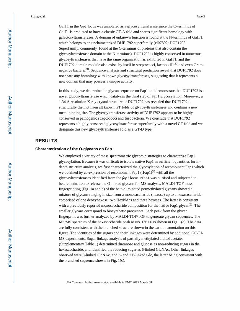
GalT1 in the fap1 locus was annotated as a glycosyltransferase since the C-terminus of
GalT1 is predicted to have a classic GT-A fold and shares significant homology with
galactosyltransferases. A domain of unknown function is found at the N-terminus of GalT1,
which belongs to an uncharacterized DUF1792 superfamily (cl07392: DUF1792
Superfamily, commonly_found at the C-terminus of proteins that also contain the
glycosyltransferase domain at the N-terminus). DUF1792 is highly conserved in numerous
glycosyltransferases that have the same organization as exhibited in GalT1, and the
DUF1792 domain module also exists by itself in streptococci, lactobacilli37 and even Gram-
negative bacteria38. Sequence analysis and structural prediction reveal that DUF1792 does
not share any homology with known glycosyltransferases, suggesting that it represents a
new domain that may possess a unique activity.
In this study, we determine the glycan sequence on Fap1 and demonstrate that DUF1792 is a
novel glucosyltransferase which catalyzes the third step of Fap1 glycosylation. Moreover, a
1.34 Å resolution X-ray crystal structure of DUF1792 has revealed that DUF1792 is
structurally distinct from all known GT folds of glycosyltransferases and contains a new
metal binding site. The glycosyltransferase activity of DUF1792 appears to be highly
conserved in pathogenic streptococci and fusobacteria. We conclude that DUF1792
represents a highly conserved glycosyltransferase superfamily with a novel GT fold and we
designate this new glycosyltransferase fold as a GT-D type.
RESULTS
Characterization of the O-glycans on Fap1
We employed a variety of mass spectrometric glycomic strategies to characterize Fap1
glycosylation. Because it was difficult to isolate native Fap1 in sufficient quantities for in-
depth structure analysis, we first characterized the glycosylation of recombinant Fap1 which
we obtained by co-expression of recombinant Fap1 (rFap1)35 with all the
glycosyltransferases identified from the fap1 locus. rFap1 was purified and subjected to
beta-elimination to release the O-linked glycans for MS analysis. MALDI-TOF mass
fingerprinting (Fig. 1a and b) of the beta-eliminated permethylated glycans showed a
mixture of glycans ranging in size from a monosaccharide (hexose) up to a hexasaccharide
comprised of one deoxyhexose, two HexNAcs and three hexoses. The latter is consistent
with a previously reported monosaccharide composition for the native Fap1 glycan22. The
smaller glycans correspond to biosynthetic precursors. Each peak from the glycan
fingerprint was further analyzed by MALDI-TOF/TOF to generate glycan sequences. The
MS/MS spectrum of the hexasaccharide peak at m/z 1361.6 is shown in Fig. 1(c). The data
are fully consistent with the branched structure shown in the cartoon annotation on this
figure. The identities of the sugars and their linkages were determined by additional GC-EI-
MS experiments. Sugar linkage analysis of partially methylated alditol acetates
(Supplementary Table 1) determined rhamnose and glucose as non-reducing sugars in the
hexasaccharide, and identified the reducing sugar as 6-linked GlcNAc. Other linkages
observed were 3-linked GlcNAc, and 3- and 2,6-linked Glc, the latter being consistent with
the branched sequence shown in Fig. 1(c).
Zhang et al. Page 3
Nat Commun. Author manuscript; available in PMC 2015 March 08.
Author M
anuscriptA
uthor Manuscript
Author M
anuscriptA
uthor Manuscript

Collectively, the glycomics data show that the largest Fap1 glycan has the sequence
Rha1-3Glc1-(Glc1-3GlcNAc1-)2,6Glc1-6GlcNAc. Moreover, the absence of a disaccharide
intermediate in the glycomic fingerprints (Fig. 1a and b) suggests that there is a rapid
incorporation of the second glucose in the biosynthetic pathway leading to the
hexasaccharide. Also, since the same glycan fingerprint was observed in the native Fap1
purified from S. parasanguinis (Supplementary Fig. 1), we conclude that the latter shares the
O-glycan sequences identified in our in-depth studies of recombinant samples.
DUF1792 is required for the third step of Fap1 glycosylation
While we have determined the first two steps of Fap1 glycosylation34, 36 the remaining
glycosylation steps are unknown. In a search for proteins responsible for the subsequent
steps of Fap1 glycosylation we identified dGT1 (previously named GalT1 because of its
annotated function; we rename it as dGT1 as it has two functional domains). dGT1 is
predicted to be a glycosyltransferase since it possesses a putative GT-A type
glycosyltransferase domain at the C-terminus. Interestingly, dGT1 also contains a distinct
domain of unknown function DUF1792 at the N-terminus (Fig. 2a). In vitro glycosylation
assays revealed that full-length dGT1 has a glucosyltransferase activity, transferring glucose
residues to Glc-GlcNAc modified Fap1 (Fig. 2b), suggesting dGT1 is involved in the third
step of Fap1 glycosylation. To dissect the individual dGT1 domain(s) involved, we
expressed both the N-terminal DUF1792 domain (amino acids 1–272) and the C-terminal
domain (amino acids 273–582), and determined their activity. Unexpectedly, the N-terminal
DUF1792 domain, but not the predicted C-terminal glycosyltransferase GT-A domain is
responsible for the in vitro glucosyltransferase activity (Fig. 2b). Moreover, the
glucosyltransferase activity of DUF1792 is dependent on the presence of metal ions (Fig.
2c). Mn2+ maximized the activity. However DUF1792 does not have the classic metal
binding motif, DXD, found in GT-A family of glycosyltransferases, suggesting that
DUF1792 represents a new type of glycosyltransferase.
To further define the function of DUF1792, we examined the ability of DUF1792 to catalyze
the third step of Fap1 glycosylation using a well-established E. coli glycosylation system35.
Since we have demonstrated that Gtf1/2 and Gtf3 catalyze the first two steps of Fap1
glycosylation respectively, we co-expressed either DUF1792 or the full-length dGT1 with
Gtf1/2, 3 and recombinant Fap1 (rFap1)35 to determine whether dGT1 or DUF1792 further
glycosylates the Gtf1/2,3 modified rFap1. Indeed, dGT1 retarded the migration of the
Gtf1/2,3 modified rFap1 (Fig. 3a, lane3 versus 2), suggesting additional modification by
dGT1. Interestingly, the migration of the modified rFap1 was further retarded when co-
expressed with the DUF1792 domain itself (Fig. 3a, lane 4). This is also true for the in vitro
glycosyltransferase activity (Fig. 2b). The activity of DUF1792 is consistently higher than
that from the full-length dGT1, suggesting the dGT1 C-terminus may have an additional
unknown glycosyltransferase activity that coordinates with the function of DUF1792 in
vitro. To further determine the relative contribution of DUF1792 and C-terminal dGT1 to
Fap1 glycosylation in the native host S. parasanguinis, the dGT1 mutant of S. parasanguinis
was complemented by either DUF1792 or C-terminal dGT1, and then examined by Fap1-
specific antibody mAbE42. The DUF1792 alone significantly retarded the migration of Fap1
indicative of glycosylation (Fig. 3b, lane 5) in comparison with the dGT1 mutant (Fig. 3b,
Zhang et al. Page 4
Nat Commun. Author manuscript; available in PMC 2015 March 08.
Author M
anuscriptA
uthor Manuscript
Author M
anuscriptA
uthor Manuscript
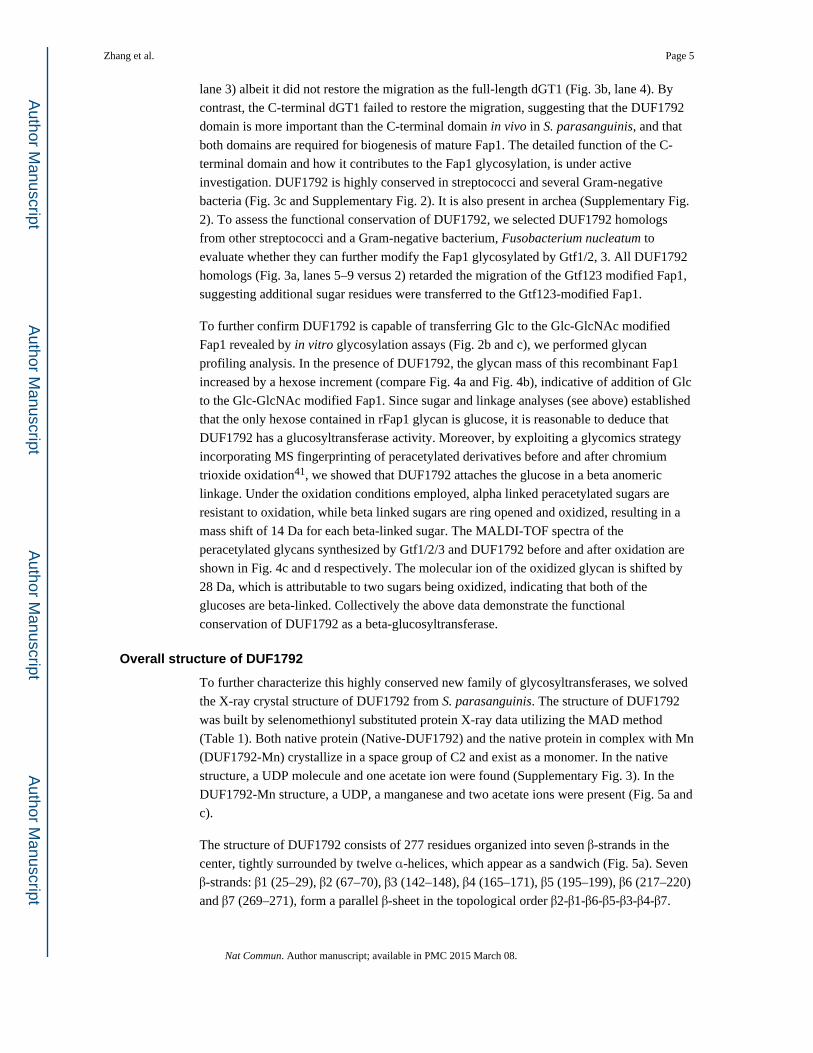
lane 3) albeit it did not restore the migration as the full-length dGT1 (Fig. 3b, lane 4). By
contrast, the C-terminal dGT1 failed to restore the migration, suggesting that the DUF1792
domain is more important than the C-terminal domain in vivo in S. parasanguinis, and that
both domains are required for biogenesis of mature Fap1. The detailed function of the C-
terminal domain and how it contributes to the Fap1 glycosylation, is under active
investigation. DUF1792 is highly conserved in streptococci and several Gram-negative
bacteria (Fig. 3c and Supplementary Fig. 2). It is also present in archea (Supplementary Fig.
2). To assess the functional conservation of DUF1792, we selected DUF1792 homologs
from other streptococci and a Gram-negative bacterium, Fusobacterium nucleatum to
evaluate whether they can further modify the Fap1 glycosylated by Gtf1/2, 3. All DUF1792
homologs (Fig. 3a, lanes 5–9 versus 2) retarded the migration of the Gtf123 modified Fap1,
suggesting additional sugar residues were transferred to the Gtf123-modified Fap1.
To further confirm DUF1792 is capable of transferring Glc to the Glc-GlcNAc modified
Fap1 revealed by in vitro glycosylation assays (Fig. 2b and c), we performed glycan
profiling analysis. In the presence of DUF1792, the glycan mass of this recombinant Fap1
increased by a hexose increment (compare Fig. 4a and Fig. 4b), indicative of addition of Glc
to the Glc-GlcNAc modified Fap1. Since sugar and linkage analyses (see above) established
that the only hexose contained in rFap1 glycan is glucose, it is reasonable to deduce that
DUF1792 has a glucosyltransferase activity. Moreover, by exploiting a glycomics strategy
incorporating MS fingerprinting of peracetylated derivatives before and after chromium
trioxide oxidation41, we showed that DUF1792 attaches the glucose in a beta anomeric
linkage. Under the oxidation conditions employed, alpha linked peracetylated sugars are
resistant to oxidation, while beta linked sugars are ring opened and oxidized, resulting in a
mass shift of 14 Da for each beta-linked sugar. The MALDI-TOF spectra of the
peracetylated glycans synthesized by Gtf1/2/3 and DUF1792 before and after oxidation are
shown in Fig. 4c and d respectively. The molecular ion of the oxidized glycan is shifted by
28 Da, which is attributable to two sugars being oxidized, indicating that both of the
glucoses are beta-linked. Collectively the above data demonstrate the functional
conservation of DUF1792 as a beta-glucosyltransferase.
Overall structure of DUF1792
To further characterize this highly conserved new family of glycosyltransferases, we solved
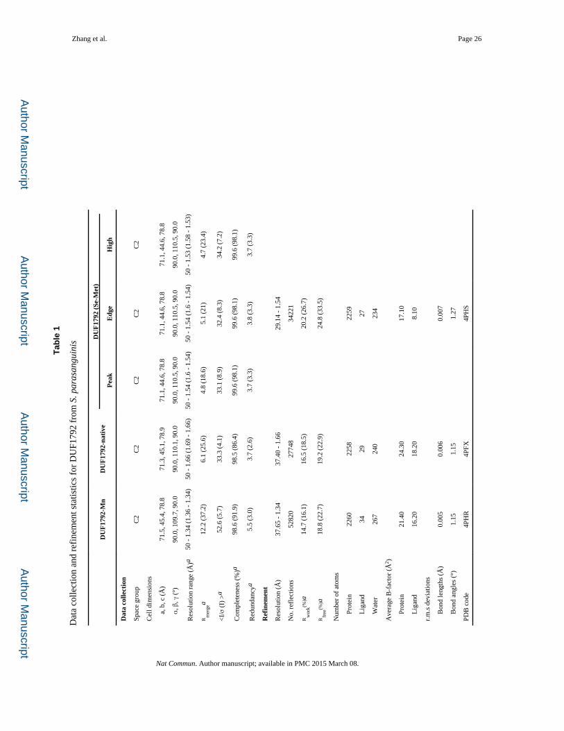
the X-ray crystal structure of DUF1792 from S. parasanguinis. The structure of DUF1792
was built by selenomethionyl substituted protein X-ray data utilizing the MAD method
(Table 1). Both native protein (Native-DUF1792) and the native protein in complex with Mn
(DUF1792-Mn) crystallize in a space group of C2 and exist as a monomer. In the native
structure, a UDP molecule and one acetate ion were found (Supplementary Fig. 3). In the
DUF1792-Mn structure, a UDP, a manganese and two acetate ions were present (Fig. 5a and
c).
The structure of DUF1792 consists of 277 residues organized into seven β-strands in the
center, tightly surrounded by twelve α-helices, which appear as a sandwich (Fig. 5a). Seven
β-strands: β1 (25–29), β2 (67–70), β3 (142–148), β4 (165–171), β5 (195–199), β6 (217–220)
and β7 (269–271), form a parallel β-sheet in the topological order β2-β1-β6-β5-β3-β4-β7.
Zhang et al. Page 5
Nat Commun. Author manuscript; available in PMC 2015 March 08.
Author M
anuscriptA
uthor Manuscript
Author M
anuscriptA
uthor Manuscript

The sheet is flanked by eight helices α1 (1–6), α5 (78–80), α6 (83–103), α2 (12–22), α7
(112–115), α8 (126–138), α11 (224–233) and α12 (261–266) on one side, and four helices
α3 (31–38), α4 (49–60), α9 (176–191) and α10 (202–214), on the other side.
The DUF1792 structure is composed of three regions: the N-terminal region formed from
helices α1, α2, α3, α4, α5 and two strands β1 and β2; α2β1α3and α4β2α5 form sandwich
domains consisting of the metal binding site, the DXE motif; α6 and α7 form a long turn-
helix-coil that connects the N-terminal region and the central region. The central region
contains a Rossmann-like fold (β4α9β5α10β6). The C-terminal region is composed of a long
coil region, a short helical turn (α12) and a short β-strand (β7). The nucleotide-binding sites
are located at the edge of the β-sheet (β1 and β6), helices α3 and α7 and the C-terminal loop.
The DUF1792 structure is distinctly different from the two classic glycosyltransferase folds,
GT-A and GT-B (Fig. 5d and e). In addition, it does not share any structural similarity with
a previously suggested GT-C fold.
The DUF1792 structure represents a new GT fold
To date, numerous structures have been solved and reported for glycosyltransferases.
Structural alignment of DUF1792 performed using the Secondary Structure Matching
server42 from the European Bioinformatics Institute revealed that the closest match is the
class B nonspecific acid phosphatase (AphA protein, PDB entry 1Z5G) from Salmonella
typhimurium, with a RMSD deviation of 4.7 Å. AphA has a haloacid dehalogenase-like fold
that is conserved in members of the DDDD superfamily of phosphohydrolases43. Upon
visual inspection, it is clear that only the so-called Rossmann-like fold of the DUF1792
structure superimposes onto the haloacid dehalogenase-like structure of AphA (Fig. 6a). A
general search using the DALI server44 indicated that the closest match to DUF1792 in the
database is 3-Dehydroquinate Synthase (DHQS) from Vibrio cholerae (PDB entry 3OKF)
with a weak Z score of 4.8 and a RMS deviation of 3.3Å (Fig. 6b). DHQS catalyzes the
formation of the first cyclic compound (3-Dehydroquinate) of the shikimate pathway, a
promising target for the design of antimicrobial compounds45. DHQS requires NAD and a
divalent cation to catalyze the reaction46. The similarity between DUF1792 and DHQS only
extends to the Rossmann-like fold. In addition, there is no sequence homology among the
three proteins. Key conserved residues important for glycosyltransferase activity among
DUF1792 homologs (Fig. 3c) are not found in either 3-Dehydroquinate Synthase or AphA,
further suggesting DUF1792 represents a new type of glycosyltransferase fold. Despite
structures of a large number of glycosyltransferases from two well defined GT folds being
resolved and documented8, 9, 10, the search failed to identify any structural homolog of
DUF1792 from this large pool of reported glycosyltransferases and only revealed a weak
homology to two non-glycosyltransferase enzymes, AphA and DHQS, further suggesting
that the structure of DUF1792 is unique. Because this structure is distinct from two currently
defined GT folds, GT-A and GT-B, and a previously designated GT-C fold39, 40,15, we have
named it as the GT-D fold.
Zhang et al. Page 6
Nat Commun. Author manuscript; available in PMC 2015 March 08.
Author M
anuscriptA
uthor Manuscript
Author M
anuscriptA
uthor Manuscript
The GT-D fold possesses UDP and manganese binding sites
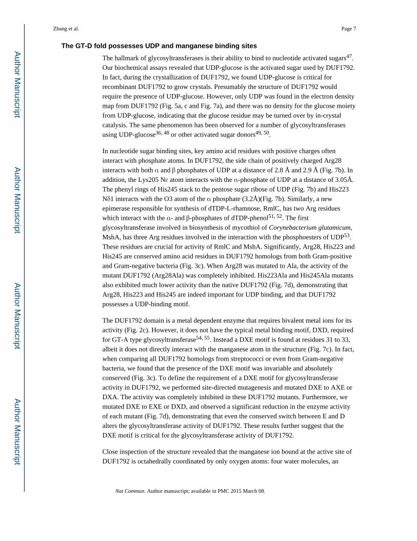
The hallmark of glycosyltransferases is their ability to bind to nucleotide activated sugars47.
Our biochemical assays revealed that UDP-glucose is the activated sugar used by DUF1792.
In fact, during the crystallization of DUF1792, we found UDP-glucose is critical for
recombinant DUF1792 to grow crystals. Presumably the structure of DUF1792 would
require the presence of UDP-glucose. However, only UDP was found in the electron density
map from DUF1792 (Fig. 5a, c and Fig. 7a), and there was no density for the glucose moiety
from UDP-glucose, indicating that the glucose residue may be turned over by in-crystal
catalysis. The same phenomenon has been observed for a number of glycosyltransferases
using UDP-glucose36, 48 or other activated sugar donors49, 50.
In nucleotide sugar binding sites, key amino acid residues with positive charges often
interact with phosphate atoms. In DUF1792, the side chain of positively charged Arg28
interacts with both α and β phosphates of UDP at a distance of 2.8 Å and 2.9 Å (Fig. 7b). In
addition, the Lys205 Nε atom interacts with the α-phosphate of UDP at a distance of 3.05Å.
The phenyl rings of His245 stack to the pentose sugar ribose of UDP (Fig. 7b) and His223
Nδ1 interacts with the O3 atom of the α phosphate (3.2Å)(Fig. 7b). Similarly, a new
epimerase responsible for synthesis of dTDP-L-rhamnose, RmlC, has two Arg residues
which interact with the α- and β-phosphates of dTDP-phenol51, 52. The first
glycosyltransferase involved in biosynthesis of mycothiol of Corynebacterium glutamicum,
MshA, has three Arg residues involved in the interaction with the phosphoesters of UDP53.
These residues are crucial for activity of RmlC and MshA. Significantly, Arg28, His223 and
His245 are conserved amino acid residues in DUF1792 homologs from both Gram-positive
and Gram-negative bacteria (Fig. 3c). When Arg28 was mutated to Ala, the activity of the
mutant DUF1792 (Arg28Ala) was completely inhibited. His223Ala and His245Ala mutants
also exhibited much lower activity than the native DUF1792 (Fig. 7d), demonstrating that
Arg28, His223 and His245 are indeed important for UDP binding, and that DUF1792
possesses a UDP-binding motif.
The DUF1792 domain is a metal dependent enzyme that requires bivalent metal ions for its
activity (Fig. 2c). However, it does not have the typical metal binding motif, DXD, required
for GT-A type glycosyltransferase54, 55. Instead a DXE motif is found at residues 31 to 33,
albeit it does not directly interact with the manganese atom in the structure (Fig. 7c). In fact,
when comparing all DUF1792 homologs from streptococci or even from Gram-negative
bacteria, we found that the presence of the DXE motif was invariable and absolutely
conserved (Fig. 3c). To define the requirement of a DXE motif for glycosyltransferase
activity in DUF1792, we performed site-directed mutagenesis and mutated DXE to AXE or
DXA. The activity was completely inhibited in these DUF1792 mutants. Furthermore, we
mutated DXE to EXE or DXD, and observed a significant reduction in the enzyme activity
of each mutant (Fig. 7d), demonstrating that even the conserved switch between E and D
alters the glycosyltransferase activity of DUF1792. These results further suggest that the
DXE motif is critical for the glycosyltransferase activity of DUF1792.
Close inspection of the structure revealed that the manganese ion bound at the active site of
DUF1792 is octahedrally coordinated by only oxygen atoms: four water molecules, an
Zhang et al. Page 7
Nat Commun. Author manuscript; available in PMC 2015 March 08.
Author M
anuscriptA
uthor Manuscript
Author M
anuscriptA
uthor Manuscript

oxygen atom from the β-phosphate of UDP, and one oxygen atom from the acetate ion most
proximal to the UDP (Fig. 7c). Together, these results demonstrate that DUF1792 contains a
metal binding (DXE) motif in this new family of glycosyltransferases.
UDP and metal binding are crucial for in vivo glycosylation of Fap1
To determine the requirement of UDP and metal binding sites in vivo in S. parasanguinis,
we selected one key residue Asp31 engaged in the metal binding, and another one His223
involved in the UDP binding, to carry out site-directed mutagenesis and determined the
impact of the mutated dGT1 alleles on biogenesis of Fap1 in S. parasanguinis. D31A, D31E
and H223A completely inhibited the production of mature Fap1 as determined by mature
Fap1 specific mAbF51 antibody (Fig. 8a, lanes 5–7). The Fap1 variants generated by the
dGT1 site-directed mutants (Fig. 8b, lanes 5–7) show a similar migration pattern to the Fap1
protein from the dGT1 non mutant (Fig. 8b, lane 3) when probed by Fap1-peptide specific
antibody mAbE42, further demonstrating the importance of these two motifs in the
glycosylation of Fap1 in vivo.
DISCUSSION
In this study we have defined a new glycosyltransferase superfamily, DUF1792, which is
involved in the biosynthesis of bacterial O-glycans. Glycomic strategies have revealed that
Fap1, a bacterial adhesin, is modified by a branched hexasaccharide with the sequence
Rha1-3Glcβ1-(Glc1-3GlcNAc1-) 2,6 Glcβ1-6GlcNAc. DUF1792 is a metal dependent beta-
glucosyltransferase, which transfers a Glc residue to the Glc-GlcNAc modified Fap1 at the
branching point. The DUF1792 domain has a Rossmann-like nucleotide-binding fold, but
does not show any sequence and structure identity with glycosyltransferases possessing
currently annotated type GT-A or GT-B folds. The domain does not share any structural
identity with a previously proposed GT-C fold either 39, 40,15. Moreover, DUF1792 has a
highly conserved DXE motif instead of the classic DXD metal-binding motif found in the
archetypical GT-A folds. Together our data lead us to propose that DUF1792 represents a
new family of glycosyltransferases that display a unique glycosyltransferase fold which we
have named GT-D.
The amino acid sequence constituting the fold is highly conserved in streptococci and even
in Gram-negative bacteria. Our biochemical and structural studies of DUF1792 have not
only defined the activity of this domain, but have also established a new family of bacterial
glycosyltransferases with a previously uncharacterized GT fold. This GT-D fold is crucial
for Fap1 biogenesis; moreover, the biogenesis of several Fap1-like proteins has been
implicated in bacterial virulence30. Thus our characterization of this new GT-D type of
glycosyltransferase may be helpful in guiding the design of antibacterial therapeutics
targeting this activity.
METHODS
Protein expression and purification
The DUF1792 domain encoding amino acids 1–272 of dGT1 (AFJ26875) was amplified
from genomic DNA of S. parasanguinis FW213 and cloned into the pET28a-sumo vector,
Zhang et al. Page 8
Nat Commun. Author manuscript; available in PMC 2015 March 08.
Author M
anuscriptA
uthor Manuscript
Author M
anuscriptA
uthor Manuscript

and transformed to E. coli BL21 Gold (DE3) cells. All strains and primers used in this study
were listed in Supplementary Table 2 and 3. The recombinant strain grown to OD600=0.8 in
LB medium was then induced with 0.1 mM IPTG at 18°C overnight. Native DUF1792
protein was purified with Histrap Column (Ni affinity) and gel filtration as described36. In
brief, the overnight grown E. coli cells were harvested by centrifugation and lysed by
sonication in binding buffer (20 mM Tris-HCl pH 8.0, 500 mM NaCl, and 25 mM
Imidazole). The clear cell lysates obtained after centrifugation (16, 500 rpm for 1 h) were
subjected to protein purification using HiTrapTM Column (Ni2+_affinity). Proteins were
eluted from the affinity resin by elution buffer (20 mM Tris-HCl, pH 8.0, 500 mM NaCl,
and 500 mM imidazole). The N-terminal His-SUMO tag was cleaved by incubating the
elution fractions with SUMO protease, ULP (ubiquitin-like protein protease), during
overnight dialysis at 4°C against 20 mM Tris-HCl, pH 8.0, 500 mM NaCl. Dialyzed protein
samples were reapplied to HiTrapTM Column to remove ULP, the cleaved His tag, and
uncleaved proteins. Flow-through was collected and further purified by using a 16/60
Superdex 75 gel filtration column (GE Healthcare) with gel filtration buffer (20 mM Tris pH
8.0, 100 mM NaCl and 1 mM DTT). Protein purity was analyzed by SDS-PAGE. Peak
fractions were collected and concentrated to 30 mg per ml for crystallization screen.
Selenomethionyl DUF1792 was obtained by growing the recombinant strain in M9 medium
with selenomethionine at 60 mg per L56. The selenomethionyl protein was purified using the
same protocol as the native protein.
Crystallization, data collection and refinement
The purified protein was concentrated to 10 mg per ml in 20 mMTris buffer (pH 8.0), 100
mM NaCl, 1mM DTT and subjected to crystallization trials. Hanging-drop vapor-diffusion
method was used for the crystallization trials. Crystals were obtained in the following
solution. 1 μL protein solution (10 mg per mL) with 10 mM UDP-Glc was mixed with 1 μL
well solution (500 uL) consisting of 100 mM Tris buffer (pH 8.5), 35% PEG 1500, 200 mM
Sodium Acetate. A single crystal was cryo-cooled in liquid nitrogen after being cryo
protected by addition of 10% glycerol. The Se-Met DUF1792 and native DUF1792 in the
complex with Mn were crystallized utilizing the same conditions. All DUF1792 crystals
appeared in three days.
Data were collected with an oscillation angle of 1° per image on beamline SER-CAT ID 22
at the Argonne National Laboratory. The structure was determined by multiwavelength
anomalous dispersion15, utilizing Se atoms as the anomalous scatterers. Se-Met-DUF1792
data were collected at wavelength of 0.97877 (Peak), 0.97907 (Edge) and 0.97142 (High).
Data of native-DUF1792 and DUF1792-Mn were collected at wavelength of 1.00000. All
data were collected under temperature 100 K. Data were processed and scaled with
HKL200057. The model building and subsequent structure refinement were performed with
the Phenix software58. Restrained individual B-factor and TLS refinement were not
performed until the last cycle. After each cycle of refinement, the model was manually
rebuilt based on the resulting 2Fo - Fc and Fo- Fc maps.
Zhang et al. Page 9
Nat Commun. Author manuscript; available in PMC 2015 March 08.
Author M
anuscriptA
uthor Manuscript
Author M
anuscriptA
uthor Manuscript

Expression and purification of rFap1-RI
We used rFap1-RI, a small fragment of Fap1 that contains the first repeat region (100 to 200
amino acid residues of Fap1), as a model to carry out glycan profiling study of Fap1.
Unglycosylated rFap1-R1 was purified from a recombinant strain that carries pET28a-
rFap1-R1. Glycosylated Fap1-R1 was purified from a recombinant BL21 strain that was
constructed by coexpression of pET28a-rFap1-R1 with pVPT-Gtf123-dGT1-GalT2 and
pHSG576-Gly, in which glycosylated rFap1-R1 was modified by all putative
glycosyltransferase, Gtf12, Gtf3, dGT1, GalT2 and Gly. The plasmids and strains were
constructed as described in Supplementary Table 2. All plasmids were confirmed by
sequencing. Unglycosylated and glycosylated rFap1-RI were purified using the same
method as recombinant DUF1792 protein.
Glycan profiling of modified rFap1-R1
Purified rFap1-R1 was subjected to reductive elimination and permethylation as described
below.
Reductive Elimination
The glycans were reductively eliminated from rFap1 and purified on a 50E-8C Dowex®
column as previously described59, and the purified glycans were subjected to permethylation
and purified according to published methods. Briefly, the freeze-dried rFap1 sample was
dissolved in 55 mg per mL potassium borohydride in a 1 mL of a 0.1 M potassium
hydroxide solution. The mixture was incubated at 45 °C for 18 hrs and quenched by adding
5–6 drops of acetic acid. The sample was loaded onto the Dowex® column and eluted with
5% acetic acid. The collected solution was concentrated and lyophilized. Excessive borates
were removed with 10% methanolic acetic acid.
Permethylation
For the permethylation reaction, sodium hydroxide (3–5 pellets per sample) was crushed in
3 mL dry dimethyl sulfoxide. The resulting slurry (0.75 mL) and methyl iodine (500 μL)
were added to the sample. The mixture was agitated for 15 min and quenched by adding 2
mL ultra-pure water with shaking. The glycans were extracted with chloroform (2mL) and
washed with ultra-pure water two times. Chloroform was removed under a stream of
nitrogen. The permethylated glycans were loaded onto a C18 Sep-pak® column, washed
with 5 mL ultra-pure water, and successively eluted with 3 mL each of 15%, 35%, 50% and
75% aq. acetonitrile. The solutions were collected and lyophilized.
Peracetylation
A previously described method was used for peracetylation60. Glycans were incubated with
200 μL pyridine and 200 μL acetic anhydride at 80°C for 3 hrs, after which the reagent was
removed under a stream of nitrogen. The acetylated glycans were dissolved in chloroform
and washed 3 times with pure water, and the chloroform was removed under a stream of
nitrogen.
Zhang et al. Page 10
Nat Commun. Author manuscript; available in PMC 2015 March 08.
Author M
anuscriptA
uthor Manuscript
Author M
anuscriptA
uthor Manuscript

CrO3 oxidation
10 mg CrO3 was added to 100 μL of acetic acid. The slurry was added to peracetylated
samples, and the mixture was heated to 50 °C and kept for 3 hrs. After quenching the
reaction with water, the product of oxidation was extracted with chloroform and washed
with water twice.
Mass Spectrometry
MS data were acquired by using either a Voyager DE-STRTM MALDI-TOF or a 4800
MALDI-TOF/TOF mass spectrometer (Applied Biosystems, Darmstadt, Germany). MS/MS
data were acquired with the latter instrument. MS mode was calibrated with 4700
Calibration standard kit (Applied Biosystems), and MS/MS mode was calibrated with
fibrinopeptide B human (Sigma). For MS/MS studies, the collision energy was set to 1 kV,
and the collision gas was argon. 2, 5-dihydroxybenzoic acid was used as matrix.
Permethylated samples were dissolved in 10 μL methanol, 1 μL of this solution was
premixed with 1 μL matrix and1 μL of the mixture was spotted onto the plate.
GC-MS Trimethylsilyl (TMS) analysis
The glycan sample was incubated with 1.0 M methanolic HCl at 80 °C for 14 hrs. The
reagent was removed under a stream of nitrogen. 500 μL methanol, 10 μL pyridine and 50
μL acetic anhydride and were successively added to the sample. The mixture was kept at
room temperature for 15 min and the reagent was removed under a stream of nitrogen. 200
μL Tri-Sil Z reagent was added to the sample, and the mixture was kept at room temperature
for 15 min. After removing the reagent under a stream of nitrogen, the sample was washed
by hexane twice. A PerkinElmer Clarus 500 instrument fitted with a RTX-5 fused silica
capillary column was used for carrying out the analysis. The following temperature
programme was used for eluting the sample. The oven temperature was initially 65 °C, and
heated to 140 °C at the rate of 25 °C per min, and heated to 200 °C at the rate of 5 °C per
min. The temperature was finally raised to 300 °C at a rate of 10 °C per min and is held for 5
min.
GC-MS Linkage Analysis
Partially methylated alditol acetates were prepared as previously described61. A
PerkinElmer Clarus 500 instrument fitted with a RTX-5 fused silica capillary column was
used for carrying out linkage analysis. A linear gradient temperature programme was used:
the sample was injected into the column at 60 °C, and the temperature increases to 300 °C
over 30 min at a rate of 8 °C per min.
Construct of a dgT1 knock-out mutant
A non-polar dgT1 knock-out mutant was generated by insertional mutagenesis with a
kanamycin resistance cassette (Kanr). Briefly, the dgT1 gene and its flanking regions
including the 600-bp upstream and 600-bp downstream regions were amplified from
genomic DNA of S. parasanguinis FW213. The PCR fragment was purified and cloned into
pGEM-T Easy vector (Promega, Madison, WI). A 1500-bp dgT1 internal fragment was
replaced with an 830-bp nonpolar kanamycin resistance cassette (aphA3) isolated from
Zhang et al. Page 11
Nat Commun. Author manuscript; available in PMC 2015 March 08.
Author M
anuscriptA
uthor Manuscript
Author M
anuscriptA
uthor Manuscript

pALH12462 by an inverse PCR strategy. Plasmid was confirmed by sequencing, and then
transformed into the FW213 strain by electroporation. The transformants were selected on
TH agar plates containing kanamycin. The dgT1 allelic replacement mutant was selected by
its ability to resist kanamycin and its susceptibility to tetracycline, and was further verified
by PCR and sequencing analysis. The confirmed dGT1 allelic replacement mutant was used
in this study.
Western blot analysis
For all S. parasanguinis strains, bacteria grown to an optical density at 470 nm (OD470) of
0.5 to 0.6 were harvested by centrifugation. The cell pellets were treated with amidase to
lyse the cells31. For E. coli strains, bacteria grown to an optical density at 600nm (OD600)
of 0.6 to 0.7 were harvested by centrifugation. Cell lysates were prepared by boiling the cell
pallets collected in sample buffer (0.0625 M Tris HCl [pH 6.8], 2% sodium dodecyl sulfate
[SDS], 10% glycerol, 0.01% bromophenol blue) for 10 min before being loaded onto 8%
SDS-polyacrylamide gel electrophoresis (PAGE) gels and subjected to Western blotting.
Two monoclonal anti-mouse antibodies (MAbs) were used to detect Fap1: MAb E42
(1:3000), which is specific to the peptide backbone of Fap1, and MAb F51(1:5000), which is
specific to mature Fap163. A polyclonal anti-rabbit antibody against DNAK (a gift from José
Lemos at University of Rochester) was used to standardize the protein loading of S.
parasanguinis lysates. All western blot figures shown in this paper were cropped.
Uncropped figures are supplied in Supplementary Figure 4.
Site-direct mutagenesis, in vitro and in vivo Fap1 glycosylation
Site-direct mutagenesis was carried out using QuickChange mutagenesis kit (Stratagene) as
described36. The plasmid pET28a-sumo-DUF1792 and pVPT-dGT1 were used as templates.
Mutant constructs were identified and confirmed by sequencing. Mutated DUF1792 proteins
were purified using the same method as described above. Gtf1/2,3 modified rFap1 was
purified using Glutathione Sepharose 4B beads according to the manufacturer’s protocol
(Amersham) and used as a substrate for the in vitro glycosyltransferase assays as described
previously35. In brief, the substrate and enzyme bound to glutathione-Sepharose beads were
washed five times with glycosylation buffer (50 mM Hepes, pH 7.0, 10 mM MnCl2, 0.01%
bovine serum albumin). 20 μg of recombinantdGT1 or its variants and substrate Fap1 were
mixed with 0.2 μCi of UDP-[3H]glucose (28 Ci/mmol; Amersham Biosciences) or 0.2 μCi
of UDP-[3H]GlcNAc (2.8 Ci/mmol; Amersham Biosciences) in a final volume of 200 μL of
glycosylation buffer and incubated for 1 h at 37°C. The beads in the glycosylation assays
were washed three times with NETN buffer (20 mM Tris-HCl, 100 mM NaCl, 1mM EDTA,
0.2% NP40, pH 7.0) and then transferred to scintillation vials to measure radioactivity
transferred to the Fap1 substrates from the radiolabeled activated sugars. The assays were
performed in triplicate in three independent experiments.
DUF1792 homologs from S. agalactiae J48, S. pneumonia TIGR4, S. sanguinis SK36 and
Fusobacterium nucleatum were amplified from each strain and cloned to pGEX-6p-1 to
generate pGEX-DUF1792 respectively. pHSG576-rFap1 (pAL80), pVPT-gtf1-2-3 and
pGEX-DUF1792 were co-transformed into E. coli. The ability of DUF1792 homologs to
Zhang et al. Page 12
Nat Commun. Author manuscript; available in PMC 2015 March 08.
Author M
anuscriptA
uthor Manuscript
Author M
anuscriptA
uthor Manuscript
glycosylate Gtf1/2, 3-modified rFap1 was examined using Western blotting analysis with
Fap1 peptide-specific monoclonal antibody E42 at 1:3000 dilution.
The construct pVPT-dGT1, pVPT-DUF1792, pVPT-dGT1-Cterminus and site-directed
mutants of dGT1 variants were used to transform the dGT1 knockout to determine the effect
of site-directed mutations within this DUF1792domain on Fap1 glycosylation in vivo.
Biogenesis of Fap1 was detected by Western blotting analysis using the Fap1-specific
monoclonal antibodies E42 (peptide-specific) and F51 (mature Fap1).
Statistics
The two-tailed Student’s t-test was used to determine statistically significant differences
between groups and statistically significant differences with p-values below 0.006 are
indicated with two asterisk (**) and below 0.0006 are indicated with three asterisk (***).
Supplementary Material
Refer to Web version on PubMed Central for supplementary material.
Acknowledgments
This study was supported by NIH/NIDCR F33DE022215 and R01DE017954 (H. Wu) and the Biotechnology and Biological Sciences Research Council (AD and SMH).
References
1. Nothaft H, Szymanski CM. Protein glycosylation in bacteria: sweeter than ever. Nature Reviews Microbiology. 2010; 8:765–778. [PubMed: 20948550]
2. Schwarz F, Aebi M. Mechanisms and principles of N-linked protein glycosylation. Current Opinion in Structural Biology. 2011; 21:576–582. [PubMed: 21978957]
3. Fletcher CM, Coyne MJ, Villa OF, Chatzidaki-Livanis M, Comstock LE. A general O-glycosylation system important to the physiology of a major human intestinal symbiont. Cell. 2009; 137:321–331. [PubMed: 19379697]
4. Ohtsubo K, Marth JD. Glycosylation in cellular mechanisms of health and disease. Cell. 2006; 126:855–867. [PubMed: 16959566]
5. Varki A. Nothing in glycobiology makes sense, except in the light of evolution. Cell. 2006; 126:841–845. [PubMed: 16959563]
6. Campbell J, Davies G, Bulone V, Henrissat B. A classification of nucleotide-diphospho-sugar glycosyltransferases based on amino acid sequence similarities. Biochemical Journal. 1997; 326:929. [PubMed: 9334165]
7. Drickamer K, Taylor ME. Evolving views of protein glycosylation. Trends in Biochemical Sciences. 1998; 23:321–324. [PubMed: 9787635]
8. Lairson L, Henrissat B, Davies G, Withers S. Glycosyltransferases: structures, functions, and mechanisms. Biochemistry. 2008; 77:521.
9. Liu J, Mushegian A. Three monophyletic superfamilies account for the majority of the known glycosyltransferases. Protein Science. 2003; 12:1418–1431. [PubMed: 12824488]
10. Coutinho PM, Deleury E, Davies GJ, Henrissat B. An evolving hierarchical family classification for glycosyltransferases. Journal of Molecular Biology. 2003; 328:307–317. [PubMed: 12691742]
11. Busch C, Hofmann F, Selzer J, Munro S, Jeckel D, Aktories K. A common motif of eukaryotic glycosyltransferases is essential for the enzyme activity of large clostridial cytotoxins. Journal of Biological Chemistry. 1998; 273:19566–19572. [PubMed: 9677381]
Zhang et al. Page 13
Nat Commun. Author manuscript; available in PMC 2015 March 08.
Author M
anuscriptA
uthor Manuscript
Author M
anuscriptA
uthor Manuscript
12. Charnock SJ, Davies GJ. Structure of the nucleotide-diphospho-sugar transferase, SpsA from Bacillus subtilis, in native and nucleotide-complexed forms. Biochemistry. 1999; 38:6380–6385. [PubMed: 10350455]
13. Vrielink A, Rüger W, Driessen H, Freemont PS. Crystal structure of the DNA modifying enzyme beta-glucosyltransferase in the presence and absence of the substrate uridine diphosphoglucose. The EMBO Journal. 1994; 13:3413. [PubMed: 8062817]
14. Moréra S, et al. High resolution crystal structures of T4 phage [beta]-glucosyltransferase: induced fit and effect of substrate and metal binding1. Journal of Molecular Biology. 2001; 311:569–577. [PubMed: 11493010]
15. Igura M, et al. Structure-guided identification of a new catalytic motif of oligosaccharyltransferase. The EMBO Journal. 2007; 27:234–243. [PubMed: 18046457]
16. Lovering AL, De Castro LH, Lim D, Strynadka NCJ. Structural insight into the transglycosylation step of bacterial cell-wall biosynthesis. Science. 2007; 315:1402. [PubMed: 17347437]
17. Yuan Y, Barrett D, Zhang Y, Kahne D, Sliz P, Walker S. Crystal structure of a peptidoglycan glycosyltransferase suggests a model for processive glycan chain synthesis. Proceedings of the National Academy of Sciences of the United States of America. 2007; 104:5348. [PubMed: 17360321]
18. Froeliger EH, Fives-Taylor P. Streptococcus parasanguis fimbria-associated adhesin Fap1 is required for biofilm formation. Infection and Immunity. 2001; 69:2512. [PubMed: 11254614]
19. Mistou MY, Dramsi S, Brega S, Poyart C, Trieu-Cuot P. Molecular dissection of the secA2 locus of group B Streptococcus reveals that glycosylation of the Srr1 LPXTG protein is required for full virulence. Journal of Bacteriology. 2009; 191:4195. [PubMed: 19395494]
20. Sanchez CJ, et al. The pneumococcal serine-rich repeat protein is an intra-species bacterial adhesin that promotes bacterial aggregation in vivo and in biofilms. PLoS Pathogens. 2010; 6:e1001044. [PubMed: 20714350]
21. Wu H, Mintz KP, Ladha M, Fives-Taylor PM. Isolation and characterization of Fap1, a fimbriae-associated adhesin of Streptococcus parasanguis FW213. Molecular Microbiology. 1998; 28:487–500. [PubMed: 9632253]
22. Bu S, et al. Interaction between two putative glycosyltransferases is required for glycosylation of a serine-rich streptococcal adhesin. Journal of Bacteriology. 2008; 190:1256–1266. [PubMed: 18083807]
23. Wu H, Fives-Taylor PM. Identification of dipeptide repeats and a cell wall sorting signal in the fimbriae-associated adhesin, Fap1, of Streptococcus parasanguis. Molecular Microbiology. 1999; 34:1070–1081. [PubMed: 10594831]
24. Bensing BA, Sullam PM. An accessory sec locus of Streptococcus gordonii is required for export of the surface protein GspB and for normal levels of binding to human platelets. Molecular Microbiology. 2002; 44:1081–1094. [PubMed: 12010500]
25. Xu P, et al. Genome of the opportunistic pathogen Streptococcus sanguinis. Journal of Bacteriology. 2007; 189:3166. [PubMed: 17277061]
26. Seifert KN, Adderson EE, Whiting AA, Bohnsack JF, Crowley PJ, Brady LJ. A unique serine-rich repeat protein (Srr-2) and novel surface antigen (ε) associated with a virulent lineage of serotype III Streptococcus agalactiae. Microbiology. 2006; 152:1029. [PubMed: 16549667]
27. Zhang YQ, et al. Genome-based analysis of virulence genes in a non-biofilm-forming Staphylococcus epidermidis strain (ATCC 12228). Molecular Microbiology. 2003; 49:1577–1593. [PubMed: 12950922]
28. Takeuchi F, et al. Whole-genome sequencing of Staphylococcus haemolyticus uncovers the extreme plasticity of its genome and the evolution of human-colonizing staphylococcal species. Journal of Bacteriology. 2005; 187:7292–7308. [PubMed: 16237012]
29. Siboo IR, Chaffin DO, Rubens CE, Sullam PM. Characterization of the accessory Sec system of Staphylococcus aureus. Journal of Bacteriology. 2008; 190:6188. [PubMed: 18621893]
30. Zhou M, Wu H. Glycosylation and biogenesis of a family of serine-rich bacterial adhesins. Microbiology. 2009; 155:317. [PubMed: 19202081]
Zhang et al. Page 14
Nat Commun. Author manuscript; available in PMC 2015 March 08.
Author M
anuscriptA
uthor Manuscript
Author M
anuscriptA
uthor Manuscript

31. Wu H, Bu S, Newell P, Chen Q, Fives-Taylor P. Two gene determinants are differentially involved in the biogenesis of Fap1 precursors in Streptococcus parasanguis. Journal of Bacteriology. 2007; 189:1390. [PubMed: 16997950]
32. Wu H, Zeng M, Fives-Taylor P. The glycan moieties and the N-terminal polypeptide backbone of a fimbria-associated adhesin, Fap1, play distinct roles in the biofilm development of Streptococcus parasanguinis. Infection and Immunity. 2007; 75:2181–2188. [PubMed: 17296746]
33. Wu R, Wu H. A Molecular Chaperone Mediates a Two-protein Enzyme Complex and Glycosylation of Serine-rich Streptococcal Adhesins. Journal of Biological Chemistry. 2011; 286:34923–34931. [PubMed: 21862581]
34. Wu R, Zhou M, Wu H. Purification and characterization of an active N-acetylglucosaminyltransferase enzyme complex from Streptococci. Applied and Environmental Microbiology. 2010; 76:7966–7971. [PubMed: 20971868]
35. Zhou M, Zhu F, Dong S, Pritchard DG, Wu H. A novel glucosyltransferase is required for glycosylation of a serine-rich adhesin and biofilm formation by Streptococcus parasanguinis. Journal of Biological Chemistry. 2010; 285:12140. [PubMed: 20164186]
36. Zhu F, et al. Structural and functional analysis of a new subfamily of glycosyltransferases required for glycosylation of serine-rich streptococcal adhesins. Journal of Biological Chemistry. 2011; 286:27048–27057. [PubMed: 21653318]
37. Forde BM, et al. Genome sequences and comparative genomics of two Lactobacillus ruminis strains from the bovine and human intestinal tracts. Microbial Cell Factories. 2011; 10:S13. [PubMed: 21995554]
38. Kuwahara T, et al. Genomic analysis of Bacteroides fragilis reveals extensive DNA inversions regulating cell surface adaptation. Proceedings of the National Academy of Sciences of the United States of America. 2004; 101:14919. [PubMed: 15466707]
39. Takahashi M, et al. PIG-B, a membrane protein of the endoplasmic reticulum with a large lumenal domain, is involved in transferring the third mannose of the GPI anchor. The EMBO Journal. 1996; 15:4254. [PubMed: 8861954]
40. Maeda Y, et al. PIG-M transfers the first mannose to glycosylphosphatidylinositol on the lumenal side of the ER. The EMBO Journal. 2001; 20:250–261. [PubMed: 11226175]
41. Khoo K-H, Dell A. Assignment of anomeric configurations of pyranose sugars in oligosaccharides using a sensitive FAB-MS strategy. Glycobiology. 1990; 1:83–91. [PubMed: 2136384]
42. Krissinel E, Henrick K. Secondary-structure matching (SSM), a new tool for fast protein structure alignment in three dimensions. Acta crystallographica Section D, Biological crystallography. 2004; 60:2256–2268.
43. Makde RD, Gupta GD, Mahajan SK, Kumar V. Structural and mutational analyses reveal the functional role of active-site Lys-154 and Asp-173 of Salmonella typhimurium AphA protein. Archives of Biochemistry and Biophysics. 2007; 464:70–79. [PubMed: 17570338]
44. Holm L, Rosenstrom P. Dali server: conservation mapping in 3D. Nucleic acids research. 2010; 38:W545–549. [PubMed: 20457744]
45. Hawkins AR, Lamb HK. The Molecular Biology of Multidomain Proteins Selected Examples. European Journal of Biochemistry. 1995; 232:7–18. [PubMed: 7556173]
46. Barten R, Meyer T. Cloning and characterisation of the Neisseria gonorrhoeaearoB gene. Molecular and General Genetics. 1998; 258:34–44. [PubMed: 9613570]
47. Breton C, Imberty A. Structure/function studies of glycosyltransferases. Current Opinion in Structural Biology. 1999; 9:563–571. [PubMed: 10508766]
48. Jinek M, Chen YW, Clausen H, Cohen SM, Conti E. Structural insights into the Notch-modifying glycosyltransferase Fringe. Nature Structural and Molecular Biology. 2006; 13:945–946.
49. Chiu CPC, et al. Structural analysis of the sialyltransferase CstII from Campylobacter jejuni in complex with a substrate analog. Nature Structural and Molecular Biology. 2004; 11:163–170.
50. Chang A, et al. Complete set of glycosyltransferase structures in the calicheamicin biosynthetic pathway reveals the origin of regiospecificity. Proceedings of the National Academy of Sciences of the United States of America. 2011; 108:17649–17654. [PubMed: 21987796]
Zhang et al. Page 15
Nat Commun. Author manuscript; available in PMC 2015 March 08.
Author M
anuscriptA
uthor Manuscript
Author M
anuscriptA
uthor Manuscript
51. Giraud MF, Leonard GA, Field RA, Berlind C, Naismith JH. RmlC, the third enzyme of dTDP-L-rhamnose pathway, is a new class of epimerase. Nature Structural & Molecular Biology. 2000; 7:398–402.
52. Ramakrishnan B, Boeggeman E, Ramasamy V, Qasba PK. Structure and catalytic cycle of [beta]-1, 4-galactosyltransferase. Current Opinion in Structural Biology. 2004; 14:593–600. [PubMed: 15465321]
53. Vetting MW, Frantom PA, Blanchard JS. Structural and Enzymatic Analysis of MshA from Corynebacterium glutamicum. Journal of Biological Chemistry. 2008; 283:15834–15844. [PubMed: 18390549]
54. Tarbouriech N, Charnock SJ, Davies GJ. Three-dimensional structures of the Mn and Mg dTDP complexes of the family GT-2 glycosyltransferase SpsA: a comparison with related NDP-sugar glycosyltransferases1. Journal of Molecular Biology. 2001; 314:655–661. [PubMed: 11733986]
55. Persson K, Ly HD, Dieckelmann M, Wakarchuk WW, Withers SG, Strynadka NCJ. Crystal structure of the retaining galactosyltransferase LgtC from Neisseria meningitidis in complex with donor and acceptor sugar analogs. Nature Structural and Molecular Biology. 2001; 8:166–175.
56. Doublié S. Preparation of selenomethionyl proteins for phase determination. Methods in Enzymology. 1997; 276:523–530. [PubMed: 9048379]
57. Otwinowski Z, Minor W. Processing of X-ray diffraction data collected in oscillation mode. Methods in Enzymology. 1997; 276:307–326.
58. Adams PD, et al. PHENIX: a comprehensive Python-based system for macromolecular structure solution. Acta crystallographica Section D, Biological crystallography. 2010; 66:213–221.
59. North SJ, et al. Chapter Two-Mass Spectrometric Analysis of Mutant Mice. Methods in Enzymology. 2010; 478:27–77. [PubMed: 20816474]
60. Haslam SM, Khoo K-H, Houston KM, Harnett W, Morris HR, Dell A. Characterisation of the phosphorylcholine-containing N-linked oligosaccharides in the excretory-secretory 62 kDa glycoprotein ofAcanthocheilonema viteae. Molecular and biochemical parasitology. 1997; 85:53–66. [PubMed: 9108548]
61. Jang-Lee J, et al. Glycomic profiling of cells and tissues by mass spectrometry: fingerprinting and sequencing methodologies. Methods in Enzymology. 2006; 415:59–86. [PubMed: 17116468]
62. Kremer BH, van der Kraan M, Crowley PJ, Hamilton IR, Brady LJ, Bleiweis AS. Characterization of the sat operon in Streptococcus mutans: evidence for a role of Ffh in acid tolerance. Journal of Bacteriology. 2001; 183:2543–2552. [PubMed: 11274114]
63. Stephenson AE, Wu H, Novak J, Tomana M, Mintz K, Fives-Taylor P. The Fap1 fimbrial adhesin is a glycoprotein: antibodies specific for the glycan moiety block the adhesion of Streptococcus parasanguis in an in vitro tooth model. Mol Microbiol. 2002; 43:147–157. [PubMed: 11849543]
Zhang et al. Page 16
Nat Commun. Author manuscript; available in PMC 2015 March 08.
Author M
anuscriptA
uthor Manuscript
Author M
anuscriptA
uthor Manuscript
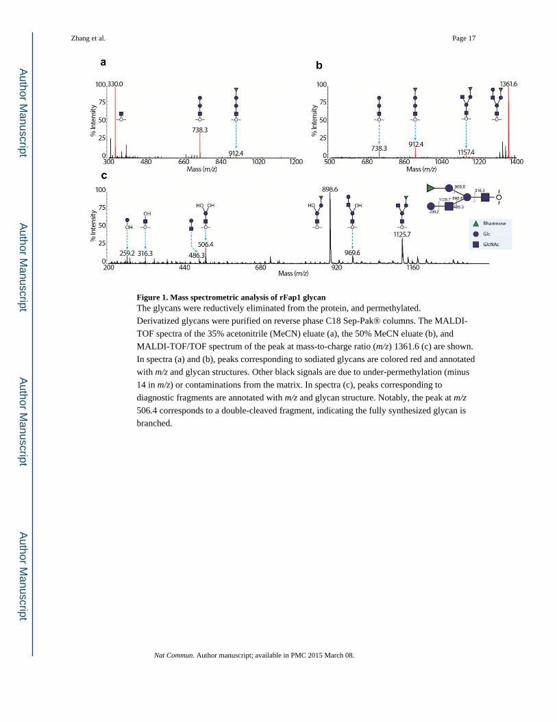
Figure 1. Mass spectrometric analysis of rFap1 glycanThe glycans were reductively eliminated from the protein, and permethylated.
Derivatized glycans were purified on reverse phase C18 Sep-Pak® columns. The MALDI-
TOF spectra of the 35% acetonitrile (MeCN) eluate (a), the 50% MeCN eluate (b), and
MALDI-TOF/TOF spectrum of the peak at mass-to-charge ratio (m/z) 1361.6 (c) are shown.
In spectra (a) and (b), peaks corresponding to sodiated glycans are colored red and annotated
with m/z and glycan structures. Other black signals are due to under-permethylation (minus
14 in m/z) or contaminations from the matrix. In spectra (c), peaks corresponding to
diagnostic fragments are annotated with m/z and glycan structure. Notably, the peak at m/z
506.4 corresponds to a double-cleaved fragment, indicating the fully synthesized glycan is
branched.
Zhang et al. Page 17
Nat Commun. Author manuscript; available in PMC 2015 March 08.
Author M
anuscriptA
uthor Manuscript
Author M
anuscriptA
uthor Manuscript
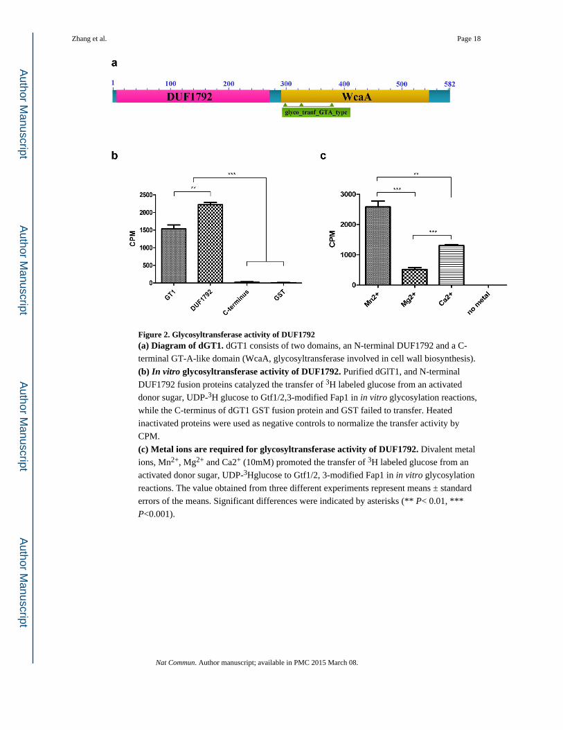
Figure 2. Glycosyltransferase activity of DUF1792(a) Diagram of dGT1. dGT1 consists of two domains, an N-terminal DUF1792 and a C-
terminal GT-A-like domain (WcaA, glycosyltransferase involved in cell wall biosynthesis).
(b) In vitro glycosyltransferase activity of DUF1792. Purified dGlT1, and N-terminal
DUF1792 fusion proteins catalyzed the transfer of 3H labeled glucose from an activated
donor sugar, UDP-3H glucose to Gtf1/2,3-modified Fap1 in in vitro glycosylation reactions,
while the C-terminus of dGT1 GST fusion protein and GST failed to transfer. Heated
inactivated proteins were used as negative controls to normalize the transfer activity by
CPM.
(c) Metal ions are required for glycosyltransferase activity of DUF1792. Divalent metal
ions, Mn2+, Mg2+ and Ca2+ (10mM) promoted the transfer of 3H labeled glucose from an
activated donor sugar, UDP-3Hglucose to Gtf1/2, 3-modified Fap1 in in vitro glycosylation
reactions. The value obtained from three different experiments represent means ± standard
errors of the means. Significant differences were indicated by asterisks (** P< 0.01, ***
P<0.001).
Zhang et al. Page 18
Nat Commun. Author manuscript; available in PMC 2015 March 08.
Author M
anuscriptA
uthor Manuscript
Author M
anuscriptA
uthor Manuscript

Figure 3. The glycosyltransferase activity and DXE motif of DUF1792 are conserved(a) DUF1792 is functionally conserved. DUF1792 homologs from a variety of bacterial
species were cloned into vector pGEx-6p-1 and co-transformed in E. coli carrying rFap1 and
Gtf1/2, 3 to determine the ability of DUF1792 to catalyze the transfer of additional sugar
residues to Gtf1/2, 3 modified rFap1. Cell lysates from rFap1 (1), Gtf1/2, 3 modified rFap1
(2) and Gtf1/2, 3 modified rFap1coexpressed with dGT1 of S. parasanguinis (3), or with
DUF1792 from S. parasanguinis (4), S. agalactiaeCOH1 (5); S. sanguinis SK36 (6), S.
agalactiae 2603 V/R (7), S. pneumoniae TIGR4 (8) and F. nucleatum F0401 (9) were
subjected to western blotting analysis with Fap1 specific antibody E42. All DUF1792
homologs retarded the migration of rFap1, suggesting they promoted the transfer of
additional sugar residues to Gtf1/2, 3 modified Fap1.
(b) Contribution of DUF1792 to glycosylation of Fap1. Wild type dGT1, N-terminal dGT1-
DUF1792, C-terminal dGT1 constructs were used to complement the dGT1mutant in S.
parasanguinis. Cell lysates from wild type S. parasanguinis (1); Fap1 mutant (2); dGT1
mutant (3); the dGT1 mutant complemented with the dGT1 full-length gene (4), DUF1792
(5); and C-terminal dGT1 (6) were subjected to western blotting analysis with Fap1-peptide
specific mAbE42 (top) and anti-DNAK antibody (bottom)as a sample loading control.
Complementation of DUF1792 produces an intermediate of Fap1, while the
complementation with the C-terminus alone has the same phenotype as dGT1 mutant.
(c)The DXE motif and UDP binding sites are highly conserved in bacteria. DUF1792
homologs were identified from a group of streptococci, Lactobacillus lactis and several
Gram-negative bacteria. The regions flanking the DGE motif, and UDP binding sites were
Zhang et al. Page 19
Nat Commun. Author manuscript; available in PMC 2015 March 08.
Author M
anuscriptA
uthor Manuscript
Author M
anuscriptA
uthor Manuscript
compared. The invariant amino acid residues highlighted in red and consensus sequence was
deduced.
Zhang et al. Page 20
Nat Commun. Author manuscript; available in PMC 2015 March 08.
Author M
anuscriptA
uthor Manuscript
Author M
anuscriptA
uthor Manuscript

Figure 4. MS spectra confirming in vivo glycosyltransferase activity of DUF1792The MALDI-TOF spectra of permethylated recombinant Fap1 glycans modified by
Gtf1/2/3, and Gtf1/2/3 plus DUF1792 are shown in (a) and (b) respectively. The shift of 204
in m/z corresponding to a permethylated hexose is consistent with the glucosyltransferase
activity of DUF1792. Recombinant Fap1 modified by Gtf1/2/3 shown in (b) were
peracetylated and oxidized by CrO3. MALDI-TOF spectra of the tri-saccharide from
recombinant Fap1 expressed with Gtf1/2/3 and DUF1792, which was analyzed as its
peracetylated derivative before and after CrO3 oxidation, are shown in (c) and (d)
respectively. A shift of 28 in m/z corresponding to two oxidations indicates both linkages are
β.
Zhang et al. Page 21
Nat Commun. Author manuscript; available in PMC 2015 March 08.
Author M
anuscriptA
uthor Manuscript
Author M
anuscriptA
uthor Manuscript
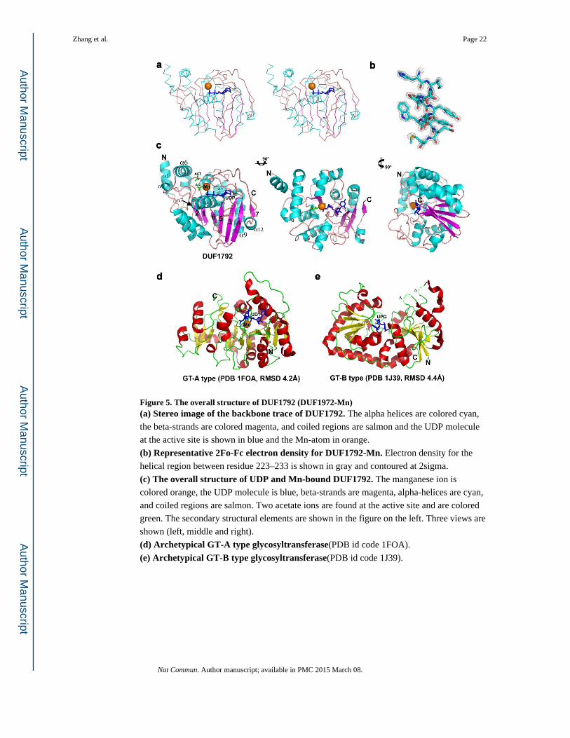
Figure 5. The overall structure of DUF1792 (DUF1972-Mn)(a) Stereo image of the backbone trace of DUF1792. The alpha helices are colored cyan,
the beta-strands are colored magenta, and coiled regions are salmon and the UDP molecule
at the active site is shown in blue and the Mn-atom in orange.
(b) Representative 2Fo-Fc electron density for DUF1792-Mn. Electron density for the
helical region between residue 223–233 is shown in gray and contoured at 2sigma.
(c) The overall structure of UDP and Mn-bound DUF1792. The manganese ion is
colored orange, the UDP molecule is blue, beta-strands are magenta, alpha-helices are cyan,
and coiled regions are salmon. Two acetate ions are found at the active site and are colored
green. The secondary structural elements are shown in the figure on the left. Three views are
shown (left, middle and right).
(d) Archetypical GT-A type glycosyltransferase(PDB id code 1FOA).
(e) Archetypical GT-B type glycosyltransferase(PDB id code 1J39).
Zhang et al. Page 22
Nat Commun. Author manuscript; available in PMC 2015 March 08.
Author M
anuscriptA
uthor Manuscript
Author M
anuscriptA
uthor Manuscript

Figure 6. Structural alignment of DUF1792(a) DUF1792 superimposed onto molecule A of AphA(PDB id code 1Z5G). DUF1792 is
shown in magenta cartoon, and AphA is shown in cyan cartoon. The UDP molecule in
DUF1792 is shown as stick and colored in blue and the Mn ion shown as an orange sphere.
(b) DUF1792 superimposed onto molecule A of 3-Dehydroquinate Synthase (DHQS) (PDB id code 3OKF). DUF1792 is shown in magenta cartoon, and 3-Dehydroquinate
Synthase is shown in green cartoon. The UDP molecule in DUF1792 is shown as blue stick
and the Mn ion shown as an orange sphere. The nicotinamide-adenine-dinucleotide is shown
in sand color stick along with the phosphate ion.
Zhang et al. Page 23
Nat Commun. Author manuscript; available in PMC 2015 March 08.
Author M
anuscriptA
uthor Manuscript
Author M
anuscriptA
uthor Manuscript

Figure 7. DUF1792 possesses UDP and Manganese binding sites(a) Cross-eyed stereo view showing the electron density of UDP and the manganese ion at the active site. The map shown is a simulated annealing composite omit electron density
map calculated in Phenix and contoured at 2.0σ.
(b) The UDP binding site. UDP and the amino acid residues involved in UDP binding are
labeled and atomic distances are shown in Ångströms.
(c) Manganese binding sites. Manganese and amino acid residues involved in manganese
binding are shown and labeled. The atomic distances are shown in Ångströms.
(d) Critical residues within UDP and metal binding sites required for glycosyltransferase activity of DUF1792. Site-direct mutagenesis was carried out to mutate
critical residues that are involved in binding to UDP and Mn2+, the mutant DUF1792 protein
variants were assayed for their in vitro glycosyltransferase activity. The value obtained from
three different experiments represent means ± standard errors of the means. Significant
differences were indicated by asterisks (*** P<0.001).
Zhang et al. Page 24
Nat Commun. Author manuscript; available in PMC 2015 March 08.
Author M
anuscriptA
uthor Manuscript
Author M
anuscriptA
uthor Manuscript

Figure 8. UDP and metal binding are required for Fap1 glycosylation in vivoWild type and dGT1 site-directed mutant constructs were used to complement the
dGT1mutant in S. parasanguinis. Cell lysates from S. parasanguinis (1); Fap1 mutant (2);
dGT1 mutant (3); the dGT1 mutant complemented with the dGT1 full-length gene (4), dGT1
(D31A) (5); dGT1(D31E) (6); and dGT1(H223A) (7) were subjected to western blotting
analysis with Fap1-peptide specific mAbE42 (a), mature Fap1-specific mAbF51 (b) to
determine Fap1 production, and anti-DNAK antibody (c) as a sample loading control.
Zhang et al. Page 25
Nat Commun. Author manuscript; available in PMC 2015 March 08.
Author M
anuscriptA
uthor Manuscript
Author M
anuscriptA
uthor Manuscript
Author M
anuscriptA
uthor Manuscript
Author M
anuscriptA
uthor Manuscript
Zhang et al. Page 26
Tab
le 1
Dat
a co
llect
ion
and
refi
nem
ent s
tatis
tics
for
DU
F179
2 fr
om S
. par
asan
guin
is
DU
F17
92-M
nD
UF
1792
-nat
ive
DU
F17
92 (
Se-M
et)
Pea
kE
dge
Hig
h
Dat
a co
llect
ion
Spac
e gr
oup
C2
C2
C2
C2
C2
Cel
l dim
ensi
ons
a,
b, c
(Å
)71
.5, 4
5.4,
78.
871
.3, 4
5.1,
78.
971
.1, 4
4.6,
78.
871
.1, 4
4.6,
78.
871
.1, 4
4.6,
78.
8
α
, β, γ
(°)
90.0
, 109
.7, 9
0.0
90.0
, 110
.1, 9
0.0
90.0
, 110
.5, 9
0.0
90.0
, 110
.5, 9
0.0
90.0
, 110
.5, 9
0.0
Res
olut
ion
rang
e (Å
)a50
- 1
.34
(1.3
6 -
1.34
)50
- 1
.66
(1.6
9 -
1.66
)50
- 1
.54
(1.6
- 1
.54)
50 -
1.5
4 (1
.6 -
1.5
4)50
- 1
.53
(1.5
8 -
1.53
)
Rm
erge
a12
.2 (
37.2
)6.
1 (2
5.6)
4.8
(18.
6)5.
1 (2
1)4.
7 (2
3.4)
<I/
σ (
I) >
a52
.6 (
5.7)
33.3
(4.
1)33
.1 (
8.9)
32.4
(8.
3)34
.2 (
7.2)
Com
plet
enes
s (%
)a98
.6 (
91.9
)98
.5 (
86.4
)99
.6 (
98.1
)99
.6 (
98.1
)99
.6 (
98.1
)
Red
unda
ncya
5.5
(3.0
)3.
7 (2
.6)
3.7
(3.3
)3.
8 (3
.3)
3.7
(3.3
)
Ref
inem
ent
Res
olut
ion
(Å)
37.6
5 -
1.34
37.4
0 -
1.66
29.1
4 -
1.54
No.
ref
lect
ions
5282
027
748
3422
1
Rw
ork(%
)a14
.7 (
16.1
)16
.5 (
18.5
)20
.2 (
26.7
)
Rfr
ee(%
)a18
.8 (
22.7
)19
.2 (
22.9
)24
.8 (
33.5
)
Num
ber
of a
tom
s
Pr
otei
n22
6022
5822
59
L
igan
d34
2927
W
ater
267
240
234
Ave
rage
B-f
acto
r (Å
2 )
Pr
otei
n21
.40
24.3
017
.10
L
igan
d16
.20
18.2
08.
10
r.m
.s d
evia
tions
B
ond
leng
ths
(Å)
0.00
50.
006
0.00
7
B
ond
angl
es (
°)1.
151.
151.
27
PDB
cod
e4P
HR
4PFX
4PH
S
Nat Commun. Author manuscript; available in PMC 2015 March 08.

Author M
anuscriptA
uthor Manuscript
Author M
anuscriptA
uthor Manuscript
Zhang et al. Page 27a V
alue
s in
par
enth
eses
are
for
the
high
est-
reso
lutio
n sh
ell.
Nat Commun. Author manuscript; available in PMC 2015 March 08.