new department of radiology, university of washington · 2017. 9. 7. · 4/2/2015 1 white matter,...
TRANSCRIPT

4/2/2015
1
White Matter, Metabolic,
and Degenerative Disease
UW Radiology Review 2015
Dean Shibata, M.DNeuroradiology
Department of Radiology,
University of Washington
• MS (Multiple Sclerosis)
• ADEM (Acute DisseminatedEncephalomyelitis)
• CPM (Central Pontine Myelinolysis)
• PRES (Posterior Reversible Encephalopathy Syndrome)
• PML (Progressive Multifocal Leukoencephalopathy)

4/2/2015
2
WM Dz: Classific by Mechanism
• Primary demyelinating disease¯ Multiple Sclerosis
• Secondary demyelinating disease¯ Allergic
¯ Viral
¯ Vascular
¯ Metabolic
¯ Toxic
WM Dz: Classific by Mechanism
• Allergic (immunologic)¯ Acute disseminated encephalomyelitis - ADEM
• Viral¯ HIV-associated encephalitis
¯ Prog multifocal leukoencephalopathy - PML
¯ Subacute sclerosing Panencephalitis y – SSPE
• Vascular¯ Binswanger’s disease – (WM sml vessel dz)
¯ Postanoxic encephalopathy
¯ Toxic

4/2/2015
3
WM Dz: Classific by Mechanism
• Toxic¯ Radiation
¯ Marchiafava-Bignami Disease (Corpus Collosum)
¯ Disseminated necrotizing leukoencephalopathy
¯ Drugs (chemoRx, metamphetamine, cocaine)
¯ Toxins (triethyl, tin, lead)
• Metabolic¯ central pontine myelinolysis (osmotic) - CPM
• Traumatic: Diffuse axonal shear injury - DAI
WM Dz: Classific by Mechanism
• Dysmyelinating Disease¯ Adrenoleukodystrophy – Males, posterior,
Pons/Medulla, Gad+
¯ Metachromatic Leukodystrophy (MLD) –cerebellum, spares subcortical U fibers and BG
¯ Krabbe’s Disease – Ca++ BG
¯ Alexander Disease – Big head, Anterior
¯ Canavan’s Disease – Big head, NAA, stem/BG
¯ Pelizaeus-Merzbacher Disease (PMD) – tigroid WM
¯ Toxins (solvents, tin, lead)

4/2/2015
4
Metachromatic leukodystrophy
Cheon J et al. Radiographics 2002;22:461-476©2002 by Radiological Society of North America
. Tigroid WM
(AlsoPelizaeus-Merzbacher)
Metachromatic leukodystrophy
Cheon J et al. Radiographics 2002;22:461-476©2002 by Radiological Society of North America
Leopard Skin WM

4/2/2015
5
Hallervorden-SpatzPantothenate kinase-associated neurodegeneration (PKAN)
©2002 by Radiological Society of North AmericaGuillerman R P Radiology 2000;217:895-896
Canavan’sBarkovich 1995

4/2/2015
6
Which one is Adrenoleukodystrophy?
1 2
Adrenoleukodystrophy?
1. Post Dz
2. Ant Dz

4/2/2015
7
ALD Alexander
WM Small Vessel Dz
• Risk factors¯ Hypertension¯ APOE4 ¯ Responds to Rx
• Predicts future clinical strokes
• Correlates with cognitive decline
• Clinical Reporting variable
4/2/2015
8
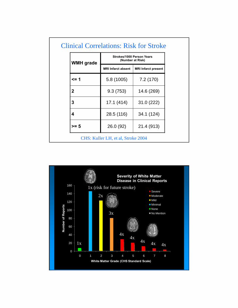
Clinical Correlations: Risk for Stroke
WMH grade
Strokes/1000 Person Years (Number at Risk)
MRI Infarct absent MRI Infarct present
<= 1 5.8 (1005) 7.2 (170)
2 9.3 (753) 14.6 (269)
3 17.1 (414) 31.0 (222)
4 28.5 (116) 34.1 (124)
>= 5 26.0 (92) 21.4 (913)
CHS: Kuller LH, et al, Stroke 2004
0
20
40
60
80
100
120
140
160
0 1 2 3 4 5 6 7 8
Nu
mb
er o
f R
epo
rts
White Matter Grade (CHS Standard Scale)
Severity of White Matter Disease in Clinical Reports
Severe
Moderate
Mild
Minimal
None
No Mention
2x
1x (risk for future stroke)
3x
4x4x
4x 4x 4x1x

4/2/2015
9
1 3
5 7
2 4
6 8
White Matter Dz Standards
Severity of White Matter Disease in Clinical Reports
0
20
40
60
80
100
120
140
160
0 1 2 3 4 5 6 7 8
White Matter Grade (CHS Standard Scale)
Nu
mb
er o
f R
epo
rts
Severe
Moderate
Mild
Minimal
None
No Mention
Severity of White Matter Disease in Clinical Reports
0
20
40
60
80
100
120
140
160
0 1 2 3 4 5 6 7 8
White Matter Grade (CHS Standard Scale)
Nu
mb
er o
f R
epo
rts
Severe
Moderate
Mild
Minimal
None
No Mention

4/2/2015
10
Severity of White Matter Disease in Clinical Reports
0%
20%
40%
60%
80%
100%
0 1 2 3 4 5 6 7 8
White Matter Grade
Cli
nic
al R
epo
rt D
escr
ipti
on
Severe
Moderate
Mild
Minimal
None
No Mention
Severity of White Matter Disease in Clinical Reports
Multiple Sclerosis
• Variants:¯ “Acute” - death in 10 months
¯ Devics = Neuromyelitis optica
¯ Balo’s Disease - Concentric Sclerosis
• 85 % have ovoid perivent lesions (Dawson’s fingers)
• 50 to 90 % w “definite” MS have CC lesions
• 10% of adults have PF lesions
• MS lesions generally lack mass effect

4/2/2015
11
44 year old numbnesslegs and feet
35 year old R body numbnessR leg weakness
Dawson’s Fingers
Flame shaped periventricular
4/2/2015
12
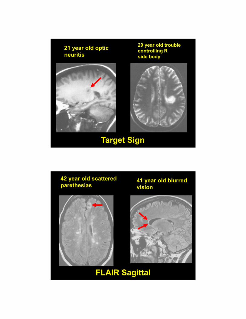
21 year old opticneuritis
29 year old troublecontrolling R side body
Target Sign
42 year old scatteredparethesias
41 year old blurredvision
FLAIR Sagittal

4/2/2015
13
Corpus Collosum
Punched out lesions on T1W
4/2/2015
14
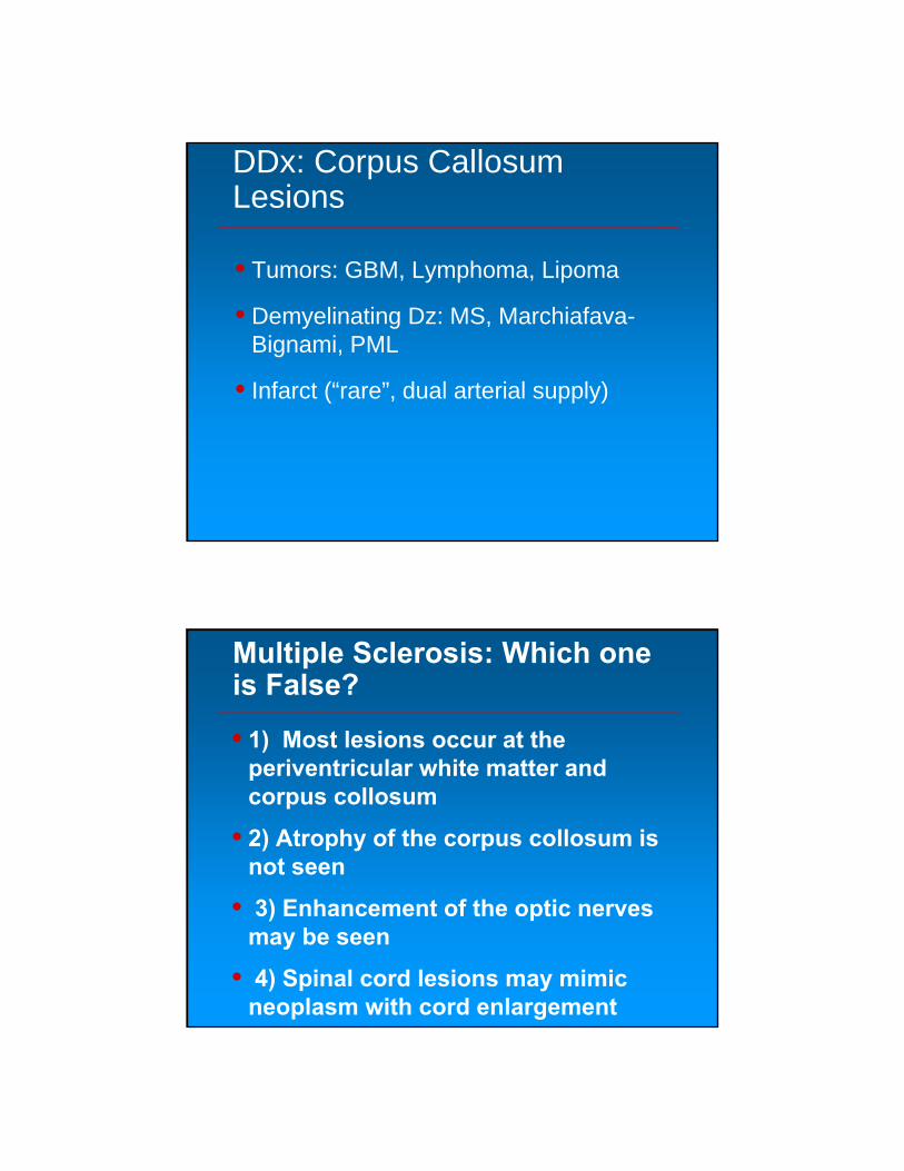
DDx: Corpus Callosum Lesions
• Tumors: GBM, Lymphoma, Lipoma
• Demyelinating Dz: MS, Marchiafava-Bignami, PML
• Infarct (“rare”, dual arterial supply)
Multiple Sclerosis: Which one is False?
• 1) Most lesions occur at the periventricular white matter and corpus collosum
• 2) Atrophy of the corpus collosum is not seen
• 3) Enhancement of the optic nerves may be seen
• 4) Spinal cord lesions may mimic neoplasm with cord enlargement

4/2/2015
15
Multiple Sclerosis: which is false?
1. Periventricular white matter and corpus collosum lesions
2. No corpus collosum atrophy
3. Optic nerve enhancement
4. Spinal cord lesions may mimic neoplasm
Tumefactive MS

4/2/2015
16
Tumefactive MS
PreGad PostGad
24 year old lethargy p
respiratory illness
24 year old scatteredparethesias
Which one is Multiple Sclerosis?
1 2

4/2/2015
17
Which is Multiple Sclerosis?
1. 1
2. 2
24 year old lethargy prespiratory illness
24 year old scatteredparethesias
Histoplasmosis MS

4/2/2015
18
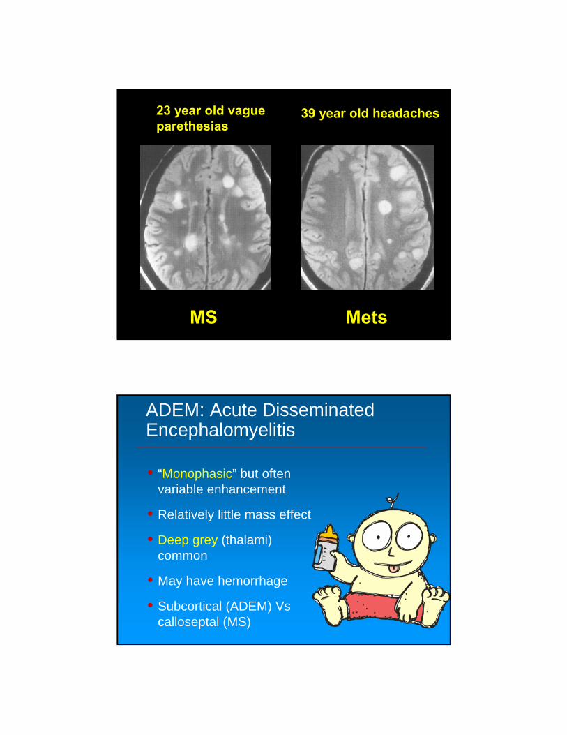
23 year old vagueparethesias
39 year old headaches
Which one is Multiple Sclerosis?
1 2
Which one is Multiple Sclerosis?
1. 1
2. 2
4/2/2015
19
23 year old vagueparethesias
39 year old headaches
MS Mets
ADEM: Acute DisseminatedEncephalomyelitis
• “Monophasic” but often variable enhancement
• Relatively little mass effect
• Deep grey (thalami) common
• May have hemorrhage
• Subcortical (ADEM) Vs calloseptal (MS)

4/2/2015
20
11 year old lethargyhemiparesis
3 year old rapidly ↓mental status
ADEM
ADEM

4/2/2015
21
5 year old drowsinessand seizure
ADEM
6 year old Bilat armand leg numbness
9 year old 9 days of L hemiparesis
ADEM
3 year old ↓ level ofconciousness

4/2/2015
22
ADEM: Which one is False?
• 1) It is a viral infection
• 2) It is more common in children than adults
• 3) Optic neuritis can be seen
• 4) Long-term f/u may be need to r/o MS
• 5) Both cerebral and cerebellar WM may be involved
ADEM: Which is False?
1. Viral Infection
2. Children > Adults
3. Optic Neuritis
4. F/U imaging to
R/O MS
5. Cerebral &
Cerebellar WM

4/2/2015
23
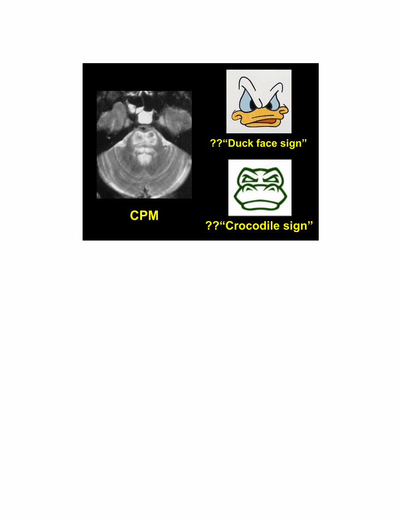
CPM: Central Pontine Myelinolysis
• Transverse pontine fibers most severely involved Vs corticospinal tract
• Extra PM in 50%
• >75% EtOH or hyponatremia correction
• DWI bright early, may enhance
• Variable resolution
• Spastic quadriparesis, pseudobulbar palsy
CPM

4/2/2015
24
CPM
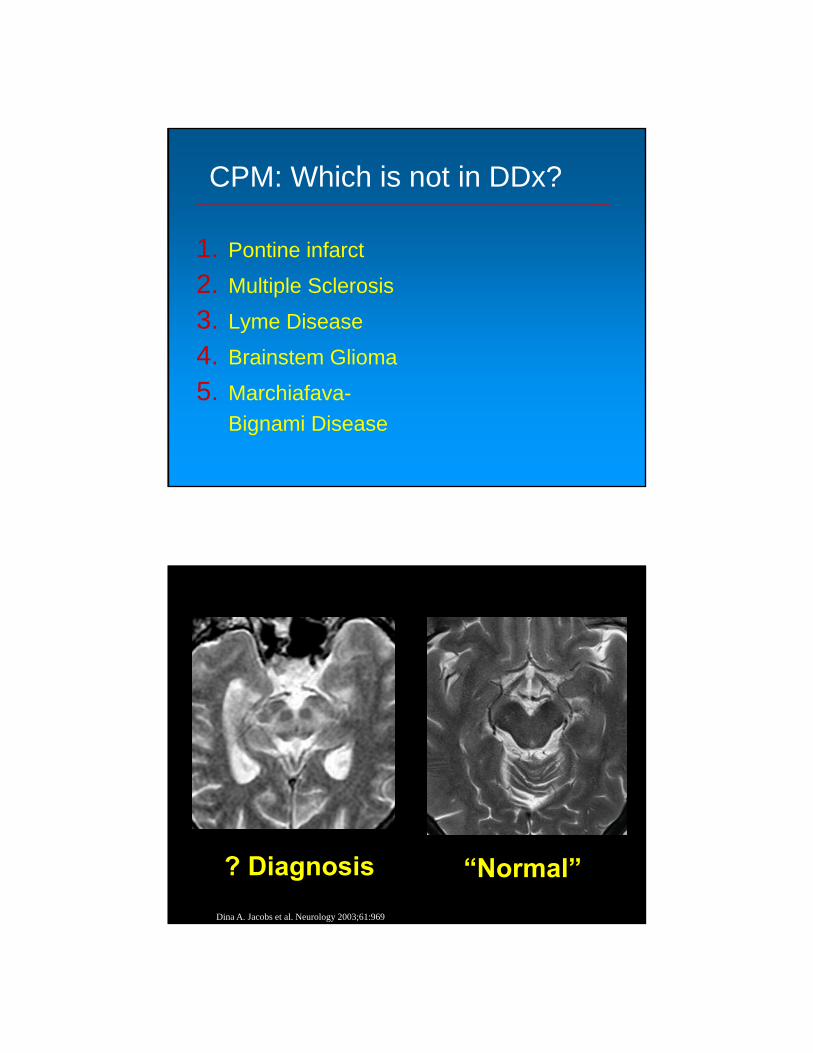
CPM: Which is NOT in DDX ?
• 1) Pontine Infarct
• 2) Multiple sclerosis
• 3) Lyme Disease
• 4) Brainstem Glioma
• 5) Marchiafava-Bignami Disease
4/2/2015
25
CPM: Which is not in DDx?
1. Pontine infarct
2. Multiple Sclerosis
3. Lyme Disease
4. Brainstem Glioma
5. Marchiafava-
Bignami Disease
? Diagnosis “Normal”
Dina A. Jacobs et al. Neurology 2003;61:969

4/2/2015
26
Brain stem disease:
1. CPM
2. Multiple System
Atrophy
3. Wilson’s Disease
4. ADEM
5. Olivary Pontine
Degenerative
Wilsons “Normal”

4/2/2015
27
Wilson’s Disease
• AKA: Hepatolenticular Degeneration
• Autosomal recessive: Copper accumulation
• Hand weakness, dysarthria, pseudo-Parkinsons
• High T2: Lentiform nuclei and midbrain
• Red nucleus and substania nigra surrounded by high T2 signal
• Midbrain: “Panda sign”
Wilson’s “Gourmet”

4/2/2015
28
Wilson’s
? Diagnosis “Normal”

4/2/2015
29
Brain stem disease:
1. CPM
2. Multiple System
Atrophy
3. Huntington’s
4. ADEM
5. Progressive
supranuclear palsy
MultisystemAtrophy
“Normal”

4/2/2015
30
Multisystem Atrophy (MSA)
• Sporadic neurodegenerative disease
• Synucleinopathy: alpha-synuclein metabolism
• Autonomic (Shy-Drager), Striaonigral, Olivopontocerebellar
• High T2: pontocerebellar tracts
• Pons: “Hot Cross Bun sign”
MultisystemAtrophy
“Hot cross bun”

4/2/2015
31
? Diagnosis “Normal”
Brain stem disease:
1. CPM
2. Multiple System
Atrophy
3. Huntington’s
4. Progressive
supranuclear palsy
5. Olivary Pontine
Degenerative

4/2/2015
32
Progressive supranuclear palsy
“Normal”
Progressive supranuclear palsy
“Hummingbirdsign”

4/2/2015
33
Progressive Supranuclear Palsy (PSP)
• AKA: Steele-Richardson-Olszewski syndrome
• Sporadic neurodegenerative disease
• Cognitive decline, abnormal eye movements
• Midbrain atrophy (“Hummingbird sign”)
• High T2: Pontine tegmentum, midbrain tectum
Reversible Post Leukoencephalopathy Syndrome (RPLS or PRES)
• Most often caused by abrupt changes in blood pressure, seizures, or certain immunosuppressive medications.
• Vasogenic edema predom in WM related to loss of autoregulatory ability
• Overall prognosis is good.
• DWI negative (Vs acute infarct cytotoxic edema)
• Usually in occipital, parietal, and temporal areas

4/2/2015
34
PRES
48 year old postseizure
2 months later
HypertensiveEncephalopathy

4/2/2015
35
HypertensiveEncephalopathy
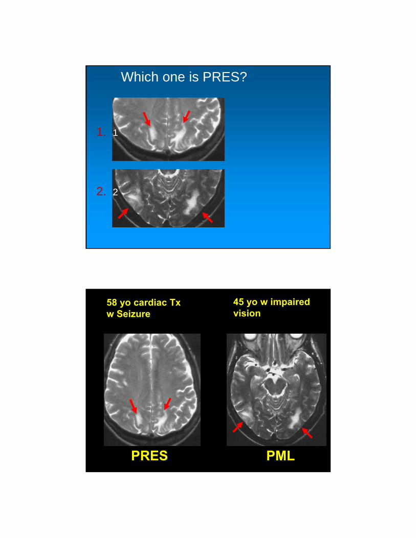
58 yo cardiac Txw Seizure
45 yo w impairedvision
Which one is PRES?
1 2
4/2/2015
36
Which one is PRES?
1. 1
2. 2
58 yo cardiac Txw Seizure
45 yo w impairedvision
PRES PML
4/2/2015
37
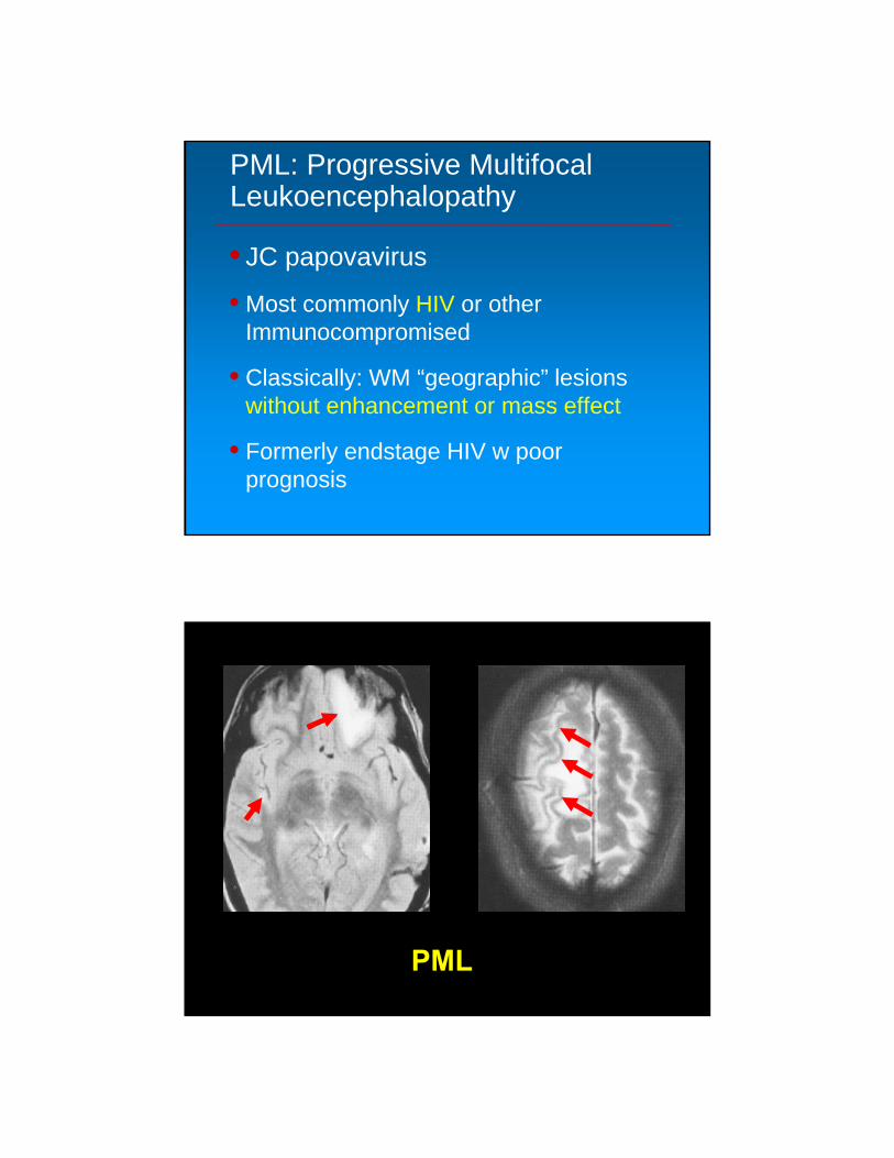
PML: Progressive Multifocal Leukoencephalopathy
• JC papovavirus
• Most commonly HIV or other Immunocompromised
• Classically: WM “geographic” lesions without enhancement or mass effect
• Formerly endstage HIV w poor prognosis
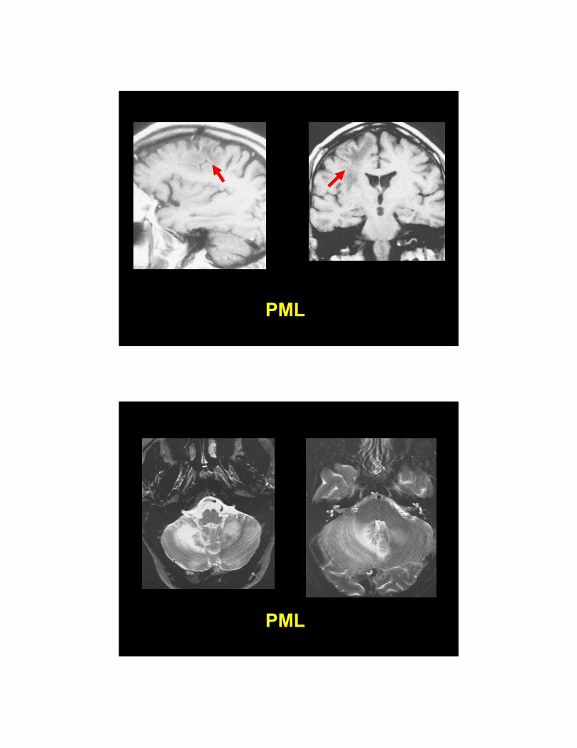
PML
4/2/2015
38
PML
PML

4/2/2015
39
PML: Which is FALSE?
• 1) It is caused by the JC virus
• 2) It is more common in males than females
• 3) CT frequently shows ringlike enhancement
• 4) Predilection for the parietal and occipital lobes
• 5) The spinal cord is rarely involved
PML: Which is False?
1. JC Virus
2. Males > Females
3. Ring Enhances
4. Parietal/Occipital
5. Spinal Cord rarely

4/2/2015
40
Signs:
• Tigroid WM: Metachromatic leukodystrophy
• Eye of the Tiger: Hallervorden-Spatz (PKAN)
• Panda sign: Wilsons
• Hot Cross Bun: Multiple system atrophy
• Hummingbird Sign: Progressive Supranuclear palsy
Metachromatic leukodystrophy(MLD)
Cheon J et al. Radiographics 2002;22:461-476©2002 by Radiological Society of North America
. Tigroid WM

4/2/2015
41
Hallervorden-SpatzPantothenate kinase-associated neurodegeneration (PKAN)
©2002 by Radiological Society of North AmericaGuillerman R P Radiology 2000;217:895-896
Progressive supranuclear palsy
“Hummingbirdsign”

4/2/2015
42
Wilson’s “Panda”
MultisystemAtrophy
“Hot cross bun”
4/2/2015
43
CPM
??“Duck face sign”
??“Crocodile sign”