new challenge of nuclear medicine in oncology -the dawn of ... · imaging of regional tumor biology...
TRANSCRIPT

1SUMITOMO KAGAKU 2004-II
This paper is translated from R&D Report, “SUMITOMO KAGAKU”, vol. 2004-II.
Introduction
In many countries within Europe and North Ameri-
ca, as well as in Japan, although there has been a grad-
ual decrease in the number of people dying of brain
and heart diseases, the number of deaths from malig-
nant neoplasms (referred to as cancer from this point
forward) has been increasing. During the year 2000,
there were 295,399 cancer-related deaths in Japan, rep-
resenting a mortality rate of 235.2 deaths per 100,000
people, meaning that 1 out of every 3 deaths were due
to cancer1). According to the “Study on Accuracy
Improvement and Usage of Local Cancer Registries” 2)
(Research Leader: Hideaki Tsuguma) conducted by
the Ministry of Health, Labour and Welfare, a break-
down of the number of cancer patients predicted in the
year 2020, was as follows: 500,000 males and 350,000
females, for a total of 850,000 patients. When the num-
ber of cancer patients predicted in 2020 is compared to
the number of actual cases in 2000, the annual rate of
New Challenge of Nuclear Medicinein Oncology-The dawn of functional imaging-
Today, imaging diagnosis in oncology is based on the regional anatomical information. However, anatomy-based imaging provides limited functional information and can only differentiate tumors from normal tissuebased on shape, density, vascularity, fat and water. Imaging of regional tumor biology by nuclear medicine pro-vides quantitative estimates of regional biochemistry and receptor status and can overcome the sampling errorand difficulty in performing serial studies inherent with biopsy. In this review, current status of the functionalimaging of tumor by nuclear medicine will be described.
Nihon Medi-Physics Co., Ltd.
Corporate Planning and Coordination Office
Strategic Marketing
Hideshi HATTORI
Research and Development Division
Research Center
Jun TOYOHARA
Fig. 1 Prediction of incident cases according to primary site, Japan (Data from reference1))
1
3
10
30
100
Male All sites
Stomach
Kidney, etc.
Num
ber
of in
cide
nt c
ase
LungProstate
Colon
LiverRectumEsophagusGallbladder
GallbladderPancreas
BladderLymphatic tissue
Leukemia
300
×103
1975 1980 1985 1990 1995 2000 2005 2010 2015 2020
1
3
10
30
100
Female (*includes CIS.)All sites*
Stomach
Kidney, etc.
Lung
Breast*Colon
LiverRectumUterus
Esophagus
Ovary
Pancreas
BladderLymphatic tissue
Leukemia
300
×103
1975 1980 1985 1990 1995 2000 2005 2010 2015 2020
2SUMITOMO KAGAKU 2004-II
New Challenge of Nuclear Medicine in Oncology –The dawn of functional imaging–
(NM). In particular, the function of diagnostic imaging
in the area of nuclear medicine can be classified into
three modalities:
1)Localization diagnosis: detection of the tumor loca-
tion (primary, recurrent, metastatic)
2)Identification diagnosis: differentiating between
malignant and benign lesions, evaluation of the
degree of malignancy, determination of treatment
regimen
3)Evaluation of treatment effectiveness and response
(follow-up): confirmation of tumor regression /
disappearance, probability of recurrence (micro-
metastasis)
Localization Diagnosis
Since localization diagnosis essentially requires high
definition imaging, the role of nuclear medicine is natu-
rally limited, as it is only able to provide images of low
definition. At this present stage, nuclear medicine is
useful in indicating which particular areas should be
further investigated using other testing methods, in
other words, for screening of the entire body. So far,
the most commonly utilized NM procedures are 99mTc-
Bone imaging, which is used to detect bone metastasis
and 67Ga imaging, which is used to perform tumor
localization diagnosis. In addition, another method of
localization diagnosis that employs positron emission
tomography (PET), has gradually gained popularity in
the field of clinical medicine. Thus, it is expected that
the role of nuclear medicine will continue to expand
further, in the future.
Identification Diagnosis
Subsequent to the confirmation of the location and
increase is as follows: males, 1.51; females, 1.47; for an
average annual rate of increase of 1.49. These figures
indicate that the number of cancer patients is expected
to continue rising. Fig. 1 depicts the prediction of inci-
dent cases according to primary site.
For males, although the number of incident cases
has increased for the areas of the prostate, lung, colon,
esophagus and rectum, the incidences of cancer of the
stomach and liver have remained unchanged for 1995
and subsequent years. Therefore, in 2020, the number
of incident cases in the lung, prostate and stomach
areas are expected to remain almost exactly the same.
For females, increases are seen for the areas of the
colon, breast, lung, uterus, rectum and liver. However,
as with males, the number of incident cases of stomach
cancer has remained unchanged for 1995 and subse-
quent years. In the future, the number of incident
cases of breast cancer is therefore predicted to exceed
that of stomach cancer.
Current Status of Tumor Image Diagnosis
The relationship between trends in cancer diagnosis
/ treatment and diagnostic imaging varies according to
the particular type of cancer and its primary site. In
this section, Table 1 depicts basic the role of diagnos-
tic imaging throughout the various stages of cancer
diagnosis and treatment.
The role of diagnostic imaging can be categorized
based upon the technical characteristics of each area of
application: X-ray imaging (including computed radiog-
raphy: CR ; and digital subtraction angiography: DSA),
X-ray computed tomography (CT), magnetic resonance
imaging (MRI), ultrasound (US) and nuclear medicine
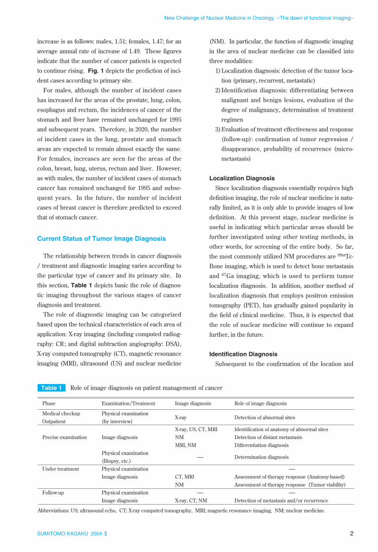
Medical checkupOutpatient
Phase Image diagnosis Role of image diagnosis
Physical examination (by interview)
X-ray Detection of abnormal sites
Physical examination(Biopsy, etc.)
– Determination diagnosis
Precise examination Image diagnosisX-ray, US, CT, MRINMMRI, NM
Identification of anatomy of abnormal sitesDetection of distant metastasisDifferentiation diagnosis
Under treatment Physical examinationImage diagnosis CT, MRI
NM
–Assessment of therapy response (Anatomy-based)Assessment of therapy response (Tumor viability)
Follow-up Physical examinationImage diagnosis
–X-ray, CT, NM
–Detection of metastasis and/or recurrence
Examination/Treatment
Table 1 Role of image diagnosis on patient management of cancer
Abbreviations: US; ultrasound echo, CT; X-ray computed tomography, MRI; magnetic resonance imaging, NM; nuclear medicine.

3SUMITOMO KAGAKU 2004-II
New Challenge of Nuclear Medicine in Oncology –The dawn of functional imaging–
all other areas of diagnostic technology. The only
method of diagnostic imaging that can presently
respond to this demand is 18F-FDG-PET imaging.
Other existing methodologies (including enhanced CT
/ CT, enhanced MRI / MRI, monoclonal antibodies /
SPECT) have not yet reached a satisfactory level of util-
ity due to their own particular shortcomings.
� Determination of Treatment Regimen
Chemotherapy is generally performed in the event
that surgical treatment is not applicable. Conventional
anti-cancer drugs almost always cause side effects, as
they target the difference in proliferation rates between
cancer cells and normal cells. However, accompanying
advances in molecular biology, specific molecules have
been identified that are associated with the invasion,
proliferation, vascularization and metastasis of cancer
cells. Anti-cancer drugs that target these molecules
(molecularly-targeted anti-cancer agents) have now
been developed and put into clinical usage. It is impor-
tant to carefully choose patients for whom these drugs
are expected to have the most beneficial effects. It is
crucial to diagnosis the patient in terms of the degree
of expression of the target molecules, prior to the
administration of such drugs. For example, it is now
commonly known that dual HER-2 / EGFR tyrosine
kinase inhibitors can be quite effective for treating can-
cer patients who are experiencing overexpression of
target molecules.
In addition to chemotherapy, radiation therapy (or
radiotherapy), is another useful treatment for cancer.
It is known that cancer cells gradually become starved
for oxygen (hypoxia) as the cancer progresses, thus
causing a corresponding decrease in their sensitivity to
radiation and thus reducing the effectiveness of radio-
therapy. On the other hand, if a large dose of radiation
is provided, undesirable side effects may occur, such as
the bone marrow toxicity. Drugs that may help to com-
bat these problems (radiosensitizers) are currently
under development. In the future, it will become
increasingly important to gain a more precise knowl-
edge of the locations and conditions associated with
cancer cell hypoxia.
Evaluating the Effects of Treatment
In principle, the first choice for tumor treatment is
surgical removal. Cancer treatment is usually conduct-
ed using a combination of surgical removal, radiothera-
spread of lesions using CT or MRI, the following proce-
dures are undertaken: differentiation between malig-
nant and benign lesions, determination of cell types,
malignancy diagnosis and specification of the treat-
ment regimen. The first choice for diagnostic action is
usually a pathological diagnosis conducted on a biopsy
tissue sample. However, due to the high degree of het-
erogeneity found within tumor tissue (tumor cells hav-
ing very different characteristics and are distributed
unevenly throughout a single tumor mass), the results
of the pathological diagnosis can be affected by the
location of the biopsy sample. Therefore, the accuracy
of this type of diagnosis is considered to be only about
80%. In the event that a tissue sample cannot be
obtained via biopsy, non-invasive diagnostic imaging is
another possible diagnostic choice. In this case, the
type of identification required, in terms of clinical medi-
cine, consists of differentiating between malignant and
benign lesions, performing a malignancy diagnosis and
specifying the treatment regimen.
� Differentiation Between Malignant and Benign
Lesions ; and Malignancy Diagnosis
Since high-resolution imaging is generally required
to correctly differentiate between malignant and benign
lesions, enhanced -CT (or enhanced MRI) is usually
utilized. However, the accuracy of diagnosis is limited
when CT or MRI techniques are utilized, as it is often
difficult to determine whether any lymphadenopathy
found by CT is actually due to metastasis or lym-
phadenitis. As this limitation is noted most frequently
for cancers in the thoracic and abdominal areas, it still
makes sense to utilize nuclear medicine for the diagno-
sis of cancers within these locations. However, the
only truly practical method of identification at the pre-
sent time is through 67Ga imaging, which is used to
identify lymphomas. Thus, nuclear medicine has not
yet entered the stage in which its utility has been fully
proven. Nuclear medicine-based examinations do pos-
sess the potential to replace CT and MRI, once the res-
olution has been improved for single photon emission
computed tomography (SPECT) and PET, and after
suitable second-generation tumor-specialized agents
have been developed.
Furthermore, since malignancy diagnosis is a type of
diagnosis that is one step beyond localization diagno-
sis, which is merely the anatomical diagnosis of
tumors, malignancy diagnosis is now in great demand,
not only in the area of diagnostic imaging, but also in

4SUMITOMO KAGAKU 2004-II
New Challenge of Nuclear Medicine in Oncology –The dawn of functional imaging–
py and the administration of anti-cancer drugs. The
effects of cancer treatment are evaluated using a vari-
ety of standards, such as improvement in the patient’s
overall condition, recovery of specific functions, verifi-
cation of remission / disappearance of tumors and the
disappearance of tumor markers within the patient’s
blood. The verification of tumor remission / disappear-
ance is a process that must always be conducted subse-
quent to the implementation of surgical removal. Diag-
nostic imaging is utilized to facilitate this process. In
general, since high-resolution images are required for
proper verification of tumor remission, either CT or
MRI are utilized and nuclear medicine examination is
not usually the first choice.
However, CT and MRI do not provide any informa-
tion with respect to the presence of any tumor cells
within a tumor mass (= focus of necrosis or scarred
connective tissue) that may still remain after treatment.
In addition, as morphological changes are non-specific
and occur subsequent to a functional change, a certain
amount of time is required in order to evaluate the
effectiveness of the treatment. Since the treatment
itself is continued until its effectiveness has been evalu-
ated, an ineffective treatment may result in economic
hardship for the patient, lost opportunity to administer
an alternate treatment, as well as a decrease in quality
of life due to the continued administration of ineffective
drugs. Thus, it is anticipated that the early stage detec-
tion of functional changes (= functional imaging) will
solve these problems. As described above, although
diagnostic imaging of tumor areas is conducted based
on morphological information, its limitations have also
been pointed out. The use of functional imaging to
image the metabolic characteristics of a tumor locus is
a process that can be used to solve essential problems
associated with biopsies. Nuclear medicine diagnostics
has the potential to solve these problems. This paper
introduces the current status of ongoing radiopharma-
ceutical research conducted for the purpose of facilitat-
ing effective functional imaging.
Current Status of Radiopharmaceutical
Research in the Field of Oncology
Table 2 lists certain radiopharmaceuticals that are
utilized for the functional imaging of tumor biology.
Fig. 2 depicts the chemical structures of various
positron-labeled radiopharmaceuticals.
1. Imaging of Glucose Metabolism
In a malignant tissue, the proliferation of neoplastic
cells usually progresses at a greater rate than the cor-
responding tissue vascularization, thus producing a
hypoxic tissue fraction. As tumor cells continue to mul-
tiply within this hypoxic environment, they are forced
Glucose metabolismAmino acid transport and incorporationMembrane/lipid biosynthesisDNA synthesisHypoxiaApoptosisReceptor expressionP-glycoprotein
Biological/biochemical function
18F-FDG18F-FACBC11C-acetate, 11C-choline,18F-FCH18F-FLT 18F-fluoromisonidazole, 62Cu-ATSM99mTc-AnnexinV18F-FES, 123I-MIBG, 111In-DTPA-octreotide99mTc-MIBI, 99mTc-tetrofosmine
Radiopharmaceutical
Table 2 Radiopharmaceuticals for functional imag-ing of tumor biology
See text for definition of abbreviations
Fig. 2 Chemical structures of positron labeled radiopharmaceutical for tumor functional imaging
OHO
N
NH
O
O
OH18F
OH
HO
OH
H NH2
COOH18F
18F-FACBC
NN
NO2
OH
HO
OH
CH3 11COOH NCH3 CH3
CH3
CH3CH3
CH3
11CH3
CH2CH2OH CH2CH2OHN
18FCH2
N N
CH3 CH3
CH3HN NHCH3SS
62Cu
62Cu-ATSM
18F-FCH
18F-FDG
18F-FLT
18F-Fluoromisonidazole 18F-FES
11C-Acetate
11C-Choline
18F
18F
18F

5SUMITOMO KAGAKU 2004-II
New Challenge of Nuclear Medicine in Oncology –The dawn of functional imaging–
to obtain energy by taking advantage of the anaerobic
glycolysis of glucose.3) For this reason, an increase in
glucose transporters and hexokinase activity can be
recognized in tumor cells. These increases demon-
strate a characteristic increase in the ability of tumor
cells to uptake large quantities of glucose.4) Further-
more, tumor cells also have a lower incidence of glu-
cose-6-phosphatase activity (phosphatase of glucose-6-
phosphoric acid).4) The radiopharmaceutical known as
2-deoxy-2-[18F] fluoro-D-glucose (18F-FDG) (Fig. 2), in
which the secondary hydroxyl group has been
replaced with 18F, accumulates rapidly in a variety of
different tumor types. Since April 1 of 2002, 18F-FDG,
which can be used to diagnose lung cancer, breast can-
cer, colon cancer, head and neck cancer, brain tumors,
pancreatic cancer, malignant lymphoma, metastatic
liver cancer, non-site-specific cancers and malignant
melanoma, with these patients also fulfill given specific
requirements, have been utilized in a number of appli-
cations involving medical examinations. In general,
since 18F-FDG is more readily taken up by tumors hav-
ing a high degree of malignancy, it is often utilized in
the following areas: identification diagnosis; staging
diagnosis; and in diagnosis for metastasis and recur-
rence. In addition, as the rate of tissue accumulation of18F-FDG is proportional to the number of cells that are
active in glucose metabolism (= living cells)5), 18F-FDG
has been utilized in quantitative evaluations of the
effects of both chemotherapy and radiotherapy.
Wieder et al., had conducted testing with positron
emission tomography 18F-FDG (18F-FDG-PET) on 38
cancer patients (cT3, cN0/+, cM0) with esophageal
squamous cell carcinoma (whose esophagectomies
were scheduled after preoperative chemoradiation
therapy had been conducted for a period of 4 weeks)
prior to their preoperative chemoradiation therapy and
subsequent operations. The changes observed in glu-
cose metabolism were then compared to response eval-
uation criteria according to histopathological
response.6) The following results were obtained from
this comparison testing: in histopathologically respond-
ing cases, 18F-FDG-PET studies indicated that glucose
metabolism had decreased by 70±11%. In histopatho-
logically non-responding cases, glucose metabolism
was seen to have decreased by 51±21%. In 18F-FDG-
PET studies conducted on the 14th day after the initia-
tion of preoperative treatment, tumor glucose metabo-
lism changes in histopathologically responding cases
were detected at a sensitivity of 93% and a specificity of
88%. Furthermore, such changes correlated signifi-
cantly with patient survival time (p = 0.011). Fig. 3
depicts examples of images obtained from 18F-FDG-
PET studies (coronal slices). The upper images (A)
depict the histopathologically responding tumors, with
the lower images (B) depicting non-responding
tumors. In the histopathologically responding tumors,
a significant decrease was observed in the accumula-
tion of 18F-FDG after the 14th day of continued preoper-
ative chemoradiation therapy. However, no corre-
sponding decrease in 18F-FDG accumulation was rec-
ognized in the non-responding tumors. According to
the treatment response evaluation of chemotherapy,
the utility of 18F-FDG has been proven for use in the
diagnosis of stomach cancer and breast cancer.7, 8)
Glucose metabolism comparison tests were conduct-
ed during chemotherapy treatment of a progressive
cancer, as follows: 18F-FDG-PET examinations were
conducted on 57 patients with progressive non-small
cell lung cancer (stage IIIb or IV), both prior to the
administration of cisplatin-based chemotherapy and
after the administration of a single treatment cycle.
Changes in tumor glucose metabolism were then com-
pared to Response Evaluation Criteria in Solid Tumors
(RECIST)9) during the follow-up process.10) As a result
of this comparison, the changes in tumor glucose
metabolism were considered to be predictable indica-
Fig. 3 Example of 18F-FDG-PET studies (coronal slices) in (A) histophathologically re-sponding and (B) nonresponding tumors. RCTx, chemotherapy; SUV, standardized uptake value. (Data from reference6)).
A
B
6SUMITOMO KAGAKU 2004-II
New Challenge of Nuclear Medicine in Oncology –The dawn of functional imaging–
tioning of this mechanism have not yet been identified.
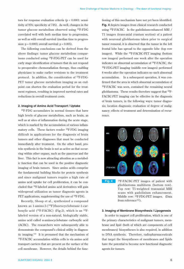
Fig. 4 depicts images from clinical research conducted
using 18F-FACBC. In the gadolinium-enhanced MRI /
T1 images (trans-axial cranium section) of a patient
with neuronal glioblastoma taken prior to surgical
tumor removal, it is observed that the tumor in the left
frontal lobe has spread to the opposite lobe (top row
images). While the 18F-FACBC-PET imaging (bottom
row images) performed one week after the operation
indicates an abnormal accumulation of 18F-FACBC, the18F-FDG-PET imaging (middle row images) performed
8 weeks after the operation indicates no such abnormal
accumulation. In a subsequent operation, it was con-
firmed that the area in which abnormal accumulation of18F-FACBC was seen, contained the remaining neural
glioblastoma. These results therefore suggest that 18F-
FACBC-PET imaging can be effective in the diagnosis
of brain tumors, in the following ways: tumor diagno-
sis; location diagnosis; evaluation of degree of malig-
nancy; effects of treatment and determination of recur-
rence.
3. Imaging of Membrane Biosynthesis / Lipogenesis
In order to support cell proliferation, which is one of
the primary characteristics of malignant tumors, mem-
brane and lipid (both of which are components of cell
membranes) biosyntheses is also required, in addition
to DNA synthesis. Therefore, radiopharmaceuticals
that target the biosyntheses of membranes and lipids
have the potential to become new functional diagnostic
agents for tumors.
tors for response evaluation criteria (p < 0.0001: sensi-
tivity of 95%; specificity of 74%). As well, changes in the
tumor glucose metabolism observed using 18F-FDG
correlated well with both median time to progression,
as well as with overall survival (median time to progres-
sion: p = 0.0003; overall survival: p = 0.005).
The following conclusions can be derived from the
above findings: tumor glucose metabolism compar-
isons conducted using 18F-FDG-PET can be used for
early stage identification of tumors that do not respond
to preoperative chemoradiation therapy, thus enabling
physicians to make earlier revisions to the treatment
protocol. In addition, the consideration of 18F-FDG-
PET tumor glucose metabolism imaging as an end-
point can shorten the evaluation period for the treat-
ment regimen, resulting in improved survival rates and
reductions in overall treatment costs.
2. Imaging of Amino Acid Transport / Uptake18F-FDG accumulates in normal tissues that have
high levels of glucose metabolism, such as brain, as
well as at sites of inflammation during the acute stage,
which is marked by the accumulation of various inflam-
matory cells. These factors render 18F-FDG imaging
difficult in applications for the diagnosis of brain
tumors and other diagnoses that must be conducted
immediately after treatment. On the other hand, pro-
tein synthesis in the brain is not as active as that occur-
ring within other organs, such as the pancreas and the
liver. This fact is now attracting attention as a metabol-
ic function that can be used in the positive diagnostic
imaging of brain tumors. Since amino acids comprise
the fundamental building blocks for protein synthesis
and since malignant tumors require a high rate of
amino acid uptake for cell proliferation, it can be con-
cluded that 18F-labeled amino acid derivatives will gain
widespread utilization as tumor diagnosis agents in
PET applications, supplementing the use of 18F-FDG.
Recently, Shoup et al., synthesized a compound
known as 1-amino-3-[18F]fluorocyclobutane-1-car-
boxylic acid (18F-FACBC) (Fig.2), which is an 18F-
labeled version of a non-natural, biologically stable,
amino acid called α-aminocyclobutane carboxylic acid
(ACBC). The researchers were subsequently able to
demonstrate the compound’s clinical utility in diagnos-
tic imaging.11) It is presumed that the mechanism of18F-FACBC accumulation within cells is via amino acid
transport carriers that are present on the surface of the
cell membrane. However, the details behind the func-
Fig. 4 18F-FACBC-PET images of patient with glioblastoma multiform (bottom row). Top row: T1-weighted transaxial MRI scans with gadolinium enhancement. Middle row: 18F-FDG-PET images. (Data from reference11)).

7SUMITOMO KAGAKU 2004-II
New Challenge of Nuclear Medicine in Oncology –The dawn of functional imaging–
prostate.15) In the 18F-FDG-PET images shown in the
upper row, no abnormalities were detected, as the level
of 18F-FDG accumulation in the primary lesion (A), was
low. The 18F-FDG-PET images did indicate lymph
node metastasis to the left ilium (B) and bone metasta-
sis to the right pubic bone. The 11C-Acetate-PET
images shown in the bottom row clearly indicate the
primary lesion (D), left ilium lymph node metastasis
(E) and bone metastasis to the right pubic bone (F).
Furthermore, an acceleration of choline metabolism
has been confirmed in malignant tumors, correspond-
ing to the acceleration of cell membrane phosphatide
biosynthesis. The dynamics of choline metabolism
within tumor tissue have been increasingly clarified
through numerous studies involving MR spectroscopy
and biochemical research. Therefore, in tumor cells
that are undergoing vigorous cell division, there is an
acceleration of choline kinase activity, which causes
increased concentrations of choline, phosphocholine
and phosphoethanolamine within the cell. It has been
speculated that after uptake by the cell, the choline
undergoes phosphorylation by choline kinase and then
remains within the tumor tissue.
Hara et al., in studies employing 11C-choline (Fig. 2),
reported that certain choline derivatives can be
employed as imaging agents for brain tumors and
prostate cancer.16, 17) Furthermore, DeGrado et al.,
have developed a very useful compound known as
[18F]fluoromethyl-dimethyl-2-hydroxyethyl-ammonium
(18F-FCH) (Fig. 2).18) As with [1-11C-]acetate, 11C-
choline does not release much radioactivity into the
urine, thus it is expected to find application in prostate
cancer diagnosis. 18F-FCH was found to have limited
utility in applications for prostate cancer diagnosis, as
unlike 11C-choline, it releases radioactivity into the
urine immediately.
4. Imaging of DNA Synthesis
Research into the imaging of DNA synthesis using
nucleic acid derivatives is being conducted in order to
evaluate the effectiveness of chemotherapy and radio-
therapy treatments for malignant tumors. The use of11C-thymidine has been studied, based on a method of
evaluating DNA synthesis that utilizes [methyl-3H]thymidine as a cell proliferation marker. However,
certain problems, including excess noise due to the
short physical half-life of the 11C (approximately 20
minutes) and the secondary distribution of 11C-labeled
metabolites, made it difficult to conduct a quantitative
[1-11C]-Acetate (Fig. 2) is a radiopharmaceutical that
has been developed for the purpose of evaluating the
aerobic metabolism of the TCA cycle in cardiac mus-
cle. However, it is known that the acetyl-CoA produced
during acetate metabolism is used not only for aerobic
energy production, but also for the biosynthesis of
lipids, such as phospholipids.12, 13) Yoshimoto et al.,
therefore hypothesized that within malignant tumor
cells having a high degree of proliferation, acetate
metabolism has actually shifted to lipogenesis, since
energy metabolism in the tumor depends greatly upon
the aerobic energy produced via the glycolytic path-
way. Based on this hypothesis, the researchers con-
ducted a fundamental examination into the relationship
between acetate metabolism within the tumor and the
degree of tumor cell proliferation.14) The results indi-
cated that the [1-14C]-acetate taken into tumor cells was
a component of lipophilic metabolites (phosphatidyl
choline and glyceride), as had been predicted. In addi-
tion, the quantity of radioactivity absorbed by the [1-14C]-acetate in the lipophilic fraction showed a strong
correlation with the amount of [methyl-3H]thymidine
(used as an index of cell proliferation) incorporated
into the DNA (p = 0.0016).
Unlike 18F-FDG, [1-11C]-acetate releases only a mini-
mal amount of radioactivity into urine, which has made
it useful in clinical studies for both the detection and
disease stage diagnosis of pelvic neoplasms adjacent to
the bladder, particularly prostate cancer. Fig. 5
depicts the clinical images of the 11C-acetate in a
patient with poorly differentiated adenocardinoma of
Fig. 5 PET images of prostate, lymph node, and bone metastases obtained using 18F-FDG (upper row) and 11C-acetate (bottom row) from 73-y-old man with poorly differenti-ated (Gleason score 7) adenocarcinoma of prostate. bn=bone; ly=lymph node; pr=prostate. (Data from reference15)).
A
18F-FDG PET
11C-acetate PET
18F-FDG PET
11C-acetate PET
18F-FDG PET
11C-acetate PET
B C
D E F

8SUMITOMO KAGAKU 2004-II
New Challenge of Nuclear Medicine in Oncology –The dawn of functional imaging–
Toyohara et al., utilized 22 tumor cell lines, each pos-
sessing a different degree of proliferation, in a study to
determine whether the intercellular accumulation of
FLT that reflects TK1 activity would also reflect the
degree of cell proliferation.20) The results of this study
demonstrated that cellular FLT uptake is strongly cor-
related with the uptake of [methyl-3H]thymidine and
the %S-phase, cell proliferation markers. In addition,
Rasey et al., used non-small cell lung cancer cell lines
having synchronized cell cycles, to study whether the
accumulation of FLT is dependent upon cell cycle.
They were able to confirm that the pattern of FLT
uptake indicates that FLT accumulation is dependent
upon cell cycle. 19) Furthermore, an examination of the
histopathology showed that there was a strong correla-
tion (r = 0.87; p < 0.0001) between the accumulated 18F-
FLT radioactivity levels in solitary human pulmonary
nodules and the degree of staining in the cell prolifera-
tion marker Ki-67 (Fig. 7).22)
These findings are the result of proactive support for
the hypothesis that the proliferation dynamics of malig-
nant tumor cells can be imaged non-invasively, using18F-FLT. However, the following phenomenon has also
been reported: the level of 18F-FLT accumulation in
each cell increases in the presence of either the de
novo synthesis inhibitor 5-fluorouracil (5-FU), as well
as methotrexate (MTX), thus causing discrepancy
between 18F-FLT accumulation and cell proliferation
dymanics.23) This phenomenon is derived from the
fact that the level of 18F-FLT accumulation in each cell
increases as TK1 (a salvage pathway enzyme) activity
accelerates, due to enzymatic inhibition of de novo syn-
evaluation of tumor proliferation from the images
obtained. In order to solve these problems, Shields
and researchers attempted to use 18F-labeled 3´-deoxy-
3´-fluorothymidine (FLT), which was originally devel-
oped as an anti-viral agent, resulting in the successful
synthesis of 18F-FLT (Fig. 2).19) Fig. 6 depicts the first
clinical case in which 18F-FLT was administered to a
patient with non-small cell lung cancer. Fig. 6 shows a
coronal image on the left, a sagittal image on the right
and transverse scans in the middle. 18F-FLT accumula-
tion can be observed in the following locations: tumors
having high degree of proliferation, bone marrow, the
liver and the bladder; the latter two organs functioning
in metabolism / excretion. The accumulation of 18F-
FDG was also observed in the tumor, however, no cor-
responding accumulation of radioactivity due to 18F-
FDG was observed in the bone marrow.
18F-FLT is incorporated into DNA synthesis path-
ways subsequent to the phosphorylation of the 5´-
hydroxyl group. However, as the 3´-hydroxyl group
has been replaced with fluorine, no phosphodiester
bond is formed, thus preventing the replication of the
DNA chain. On the other hand, the 18F-FLT accumu-
lated after phosphorylation cannot easily be dispersed
outside of the cell due to its negative charge, thus it
remains within the cell. The intracellular level of 18F-
FLT accumulation therefore reflects the thymidine
kinase (TK1) activity, thus providing imaging that
expresses an indirect indication of the level of DNA
synthesis.
Fig. 6 18F-FLT images of patient with non-small cell lung cancer. Left: coronal image. Right: sagital image. Center: transverse image. (Data from reference18)).
FLT
FDG
CT
0
10
20
30
40
50
60
70
0 1 2 3 4 5 6 7
Ki-6
7 la
belin
g in
dex
(%)
FLT-SUV
Fig. 7 Linear regression analysis demonstrated a significant correlation between prolifera-tive fraction and 18F-FLT uptake in solitary pulmonary nodule. (Data from refer-ence22)).

9SUMITOMO KAGAKU 2004-II
New Challenge of Nuclear Medicine in Oncology –The dawn of functional imaging–
can be used to determine the degree of tumor cell
apoptosis after anti-cancer drug administration, then
this imaging may facilitate the evaluation of tumor sen-
sitivity to such drugs, thus allowing for better predic-
tion of treatment effectiveness.
Blankenberg et al., sought a method for detecting
apoptosis in vivo. They initially synthesized 99mTc-
HYNIC-AnnexinV, which possesses a strong affinity
for PS. The phosphatidylserine (PS) expressed on
the surface of cell membranes, which can be observed
in the early stages of apoptosis. Blankenberg et al.,
subsequently used a variety of animal models to
demonstrate that apoptosis can be detected in vivo
using 99mTc-HYNIC-AnnexinV.27) Furthermore, Bel-
hocine et al., conducted phase 1/2 clinical testing
with 99mTc-AnnexinV on 10 lung cancer patients, 2
breast cancer patients and 3 patients with malignant
lymphomas28). Throughout this testing, the changes
in the accumulations of 99mTc-AnnexinV were exam-
ined both before and after a single course of
chemotherapy. The chemotherapy proved effective in
8 cases of lung cancer and malignant lymphoma, in
which 99mTc-AnnexinV accumulation levels had been
seen to increase (= apoptosis was detected). The
chemotherapy was not effective for the 5 such cases
in which 99mTc-AnnexinV accumulation levels were
not seen to increase. For the 2 cases of breast cancer,
no correlation was found between changes in 99mTc-
AnnexinV accumulation levels and the effectiveness of
the chemotherapy.28) These results suggest that for
lung cancer and malignant lymphomas, the effective-
ness of chemotherapy can be predicted using 99mTc-
AnnexinV. Fig. 8 depicts the images taken during
phase 1/2 clinical testing with 99mTc-AnnexinV. In
the non-responding case (PD) of non-small cell lung
cancer (A), no accumulation of raioactivity was
observed in the 99mTc-AnnexinV image taken after
therapy (A2), as compared to the 99mTc-AnnexinV
image taken prior to therapy (A1). In the responding
case (CR) of non-small cell lung cancer (B), the 99mTc-
AnnexinV image taken after therapy (B2) showed a
greater accumulation of radioactivity in the metastasis
lesions of the cervical area, as well as in the bilateral
bronchoplumonary lymph nodes (areas indicated by
arrows), as compared to the 99mTc-AnnexinV image
taken prior to therapy (B1). In the responding case
(CR) of non-Hodgkin’s lymphoma, the 99mTc-Annex-
inV image taken after therapy (C2) showed a greater
accumulation of radioactivity in the metastasis lesions
thesis. Therefore, further testing is needed before 18F-
FLT can be properly used in the evaluation of anti-
tumor drug effectiveness.
5. Imaging of Hypoxia
Hypoxic tissues can be defined as “tissues that are
still within the range of structural normality, yet in
which reversible changes are occurring due to a lack of
oxygen.” In a malignant tumor, the rate of tissue vas-
cularization is slower than that required by the vigor-
ous proliferation of tumor cells, thus creating hypoxic
regions. Since hypoxic tumors are considered to have
low radiosensitivity, it is difficult to obtain satisfactory
effects from radiotherapy. Therefore, it is expected
that the imaging of hypoxic regions within tumors may
provide information that is useful for planning dose dis-
tributions and irradiation target sites during Intensity
Modulated Radiation Therapy (IMRT).
With respect to imaging agents for hypoxic tissues,18F-fluoromisonidazole (Fig. 2) has been commonly uti-
lized. 18F-Fluoromisonidazole is a derivative of a
nitroimidazole compound that was developed as a
radiosensitizing agent. However, as the nitroimidazole
compound was originally developed as a scavenger for
unstable radicals produced within the cell due to radia-
tion exposure, numerous factors pertaining to its mech-
anism of accumulation are still unknown. Focusing
upon the fact that copper (Cu) contributes to oxidation-
reduction reactions within living organisms,
Fujibayashi et al., synthesized a hypoxic tissue diagnos-
tic agent using a 62Zn / 62Cu generator known as 62Cu-
diacetyl-bis (N4-methylthiosemicarbazone) (62Cu-
ATSM) (Fig. 2). The agent was labeled with 62Cu,
which has a positron emitting nucleus, and was subse-
quently utilized in the imaging of tumors.24, 25) The62Cu-ATSM showed a high level of accumulation in the
tumor. As well, unlike 18F-FDG, it also resulted in
images that appeared to reflect non-uniformities in the
blood flow. Furthermore, Obata et al., have deter-
mined that Cu-ATSM accumulates and remains within
tumor cells due to reduction caused by the electron
transfer system enzyme of microsomes that express
vigorously within tumor cells.26)
6. Imaging of Apoptosis
Based on the fact that anti-cancer drugs induce apop-
tosis in cancer cells, the phenomenon of apoptosis is
considered important as it directly produces the suc-
cessful effects of chemotherapy. Therefore, if imaging

10SUMITOMO KAGAKU 2004-II
New Challenge of Nuclear Medicine in Oncology –The dawn of functional imaging–
in neuroendocrine tumor cells, allowing radiolabeled
compounds that bond to somatostatin receptors to be
utilized as highly specific radiopharmaceuticals.
Although somatostatin is a peptide composed of 14
amino acids, the location of receptor binding occurs at
Phe7-Trp-Lys-Thr10. (D)-Phe-Cys-Phe-(D)-Trp-Lys-
Thr-Cys-The(ol) (octreotide) was utilized as a precur-
sor compound (peptidase resistance was added
through conversion of some constituent amino acids
into D-types) in the synthesis of 111In-DTPA-octreotide,
through the following process: DTPA was introduced
as a chelation site to the D-Phe residue, located at the
N-end. The radiolabeleing of 111In then occurred via
this DTPA.30) Octreotide demonstrates a high positive
rate of binding to neuroendocrine tumors, such as
hypophyseal adenomas, pancreas endocrine tumors,
pheochromocytomas, neuroblastomas, paragan-
gliomas, carcinoids, thyroid medullary carcinomas and
small cell lung cancers. In particular, 111In-DTPA-
octreotide is extremely useful in the diagnoses of pri-
mary and metastastic lesions in pancreas endocrine
tumors and carcinoids, all of which are difficult to
detect using only CT or MRI.
Today, it is estimated that two-thirds of all breast
cancer cases will respond to hormone treatments. It is
also presumed that the degree of response can be pre-
dicted by observing the expression of estrogen recep-
tors (ER) and progesterone receptors (PR). Therefore,
in the field of breast cancer treatment, it is expected
that receptor density may be predicted not only for pri-
mary lesions, but also for a broader range, such as for
metastatic lesions, once it becomes possible to obtain
ER and PR information through diagnosis via nuclear
medicine imaging. At present, although no radiophar-
maceuticals have yet proven effective in the imaging of
PR, a compound known as 16α-[18F]fluoroestradiol-17β(18F-FES) (Fig. 2) has been developed for the imaging
of ER. A correlation has been reported between the
accumulation of 18F-FES and the results of in vitro
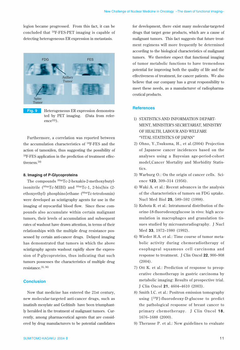
receptor binding assays.31, 32) Fig. 9 shows coronal
images taken of a breast cancer case, using 18F-FDG-
PET (left) and 18F-FES-PET (right).33) Positive ER
expression was confirmed through a biopsy of a left
axillary lymph node. Although the accumulation of 18F-
FES was observed in the metastatic left axillary lymph
nodes, an accumulation of 18F-FDG in a bone metasta-
sis within the spine could not be observed. Although
the metastastic left axillary lymph node responded to a
subsequent hormone treatment, the bone metastastic
in the cervical nodes, as well as in the left axillary
nodes (areas indicated by arrows), as compared to the99mTc-AnnexinV image taken prior to therapy (C1).
7. Imaging of Receptors
In certain tumors, the presence of specific cells and
receptors can be observed. Therefore, the imaging of
such tumors has been attempted using chemical com-
pounds that possess an affinity for the transporters and
receptors present in the subject cells.123I-m-iodobenzylguanidine (123I-MIBG) is a chemical
compound derived from guanethidine, which is an anti-
hypertension agent that blocks sympathetic nerve
action. As this compound is taken up by the adrenal
medulla and sympathetic nerve endings via the same
mechanism as that of norepinephrine, it has been uti-
lized for scintigraphy of the adrenal medulla and for
the diagnosis of sympathetic nerve function in cardiac
muscle. The first known application of MIBG in tumor
imaging was for the scintigraphy of a pheochromocy-
toma using 131I-MIBG, as reported by Sissonn et al.29)
Since then, many instances of MIBG usage have been
reported in the diagnoses of pheochromocytomas, as
well as in regional diagnosis of neuronal glioblastomas,
thyroid medullary carcinomas, carcinoids and lung can-
cers.
Somatostatin receptors are found in abundance with-
Fig. 8 Example of 99mTc-AnnexinV imaging stud-ies in responding (B and C) and nonres-ponding (A) tumors. A and B: Case of non-small cell lung cancers. C: A case of non-Hodgkin’s lymphoma. (Data form ref-erence28)).
A1 A2
B1 B2
C1 C2
11SUMITOMO KAGAKU 2004-II
New Challenge of Nuclear Medicine in Oncology –The dawn of functional imaging–
legion became progressed. From this fact, it can be
concluded that 18F-FES-PET imaging is capable of
detecting heterogeneous ER expression in metastasis.
Furthermore, a correlation was reported between
the accumulation characteristics of 18F-FES and the
action of tamoxifen, thus suggesting the possibility of18F-FES application in the prediction of treatment effec-
tiveness.34)
8. Imaging of P-Glycoproteins
The compounds 99mTc-2-hexakis-2-methoxybutyl-
isonitrile (99mTc-MIBI) and 99mTc-1, 2-bis[bis (2-
ethoxyethyl) phosphino]ethane (99mTc-tetrofosmin)
were developed as scintigraphy agents for use in the
imaging of myocardial blood flow. Since these com-
pounds also accumulate within certain malignant
tumors, their levels of accumulation and subsequent
rates of washout have drawn attention, in terms of their
relationships with the multiple drug resistance pos-
sessed by certain anti-cancer drugs. Delayed imaging
has demonstrated that tumors in which the above
scintigraphy agents washout rapidly show the expres-
sion of P-glycoproteins, thus indicating that such
tumors possesses the characteristics of multiple drug
resistance.35, 36)
Conclusion
Now that medicine has entered the 21st century,
new molecular-targeted anti-cancer drugs, such as
imatinib mesylate and Gefitinib have been triumphant-
ly heralded in the treatment of malignant tumors. Cur-
rently, among pharmaceutical agents that are consid-
ered by drug manufacturers to be potential candidates
for development, there exist many molecular-targeted
drugs that target gene products, which are a cause of
malignant tumors. This fact suggests that future treat-
ment regimens will more frequently be determined
according to the biological characteristics of malignant
tumors. We therefore expect that functional imaging
of tumor metabolic functions to have tremendous
potential for improving both the quality of life and the
effectiveness of treatment, for cancer patients. We also
believe that our company has a great responsibility to
meet these needs, as a manufacturer of radiopharma-
ceutical products.
References
1) STATISTICS AND INFORMATION DEPART-
MENT, MINISTER’S SECRETARIAT, MINISTRY
OF HEALTH, LABOUR AND WELFARE
“VITAL STATISTICS OF JAPAN”
2) Ohno, Y.,Tsukuma, H., et al.(2004) Projection
of Japanese cancer incidences based on the
analyses using a Bayesian age-period-cohort
model,Cancer Mortality and Morbidity Statis-
tics.
3) Warburg O.: On the origin of cancer cells. Sci-
ence 123, 309–314 (1956).
4) Waki A. et al.: Recent advances in the analysis
of the characteristics of tumors on FDG uptake.
Nucl Med Biol 25, 589–592 (1998).
5) Kubota R. et al.: Intratumoral distribution of flu-
orine-18-fluorodeoxyglucose in vivo: high accu-
mulation in macrophages and granulation tis-
sues studied by microautoradiography. J Nucl
Med 33, 1972–1980 (1992).
6) Wieder H.A. et al.: Time course of tumor meta-
bolic activity during chemoradiotherapy of
esophageal squamous cel l carcinoma and
response to treatment. J Clin Oncol 22, 900–908
(2004).
7) Ott K. et al.: Prediction of response to preop-
erative chemotherapy in gastric carcinoma by
metabolic imaging: Results of prospective trial.
J Clin Oncol 21, 4604–4610 (2003).
8) Smith I.C. et al.: Positron emission tomography
using [18F]-fluorodeoxy-D-glucose to predict
the pathological response of breast cancer to
primary chemotherapy. J Cl in Oncol 18 ,
1676–1688 (2000).
9) Therasse P. et al.: New guidelines to evaluate
Fig. 9 Heterogeneous ER expression demonstra-ted by PET imaging. (Data from refer-ence33)).
FDG
AxillaryTumor
SpineTumor
FES

12SUMITOMO KAGAKU 2004-II
New Challenge of Nuclear Medicine in Oncology –The dawn of functional imaging–
the response to treatment in solid tumors: Euro-
pean Organization for Research and Treatment
of CancerNational Cancer Institute of the Unit-
ed States National Cancer Institute of Canada.
J Natl Cancer Inst 92, 205–216 (2000).
10) Wolfgang A. et al.: Positron emission tomog-
raphy in non-small-cell lung cancer: prediction
of response to chemotherapy by quantitative
assessment of glucose use. J Clin Oncol 21,
2651–2657 (2003).
11) Shoup T.M. et al.: Synthesis and evaluation of
[18F]1-amino-3-fluorocyclobutane-1-carboxylic
acid to image brain tumors. J Nucl Med 40,
331–338 (1999).
12) Howard B.V.: Acetate as a carbon source for
lipid synthesis in cultured cells. Biochem Bio-
phys Acta 488, 145–151 (1977).
13) Long V.J.W.: Incorporation of 1-11C-acetate into
the lipids of isolated epidermal cells. Br J Der-
matol 94, 243–252 (1976).
14) Yoshimoto M. et al.: Characterization of acetate
metabolism in tumor cells in relation to cell pro-
liferation: Acetate metabolism in tumor cells.
Nucl Med Biol 28, 117–122 (2001).
15) Oyama N. et al.: 11C-acetate PET imaging of
prostate cancer. J Nucl Med 43 , 181–186
(2000).
16) Hara T. et al.: PET imaging of brain tumor with
[methyl-11C]choline. J Nucl Med 38, 842–847
(1997).
17) Hara T. et al.: PET imaging of prostate cancer
using carbon-11-choline. J Nucl Med 39 ,
990–995 (1998).
18) DeGrado T.R. et al.: Synthesis and evaluation of18F-labeled choline as an oncologic tracer for
positron emission tomography: initial findings in
prostate cancer. Cancer Res 61, 110–117 (2001).
19) Shields et al.: Imaging proliferation in vivo with
[18F]FLT and positron emission tomography.
Nat Med 4, 1334–1336 (1998).
20) Toyohara et al.: Basis of FLT as a cell prolifer-
ation marker: comparative uptake studies with
[3H]thymidine and [3H]arabinothymidineand
cell analysis in 22 asynchronously growing tumor
cell lines. Nucl Med Biol 29, 281–287 (2002).
21) Rasey J.S. et al.: Validation of FLT uptake as a
measure of thymidine kinase-1 activity inA549
carcinoma cells. J Nucl Med 43, 1210–1217
(2002).
22) Buck A.K. et al.: 3-deoxy-3-[18F]fluorothymi-
dine-positron emission tomography for non-
invasive assessment of proliferation in pul-
monary nodules. Cancer Res 62, 3331–3334
(2002).
23) Dittmann H. et al.: Early changes in [18F]FLT
uptake after chemotherapy: an early experi-
mental study. Eur J Nucl Med Mol Imaging 29,
1462–1469 (2002).
24) Fujibayashi Y. et al.: Copper-62-ATSM: a new
hypoxia imaging agent with high membrane
permeability and low redox potential. J Nucl
Med 38, 1155–1160 (1997).
25) Takahashi N. et al.: Evaluation of 62Cu labeled
diacetyl-bis(N4-methylthiosemicarbazone) as a
hypoxic tissue tracer in patients with lung can-
cer. Ann Nucl Med 14, 323–328 (2000).
26) Obata A. et al.: Retention mechanism of hypox-
ia selective nuclear imaging/radiotherapeutic
agent Cu-diacetyl-bis(N4-methylthiosemicar-
bazone) (Cu-ATSM) in tumor cells. Ann Nucl
Med 15, 499–504 (2001).
27) Blankenberg F.G. et al.: In vivo detection and
imaging of phsophatidylserine expression dur-
ing programmed cell death. Proc Natl Acad Sci
95, 6349–6354 (1998).
28) Belhocine T. et al.: In vivo imaging of chemother-
apy-induced apoptosis in human cancers. Ann
NY Acad Sci 1010, 525–529 (2003).
29) Sisson J.C. et al.: Scintigraphic localization of
pheochrocytoma. N Eng J Med 305, 12–17
(1981).
30) Bakker W.H. et a l . : [1 1 1In-DTPA-D-Phe1] -
octreotide a potential radiopharmaceutical for
imaging of somatostat in receptor-posit ive
tumors: synthesis radiolabeling and in vitro
validation. Life Sci 49, 1583–1591 (1991).
31) Kiesewetter D.O. et al.: Preparation of fluo-
rine-18-labeled estrogens and their selective
uptakes in target tissues of immature rats. J
Nucl Med 25, 1212–1221 (1984).
32) Mintun M.A. et al.: Breast cancer: PET imag-
ing of estrogen receptors. Radiology 169, 45–48
(1988).
33) Mankoff D.A. et al.: Characterizing tumors
using metabolic imaging: PET imaging of cel-
lular proliferation and steroid receptors. Neo-
plasia 2, 71–88 (2000).
34) Dehdashti D. et al.: Positron emission tomo-

13SUMITOMO KAGAKU 2004-II
New Challenge of Nuclear Medicine in Oncology –The dawn of functional imaging–
graphic assessment of “metabolic flare” to pre-
dict response of metastatic breast cancer to
antiestrogen therapy. Eur J Nucl Med 26,
51–56 (1999).
35) Kao C.H. et al.: P-Glycoprotein and multidrug
resistance-related protein expressions in rela-
tion to technetium-99m methoxyisobutylisoni-
trile scintimammography findings. Cancer Res
61, 1412–1414 (2001).
36) Fuster D. et al.: Tetrofosmin as predictors of
tumour response. Q J Nucl Med 47, 58–62
(2003).
P R O F I L E
Hideshi HATTORI
Nihon Medi-Physics Co., Ltd.Corporate Planning and Coordination OfficeStrategic Marketing
Jun TOYOHARA
Nihon Medi-Physics Co., Ltd.Research and Development DivisionResearch CenterPh. D.