neuroscience tutorial - cu-boulder computer science
TRANSCRIPT

Neuroscience Tutorial

Brain Organization
: cortex, basal ganglia, limbic lobe
: medulla oblongata,
midbrain, pons,
cerebellum
: thalamus, hypothal.,
pituitary gland

Cortical Organization
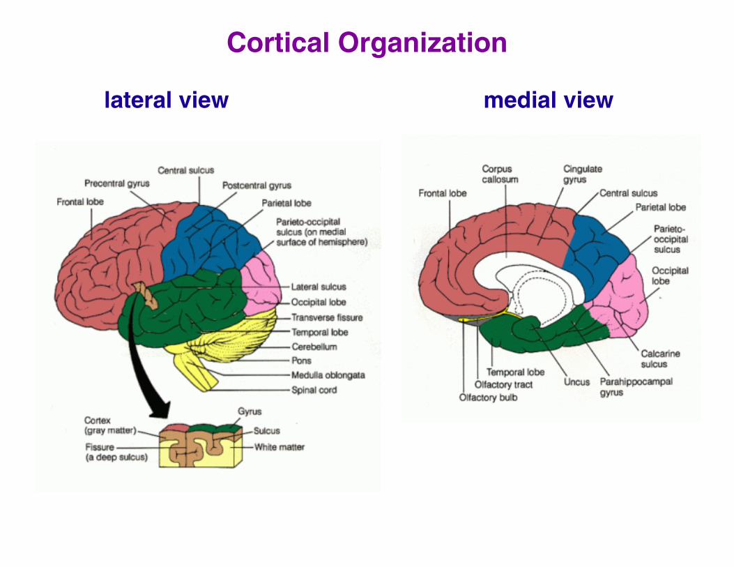
Cortical Organization
lateral view medial view
Brain Terminology
anterior toward the front
posterior toward the back
medial toward the middle (midline)
lateral toward the side
dorsal toward the top
ventral toward the bottom
superior above
inferior below

Visual System
~30% of brain devoted to vision
~8% touch
~3% hearing

Visual System Organization
Dorsal stream
“where” pathway: parietal cortex (V5, MT, LIP)
location of objects, attention, motion
Ventral stream
“what” pathway: temporal cortex (V4, IT)
color and shape perception, object identification
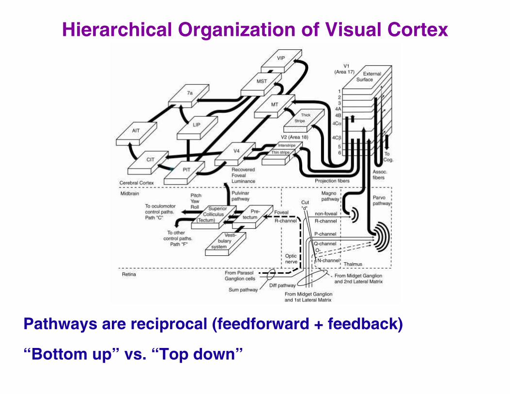
Hierarchical Organization of Visual Cortex
Pathways are reciprocal (feedforward + feedback)
“Bottom up” vs. “Top down”

Stages of Visual Processing
Lateral Geniculate Nucleus
90% of projections to V1, 10% to superior colliculus -> pulvinar -> extrastriate
small, center-surround receptive fields

V1 (primary visual cortex, striate cortex, area 17)
receives feedback projections from IT, FEF, MST, MT (not reciprocal)
receptive fields tuned to color, spatial frequency, contrast, orientation, motion direction, eye, binocular disparity
actual size
rescaled to same size

Topographic organization of V1

V2, V3, V3A (extrastriate)
V4 (extrastriate)
color perception
IT (inferotemporal)
form perception
broad receptive fields
stages within IT: posterior, central, anterior (PIT, CIT, AIT)
MT, MST
projections from V1
process motion
parietal, frontal attentional areas
projections from V4, MT

Hierarchical Architecture
Early stages: simple features, small receptive fields
Late stages: complex features, large receptive fields
e.g., Stringer and Rolls (2002)

Hierarchical Architecture
Early stages: simple features, small receptive fields
Late stages: complex features, large receptive fields
e.g., Reisenhuber & Poggio model

Two Theories: The Role of V1 in Conscious Vision
Hierarchical theories
Awareness arises at later stages of visual system
Damage to V1 disrupts flow of information to high-level areas
Because V1 lacks direct projections to higher visual areas and frontal cortex (response initiation), it is not essential.
Prediction: awareness more tightly correlated with activity in extrastriate areas than in V1
Prediction: disruption of V1 activity should not impair awareness if extrastriate activity remains the same.
Interactive theories
V1 takes part in dynamic recurrent circuits with extrastriate areas necessary for awareness, possibly as a high-resolution ‘master map’ for binding
Disruption of V1 activity should always impair awareness even if extrastriate areas remain intact.
Prediction: disruption of V1 activity should always impair awareness

Evidence Consistent With Interactive Theories
Blindsight
V1 lesions -> loss of awareness in region of visual field
residual visual ability, e.g., forced choice discrimination above chance
visual information still reaches extrastriate areas (e.g., V3A, Mt, V4/V8, LOC)
lesionLVF RVF
conscious lefthemifield stimulus
unconscious righthemifield stimulustime
only only
Extrastriate lesions are more restricted, less devastating than V1 lesions
e.g., loss of motion perception with MT lesions
e.g., loss of color vision with V4/V8 lesions
e.g., prosopagnosia
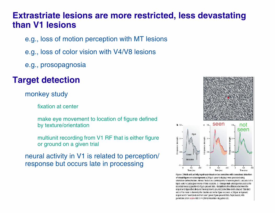
Target detection
monkey study
fixation at center
make eye movement to location of figure defined by texture/orientation
multiunit recording from V1 RF that is either figure or ground on a given trial
neural activity in V1 is related to perception/response but occurs late in processing
seen notseen

Transcranial Magnetic Stimulation (TMS) studies
Perception of briefly flashed stimulus disrupted by TMS pulse over V1/V2
TMS produces maximal disruption 70-120 ms after stimulus onset, which might be due to disruption of feedback signals from higher areas.
TMS of higher areas (MT) is most effective at stimulus onset
Eliciting motion phosphenes by applying TMS to MT
Second TMS pulse applied either to V1 or MT at various times before or after phosphene eliciting pulse
Phosphene disrupted by V1 pulse if lag is ~25 msec
TMS to MT fails to elicit motion phosphene to patient with ipsilesional V1 damage.
strong
weak

Evidence Consistent with Hierarchical Theories
Bistable perception (reversible figures)
Increased activity of extrastriate, parietal, frontal areas increased at time of reported alternation; decreased activity of striate area
Are higher areas directing lower areas to reorganize?
Internally generated visual experiences (hallucinations, dreams, migraines, synesthesia, imagery)
Produce awareness but V1 activity is greatly reduced relative to real-world input.
Stimulation of temporal lobe can elicit hallucinations of people, scenes, objects.

Evidence Consistent With Both Theories
Binocular rivalry
presentation of incompatible images to the two eyes
comparison of rivalry vs. stimulus alternation with same statistics

Probe monocular region of V1 corresponding to blind spot (all activity in this region is internally generated)
fMRI modulations during rivalry were as large as those evoked by physical alternation-> rivalry might be resolved in V1
more consistent with interactive view
Rivalry with low and high contrast gratings
effects of rivalry are no stronger in higher visual areas
also more consistent with interactive view
These human studies suggest that neural activity in V1 linked tightly to perception.
But monkey data...
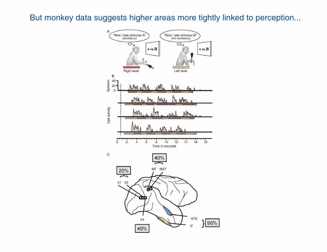
But monkey data suggests higher areas more tightly linked to perception...

Conclusions
V1 is the only single cortical visual area necessary for awareness.
V1 is not sufficient for awareness
e.g., V1 response to gratings whose frequency is too high to be perceived
Late component of V1 activity reflects top-down feedback
Data are probably more consistent with interactive theory.
Strength of qualia depend on V1
e.g., adaptation effects: adaptation to orientation, color, motion -> decrease in V1 activity
e.g., internally generated visual experiences lead to some V1 activity, but not as much as with real-world input

Defining Consciousness:A Psychological Perspective
(Allport, 1977)
Criteria for the presence or absence of awareness of X
e.g., X = black ink on page, letters, words, commands
1. Potential action
X can directly guide or control choice of actions
However, many types of actions: speech, reaching for object, pupil dilation
2. Memory
Individual must be able to overtly recall or recognize X later
3. Self-evaluation
Individual must be able to indicate confidence in awareness of X, as well as lack of confidence on occasion.
Indication can be verbal or nonverbal (betting, willingness to act on)