neuropsychiatric systemic lupus erythematosus : stress, stroke, and seizures
TRANSCRIPT

Neuropsychiatric Systemic Lupus Erythematosus
Stress, Stroke, and Seizures
PATRICIA M. MOORE" Department of Neurology
Wayne State University School of Medicine 4201 St. Antoine
Detroit, Michigan 48201
Neuropsychiatric systemic lupus erythematosus (NP-SLE) refers to the spectrum of neurologic, psychiatric, and behavioral abnormalities that occur in patients with systemic lupus erythematosus. These clinical disturbances are particularly frustrating because we currently understand little of the pathogenic mechanisms and have few specific tools for diagnosis. Traditionally, identifiable secondary causes of disease, such as infections, metabolic abnormalities, and toxins, including side effects of medications (which may account for the clinical features of up to half of the SLE patients seen in neurologic consultation), are excluded. The range and acuity of NP- SLE abnormalities are broad; some features are severe and dramatically increase the morbidity and mortality of disease. Other features are transient or, if persistent, create only a minor disruption of lifestyle. Abnormalities of almost every region of the neuraxis are reported-but certain features predominate (TABLE 1). The frequent appearance of seizures, encephalopathies, behavioral abnormalities, affective disor- ders, cognitive disorders, and cerebrovascular disease suggest that certain systems within the brain rather than specific anatomical regions provide clues to the underlying immunopathogeneses.' In this volume, we plan to review some of the clinical features of the disease, explore advances in the understanding of neurobiologic mechanisms of central nervous system (CNS) diseases, and discuss ideas for future scientific and clinical studies of NP-SLE.
Systemic lupus erythematosus is an autoimmune disease characterized by circulat- ing autoantibodies and immune complexes. Features that define the disease are particular patterns of autoantibodies (such as those to dsDNA or ribosomal proteins) and evidence of organ system damage, usually through immune complex deposition (e.g., skin, kidneys) or direct autoantibody effects (e.g., anemia, thrombocytopenia). Other nondefining aspects of disease, such as fever, arthralgias, and malaise, appear to be mediated by cytokines and indicate the presence of systemic inflammation. The epidemiology of SLE is complex, reflecting the multiple genetic, hormonal, and environmental factors that contribute to the manifestation of disease. Although the overall prevalence of disease varies (in North American and Western Europe about 50 per lOO,OOO), the prevalence among young women approaches 1:lW. Relatives
aTel: (313) 577-1244; fax: (313) 745-4216.
1

2 ANNALS NEW YORK ACADEMY OF SCIENCES
TABLE 1. Clinical Abnormalities in NP-SLE
Seizures Abnormalities in consciousness, cognition, and behavior
Encephalopathies acute confusional state acutehbacute changes in behavior, cognition, or level of arousal dementias
mild cognitive disorder mildmoderate cognitive impairment
Other: aphasias, neglect syndromes, dressing disorders Mood disorders with or without psychoses Sleep disorders Psychological disorders
somatoform disorders anxiety disorders personality disorders
Isolated cognitive abnormalities
Cerebrovascular disease Stroke Multiinfarct disorders Microvascular disease Subarachnoid hemorrhage Cerebral venous thromboses
Cranial neuropathies Optic neuropathies Cranial neuropathies affecting extraocular muscles Trigeminal neuropathy Facial neuropathy
Ataxia Movement disorders, particularly chorea Myelopathies Peripheral neuropathies
Radiculopathy Plexopathy Mononeuropathy Mononeuropathy multiplex Polyneuropathy Autonomic neuropathy
Myasthenia gravis Myopathy
of patients with SLE have a higher incidence of other autoimmune disorders than the general population. The particular distribution of disease among woman of childbearing age, and the effects of menarche and pregnancy, supports the hypothesis that hormonal factors influence disease activity. In most patients, the disease is episodic, and exacerbations may be precipitated by exposure to ultraviolet light, medications, or infection^.^.^

MOORE: STRESS, STROKE, AND SEIZURES 3
& PERIPHERAL INFLAMMATORY NEUROENDOCRNE
LYMPH NODES
FIGURE 1. Neuroendocrine-immune system interactions.
Strokes, stress, and seizures is chosen as a title for reasons other than alliteration. Both seizures, which are very frequent in SLE, and strokes, which are less frequent overtly but could be recurrent in a clinically quiescent form, share central mechanisms of tissue injury. Glutamate, the principle excitatory amino acid (EAA) neurotransmit- ter in the brain, and its membrane receptors operate prominently in many normal neurologic functions, including cognition, mood, movement, and sensation. Excitatory amino acid neurotransmitters also guide developmental plasticity of synaptic connec- tions, thus influencing learning and brain repair. Stimulation of any of the ionotropic glutamate receptors results in membrane depolarization due to the influx of positively charged ions. Overstimulation of these receptors is a final common pathway for neuronal cell injury (through excessive influx of calcium into neurons) in diseases with diverse pathophysiologic processes. Whether the cell recovers or dies depends on many factors, including cell type, location, and other inducible molecules. Within the brain there are regions where the cells are particularly vulnerable to injury from EAA; among these regions is the hippocampus. This area is part of the limbic system, a phylogenetically old region of the brain and central to many processes of memories and emotions, as well as prominent in their influence over “mind-body” connections, neuroimmunoendocrine circuits, and the body’s adaptive responses to injury (FIG. l).4
On the cellular level, many insults to the CNS parenchyma share common mecha- nisms of injury. Recurrent depolarization, failures of energy metabolism from a lack of substrate, disruption of mitochondria, and direct neuronal injury all result in toxic levels of glutamate in the tissue. The ubiquitous EAA and dysregulated Ca2+ thus causes (albeit with different time course) injury in susceptible neurons in trauma, ischemia, and seizures. EAA-mediated cell damage is also likely in some degenerative

4 ANNALS NEW YORK ACADEMY OF SCIENCES
diseases (Huntington's chorea) and chronic viral disorders (HIV dementia).5 Some circumstances shift the balance towards increased tissue injury from minor insults. Stress, from various causes, results in increased expression of CNS cytokines, activa- tion of the HPA axis, and elevation of glucocorticoid levels in the brain. One effect of the predominant human glucocorticoid, cortisol, is to increase the amount of glutamate (apparently through reduction in uptake mechanisms in the synapse) and exacerbate EAA-mediated injury.&'O Thus, seizures, strokes, and stress all result in increased tissue EAA and may potentiate injury. Specifically, vulnerable regions of the brain bear the injury in these pathogenically dissimilar insults.
The nervous system, and endocrine and immune systems are anatomically and functionally interconnected."-14 These systems express and respond to a large number of shared regulatory molecules (steroids, cytokines, and neuropeptides). Central to the influence of the CNS in endocrine and immunologic reactions are the connections between the limbic system and the (1) hypothalamic-pituitary-adrenal axis (HPA), (2) the hypothalamic-pituitary-gonadal (HPG) axis, and, (3) the locus caeruleus- sympathetic autonomic nervous system. Each of these systems feed back upon the neocortex and the limbic system (FIG. 2).
THE SPECTRUM OF CLINICAL FEATURES
Many of the major neurologic abnormalities are apparent and definable on neuro- logic examination, and upheld by neurodiagnostic studies. These include the frequently encountered seizures, encephalopathies, strokes, cranial neuropathies, and ataxia, as well as the less frequently encountered myelopathy, peripheral neuropathies, and myasthenia g r a ~ i s . ' ~ ~ ~ Other features may be more difficult to diagnose, including abnormalities of memory, judgment, and changes in personality. Similarly, psychiatric abnormalities may be clinically obvious, such as major mood disorders with or without coexisting neurologic abn~rma l i t i e s .~ '~~ However, both neurologic and psychiatric abnormalities appear in more subtle forms that may not be conspicuous on clinical examination. The transient, patchy, or subtle symptoms associated with a change in a person's lifestyle deserve more detailed evaluation. Some investigators attribute these abnormalities to an exacerbation of a preexisting psychiatric disease or exaggera- tion of an underlying personality trait rather than distinct ~hanges.4~3~~ Further, studies reveal the complex features of a chronic illness, and the strength of social supports do influence manifest behavior. Studies can be designed to clarify many of these issues.
SPECIFIC CLINICAL FEATURES
Seizures, either focal or generalized, occur in many patients with SLE (10-20%) and may be the presenting manifestation of disease in 5 percent. Although usually easily controlled with anticonvulsants, seizures may increase the vulnerability of certain neurons to additional injury.
Encephalopathies, acute or subacute abnormalities of cognition, attention, or level of arousal, sometimes associated with agitated behavior, are frequent but are so varied

MOORE: STRESS, STROKE, AND SEIZURES 5
FIGURE 2. Some of the pathways and mediators between the central nervous, endocrine, and immune systems are shown. Notably, (1) the corticosteroids (CS) secreted by the adrenals to dampen inflammation also have prominent actions in the brain; (2) the hypothalamic hormones corticotropin-releasing hormone (CRH) and arginine vasopressin (AVP), as well as the gonadal steroids, particularly estrogen, prominently affect behavior through their actions in the brain; (3) cytokines, such as IL-1 and IL-6, produced by brain cells in response to a variety of stressors, affect behavior as well as activating hypothalamic, pituitary, and brain stem structures; and (4) the prominent connections of the locus caeruleus in the brain stem with the hypothalamus result in inactivation of the autonomic nervous system and catecholamine (CC) stimulation to brain structures as well as to lymphocytes and blood vessels.

6 ANNALS NEW YORK ACADEMY OF SCIENCES
in presentation that they often defy classification. The most straightforward syndrome is the acute confusional disorder, which can be reproducibly diagnosed; the syndrome elicits an appropriate search for potential underlying causes, although it is often associated with the SLE itself. Chronic, multidomain changes in cognition, sometimes called dementia, do occur in SLE, but the contributing factors and incidences remain undefined. Even more frequent are the deviations in cognition, memory, attention, judgment, or manifest behavior from a person’s normal state that do not fit a pattern associated with other defined neurologic disorders. Abnormalities in the frontal- subcortical circuits, which may be evident by altered executive functions, disinhibi- tion, distractibility, or apathy, may also appear in patients with SLE. There is debate over the appropriate nomenclature for these clinical episodes. Often the disparities result from the training (neurology, psychiatry, neuropsychology) of the investigators, each bringing a different terminology to the study. We recommend that multidomain abnormalities be called encephalopathies with the appropriate descriptors rather than the use of operational and often misleading terms, such as delirium. The terms for abnormalities in higher cortical function, such as aphasias, neglect syndromes, and dressing disorders, are also currently descriptive rather than anatomical.
Isolated abnormalities in cognition or behavior reportedly appear in patients with SLE, although several studies, using different tools and methods of analysis, provide disparate results. A potentially useful working scheme is that described in a later section by Grant: (1) those with largely symptomatic abnormalities associated with changes in function and mild cognitive disorders, and (2) those with documented, substantiated abnormalities in neuropsychological testing and mild to moderate cogni- tive impairment.
Abnormalities of affect, monopolar or bipolar depression, occur frequently, but given the current estimates of the incidence of depression in the general population, it is difficult to ascertain the increased incidence in patients with SLE. Psychoses, severe disturbances in thought processes and functioning, may complicate a mood disorder and often require acute hospitalization.
Cerebrovascular disease occurs, although the histologic rarity of CNS vasculitis in SLE reveals some of the brain’s distinctive regulatory mechanisms over inflamma- tion. The patterns and consequences of cerebrovascular disease that do appear are so varied that they are usefully broken down into subsets for clinical identification.
Strokes are neurologic abnormalities secondary to ischemidinfarction in the distri- bution of one or more blood vessels. Strokes happen in SLE, although the actual frequency and the contributing factors remain undefined. Acute cerebral infarction may be overdiagnosed in some patients when, for example, focal motor abnormalities may have other causes (it?., metabolic). Similarly, it is likely underdiagnosed where the presence of diffuse microvascular disease may be mistaken for a metabolic encephalopathy. More accurate assessment is difficult with currently available tech- niques for brain imaging and measurement of blood flow. Cerebral blood flow depends on numerous factors, including, most importantly, the metabolic rate of cerebral tissue. Thus, our standard diagnostic assessments of blood flow may not measure primary vascular processes but, instead, reflect a compensatory diminished flow (oligemia) from cellular injury. Further complicating identification of early cerebro- vascular disease is the cascade of events associated with cerebral ischemia. Ischemia may initiate inflammation, which, in turn, precipitates coagulation and vasoconstric-

MOORE: STRESS, STROKE, AND SEIZURES 7
tion, leading to increased tissue ischemia and cellular damage.4%s1 The roles of other factors, such as coagulopathies, hypertension, chronic corticosteroid medications, and lipid disorders in the genesis of cerebrovascular disease, warrant further study.
Subarachnoid hemorrhages result from several different processes. The presence of a subarachnoid hemorrhage usually initiates a search for a leaking or ruptured aneurysm.
Microvascular disease is difficult to diagnose because both the mechanisms of tissue injury and the clinical features overlap with other disorders. The clinical features are variable, potentially presenting as global dysfunction or extension of focal injury after other tissue damage. As we understand more about the local events between arterioles, capillaries, and neurons, our approach to microvasculature will e~olve.’~-’~
Cortical vein thromboses are dramatic events in their complete form, manifest by seizures, subarachnoid hemorrhage, and increased intracranial pressure. Because neuroimaging of occluded veins is so difficult, it appears evident that the more subtle manifestations of cortical vein disease are underdiagnosed. With the increased sensitivity of imaging techniques for detecting hemorrhages and potential venous stases, we detect cortical vein occlusions more frequently than before.
Some clinical features, such as headache, are present in patients with SLE but are so frequent a feature in the general population and result from so many mechanisms that, in the absence of other abnormalities, they cannot be used as a defining feature
In the investigation of clinical frequency and pathogenic mechanisms of NP- SLE, areas of concern are the abnormal neurodiagnostic studies in asymptomatic people. Examples of this are brain magnetic resonance images (MRI), revealing multifocal areas of increased intensity; SPECT scans suggesting asymmetry of blood flow; and head CT scans showing These results obtained when patients with SLE without NP features are used, appropriately, as controls in studies of patients with NP-SLE, caution us in our interpretation of studies. Whether the studies reveal preclinical tissue injury or represent physiologic changes in water diffusion remains an area of keen interest.
of NP-SLE.
MECHANISMS THAT INITIATE OR CONTRIBUTE TO ABNORMALITIES
Immune-mediated neurologic and psychiatric abnormalities may be divided into several groups: (1) those conditions in which the autoimmune pathogenesis is well characterized and in which the immune effector cells directly cause the nervous system tissue damage, (2) conditions in which there is a temporal or spatial intermedi- ary between the immunologic damage and the neurologic damage, and (3) conditions associated with acute or chronic inflammation without specific known antigens. In the processes discussed here, we will focus on autoantibody-mediated neurologic disease (TABLE 2) .
Three immunopathogenic mechanisms appear likely to play a role in the central nervous system changes associated with SLE: autoantibodies; cytokine-mediated changes in neuronal, vascular, or glial function; and EAA-initiated tissue damage.
Antibodies serve many roles in mammalian physiology. Autoantibodies, which are normally a transient phenomenon, are prominent in chronic inflammatory and

8 ANNALS NEW YORK ACADEMY OF SCIENCES
TABLE 2. Characteristics of Autoantibody-mediated Neurologic Disease
Direct Effects The autoantigen is known and resides on a cell in the nervous system. There is a specific antibody response against, and cognate binding to, the autoantigen. The defined autoantibody will transfer the disease or process to experimental animals. The consequences of binding of the autoantibody to the autoantigen are defined in vitro. Removal of autoantibodies or cells producing antibodies ameliorates the disease.
Examples: myasthenia gravis, Lambert-Eaton myasthenic syndrome.
Indirect Effects Autoantigens are well defined and may be in or outside the nervous system. The pathogenic effects of the antibody occur through its binding to the antigen. Consequences of antibody-antigen binding, secondarily injuring neural tissue by:
ischemia: antibody: platelet binding
vascular injlammatioddegeneration:
cytokine-mediated injury: stimulation HPA axis leads to increased hormones.
immune complex deposition ~ induced vasculopathy
increased inflammation, coagulation, and vasoconstriction in blood vessels
The disease process can be reproduced in experimental animals. Suppression of the antibody response ameliorates the disease.
Examples: serum sickness, thrombotic thrombocytopenic purpura (TTP), and dermatomy- ositis/fever/encephalopathy associated with sepsis.
certain autoimmune diseases .6'-63 Specific autoantibodies may be pathogenic, markers of disease, or neither. In SLE, most pathogenic autoantibodies exert their effects by immune complex formation with subsequent recruitment of an inflammatory cascade. Tissue destruction results from the accrual of complement, leukocytes, macrophages, and the effects of their products on the vessel wall. These and the associated ischemia injure the surrounding tissue. Immune complex deposition in the vasculature may result in vasculitis in the skin and viscera as well as other forms of vasculopathy, depending upon features, including genetic characteristics of the host and immuno- chemical features of the complexes. In the nervous system, inflammation from immune complex deposition may result in tissue ischemia (from degenerative vascular disease or microvascular changes) as well as disruption of autonomic, endocrine, and secretory properties of the choroid p le~us .~" '
Cytokines acting in the brain may come from the blood, in which case their effects are largely on the hypothalamus (a region without a blood- brain barrier) and the vasculature. Cytokines are also produced within the CNS where they exert autocrinel paracrine effects as well as having an activating effect on the hypothalamic nuclei modulating pituitary-adrenal function.68
The action of systemic cytokines on the brain is clearly evident in sepsis. Here, IL- 1 by its effect on the hypothalamus (inducing corticotrophin-releasing hormone and arginine vasopressin) and pituitary (inducing ACTH), mediates fever, sickness behavior (leading to rest and quiet), and increased production of cortisol from the adrenals.@'* The elevated steroids serve to dampen or limit the underlying inflamma-
MOORE: STRESS, STROKE, AND SEIZURES 9
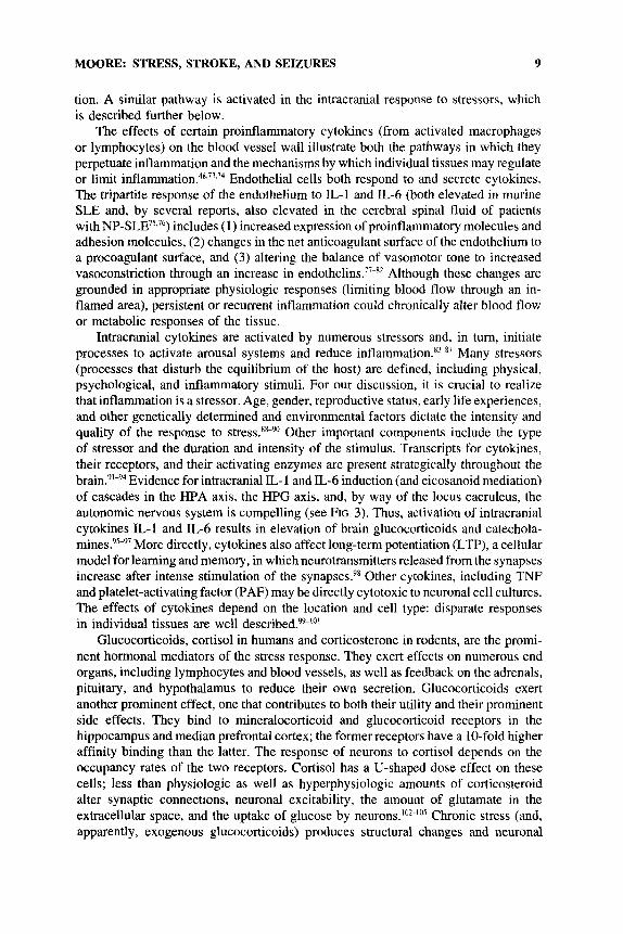
tion. A similar pathway is activated in the intracranial response to stressors, which is described further below.
The effects of certain proinflammatory cytokines (from activated macrophages or lymphocytes) on the blood vessel wall illustrate both the pathways in which they perpetuate inflammation and the mechanisms by which individual tissues may regulate or limit i n f l a m m a t i ~ n . ~ ~ . ~ ~ . ~ ~ Endothelial cells both respond to and secrete cytokines. The tripartite response of the endothelium to IL-1 and IL-6 (both elevated in murine SLE and, by several reports, also elevated in the cerebral spinal fluid of patients with NP-SLE75.76) includes ( 1) increased expression of proinflammatory molecules and adhesion molecules, ( 2 ) changes in the net anticoagulant surface of the endothelium to a procoagulant surface, and ( 3 ) altering the balance of vasomotor tone to increased vasoconstriction through an increase in e n d o t h e l i n ~ . ~ ~ - ~ ~ Although these changes are grounded in appropriate physiologic responses (limiting blood flow through an in- flamed area), persistent or recurrent inflammation could chronically alter blood flow or metabolic responses of the tissue.
Intracranial cytokines are activated by numerous stressors and, in turn, initiate processes to activate arousal systems and reduce Many stressors (processes that disturb the equilibrium of the host) are defined, including physical, psychological, and inflammatory stimuli. For our discussion, it is crucial to realize that inflammation is a stressor. Age, gender, reproductive status, early life experiences, and other genetically determined and environmental factors dictate the intensity and quality of the response to Other important components include the type of stressor and the duration and intensity of the stimulus. Transcripts for cytokines, their receptors, and their activating enzymes are present strategically throughout the brain.91-94 Evidence for intracranial IL- 1 and IL-6 induction (and eicosanoid mediation) of cascades in the HPA axis, the HPG axis, and, by way of the locus caeruleus, the autonomic nervous system is compelling (see FIG. 3) . Thus, activation of intracranial cytokines IL-1 and IL-6 results in elevation of brain glucocorticoids and catechola- mine^?^-^^ More directly, cytokines also affect long-term potentiation (LTP), a cellular model for learning and memory, in which neurotransmitters released from the synapses increase after intense stimulation of the synapses.98 Other cytokines, including TNF and platelet-activating factor (PAF) may be directly cytotoxic to neuronal cell cultures. The effects of cytokines depend on the location and cell type: disparate responses in individual tissues are well described?'"'
Glucocorticoids, cortisol in humans and corticosterone in rodents, are the promi- nent hormonal mediators of the stress response. They exert effects on numerous end organs, including lymphocytes and blood vessels, as well as feedback on the adrenals, pituitary, and hypothalamus to reduce their own secretion. Glucocorticoids exert another prominent effect, one that contributes to both their utility and their prominent side effects. They bind to mineralocorticoid and glucocorticoid receptors in the hippocampus and median prefrontal cortex; the former receptors have a 10-fold higher affinity binding than the latter. The response of neurons to cortisol depends on the occupancy rates of the two receptors. Cortisol has a U-shaped dose effect on these cells; less than physiologic as well as hyperphysiologic amounts of corticosteroid alter synaptic connections, neuronal excitability, the amount of glutamate in the extracellular space, and the uptake of glucose by n e ~ r o n s . ' ~ ~ - ~ ~ ~ Chronic stress (and, apparently, exogenous glucocorticoids) produces structural changes and neuronal

10 ANNALS NEW YORK ACADEMY OF SCIENCES
- bflammation
increased expression adhesion molecules r-- secretion - cytokines
effect to pro-coagulant
endothelinslnltrlc oxide
ENDOTHELIAL CELL
FIGURE 3. Endothelial cells respond to and secrete cytokines.
damage in specific areas of the neuraxis. The consequences of excessive and persistent glucocorticoid production include hippocampal atrophy, which will affect (1) certain areas of behavior and memory, particularly declarative memory and (2) the normal modulating effects of the hippocampus and other limbic system structures on the regulation of the HPA a x i ~ . ~ ” ~ ~ ~ ~ ’ ~ ~ ~ ” ’ ’ ~ The associated clinical features emerge in Cushing’s disease, posttraumatic stress disorders, and, possibly, aging. Numerous animal models of restraint stress, inflammatory stress, and adrenalectomy plus incre- mental pharmacological steroid replacement support these findings.9,13,113J14
Other factors also participate in the regulation of the stress response, including neurotransmitters, neuropeptides, and other cytokines; some of this work is presented in later sections. The potential role of antiinflammatory cytokines, such as TGF-p and IL-10, in tissue injury and healing are being investigated.
In patients with SLE, where these conditions may coexist, minor insults may result in more damage than when these injuries occur in isolation. A current hypothesis in our laboratory is that the clinical expression of NP-SLE results from “multiple hits.’ ’ This may partly explain the temporal dissociation of clinical NP features from evidence of active system disease. Clinical features appear after a minor event superimposed upon several previous injuries.
FUTURE INVESTIGATIONS AND APPROACHES
In this volume we bring together investigators from different disciplines whose work may help us better understand the clinical features and basic mechanisms of
MOORE: STRESS, STROKE, AND SEIZURES 11
tissue damage in NP-SLE. The following are some of the questions w e could profitably address. (1) Using predefined terminology in a multicenter neuroepidemiologic study, what is the incidence and prevalence of specific abnormalities in neuropsychiatric function? (2) What is the role of endogenous and pharmacological glucocorticoids in the neuroendocrine and behavioral aspects of SLE? (3) How can we distinguish the occurrence of ischemia from changes in cerebral metabolism?
1.
2. 3.
4.
5.
6.
7.
8.
9.
10.
11.
12.
13.
14.
15.
16.
17.
REFERENCES
MOORE, P. M. & R. P. LISAK. 1995. Systemic lupus erythematosus: Immunopathogenesis
MILLS, J. A. 1994. Systemic lupus erythematosus. N. Engl. J. Med. 330: 1871-1879. of neurologic dysfunction. Springer Semin. Imrnunopathology 17: 43 -60.
CERVERA, R., M. A. KHAMASHTA, J. FONT, G. D. SEBASTIANI, A. GIL, P. LAVILLA, I. DOMENECH, A. 0. AYDINTUG, A. J-G, E. DE RAMON, M. GALEAZZI, H. HAGA, A. MATHIEU, F. HOUSSIAU, M. INGELMO & G. R. V. HUGHES. 1993. Systemic lupus erythematosus: Clinical and immunologic patterns of disease expression in a cohort of 1,000 patients. Medicine 72: 113-123.
JACOBSON, L. & R. SAPOLSKY. 1991. The role of the hippocampus in feedback regulation of the hypothalamic-pituitary-adrenocortical axis. Endocr. Rev. 12: 118- 134.
LIPTON, S. A. & P. A. ROSENBERG. 1994. Excitatory amino acids as a final common pathway for neurologic disorders. N. Engl. J. Med. 330: 613-622.
LOWY, M. T., L. WITTENBERG & B. K. YAMAMOTO. 1995. Effect of acute stress on hippocampal glutamate levels and spectrin proteolysis in young and aged rats. J. Neurochem. 65: 268-274.
MOGHADDAM, B., M. L. BOLINAO, B. STEIN-BEHRENS & R. SAPOLSKY. 1994. Glucocorti- coids mediate the stress-induced extracellular accumulation of glutamate. Brain Res.
STEIN-BEHRENS, B. A,, W. J. LIN & R. M. SAPOLSKY. 1994. Physiological elevations of glucocorticoids potentiate glutamate accumulation in the hippocampus. J. Neurochem.
LOWY, M. T., L. GAULT & B. K. YAMAMOTO. 1993. Adrenalectomy attenuates stress- induced elevations in extracellular glutamate concentrations in the hippocampus. J . Neurochem. 61: 1957-1959.
SAPOLSKY, R. M. 1994. The physiological relevance of glucocorticoid endangerment of the hippocampus. Ann. N. Y. Acad. Sci. 746: 294-307.
REICHLIN, S. 1993. Neuroendocrine-immune interactions. N. Engl. J. Med. 329:
WICK, G., Y. Hu, S. SCHWARZ & G. KROEMER. 1993. Immunoendocrine communication via the hypothalamo-pituitary-adrenal axis in autoimmune diseases. Endocr. Rev. 14:
HARBUZ, M. S. & S. L. LIGHTMAN. 1992. Stress and the hypothalamo-pituitary-adrenal axis: Acute, chronic and immunological activation. J. Endocrinol. 134: 327-339.
MCEWEN, B. S. 1996. Gonadal and adrenal steroids regulate neurochemical and structural plasticity of the hippocampus via cellular mechanisms involving NMDA receptors. Cell. Mol. Neurobiol. 16: 103-116.
WEST, S. G., W. EMLEN, M. K. WENER & B. L. KOTZIN. 1995. Neuropsychiatric lupus erythematosus: A 10-year prospective study on the value of diagnostic tests. Am. J. Med. 9 9 153-163.
MACKWORTH-YOUNG, C. G. & G. R. V. HUGHES. 1985. Epilepsy, an early symptom of systemic lupus erythematosus. J. Neurol. Neurosurg. Psychiatry 48: 185.
HAY, E. M., A. HUDDY, D. BLACK, P. MBAYA, B. TOMENSOV, R. M. BERNSTEIN, P. 5 . L. HOLT & F. CREED. 1994. A prospective study of psychiatric disorder and cognitive function in systemic lupus erythematosus. Ann. Rheum. Dis. 53: 298-303.
655: 25 1-254.
63: 596-601.
1246- 1253.
539 - 563.

12 ANNALS NEW YORK ACADEMY OF SCIENCES
18.
19.
20.
21.
22.
23.
24.
25.
26.
27.
28.
29.
30.
31.
32.
33.
34.
35.
36.
KUROE, K., K. KURAHASHI, I. NAKANO, Y. MORIMATSU & H. TAKEMORI. 1994. A neum- pathological study of a case of lupus erythematosus with chorea. J. Neurol. Sci. 123:
SMITH, R. W., D. W. ELLISON, E. A. JENKINS, P. J. GALLAGHER & M. I. CAWLEY. 1994. Cerebellum and brainstem vasculopathy in systemic lupus erythematosus: Two clinico- pathological cases. Ann. Rheum. Dis. 5 3 327-330.
FISK, J. D., B. EASTWOOD, G. SHERWCOD & J. G. HANLY. 1993. Patterns of cognitive impairment in patients with systemic lupus erythematosus. Br. J. Rheumatol. 32:
HIETAHARJU, A,, V. JANTTI, M. KORPELA & H. FREY. 1993. Nervous system involvement in systemic lupus erythematosus, Sjogren syndrome and sclerodema. Acta Med.
WONG, K. L., E. K. Woo, Y. L. Yu & R. W. WONG. 1991. Neurological manifestations of systemic lupus erythematosus: A prospective study. Q. J. Med. 81: 857-870.
KINNUNEN, E., P. JARVINEN, L. KETONEN & R. SEPPONEN. 1993. Co-twin control study on cerebral manifestations of systemic lupus erythematosus. Acta Med. Scand. 88:
FUTRELL, N., L. R. SCHULTZ & C. MILLIKAN. 1992. Central nervous system disease in patients with systemic lupus erythematosus. Neurology 40: 1649- 1657.
GINSBURG, K. S., E. A. WRIGHT, M. G. LARSON, A. H. FOSSEL, M. ALBERT, P. H. SCHUR & M. H. LIANG. 1992. A controlled study of the prevalence of cognitive dysfunction in randomly selected patients with systemic lupus erythematosus. Arthritis Rheum. 35: 776-782.
HAMMAD, A., Y. TSUKADA & N. TORRE. 1992. Cerebral occlusive vasculopathy in systemic lupus erythematosus and speculation on the part played by complement. Ann. Rheum. Dis. 51: 550-552.
HAY, E. M., D. BLACK, A. HUDDY, F. CREED, B. TOMENSON, R. M. BERNSTEIN & P. .I. L. HOLT. 1992. Psychiatric disorder and cognitive impairment in systemic lupus erythematosus. Arthritis Rheum. 35: 411-416.
LIGHTMAN, S. L. & M. S. HARBUZ. 1993. Expression of corticotropin-releasing factor mRNA in response to stress. Ciha Found. Symp. 172: 173-187.
SIBLEY, J. T., W. P. OLSZYNSKI, W. E. DECOTEAU & M. B. SUNDARAM. 1992. The incidence and prognosis of central nervous system disease in systemic lupus erythematosus. J. Rheumatol. 19: 47-52.
TOLA, M. R., E. GRANIERI, L. CANIATTI, E. PAOLINO, C. MONETTI, L. DOVIGO, R. SCOLOZZI, P. DE BASTIANI & M. CARRERAS. 1992. Systematic lupus erythematosus presenting with neurological disorders. J. Neurol. 239: 61 -64.
EUSTACE, S., M. HUTCHINSON & B. BRESNIHAN. 1991. Acute cerebrovascular episodes in systemic lupus erythematosus. Q. J. Med. 81: 739-750.
KIRK, A., A. KERTESZ & M. J. POLK. 1991. Dementia with leukoencephalopathy in systemic lupus erythematosus. Can. J. Neurol. Sci. 18: 344-348.
OMDAL, R., S. I. MELLGREN, G. HUSSY, R. SALVESEN, 0. A. HENRIKSEN & T. A. TORBERGSEN. 1993. A controlled study of peripheral neuropathy in systemic lupus erythematosus. Acta Neurol. Scand. 88: 41-46.
TSOKOS, G. C. , M. TSOKOS, N. G. H. LE RCHE & J. H. KLIPPEL. 1986. A clinical and pathologic study of cerebrovascular disease in patients with systemic lupus erythemato- sus. Semin. Arthritis Rheum. 16: 70-78.
MARKUSSE, €3. M. & C. J. VECHT. 1986. Is neurological disease with positive lupus serology sufficient for a diagnosis of systemic lupus erythematosus? Br. J. Rheumatol.
ADELMAN, D. C., E. SALTrEL & J. R. KLINENBERG. 1986. The neuropsychiatric manifesta- tions of systemic lupus erythematosus: An overview. Semin. Arthritis Rheum. 15:
59-63.
458 -462.
Scand. 88: 299-308.
422-426.
25: 302-305.
185-199.

MOORE: STRESS, STROKE, AND SEIZURES 13
37.
38.
39.
40.
41.
42.
43.
44.
45.
46.
47. 48.
49.
50.
51.
52.
53.
54.
55.
56.
DENBURG, S. D., R. M. CARBOTTE & J. A. DENBURG. 1987. Cognitive impairment in systemic lupus erythematosus: A neuropsychological study of individual and group deficits. J. Clin. Exp. Neuropsychol. 9: 323-339.
SINGH, R. R., K. PRASAD, A. KUMAR, A. MISRA, K. PADMAKUMAR & A. N. MALAVIYA. 1988. Cerebellar ataxia in systemic lupus erythematosus: Three case reports. Ann. Rheum. Dis. 47: 954-956.
HOYLE, C., D. J. EWING & A. C. PARKER. 1985. Acute autonomic neuropathy in association with systemic lupus erythematosus. Ann. Rheum. Dis. 44: 420-424.
HELLMANN, D. B., T. J. LAING, M. PETRI, Q. WHITING-O'KEEFE & G. J. PARRY. 1988. Mononeuritis multiple: The yield of evaluations for occult rheumatic diseases. Medi- cine 67: 145-153.
MIGUEL, E. C., R. M. R. PEREIRA, C. A. D. B. PEREIRA, L. BAER, R. E. GOMES, L. C. F. DE SA, R. HIRSCH, N. G. DE BARROS, J. M. DE NAVARRO & V. GENTIL. 1994. Psychiatric manifestations of systemic lupus erythematosus: Clinical features, symp- toms, and signs of central nervous system activity in 43 patients. Medicine 73: 224-232.
WARD, M. M. & S. STUDENSKI. 1991. The time course of acute psychiatric episodes in systemic lupus erythematosus. J. Rheumatol. 18: 535-539.
WEKKING, E. M., 3. C . NOSSENT, A. P. VAN DAM & A. J. SWAAK. 1991. Cognitive and emotional disturbances in systemic lupus erythematosus. Psychother. Psychosom. 55:
IVERSON, G. L. 1993. Psychopathology associated with systemic lupus erythematosus:
SHORTALL, E., D. ISENBERG & S. P. NEWMAN. 1995. Factors associated with mood and
HAMANN, G. F. & G. J. DEL ZOPPO. 1994. Leukocyte involvement in vasomotor reactivity
HARLAN, J. M. 1985. Leukocyte-endothelial interactions. Blood 65: 513-525. BRAQUET, P., D. HOSFORD, M. BRAQUET, R. BOURGAIN & F. BUSSOLINO. 1989. Role of
cytokines and platelet-activating factor in microvascular immune injury. Int. Arch. Allergy Appl. Immunol. 88: 88-100.
YAMASAKI, Y.. N. MATSUURA, H. SHOZUHARA, H. ONODERA, Y. ITOYAMA & K. KOGURE. 1995. Interleukin-I as a pathogenetic mediator of ischemic brain damage in rats. Stroke 26: 676-681.
GOLIGORSKY, M. S., H. TSUKAHARA, H. MAGAZINE, T. T. ANDERSEN A. B. MALIK & W. F. BAHOU. 1994. Termination of endothelin signaling: Role of nitric oxide. J. Cell Physiol. 158: 485-494.
SMITH-SWINTOSKY,V. L.,L. C. FETTIGREW,R. M. SAPOLSKY,~. PHARES, S. D. CRADDOCK, S . M. BROOKE & M. P. MAITSON. 1996. Metyrapone, an inhibitor of glucocorticoid production, reduces brain injury induced by focal and global ischemia and seizures. J. Cereb. Blood Flow Metab. 16: 585-598.
ELLISON, D., K. GATTER, A. HERYET & M. ESIRI. 1993. Intramural platelet deposition in cerebral vasculopathy of systemic lupus erythematosus. J. Clin. Pathol. 46: 37-40.
WANG, X., T. L. YUE, F. C. BARONE & G. Z . FEUERSTEIN. 1995. Monocyte chemoattractant protein-1 messenger RNA expression in rat ischemic cortex. Stroke 26: 661 -666.
GIBBONS, G. H. & V. J. DZAU. 1994. The emerging concept of vascular remodeling. N. Engl. J. Med. 330: 1431 -1438.
YAMAGATA, K., K. I. ANDREASSON, W. E. KAUFMANN, C. A. BARNES & P. F. WORLEY. 1993. Expression of a mitogen-inducible cyclooxygenase in brain neurons: Regulation by synaptic activity and glucocorticoids. Neuron 11: 371 -386.
JAREK, M. J., S. G. WEST, M. R. BAKER & K. M. RAK. 1994. Magnetic resonance imaging in systemic lupus erythematosus patients without a history of neuropsychiatric lupus erythematosus. Arthritis Rheum. 37: 1609- 1613.
126-131.
A methodological review. Semin. Arthritis Rheum. 22(4): 242-25 1.
mood disorders in SLE. Lupus 4: 272-279.
of the cerebral vasculature. Stroke 25: 21 17-2119.

14 ANNALS NEW YORK ACADEMY OF SCIENCES
57.
58.
59.
60.
61. 62.
63.
64.
65.
66.
67.
68.
69.
70.
71.
72.
73.
74.
75.
76.
77.
EMMI, L., M. BRAMATI, M. T. R. DECRISTOFARO, M. MASCALCHI, G . DAL Pozzo, G . P. MARCONI, G. MASSAI & A. PASSALEVA. 1993. MRI and SPECT investigations of the CNS in SLE patients. Clin. Exp. Rheumatol. 11: 13-20.
PRICHARD, J. W. & L. M. BRASS. 1992. New anatomical and functional imaging methods. Ann. Neurol. 3 2 395-400.
YAFFE, K., D. FERRIERO, A. J. BARKoviCH & H. ROWLEY. 1995. Reversible MRI abnormal- ities following seizures. Neurology 45: 104- 108.
VERN, B. A. & M. BUTLER. 1983. Transient thalamic hypodensity in lupus erythematosus with generalized seizures. Neurology 33: 1081 - 1083.
TAN, E. M. 1991. Autoantibodies in pathology and cell biology. Cell 67: 841-842. PORTANOVA, J. P., R. E. ARNDT & B. L. KOTZIN. 1988. Selective production of autoanti-
bodies in graft-vs-host-induced and spontaneous murine lupus. J. Immunol. 140:
EPSTEIN, F. H. 1990. Receptor autoimmunity in endocrine disorders. N. Engl. J. Med.
PERESS, N. S., E. PERILLO & J. D. FENSTERMACHER. 1989. Circumventricular organs in chronic serum sickness: A model for cerebral lupus. Biol. Psychiatry 26: 397-407.
NILSON, C., M. LINDVALL-AXELSSON & C. OWMAN. 1992. Neuroendocrine regulatory mechanisms in the choroid plexus-cerebrospinal fluid system. Brain Res. Brain Res. Rev. 17: 109- 138.
PERESS, N. S., V. A. ROXBURGH, & M. C. GLENFAND. 1981. Binding sites for immune components in human choroid plexus. Arthritis Rheum. 24: 520-526.
NILSSON, C., B. M. HULTBERG & S. GAMMELTOFT. 1996. Autocrine role of insulin-like growth factor I1 secretion by the rat choroid plexus. Eur. J. Neurosci. 8: 629-635.
feedback between interleukin-I and glucocorticoid hormones. Science 233: 652-654. DINARELLO, C. A. & S. M. WOLFF. 1993. The role of interleukin-1 in disease. N. Engl.
J. Med. 328: 106-113. GOUJON, E., P. PARNET, S. CREMONA & R. DANTZER. 1995. Endogenous glucocorticoids
downregulate central effects of interleukin- 1 beta on body temperature and behaviour in mice. Brain Res. 702: 173- 180.
NAVARRA, P., S. TSAGARAKIS, M. S. FARIA, L. H. REES, G. M. BESSER & A. B. GROSSMAN. 1991. Interleukins-1 and -6 stimulate the release of corticotropin-releasing hormone- 4 1 from rat hypothalamus in vitro via the eicosanoid cyclooxygenase pathway. Endocri- nology 128: 37-44.
CALOGERO, A. E., W. T. GALLUCCI, P. W. GOLD & G. P. CHROUSOS. 1988. Multiple feedback regulatory loops upon rat hypothalamic corticotropin-releasing hormone secretion. J. Clin. Invest. 82: 767-774.
FABRY, Z., C. S. RAINE & M. N. HART. 1994. Nervous tissue as an immune compartment: The dialect of the immune response in the CNS. Immunol Today 15: 218-224.
DE VRIES, H. E., M. C. M. BLOM-ROOSEMALEN, M. VAN OOSTEN, A. DE BOER, T. J. C. VAN BERKEL, D. D. BREIMER & J. KUIPER. 1996. The influence of cytokines on the integrity of the blood-brain barrier in vitro. J. Neuroimmunol. 64: 37-43.
TSAI, C.-Y., T.-H. Wu, S.-T. TsAi, K.-H. CHEN, P. THAJEB, W.-M. LIN, H.3. Yu & C.-L. Y u. 1994. Cerebrospinal fluid interleukin-6, prostaglandin E2 and autoantibodies in patients with neuropsychiatric systemic lupus erythematosus and central nervous system infections. Scand. J. Rheumol 23: 57-63.
HIROHATA, s. & T. MIYAMOTO. 1990. Elevated levels of interleuhn-6 in cerebrospinal fluid from patients with systemic lupus erythematosus and central nervous system involvement. Arthritis Rheum. 33: 644649.
FABRY, Z., K. M. FITZSIMMONS, J. A. HERLEIN, T. 0. MONINGER, M. B. DOBBS & M. N. HART. 1993. Production of the cytokines interleukin 1 and 6 by murine brain microvessel endothelium and smooth muscle pericytes. J. Neuroimmunol. 47: 23-34.
755 -760.
323: 1318-1324.
BESEDOVSKY, H., A. DELREY, E. SORKIN & c . A. DINARELLO. 1986. Immunoregulatory

MOORE: STRESS, STROKE, AND SEIZURES 15
78.
79.
80.
81.
82.
83.
84.
85.
86.
87.
88.
89.
90.
91.
92.
93.
94.
95.
LUSCHER, T. F., C. M. Boui.ANGER, Z. YANG, G. NULL & Y. DOHI. 1993. Interactions between endothelium-derived relaxing and contracting factors in health and cardiovas- cular disease. Circulation 87: 36-44.
BRENNER, B. M., J. L. TROY & B. J. BALLERMANN. 1989. Endothelium-dependent vascular responses. J. Clin. Invest. 84: 1373-1378.
MALE, D., G. PRYCE, C. HUGHES & P. LANros. 1990. Lymphocyte migration into brain modelled in vitro: Control by lymphocyte activation, cytokines, and antigen. Cell. Immunol. 127: 1-11.
Regulation of the fibrinolytic system of cultured vascular endothelium by interleukin 1. J. Clin. Invest. 78: 587-591.
endothelial cells enhance T cell responses by markedly augmenting IL-2 concentra- tions. Cell. Immunol. 118: 166- 177.
Effects of glucocorticoids and chronic inflammatory stress upon anterior pituitary interleuhn-6 mRNA expression in the rat. Br. J. Rheumatol. 32: 653-657.
R. KATO & M. ASAi. 1993. Interleukin-1 beta augments release of norepinephrine, dopamine, and serotonin in the rat anterior hypothalamus. J. Neurosci. 13: 3574.
BENvmisTE, E. N. 1992. Inflammatory cytokines within the central nervous system: Sources, function, and mechanism of action. Am. J. Physiol. 263: CI -C16.
SAPERSTEIN, A,, H. BRAND, T. AUDHYA, D. NAHRISKI, B. HUTCHINSON, S. ROSENZWEIG & C. S. HOLLANDER. 1992. Interleukin 1B mediates stress-induced immunosuppression via corticotropin-releasing factor. Endocrinology 130: 152- 158.
DUNN, A. J. & J. WANG. 1995. Cytokine effects on CNS biogenic amines. Neuroimmuno- modulation 2: 319-328.
SHANKS, N., S. LAROCQUE & M. J. MEANEY. 1995. Neonatal endotoxin exposure alters the development of the hypothalamic-pituitary-adrenal axis: Early illness and later responsivity to stress. J. Neurosci. 15: 376-384.
SHANKS, N., C. M. MCCORMICK & M. J. MEANEY. 1994. Sex differences in hypothalamic- pituitary-adrenal responding to endotoxin challenge in the neonate: Reversal by gonad- ectomy. Brain Res. Dev. Brain Res. 79: 260-266.
CAREY, M. P., C. H. DETERD, K. J. DE KUNING, F. HELMERHORST & K. E. R. DE KLOET. 1995. The influence of ovarian steroids on hypothalamic-pituitary-adrenal regulation in the female rat. J. Endocrinol. 144: 31 1-321.
S C H O B ~ ~ L , B., E. R. DE KLOET, W. SUTANTU & F. HOLSBOER. 1993. Cellular localization of interleukin 6 mRNA and interleukm 6 receptor mRNA in rat brain. Eur. J. Neurosci.
BEVILACQUA, M. P., R. R. SCHL~EF, M. A. GIMBRONE, JR. & D. J. LOSKUTOFF. 1986.
GUINAN, E. C., B. R. SMITH, J. T. DOUKAS, R. A. MILLER & J. S. POBER. 1989. Vascular
SARLIS, N. J., A. ~ T E P H A N O U , R. A. KNIGHT, S. L. LIGHTMAN & H. S. CHOWDREY. 1993.
SHINTANI, F., s. ~ N B A , T. NAKAKI, M. NIBUYA, N. KINOSHITA, E. SUZUKI, G. YAGI,
5: 1426-1435. SEBIRE, G., D. EMILIE, C. WALLON, C. HtRY, 0. DEVERGNE, J. DELFRAISSY, P. GALA-
NAUD & M. TARDIEU. 1993. In vitro production of IL-6, IL-lB, and tumor necrosis factor-a by human embryonic microglial andneural cells. J. Immunol. 150: 1517- 1523.
CUNNINGHAM JR., E. T., E. WADA, D. B. CARTER, D. E. TRACEY, J. F. BATTEY & E. B. DE SOUZA. 1992. In situ histochemical localization of type 1 interleukin-1 receptor messenger RNA in the central nervous system, pituitary, and adrenal gland of the mouse. .I. Neurosci. 12: 1101 ~ 11 14.
LYSON, K. AND S. M. MCCANN. 1992. Involvement of arachidonic acid cascade pathways in interleukin-6-stimulated corticotropin-releasing factor release in vitro. Neuroendo- crinology 55: 708 -7 13.
DE KLOET, E. R., M. S. OITZL & B. ScHoBrrz. 1994. Cytokines and the brain corticosteroid receptor balance: relevance to pathophysiology of neuroendocrine-immune communi- cation. Psychoneuroendoctinology 19: 121 ~ 134.

16 ANNALS NEW YORK ACADEMY OF SCIENCES
96.
97.
98.
99.
100.
101.
102.
103.
104.
105.
106.
107.
108.
109.
110.
111.
112.
113.
YASIN, S. A., A. COSTA, M. L. FORSLING & A. GROSSMAN. 1994. Interleukin-1 beta and interleukin-6 stimulate neurohypophysial hormone release in vitro. J. Neuroendocrinol.
DA SILVA, J. A. P., S. H. PEERS, M. PERRETI & D. A. WILLOUGHBY. 1993. Sex steroids affect glucocorticoid response to chronic inflammation and to interleukin- 1. J. Endocrinol. 136: 389-397.
KATSUKI, H., S. NAKAI, Y. HIRAI, K. AKAJI, Y. Krso & M. SATOH. 1990. Interleukin 1B inhibits long-term potentiation in the CA3 region of mouse hippocampal slices. Eur. J. Pharmacol. 181: 323-326.
LINDSBERG, P. J., J. M. HALLENBECK & G. FEUERSTEIN. 1991. Platelet-activating factor in stroke and brain injury. Ann. Neurol. 30: 117- 129.
CHULUYAN, H. E., D. SAPHIER, W. M. ROHN & A. J. DUNN. 1992. Noradrenergic innervation of the hypothalamus participates in adrenocortical responses to interleukin- 1. Neuroendocrinology 56: 106- 11 1.
WIEDMEIER, S. E., H. Mu, B. A. ARANEO & R. A. DAYNES. 1994. Age- and microenviron- ment-associated influences by platelet-derived growth factor on T cell function. J. Immunol. 152: 3417-3426.
DE KLOET, E. R., M. S. OITZL & M. JOELS. 1993. Functional implications of brain corticosteroid receptor diversity. Cell. Mol. Neurobiol. 13: 433.
MCEWEN, B. S., H. CAMERON, H. M. CHAO, E. GOULD, A. M. MAGARINOS, Y . WATA- NABE & C. S. WOOLLEY. 1993. Adrenal steroids and plasticity of hippocampal neurons: Toward an understanding of underlying cellular and molecular mechanisms. Cell. Mol. Neurobiol. 13: 457.
KERR, D. S., L. W. CAMPBELL, S.-Y. HAO & P. W. LANDFIELD. 1989. Corticosteroid modulation of hippocampal potentials: Increased effect with aging. Science 245:
JOELS, M. & K. E. R. DE KLOET. 1989. Effects of glucocorticoids and norepinephrine on the excitability in the hippocampus. Science 245: 1502- 1505.
SAPOLSKY, R. M. & J. ALTMANN. 1991. Incidence of hypercortisolism and dexamethasone resistance increases with age among wild baboons. Biol. Psychiatry 30: 1008- 1016.
LAYE, S., E. GOUJON, C. COMBE, R. VAN HOY, K. W. KELLEY, P. PARNET & R. DANTZER. 1996. Effects of lipopolysaccharide and glucocorticoids on expression of interleukin- 1 beta converting enzyme in the pituitary and brain of mice. J. Neuroimmunol. 68:
MEANEY, M. J., J. DIORIO, D. FRANCIS, J. WIDDOWSON, P. LAPLANTE, C. CALDJI, S. SHARMA, J. R. SECKL & P. M. PLOTSKY. 1996. Early environmental regulation of forebrain glucocorticoid receptor gene expression: Implications for adrenocortical responses to stress. Dev. Neurosci. 18: 49-72.
J. SCHULKIN. 1994. Glucocorticoid treatment increases the ability of CRH to induce seizures. Neurosci. Lett. 174: 113- 116.
Hu, S. B., L. A. TANNAHILL & S. L. LIGHTMAN. 1993. Regulation of arginine vasopressin mRNA in rat fetal hypothalamic cell culture. Role of protein kinases and glucocorti- coids. J. Mol. Endocrinol. 10: 51-57.
SAPOLSKY, R. M., H. UNO, C. S. REBERT & C. E. FINCH. 1990. Hippocampal damage associated with prolonged glucocorticoid exposure in primates. J. Neurosci. 10:
DE KLOET, K. E. R., M. S. OITZL & B. SCHOBITZ. 1994. Cytokines and the brain corticosteroid receptor balance: Relevance to pathophysiology of neuroendocrine- immune communication. Psychoneuroendocrinology 19: 121 - 134.
HARBUZ, M. S., S. A. NICHOLSON, B. GILLHAM & S. L. LIGHTMAN. 1990. Stress respon- siveness of hypothalamic corticotrophin-releasing factor and pituitary proopiomelano-
6: 179-184.
1505-1508.
61-66.
ROSEN, J. B., S. K. PISHEVAR, S. R. WEISS, M. A. SMITH, M. A. KLING, P. W. GOLD &
2897-2902.

MOORE: STRESS, STROKE, AND SEIZURES 17
cortin mRNAs following high-dose glucocorticoid treatment and withdrawal in the rat. J. Endocnnol. 127: 407-415.
VAN HAARST, H. A. D., M. S . OITZL, J. 0. WORKEL & K. E. R. DE KLOET. 1996. Chronic brain glucocorticoid receptor blockade enhances the rise in circadian and stress- induced pituitary-adrenal activity. Endocrinology 137: 4935 -4943.
114.