neuroanatomical correlates of neuropsychiatric
TRANSCRIPT

8/3/2019 Neuroanatomical Correlates of Neuropsychiatric
http://slidepdf.com/reader/full/neuroanatomical-correlates-of-neuropsychiatric 1/9
Neuroanatomical correlates of neuropsychiatricsymptoms in Alzheimer’s disease
Peita D. Bruen,1,2 William J. McGeown,1 Michael F. Shanks1 and Annalena Venneri1,3
1Clinical Neuroscience Centre, University of Hull, Hull, UK, 2Department of Neuroscience, University of Parma, Parma and3Department of Neuroscience, University of Modena and Reggio Emilia, Modena, Italy
Correspondence to: Prof. Annalena Venneri, Clinical Neuroscience Centre, University of Hull, Cottingham Road, Hull HU6
7RX, England, UK
E-mail: [email protected]
Alzheimer’s disease research has largely concentrated on the study of cognitive decline, but the associated
behavioural and neuropsychiatric symptoms are of equal importance in the clinical profile of the disease.
There is emerging evidence that regional differences in brain atrophy may align with variant disease presenta-
tions.The objective of this study was to identify the regions of decreased grey matter (GM) volume which were
associated with specific neuropsychiatric behaviours in patients with mild Alzheimer’s disease.Voxel-based mor-
phometry was used to correlate GM derived fromT1-weighted MRI images of 31 patients with mild Alzheimer’s
disease and specific neuropsychiatric symptoms and behaviours measured by the Neuropsychiatric Inventory.
Delusions were associated with decreased GM density in the left frontal lobe, in the right frontoparietal cortex
and in the left claustrum. Apathy was associated with GM density loss in the anterior cingulate and frontal
cortex bilaterally, the head of the left caudate nucleus and in bilateral putamen. Agitation was associated with
decreased GM values in the left insula, and in anterior cingulate cortex bilaterally. Neuropsychiatric symptoms
of Alzheimer’s disease seem to associate with neurodegeneration of specific neural networks supporting perso-
nal memory, reality monitoring, processing of reward, interoceptive sensations and subjective emotional
experience. The study of neurodegenerative disorders such as Alzheimer’s disease using voxel-based morpho-
metry and other imaging modalities may further the understanding of the neural structures that mediate the
genesis of abnormal behaviours.
Keywords: delusions; apathy; agitation; dementia; voxel-based morphometry; MRI
Abbreviations: GM= grey matter; MMSE = Mini-Mental State Examination; NPI= Neuropsychiatric Inventory
Received March 31, 2008. Revised May 21, 2008. Accepted June 16, 2008. Advance Access publication July11, 2008
Introduction
The neuropsychiatric symptoms seen in the course of
Alzheimer’s disease are usually persistent, although with
fluctuating intensity, and can be resistant to treatment
(Ryu et al ., 2005). Such symptoms, particularly when there
is a psychotic driver for abnormal behaviour, are linked to
higher rates of institutional care and more rapid cognitivedecline. An important component of effective intervention
to reduce patient and carer distress, therefore, is the
appropriate management of abnormal thought content and
related behaviours (Yaffe et al ., 2002; Cummings, 2003;
Perez-Madrinan et al ., 2004; Scarmeas et al ., 2005). The
pathophysiological and psychological mechanisms involved
in the development of neuropsychiatric symptoms are still
poorly understood.
Initially, syndromes of abnormal thinking and behaviour
in the dementias were seen as an inevitable and non-specific
consequence of neurodegeneration with global cognitive
impairment. The different neuropsychiatric syndromes seen
in the course of Alzheimer’s disease, however, have
stimulated both genetic and neuroanatomical correlative
studies in patient groups using different rating scales (Megaet al ., 2000; Sweet et al ., 2003; Rosen et al ., 2005). Discrete
regional associations with a range of symptoms have been
identified using indices of brain metabolism, blood flow
and brain structure. Experimental studies have begun to
delineate more specific neurological associations with
discrete symptoms or syndromes, and more substantial
theoretical accounts have emerged. For example, it is now
clear that delusional symptoms are not simply incidental
doi:10.1093/brain/awn151 Brain (2008), 131, 2455^2463
ß The Author (200 8). Publishedby Oxford University Press on behalfof the Guarantors of Brain. Allrightsreserved. For Permissions, please email: [email protected]

8/3/2019 Neuroanatomical Correlates of Neuropsychiatric
http://slidepdf.com/reader/full/neuroanatomical-correlates-of-neuropsychiatric 2/9
to multiple cognitive deficits but are likely associated
with discrete regional pathologies which often involve
the anterior part of the right hemisphere (Sultzer et al .,
2003; Shanks and Venneri, 2004). Alzheimer’s disease
patients with apathy, on the other hand, are more likely
to show damage to the medial frontal and anterior
cingulate regions (Craig et al ., 1996; Migneco et al ., 2001;Rosen et al ., 2005). Neurofibrillary tangle burden was
higher in the left orbitofrontal cortex of Alzheimer’s
disease patients who had higher agitation scores (Tekin
et al ., 2001).
These and other regional associations have been inferred
either from measures of regional brain dysfunction (brain
metabolism or regional blood flow) or gross estimates of
structural or neuropathological damage. This study was
designed to establish more specific and focal anatomical
correlations between regional atrophy and early or emer-
ging neuropsychiatric symptoms, using the method of
voxel-based correlation analysis on 3D structural MRI scans
of the brains of patients with mild Alzheimer’s disease.Improved anatomical definition of structural correlations
with specific neuropsychiatric symptoms may be more
easily established in patients with mild Alzheimer’s disease.
At the mild stage of Alzheimer’s disease, disorders of
experience or behaviour often appear in relative isolation.
At a more advanced stage of the disease, the detection of
discrete symptom/atrophy associations is less likely when
such symptoms occur as part of a cluster of neuropsychia-
tric disturbances.
MethodsSampleThirty one patients with Alzheimer’s disease (19 male and 12
female) were recruited from referrals to our old age psychiatry
memory clinic. Their mean age was 77.1 years (SD = 8.6, range
56–95) and their mean education was 11.3 years (SD = 3.1, range
9–17). All patients had extensive neuropsychological screening (see
Table 1 for their full neuropsychological profile), structural MRI
scanning, neuropsychiatric assessment and neurological examina-
tion. The patients were selected from a larger group of $100
patients who had been referred by local general practitioners to an
NHS memory clinic. All patients in the study fulfilled the
NINCDS-ADRDA criteria for probable Alzheimer’s disease of
mild severity (McKhann et al ., 1984). Selected patients were first
referrals with a 6–12 month history of progressive cognitivedecline. They were clinically, neuropsychologically and radiologi-
cally (MRI) re-examined after at least 6 months and had their
clinical diagnosis confirmed. Exclusion criteria included significant
symptoms of depression, claustrophobia, radiological evidence of
ischemic brain disease, Hachinski Ischaemia score 44, Mini-
Mental State Examination (MMSE) score518/30 and evidence of
other degenerative or secondary dementias. Published clinical
criteria were used to exclude other potential causes of dementia
(Roman et al ., 1993; Brun et al ., 1994; McKeith et al ., 1996) and
no patient with a Hachinski scale (Hachinski et al ., 1975) score42
was entered in the study. None of the patients had a premorbid
psychiatric history. None of the patients was taking any
antipsychotic or psychoactive medication and they had not yet
begun cholinesterase inhibitor therapy at the time of investigation.
All patients included in the study had widespread atrophy with
more prominent neuronal loss in the medial temporal structures
(hippocampus and other structures in the hippocampal complex)
and no detectable significant ischemic white matter changes with a
T2-weighted MRI scan.
Before entering the study, a brief interview was carried out with
each patient and their relative/guardian. Only those patients who
were able to give informed consent were enrolled. Ethics approval
for the current study was granted by the NHS Trust Regional
Ethics Committee.
Neuropsychiatric assessmentEach patient and their caregiver had a semistructured interview
with a consultant psychiatrist (M.F.S) who also completed the
Neuropsychiatric Inventory (NPI) (Cummings et al ., 1994). The
NPI is a caregiver-based behavioural rating system developed and
validated for the assessment of mental state and behavioural
abnormalities in dementia. The NPI records the presence or
absence, severity (rated 1–3) and frequency (rated 1–4) of 12
symptom fields. An index of severity is created for each
behavioural variable by multiplying the frequency and severity
scores. NPI values were then correlated with grey matter (GM)
density values extracted from the patients’ 3D MRI brain scans.
Structural MRI scanning: acquisition and analysisThree dimensional T1-weighted MRI images were acquired
on a 1.5 tesla MRI scanner. Voxel dimensions were
Table 1 Mean (and SD) scores of the Alzheimer’s diseasepatients on the neuropsychological tests
Tests Patients Cut-off
Mini Mental State Examination 23.30 (2.8) 527.9ADAS-Cog 19.80 (5.6) NAConfrontation naming 17.58 (1.75) 519.53Pyramids and palm trees test 47.73 (3.45) 549.73Verbal paired associates 7.81 (2.95) 58.94Rey complex figure
Direct copy 18.42 (9.93) 520.45Delayed copy 1.21 (1.70) 57.11
Semantic fluency 27.50 (9.14) 542.63Phonemic fluency 25.57 (10.87) 529.67Digit span
Forward 6.41 (1.21) 56.29Backward 4.14 (0.95) 53.94
Raven’s coloured progressivematrices (PM47)
22.45 (5.03) 528.88
Prose memoryImmediate recall 4.5 (3.08) 512.33Delayed recall (10min) 3.12 (3.49) 512.80
Digit cancellation 42.03 (10.6) 548.55Stroop test
Error interference effect 5.48 (8.19) 40.25Time interference effect (sec) 47.43 (32.29) 45.25
Token test 30.00 (2.61) 530.66Visuoconstructive apraxia test 12.18 (2.19) 511.38
Cut-off scores are derived from a local reference sample.NA = Not available.
2456 Brain (2008), 131, 2455^2463 P. D. Bruen et al.
8/3/2019 Neuroanatomical Correlates of Neuropsychiatric
http://slidepdf.com/reader/full/neuroanatomical-correlates-of-neuropsychiatric 3/9
0.937Â 0.937Â 1.6 mm3. The field of view was 240 mm with a
matrix size of 256Â 256Â 124.
A number of pre-processing steps were followed to isolate the
GM from the 3D T1-weighted structural scans before performing
the statistical analysis using SPM5 (Wellcome Department of
Imaging Neuroscience, UCL, London, UK).
To correct for global differences in brain shape, structural
images were warped to standard stereotactic space and segmented
to extract GM, white matter and CSF. The GM segments were
then modulated to correct for changes in volume induced by non-
linear normalization and smoothed using a Gaussian filter set at
12 mm to reduce possible error from between subject variability in
local anatomy and render the data more normally distributed.
Finally, smoothed GM segments were entered into a voxel-based
multiple regression analysis to investigate linear correlations
between GM concentration and severity indices for the symptoms
assessed by the NPI. Age, number of years of education and
MMSE score were also included in the model as covariates for all
symptom fields. A measure of executive functions was also
included in the model for apathy and agitation, since both of these
symptoms have been associated with executive dysfunction (Chen
et al ., 1998). The x , y , z coordinates of the areas of significant
correlation obtained from the analyses were first converted into
Talairach coordinates and then identified using the Talairach
Daemon Client (http://ric.uthscsa.edu/projects/tdc/).
A T2-weighted axial scan was also acquired prior to the 3D scan
acquisition to better highlight any vascular load and to ensure that
all participants included in the 3D structural imaging study had
no significant vascular burden.
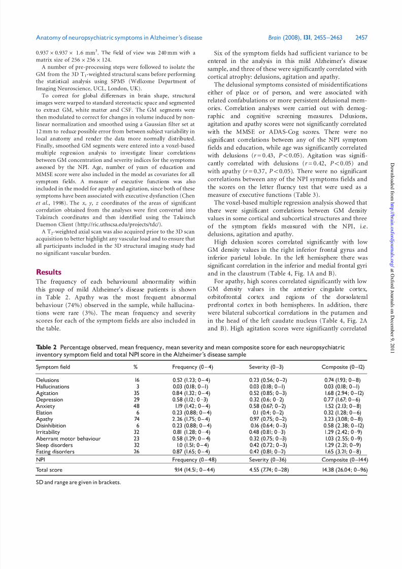
ResultsThe frequency of each behavioural abnormality within
this group of mild Alzheimer’s disease patients is shown
in Table 2. Apathy was the most frequent abnormalbehaviour (74%) observed in the sample, while hallucina-
tions were rare (3%). The mean frequency and severity scores for each of the symptom fields are also included in
the table.
Six of the symptom fields had sufficient variance to be
entered in the analysis in this mild Alzheimer’s disease
sample, and three of these were significantly correlated with
cortical atrophy: delusions, agitation and apathy.
The delusional symptoms consisted of misidentifications
either of place or of person, and were associated with
related confabulations or more persistent delusional mem-ories. Correlation analyses were carried out with demog-
raphic and cognitive screening measures. Delusions,
agitation and apathy scores were not significantly correlated
with the MMSE or ADAS-Cog scores. There were no
significant correlations between any of the NPI symptom
fields and education, while age was significantly correlated
with delusions (r = 0.43, P 50.05). Agitation was signifi-
cantly correlated with delusions (r = 0.42, P 50.05) and
with apathy (r = 0.37, P 50.05). There were no significant
correlations between any of the NPI symptoms fields and
the scores on the letter fluency test that were used as a
measure of executive functions (Table 3).
The voxel-based multiple regression analysis showed that
there were significant correlations between GM density
values in some cortical and subcortical structures and three
of the symptom fields measured with the NPI, i.e.
delusions, agitation and apathy.
High delusion scores correlated significantly with low
GM density values in the right inferior frontal gyrus and
inferior parietal lobule. In the left hemisphere there was
significant correlation in the inferior and medial frontal gyri
and in the claustrum (Table 4, Fig. 1A and B).
For apathy, high scores correlated significantly with low
GM density values in the anterior cingulate cortex,
orbitofrontal cortex and regions of the dorsolateralprefrontal cortex in both hemispheres. In addition, there
were bilateral subcortical correlations in the putamen and
in the head of the left caudate nucleus (Table 4, Fig. 2A
and B). High agitation scores were significantly correlated
Table 2 Percentage observed, mean frequency, mean severity and mean composite score for each neuropsychiatricinventory symptom field and total NPI score in the Alzheimer’s disease sample
Symptom field % Frequency (0 ^ 4) Severity (0 ^3) Composite (0 ^12)
Delusions 16 0.52 (1.23; 0 ^ 4) 0.23 (0.56; 0 ^2) 0.74 (1.93; 0 ^ 8)Hallucinations 3 0.03 (0.18; 0 ^1) 0.03 (0.18; 0 ^1) 0.03 (0.18; 0 ^1)Agitation 35 0.84 (1.32; 0 ^ 4) 0.52 (0.85; 0 ^3) 1.68 (2.94; 0 ^12)
Depression 29 0.58 (1.12; 0 ^3) 0.32 (0.6; 0 ^2) 0.77 (1.67; 0 ^ 6)Anxiety 48 1.19 (1.42; 0 ^ 4) 0.58 (0.67; 0 ^2) 1.52 (2.13; 0 ^ 8)Elation 6 0.23 (0.88; 0 ^ 4) 0.1 (0.4; 0 ^2) 0.32 (1.28; 0 ^ 6)Apathy 74 2.26 (1.75; 0 ^ 4) 0.97 (0.75; 0 ^2) 3.23 (3.08; 0 ^ 8)Disinhibition 6 0.23 (0.88; 0 ^ 4) 0.16 (0.64; 0 ^3) 0.58 (2.38; 0 ^12)Irritability 32 0.81 (1.28; 0 ^ 4) 0.48 (0.81; 0 ^3) 1.29 (2.42; 0 ^9)Aberrant motor behaviour 23 0.58 (1.29; 0 ^ 4) 0.32 (0.75; 0 3̂) 1.03 (2.55; 0 9̂)Sleep disorders 32 1.0 (1.51; 0 ^ 4) 0.42 (0.72; 0 ^3) 1.29 (2.21; 0 ^9)Eating disorders 26 0.87 (1.65; 0 ^ 4) 0.42 (0.81; 0 ^2) 1.65 (3.21; 0 ^ 8)
NPI Frequency (0 ^ 48) Severity (0 ^36) Composite (0 ^144)
Total score 9.14 (14.51; 0 ^ 44) 4.55 (7.74; 0 ^28) 14.38 (26.04; 0 ^96)
SD and range are given in brackets.
Anatomy of neuropsychiatric symptoms in Alzheimer’s disease Brain (2008), 131, 2455^2463 2457

8/3/2019 Neuroanatomical Correlates of Neuropsychiatric
http://slidepdf.com/reader/full/neuroanatomical-correlates-of-neuropsychiatric 4/9
with low GM density in the left insula and bilateral anterior
cingulate cortex (Table 4 and Fig. 3).
Discussion
This study has established that some of the neuropsychiatric
symptoms seen in early Alzheimer’s disease are associated
with atrophy of well-defined brain structures. Some of these
symptoms, delusions and agitation for example, are fre-
quent in the later stage of the disease, but less common at
the mild stage, although one study reported agitation in
47% of their subsample of mild Alzheimer’s disease patients
(Mega et al ., 1996). Significant apathy, on the other hand,
is often seen in early presentations of Alzheimer’s disease
and is sometimes described even before memory deficits
become noticeable. This symptom frequency pattern was
reflected in this group of early Alzheimer’s disease patients.This study identified those discrete atrophic sites that are
most strongly associated with the presence and severity of
these symptoms. The association of delusions with atrophy
primarily in the right frontoparietal regions supports
previous findings from metabolic and regional cerebral
blood flow (rCBF) studies (Staff et al ., 1999; Sultzer et al .,
2003). There was a smaller cluster of significant correlation
in the left frontal cortex, but nearly twice as many voxels
showed significant association in the right hemisphere
than in the left. A left/right asymmetry with relative
preservation of the left frontal lobe had already been
Table 4 Areas of significant correlation between GM density values and delusions; apathy; and agitation
Brain area Right/Left Brodmann’s area Talairach Co-ordinates Z -value at local
x y z maximum
Delusions (P50.01)
Inferior frontal gyrus R 45 50 18 12 3.49Inferior parietal lobule R 40 34 À50 41 3.0442 À52 43 2.79
Inferior frontal gyrus L 45 À55 22 15 3.14À51 26 21 2.88
Medial frontal gyrus L 11 À8 61 À15 2.86À1 56 À15 2.66
Claustrum L À38 À2 4 2.75
Apathy (P50.001)Lentiform nucleus (putamen) R 24 10 1 4.12Inferior frontal gyrus R 47 18 20 À21 3.18Middle frontal gyrus R 9 28 25 30 3.58Superior frontal gyrus R 10 24 47 À1 3.26Anterior cingulate R 24 12 À6 37 3.58Caudate nucleus (head) L À12 10 1 3.60
Lentiform nucleus (putamen) LÀ
30 0 7 3.60Inferior frontal gyrus L 47 À16 18 À19 3.3245 À34 29 6 3.58
Middle frontal gyrus L 9 À30 27 26 3.37Anterior cingulate L 24 À14 2 40 3.19
Agitation (P50.001)Anterior cingulate R 24 12 À2 42 3.49Insula L 13 À48 10 1 3.69Anterior cingulate L 24 À14 À4 39 3.45
Table 3 Correlations between demographic and cognitive variables and NPI symptom field scores
Age Education MMSE ADAS-Cog Delusions Agitation Apathy Letter Fluency
Age ^ À0.37 0.105 À0.111 0.430Ã À0.61 À0.075 À0.079Education À0.37 ^ 0.223 À0.232 À0.180 0.047 0.002 0.269MMSE 0.105 0.223 ^ À0.664ÃÃ À0.172 À0.160 À0.60 0.260
ADAS^ CogÀ
0.111À
0.232À
0.664
ÃÃ
^ 0.149 0.272 0.348À
0.422
Ã
Delusions 0.430Ã À0.180 À0.172 0.149 ^ 0.420Ã 0.230 0.041Agitation À0.61 0.047 À0.160 0.272 0.420Ã ^ 0.368Ã
À0.003Apathy À0.075 0.002 À0.60 0.348 0.230 0.368Ã ^ À0.035Letter fluency À0.079 0.269 0.260 0.422Ã 0.041 À0.003 À0.035 ^
Ã
Value is significant at P50.05 (two-tailed). ÃÃValue is significant at P50.01 (two-tailed).
2458 Brain (2008), 131, 2455^2463 P. D. Bruen et al.

8/3/2019 Neuroanatomical Correlates of Neuropsychiatric
http://slidepdf.com/reader/full/neuroanatomical-correlates-of-neuropsychiatric 5/9
associated with misidentification delusions in a CT study
(Forstl et al ., 1991). Right fronto-parietal dysfunction
was regularly reported in studies of single cases with avariety of misidentification delusions (Feinberg et al ., 1999;
Shanks and Venneri, 2002; Abe et al ., 2007). It seems,
therefore, that an unusually severe impairment of the
anterior right hemisphere may appear in a few patients at a
relatively early disease stage in association with delusional
symptoms. This relatively focal atrophy might either follow
from regional variance in Alzheimer’s disease neuropathol-
ogy (reflecting the well-known clinical variance at onset), or
perhaps more likely, indicate regional genetic and/or
neurodevelopmental susceptibility. This latent variable
might then become expressed endophenotypically in the
course of neurodegeneration. In either case, damage to the
corresponding anatomical network presumably disrupts this
region’s functional role in aligning mental contents with
veridical reality (Shanks and Venneri, 2004). Delusional
misidentifications in this sample were associated with
content-related confabulations and delusional memories.
The region of correlation, especially in the frontal lobe,
involves areas which are relevant for personal episodic
memory retrieval. There is evidence from behavioural
studies that confabulations and delusional memories are
strongly correlated with failure of personal episodic
A
B
Fig. 2 Areas of significant correlation with the presence andseverity of apathy in: (A) bilateral striatal structures (axial view)and (B) in the left anterior cingulate cortex (sagittal view). Theaxial image is presented in neurological orientation (L/L).
A
B
Fig. 1 (A) Sagittal view of the right frontal and parietal regionswhere a significant correlation with the presence and severity of delusions was found. (B) Coronal view showing the cluster of significant correlation in the left claustrum. The coronal image ispresented in neurological orientation (L/L).
Anatomy of neuropsychiatric symptoms in Alzheimer’s disease Brain (2008), 131, 2455^2463 2459

8/3/2019 Neuroanatomical Correlates of Neuropsychiatric
http://slidepdf.com/reader/full/neuroanatomical-correlates-of-neuropsychiatric 6/9
memory retrieval for late adulthood and recent life periods
and relatively preserved personal semantic information
(Cooper et al ., 2006; Lee et al . , 2007). A cluster of
significant correlation was also found in the left claustrum.
An association between damage to this subcortical struc-ture and delusions has not previously been reported to
our knowledge. At first sight, this association seemed
difficult to interpret in any theoretical model. The
claustrum, however, has unique connectivity, in that the
target areas of projections from the cortex overlap only
when the cortical areas concerned are themselves inter-
connected (Pearson et al ., 1982). Projections from the
claustrum are extensive, mainly ipsilateral but with some
neurons projecting contralaterally. Representations in the
claustrum, therefore, will be more faithful to a regionally
sourced polymodal convergence than to more precise
homotopic mapping. The claustrum is well placed toperform higher order, integrative functions and to facilitate
rapid transfer of information along its anterio-posterior and
ventral-dorsal axes and in this way instantiate multimodal
(cognitive, perceptual, motor) syntheses (Crick and Koch,
2005). It follows that damage to this structure might impair
the normal synchronization of perceptual and cognitive
experiences in Alzheimer’s disease patients, contributing to
delusional misinterpretation of percepts, memories or
mental images.
Apathy was the most frequent symptom in this sample of
early Alzheimer’s disease patients. This symptom may be an
early diagnostic indicator of the disease, and is detectable
in a high proportion of patients with mild cognitive
impairment (Lyketsos et al ., 2002; Palmer et al ., 2007).
The presence and severity of apathy was significantly
correlated with low GM density in subcortical nuclei, the
putamen bilaterally and the left caudate nucleus. There were
also significant correlations in bilateral anterior cingulatecortex and inferior frontal and orbitofrontal regions of both
hemispheres. Atrophy of elements of the limbic circuit,
including the cingulate gyrus, together with the more
prominent atrophy in mediotemporal structures, is
observed early in Alzheimer’s disease. Atrophic changes in
the periventricular nuclei and the striatum also appear early
(Gado et al ., 1983; Jack et al ., 2005; Shiino et al ., 2006). The
association of apathy with atrophy in these regions is in
accord with the temporal coincidence between the early
appearance of this symptom and the first stages of regional
neuropathology in Alzheimer’s disease (Tekin et al ., 2001).
Previous studies using dynamic measures of brain
function in Alzheimer’s disease patients with apathy havepredominantly reported dysfunction in anterior cingulate/
medial frontal areas, although one study showed involve-
ment of the medial thalamus (Migneco et al ., 2001; Lanctot
et al ., 2007; Marshall et al ., 2007). Neuropathological and
structural studies showed involvement of similar cortical
regions in apathetic Alzheimer’s disease patients (Tekin
et al ., 2001; Rosen et al ., 2005; Marshall et al ., 2006;
Apostolova et al ., 2007). A crucial function of the anterior
cingulate cortex relates to the initiation and motivational
drivers for goal directed activities, particularly when
these are effortful, and damage to this cortical structure
would likely, therefore, lead to a degree of behavioural and
cognitive inertia (Devinsky et al ., 1995; Allman et al ., 2001).
Apathy has also been linked to dysfunction or atrophy of
ventral frontal areas (Rosen et al ., 2005; Marshall et al .,
2007), and the present finding of an association with an
area of atrophy in ventrolateral prefrontal cortex (BA47)
may relate to the role of this region in executive functions
and social cognition (Blackwood et al ., 2000; Wager and
Smith, 2003). The other clusters of significant correlation in
the present study involved the dorsal striatum predomi-
nantly in the left hemisphere. The dorsal portion of the
caudate nuclei, particularly the caudate head, is critical for
the executive function of a fronto-striatal network. This
network, in brief summary, is organized as a feed-forwardloop from frontal cortical areas to the caudate nucleus
and putamen, which then project to the medial thalamus
and back to the prefrontal cortex (Mesulam, 2000). Front-
ostriatal mechanisms are important for executive functions
including planning, set shifting and strategic thinking, and
damage will lead to impairment of these processes as well as
a blunting of response to rewarding stimuli. Clearly
significant disruption of this network could also contribute
to apathy through cognitive, emotional and motivational
deficits (Levy and Dubois, 2006). Apathy in a variety of
other neuropsychiatric disorders including frontotemporal
Fig. 3 Areas of significant correlation with the presence andseverity of agitation in the left insula (axial view).The image ispresented in neurological orientation (L/L).
2460 Brain (2008), 131, 2455^2463 P. D. Bruen et al.

8/3/2019 Neuroanatomical Correlates of Neuropsychiatric
http://slidepdf.com/reader/full/neuroanatomical-correlates-of-neuropsychiatric 7/9
dementia, schizophrenia and mood disorders may result
from impaired functioning of such frontal-subcortical
circuits (Bonelli and Cummings, 2007). In addition, a
small number of case studies of patients with caudate
lesions report apathy with impaired motivation (Habib
and Poncet, 1988; Mendez et al ., 1989; Trillet et al ., 1990;
Wang, 1991).Agitation was observed in a third of the patients and the
degree of agitation was associated with severity of atrophy
in the left insula and in bilateral anterior cingulate cortex.
The insula mediates the experience of the basic emotions,
and while the right hemisphere is dominant for the
regulation of higher social and emotional behaviours like
self awareness, there is less evidence for lateralization of
basic emotional processes (Davidson and Irwin, 1999). The
insula also has a role in body representation, subjective
emotional experience, the integration of sensory informa-
tion and the processing of convergent information to make
sensory experience emotionally salient (Damasio, 1996). In
this respect, it may have an important role in theexperience of pain, as interoceptive sensations are trans-
mitted to the insular cortex. Patients with dementia have
difficulty in the identification and expression of somatic
symptoms including pain, and insular damage may
contribute to replacing normal complaint and help seeking
with undirected agitation, and more generally to an
exaggerated behavioural response to somatosensory and
environmental stimuli. Additional support for a possible
role of insular damage in causing agitation comes from
studies of epileptic patients where insular seizures can
mimic the features, including severe agitation, of temporal
lobe epilepsy and nocturnal frontal lobe epilepsy (Ryvlin,
2006; Ryvlin et al ., 2006). Finally, the insula is the most
significantly thickened brain area in meditators, suggesting
that training in mental tranquillity may reinforce insular
activity as opposed to its suggested impoverishment in
states of agitation (Lazar et al ., 2005).
There were also significant correlations between agitation
scores and bilateral atrophy of the anterior cingulate gyrus,
a structure which together with damage to orbitofrontal
areas had already been associated with agitation in
neuropathological studies of Alzheimer’s disease (Tekin
et al ., 2001). The present study, while confirming the link
between agitation and anterior cingulate atrophy, found no
significant correlations between agitation and tissue loss inorbital frontal regions. The neuropathological evidence,
however, was of course not contemporary with the
appearance of the symptom. Any associations established
in that way are likely to be less specific than those observed
when the MRI and neuropsychiatric data are collected at
the same time (with a one week delay in the present study).
The validity of these results is potentially influenced by
the method that was used. Criticisms of the voxel-based
morphometry technique include concerns that segmenta-
tion inaccuracy may survive the automated processes
involved. The method seems reliable, however, when used
to identify the neural substrate of specific cognitive deficits
using a correlational approach. By treating each individual
voxel value in the brain as a continuous variable, a
correlation between differences in GM density values and
continuous behavioural data can be obtained. The resulting
data can better establish associations between brain regions
or equivalent damaged structures and related cognitivefunctions and behaviours (Tyler et al ., 2005; Venneri et al .,
2008). These more detailed clinical/pathological correlations
would not be feasible using the more traditional region of
interest approach, given the a priori assumptions about
location and extent that the method requires.
The relatively small sample size might also be seen as a
limitation and another study included 148 patients with
different types and stages of dementia studied with the
same methods (Rosen et al ., 2005). The present study,
however, is specific to Alzheimer’s disease and focused on
the milder level of severity. The investigation of patients
only at the earlier stage of the disease might also be seen as
a limitation in terms of the full development of neuro-psychiatric symptomatology. Previous studies have estab-
lished that neuropsychiatric symptoms increase in
frequency with disease severity (Mega et al ., 1996). The
inclusion of patients at a more advanced stage of
Alzheimer’s disease would most likely, therefore, have
resulted in a wider spectrum of symptom fields observed
as well as greater frequency and severity of symptoms. In
this mild Alzheimer’s disease group, there was no correla-
tion between any of the symptom field scores and the
MMSE scores. Significant correlations with disease severity
would probably have emerged if patients with a wider range
of severity had been included in the sample. On the other
hand, identification of discrete anatomical correlations with
specific neuropsychiatric symptoms is more likely to be
established in Alzheimer’s disease patients at the mild stage
when disorders of experience or behaviour may appear in
relative isolation. At the moderate stage, aggregated effects
of disease severity and clustering of neuropsychiatric
symptoms would make discriminative correlations of this
kind more challenging. Finally, there was no significant
correlation between neuropsychiatric symptom scores and
letter fluency scores. Executive dysfunction had been
associated with agitation and apathy in Alzheimer’s disease
(Chen et al ., 1998). The absence of a similar correlation in
this sample may follow from the more recent inception of these neuropsychiatric symptoms in these mild patients
which may have contributed to the specificity and more
focal nature of these anatomical associations.
In conclusion, this study has identified a number of
associations between certain foci of GM loss and the
presence of different neuropsychiatric symptoms. Discrete
regional associations of this kind may be more clearly
established in patients with Alzheimer’s disease as the
disease, while unfolding in a broadly predictable anatomical
pattern, can selectively affect discrete brain regions in
individual patients with corresponding variation in onset
Anatomy of neuropsychiatric symptoms in Alzheimer’s disease Brain (2008), 131, 2455^2463 2461

8/3/2019 Neuroanatomical Correlates of Neuropsychiatric
http://slidepdf.com/reader/full/neuroanatomical-correlates-of-neuropsychiatric 8/9
symptoms. Particularly in the early stages, therefore, the
architectonic distribution of neurodegenerative damage
and/or predisposing developmental changes may be subtle
and limited, allowing more precise correlative analysis than
in cases of vascular or traumatic damage. Even so, the
evidence obtained from clinicopathological correlation after
different pathological processes is remarkably convergent.Correlations of this kind may help to clarify the mech-
anisms and brain circuits involved in the control of certain
social and emotional behaviours, and will guide more
rational psychopharmacological and behavioural interven-
tions to alleviate these distressing symptoms.
AcknowledgementsThis study was partially funded by a grant from MIUR to
AV and support from Humber Mental Health Trust to
M.F.S. This study was also supported by the Marie Curie
Research Training Network on Language and Brain funded
by the European Commission under Framework 6 of whichP.D.B. and A.V. are members.
References
Abe N, Ishii H, Fujii T, Ueno A, Lee E, Ishioka T, et al. Selective
impairment in the retrieval of family relationships in person identifica-
tion: a case study of delusional misidentification. Neuropsychologia
2007; 45: 2902–9.
Allman JM, Hakeem A, Erwin JM, Nimchinsky E, Hof P. The anterior
cingulate cortex. The evolution of an interface between emotion and
cognition. Ann N Y Acad Sci 2001; 935: 107–17.
Apostolova LG, Akopyan GG, Partiali N, Steiner CA, Dutton RA,
Hayashi KM, et al. Structural correlates of apathy in Alzheimer’s
disease. Dement Geriatr Cogn Disord 2007; 24: 91–7.
Blackwood NJ, Howard RJ, ffytche DH, Simmons A, Bentall RP,Murray RM. Imaging attentional and attributional bias: an fMRI
approach to the paranoid delusion. Psychol Med 2000; 30: 873–83.
Bonelli RM, Cummings JL. Frontal-subcortical circuitry and behavior.
Dialogues Clin Neurosci 2007; 9: 141–51.
Brun A, Englund B, Gustafson L, Passant V, Mann DMA, Neary D, et al.
Clinical and neuropathological criteria for frontotemporal dementia.
J Neurol Neurosurg Psychiatry 1994; 57: 416–8.
Chen ST, Sultzer DL, Hinkin CH, Mahler ME, Cummings JL. Executive
dysfunction in Alzheimer’s disease: association with neuropsychiatric
symptoms and functional impairment. J Neuropsychiatry Clin Neurosci
1998; 10: 426–32.
Cooper JM, Shanks MF, Venneri A. Provoked confabulations in
Alzheimer’s disease. Neuropsychologia 2006; 44: 1697–707.
Craig AH, Cummings JL, Fairbanks L, Itti L, Miller BL, Li J, et al. Cerebral
blood flow correlates of apathy in Alzheimer disease. Arch Neurol 1996;53: 1116–20.
Crick FC, Koch C. What is the function of the claustrum? Philos Trans R
Soc Lond B Biol Sci 2005; 360: 1271–9.
Cummings JL, editor. The neuropsychiatry of Alzheimer’s disease and
related dementias. London: Martin Dunitz; 2003.
Cummings JL, Mega M, Gray K, Rosenberg-Thompson S, Carusi DA,
Gornbein J. The neuropsychiatric inventory: comprehensive assessment
of psychopathology in dementia. Neurology 1994; 44: 2308–14.
Damasio AR. The somatic marker hypothesis and the possible functions of
the prefrontal cortex. Philos Trans R Soc Lond B Biol Sci 1996; 351:
1413–20.
Davidson RJ, Irwin W. The functional neuroanatomy of emotion and
affective style. Trends Cogn Sci 1999; 3: 11–21.
Devinsky O, Morrell MJ, Vogt BA. Contributions of anterior cingulate
cortex to behaviour. Brain 1995; 118: 279–306.
Feinberg TE, Eaton LA, Roane DM, Giacino JT. Multiple Fregoli delusions
after traumatic brain injury. Cortex 1999; 35: 373–87.
Forstl H, Burns A, Jacoby R, Levy R. Neuroanatomical correlates of clinical
misidentification and misperception in senile dementia of the Alzheimer
type. J Clin Psychiatry 1991; 52: 268–71.
Gado M, Hughes CP, Danziger W, Chi D. Aging, dementia, and brainatrophy: a longitudinal computed tomographic study. AJNR Am J
Neuroradiol 1983; 4: 699–702.
Habib M, Poncet M. Loss of vitality, of interest and of the affect
(athymhormia syndrome) in lacunar lesions of the corpus striatum.
Rev Neurol 1988; 144: 571–7.
Hachinski VC, Iliff LD, Zilhka E, DuBoulay GH, McAllister VL, Marshall J,
et al. Cerebral blood flow in dementia. Archives of Neurology 1975; 32:
632–7.
Jack CR Jr, Shiung MM, Weigand SD, O’Brien PC, Gunter JL, Boeve BF,
et al. Brain atrophy rates predict subsequent clinical conversion in
normal elderly and amnestic MCI. Neurology 2005; 65: 1227–31.
Lanctot KL, Moosa S, Herrmann N, Leibovitch FS, Rothenburg L,
Cotter A, et al. A SPECT study of apathy in Alzheimer’s disease. Dement
Geriatr Cogn Disord 2007; 24: 65–72.
Lazar SW, Kerr CE, Wasserman RH, Gray JR, Greve DN, Treadway MT,et al. Meditation experience is associated with increased cortical
thickness. Neuroreport 2005; 16: 1893–7.
Lee E, Meguro K, Hashimoto R, Meguro M, Ishii H, Yamaguchi S, et al.
Confabulations in episodic memory are associated with delusions in
Alzheimer’s disease. J Geriatr Psychiatry Neurol 2007; 20: 34–40.
Levy R, Dubois B. Apathy and the functional anatomy of the prefrontal
cortex-basal ganglia circuits. Cereb Cortex 2006; 16: 916–28.
Lyketsos CG, Lopez O, Jones B, Fitzpatrick AL, Breitner J, DeKosky S.
Prevalence of neuropsychiatric symptoms in dementia and mild
cognitive impairment: results from the cardiovascular health study.
JAMA 2002; 288: 1475–83.
Marshall GA, Fairbanks LA, Tekin S, Vinters HV, Cummings JL.
Neuropathologic correlates of apathy in Alzheimer’s disease. Dement
Geriatr Cogn Disord 2006; 21: 144–7.
Marshall GA, Monserratt L, Harwood D, Mandelkern M, Cummings JL,Sultzer DL. Positron emission tomography metabolic correlates of
apathy in Alzheimer disease. Arch Neurol 2007; 64: 1015–20.
McKeith IG, Galasko D, Kosaka K, Perry EK, Dickson DW, Hansen LA,
et al. Consensus guidelines for the clinical and pathologic diagnosis of
dementia with Lewy bodies (DLB): report of the consortium on DLB
international workshop. Neurology 1996; 47: 1113–24.
McKhann G, Drachman D, Folstein M, Katzman R, Price D, Stadlan EM.
Clinical diagnosis of Alzheimer’s disease: report of the NINCDS-
ADRDA work group under the auspices of department of health and
human services task force on Alzheimer’s disease. Neurology 1984; 34:
939–44.
Mega MS, Cummings JL, Fiorello T, Gornbein J. The spectrum of
behavioral changes in Alzheimer’s disease. Neurology 1996; 46: 130–5.
Mega MS, Lee L, Dinov ID, Mishkin F, Toga AW, Cummings JL. Cerebral
correlates of psychotic symptoms in Alzheimer’s disease. J Neurol
Neurosurg Psychiatry 2000; 69: 167–71.
Mendez MF, Adams NL, Lewandowski KS. Neurobehavioral changes
associated with caudate lesions. Neurology 1989; 39: 349–54.
Mesulam MM. Behavioral Neuroanatomy. Large-scale networks, associa-
tion cortex, frontal syndromes, the limbic system, and hemispheric
specializations. In: Mesulam MM, editor.. Principles of Behavioral and
Cognitive Neurology. New York: Oxford University Press; 2000.
p. 1–120.
Migneco O, Benoit M, Koulibaly PM, Dygai I, Bertogliati C, Desvignes P,
et al. Perfusion brain SPECT and statistical parametric mapping
analysis indicate that apathy is a cingulate syndrome: a study in
Alzheimer’s disease and nondemented patients. Neuroimage 2001; 13:
896–902.
2462 Brain (2008), 131, 2455^2463 P. D. Bruen et al.

8/3/2019 Neuroanatomical Correlates of Neuropsychiatric
http://slidepdf.com/reader/full/neuroanatomical-correlates-of-neuropsychiatric 9/9