neuropsychiatric symptoms in patients with illness ......• thanks to david ross md of virginia...
TRANSCRIPT

Ritchie Shoemaker, M.D. Center for Research on Biotoxin
Associated Illnesses Pocomoke, Maryland
3/14 /2013 Gallaway, NJ
Neuropsychiatric symptoms in patients with illness acquired following exposure to WDB are
associated with structural abnormalities: a volumetric MRI study using NeuroQuant

Co-authors
David Ross MD, Alfred Ochs PhD, Megan DeSmit; Virginia Institute of Neuropsychiatry
James Ryan PhD, Proteogenomics Dennis House, CRBAI

Before I start
• Thanks to David Ross MD of Virginia Neuropsychiatry Institute and Virginia Commonwealth University
• Mentor • Provider of academic papers • Author of NeuroQuant slides used
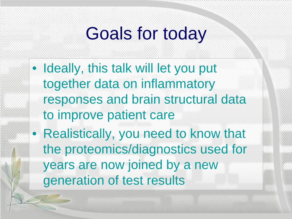
Goals for today
• Ideally, this talk will let you put together data on inflammatory responses and brain structural data to improve patient care
• Realistically, you need to know that the proteomics/diagnostics used for years are now joined by a new generation of test results

The Biotoxin Pathway
High levels of cytokines produce flu-like symptoms: Headaches, muscle aches, fatigue, unstable temperature, difficulty concentrating and more. High levels of cytokines also result in increased levels of several other immune-response related substances, including TGF B-1, MMP-9, IL-1B, and PAI-1. MMP-9 delivers inflammatory elements from blood to brain, nerve, muscle, lungs, and joints. It combines with PAI-1 in increasing clot formation and arterial blockage.
Inflammation-related symptoms
Reduced MSH
Hypothalamus
VIP MSH
AVP
Leptin receptor
Damaged leptin receptors lead to reduced production by the hypothalamus of MSH, a hormone with many functions.
In genetically susceptible people, biotoxins bind to pattern receptors, causing continuing, unregulated production of cytokines.
Dendritic Cells
HLA-DR
Surface Receptors
(Toll; C-type lectin;
mannose & others)
Fat cells then produce more leptin, leading to obesity (which doesn’t respond to exercise and diet).
Excessive cytokine levels can damage leptin receptors in the hypothalamus.
Removal from the
body In most people, biotoxins are either removed from the blood by the liver or attached by the immune system, broken down, and excreted harmlessly. In people who don’t have the right immune response genes, however, biotoxins can remain in the body indefinitely.
Nerve cell/axon
Biotoxins have direct effects, including impairment of nerve cell function.
Resistant Coag-negative Staph Bacteria
Colonies of MARCoNS with resistance to multiple antibiotics may develop in biofilm or mucus membranes. The bacteria produce substances that aggravate both the high cytokine levels and low MSH levels.
Reduced ADH Reduced MSH can cause the pituitary to produce lower levels of anti-diuretic hormone (ADH), leading to thirst, frequent urination, and susceptibility to shocks from static electricity.
Reduced Androgens Reduced MSH can cause the pituitary to lower its production of sex hormones.
Changes in Cortisol and ACTH levels
The pituitary may produce elevated levels of cortisol and ACTH in early stages of illness, then drop to excessively low levels later. (Patients should avoid steroids such as prednisone, which can lower levels of ACTH)
Sleep Disturbance Production of melatonin is reduced, leading to chronic, non-restorative sleep.
Chronic Pain Endorphin production is suppressed. This can lead to chronic, sometimes unusual, pain.
Gastrointestinal Problems
Lack of MSH can cause malabsorption in the gut, resulting in diarrhea. This is sometimes called “leaky gut” and resembles (but is not) celiac disease. IBS is often present.
White blood cells lose regulation of cytokine response, so that recovery from other illnesses, including infections diseases, may be slowed.
Prolonged Illness
c R. Shoemaker, 2011
Split Products of Complement Activation
C4a: capillary hypoperfusion C3a: bacterial membranes
Immune System Symptoms Patients with certain HLA genotypes (immune response genes) may develop inappropriate immunity. Most common are antibodies to: -Gliadin (affects digestion) -Cardiolipins (affects blood clotting) Treg cells: Pathogenic T cells
High cytokine levels in the capillaries attract white blood cells, leading to restricted blood flow, and lower oxygen levels. HIF stimulates VEGF and TGF B-1. Reduced VEGF leads to fatigue, muscle cramps, and shortness of breath (may be over-ridden by replacement with erythropoietin). TGF B-1 changes cell type and interacts with Treg cells.
Capillaries HIF
Increased Cytokines
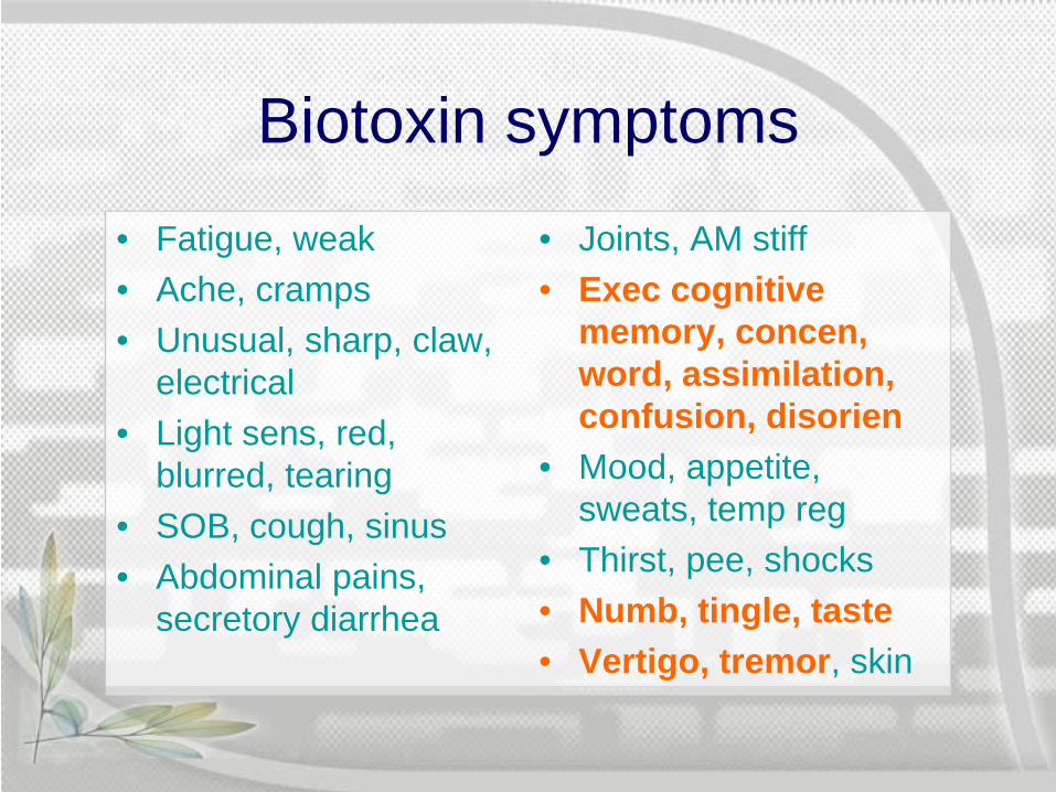
Biotoxin symptoms
• Fatigue, weak • Ache, cramps • Unusual, sharp, claw,
electrical • Light sens, red,
blurred, tearing • SOB, cough, sinus • Abdominal pains,
secretory diarrhea
• Joints, AM stiff • Exec cognitive
memory, concen, word, assimilation, confusion, disorien
• Mood, appetite, sweats, temp reg
• Thirst, pee, shocks • Numb, tingle, taste • Vertigo, tremor, skin
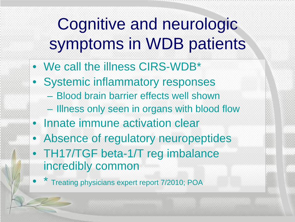
Cognitive and neurologic symptoms in WDB patients
• We call the illness CIRS-WDB* • Systemic inflammatory responses
– Blood brain barrier effects well shown – Illness only seen in organs with blood flow
• Innate immune activation clear • Absence of regulatory neuropeptides • TH17/TGF beta-1/T reg imbalance
incredibly common • * Treating physicians expert report 7/2010; POA

Add to the list
• Absence of executive inhibition • Tics • Atypical seizures • OCD • What looks like depression • What looks like anxiety/panic • ALL REPORTED IN CAUDATE
NUCLEUS ATROPHY SYNDROMES

How do I know these so many neuropsychiatric symptoms are
inflammatory? • Seen in cases • Not seen in controls • Differences are p < 0.001 • Abate with Rx only (not self-healing) • Recur with relapse (prospective!!) • Genomics (mRNA and MicroRNA) • TGF beta-1, VEGF, MMP9, C4a key

How does a “Neuro-Naysayer” know otherwise?
• Data on thousands of patients affirms • Data on treatment of thousands of patients
affirms • No data from anyone (ever) to deny • No research (ever) to deny • All the Naysayer has are empty words; no
data, no research, no prospective studies • Countless mRNA markers affirm • US Patent 8/21/2012
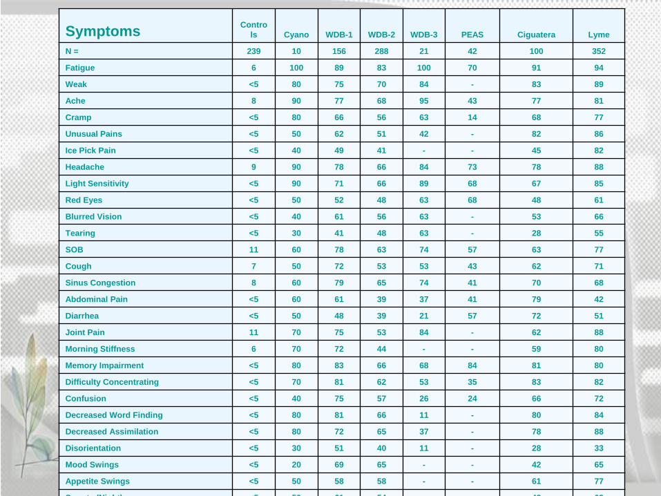
Symptoms Controls Cyano WDB-1 WDB-2 WDB-3 PEAS Ciguatera Lyme
N = 239 10 156 288 21 42 100 352
Fatigue 6 100 89 83 100 70 91 94
Weak <5 80 75 70 84 - 83 89
Ache 8 90 77 68 95 43 77 81
Cramp <5 80 66 56 63 14 68 77
Unusual Pains <5 50 62 51 42 - 82 86
Ice Pick Pain <5 40 49 41 - - 45 82
Headache 9 90 78 66 84 73 78 88
Light Sensitivity <5 90 71 66 89 68 67 85
Red Eyes <5 50 52 48 63 68 48 61
Blurred Vision <5 40 61 56 63 - 53 66
Tearing <5 30 41 48 63 - 28 55
SOB 11 60 78 63 74 57 63 77
Cough 7 50 72 53 53 43 62 71
Sinus Congestion 8 60 79 65 74 41 70 68
Abdominal Pain <5 60 61 39 37 41 79 42
Diarrhea <5 50 48 39 21 57 72 51
Joint Pain 11 70 75 53 84 - 62 88
Morning Stiffness 6 70 72 44 - - 59 80
Memory Impairment <5 80 83 66 68 84 81 80
Difficulty Concentrating <5 70 81 62 53 35 83 82
Confusion <5 40 75 57 26 24 66 72
Decreased Word Finding <5 80 81 66 11 - 80 84
Decreased Assimilation <5 80 72 65 37 - 78 88
Disorientation <5 30 51 40 11 - 28 33
Mood Swings <5 20 69 65 - - 42 65
Appetite Swings <5 50 58 58 - - 61 77
S t (Ni ht) <5 50 61 54 42 68

Chronic cognitive abnormalities in CIRS-WDB patients
• Executive cognitive functions – Recent memory – Concentration – Word finding; assimilation of new – Confusion – Disorientation NOT SPECIFIC FOR A GIVEN BIOTOXIN
ILLNESS

No differences between CIRS-WDB and other biotoxin illnesses • Mold (think water-damaged buildings) • Dinoflagellates (Pfiesteria, ciguatera,
Chattonella, ?? Karenina) • Apicomplexans (Babesia, Sarcocystis
and Eimeria) • Cyanobacteria (Microcystis, Lyngbya,
cylindrospermopsis)

Sx CLUSTER ANALYSIS
– Fatigue – Weak, assimilation,
aching, headache, light sensitivity
– Memory, words – Concentration – Joint, AM stiff, cramps – Unusual skin
sensations, tingling – Shortness of breath,
sinus – Cough, thirst, confusion
– Appetite, body temperature regulation, urinary freq.
– Red eyes, blurred vision, sweats, mood, ice-pick pains
– Abdominal pain, diarrhea, numbness
– Tearing, disorientation, metallic taste
– Static shocks, vertigo

8 clusters are only seen in biotoxin illnesses to date
• History must be taken by the health care provider
• No check lists • No self reporting • Use techniques of attorneys: multiple
re-asking of same question to confirm reliability of history

Logistic Regression Model - 8 Factor Score Combining Symptoms: Predicting Membership in the Group of Cases or
Controls PREDICTED CONTROLS
PREDICTED CASES
ROW TOTAL
Observed Controls
238 1 239 Observed Cases
5 277 282 COLUMN TOTAL
243 278 521
Percent Agreement 98.85 Agreement Odds 515/521 Disparities 6 Standard Deviation 0.47%
Confidence Limits 2-tailed Z at α=0.05=1.960
% Agreement
Disparities
Lower Confidence Limit=
97.03 11
Upper Confidence Limit=
99.76 1
- +
- Specificity False Positive
+ False Negative
Sensitivity
Significance Test of Agreement Test ChiSquare Prob. of Chi Square
Likelihood 656.722 <0.00000000001
Pearson 497.264 <0.00000000001

Biotoxins are ionophores
• Forget relying on antibody testing if epitope separation isn’t confirmed
• Move from cell to cell • Very small, many less than 1000 daltons • Secreted against a gradient into bile by
organic anion transport system (OATP) • News: OATP is also found along blood
brain barrier!!

Back to the brain
• Are executive symptoms telling us about abnormal brain structures?
• Brain physiology? • What we need is a dynamic imaging
study that correlates with symptoms and physiology!
• And we have one: NeuroQuant

NeuroQuant
• Volumetric study of 11 brain regions – Can expand to 15 – Changes over time key
• FDA approved in 2007 • Software added to MRI of brain • Takes 10 minutes • Reproducibly reliable • Controls data sets available

What do changes from normal in NeuroQuant mean?
• Changes in volume – Interstitial edema will increase – Atrophy or pruning will decrease
• Analyzed sequentially • Correlate with clinical studies • Correlate with genomics! • We can link mRNA to changes in brain
volumes with changes in clinical status

Review: what is the blood brain barrier?
• Endothelial cells and tight junctions • Basement membrane • Pericytes • Astrocytes • “The fence and the gate” • Breached by VEGF, MMP9 • TGF beta-1 dual role

BLOOD BRAIN BARRIER
Adapted from: Karen Francis, Johan van Beek, Cecile Canova, Jim W. Neal and Philippe Gasque, The Blood Brain Barrier. Expert Reviews in Molecular Medicine, Vol 5, 23 May, 2003

BLOOD BRAIN BARRIER
Adapted from: Karen Francis, Johan van Beek, Cecile Canova, Jim W. Neal and Philippe Gasque, The Blood Brain Barrier. Expert Reviews in Molecular Medicine, Vol 5, 23 May, 2003

What are we talking about when we say brain fog?
• Executive cognitive disruption from neuronal dysfunction?
• What injures the neuron? Toxin, infection
• How about pressure up or down? • How about loss of dendritic
connections (“pruning”) • Atrophy

Work with C4a and epo says the injury isn’t permanent
• 2006 study, CFS in Fort Lauderdale • Certainly high C4a associated with
high lactate= capillary hypoperfusion • Correction of lactate resolved “fog” • Of 8 measurements on MRS, cases
were 5.2 before and 1.2 after • Controls were 0.9

Dendritic pruning
• Hot topic in neurology • Ranges from PTSD to MS • Loss of volume with pruning and then
replacement of lost volume with correction of inflammation
• Plasticity of dendritic connections • What do we know about remodeling?

Glial fibrillary acidic protein
• Release from astrocytes after TGF beta-1 stimulation
• Effects can come from luminal and abluminal sides of BBB!
• Suppression neuronal re-growth • Suppression reformation of axonal
connections

Atrophy
• Loss of neuronal tissue • Atrophy is permanent unless it is
actually dendritic pruning • How can one tell? • MRS, NAA and creatine help

History of Structural Brain Imaging
• 1970s: Computerized tomography (CT) scans

MRI scan
A patient undergoing an MRI examination of the head.

MRI in TBI
• Summary of MRI brain volumetry through 2000 • Traumatic brain injury causes brain atrophy. • Brain volumetry was performed by human operator
with computer assistance. • Brain volumetry took about 15 hr per subject/MRI. • Brain volumetry was confined to research settings.
References Bigler, E. D. (2005). Structural imaging. Textbook of traumatic brain injury. J. M. Silver, T. W. McAllister and S. C. Yudofsky. Washington, DC, American Psychiatric Publishing, Inc.: 79-105. Bigler, E. D. (2011). Structural imaging. Textbook of Traumatic Brain Injury. J. M. Silver, T. W. McAllister and S. C. Yudofsky. Washington, DC, American Psychiatric Publishing, Inc.: 73-90.

FreeSurfer Methods Segmentation and Volumetry


FreeSurfer Methods
a) Inflation and spherification
b) Mapping to common space and comparison to brain atlas
c) Return with brain regions mapped


History of Structural Brain Imaging
• 1970s: CT scans • 1980s: MRI scans • 1990s: Brain volume measurement • 2000s: Automated brain volume measurement
• FreeSurfer • ADNI
• 2007: NeuroQuant®

History of Structural Brain Imaging
• 2007: NeuroQuant®
• Developed by CorTechs Labs • Based on FreeSurfer
• Computer-automated analysis of brain MRI volume
• Commercially available • FDA-approved method
References Birk, S. (2009). "Hippocampal Atrophy: Biomarker for Early AD? : Hippocampal volume in patients with AD is typically two standard deviations below normal." Retrieved 02/25/12, 2012, from http://www.internalmedicinenews.com/index.php?id=2049&type=98&tx_ttnews%5Btt_news%5D=10034&cHash=da03e20e36. Fischl, B. (2011). "[Freesurfer] general info about FS." Retrieved 02/25/12, 2012, from https://mail.nmr.mgh.harvard.edu/pipermail//freesurfer/2011-March/017501.html.

NeuroQuant® Segmented Brain Image

History of Structural Brain Imaging
• 2007: NeuroQuant® • FDA-approved method
• Cleared for marketing by the US FDA [510(k) K061855] as a medical device to measure brain MRI volume in human subjects
• Highly reliable with the earlier approach based on computer-assisted, manual identification of brain regions
• “Brain ruler”
Reference http://www.cortechs.net/products/neuroquant.php

NeuroQuant® Standard Report Page 1
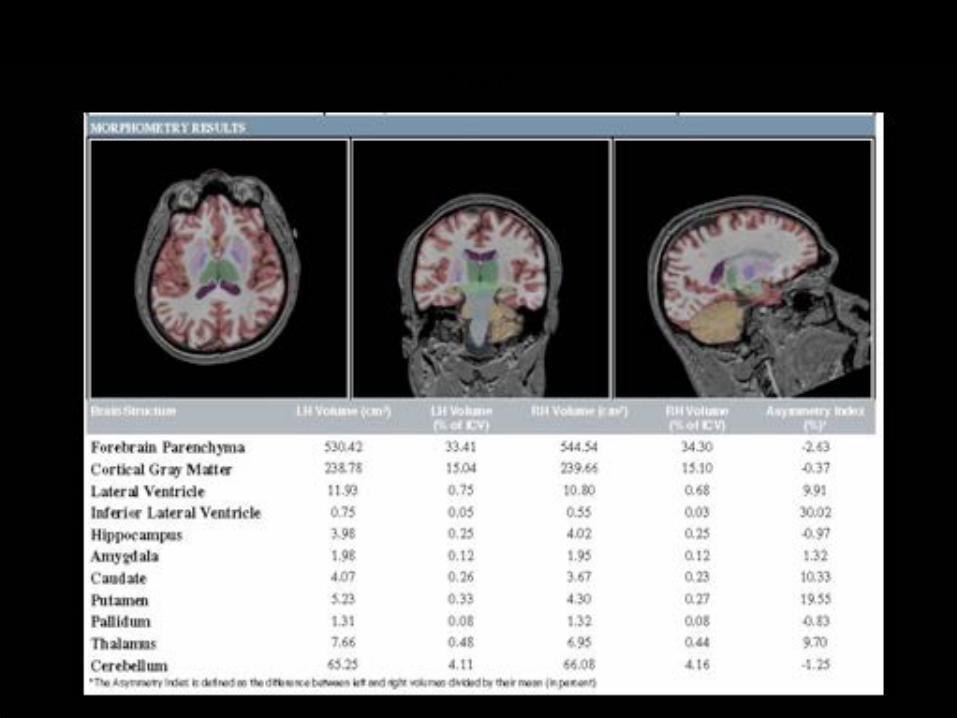
NeuroQuant® Standard Report Page 2

Reliability of NeuroQuant®
• NeuroQuant is reliable with FreeSurfer (Kovacevic, Rafii et al. 2009).
• NeuroQuant® is reliable with a computer-supported manual technique using NeuroMorphometric software (Brewer, Magda et al. 2009).
• The segmentation error rate of NeuroQuant® was found to be very low (9 out of 822) (Heister, Brewer et al. 2011).
Reference Brewer, J. B., S. Magda, et al. (2009). "Fully-automated quantification of regional brain volumes for improved detection of focal atrophy in Alzheimer disease." Am J Neuroradiol 30(3): 578-580. Heister, D., J. B. Brewer, et al. (2011). "Predicting MCI outcome with clinically available MRI and CSF biomarkers." Neurology 77(17): 1619-1628. Kovacevic, S., M. S. Rafii, et al. (2009). "High-throughput, Fully Automated Volumetry for Prediction of MMSE and CDR Decline in Mild Cognitive Impairment." Alzheimer Dis Assoc Disord 23(2): 139–145.

Experience with NeuroQuant® at the Virginia Institute of Neuropsychiatry
• Quality control • Prior to data collection, communicate with radiology center • NeuroQuant® software automatically checks several
parameters • Visual inspection of each set of segmented brain images • Inspection of the numerical and statistical results of the
analyses

Radiologist vs. NeuroQuant®
N positive/ Total N % positive for atrophy
Radiologist atrophy 2/20 10%
NQ Extended atrophy 10/20 50%
Paired sign test, test statistic M =-4.00, P=0.02
Reference Ross DE, Ochs AL, Seabaugh JM, Shrader CR (submitted): Man vs. Machine: Comparison of Radiologists’ Interpretations and NeuroQuant® Volumetric Analyses of Brain MRIs in Patients with Traumatic Brain Injury . Journal of Neuropsychiatry and Clinical Neurosciences.
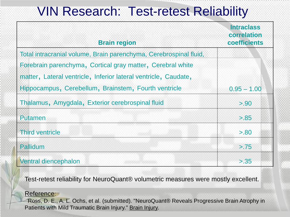
VIN Research: Test-retest Reliability
Brain region
Intraclass correlation coefficients
Total intracranial volume, Brain parenchyma, Cerebrospinal fluid,
Forebrain parenchyma, Cortical gray matter, Cerebral white
matter, Lateral ventricle, Inferior lateral ventricle, Caudate, Hippocampus, Cerebellum, Brainstem, Fourth ventricle 0.95 – 1.00
Thalamus, Amygdala, Exterior cerebrospinal fluid >.90
Putamen >.85
Third ventricle >.80
Pallidum >.75
Ventral diencephalon >.35
Test-retest reliability for NeuroQuant® volumetric measures were mostly excellent.
Reference: Ross, D. E., A. L. Ochs, et al. (submitted). "NeuroQuant® Reveals Progressive Brain Atrophy in Patients with Mild Traumatic Brain Injury." Brain Injury.

Community Acceptance of NeuroQuant® • NeuroQuant® is currently used in at least a dozen clinics and
radiology centers across the USA:
East River Medical Imaging, PC 519/523 East 72nd Street, New York, NY 10021
Lenox Hill Radiology & Medical Imaging Associates 61 East 77th Street, New York, NY 10075
Advanced Radiology 888 Bestgate Rd, Ste 101, Annapolis 21401
Washington Radiology Associates 2141 K St. NW, Washington, DC 20037
Virginia Institute of Neuropsychiatry 364 Browns Hill Court, Midlothian, VA 23114
Center for Neurorehabilitation Services 10710 Midlothian Turnpike, Suite 125, Richmond, VA 23235
MRI CT Diagnostics 4668 Pembroke Blvd, Virginia Beach, VA 23455
Santa Rosa Imaging Center 3536 Mendocino Ave., Suite 280 Santa Rosa, CA 95403
Liberty Pacific Advanced Imaging 16130 Ventura Blvd., Suite 100, Encino, CA 91436
San Joaquin Community Hospital 2615 Chester Avenue, Bakersfield, CA 93301
Dr. James Brewer University of California, San Diego, CA
Radnet, www.radnet.com Sites in California
East West
South

Acknowledgements
Alfred L. Ochs, Ph.D. Biomedical Engineer, Clinical Neurophysiologist
Virginia Institute of Neuropsychiatry Virginia Commonwealth University
Michael Havranek, MAMS, CMI Medical Illustrator
Amicus Visual Solutions
Jan Seabaugh, M.A. Assistant Director
Virginia Institute of Neuropsychiatry
Jennifer Marwitz, M.A. Virginia Commonwealth University
Michael DeMark, M.A. Vocational Rehabilitation Intern
Virginia Institute of Neuropsychiatry
Carole R. Shrader, B.A. Case Manager
Virginia Institute of Neuropsychiatry
Megan DeSmit, B.S. Clinical Coordinator
Virginia Institute of Neuropsychiatry
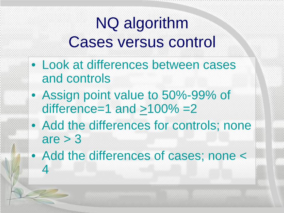
NQ algorithm Cases versus control
• Look at differences between cases and controls
• Assign point value to 50%-99% of difference=1 and >100% =2
• Add the differences for controls; none are > 3
• Add the differences of cases; none < 4

Creating the mold NQ Index (MNQI)
• Now look at the sum of abnormalities for each measure between cases and controls
• Select top five > 3.0 • Return to the top five • Create scoring on the top five • Stratify scores by illness type

For CIRS-WDB, the five areas are not seen in any other illness to date
• Forebrain parenchyma increased • Cortical gray increased • Hippocampus increased • Caudate decreased • Pallidum increased

Scoring system 0, 1, 2
• Forebrain >31.7 and 32.3 • Cortical gray >16.4 and 17.0 • Hippocampus > .28 and .30 • Caudate < .24 and < .23 • Pallidum > .066 and > .071

No other combination like this in neurology known
• Selective increase in multiple areas • Reduced lateral ventricle size • Reduced caudate size but no other
grey matter or basal ganglia • Role of BBB in edema? Corrected by
Rx illness • Caudate atrophy responds slowly to
VIP and nothing else to date

MNQI values in two cohorts
• Controls 2.5 and 1.9 • Untreated cases 6.6 and 6.4 • Partially treated cases 4.0 and 3.8 • Treated cases 2.0 and 2.1 • Relapse 7.0 and 6.5 • Cohort 1 N= 19 • Cohort 2 N= 65

Edema?
• BBB leak? • Reduced albumin? • Dehydration preventing measurement
of edema • Is edema macroscopic i.e. seen on
MRI?

So what is going on? • Microscopic interstitial edema means
loosening of BBB • Caudate atrophy is not permanent! • Pruning! • How do we say that pallidum increases
and caudate decreases yet no other posterior gray is altered
• Diffuse gray reduction is typical of MS, other CNS illnesses (Huntington’s chorea and PSTD)

BBB
• VEGF increases capillary permeability
• So does MMP9 and OATP • TGF beta-1 has dual role (what a
surprise); can stiffen tight junctions and reduce inflow

Inflammation effects-1
• Loosening of blood brain barrier permits intrusion of peripheral elements into CNS
• Major players are TGF beta-1, VEGF, MMP9
• C4a is less of a problem

Inflammation effects-2
• The new buzz phrase is “TH17/Treg imbalance”
• Rising TGF beta-1 will direct T regs into tissue to suppress inflammation if there is ROR receptor present but if not, and IL-1, IL-17, IL-23 are, the T regs are converted into pathogenic T cells that make more TGF beta-1
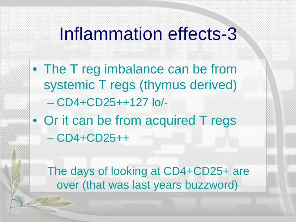
Inflammation effects-3
• The T reg imbalance can be from systemic T regs (thymus derived) – CD4+CD25++127 lo/-
• Or it can be from acquired T regs – CD4+CD25++
The days of looking at CD4+CD25+ are
over (that was last years buzzword)

What do I need to know to treat?
• No single attack corrects BBB or pruning
• Multiple sequential steps based on BT pathway
• Relapse with re-exposure wreaks havoc with sequential intervention

A bit of review
• We are looking at specific brain areas as targets of inflammatory processes
• We cannot forget that similar kinds of changes in tissues are ongoing
• We know a lot or about peripheral innate immune abnormalities

Innate immune effects are systemic
• Pre-formed proteins, ready to go • Pattern receptors pick up foreign signals • Activation is instantaneous
– Imagine what turns off such activation • Initiates the formation of antibodies, in a
different type immune response called acquired immunity
• The names of innate immunity might be new: cytokines, complement, VEGF, TGF beta-1, MMP9, CD4+CD25++ (cellular)

Innate immunity isn’t new • It was 1989 at the CSH Symposia • Charles Janeway, looking at immunology,
predicted an expansion of insight in innate immunity
• 1972 Lewis Thomas talked about the peculiar over-reaction of the host to toxins, thinking of bacterial toxins
• 1985 first description of TNF • By 2000, over 50,000 papers published • 2011 Nobel Prize, Bruce Beutler, Scripps

Defining what is wrong brings effective treatment
• Lowering levels of inflammagens: C3a, C4a, MMP9 and TGF beta-1
• Correct hormonal dysregulation • Deal with auto-immunity • Improve capillary hypoperfusion • Eradicate commensal staphs • Correct cellular immunity


Treatment steps
VIP
TGF beta-1
Correct C4a Correct C3a
Correct VEGF Correct MMP9
Correct ADH/osmolality
Correct androgens Correct antigliadin
Eradicate MARCoNS CSM/Welchol
Remove from exposure

Look for the final common pathway
• Abnormalities in innate immune responses (non-specific for cause)
• Call it the host response • Incredible amplification of multiple
pathways following initiation • CHRONIC INFLAMMATORY
RESPONSE SYNDROMES!

CIRS is systemic, interacting
• No way to say just one lab as source of fatigue, cognitive abnormalities, joints, respiratory problems
• All of the putative diagnoses have the same final common pathway
• You will see the same combo of multisymptom illness and labs
• Differential diagnosis key
CIRS-1
• Once you see it once your life as a physician will be changed forever
• Lack of regulation of inflammation • Enhanced innate inflammatory parameters
(C4a, TGF beta-1, MMP9 and more) • Hormonal dysregulation • Hypoxia from capillary hypoperfusion • And now T regs too

CIRS-2
• Colonizing commensal MARCoNS • Von Willebrand’s factor-66%
abnormal: Acute reactants? NO • Autoimmunity like crazy! AGA,
ACLA, ANA, ANCA, actin • Cellular immunity: TGF beta-1 • Activated complement split products
(C3a, C4a)

VEGF
• Vascular endothelial growth factor • Responsive to hypoxia inducible
factor; feedback from TGF beta-1 • Increases O2 and increases new
blood vessel formation • Judah Folkman and anti-
angiogenesis knew about VEGF

VEGF 2
• Blockade of VEGF a big deal in chemotherapy now; most effective at VEGF + receptor tumors
• But low VEGF is the norm in the worst biotoxin people
• Sure some U-shaped skew but low VEGF means cell-based starvation

Capillary hypoperfusion
• Bottom line is decreased delivery of nutrients and oxygen into capillaries
• ABG won’t help; I don’t see where venous gases have academic basis in these illnesses
• Use VO2 max from PST • Use lactate in MR spectroscopy

VO2 max
• Disability uses this measure a lot • Look for over 35 in healthy younger
person; nomograms available • 12 ml/kg/min is stage 4 CHF • So many have V02 max < 20 • Conversely training to raise VO2 max
that doesn’t go beyond anaerobic threshold works in biotoxin people

Raising VO2 max shows benefit
• Correcting VEGF must happen • Anaerobic threshold is measured • At exercises start low, go slow • Defined exercise EVERY DAY • Bike, treadmill; work up to 15’ • Add floor; build up to 15’; then free • Go back to first defined work,
increase sequentially

Post-exertional malaise
• Measure VO2 max in pulm stress test • It will be low • What about glycogen in exercise
– Remarkably inefficient glucose burn • No O2; no efficiency
– Can’t say this is mitochondrial illness! • Fat storage (look at leptin) • Protein burning (alanine and glutamine)

TGF beta-1
• Will have its own section • Here is the key advancement in
assessment of inflammatory illness • Lung symptoms? Ask re TGF beta-1 • Neuro problems? Ask re TGF beta-1 • Autoimmune? Ask re TGF beta-1 • Learning disability? MS? TM? Same

TGF beta-2
• First found to have increased tissue effect in those with mutate fibrillin-1
• Then the switch to plasma measures • Normal is < 2380; over 5000 I worry • Over 10,000 essentially guaranteed
restrictive lung disease, tremor, cognitive issues and joint problems

TGF beta-3
• Must be double spun plasma • Platelet contamination common • If result over 40,000, not properly
handled • Always have second specimen saved • Cambridge runs the assay

MR spectroscopy
• 3 Tesla coil; single voxel • Frontal lobes and hippocampi • Same spots! Measure same
compounds • High lactate (> 1.29) too high • Ratio of glutamate to glutamine (G/G)
< 2.19 too low

MRS-2
• Change in cognition is a tip-off • Reversal of high lactate reverses
suppressed G/G • And Voila! Reversal of cognitive too • Key concept is that the cellular
neuronal mechanisms are not permanently injured

VIP-1
• Vasoactive intestinal polypeptide • Neuroregulatory • Agonist in suprachiasmatic nucleus
– Primarily olfactory! • Binds to membrane receptors • Activates cellular regulation • Downregulates cytokines (MMP9)

VIP-2
• Downregulates MASP2 • Restores balance of Vitamin D3 • Downregulates aromatase • Up-regulates VEGF • Warning re lipase • Main effect immediately is endorphin • Followed by lowering PASP in
exercise

PASP and VIP
• 50 mcg QID corrects paradoxical rise in PASP in exercise in days, not weeks, with durable effects with titration to BID and over time: off!
• So many people aren’t diagnosed with acquired PASP even if they have stress echo: must measure TR!
• Don’t accept “normal”

Looks like asthma, but isn’t
• Measure PASP in exercise • Should not rise more than 8 mm Hg • Source of palpitations and SOB • Won’t get better with beta-2 agonists • Don’t forget EMT and TGF beta-1 • Remodeling in heart, CNS, liver • Fibrotic change

VIP-3
• Immunoregulatory aspects • Drives up CD4+CD25++ • Here is link from neuropeptides to
humoral factors to T-cell physiology • Role of downregulation of TGF beta-1
has no obvious upper limit in its application

What you need to know
• Symptoms must be there • Labs must be there to show what is
– And labs must show what is not • Differential diagnosis must be there • The labs will show you the way
– Start looking at innate immunity as a target – Start looking at targets that you can fix – Fix the targets; watch the illness disappear – Wait for relapse
Here’s my treatment message
• Look for environmental exposures • Establish a decent baseline of results of
innate immunity testing • Look for biofilm formers; they must go! • Treat the inflammatory physiology • What do you have left? • What happens when the injured patient is
exposed next week? Repeat illness!

For more information:
www.survivingmold.com
www.chronicneurotoxins.com
Surviving Mold December, 2010
Mold Warriors 2005, 2007, 2010
Desperation Medicine 2001, 2006, 2009
Lose the Weight You Hate 2002, 2005