neurobiologyofdisease minocycline ... minocycline-preconditionedneuralstemcellsenhance...
TRANSCRIPT
Neurobiology of Disease
Minocycline-Preconditioned Neural Stem Cells EnhanceNeuroprotection after Ischemic Stroke in Rats
Hiroyuki Sakata,1 Kuniyasu Niizuma,1 Hideyuki Yoshioka,1 Gab Seok Kim,1 Joo Eun Jung,1 Masataka Katsu,1
Purnima Narasimhan,1 Carolina M. Maier,1 Yasuhiro Nishiyama,2 and Pak H. Chan1
1Department of Neurosurgery, Department of Neurology and Neurological Sciences, and Program in Neurosciences and 2Department of Neurosurgery,Stanford University School of Medicine, Stanford, California 94305
Transplantation of neural stem cells (NSCs) offers a novel therapeutic strategy for stroke; however, massive grafted cell death followingtransplantation, possibly due to a hostile host brain environment, lessens the effectiveness of this approach. Here, we have investigatedwhether reprogramming NSCs with minocycline, a broadly used antibiotic also known to possess cytoprotective properties, enhancessurvival of grafted cells and promotes neuroprotection in ischemic stroke. NSCs harvested from the subventricular zone of fetal rats werepreconditioned with minocycline in vitro and transplanted into rat brains 6 h after transient middle cerebral artery occlusion. Histolog-ical and behavioral tests were examined from days 0 –28 after stroke. For in vitro experiments, NSCs were subjected to oxygen–glucosedeprivation and reoxygenation. Cell viability and antioxidant gene expression were analyzed. Minocycline preconditioning protected thegrafted NSCs from ischemic reperfusion injury via upregulation of Nrf2 and Nrf2-regulated antioxidant genes. Additionally, precondi-tioning with minocycline induced the NSCs to release paracrine factors, including brain-derived neurotrophic factor, nerve growth factor,glial cell-derived neurotrophic factor, and vascular endothelial growth factor. Moreover, transplantation of the minocycline-preconditioned NSCs significantly attenuated infarct size and improved neurological performance, compared with non-preconditionedNSCs. Minocycline-induced neuroprotection was abolished by transfecting the NSCs with Nrf2-small interfering RNA before transplan-tation. Thus, preconditioning with minocycline, which reprograms NSCs to tolerate oxidative stress after ischemic reperfusion injury andexpress higher levels of paracrine factors through Nrf2 up-regulation, is a simple and safe approach to enhance the effectiveness oftransplantation therapy in ischemic stroke.
IntroductionA growing number of experimental studies highlights the poten-tial of stem cell transplantation as a novel therapeutic approachfor stroke (Savitz et al., 2002; Bliss et al., 2007). Moreover, avariety of clinical trials have been performed and others are cur-rently ongoing (Banerjee et al., 2011). Transplantation of neuralstem cells (NSCs) in the acute stage of stroke often reduces lesionsize and inhibits apoptosis in the penumbra area by providingneuroprotective paracrine factors that enhance host cell survivaland function (Bliss et al., 2007; Harms et al., 2010). However, ahostile microenvironment in the ischemic brain offers a signifi-cant challenge to survival of transplanted cells. Only a small frac-tion of grafted cells (1–3%) survived in the ischemic brain 28 dafter grafting (Hicks et al., 2009; Nakagomi et al., 2009). The
accelerated death of grafted cells might be influenced by produc-tion of reactive oxygen species after ischemic reperfusion injuryand host inflammatory response mediators (Savitz et al., 2002; Loet al., 2003). This massive loss of stem cells post-engraftment is animpediment that lessens the effectiveness of cell transplantationtherapy.
Considering that cell survival may greatly enhance the effec-tiveness of transplantation therapy, several remedial approacheshave been suggested. Ex vivo gene modification of stem cells foroverexpression of pro-survival signaling molecules, such as Bcl-2,reduces grafted cell loss (Wei et al., 2005). An alternative strategyis to genetically modulate them for overexpression of the para-crine factors of interest, such as placental growth factor (Liu et al.,2006). These cells serve as a continuous source of paracrine fac-tors that enhance neuroprotection in the host brain. However,while these methods exhibit a better transplantation outcome, amore beneficial, simpler, and safer approach is needed for futureclinical application.
Minocycline, a semisynthetic tetracycline, has been clinicallyused as an antibiotic and anti-inflammatory drug. Previously, weshowed the neuroprotective potential of minocycline in animalmodels of cerebral ischemia (Yrjanheikki et al., 1999). One of themain biological effects of minocycline is its cytoprotective properties(Zhu et al., 2002). Minocycline selectively manipulates expression ofgenes, such as Bcl-2 and X chromosome-linked inhibitor-of-apoptosis protein (Keilhoff et al., 2008; Kernt et al., 2010). This find-
Received Nov. 11, 2011; revised Jan. 12, 2012; accepted Jan. 20, 2012.Author contributions: H.S., K.N., H.Y., G.S.K., J.E.J., M.K., P.N., C.M.M., Y.N., and P.H.C. designed research; H.S.
performed research; H.S. analyzed data; H.S. wrote the paper.This work was supported by National Institutes of Health Grants PO1 NS014543, RO1 NS025372, and RO1
NS038653, and by the James R. Doty Endowment (P.H.C.). We thank Liza Reola and Bernard Calagui for technicalassistance, Cheryl Christensen for editorial assistance, and Elizabeth Hoyte for assistance with figure preparation. Wealso thank Dr. Carlos Lois (California Institute of Technology) for supplying SD-Tg(GFP)2BalRrrc rats.
The authors declare no competing financial interests.Correspondence should be addressed to Pak H. Chan, Neurosurgical Laboratories, Stanford University, 1201
Welch Road, MSLS #P314, Stanford, CA 94305-5487. E-mail: [email protected]:10.1523/JNEUROSCI.5686-11.2012
Copyright © 2012 the authors 0270-6474/12/323462-12$15.00/0
3462 • The Journal of Neuroscience, March 7, 2012 • 32(10):3462–3473
ing supports our study rationale that minocycline preconditioningmay induce reprogramming of NSCs and promote neuropro-tection after transplantation. Therefore, the purpose of thepresent study was to determine whether preconditioning withminocycline protects grafted cells from ischemic reperfusioninjury and enhances the effectiveness of transplantation ther-apy in ischemic stroke. We also sought to elucidate the under-lying mechanisms of minocycline preconditioning in NSCs.
Materials and MethodsIsolation and culturing of fetal NSCs. All animals were treated in accor-dance with Stanford University Guidelines and the animal protocolswere approved by Stanford University’s Administrative Panel on Labo-ratory Animal Care (Stanford, CA). NSCs were harvested from greenfluorescent protein (GFP) transgenic Sprague Dawley rats (SD-Tg(GFP)2BalRrrc) as described previously (Blurton-Jones et al., 2009),with some modification. In brief, bilateral subventricular zones frompostnatal day 1 rat brains were dissected in Dulbecco’s PBS (14040 –182;Invitrogen) and mechanically dissociated. The cells were collected andresuspended in Neurobasal-A medium (catalog no. 10888-022; Invitro-gen) containing B-27 supplement (catalog no. 12587-010; Invitrogen),L-glutamine (catalog no. 25030-081; Invitrogen), 20 ng/ml rat fibroblastgrowth factor-basic (catalog no. 400-29; PeproTech), and 10 ng/ml ratepidermal growth factor (400-25; PeproTech). Cells were grown on a 10cm plastic dish precoated with poly-L-ornithine hydrobromide (catalogno. P3655-100MG; Sigma-Aldrich) and laminin (L2020 –1MG; Sigma-Aldrich) at 37°C and 5% CO2 as adherent monolayers. The medium waschanged every 2 d, and cells were passaged once a week. Cells that hadbeen passaged 5–10 times were used for the experiments.
The NSCs were preconditioned with minocycline before the in vitroexperiments or transplantation. Minocycline hydrochloride (catalog no.M9511; Sigma-Aldrich) was added to the cell culture medium (finalconcentration: 0, 1, 3, or 10 �M) for 24 h, followed by drug washoutbefore experiments.
Treatment of the cultures with oxygen– glucose deprivation. We usedoxygen– glucose deprivation (OGD) and reoxygenation, an in vitro
model that best mimics in vivo cerebral ischemia–reperfusion. The NSCswere subjected to OGD by replacing the medium with a buffered saltsolution without glucose (in mM): 116 NaCl, 1.8 CaCl2, 0.8 MgSO4, 5.4KCl, 1 NaH2PO4, 14.7 NaHCO3, and 10 HEPES (375368; EMD Chemi-cals; pH 7.4). The plates were placed in an anaerobic chamber (Plas Labs)at 37°C. After 8 h, the medium was replaced with the regular NSC me-dium (Neurobasal-A) with glucose, and the plates were returned to a 5%CO2/95% air incubator for various reoxygenation periods.
Assessment of cell death and cell viability in vitro. Cell death was quantifiedby a standard measurement of lactate dehydrogenase (LDH) release using aLDH-cytotoxicity assay kit (catalog no. K311-400; BioVision). Cell viabilitywas assessed with a cell proliferation reagent using a WST-1 assay kit (catalogno. 05015944001; Roche Diagnostics). For in situ labeling of DNA fragmen-tation, an in situ cell death detection kit, TMR red (12156792910; RocheDiagnostics), was used according to the manufacturer’s instructions. Slideswere covered with VECTASHIELD mounting medium with 4�,6-diamidino-2-phenylindole (DAPI) (catalog no. H-1500; Vector Laborato-ries) and observed with confocal microscopy (LSM 510; Carl Zeiss).
Immunofluorescent staining. For immunocytochemistry, NSCs cul-tured on eight-well chamber slides (catalog no. 154941; Thermo FisherScientific) were washed with PBS and fixed with 4% paraformaldehyde inPBS for 15 min. They were then washed with PBS and incubated for 1 h inblocking solution (PBS containing 3% bovine serum albumin and 0.3%Triton X-100). For immunohistochemistry, the animals were anesthe-tized and perfused with PBS followed by 4% paraformaldehyde in PBS,pH 7.4. The brains were postfixed overnight in the same fixative at 4°C.For cryosectioning, fixed tissues were cryoprotected in 10% sucrose inPBS overnight at 4°C, then in 20% sucrose in PBS overnight at 4°C, andembedded in Tissue-Tek O.C.T. compound (catalog no. 4583; SakuraFinetek). Cryostat sections (20 �m) were cut and affixed to glass slides.Cells or tissue sections were subsequently incubated overnight at 4°C inan appropriate mixture of primary antibodies. The following antibodieswere used: rabbit anti-GFP (1:100; catalog no. G10362; Invitrogen), goatanti-GFP (1:100; catalog no. LS-C67095; LifeSpan BioSciences), mouseanti-nestin (1:100; catalog no. 556309; BD Biosciences PharMingen),mouse anti-Sox2 (a NSC marker) (1:50; catalog no. 4900; Cell Signaling
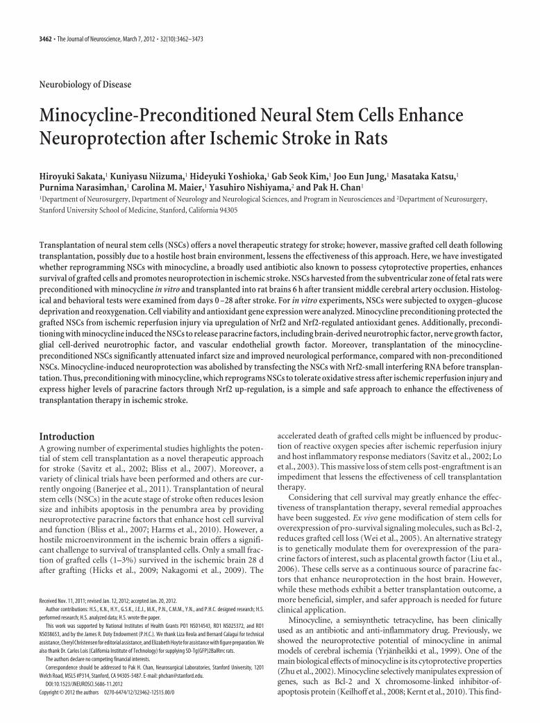
Figure 1. Reduced NSC death with minocycline preconditioning in vitro. A, The NSCs grown as adherent cultures were examined by immunocytochemistry for GFP (green) and the NSC markernestin (red). Nearly all the GFP-positive cells colocalized with nestin (yellow). Nuclei were counterstained with DAPI (blue). Scale bar, 20 �m. B, After culturing in differentiation medium containing1 �M retinoic acid and 1% fetal bovine serum for 5 d, GFP-positive cells (green) expressed the neuronal marker �-tubulin, the astrocytic marker GFAP, and the oligodendrocytic marker NG2 (red)(arrows). Nuclei were counterstained with DAPI (blue). Scale bars, 20 �m. C, LDH assay demonstrated a significant reduction in NSC death with minocycline preconditioning (10 �M) after 8 h of OGDand 24 h of reoxygenation (n � 4). Mino, Minocycline; RO, reoxygenation. D, WST-1 assay showed significantly increased cell viability with 10 �M minocycline after 8 h of OGD and 24 h ofreoxygenation (n � 4). E, NSCs analyzed by TUNEL staining (red) and DAPI (blue) after 8 h of OGD and 24 h of reoxygenation. Scale bar, 50 �m. The cell-counting study revealed a significant decreasein TUNEL-positive cells with minocycline preconditioning (10 �M) (n � 4). F, Fluorescent staining of cytochrome c (red) and DAPI (blue) after 8 h of OGD and 3 h of reoxygenation. The NSCs in whichcytochrome c was released from mitochondria to the cytoplasm are indicated by arrows. Scale bar, 20 �m. G, H, Preconditioning with 10 �M minocycline significantly reduced the release of LDH fromthe NSCs under the oxidative stimuli of 200 �M H2O2 (G) and 250 �M diethylenetriamine/nitric oxide (DETA/NO) (H ) (n � 4). *p � 0.05; ‡p � 0.005; §p � 0.001.
Sakata et al. • Minocycline-Preconditioned NSCs in Ischemic Stroke J. Neurosci., March 7, 2012 • 32(10):3462–3473 • 3463
Technology), rabbit anti-�-tubulin (1:500; catalog no. PRB-435P; Cova-nce), rabbit anti-microtubule-associated protein 2 (MAP2) (1:200; cata-log no. AB5622; Millipore), mouse anti-glial fibrillary acidic protein(GFAP) (1:100; catalog no. MAB360; Millipore), rabbit anti-NG2 (anoligodendrocytic marker) (1:100 catalog no. AB5320; Millipore), mouseanti-cytochrome c (1:100; catalog no. 556433; BD Biosciences PharMin-gen), and rabbit anti-Ki-67 (1:500; catalog no. ab16667; Abcam). Afterthree washes in PBS, cells or tissue sections were incubated for 1 h with a1:500 dilution of the following secondary antibodies: Alexa Fluor 488-conjugated donkey anti-rabbit IgG (A21206), Alexa Fluor 594-conjugateddonkey anti-rabbit IgG (A21207), Alexa Fluor 488-conjugated donkey anti-goat IgG (A11055), Alexa Fluor 594-conjugated donkey anti-mouse IgG(A21203; all from Invitrogen). After three washes with PBS, samples werecovered with VECTASHIELD mounting medium with DAPI. The sampleswere examined by confocal microscopy or fluorescence microscopy (Ax-ioplan 2; Carl Zeiss).
In situ detection of superoxide anion production. Early production ofsuperoxide anions was investigated with the use of hydroethidine (HEt)as described previously (Murakami et al., 1998). Production of superox-ide anions was shown by oxidized HEt as diffuse signals and small parti-cles in the cytosol. For the in vitro study, 5 �M HEt solution (D23107;Invitrogen) was added to the cell culture medium. The cells were incu-bated for 5 min, followed by fixation with 4% paraformaldehyde in PBSfor 15 min. For the in vivo study, HEt solution (1 ml of 1 mg/ml in 1%dimethyl sulfoxide with saline) (D11347; Invitrogen) was administeredintravenously immediately after transplantation of the NSCs. Animalswere killed 1 h after administration, and tissue sections were prepared.For fluorescent double staining of the HEt signals and GFP, the sectionswere incubated with anti-GFP (1:100; catalog no. G10362; Invitrogen),followed by Alexa Fluor 488-conjugated donkey anti-rabbit IgG (catalogno. A21206; Invitrogen). Slides were covered with VECTASHIELDmounting medium with DAPI. The sections were observed with a fluo-
rescence microscope, and oxidized HEt fluorescence was examined at anexcitation of 510 nm and emission of �580 nm and quantified withImageJ software (version 1.42q; NIH).
Gene expression study. After 8 h of OGD and 3 h of reoxygenation, totalRNA was isolated from the NSCs using the RNeasy Mini kit (catalog no.74104; SABiosciences), including DNase digestion. At the end, RNAsamples were eluted from the columns using 30 �l of RNase-free water,and their concentrations were determined spectrophotometrically byA260. cDNA was synthesized from 1 �g of total RNA using an RT 2 first-strand kit (catalog no. C-03; SABiosciences). Quantitative real-time RT-PCR was performed in triplicate in a 25 �l final volume containingtemplate cDNA, RT 2 SYBR Green/ROX qPCR master mix (catalog no.PA-012; SABiosciences) and specific primers (SABiosciences), using aMx3000p qPCR system (Agilent Technologies). For real-time RT-PCRarray, a modified oxidative stress and antioxidant defense PCR array(catalog no. PARN-065; SABiosciences) was used. Data were collectedwith MxPro qPCR software (Agilent Technologies) and analyzed usingthe �� threshold cycle (CT) method. In this study, �-actin was usedexclusively as a housekeeping gene. As calibrator samples, we used thenon-preconditioned NSCs under normal conditions.
Western blot analysis. After 8 h of OGD and 3 h of reoxygenation, theNSCs were treated with cell lysis buffer (catalog no. 9803; Cell SignalingTechnology) and used as whole-cell lysate samples. Protein concentra-tions were determined by comparison with a known concentration ofbovine serum albumin using a kit (catalog no. 23225; Thermo FisherScientific). Equal amounts of the samples (20 �g) were loaded per laneand analyzed by SDS-PAGE on a 10% NuPAGE Bis-Tris gel (catalog no.NP0303; Invitrogen) and then immunoblotted. The primary antibodieswere a 1:100 dilution of a rabbit polyclonal anti-Nrf2 antibody (molec-ular weight, �100 kDa) (catalog no. sc-13032; Santa Cruz Biotechnol-ogy), a 1:1000 dilution of a rabbit polyclonal anti-heme oxygenase-1(HO-1) antibody (molecular weight, �28 kDa) (catalog no. 5141; Cell
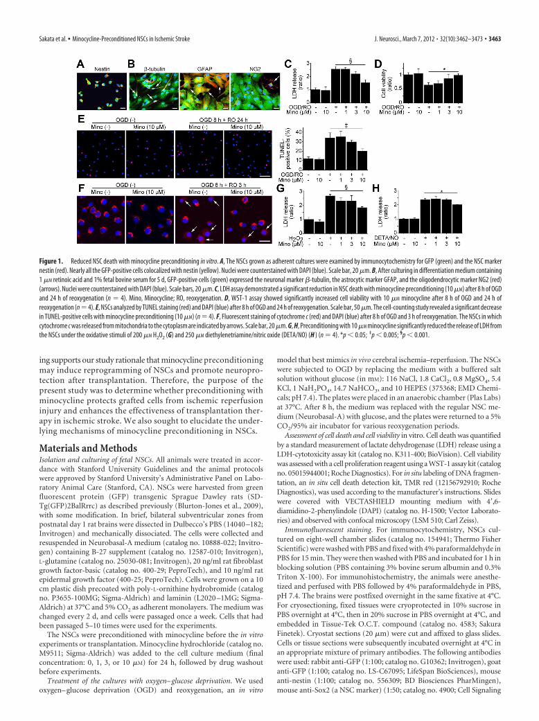
Figure 2. Upregulation of antioxidant genes in NSCs with minocycline preconditioning in vitro. A, Fluorescent staining of NSCs with HEt (red) and DAPI (blue) in vitro. Production of superoxideanions in the NSCs is shown by oxidized HEt as diffuse signals and small particles in the cytosol. Preconditioning with 10 �M minocycline reduced the increase in HEt signals after 8 h of OGD and 15min of reoxygenation (n � 4). Scale bar, 20 �m. B, Expression of antioxidant genes in non-PCNSCs and PCNSCs under normal conditions or after OGD and reoxygenation analyzed by a real-timeRT-PCR array (n � 3). Data are expressed as mean fold induction (FI), compared with the non-PCNSCs under normal conditions (�/�). C, D, Real-time RT-PCR assays (C) (n � 3) and Western blotanalyses of whole-cell lysate from the NSCs (D) (n � 4) revealed that preconditioning with 10 �M minocycline induced higher expression of Nrf2 and the Nrf2-regulated antioxidant genes NQO1 andHO-1. �-Actin was used as an internal control. Mino, Minocycline; RO, reoxygenation; SOD2, manganese-superoxide dismutase; OD, optical density. *p � 0.05; †p � 0.01; ‡p � 0.005; §p � 0.001.
3464 • J. Neurosci., March 7, 2012 • 32(10):3462–3473 Sakata et al. • Minocycline-Preconditioned NSCs in Ischemic Stroke
Signaling Technology), a 1:1000 dilution of a rabbit polyclonal anti-NAD(P)H:quinone oxidoreductase1 (NQO1) antibody (molecularweight: �31 kDa) (catalog no. sc-25591; Santa Cruz Biotechnology), a1:1000 dilution of a rabbit polyclonal anti-manganese-superoxide dis-mutase antibody (molecular weight, �25 kDa) (catalog no. SOD-110;Enzo Life Sciences), and a 1:100,000 dilution of a mouse monoclonalanti-actin antibody (molecular weight, �42 kDa) (catalog no. A5441;Sigma-Aldrich). After incubation with horseradish peroxidase-conjugatedanti-mouse IgG (catalog no. 7076; Cell Signaling Technology) or anti-rabbitIgG (catalog no. 7074; Cell Signaling Technology), the antigen was detectedby SuperSignal West Pico and/or Femto substrate (catalog nos. 1856135/1856189; Thermo Fisher Scientific). Images were scanned with a GS-700imaging densitometer (Bio-Rad Laboratories), and the results were quanti-fied using MultiAnalyst software (Bio-Rad).
Transfection of small interfering RNA. NSCs, which were 60% con-fluent, were transfected with 20 nM stealth Nrf2-small interferingRNA (siRNA) (Invitrogen) or nonfunctioning negative-control siRNA(catalog no. 45-2001; Invitrogen) using Lipofectamine RNAiMAX (cat-alog no. 13778; Invitrogen) according to the manufacturer’s protocols.
The target sequence of the rat-specific Nrf2-siRNA mixture was as follows: UUUAAGUG-GCCCAAGUCUUGCUCCA. After 48 h ofincubation, the NSCs were used for variousexperiments and transplantation.
Detection of paracrine factors. For the in vitrostudy, culture supernatants were collected foranalysis after 4 h of OGD and 48 h of reoxygen-ation. For the in vivo study, fresh brain tissue wasremoved 2 d after transplantation. The rectangu-lar cuboid tissue block of the cortex, 2 mm oneither side of the NSC-transplanted regions, wasdissected (width 4 mm � length 10 mm) andused as a sample. Protein was extracted as de-scribed in the Western blot analysis section.Commercial brain-derived neurotrophic factor(BDNF), glial cell-derived neurotrophic factor(GDNF) (catalog nos. G7610 and G7620; Pro-mega), and vascular endothelial growth fac-tor (VEGF) (catalog no. RRV00; R&DSystems) ELISA kits were used to quantifythe concentration of BDNF, GDNF, andVEGF in each of the samples.
Focal cerebral ischemia. Adult male SpragueDawley rats (260 –280 g) were subjected totransient focal cerebral ischemia by intralumi-nal middle cerebral artery blockade with a su-ture, as described previously (Fujimura et al.,1998), with some modifications. The rats wereanesthetized with 2.0% isoflurane in 30% oxy-gen and 70% nitrous oxide using a face mask.The rectal temperature was controlled at 37°Cwith a homeothermic blanket. Physiologicalparameters were monitored throughout thesurgeries. After a midline skin incision, theright external carotid artery was exposed andits branches were electrocoagulated. A 22 mm4-0 monofilament nylon suture coated withsilicon rubber (catalog no. 4037PK5Re; Doc-col) was introduced into the right internal ca-rotid artery through the external carotid arterystump. After 90 min of middle cerebral arteryocclusion, blood flow was restored by with-drawal of the suture. The animals were main-tained in an air-conditioned room at 20°C withad libitum access to food and water before andafter surgery.
Intracerebral transplantation. The rats wereanesthetized 6 h after stroke, and the NSCswere transplanted using a 10 �l Hamilton sy-ringe with a 26 G needle attached to a stereo-
taxic apparatus (David Kopf Instruments). The rats were given four 1.0 �ldeposits of single cell suspension in Dulbecco’s PBS (1 � 105 cells per �l) alongthe anterior–posterior axis into the cortex at these coordinates: (1) anterior–posterior (A–P), 1.0; medial–lateral (M–L), 3.0; dorsal–ventral (D–V),�3.0; (2) A–P, �1.0; M–L, 3.0; D–V, �3.0; (3) A–P, �3.0; M–L, 3.0; D–V,�2.5; (4) A–P, �5.0; M–L, 3.0; D–V, �2.5. These targets approximated thepenumbra area in the cortex. Deposits were delivered at 0.5 �l/min, and theneedle was left in situ for 5 min post-injection before being slowly removed.
In situ labeling of DNA fragmentation of the transplanted NSCs. Everyeighth section (160 �m apart) containing the graft region (A–P, �1.0;M–L, 3.0; D–V, �3.0) was chosen for staining using the in situ celldeath detection kit, TMR red (Roche Diagnostics). The sections werethen incubated with rabbit anti-GFP (1:100; catalog no. G10362; Invit-rogen) and mouse anti-nestin (1:100; catalog no. 556309; BD BiosciencesPharMingen), followed by Alexa Fluor 488-conjugated donkey anti-rabbit IgG (catalog no. A21206; Invitrogen) and Alexa Fluor 647-conjugated donkey anti-mouse IgG (catalog no. A31571; Invitrogen).Slides were covered with VECTASHIELD mounting medium with
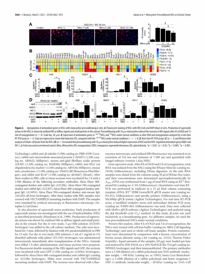
Figure 3. Inhibition of Nrf2 abolished minocycline-induced cytoprotection in vitro. non-PCNSCs and PCNSCs (10 �M) werepretreated with control- or Nrf2-siRNA before OGD and reoxygenation. A–C, Real-time RT-PCR assays of the NSCs. Expression ofNrf2 (A), together with NQO1 (B) and HO-1 (C), was downregulated by transfection with Nrf2-siRNA (n � 3). D, Reduced HEtsignals with minocycline preconditioning after 8 h of OGD and 15 min of reoxygenation were reversed by Nrf2-siRNA (n � 4). E, F,Death and viability of the NSCs analyzed by LDH assay (E) and WST-1 assay (F ) after 8 h of OGD and 24 h of reoxygenation.Nrf2-siRNA abolished cytoprotection offered by minocycline preconditioning (n � 4). Mino, Minocycline; c, control-siRNA. *p �0.05; †p � 0.01; ‡p � 0.005; §p � 0.001.
Sakata et al. • Minocycline-Preconditioned NSCs in Ischemic Stroke J. Neurosci., March 7, 2012 • 32(10):3462–3473 • 3465
DAPI. Terminal deoxynucleotidyl transferase-mediated uridine 5�-triphosphate-biotin nickend labeling (TUNEL)-positive cells, alsostained with GFP, were counted under highmagnification (�1000) using unbiased com-putational stereology [fractionator method,using STEREOINVESTIGATOR software(MicroBrightfield)], as described previously(Kelly et al., 2004).
Quantification of survival of the transplantedGFP-positive NSCs. The transplanted GFP-positive cells were counted using unbiasedcomputational stereology as described above.All the GFP-positive cells were counted on fiveserial coronal sections per brain (2 mm apart)and stained with GFP and DAPI.
Assessment of NSC proliferation and differen-tiation profiles. The proportion of GFP-labeledcells, also stained with a proliferation marker(Ki-67) or lineage-specific phenotype markers(�-tubulin, MAP2, and GFAP), was deter-mined by confocal microscopy. Split panel andz-axis analyses were used for all counting. Onehundred or more GFP-positive cells werescored for each marker per animal.
Measurement of infarct size. The brain sec-tions were stained with H&E. We estimated theinfarct size in the cortex/striatum as a percent-age of the ipsilateral cortex/striatum using thefollowing: [(area of contralateral cortex/stria-tum) � (area of remaining ipsilateral cortex/striatum)/(area of contralateral cortex/striatum) � 100]. The area of both sides of thecortex and striatum was measured on nine se-rial coronal sections per brain (1 mm apart),and the area of the infarct was quantified overthese nine levels using Adobe Photoshop(Adobe Systems).
Behavioral analysis. A rotarod test andbeam-balance test were evaluated by two indi-viduals blinded to the rat treatment status onthe day of transplantation before the strokesurgery and at 1, 7, 14, 21, and 28 d after trans-plantation. Beam-balance performance was as-sessed on a six-point scale: 0, balances withsteady posture; 1, grasps side of the beam; 2,hugs the beam and one limb falls from thebeam; 3, hugs the beam and two limbs fall fromthe beam or spins on the beam (�60 s); 4, attempts to balance on thebeam but falls off (�40 s); 5, attempts to balance on the beam but falls off(�20 s); and 6, falls off with no attempt to balance or hang onto the beam(�20 s) (Schabitz et al., 2004).
For the rotarod test, after 3 d of training before the stroke surgery, therats were placed on the cylinder and the time the animals remained on therotarod was recorded. The speed was slowly increased from 10 to 40 rpmwithin a period of 4 min. The trial was ended if the animal fell off therungs. The maximum duration on the device was recorded with threerotarod measurements before the surgery. Rotarod test data are pre-sented as percentages of the maximal duration, compared with the inter-nal baseline control.
Statistical analysis. Behavioral data were assessed using repeated-measures ANOVA. We used Scheffe’s post hoc analysis of the rotarodtest and analyzed the beam-balance test using the Steel–Dwass test.For other experimental data, comparisons among multiple groupswere performed with one-way ANOVA, followed by Scheffe’s post hocanalysis. Comparisons between two groups were achieved with a Stu-dent’s unpaired t test. Data are expressed as median for the beam-balance test and mean SD for the other experiments. Significancewas accepted with p � 0.05.
ResultsMinocycline preconditioning conferred cytoprotection onNSCs in vitroWe used self-renewing and multipotent NSCs isolated fromfetal GFP transgenic rats (Fig. 1 A,B). We first investigatedwhether minocycline preconditioning could reduce NSCdeath under ischemic reperfusion injury in vitro. After 8 h ofOGD and 24 h of reoxygenation, a LDH assay revealed thatminocycline-preconditioned NSCs ( PCNSCs) (10 �M) had asignificant reduction in death (41%), compared with the non-preconditioned NSCs ( non-PCNSCs) (Fig. 1C). This cytopro-tective effect was supported by a WST-1 assay (Fig. 1 D) andTUNEL staining (Fig. 1 E), which showed an increased viabil-ity and reduced TUNEL positivity in the PCNSCs comparedwith the non-PCNSCs. Moreover, fluorescent staining of cyto-chrome c revealed that a smaller number of PCNSCs showed arelease of cytochrome c from mitochondria to the cytoplasmthan non-PCNSCs after 8 h of OGD and 3 h of reoxygenation(Fig. 1 F). Minocycline also reduced the death of NSCs sub-
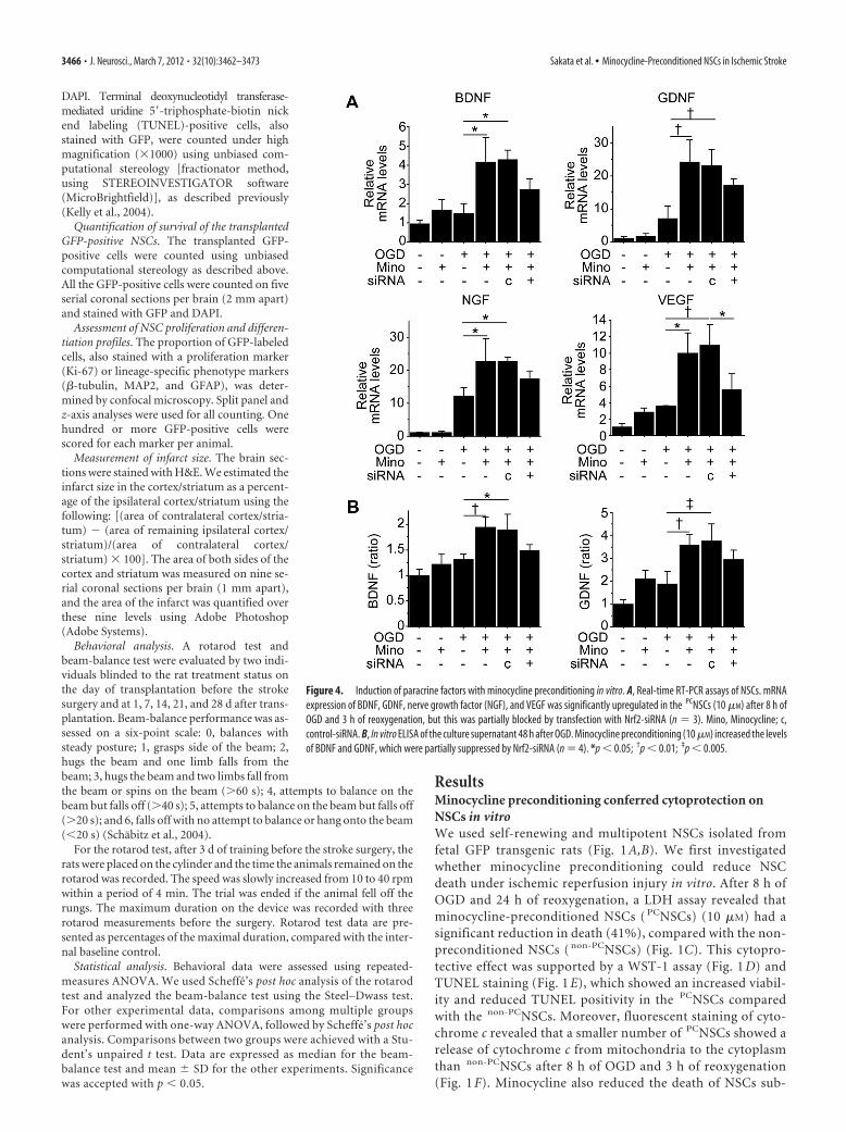
Figure 4. Induction of paracrine factors with minocycline preconditioning in vitro. A, Real-time RT-PCR assays of NSCs. mRNAexpression of BDNF, GDNF, nerve growth factor (NGF), and VEGF was significantly upregulated in the PCNSCs (10 �M) after 8 h ofOGD and 3 h of reoxygenation, but this was partially blocked by transfection with Nrf2-siRNA (n � 3). Mino, Minocycline; c,control-siRNA. B, In vitro ELISA of the culture supernatant 48 h after OGD. Minocycline preconditioning (10 �M) increased the levelsof BDNF and GDNF, which were partially suppressed by Nrf2-siRNA (n � 4). *p � 0.05; †p � 0.01; ‡p � 0.005.
3466 • J. Neurosci., March 7, 2012 • 32(10):3462–3473 Sakata et al. • Minocycline-Preconditioned NSCs in Ischemic Stroke
jected to H2O2 and diethylenetriamine/nitric oxide, as deter-mined by LDH assay (Fig. 1G,H ).
Minocycline preconditioning upregulated antioxidant genesand reduced oxidative stress in vitroWe next sought to elucidate the underlying mechanism ofminocycline-induced cytoprotection. NSCs subjected to 8 h ofOGD and 15 min of reoxygenation exhibited a remarkable in-crease in HEt signals in the cytosol, which represents superoxideanion production (Fig. 2A) (Murakami et al., 1998). However,
this signal increase was reduced by preconditioning with 10 �M
minocycline. This finding suggests that resistance to oxidativestress is one of the mechanisms of minocycline-induced cytopro-tection. We investigated the expression of antioxidant genesusing a RT-PCR array. This analysis identified that with precon-ditioning, Nrf2, the principal transcription factor that regulatesthe basal and inducible expression of a battery of antioxidantgenes, was up-regulated together with the Nrf2-regulated antiox-idant genes, including NQO1 and HO-1 (Fig. 2B). Real-timeRT-PCR assays showed that minocycline preconditioning signif-
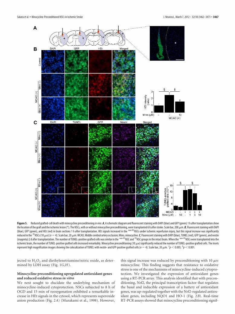
Figure 5. Reduced grafted-cell death with minocycline preconditioning in vivo. A, A schematic diagram and fluorescent staining with DAPI (blue) and GFP (green) 1 h after transplantation showthe location of the graft and the ischemic lesion (*). The NSCs, with or without minocycline preconditioning, were transplanted 6 h after stroke. Scale bar, 200 �m. B, Fluorescent staining with DAPI(blue), GFP (green), and HEt (red) in brain sections 1 h after transplantation. HEt signals increased in the non-PCNSCs under ischemic reperfusion injury, but this signal increase was significantlyreduced in the PCNSCs (10 �M) (n � 4). Scale bar, 20 �m. MCAO, Middle cerebral artery occlusion; Mino, minocycline. C, Fluorescent staining with DAPI (blue), TUNEL (red), GFP (green), and nestin(magenta) 2 d after transplantation. The number of TUNEL-positive grafted cells was similar in the non-PCNSC and PCNSC groups in the intact brain. When the non-PCNSCs were transplanted into theischemic brain, the number of TUNEL-positive grafted cells increased remarkably. Minocycline preconditioning (10 �M) significantly reduced the number of TUNEL-positive grafted cells. The insetsrepresent high magnification images showing the colocalization of TUNEL with nestin- and GFP-positive grafted cells (n � 4). Scale bar, 20 �m. ‡p � 0.005; §p � 0.001.
Sakata et al. • Minocycline-Preconditioned NSCs in Ischemic Stroke J. Neurosci., March 7, 2012 • 32(10):3462–3473 • 3467
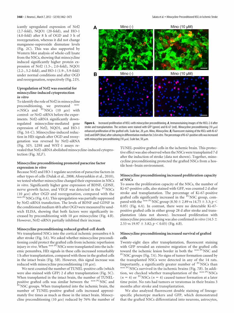
icantly upregulated expression of Nrf2(2.7-fold), NQO1 (20-fold), and HO-1(4.0-fold) after 8 h of OGD and 3 h ofreoxygenation, whereas it did not changemanganese-superoxide dismutase levels(Fig. 2C). This was also supported byWestern blot analysis of whole-cell lysatefrom the NSCs, showing that minocyclineinduced significantly higher protein ex-pression of Nrf2 (1.5-, 2.0-fold), NQO1(2.2-, 5.2-fold), and HO-1 (1.9-, 5.9-fold)under normal conditions and after OGDand reoxygenation, respectively (Fig. 2D).
Upregulation of Nrf2 was essential forminocycline-induced cytoprotectionin vitroTo identify the role of Nrf2 in minocyclinepreconditioning, we pretreated non-
PCNSCs and PCNSCs (10 �M) withcontrol- or Nrf2-siRNA before the exper-iments. Nrf2-siRNA significantly down-regulated minocycline-mediated geneexpression of Nrf2, NQO1, and HO-1(Fig. 3A-C). Minocycline-induced reduc-tion in HEt signals after OGD and reoxy-genation was reversed by Nrf2-siRNA(Fig. 3D). LDH and WST-1 assays re-vealed that Nrf2-siRNA abolished minocycline-induced cytopro-tection (Fig. 3E,F).
Minocycline preconditioning promoted paracrine factorexpression in vitroBecause Nrf2 and HO-1 regulate secretion of paracrine factors inother types of cells (Dulak et al., 2008; Afonyushkin et al., 2010),we tested whether minocycline changed their expression in NSCsin vitro. Significantly higher gene expression of BDNF, GDNF,nerve growth factor, and VEGF was detected in the PCNSCs(10 �M) after OGD and reoxygenation, compared with thenon-PCNSCs (Fig. 4A). This upregulation was partially suppressedby Nrf2-siRNA transfection. The levels of BDNF and GDNF inthe conditioned medium 48 h after OGD were measured by sand-wich ELISA, showing that both factors were significantly in-creased by preconditioning with 10 �M minocycline (Fig. 4B).However, Nrf2-siRNA partially inhibited their increase.
Minocycline preconditioning reduced grafted-cell deathWe transplanted NSCs into the cortical ischemic penumbra 6 hafter stroke (Fig. 5A). We asked whether minocycline precondi-tioning could protect the grafted cells from ischemic reperfusioninjury in vivo. When non-PCNSCs were transplanted into the isch-emic penumbra, HEt signals in these cells increased remarkably1 h after transplantation, compared with those in the grafted cellsin the intact brain (Fig. 5B). However, this signal increase wasreduced with minocycline preconditioning (10 �M).
We next counted the number of TUNEL-positive cells (whichwere also stained with GFP) 2 d after transplantation (Fig. 5C).When transplanted in the intact brain, the number of TUNEL-positive grafted cells was similar between the non-PCNSC andPCNSC groups. When transplanted into the ischemic brain, thenumber of TUNEL-positive grafted cells increased approxi-mately five times as much as those in the intact brain. Minocy-cline preconditioning (10 �M) reduced by 76% the number of
TUNEL-positive grafted cells in the ischemic brain. This protec-tive effect was also observed when the NSCs were transplanted 7 dafter the induction of stroke (data not shown). Together, mino-cycline preconditioning protected the grafted NSCs from a hos-tile host– brain environment.
Minocycline preconditioning increased proliferation capacityof NSCsTo assess the proliferation capacity of the NSCs, the number ofKi-67-positive cells, also stained with GFP, was counted 2 d afterstroke and transplantation. The percentage of Ki-67-positivegrafted cells significantly increased in the PCNSC group, com-pared with the non-PCNSC group (8.50 2.89 vs 14.75 3.3; p �0.05) (Fig. 6A). In contrast, there were no detectable Ki-67-positive grafted cells in either group 28 d after stroke and trans-plantation (data not shown). Increased proliferation withminocycline preconditioning was also confirmed in vitro (14.5 2.53 vs 19.97 3.82; p � 0.05) (Fig. 6B).
Minocycline preconditioning increased survival of graftedcellsTwenty-eight days after transplantation, fluorescent stainingwith GFP revealed an extensive migration of the grafted cellstoward the ischemic lesion border in both the non-PCNSC andPCNSC groups (Fig. 7A). No signs of tumor formation caused bythe transplanted NSCs were detected in any of the 14 rats.Importantly, a significantly greater number of PCNSCs thannon-PCNSCs survived in the ischemic brains (Fig. 7B). In addi-tion, we checked whether transplantation of the non-PCNSCs(n � 4) or PCNSCs (n � 4) caused tumor formation at a latertime point. No rats had tumors or teratomas in their brains 3months after stroke and transplantation.
We also analyzed fluorescent double staining of lineage-specific phenotype markers and GFP, which demonstratedthat the grafted NSCs differentiated into neurons, astrocytes,
Figure 6. Increased proliferation of NSCs with minocycline preconditioning. A, Immunostaining images of the NSCs 2 d afterstroke and transplantation. The sections were stained with GFP (green) and Ki-67 (red). Minocycline preconditioning (10 �M)enhanced proliferation of the grafted cells. Scale bar, 20 �m. Mino, Minocycline. B, Fluorescent staining of the NSCs with Ki-67(red) and DAPI (blue) after culturing in differentiation medium for 2 d in vitro. The percentage of Ki-67-positive cells was increasedwith minocycline preconditioning (10 �M). Scale bar, 50 �m.
3468 • J. Neurosci., March 7, 2012 • 32(10):3462–3473 Sakata et al. • Minocycline-Preconditioned NSCs in Ischemic Stroke
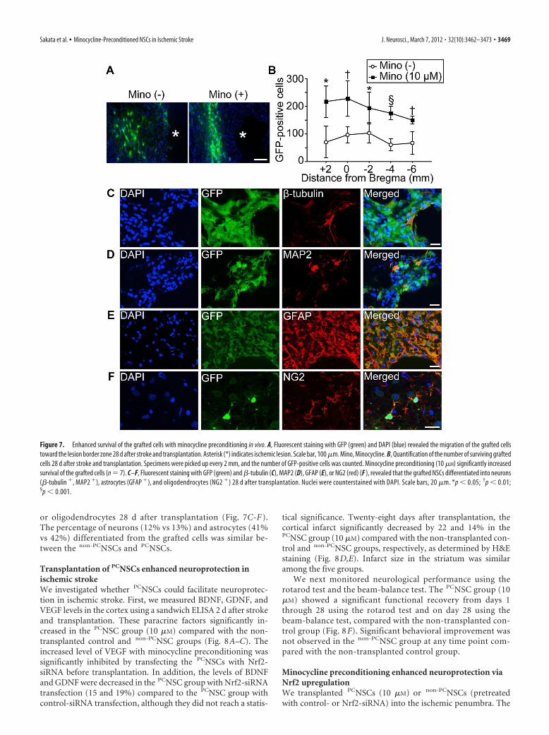
or oligodendrocytes 28 d after transplantation (Fig. 7C-F ).The percentage of neurons (12% vs 13%) and astrocytes (41%vs 42%) differentiated from the grafted cells was similar be-tween the non-PCNSCs and PCNSCs.
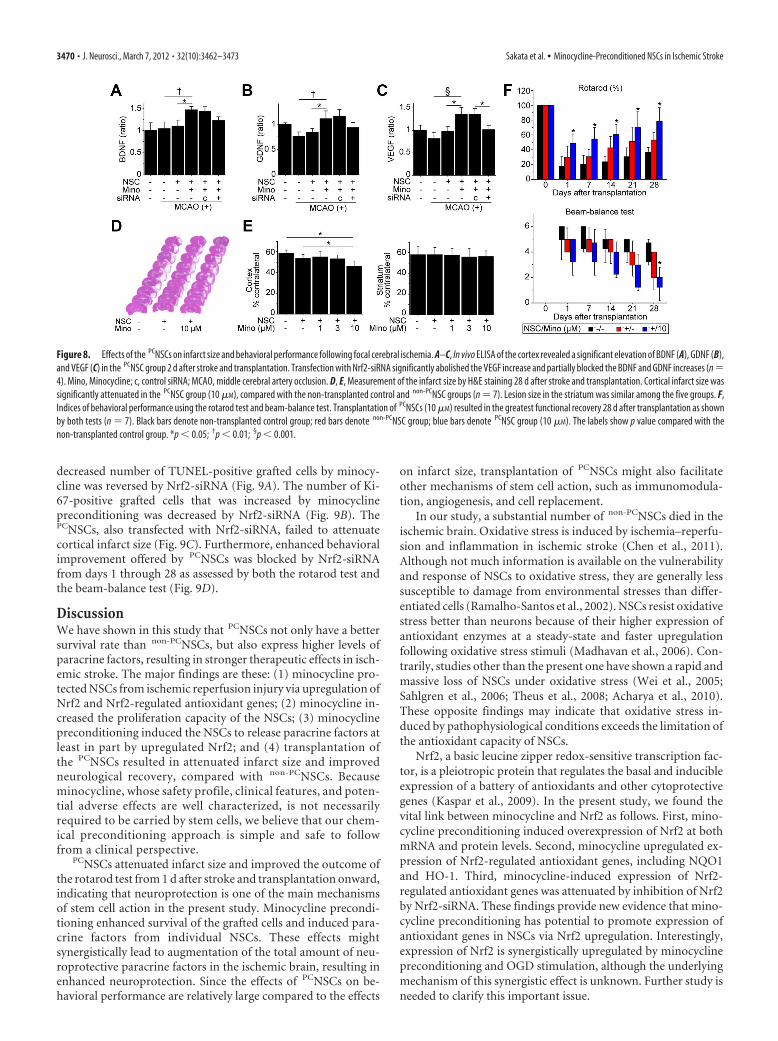
Transplantation of PCNSCs enhanced neuroprotection inischemic strokeWe investigated whether PCNSCs could facilitate neuroprotec-tion in ischemic stroke. First, we measured BDNF, GDNF, andVEGF levels in the cortex using a sandwich ELISA 2 d after strokeand transplantation. These paracrine factors significantly in-creased in the PCNSC group (10 �M) compared with the non-transplanted control and non-PCNSC groups (Fig. 8A–C). Theincreased level of VEGF with minocycline preconditioning wassignificantly inhibited by transfecting the PCNSCs with Nrf2-siRNA before transplantation. In addition, the levels of BDNFand GDNF were decreased in the PCNSC group with Nrf2-siRNAtransfection (15 and 19%) compared to the PCNSC group withcontrol-siRNA transfection, although they did not reach a statis-
tical significance. Twenty-eight days after transplantation, thecortical infarct significantly decreased by 22 and 14% in thePCNSC group (10 �M) compared with the non-transplanted con-trol and non-PCNSC groups, respectively, as determined by H&Estaining (Fig. 8D,E). Infarct size in the striatum was similaramong the five groups.
We next monitored neurological performance using therotarod test and the beam-balance test. The PCNSC group (10�M) showed a significant functional recovery from days 1through 28 using the rotarod test and on day 28 using thebeam-balance test, compared with the non-transplanted con-trol group (Fig. 8 F). Significant behavioral improvement wasnot observed in the non-PCNSC group at any time point com-pared with the non-transplanted control group.
Minocycline preconditioning enhanced neuroprotection viaNrf2 upregulationWe transplanted PCNSCs (10 �M) or non-PCNSCs (pretreatedwith control- or Nrf2-siRNA) into the ischemic penumbra. The
Figure 7. Enhanced survival of the grafted cells with minocycline preconditioning in vivo. A, Fluorescent staining with GFP (green) and DAPI (blue) revealed the migration of the grafted cellstoward the lesion border zone 28 d after stroke and transplantation. Asterisk (*) indicates ischemic lesion. Scale bar, 100 �m. Mino, Minocycline. B, Quantification of the number of surviving graftedcells 28 d after stroke and transplantation. Specimens were picked up every 2 mm, and the number of GFP-positive cells was counted. Minocycline preconditioning (10 �M) significantly increasedsurvival of the grafted cells (n � 7). C–F, Fluorescent staining with GFP (green) and �-tubulin (C), MAP2 (D), GFAP (E), or NG2 (red) (F ), revealed that the grafted NSCs differentiated into neurons(�-tubulin , MAP2 ), astrocytes (GFAP ), and oligodendrocytes (NG2 ) 28 d after transplantation. Nuclei were counterstained with DAPI. Scale bars, 20 �m. *p � 0.05; †p � 0.01;§p � 0.001.
Sakata et al. • Minocycline-Preconditioned NSCs in Ischemic Stroke J. Neurosci., March 7, 2012 • 32(10):3462–3473 • 3469
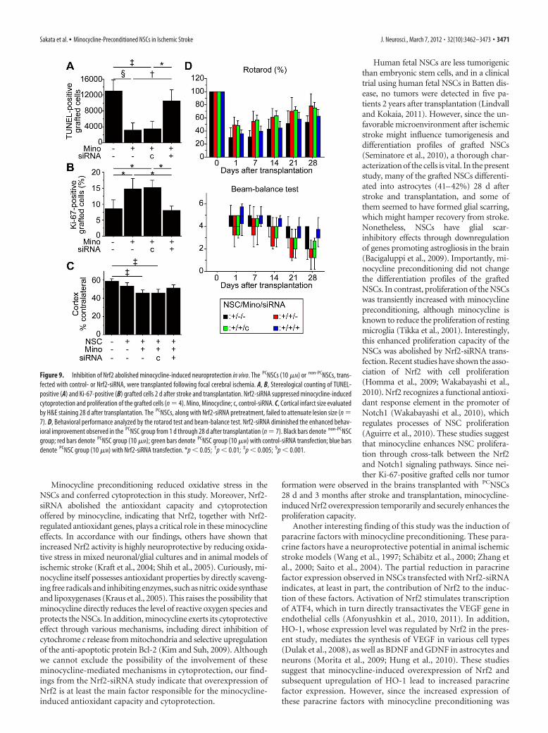
decreased number of TUNEL-positive grafted cells by minocy-cline was reversed by Nrf2-siRNA (Fig. 9A). The number of Ki-67-positive grafted cells that was increased by minocyclinepreconditioning was decreased by Nrf2-siRNA (Fig. 9B). ThePCNSCs, also transfected with Nrf2-siRNA, failed to attenuatecortical infarct size (Fig. 9C). Furthermore, enhanced behavioralimprovement offered by PCNSCs was blocked by Nrf2-siRNAfrom days 1 through 28 as assessed by both the rotarod test andthe beam-balance test (Fig. 9D).
DiscussionWe have shown in this study that PCNSCs not only have a bettersurvival rate than non-PCNSCs, but also express higher levels ofparacrine factors, resulting in stronger therapeutic effects in isch-emic stroke. The major findings are these: (1) minocycline pro-tected NSCs from ischemic reperfusion injury via upregulation ofNrf2 and Nrf2-regulated antioxidant genes; (2) minocycline in-creased the proliferation capacity of the NSCs; (3) minocyclinepreconditioning induced the NSCs to release paracrine factors atleast in part by upregulated Nrf2; and (4) transplantation ofthe PCNSCs resulted in attenuated infarct size and improvedneurological recovery, compared with non-PCNSCs. Becauseminocycline, whose safety profile, clinical features, and poten-tial adverse effects are well characterized, is not necessarilyrequired to be carried by stem cells, we believe that our chem-ical preconditioning approach is simple and safe to followfrom a clinical perspective.
PCNSCs attenuated infarct size and improved the outcome ofthe rotarod test from 1 d after stroke and transplantation onward,indicating that neuroprotection is one of the main mechanismsof stem cell action in the present study. Minocycline precondi-tioning enhanced survival of the grafted cells and induced para-crine factors from individual NSCs. These effects mightsynergistically lead to augmentation of the total amount of neu-roprotective paracrine factors in the ischemic brain, resulting inenhanced neuroprotection. Since the effects of PCNSCs on be-havioral performance are relatively large compared to the effects
on infarct size, transplantation of PCNSCs might also facilitateother mechanisms of stem cell action, such as immunomodula-tion, angiogenesis, and cell replacement.
In our study, a substantial number of non-PCNSCs died in theischemic brain. Oxidative stress is induced by ischemia–reperfu-sion and inflammation in ischemic stroke (Chen et al., 2011).Although not much information is available on the vulnerabilityand response of NSCs to oxidative stress, they are generally lesssusceptible to damage from environmental stresses than differ-entiated cells (Ramalho-Santos et al., 2002). NSCs resist oxidativestress better than neurons because of their higher expression ofantioxidant enzymes at a steady-state and faster upregulationfollowing oxidative stress stimuli (Madhavan et al., 2006). Con-trarily, studies other than the present one have shown a rapid andmassive loss of NSCs under oxidative stress (Wei et al., 2005;Sahlgren et al., 2006; Theus et al., 2008; Acharya et al., 2010).These opposite findings may indicate that oxidative stress in-duced by pathophysiological conditions exceeds the limitation ofthe antioxidant capacity of NSCs.
Nrf2, a basic leucine zipper redox-sensitive transcription fac-tor, is a pleiotropic protein that regulates the basal and inducibleexpression of a battery of antioxidants and other cytoprotectivegenes (Kaspar et al., 2009). In the present study, we found thevital link between minocycline and Nrf2 as follows. First, mino-cycline preconditioning induced overexpression of Nrf2 at bothmRNA and protein levels. Second, minocycline upregulated ex-pression of Nrf2-regulated antioxidant genes, including NQO1and HO-1. Third, minocycline-induced expression of Nrf2-regulated antioxidant genes was attenuated by inhibition of Nrf2by Nrf2-siRNA. These findings provide new evidence that mino-cycline preconditioning has potential to promote expression ofantioxidant genes in NSCs via Nrf2 upregulation. Interestingly,expression of Nrf2 is synergistically upregulated by minocyclinepreconditioning and OGD stimulation, although the underlyingmechanism of this synergistic effect is unknown. Further study isneeded to clarify this important issue.
Figure 8. Effects of the PCNSCs on infarct size and behavioral performance following focal cerebral ischemia. A–C, In vivo ELISA of the cortex revealed a significant elevation of BDNF (A), GDNF (B),and VEGF (C) in the PCNSC group 2 d after stroke and transplantation. Transfection with Nrf2-siRNA significantly abolished the VEGF increase and partially blocked the BDNF and GDNF increases (n �4). Mino, Minocycline; c, control siRNA; MCAO, middle cerebral artery occlusion. D, E, Measurement of the infarct size by H&E staining 28 d after stroke and transplantation. Cortical infarct size wassignificantly attenuated in the PCNSC group (10 �M), compared with the non-transplanted control and non-PCNSC groups (n � 7). Lesion size in the striatum was similar among the five groups. F,Indices of behavioral performance using the rotarod test and beam-balance test. Transplantation of PCNSCs (10 �M) resulted in the greatest functional recovery 28 d after transplantation as shownby both tests (n � 7). Black bars denote non-transplanted control group; red bars denote non-PCNSC group; blue bars denote PCNSC group (10 �M). The labels show p value compared with thenon-transplanted control group. *p � 0.05; †p � 0.01; §p � 0.001.
3470 • J. Neurosci., March 7, 2012 • 32(10):3462–3473 Sakata et al. • Minocycline-Preconditioned NSCs in Ischemic Stroke
Minocycline preconditioning reduced oxidative stress in theNSCs and conferred cytoprotection in this study. Moreover, Nrf2-siRNA abolished the antioxidant capacity and cytoprotectionoffered by minocycline, indicating that Nrf2, together with Nrf2-regulated antioxidant genes, plays a critical role in these minocyclineeffects. In accordance with our findings, others have shown thatincreased Nrf2 activity is highly neuroprotective by reducing oxida-tive stress in mixed neuronal/glial cultures and in animal models ofischemic stroke (Kraft et al., 2004; Shih et al., 2005). Curiously, mi-nocycline itself possesses antioxidant properties by directly scaveng-ing free radicals and inhibiting enzymes, such as nitric oxide synthaseand lipoxygenases (Kraus et al., 2005). This raises the possibility thatminocycline directly reduces the level of reactive oxygen species andprotects the NSCs. In addition, minocycline exerts its cytoprotectiveeffect through various mechanisms, including direct inhibition ofcytochrome c release from mitochondria and selective upregulationof the anti-apoptotic protein Bcl-2 (Kim and Suh, 2009). Althoughwe cannot exclude the possibility of the involvement of theseminocycline-mediated mechanisms in cytoprotection, our find-ings from the Nrf2-siRNA study indicate that overexpression ofNrf2 is at least the main factor responsible for the minocycline-induced antioxidant capacity and cytoprotection.
Human fetal NSCs are less tumorigenicthan embryonic stem cells, and in a clinicaltrial using human fetal NSCs in Batten dis-ease, no tumors were detected in five pa-tients 2 years after transplantation (Lindvalland Kokaia, 2011). However, since the un-favorable microenvironment after ischemicstroke might influence tumorigenesis anddifferentiation profiles of grafted NSCs(Seminatore et al., 2010), a thorough char-acterization of the cells is vital. In the presentstudy, many of the grafted NSCs differenti-ated into astrocytes (41–42%) 28 d afterstroke and transplantation, and some ofthem seemed to have formed glial scarring,which might hamper recovery from stroke.Nonetheless, NSCs have glial scar-inhibitory effects through downregulationof genes promoting astrogliosis in the brain(Bacigaluppi et al., 2009). Importantly, mi-nocycline preconditioning did not changethe differentiation profiles of the graftedNSCs. In contrast, proliferation of the NSCswas transiently increased with minocyclinepreconditioning, although minocycline isknown to reduce the proliferation of restingmicroglia (Tikka et al., 2001). Interestingly,this enhanced proliferation capacity of theNSCs was abolished by Nrf2-siRNA trans-fection. Recent studies have shown the asso-ciation of Nrf2 with cell proliferation(Homma et al., 2009; Wakabayashi et al.,2010). Nrf2 recognizes a functional antioxi-dant response element in the promoter ofNotch1 (Wakabayashi et al., 2010), whichregulates processes of NSC proliferation(Aguirre et al., 2010). These studies suggestthat minocycline enhances NSC prolifera-tion through cross-talk between the Nrf2and Notch1 signaling pathways. Since nei-ther Ki-67-positive grafted cells nor tumor
formation were observed in the brains transplanted with PCNSCs28 d and 3 months after stroke and transplantation, minocycline-induced Nrf2 overexpression temporarily and securely enhances theproliferation capacity.
Another interesting finding of this study was the induction ofparacrine factors with minocycline preconditioning. These para-crine factors have a neuroprotective potential in animal ischemicstroke models (Wang et al., 1997; Schabitz et al., 2000; Zhang etal., 2000; Saito et al., 2004). The partial reduction in paracrinefactor expression observed in NSCs transfected with Nrf2-siRNAindicates, at least in part, the contribution of Nrf2 to the induc-tion of these factors. Activation of Nrf2 stimulates transcriptionof ATF4, which in turn directly transactivates the VEGF gene inendothelial cells (Afonyushkin et al., 2010, 2011). In addition,HO-1, whose expression level was regulated by Nrf2 in the pres-ent study, mediates the synthesis of VEGF in various cell types(Dulak et al., 2008), as well as BDNF and GDNF in astrocytes andneurons (Morita et al., 2009; Hung et al., 2010). These studiessuggest that minocycline-induced overexpression of Nrf2 andsubsequent upregulation of HO-1 lead to increased paracrinefactor expression. However, since the increased expression ofthese paracrine factors with minocycline preconditioning was
Figure 9. Inhibition of Nrf2 abolished minocycline-induced neuroprotection in vivo. The PCNSCs (10 �M) or non-PCNSCs, trans-fected with control- or Nrf2-siRNA, were transplanted following focal cerebral ischemia. A, B, Stereological counting of TUNEL-positive (A) and Ki-67-positive (B) grafted cells 2 d after stroke and transplantation. Nrf2-siRNA suppressed minocycline-inducedcytoprotection and proliferation of the grafted cells (n � 4). Mino, Minocycline; c, control-siRNA. C, Cortical infarct size evaluatedby H&E staining 28 d after transplantation. The PCNSCs, along with Nrf2-siRNA pretreatment, failed to attenuate lesion size (n �7). D, Behavioral performance analyzed by the rotarod test and beam-balance test. Nrf2-siRNA diminished the enhanced behav-ioral improvement observed in the PCNSC group from 1 d through 28 d after transplantation (n � 7). Black bars denote non-PCNSCgroup; red bars denote PCNSC group (10 �M); green bars denote PCNSC group (10 �M) with control-siRNA transfection; blue barsdenote PCNSC group (10 �M) with Nrf2-siRNA transfection. *p � 0.05; †p � 0.01; ‡p � 0.005; §p � 0.001.
Sakata et al. • Minocycline-Preconditioned NSCs in Ischemic Stroke J. Neurosci., March 7, 2012 • 32(10):3462–3473 • 3471
not completely inhibited by Nrf2-siRNA transfection, otherminocycline-mediated pathways might also be involved in theirupregulation. A future study using NSCs would address this im-portant issue. In light of earlier study results showing that ex vivogene modification of stem cells for overexpression of individualparacrine factors is neuroprotective (Liu et al., 2006), we antici-pated that delivery of stem cells capable of releasing a multitudeof pro-survival paracrine factors would be more appealing.
In the present study, we administered an intracerebral injec-tion of NSCs in the acute setting of stroke, which might not be aclinically relevant scenario. Evaluating the effects of minocyclinepreconditioning using other routes/timing of stem cell delivery iswarranted in future studies. In addition, we expect that thischemical preconditioning approach might be applied to othertypes of cells, such as mesenchymal stem cells.
In conclusion, we have demonstrated that minocycline pre-conditioning reprograms NSCs to tolerate oxidative stress and toexpress higher levels of paracrine factors, resulting in enhancedeffectiveness of transplantation therapy in ischemic stroke. Thebeneficial effects of minocycline preconditioning, as well as thesimplicity, easy adoptability, and lack of safety issues, make thisapproach highly appealing for future clinical applications.
ReferencesAcharya MM, Lan ML, Kan VH, Patel NH, Giedzinski E, Tseng BP, Limoli CL
(2010) Consequences of ionizing radiation-induced damage in humanneural stem cells. Free Radic Biol Med 49:1846 –1855.
Afonyushkin T, Oskolkova OV, Philippova M, Resink TJ, Erne P, Binder BR,Bochkov VN (2010) Oxidized phospholipids regulate expression ofATF4 and VEGF in endothelial cells via NRF2-dependent mechanism:novel point of convergence between electrophilic and unfolded proteinstress pathways. Arterioscler Thromb Vasc Biol 30:1007–1013.
Afonyushkin T, Oskolkova OV, Binder BR, Bochkov VN (2011) Involve-ment of CK2 in activation of electrophilic genes in endothelial cells byoxidized phospholipids. J Lipid Res 52:98 –103.
Aguirre A, Rubio ME, Gallo V (2010) Notch and EGFR pathway interactionregulates neural stem cell number and self-renewal [Letter]. Nature467:323–327.
Bacigaluppi M, Pluchino S, Peruzzotti-Jametti L, Kilic E, Kilic U, Salani G,Brambilla E, West MJ, Comi G, Martino G, Hermann DM (2009) De-layed post-ischaemic neuroprotection following systemic neural stem celltransplantation involves multiple mechanisms. Brain 132:2239 –2251.
Banerjee S, Williamson D, Habib N, Gordon M, Chataway J (2011) Humanstem cell therapy in ischaemic stroke: a review. Age Ageing 40:7–13.
Bliss T, Guzman R, Daadi M, Steinberg GK (2007) Cell transplantationtherapy for stroke. Stroke 38:817– 826.
Blurton-Jones M, Kitazawa M, Martinez-Coria H, Castello NA, Muller FJ,Loring JF, Yamasaki TR, Poon WW, Green KN, LaFerla FM (2009)Neural stem cells improve cognition via BDNF in a transgenic model ofAlzheimer disease. Proc Natl Acad Sci U S A 106:13594 –13599.
Chen H, Yoshioka H, Kim GS, Jung JE, Okami N, Sakata H, Maier CM,Narasimhan P, Goeders CE, Chan PH (2011) Oxidative stress in isch-emic brain damage: mechanisms of cell death and potential moleculartargets for neuroprotection. Antioxid Redox Signal 14:1505–1517.
Dulak J, Deshane J, Jozkowicz A, Agarwal A (2008) Heme oxygenase-1 andcarbon monoxide in vascular pathobiology. Focus on angiogenesis. Cir-culation 117:231–241.
Fujimura M, Morita-Fujimura Y, Murakami K, Kawase M, Chan PH (1998)Cytosolic redistribution of cytochrome c after transient focal cerebralischemia in rats. J Cereb Blood Flow Metab 18:1239 –1247.
Harms KM, Li L, Cunningham LA (2010) Murine neural stem/progenitorcells protect neurons against ischemia by HIF-1�–regulated VEGF signal-ing. PLoS One 5:e9767.
Hicks AU, Lappalainen RS, Narkilahti S, Suuronen R, Corbett D, Sivenius J,Hovatta O, Jolkkonen J (2009) Transplantation of human embryonicstem cell-derived neural precursor cells and enriched environment aftercortical stroke in rats: cell survival and functional recovery. Eur J Neurosci29:562–574.
Homma S, Ishii Y, Morishima Y, Yamadori T, Matsuno Y, Haraguchi N,
Kikuchi N, Satoh H, Sakamoto T, Hizawa N, Itoh K, Yamamoto M(2009) Nrf2 enhances cell proliferation and resistance to anticancerdrugs in human lung cancer. Clin Cancer Res 15:3423–3432.
Hung SY, Liou HC, Fu WM (2010) The mechanism of heme oxygenase-1action involved in the enhancement of neurotrophic factor expression.Neuropharmacology 58:321–329.
Kaspar JW, Niture SK, Jaiswal AK (2009) Nrf2:INrf2 (Keap1) signaling inoxidative stress. Free Radic Biol Med 47:1304 –1309.
Keilhoff G, Schild L, Fansa H (2008) Minocycline protects Schwann cellsfrom ischemia-like injury and promotes axonal outgrowth in bioartificialnerve grafts lacking Wallerian degeneration. Exp Neurol 212:189 –200.
Kelly S, Bliss TM, Shah AK, Sun GH, Ma M, Foo WC, Masel J, Yenari MA,Weissman IL, Uchida N, Palmer T, Steinberg GK (2004) Transplantedhuman fetal neural stem cells survive, migrate, and differentiate in isch-emic rat cerebral cortex. Proc Natl Acad Sci U S A 101:11839 –11844.
Kernt M, Hirneiss C, Neubauer AS, Kampik A (2010) Minocycline is cyto-protective in human corneal endothelial cells and induces anti-apoptoticB-cell CLL/lymphoma 2 (Bcl-2) and X-linked inhibitor of apoptosis(XIAP). Br J Ophthalmol 94:940 –946.
Kim HS, Suh YH (2009) Minocycline and neurodegenerative diseases.Behav.
Kraft AD, Johnson DA, Johnson JA (2004) Nuclear factor E2-relatedfactor 2-dependent antioxidant response element activation by tert-butylhydroquinone and sulforaphane occurring preferentially in as-trocytes conditions neurons against oxidative insult. J Neurosci24:1101–1112.
Kraus RL, Pasieczny R, Lariosa-Willingham K, Turner MS, Jiang A, TraugerJW (2005) Antioxidant properties of minocycline: neuroprotection inan oxidative stress assay and direct radical-scavenging activity. J Neuro-chem 94:819 – 827.
Lindvall O, Kokaia Z (2011) Stem cell research in stroke. How far from theclinic? Stroke 42:2369 –2375.
Liu H, Honmou O, Harada K, Nakamura K, Houkin K, Hamada H, Kocsis JD(2006) Neuroprotection by PIGF gene-modified human mesenchymalstem cells after cerebral ischaemia. Brain 129:2734 –2745.
Lo EH, Dalkara T, Moskowitz MA (2003) Mechanisms, challenges and op-portunities in stroke. Nat Rev Neurosci 4:399 – 415.
Madhavan L, Ourednik V, Ourednik J (2006) Increased “vigilance” of anti-oxidant mechanisms in neural stem cells potentiates their capability toresist oxidative stress. Stem Cells 24:2110 –2119.
Morita K, Lee MS, Her S (2009) Possible relation of hemin-induced HO-1expression to the upregulation of VEGF and BDNF mRNA levels in rat C6glioma cells. J Mol Neurosci 38:31– 40.
Murakami K, Kondo T, Kawase M, Li Y, Sato S, Chen SF, Chan PH (1998)Mitochondrial susceptibility to oxidative stress exacerbates cerebral in-farction that follows permanent focal cerebral ischemia in mutant micewith manganese superoxide dismutase deficiency. J Neurosci 18:205–213.
Nakagomi N, Nakagomi T, Kubo S, Nakano-Doi A, Saino O, Takata M,Yoshikawa H, Stern DM, Matsuyama T, Taguchi A (2009) Endothelialcells support survival, proliferation, and neuronal differentiation of trans-planted adult ischemia-induced neural stem/progenitor cells after cere-bral infarction. Stem Cells 27:2185–2195.
Ramalho-Santos M, Yoon S, Matsuzaki Y, Mulligan RC, Melton DA (2002)“Stemness”: transcriptional profiling of embryonic and adult stem cells.Science 298:597– 600.
Sahlgren CM, Pallari HM, He T, Chou YH, Goldman RD, Eriksson JE (2006)A nestin scaffold links Cdk5/p35 signaling to oxidant-induced cell death.EMBO J 25:4808 – 4819.
Saito A, Narasimhan P, Hayashi T, Okuno S, Ferrand-Drake M, Chan PH(2004) Neuroprotective role of a proline-rich Akt substrate in apoptoticneuronal cell death after stroke: relationships with nerve growth factor.J Neurosci 24:1584 –1593.
Savitz SI, Rosenbaum DM, Dinsmore JH, Wechsler LR, Caplan LR (2002)Cell transplantation for stroke. Ann Neurol 52:266 –275.
Schabitz WR, Sommer C, Zoder W, Kiessling M, Schwaninger M, Schwab S(2000) Intravenous brain-derived neurotrophic factor reduces infarctsize and counterregulates Bax and Bcl-2 expression after temporary focalcerebral ischemia. Stroke 31:2212–2217.
Schabitz WR, Berger C, Kollmar R, Seitz M, Tanay E, Kiessling M, Schwab S,Sommer C (2004) Effect of brain-derived neurotrophic factor treatmentand forced arm use on functional motor recovery after small corticalischemia. Stroke 35:992–997.
3472 • J. Neurosci., March 7, 2012 • 32(10):3462–3473 Sakata et al. • Minocycline-Preconditioned NSCs in Ischemic Stroke
Seminatore C, Polentes J, Ellman D, Kozubenko N, Itier V, Tine S, TritschlerL, Brenot M, Guidou E, Blondeau J, Lhuillier M, Bugi A, Aubry L, Jende-lova P, Sykova E, Perrier AL, Finsen B, Onteniente B (2010) The post-ischemic environment differentially impacts teratoma or tumorformation after transplantation of human embryonic stem cell-derivedneural progenitors. Stroke 41:153–159.
Shih AY, Li P, Murphy TH (2005) A small-molecule-inducible Nrf2-mediated antioxidant response provides effective prophylaxis against ce-rebral ischemia in vivo. J Neurosci 25:10321–10335.
Theus MH, Wei L, Cui L, Francis K, Hu X, Keogh C, Yu SP (2008) In vitrohypoxic preconditioning of embryonic stem cells as a strategy of promot-ing cell survival and functional benefits after transplantation into theischemic rat brain. Exp Neurol 210:656 – 670.
Tikka T, Fiebich BL, Goldsteins G, Keinanen R, Koistinaho J (2001) Mino-cycline, a tetracycline derivative, is neuroprotective against excitotoxicityby inhibiting activation and proliferation of microglia. J Neurosci21:2580 –2588.
Wakabayashi N, Shin S, Slocum SL, Agoston ES, Wakabayashi J, Kwak MK,Misra V, Biswal S, Yamamoto M, Kensler TW (2010) Regulation ofNotch1 signaling by Nrf2: implications for tissue regeneration. Sci Signal3:ra52.
Wang Y, Lin SZ, Chiou A-L, Williams LR, Hoffer BJ (1997) Glial cell line-derived neurotrophic factor protects against ischemia-induced injury inthe cerebral cortex. J Neurosci 17:4341– 4348.
Wei L, Cui L, Snider BJ, Rivkin M, Yu SS, Lee CS, Adams LD, Gottlieb DI,Johnson EM Jr, Yu SP, Choi DW (2005) Transplantation of embryonicstem cells overexpressing Bcl-2 promotes functional recovery after tran-sient cerebral ischemia. Neurobiol Dis 19:183–193.
Yrjanheikki J, Tikka T, Keinanen R, Goldsteins G, Chan PH, Koistinaho J(1999) A tetracycline derivative, minocycline, reduces inflammation andprotects against focal cerebral ischemia with a wide therapeutic window.Proc Natl Acad Sci U S A 96:13496 –13500.
Zhang ZG, Zhang L, Jiang Q, Zhang R, Davies K, Powers C, van BruggenN, Chopp M (2000) VEGF enhances angiogenesis and promotesblood-brain barrier leakage in the ischemic brain. J Clin Invest106:829 – 838.
Zhu S, Stavrovskaya IG, Drozda M, Kim BY, Ona V, Li M, Sarang S, Liu AS,Hartley DM, Wu DC, Gullans S, Ferrante RJ, Przedborski S, Kristal BS,Friedlander RM (2002) Minocycline inhibits cytochrome c release anddelays progression of amyotrophic lateral sclerosis in mice [Letter]. Na-ture 417:74 –78.
Sakata et al. • Minocycline-Preconditioned NSCs in Ischemic Stroke J. Neurosci., March 7, 2012 • 32(10):3462–3473 • 3473