neurobiologyofdisease ... · neurobiologyofdisease...
TRANSCRIPT
Neurobiology of Disease
Critical Role of Increased PTEN Nuclear Translocation inExcitotoxic and Ischemic Neuronal Injuries
Shu Zhang,1,2,3 Changiz Taghibiglou,1 Kimberly Girling,1 Zhifang Dong,4 Shinn-Zong Lin,2 Wei Lee,2,3
Woei-cherng Shyu,2,3* and Yu Tian Wang1,2,4*1Brain Research Centre and Department of Medicine, Vancouver Coastal Health Research Institute, University of British Columbia, Vancouver, BritishColumbia, V6T2B5, Canada, 2Translational Medicine Research Center and Centers for Neuropsychiatry, China Medical University Hospital, Taichung, 404,Taiwan, 3Graduate Institute of Immunology, China Medical University, Taichung, 404, Taiwan, and 4Ministry of Education Key Laboratory of ChildDevelopment and Disorders, and Chongqing Key Laboratory of Translational Medical Research in Cognitive Development and Learning and MemoryDisorders, Children’s Hospital of Chongqing Medical University, Chongqing, China
Stroke is the leading cause of disability in developed countries. However, no treatment is available beyond 3 h post-ictus. Here, we reportthat nuclear translocation of PTEN (phosphatase and tensin homolog deleted on chromosome TEN) is a delayed step causatively leadingto excitotoxic (in vitro) and ischemic (in vivo) neuronal injuries. We found that excitotoxic stimulation of N-methyl-D-aspartate (NMDA)resulted in PTEN nuclear translocation in cultured neurons, a process requiring mono-ubiquitination at the lysine 13 residue (K13), asthe translocation was prevented by mutation of K13 or a short interfering peptide (Tat-K13) that flanks the K13 residue. More impor-tantly, using a rat model of focal ischemia, we demonstrated that systemic application of Tat-K13, even 6 h after stroke, not only reducedischemia-induced PTEN nuclear translocation, but also strongly protected against ischemic brain damage. Our study suggests thatinhibition of PTEN nuclear translocation may represent a novel after stroke therapy.
IntroductionAlthough numerous experimental studies have consistently dem-onstrated that overactivation of N-methyl-D-aspartate (NMDA)receptors (NMDARs) at least partially contributes to excitotoxic/ischemic neuronal damage after stroke (Lucas and Newhouse,1957; Olney, 1969; Rothman, 1983, 1984; Simon et al., 1984;Lipton and Rosenberg, 1994; Aarts et al., 2002; Arundine andTymianski, 2004; Lai et al., 2011), the results of clinical trialstesting NMDAR antagonists have proven underwhelming(Albers et al., 1995, 1999, 2001; Davis et al., 2000). Although theunderlying reasons for this apparent contradiction between basic
research results and clinical trials are still unknown, a number ofexplanations have been postulated, including the short therapeu-tic time-window of NMDAR antagonists and the significant sideeffects of these antagonists due to the blockade of the NMDAR’snormal functions (Gladstone et al., 2002; Ikonomidou andTurski, 2002; Roesler et al., 2003; Lo, 2008). Therefore, instead offocusing on the receptor, our research instead centers on identi-fying new molecules and/or pathways downstream of NMDARsspecifically involved in mediating excitotoxic/ischemic neuronaldeath signaling cascades. By focusing our efforts on these targets,we hope to develop new therapeutics possessing a strong post-stroke neuroprotective action, a wider efficacy window, and withgreatly reduced side effects.
Phosphatase and tensin homolog deleted on chromosomeTEN (PTEN), a dual lipid/protein phosphatase previously iden-tified as an important tumor suppressor (Li et al., 1997; Steck etal., 1997; Maehama and Dixon, 1998; Stambolic et al., 1998), hasrecently been implicated in contributing to neuronal damage af-ter excitotoxic/ischemic insults (Gary and Mattson, 2002; Ning etal., 2004); however, the underlying mechanisms remain poorlycharacterized. PTEN�s tumor-suppressive properties are mainlydependent on its lipid phosphatase activity, which antagonizesthe cell-survival promoting PI3K/Akt signaling pathway by de-phosphorylation of phosphatidylinositol 3,4,5-trisphosphate(PIP3) (Maehama and Dixon, 1998; Stambolic et al., 1998). Assuch, a reduction or loss of PTEN�s lipid phosphatase activity hasbeen linked to carcinogenesis in numbers of cancers, includingbrain, breast, and prostate cancer (Li et al., 1997; Steck et al., 1997;Myers et al., 1998). Interestingly, several recent cancer studiesidentified two PTEN mutants (K13E and K289E), which retain
Received Dec. 10, 2012; revised March 27, 2013; accepted April 2, 2013.Author contributions: S.Z., C.T., and Y.T.W. designed research; S.Z., C.T., K.G., Z.D., S.-Z.L., W.L., and W.-C.S.
performed research; S.Z., C.T., W.-C.S., and Y.T.W. analyzed data; Y.T.W. wrote the paper.This work was supported by the Canadian Institutes of Health Research (CIHR), CHDI Foundation, the Taiwan
Department of Health Clinical Trial and Research Center of Excellence (DOH102-TD-B-111– 004), the National Re-search Council of Taiwan (NSC100-2632-B-039-001-MY3 and NSC 101-2320-B-039-059-), the National NaturalScience Foundation of China 31040085 and 81271221, and Chongqing International Science and Technology Coop-eration Foundation cstc201110003. Y.T.W. is a Howard Hughes Medical Institute International Scholar, and Heartand Stroke Foundation of British Columbia and Yukon Chair in Stroke Research. We thank Dr. Alonzo H. Ross (Uni-versity of Massachusetts Medical School) for PTEN plasmids (GFP-PTENWT , GFP-PTENK13R , NLS-GFP-PTENWT , andNLS-GFP-PTENC124S ), Y. P. Auberson (Novartis Pharma AG) for the generous gift from NVP-AAM077, Yuping Li, Dr.Henry Martin, Dr. Lidong Liu, and Dr. Jie Lu for technical support, and Dr. Loren Oschipok and Agnes Kwok for theirexcellent editorial assistance.
The authors declare no competing financial interests.*W.-c.S. and Y.T.W. contributed equally to this work.Correspondence should be addressed to Dr. Yu Tian Wang, Brain Research Centre, 2211 Wesbrook Mall, Vancou-
ver, BC, V6T2B5, Canada. E-mail: [email protected]. Taghibiglou’s present address: Department of Pharmacology, University of Saskatchewan, Saskatoon, SK S7N
5E5, Canada.DOI:10.1523/JNEUROSCI.5661-12.2013
Copyright © 2013 the authors 0270-6474/13/337997-12$15.00/0
The Journal of Neuroscience, May 1, 2013 • 33(18):7997– 8008 • 7997
normal lipid phosphatase activity but fail to translocate into thenucleus (Georgescu et al., 2000; Walker et al., 2004; Trotman etal., 2007). This reduced nuclear translocation ability impairsPTEN�s tumor suppressing function, leading to reduced apopto-tic cell death during tumorigenesis. These findings suggest thatnuclear translocation of PTEN may be causally linked to thePTEN�s cell-death promoting activity. Therefore, we hypothesizethat increased nuclear PTEN accumulation may occur after exci-totoxic/ischemic brain insults, resulting in increased neuronaldeath, and thereby contributing to excitotoxic/ischemic neuro-nal damage. Here, we tested this hypothesis using several wellcharacterized in vitro and in vivo models of excitotoxic/ischemicneuronal injuries.
Materials and MethodsAntibodies and reagents. Mouse anti-PTEN (no. sc-7974) and goat anti-histone deacetylase 1 [(HDAC1) no. sc-6298] were obtained from SantaCruz Biotechnology. Mouse anti-TATA binding protein [(TBP) no.ab818] and rabbit anti-lamin B1 (no. ab16048) were purchased fromAbcam. Mouse anti-Hsp90 (no. 610418) was obtained from BD Trans-duction Laboratories. Anti-PIP3 was obtained from Echelon Biosciences.Mouse anti-NeuN (no. MAB377) was purchased from Millipore Corpo-ration. Hoechst 33342 nucleic acid stain, ProLong Gold mountingmedium and secondary fluorophore-bound IgGs (Alexa Fluor series)for immunocytochemistry were obtained from Life Technologies.NMDA was purchased from Ascent Scientific. AP5 was purchasedfrom Tocris Bioscience. Complete protease inhibitor cocktail tablets(no. 04693116001) and phosphatase inhibitor cocktail tablets (no.04906845001) were obtained from Roche Applied Science. Reagentsfor protein concentration assay (reagent A no. 500-0113, reagent Bno. 500-0114) were obtained from Bio-Rad Laboratories. Cell toxicitycolorimetric assay kit (LDH assay, no. TOX7) was purchased fromSigma-Aldrich. Nuclear extraction kit was purchased from Panomics(AY2002). Transfection system (calcium phosphate) was purchasedfrom Promega. Tat-fused peptides (Tat-K13, Tat-K13R, and Tat-K289) were custom synthesized by the Peptide Synthesis and Purifi-cation Core Facility at University of British Columbia.
Buffers. All buffers were sterilized by autoclaving or vacuum filtration(Corning, polyethersulfone with 0.22 �m pore size). PBS contains 137mM NaCl, 2.7 mM KCl, 8.1 mM Na2HPO4, 1.76 mM KH2PO4, pH 7.4.Radio immunoprecipitation assay (RIPA) buffer contains 150 mM NaCl,0.3% deoxycholic acid sodium, 50 mM Tris, 1 mM EDTA, 1.0% Triton-100, pH 7.4. Four times sample buffer (SB) contains 50% Glycerol, 125mM Tris-HCl, pH 6.8, 4% SDS, 0.08% bromophenol blue, 5%�-mercaptoethanol. Stripping buffer contains 2% SDS, 62.5 mM Tris-HCl, pH 6.7, 100 mM �-mercaptoethanol. poly-D-Lysine coating solu-tion contains 10 �g/ml poly-D-lysine. HBSS dissection buffer (DB)contains 5.0 g/L glucose, 1.25 g/L sucrose, 0.89 g/L HEPES, pH 7.4, os-molality to 310 –320 mOsm, stored at �20°C. Neurobasal plating (NP)media) contains 487.75 ml neurobasal media, 0.5 mM GlutaMAXTM-Isupplement, 2% B27 supplement, 25 �m of glutamic acid. Neurobasalfeeding (NF) media contains 488.75 ml of neurobasal media, 0.5 mM
GlutaMAXTM-I supplement, and 2% B27 supplement.Plasmids. PTEN plasmids (GFP-PTENWT, GFP-PTENK13R, NLS-
GFP-PTENWT, and NLS-GFP-PTENC124S) were kind gifts from Dr.Alonzo H. Ross (University of Massachusetts Medical School). Specifi-cally, the coding sequence of human PTEN was cloned into pcDNA3plasmid. An enhanced green fluorescent protein (GFP) was inserted intothe 5� end of PTEN to facilitate microscopic visualization. In addition, Nterminus of GFP-PTEN was fused with a nuclear localization signal(NLS), which can direct the newly synthesized PTEN into the nucleusthrough the nuclear pore complex. The NLS tag used in the PTEN con-structs is SPKKKRKV, which is a strong NLS based on a hexamer proteincalled SV40 Large T-antigen (Liu et al., 2005). GFP-PTENK289R plasmidwas constructed by site-directed mutagenesis of GFP-PTENWT using PfuDNA polymerase. The sequences of all plasmids were confirmed by au-tomated DNA sequencing.
Primary culture of cortical/hippocampal neurons. Pregnant SpragueDawley rats (E18) were killed by overdosing with 3.5 ml of a 25% ure-thane solution. Using a dissecting scope, the cortex or hippocampi wereisolated and digested with 2– 4 ml of prewarmed 0.25% trypsin-EDTA(37°C for 30 min). Then, the dissociated cells were washed with warmDMEM (with 10% FBS) three times, followed by gentle pipetting toensure a single cell suspension. Next, the cells were resuspended in NPmedia, counted using a hemocytometer, and plated onto poly-D-lysine-coated culture dishes at a density of 6.0 � 10 6 per dish (100 mm), 4.0 �10 5 per well (12-well plate), or 2.0 � 10 5 per well (24-well plate). Thecells were cultured in a 37°C incubator with 95% O2 and 5% CO2. At day2 in vitro, 1⁄3 NP media was replaced by equal volume of NF media. Mediaswap was performed every 4 d until cells were used for experiments.
NMDA-induced excitotoxicity. Primary cultures of mature cortical/hippocampal neurons (12–14 DIV) were used in this study. Preliminarytests showed that both cortical and hippocampal neurons reveal verysimilar results in response to NMDA challenge. Specifically, hippocam-pal neurons were used in the immunocytochemical experiments, as it iseasier to distinguish them from glia cells, whereas cortical neurons wereused to provide sufficient material for biochemical experiments. Imme-diately before NMDA treatment, half of the conditioned medium wastaken out and saved for further use. Neurons were stimulated with 25 �mNMDA through bath application, along with other drug/peptide treat-ments as specified in each individual experiment. After 60 min incuba-tion with NMDA, neurons were washed once with fresh neural basalmedium, and then returned to the previously saved conditional medium.Neurons were allowed to recover for different periods of time, rangingfrom 0 to 24 h until further experiments.
Immunocytochemistry (immunostaining). All imaging experimentswere performed double-blind. Neurons cultured on coverslips (in 12-well or 24-well plates) were fixed with 4% paraformaldehyde for 30 minat room temperature, and then incubated with DNA dye Hoechst-33342(1 �g/ml) for 20 min at room temperature. After three times washingwith PBS, the coverslips were mounted onto slides with ProLong Goldmounting medium (Life Technologies). In Figures 1A and 2C, four visualfields were randomly selected from each coverslip and imaged with aLeica DMIRE2 fluorescence microscope (20� magnification lens, air)and Openlab imaging software (PerkinElmer). The number of apoptoticnuclei and total nuclei were counted, respectively. The condensed orfragmented nuclei were regarded apoptotic, whereas the large and roundnuclei were counted as healthy ones. In Figure 1H, only neurons withGFP fluorescence (an indicator of successful transfection, 63� magnifi-cation lens, oil) were included in neuronal death evaluation. The neuro-nal death was expressed as a ratio (%) between the number of apoptoticnuclei and that of total nuclei. In Figure 2A, the region of the nucleus wasdefined by overlapping each GFP-PTEN image with its correspondingnucleus DAPI image (63� magnification lens, oil). Nuclear GFP-PTENand cytoplasmic GFP-PTEN signals were quantified separately by draw-ing irregular circles around corresponding areas and reading the meanGFP intensities with NIH ImageJ software. The relative nuclear GFP-PTEN level in each neuron was expressed as a ratio between the intensityof nuclear GFP-PTEN and that of cytoplasmic GFP-PTEN from the samecell.
Lactate dehydrogenase assay. Lactate dehydrogenase (LDH) is a cyto-plasmic enzyme that can convert nicotinamide adenine dinucleotide(NAD) into NADH (the reduced form). LDH is released from cells intoculture medium when the plasma membrane integrity is compromised.Therefore, the amount of released LDH represents the degree of celldeath. In this study, the extracellular LDH level was measured using an invitro toxicology assay kit obtained from Sigma-Aldrich (no. TOX-7). Thebasis of this LDH assay is as follows: (1) LDH reduces NAD into NADH,(2) the resulting NADH is then used in the stoichiometric conversion ofa tetrazolium dye, and (3) the resulting colored compound is measuredby a spectrophotometric microplate reader at a wavelength of 490 nm.The cell death rate was expressed as a ratio (%) between the absorbance ofthe treated group and that of the control group.
Nuclei fractionation. Cytoplasm/nuclei fractionation was performedon cultured cortical neurons (6.0 � 10 6 cells/100 mm dish) or braintissues by using the Panomics nuclear extraction kit (catalog #AY2002).
7998 • J. Neurosci., May 1, 2013 • 33(18):7997– 8008 Zhang et al. • Nuclear PTEN Mediates Neuronal Injuries
The purity of different cellular fractions was confirmed by probing eachfraction for corresponding subcellular marker proteins. As shown inFigure 1C, the cytoplasmic marker heat shock protein 90 (Hsp90) pri-marily localized in the cytoplasmic fraction, whereas the nuclear markerlamin B1 mainly resided in the nuclear fraction, confirming the success ofnuclear fractionation.
Western blotting (immunoblotting). Samples consisting of the sameamount of total protein (40 �g)were boiled with 4� sample buffer at95°C for 5 min. The samples were then separated on 10% SDS-PAGE gelsand transferred onto polyvinylidene difluoride (PVDF) membranes. Toblock nonspecific background, the membranes were incubated with 5%fat-free milk for 1 h at room temperature. The target proteins were im-munoblotted with primary antibody (overnight at 4°C) and then withcorresponding HRP-conjugated secondary antibody (1 h at room tem-perature). For sequential blotting, the membranes were stripped with
stripping buffer, and probed with another antibody. The blots were de-veloped by enhanced chemiluminescence detection system (GE Health-care), and imaged by Bio-Rad ChemiDoc XRS� system. The intensitiesof interested bands (raw data) were quantified using Bio-Rad QuantityOne software. If the interested bands were too faint to be viewed, wewould adjust the contrast (gamma is never changed) to reveal the bandsaccordingly. However, such a change in band contrast has no effect onour data quantification as the Quantity One software only analyses theraw data, and not the post-adjusted image. The relative level of targetprotein is expressed as the percentage between intensity of targetprotein and that of marker protein on the same blot, such as cytoplas-mic marker Hsp90, and nuclear markers including HDAC1, TBP,NeuN, and lamin B1.
Dot blotting. Nuclear extracts were spotted onto nitrocellulose (NC)membranes using Bio-Rad microfiltration apparatus, as instructed by
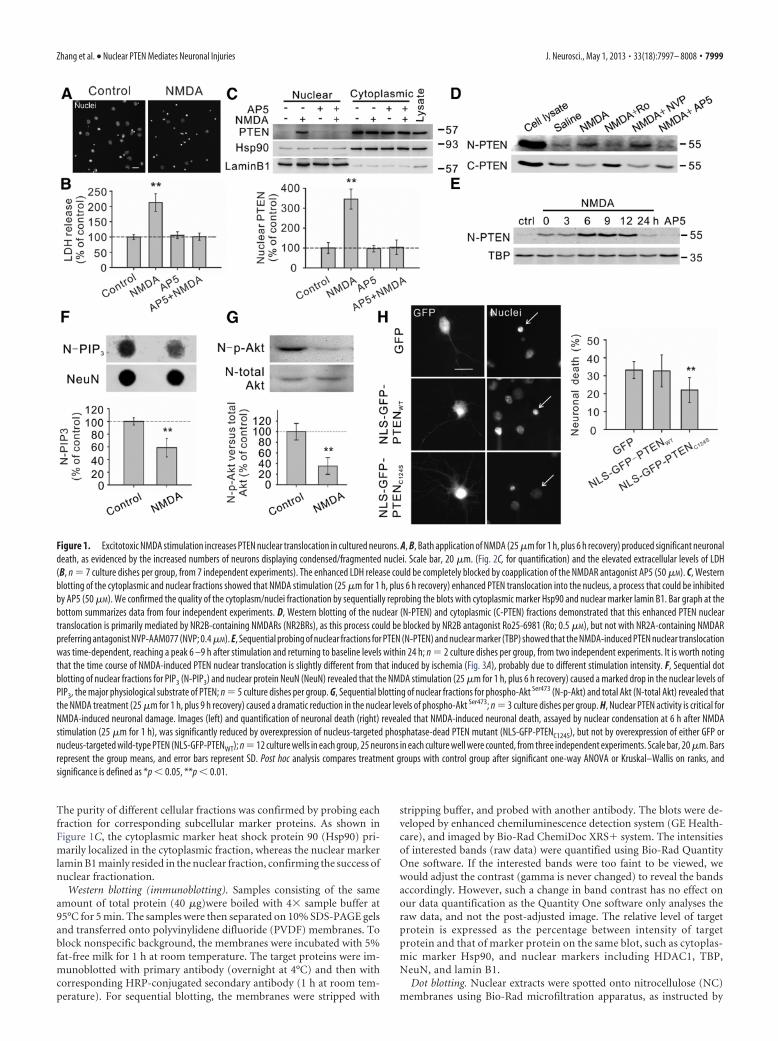
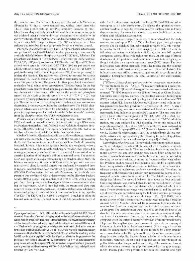
Figure 1. Excitotoxic NMDA stimulation increases PTEN nuclear translocation in cultured neurons. A, B, Bath application of NMDA (25 �m for 1 h, plus 6 h recovery) produced significant neuronaldeath, as evidenced by the increased numbers of neurons displaying condensed/fragmented nuclei. Scale bar, 20 �m. (Fig. 2C, for quantification) and the elevated extracellular levels of LDH(B, n � 7 culture dishes per group, from 7 independent experiments). The enhanced LDH release could be completely blocked by coapplication of the NMDAR antagonist AP5 (50 �M). C, Westernblotting of the cytoplasmic and nuclear fractions showed that NMDA stimulation (25 �m for 1 h, plus 6 h recovery) enhanced PTEN translocation into the nucleus, a process that could be inhibitedby AP5 (50 �M). We confirmed the quality of the cytoplasm/nuclei fractionation by sequentially reprobing the blots with cytoplasmic marker Hsp90 and nuclear marker lamin B1. Bar graph at thebottom summarizes data from four independent experiments. D, Western blotting of the nuclear (N-PTEN) and cytoplasmic (C-PTEN) fractions demonstrated that this enhanced PTEN nucleartranslocation is primarily mediated by NR2B-containing NMDARs (NR2BRs), as this process could be blocked by NR2B antagonist Ro25-6981 (Ro; 0.5 �M), but not with NR2A-containing NMDARpreferring antagonist NVP-AAM077 (NVP; 0.4 �M). E, Sequential probing of nuclear fractions for PTEN (N-PTEN) and nuclear marker (TBP) showed that the NMDA-induced PTEN nuclear translocationwas time-dependent, reaching a peak 6 –9 h after stimulation and returning to baseline levels within 24 h; n � 2 culture dishes per group, from two independent experiments. It is worth notingthat the time course of NMDA-induced PTEN nuclear translocation is slightly different from that induced by ischemia (Fig. 3A), probably due to different stimulation intensity. F, Sequential dotblotting of nuclear fractions for PIP3 (N-PIP3) and nuclear protein NeuN (NeuN) revealed that the NMDA stimulation (25 �m for 1 h, plus 6 h recovery) caused a marked drop in the nuclear levels ofPIP3, the major physiological substrate of PTEN; n � 5 culture dishes per group. G, Sequential blotting of nuclear fractions for phospho-Akt Ser473 (N-p-Akt) and total Akt (N-total Akt) revealed thatthe NMDA treatment (25 �m for 1 h, plus 9 h recovery) caused a dramatic reduction in the nuclear levels of phospho-Akt Ser473; n � 3 culture dishes per group. H, Nuclear PTEN activity is critical forNMDA-induced neuronal damage. Images (left) and quantification of neuronal death (right) revealed that NMDA-induced neuronal death, assayed by nuclear condensation at 6 h after NMDAstimulation (25 �m for 1 h), was significantly reduced by overexpression of nucleus-targeted phosphatase-dead PTEN mutant (NLS-GFP-PTENC124S), but not by overexpression of either GFP ornucleus-targeted wild-type PTEN (NLS-GFP-PTENWT); n � 12 culture wells in each group, 25 neurons in each culture well were counted, from three independent experiments. Scale bar, 20 �m. Barsrepresent the group means, and error bars represent SD. Post hoc analysis compares treatment groups with control group after significant one-way ANOVA or Kruskal–Wallis on ranks, andsignificance is defined as *p � 0.05, **p � 0.01.
Zhang et al. • Nuclear PTEN Mediates Neuronal Injuries J. Neurosci., May 1, 2013 • 33(18):7997– 8008 • 7999
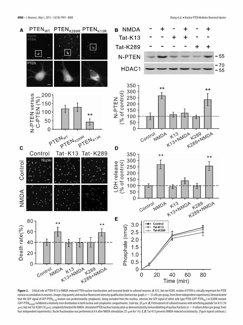
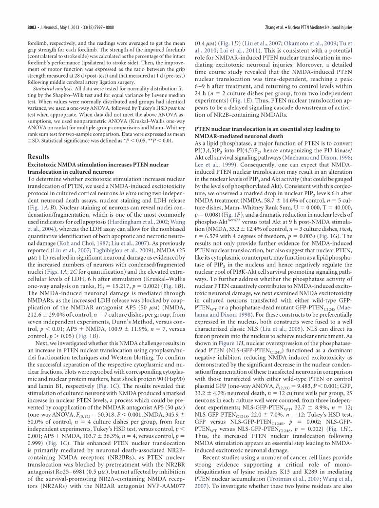
Figure 2. Critical role of PTEN K13 in NMDA-induced PTEN nuclear translocation and neuronal death in cultured neurons. A, K13, but not K289, residue of PTEN is critically important for PTENnuclear accumulation in neurons. Images (top panels) and nuclear fluorescent intensity qualification (bottom bar graph; n � 33 cells per group, from three independent experiments) demonstratedthat the GFP signal of GFP-PTENK13R mutant was predominantly cytoplasmic, being excluded from the nucleus, whereas the GFP signal of either wile type PTEN (GFP-PTENWT) or K289R mutant(GFP-PTENK289R) exhibited a relatively even distribution in both nuclear and cytoplasmic compartments. Scale bar, 20 �m. B, Pretreatment of cultured neurons with interfering peptide Tat-K13 (10�M), but not Tat-K289 (10 �M), completely blocked the NMDA-stimulated PTEN nuclear translocation as demonstrated by immunoblotting of nuclear fractions (n � 4 culture dishes per group, fromfour independent experiments). Nuclei fractionation was performed at 6 h after NMDA stimulation (25 �m for 1 h). C, D, Tat-K13 prevents NMDA-induced excitotoxicity. (Figure legend continues.)
8000 • J. Neurosci., May 1, 2013 • 33(18):7997– 8008 Zhang et al. • Nuclear PTEN Mediates Neuronal Injuries
the manufacturer. The NC membranes were blocked with 5% bovinealbumin for 60 min at room temperature, washed three times withTBS-T, then probed with an anti-PIP3 antibody, followed by HRP-labeled secondary antibody. Visualization of the immunoreactive spotswas achieved using a chemiluminescent detection system similar to theabove Western blotting method, whereas densitometric analysis was per-formed using NIH ImageJ software. The NC membranes were laterstripped and reprobed for nuclear protein NeuN as a loading control.
PTEN phosphatase activity assay. The PTEN phosphatase activity assaywas performed in a 96-well flat-bottom clear plate using a commerciallyavailable Malachite Green Phosphatase Assay Kit (K-1500, Echelon). Thephosphate standards (0 � 3 nmol/well), assay controls [buffer control,PI(3,4,5)P3 (PIP3) only control and PTEN-only control], and PTEN re-actions were setup in triplicates. GST-PTEN (100 ng per assay) wasadded, without or with either peptide Tat-K13 (10 �M) or Tat-K289 (10�M), to the assay buffer containing substrate PIP3 (3 nmol per assay) toinitiate the reaction. The reaction was allowed to proceed for variousperiods of 10, 40, or 80 min at 37°C and then terminated with 100 �l ofmalachite green solution. The green color (free phosphate) was allowedto develop for 20 min at room temperature. The absorbance for the freephosphate was measured at 620 nm in a plate reader. The standard curvewas drawn with absorbance (620 nm) on the y-axis and phosphate(nmol) on the x-axis. A best-fit curve (Y � A � B � X � C � X 2) wasgenerated by using polynomial second order nonlinear regression anal-ysis. The concentration of free phosphate in each reaction or control wasdetermined by interpolation from the standard curve. The PTEN phos-phatase activity was determined by subtracting the background phos-phate (average free phosphate value from the “substrate-only” controls)from the phosphate release by PTEN phosphatase action.
Primary culture transfection. Mature hippocampal neurons (10 –12DIV) cultured on coverslips were transfected with target DNA usingProFection Mammalian Transfection System-Calcium Phosphate (Pro-mega, PRE1200). Following transfection, neurons were returned to theincubator for an additional 48 h until further experiments.
Cerebral ischemia. All animals used in the study were housed, cared for,and used experimentally in accordance with the protocols approved bythe Ethical Committee for Animal Research at China Medical UniversityHospital, Taiwan. Adult male Sprague Dawley rats weighting �200 gwere anesthetized, and the middle cerebral artery (MCA) was exposed bymaking a craniotomy window (2 mm in diameter) 1 mm rostral to theanterior junction of the zygoma and the squamosal bone. The exposedMCA was ligated with a square knot using a 10-0 nylon suture. Next, thebilateral common carotid arteries (CCAs) were clamped with nontrau-matic arterial clips. Successful surgery was confirmed by a marked dropin regional cerebral blood flow, monitored by a laser Doppler flowmeter(PF-5010, Periflux system; Perimed AB). Moreover, the core body tem-perature was monitored with a thermometer probe (Hewlett-PackardModel 21090A probe), and maintained at 37.0 � 0.5°C with a heatingpad. Both blood pressure and blood gas levels were also monitored dur-ing the experiment. After 90 min ischemia, the suture and clips wereremoved to allow instant reperfusion. Experimental rats were subdividedinto several groups to receive different treatments of interfering peptides(Tat-K13, Tat-K13R, or Tat-K289 at a dosage of 10 mg/kg) or saline viafemoral vein injection. The first bolus of Tat-K13 was administered at
either 2 or 6 h after stroke onset, whereas Tat-K13R, Tat-K289, and salinewere given at 2 h after stroke onset. To achieve the optimal outcome,another two doses of peptides were administered on the second and thirddays, respectively. Rats were then allowed to recover for different periodsof time until additional experiments.
Magnetic resonance image. The rats were anesthetized and the bodytemperature was kept at 37.0 � 0.5°C with a heating pad during imagingprocess. The T2-weighted spin-echo imaging sequence (T2WI) was per-formed by the 3.0 T General Electric imaging system (R4, GE) with thefollowing parameters: repetition time, 4000 ms; echo time, 105 ms; 6 – 8contiguous coronal slices with each 2 mm thick. At this stage of strokedevelopment (7 d post-ischemia), brain infarct manifests as high signal(bright white) on the magnetic resonance image (MRI) images. The non-infarct areas were drawn manually from slice to slice and the volumeswere measured with Voxtool analysis software (General Electric). Theinfarct size was quantified by subtracting the noninfarct volume of theischemic hemisphere from the total volume of the contralateralhemisphere.
[18F]fluoro-2-deoxyglucose positron emission tomography examination.18F was produced by the 18O(p, n) 18F nuclear reaction in a cyclotron,and 18F-FDG ([ 18F]fluoro-2-deoxyglucose) was synthesized with an au-tomated 18F-FDG synthesis system (Nihon Kokan) at China MedicalUniversity and Hospital, Taiwan. The positron emission tomography(PET) scanning was performed by a high-resolution small-animal PETscanner (microPET, Rodent R4, Concorde Microsystems) with the sys-tem parameters described previously (Carmichael et al., 2004). At day 7post-stroke surgery, rats were anesthetized and fixed in a customizedstereotactic head holder in the microPET scanner. The rats were thengiven a bolus intravenous injection of 18F-FDG (200 –250 �Ci/rat) dis-solved in 0.5 ml of saline. Immediately following the 18F-FDG adminis-tration, rats were subjected to PET scanning for 60 min using a 3-Dacquisition protocol (Shyu et al., 2008). Data collected were analyzed byInteractive Data Language (IDL) ver. 5.5 (Research Systems) and ASIProver. 3.2 (Concorde Microsystems). Last, the deficit of brain glucose met-abolic activity was expressed using the following formula: [1-ipsilateralhemisphere signal/contralateral hemisphere signal] � 100%.
Neurological behavioral tests. Three typical sensorimotor deficit assess-ments were designed to evaluate the functional recovery of neural circuitsdamaged by ischemic insult. They are the biased body swing test, loco-motor activity test, and grip strength test. Biased body swing test is ameasurement of body asymmetric motor behavior. It was performed byelevating the rat by its tail and counting the frequency of its swing behav-ior. Previous studies revealed that ischemic rats exhibit a significantlybiased swing activity with the direction contralateral to the ischemic side,whereas the native rats have no preference for either side. Therefore, thefrequency of the biased swing activity may represent the degree of neu-rological deficits caused by ischemic stroke. The detailed experimentaldesign is as follows. The rat was lifted to �1 inch above the floor by its tail.One swing behavior was counted when the rat moved its head 10° fromthe vertical axis to either the contralateral side or ipsilateral side of isch-emia. Twenty continuous swings were counted in total, and the percent-age of recovery was normalized using the following formula: percentagerecovery � [1 � (No. of contralateral swings � 10)/10] � 100%. Loco-motor activity of the ischemic rats was monitored using the VersaMaxAnimal Activity Monitor obtained from Accuscan Instruments. Themonitor has 16 horizontal x-y and eight vertical z infrared sensors spaced2.5 cm apart. The vertical sensors were situated 10 cm from the floor of thechamber. The ischemic rat was placed in the recording chamber at night,and its vertical movement time (seconds) was automatically recorded bycomputer when the infrared beams were broken by its movement over a2 h period. The length of its vertical movement represents the recovery oflocomotor circuits injured by ischemic stroke. Grip strength is anotherindex for testing motor functions. It was recorded by a grip strengthmeter manufactured by TSE Systems. Briefly, the rat was mounted ontothe grip sensor and pulled backwards gently by its tail with a continuousmovement. Naturally, the rat clung to the grip sensor firmly to resist thepull until it could no longer hold on and let it go. The maximum force atwhich the animal released the grip was recorded by the grip strengthmeter. Twenty pulls were performed for both left forelimb and right
4
(Figure legend continued.) Tat-K13 (10 �M), but not the control peptide Tat-K289 (10 �M),decreased the number of neurons displaying nuclei condensation/fragmentation (C, n � 8culture wells per group, from three independent experiments, four visual fields were randomlyselected from each culture well and averaged; scale bar, 20 �m) and reduced LDH release (D,n � 27 culture wells per group, from three independent experiments). Both assays were per-formed at 6 h after NMDA stimulation (25 �m for 1 h). E, In vitro PTEN lipid phosphatase activityassays revealed that within the concentration tested (10 �M), neither the interfering peptideTat-K13 nor the control peptide Tat-K289 had significant effects on the production of freephosphates from the lipid substrate PIP3 by recombinant PTEN (n � 3). Bars represent thegroup means, and error bars represent SD. Post hoc analysis compares treatment groups withcontrol group after significant one-way ANOVA or Kruskal–Wallis on ranks, and significance isdefined as *p � 0.05, **p � 0.01.
Zhang et al. • Nuclear PTEN Mediates Neuronal Injuries J. Neurosci., May 1, 2013 • 33(18):7997– 8008 • 8001
forelimb, respectively, and the readings were averaged to get the meangrip strength for each forelimb. The strength of the impaired forelimb(contralateral to stroke side) was calculated as the percentage of the intactforelimb’s performance (ipsilateral to stroke side). Then, the improve-ment of motor function was expressed as the ratio between the gripstrength measured at 28 d (post-test) and that measured at 1 d (pre-test)following middle cerebral artery ligation surgery.
Statistical analysis. All data were tested for normality distribution fit-ting by the Shapiro–Wilk test and for equal variance by Levene mediantest. When values were normally distributed and groups had identicalvariance, we used a one-way ANOVA, followed by Tukey’s HSD post hoctest when appropriate. When data did not meet the above ANOVA as-sumptions, we used nonparametric ANOVA (Kruskal–Wallis one-wayANOVA on ranks) for multiple-group comparisons and Mann–Whitneyrank sum test for two-sample comparison. Data were expressed as mean�SD. Statistical significance was defined as *P � 0.05, **P � 0.01.
ResultsExcitotoxic NMDA stimulation increases PTEN nucleartranslocation in cultured neuronsTo determine whether excitotoxic stimulation increases nucleartranslocation of PTEN, we used a NMDA-induced excitotoxicityprotocol in cultured cortical neurons in vitro using two indepen-dent neuronal death assays, nuclear staining and LDH release(Fig. 1A,B). Nuclear staining of neurons can reveal nuclei con-densation/fragmentation, which is one of the most commonlyused indicators for cell apoptosis (Hardingham et al., 2002; Wanget al., 2004), whereas the LDH assay can allow for the nonbiasedquantitative identification of both apoptotic and necrotic neuro-nal damage (Koh and Choi, 1987; Liu et al., 2007). As previouslyreported (Liu et al., 2007; Taghibiglou et al., 2009), NMDA (25�M; 1 h) resulted in significant neuronal damage as evidenced bythe increased numbers of neurons with condensed/fragmentednuclei (Figs. 1A, 2C for quantification) and the elevated extra-cellular levels of LDH, 6 h after stimulation (Kruskal–Wallisone-way analysis on ranks, H3 � 15.217, p � 0.002) (Fig. 1B).The NMDA-induced neuronal damage is mediated throughNMDARs, as the increased LDH release was blocked by coap-plication of the NMDAR antagonist AP5 (50 �M) (NMDA,212.6 � 29.0% of control, n � 7 culture dishes per group, fromseven independent experiments, Dunn’s Method, versus con-trol, p � 0.01; AP5 � NMDA, 100.9 � 11.9%, n � 7, versuscontrol, p 0.05) (Fig. 1B).
Next, we investigated whether this NMDA challenge results inan increase in PTEN nuclear translocation using cytoplasm/nu-clei fractionation techniques and Western blotting. To confirmthe successful separation of the respective cytoplasmic and nu-clear fractions, blots were reprobed with corresponding cytoplas-mic and nuclear protein markers, heat shock protein 90 (Hsp90)and lamin B1, respectively (Fig. 1C). The results revealed thatstimulation of cultured neurons with NMDA produced a markedincrease in nuclear PTEN levels, a process which could be pre-vented by coapplication of the NMDAR antagonist AP5 (50 �M)(one-way ANOVA, F(3,12) � 50.318, P � 0.001; NMDA, 345.9 �50.0% of control, n � 4 culture dishes per group, from fourindependent experiments, Tukey’s HSD test, versus control, p �0.001; AP5 � NMDA, 103.7 � 36.3%, n � 4, versus control, p �0.999) (Fig. 1C). This enhanced PTEN nuclear translocationis primarily mediated by neuronal death-associated NR2B-containing NMDA receptors (NR2BRs), as PTEN nucleartranslocation was blocked by pretreatment with the NR2BRantagonist Ro25– 6981 (0.5 �M), but not affected by inhibitionof the survival-promoting NR2A-containing NMDA recep-tors (NR2ARs) with the NR2AR antagonist NVP-AAM077
(0.4 �M) (Fig. 1D) (Liu et al., 2007; Okamoto et al., 2009; Tu etal., 2010; Lai et al., 2011). This is consistent with a potentialrole for NMDAR-induced PTEN nuclear translocation in me-diating excitotoxic neuronal injuries. Moreover, a detailedtime course study revealed that the NMDA-induced PTENnuclear translocation was time-dependent, reaching a peak6 –9 h after treatment, and returning to control levels within24 h (n � 2 culture dishes per group, from two independentexperiments) (Fig. 1E). Thus, PTEN nuclear translocation ap-pears to be a delayed signaling cascade downstream of activa-tion of NR2B-containing NMDARs.
PTEN nuclear translocation is an essential step leading toNMDAR-mediated neuronal deathAs a lipid phosphatase, a major function of PTEN is to convertPI(3,4,5)P3 into PI(4,5)P2, hence antagonizing the PI3 kinase/Akt cell survival signaling pathways (Maehama and Dixon, 1998;Lee et al., 1999). Consequently, one can expect that NMDA-induced PTEN nuclear translocation may result in an alterationin the nuclear levels of PIP3 and Akt activity (that could be gaugedby the levels of phosphorylated Akt). Consistent with this conjec-ture, we observed a marked drop in nuclear PIP3 levels 6 h afterNMDA treatment (NMDA, 58.7 � 14.6% of control, n � 5 cul-ture dishes, Mann–Whitney Rank Sum, U � 0.000, T � 40.000,p � 0.008) (Fig. 1F), and a dramatic reduction in nuclear levels ofphospho-Akt Ser473 versus total Akt at 9 h post-NMDA stimula-tion (NMDA, 33.2 � 12.4% of control, n � 3 culture dishes, t test,t � 6.579 with 4 degrees of freedom, p � 0.003) (Fig. 1G). Theresults not only provide further evidence for NMDA-inducedPTEN nuclear translocation, but also suggest that nuclear PTEN,like its cytoplasmic counterpart, may function as a lipid phospha-tase of PIP3 in the nucleus and hence negatively regulate thenuclear pool of PI3K-Akt cell survival promoting signaling path-ways. To further address whether the phosphatase activity ofnuclear PTEN causatively contributes to NMDA-induced excito-toxic neuronal damage, we next examined NMDA excitotoxicityin cultured neurons transfected with either wild-type GFP-PTENWT or a phosphatase-dead mutant GFP-PTENC124S (Mae-hama and Dixon, 1998). For these constructs to be preferentiallyexpressed in the nucleus, both constructs were fused to a wellcharacterized classic NLS (Liu et al., 2005). NLS can direct itsfusion protein into the nucleus to achieve nuclear enrichment. Asshown in Figure 1H, nuclear overexpression of the phosphatase-dead PTEN (NLS-GFP-PTENC124S) functioned as a dominantnegative inhibitor, reducing NMDA-induced excitotoxicity asdemonstrated by the significant decrease in the nuclear conden-sation/fragmentation of these transfected neurons in comparisonwith those transfected with either wild-type PTEN or controlplasmid GFP (one-way ANOVA, F(2,33) � 9.483, P � 0.001; GFP,33.2 � 4.7% neuronal death, n � 12 culture wells per group, 25neurons in each culture well were counted, from three indepen-dent experiments; NLS-GFP-PTENWT, 32.7 � 8.9%, n � 12;NLS-GFP-PTENC124S, 22.0 � 7.0%, n � 12; Tukey’s HSD test,GFP versus NLS-GFP-PTENC124S, p � 0.002; NLS-GFP-PTENWT versus NLS-GFP-PTENC124S, p � 0.002) (Fig. 1H).Thus, the increased PTEN nuclear translocation followingNMDA stimulation appears an essential step leading to NMDA-induced excitotoxic neuronal damage.
Recent studies using a number of cancer cell lines providestrong evidence supporting a critical role of mono-ubiquitination of lysine residues K13 and K289 in mediatingPTEN nuclear accumulation (Trotman et al., 2007; Wang et al.,2007). To investigate whether these two lysine residues are also
8002 • J. Neurosci., May 1, 2013 • 33(18):7997– 8008 Zhang et al. • Nuclear PTEN Mediates Neuronal Injuries
essential for neuronal PTEN nuclear translocation, neurons weretransfected with PTEN mutants containing a lysine-to-argininemutation at either lysine residue (K13R and K289R). As shown inFigure 2A, in neurons transfected with either the wild-type PTEN(GFP-PTENWT) or K289R mutant (GFP-PTENK289R), the GFPsignal exhibited a relatively even distribution in both nuclear andcytoplasmic compartments (GFP-PTENWT, the ratio betweennuclear and cytoplasmic PTEN signal being 119.0 � 21.8%, n �33 neurons per group, from three independent experiments;GFP-PTENK289R, 128.2 � 24.9%, n � 33) (Fig. 2A). In contrast, inneurons transfected with the K13R mutation (GFP-PTENK13R), theGFP signal was predominantly cytoplasmic, being excluded fromthe nucleus (GFP-PTENK13R, 42.1 � 18.2%, n � 33; Kruskal–Wallis one-way analysis on ranks, H2 � 64.507, P � � 0.001; posthoc Tukey’s HSD test, GFP-PTENWT versus GFP-PTENK289R,p 0.05; GFP-PTENWT versus GFP-PTENK13R, p � 0.01) (Fig.2A). These results strongly suggest that unlike previous observa-tions in cancer cell lines (Trotman et al., 2007; Wang et al., 2007),only the K13 residue (but not the K289 residue) appears criticallyimportant for PTEN nuclear accumulation in neurons.
To further confirm the essential role of K13 residue in NMDA-induced PTEN nuclear localization, we next designed two interfer-ing peptides that flank the K13 residue (KEIVSRNK13RRYQED)and K289 residue (GPEETSEK289VENGS) of PTEN, respectively.We hypothesize that the K13 peptide may competitively inhibitPTEN mono-ubiquitination at the K13 residue, and hence blockPTEN nuclear translocation in neurons. In contrast, as the K289residue is not required for PTEN nuclear translocation in neurons(Fig. 2A), the K289 peptide may not affect PTEN nuclear transloca-tion. Both peptides were rendered membrane permeable byfusion of them with the cell-membrane transduction domainof the human immunodeficiency virus-type 1 (HIV-1) Tatprotein (GRKKRRQRRR) (Schwarze et al., 1999; Aarts et al.,2002; Borsello et al., 2003; Taghibiglou et al., 2009). Consistentwith our predictions, the results of nuclei/cytoplasm fraction-ation on primary neuronal cultures demonstrated that theNMDA-triggered PTEN nuclear accumulation could be abol-ished by Tat-K13 coapplication (10 �M), but not by Tat-K289 (10�M) (one-way ANOVA, F(5,18) � 23.809, p � 0.001; NMDA,267.3 � 25.5% of control, n � 4 culture dishes per group, fromfour independent experiments, Tukey’s HSD test, versus con-trol, p � 0.001; K13�NMDA, 126.2 � 34.1%, n � 4, versuscontrol, p � 0.828; K289 � NMDA, 234.0 � 49.6%, n � 4,versus control, p � 0.001) (Fig. 2B). These results further confirmthat the K13 residue (but not the K289 residue) is critically im-portant for NMDA-induced PTEN nuclear translocation in neu-rons, and indicate that the Tat-K13 peptide is an effectiveinhibitor for blocking PTEN nuclear translocation.
The ability of Tat-K13 to efficiently prevent NMDA-inducedPTEN nuclear translocation suggests that it may be an effectiveneuroprotectant for excitotoxic neuronal injuries. We first testedthis in neuronal cultures undergoing NMDA-induced excito-toxic neuronal damage using both nuclear condensation (Fig.2C) and LDH release (Fig. 2D) assays. Our results revealed thatpretreatment of the cultured neurons with Tat-K13 (10 �M), butnot Tat-K289 (10 �M), prevented the NMDAR-mediated neuro-nal damage. As shown in Figure 2C, NMDA treatment triggered asignificant increase in the percentage of neurons displaying con-densed/fragmented nuclei (one-way ANOVA, F(5,42) � 14.254,p � 0.001; control, 40.3 � 6.1% neuronal death, n � 8 culturewells per group, from three independent experiments; NMDA,60.0 � 8.4%, n � 8; Tukey’s HSD test, p � 0.001) (Fig. 2C). Thisenhanced apoptosis was blocked by pretreatment of neurons with
Tat-K13, but not with Tat-K289 (K13�NMDA, 39.6 � 7.9%,n � 8, versus control, p � 1.000; K289�NMDA, 58.7 � 6.9%,n � 8, versus control, p � 0.001) (Fig. 2C). Moreover, evidencefrom the LDH assay also confirmed that Tat-K13 possessed astrong neuroprotective effect against NMDA insult (Kruskal–Wallis one-way analysis on ranks, H5 � 128.957, P � � 0.001)(Fig. 2D). The NMDA-triggered LDH release was prevented bycoapplication of Tat-K13, but not Tat-K289 (NMDA, 270.0 �34.6% of control, n � 27 culture wells per group, from 3 inde-pendent experiments, Tukey’s HSD test, versus control, p � 0.01;K13�NMDA, 139.0 � 24.2%, n � 27, versus control, p 0.05;K289�NMDA, 256.5 � 35.4%, n � 27, versus control, p � 0.01)(Fig. 2D). To further confirm that the neuroprotective effect ofTat-K13 is primarily mediated by inhibiting the PTEN nucleartranslocation rather than affecting PTEN�s lipid phosphatase ac-tivity in the cytoplasm, we conducted an in vitro PTEN lipidphosphatase activity assay using the malachite green method. Theresults showed that within the concentration tested (10 �M), nei-ther the interfering peptide Tat-K13 nor the control peptide Tat-K289 had any notable effect on the lipid phosphatase activity ofPTEN toward PIP3, suggesting that the newly developed Tat-K13interfering peptide likely represents a novel therapeutic neuro-protectant which may avoid potential side effects due to alteringenzymatic functions of PTEN (Fig. 2E).
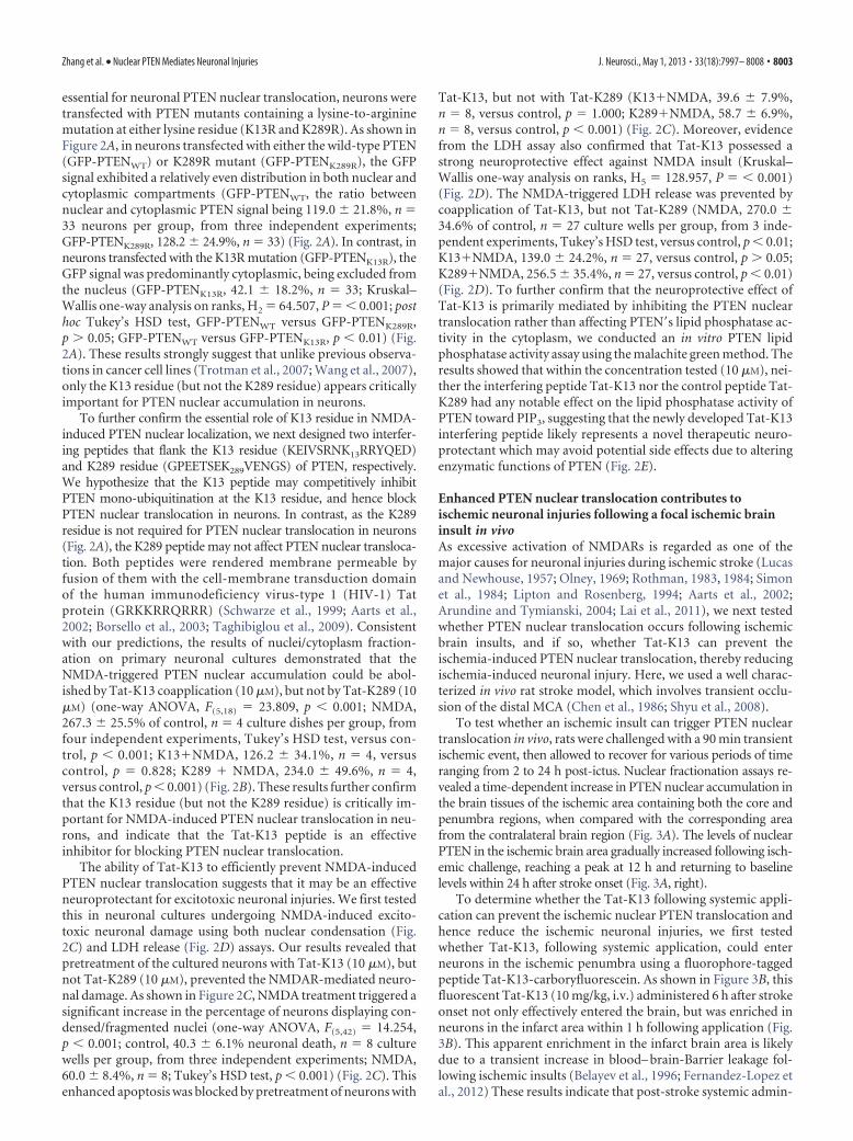
Enhanced PTEN nuclear translocation contributes toischemic neuronal injuries following a focal ischemic braininsult in vivoAs excessive activation of NMDARs is regarded as one of themajor causes for neuronal injuries during ischemic stroke (Lucasand Newhouse, 1957; Olney, 1969; Rothman, 1983, 1984; Simonet al., 1984; Lipton and Rosenberg, 1994; Aarts et al., 2002;Arundine and Tymianski, 2004; Lai et al., 2011), we next testedwhether PTEN nuclear translocation occurs following ischemicbrain insults, and if so, whether Tat-K13 can prevent theischemia-induced PTEN nuclear translocation, thereby reducingischemia-induced neuronal injury. Here, we used a well charac-terized in vivo rat stroke model, which involves transient occlu-sion of the distal MCA (Chen et al., 1986; Shyu et al., 2008).
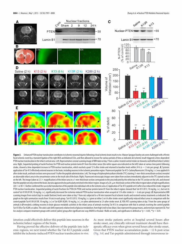
To test whether an ischemic insult can trigger PTEN nucleartranslocation in vivo, rats were challenged with a 90 min transientischemic event, then allowed to recover for various periods of timeranging from 2 to 24 h post-ictus. Nuclear fractionation assays re-vealed a time-dependent increase in PTEN nuclear accumulation inthe brain tissues of the ischemic area containing both the core andpenumbra regions, when compared with the corresponding areafrom the contralateral brain region (Fig. 3A). The levels of nuclearPTEN in the ischemic brain area gradually increased following isch-emic challenge, reaching a peak at 12 h and returning to baselinelevels within 24 h after stroke onset (Fig. 3A, right).
To determine whether the Tat-K13 following systemic appli-cation can prevent the ischemic nuclear PTEN translocation andhence reduce the ischemic neuronal injuries, we first testedwhether Tat-K13, following systemic application, could enterneurons in the ischemic penumbra using a fluorophore-taggedpeptide Tat-K13-carboryfluorescein. As shown in Figure 3B, thisfluorescent Tat-K13 (10 mg/kg, i.v.) administered 6 h after strokeonset not only effectively entered the brain, but was enriched inneurons in the infarct area within 1 h following application (Fig.3B). This apparent enrichment in the infarct brain area is likelydue to a transient increase in blood– brain-Barrier leakage fol-lowing ischemic insults (Belayev et al., 1996; Fernandez-Lopez etal., 2012) These results indicate that post-stroke systemic admin-
Zhang et al. • Nuclear PTEN Mediates Neuronal Injuries J. Neurosci., May 1, 2013 • 33(18):7997– 8008 • 8003
istration could effectively deliver this peptide into neurons in theischemic/infarct regions of the brain.
Having proved the effective delivery of the peptide into isch-emic regions, we next tested whether the Tat-K13 peptide couldinhibit the ischemia-induced PTEN nuclear translocation in vivo.
As most stroke patients arrive at hospital several hours afterstroke onset, any clinically relevant treatment should have ther-apeutic efficacy even when given several hours after stroke onset.Given that PTEN nuclear accumulation peaks �12 h post-ictus(Fig. 3A) and Tat-peptide administered through intravenous in-
Figure 3. Enhanced PTEN nuclear translocation contributes to ischemic neuronal injuries following a focal ischemic brain insult in vivo. Mature Sprague Dawley rats were challenged with a 90 minfocal ischemic event by a transient ligation of the right MCA and bilateral CCAs, and then allowed to recover for various periods of time as indicated. A, Ischemic insult triggered a time-dependentPTEN nuclear translocation in the infarct cortical area. Left, Representative coronal scanning image of MRI taken at day 7 from a saline-treated control stroke rat showed a well defined infarct (white)area. Right, Sequential probing of nuclei fractions for PTEN and nuclear protein lamin B1 from the infarct areas (the white square area indicated on the left) taken at various time points followingstroke, showed a time-dependent increase in PTEN nuclear translocation, which reached a peak 12 h after stroke and returned to baseline levels within 24 h (n � 2 rats per group). B, Systemicapplication of Tat-K13 effectively entered neurons in the brain, including neurons in the ischemic penumbra region. Fluorescent peptide Tat-K13-Carboryfluorescein (10 mg/kg, i.v.) was applied 6 hafter stroke insult, and brain sections were processed 1 h after the peptide administration. Left, The image of triphenyltetrazolium chloride (TTC) staining (1-mm-thick coronal brain section) revealedan observable infarct area in the sensorimotor cortex in the insult side of the brain. Right, Fluorescent microscopic images were taken from sections immediately adjacent to the TTC stained sectionon the left. The image (taken at 2.5� magnification) of the infarct area in a 1-mm-thick brain section corresponds to the area indicated by the white box in the TTC section on the Left, and showedthat the peptide not only entered the brain, but also appeared to be particularly enriched in the infarct region. Images of a 20-�m-thick brain section of the infarct region taken at high magnifications(20� or 40�) further confirmed the successful transduction of the peptide into individual cells in the ischemic area. C, Application of Tat-K13 peptide at 6 h after ictus reduced the stroke-triggeredPTEN nuclear translocation. Sequential probing of nuclei fractions for PTEN (N-PTEN) and nuclear protein lamin B1 from the infarct regions showed that Tat-K13 (K13; 10 mg/kg, i.v.), but not itscontrol Tat-K13R (K13R; 10 mg/kg, i.v.), significantly decreased the ischemia-induced PTEN nuclear translocation when assayed at 12 h after stroke (n � 6 rats per group). D, Representative MRIscanning images from rostral (top) to caudal (bottom) levels were taken at day 7 from rats subjected to a 90 min transient focal ischemic insults and received various treatments as indicated. Bargraph on the right summarizes results from 10 rats in each group. Tat-K13 (K13; 10 mg/kg, i.v.) given at either 2 or 6 h after stroke-onset significantly reduced infarct areas in comparison with thecontrol peptide Tat-K13R (K13R; 10 mg/kg, i.v.) or Tat-K289 (K289; 10 mg/kg, i.v.), or saline administered at 2 h after stroke onset. E, FDG-PET scanning taken at day 7 from the same groups ofanimals in D revealed a striking recovery in brain glucose metabolic activities in the infarct areas of animals receiving Tat-K13 in comparison with that in animals receiving the control peptideTat-K13R or Tat-K289, or saline. The color scale (left) represents relative levels of glucose metabolism, from high (red) to low (blue). Bars represent the group means, and error bars represent SD. Posthoc analysis compares treatment groups with control (saline) group after significant one-way ANOVA or Kruskal–Wallis on ranks, and significance is defined as *p � 0.05, **p � 0.01.
8004 • J. Neurosci., May 1, 2013 • 33(18):7997– 8008 Zhang et al. • Nuclear PTEN Mediates Neuronal Injuries
jection can quickly reach therapeutic concentrations in the brainwithin 1 h (Fig. 3B) (Aarts et al., 2002; Taghibiglou et al., 2009;Cook et al., 2012), systemic application of Tat-K13 at 6 h post-ictus should allow a sufficient time to inhibit the major portion ofthe ischemia-induced PTEN nuclear translocation. To furtherconfirm the specificity of Tat-K13 peptide, we also developedanother control peptide Tat-K13R in which the lysine 13 residue(K13) was replaced with an arginine (R13). The conversation ofK13 into R13 should render Tat-K13R incapable of competingwith endogenous PTEN mono-ubiquitination on the K13 site,and hence function as an inactive control for Tat-K13. As pre-dicted, intravenous application of Tat-K13, but not Tat-K13R(10 mg/kg, i.v.) at 6 h after stroke onset significantly suppressedthe ischemia-induced PTEN nuclear translocation when assayed12 h after stroke onset (one-way ANOVA, F(2,15) � 24.033,P � 0.001) (Fig. 3C). Nuclear PTEN levels in the Tat-K13-treatedrats was reduced to 65.3 � 7.6% of the saline-treated group,whereas nuclear PTEN levels in the Tat-K13R-treated rats werestatistically similar to saline controls (saline, n � 6 rats per group;K13, n � 6; Tukey’s HSD test, p � 0.001; K13R, 88.8 � 7.3% ofsaline control, n � 6, versus control, p � 0.106) (Fig. 3C). Theseresults confirmed that PTEN nuclear translocation is a relativelydelayed cellular process following stroke onset, one that can beinhibited by a post-stroke treatment with Tat-K13.
To examine the causative link between ischemia-inducedPTEN nuclear translocation and ischemic neuronal death, weinhibited PTEN nuclear translocation with systemic intravenousadministration of Tat-K13, or with Tat-K13R or Tat-K289 serv-ing as controls. We administered the first dose of Tat-K13 (10mg/kg, i.v.) to treatment groups at either 2 or 6 h post-ictus, andgave Tat-K13R or Tat-K289 (10 mg/kg, i.v.) to control groups at2 h post-ictus. To increase the efficacy of the peptide, we also gavetwo additional doses of each peptide at days 2 and 3 followingstroke. The long-lasting morphological neuroprotective effects ofthe peptides were then assessed using MRI 7 d after stroke onset.As shown in Figure 3D, MRI scanning revealed that rats treatedwith Tat-K13, regardless of whether it was given at 2 or 6 h post-ictus, had dramatically reduced brain infarct volumes comparedwith saline-treated rats (Kruskal–Wallis one-way analysis onranks, H4 � 38.639, P � � 0.001; saline control, 170.9 � 18.7mm 3, n � 10 rats per group; K13 (2 h), 67.7 � 17.5 mm 3, n � 10,Tukey’s HSD test, versus saline, p � 0.01; K13 (6 h), 67.4 � 15.9
mm 3, n � 10, versus saline, p � 0.01) (Fig. 3D). In contrast,post-ictus treatment of control peptides (Tat-K13R and Tat-K289), displayed little protective effects against ischemia-induced brain infarction [K13R (2 h), 160.9 � 5.7 mm 3, n � 10,versus saline, p 0.05; K289 (2 h), 151.2 � 8.0 mm 3, n � 10,versus saline, p 0.05] (Fig. 3D).
We then examined whether Tat-K13 also provides a corre-sponding functional protection of neurons from the ischemicinsults by measuring the recovery of neuronal glucose uptakewith FDG-PET 7 d after stroke onset. Consistent with the MRIresults, FDG-PET scanning also revealed a significant recovery inFDG uptake deficits in the ischemic hemisphere in the twogroups of rats treated with Tat-K13 in comparison with ratstreated with saline [one-way ANOVA, F(4,45) � 246.269, p �0.001; saline control, 78.4 � 4.8% deficit, n � 10 rats per group;K13 (2 h), 25.6 � 7.8% deficit, n � 10, Tukey’s HSD test, versussaline, p � 0.001; K13 (6 h), 20.2 � 6.3% deficit, n � 10, versussaline, p � 0.001] (Fig. 3E). In contrast, no obvious recovery wasobserved in rats treated with either control peptide [K13R (2 h),73.2 � 3.4% of deficit, n � 10, versus saline, p � 0.267; K289(2 h), 71.5 � 5.3% of deficit, n � 10, versus saline, p � 0.07](Fig. 3E). Together, MRI and PET acquisitions suggest that Tat-K13, by blocking PTEN nuclear accumulation, strongly protectsneurons against ischemic injuries.
It is clinically important to know whether a neuroprotectant,in addition to providing neuronal protection, will have a long-lasting behavioral benefit in stroke patients. We therefore alsoexamined whether the post-stroke Tat-K13 treatments provideneurobehavioral benefits using three typical sensorimotor assess-ments, the biased body swing, locomotor activity, and gripstrength measurements, which are often used to evaluate thefunctional recovery of cortical neural circuits damaged by focalischemic insult (Dunnett et al., 1998; Chang et al., 2003; Shyu etal., 2008). Over the 4 week recovery period following stroke chal-lenge, the body swing test revealed that rats treated with Tat-K13(both 2 and 6 h groups) exhibited a much faster recovery in thebody asymmetric motor behavior than those treated with salineor either of the control peptides (Fig. 4A). By day 28, rats treatedwith Tat-K13 had largely recovered from the body asymmetrydeficit [Kruskal–Wallis one-way analysis on ranks, H4 � 38.017,P � � 0.001; saline control, 44.4 � 6.0% recovery, n � 10 rats pergroup; K13 (2 h), 90.8 � 2.8% recovery, n � 10, Tukey’s HSD
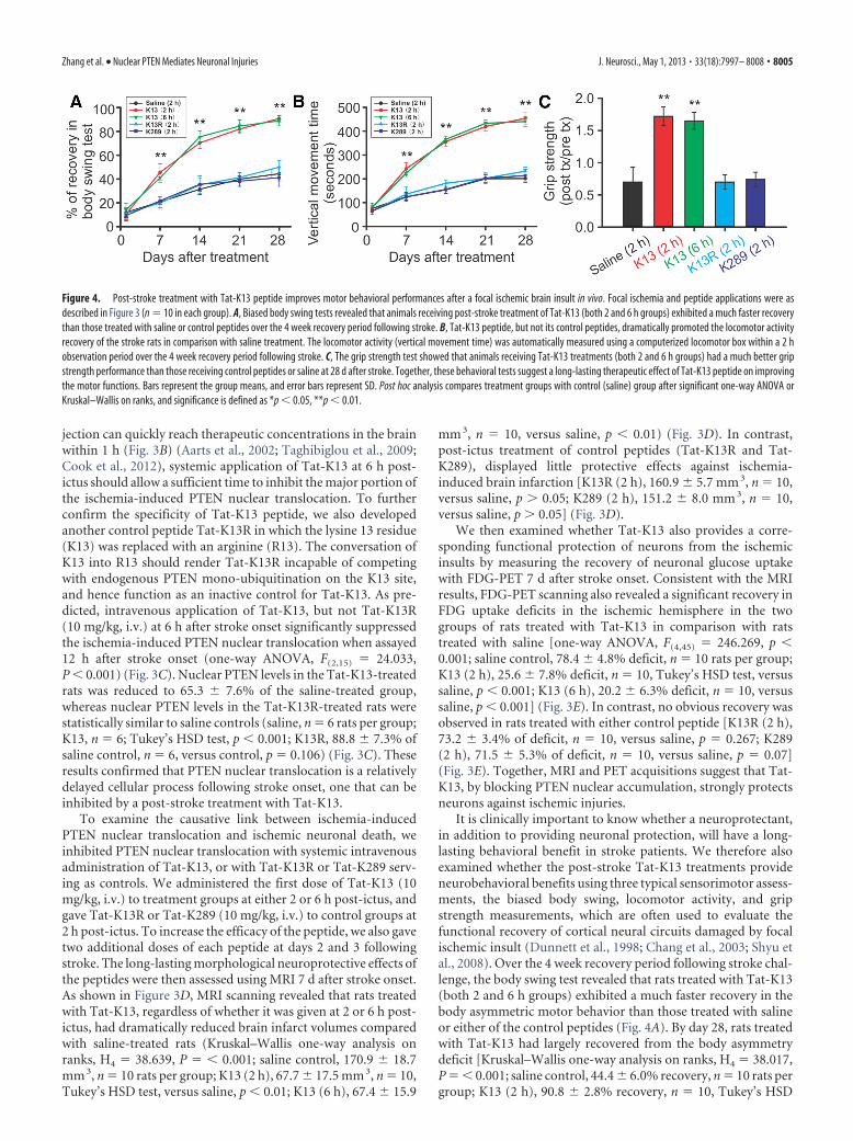
Figure 4. Post-stroke treatment with Tat-K13 peptide improves motor behavioral performances after a focal ischemic brain insult in vivo. Focal ischemia and peptide applications were asdescribed in Figure 3 (n � 10 in each group). A, Biased body swing tests revealed that animals receiving post-stroke treatment of Tat-K13 (both 2 and 6 h groups) exhibited a much faster recoverythan those treated with saline or control peptides over the 4 week recovery period following stroke. B, Tat-K13 peptide, but not its control peptides, dramatically promoted the locomotor activityrecovery of the stroke rats in comparison with saline treatment. The locomotor activity (vertical movement time) was automatically measured using a computerized locomotor box within a 2 hobservation period over the 4 week recovery period following stroke. C, The grip strength test showed that animals receiving Tat-K13 treatments (both 2 and 6 h groups) had a much better gripstrength performance than those receiving control peptides or saline at 28 d after stroke. Together, these behavioral tests suggest a long-lasting therapeutic effect of Tat-K13 peptide on improvingthe motor functions. Bars represent the group means, and error bars represent SD. Post hoc analysis compares treatment groups with control (saline) group after significant one-way ANOVA orKruskal–Wallis on ranks, and significance is defined as *p � 0.05, **p � 0.01.
Zhang et al. • Nuclear PTEN Mediates Neuronal Injuries J. Neurosci., May 1, 2013 • 33(18):7997– 8008 • 8005
test, versus saline, p � 0.01; K13 (6 h), 88.9 � 4.3% recovery, n �10, versus saline, p � 0.01] (Fig. 4A). In contrast, those treatedwith either control peptide (Tat-K13R or Tat-K289) had similarperformance as the saline controls [K13R (2 h), 50.0 � 5.4%recovery, n � 10, versus saline, p 0.05; K289 (2 h), 41.2 � 7.4%recovery, n � 10, versus saline, p 0.05] (Fig. 4A). Similarly,the vertical movement time as monitored in a computerized lo-comotor chamber showed that Tat-K13-treated rats (both 2 and6 h groups) displayed a much faster and more dramatic improve-ment in locomotor activity over the 4 week recovery period incomparison with the saline-treated or control peptide-treatedrats (Fig. 4B). On day 28, a one-way ANOVA analysis indicated asignificant difference among the groups (one-way ANOVA,F(4,45) � 361.269, p � 0.001). Post hoc comparisons (Tukey’sHSD test) revealed that the vertical movement time of Tat-K13-treated rats was significantly longer than that of saline-treatedrats [saline control, 200.9 � 17.2 s, n � 10 rats per group; K13 (2h), 453.6 � 23.7 s, n � 10, versus saline, p � 0.001; K13 (6 h),440.6 � 20.7 s, n � 10, versus saline, p � 0.001] (Fig. 4B). Onthe other hand, the locomotor activity was unaffected by controlpeptide treatments [K13R (2 h), 231.8 � 16.8 s, n � 10, versussaline, p � 0.018; K289 (2 h), 213.0 � 26.1 s, n � 10, versus saline,p � 0.708] (Fig. 4B). Furthermore, results of the grip strength testalso revealed a more dramatic recovery in rats treated with Tat-K13 at 28 d after stroke (Kruskal–Wallis one-way analysis onranks, H4 � 35.840, P � � 0.001) (Fig. 4C). Tat-K13 treatment,regardless at 2 or 6 h post-ictus, dramatically enhanced the gripstrength recovery compared with saline treatment, whereas con-trol peptides exhibited no protective effect [saline control, 0.70 �0.23 post/pre grip strength, n � 10 rats per group; K13 (2 h),1.72 � 0.14, n � 10, Tukey’s HSD test, versus saline, p � 0.01;K13 (6 h), 1.65 � 0.13, n � 10, versus saline, p � 0.01; K13R (2 h),0.70 � 0.11, n � 10, versus saline, p 0.05; K289 (2 h), 0.74 �0.11, n � 10, versus saline, p 0.05] (Fig. 4C). Together, ourresults in the rat model of stroke strongly indicate that Tat-K13not only reduces brain infarct size, but also improves the func-tional neurological circuits damaged by ischemic insults.
DiscussionOur present study identifies increased PTEN nuclear transloca-tion as an essential step in signaling cascades leading to NMDAR/ischemia-mediated neuronal injuries. We provide several piecesof evidence supporting a causative role of PTEN nuclear translo-cation in mediating neuronal injuries: (1) PTEN nuclear translo-cation is downstream of a NR2B-containing NMDAR mediatedcell death signaling pathway (Fig. 1D) (Lai et al., 2011); (2) nu-clear overexpression of dominant negative mutant PTEN reducesNMDAR-mediated excitotoxicity (Fig. 1H); and (3) most impor-tantly, specific blockade of PTEN nuclear translocation with theTat-K13 peptide prevents NMDAR-mediated excitotoxicity incultured neurons (Fig. 2) and protects against ischemic braindamage in vivo (Figs. 3, Fig. 4). Thus, increased PTEN nucleartranslocation appears an essential step leading to excitotoxic/ischemic neuronal death. Our study is in agreement with a recentreport that reduction in nuclear PTEN import promotes nuclearTDP-43-mediated neuronal survival (Zheng et al., 2012). More-over, our study is also consistent with two recent reports on ax-onal regenerative and neuroprotective effects of PTEN ablation(Domanskyi et al., 2011; Sun et al., 2011). The advantage of theneuroprotection induced by Tat-K13 peptide over PTEN abla-tion methods is that Tat-K13 does not interfere with the cytoplas-mic functions of PTEN, whereas deletion of PTEN abrogates allof its functions. It is interesting to note that a recent study using
Nedd4 family-interacting protein 1 (Ndfip1) conditional knock-out animals also reported a parallel reduction in PTEN nucleartranslocation and an increase in neuronal vulnerability followingischemic insults (Howitt et al., 2012). However, as an E3 ligaseadaptor protein, knock-out of Ndfip1 would not only affectPTEN, but numerous substrates of the E3-ligase Nedd4. Thus, itis most likely that the increased neuronal vulnerability observedin Ndfip1�/� is due to its impact on one or more cell survivalsignaling pathways, rather than decreasing PTEN nucleartranslocation.
It is also significant to note that several recent studies alsoreported an essential role of PTEN nuclear translocation inPTEN�s tumor suppressing function. Using several cancer celllines and human cancer specimens, these studies demonstratedthat impaired PTEN nuclear translocation causally contributes totumorigenesis of various types of cancers (Georgescu et al., 2000;Gimm et al., 2000; Whiteman et al., 2002; Walker et al., 2004;Trotman et al., 2007). Recent studies on human glioblastoma andCowden’s disease identified two PTEN mutants (K13E andK289E) which retain normal lipid phosphatase activity but fail totranslocate into the nucleus, leading to impaired PTEN tumorsuppressing ability during tumorigenesis (Georgescu et al., 2000;Walker et al., 2004; Trotman et al., 2007). Even more strikingly, inthe thyroid, normal follicular cells are characterized by strongerPTEN staining in the nucleus than in the cytoplasm, whereas thenuclear staining intensity of PTEN progressively diminishes dur-ing progression from normal to follicular adenoma to carcinoma(Gimm et al., 2000). A similar trend has also been observed inmelanoma progression (Whiteman et al., 2002). These results,along with that presented in this work, clearly establish that nor-mal function of PTEN nuclear translocation is a critical mecha-nism in regulating cell survival and death, one that is sharedbetween tumorigenesis and neurodegeneration. Impaired PTENnuclear translocation may lead to the failure in suppression of cellgrowth, contributing to the pathogenesis of various cancers. Onthe other hand, overexaggerated PTEN nuclear translocation canenhance cell death, leading to neurodegeneration. Thus, furthercharacterization of how PTEN nuclear translocation regulatescell survival and death may lead to a better understanding of themechanisms underlying the pathogenesis of both tumorigenesisand neurodegeneration.
Our current study revealed an essential role of PTEN nucleartranslocation in mediating excitotoxic and ischemic neuronaldeath. However, the detailed mechanisms by which nuclearPTEN increases the vulnerability of neurons to excitotoxicity andischemia remain unclear. Evidence from previous cancer studiessuggests that nuclear PTEN may function as a nuclear PIP3 phos-phatase and consequently antagonize nuclear PI3K/Akt prosur-vival signaling pathways (Gimm et al., 2000; Deleris et al., 2003;Ahn et al., 2004, 2005; Vasko et al., 2004). In pheochromocytomacells (PC12), nuclei extracts pretreated with purified PTENshowed increased DNA fragmentation in response to apoptoticstimulation, which could be rescued by an oversupply of PIP3
(Ahn et al., 2004, 2005). Moreover, during thyroid tumorigene-sis, the level of nuclear PTEN was found to be inversely correlatedto that of activated nuclear Akt during the progression from thenormal thyroid follicular cells to adenoma to carcinoma (Gimmet al., 2000; Vasko et al., 2004). These results are in harmony withour observation that NMDA stimulation resulted in a sharp dropin nuclear levels of PIP3 and phospho-Akt, and this NMDA-induced neurotoxicity was inhibited by nuclear overexpression ofdominant negative mutant PTENC124S. Therefore, it is very plau-sible to speculate that nuclear PTEN may mediate excitotoxic
8006 • J. Neurosci., May 1, 2013 • 33(18):7997– 8008 Zhang et al. • Nuclear PTEN Mediates Neuronal Injuries

neuronal death through antagonizing nuclear PIP3/Akt survivalsignaling pathways. In addition, nuclear PTEN may exert its neu-rotoxic actions via a PIP3-independent mechanism. Recent stud-ies found that PTEN can form a physical complex with p53 in thenucleus, thereby protecting p53 from ubiquitination-mediateddegradation and facilitating p53-dependent apoptosis (Freemanet al., 2003; Tang and Eng, 2006). Further examination of thedetailed mechanisms by which PTEN nuclear translocation in-creases neuronal vulnerability may lead to the identification ofnovel targets for developing therapeutic neuroprotectants.
The mechanisms mediating excitotoxic/ischemic neuronaldeath are complicated processes, and multifactors and steps havebeen suggested (Lipton, 1999; Lai et al., 2011). The rather strikingneuroprotective effectiveness of PTEN nuclear translocation in-hibition raises an interesting question as how to relate PTENnuclear translocation to these molecules and steps previously im-plicated in excitotoxic/ischemic signaling pathways. BecausePTEN nuclear translocation is a relatively delayed step causingischemic neuronal damage (reaching a peak at 12 h post-ictus),PTEN might be a later common step where several NMDARdown-stream signaling cascades, including early cytosolic signal-ing molecules such as NR2B-PSD95 (Jurado et al., 2010) and laternuclear factors, such as TPD-43 (Zheng et al., 2012), converge toinduce neuronal damage. This could partially explain why theTat-K13 peptide has an effective post-stroke therapeutic windowof 6 h. Furthermore, it is significant to point out that in addi-tion to NMDAR-mediated excitotoxicity, PTEN nuclear translo-cation is also a critical step in non-NMDAR-mediated cell deathmechanisms (Freeman et al., 2003; Chung and Eng, 2005; Tangand Eng, 2006; Shen et al., 2007). Increasing evidence supportsthe idea that some non-NMDAR-mediated mechanisms, such asfree radical generation and p53 overactivation (whether second-ary to or independent of NMDAR activation), may contributesignificantly to brain damage, particularly following severe strokeinsults (Lipton, 1999; Aarts et al., 2002; Leker et al., 2004). Thus,by blocking these non-NMDAR-dependent cell death mecha-nisms, inhibition of PTEN nuclear translocation may not onlyhave a wider therapeutic window, but may also be more effectivethan some of the previously reported NMDAR-based post-strokeneuroprotective manipulations (Lai et al., 2011).
Through characterization of the mechanisms underlyingNMDAR-mediated PTEN nuclear translocation, we developed apeptide (Tat-K13) as a specific PTEN nuclear translocation in-hibitor and demonstrated that it may represent a novel effectivestroke treatment. As observed using both MRI and PET imaging,it provides long-lasting morphological and functional protectionof neurons against ischemic insults. Moreover, behavioral analy-sis further proves its ability to promote behavioral recovery.Unlike most NMDAR antagonism-based neuroprotectants pre-viously tested in stroke clinical trials (Albers et al., 1995, 1999,2001; Davis et al., 2000), Tat-K13 does not affect NMDARs, buttargets PTEN nuclear translocation, a delayed step in theNMDAR-mediated cell death signaling cascade. Therefore, Tat-K13 likely has a much wider post-stroke therapeutic window,with reduced side effects. Finally, as NMDAR-mediated excito-toxicity is thought to contribute to the pathogenesis of a largenumber of chronic neurodegenerative disorders (such as Alzhei-mer’s and Huntington’s diseases to mental illnesses) in additionto acute brain insults, such as stroke and brain trauma (Liptonand Rosenberg, 1994), this study may have broad implicationsbeyond stroke.
ReferencesAarts M, Liu Y, Liu L, Besshoh S, Arundine M, Gurd JW, Wang YT, Salter
MW, Tymianski M (2002) Treatment of ischemic brain damage byperturbing NMDA receptor-PSD-95 protein interactions. Science 298:846 – 850. CrossRef Medline
Ahn JY, Rong R, Liu X, Ye K (2004) PIKE/nuclear PI 3-kinase signalingmediates the antiapoptotic actions of NGF in the nucleus. EMBO J 23:3995– 4006. CrossRef Medline
Ahn JY, Liu X, Cheng D, Peng J, Chan PK, Wade PA, Ye K (2005) Nucleo-phosmin/B23, a nuclear PI(3,4,5)P(3) receptor, mediates the antiapop-totic actions of NGF by inhibiting CAD. Mol Cell 18:435– 445. CrossRefMedline
Albers GW, Atkinson RP, Kelley RE, Rosenbaum DM (1995) Safety, tolera-bility, and pharmacokinetics of the N-methyl-D-aspartate antagonist dex-trorphan in patients with acute stroke: dextrorphan study group. Stroke26:254 –258. CrossRef Medline
Albers GW, Clark WM, Atkinson RP, Madden K, Data JL, Whitehouse MJ(1999) Dose escalation study of the NMDA glycine-site antagonist li-costinel in acute ischemic stroke. Stroke 30:508 –513. CrossRef Medline
Albers GW, Goldstein LB, Hall D, Lesko LM (2001) Aptiganel hydrochlo-ride in acute ischemic stroke: a randomized controlled trial. JAMA 286:2673–2682. CrossRef Medline
Arundine M, Tymianski M (2004) Molecular mechanisms of glutamate-dependent neurodegeneration in ischemia and traumatic brain injury.Cell Mol Life Sci 61:657– 668. CrossRef Medline
Belayev L, Busto R, Zhao W, Ginsberg MD (1996) Quantitative evaluationof blood-brain barrier permeability following middle cerebral artery oc-clusion in rats. Brain Res 739:88 –96. CrossRef Medline
Borsello T, Clarke PG, Hirt L, Vercelli A, Repici M, Schorderet DF, Bogous-slavsky J, Bonny C (2003) A peptide inhibitor of c-Jun N-terminal ki-nase protects against excitotoxicity and cerebral ischemia. Nat Med9:1180 –1186. CrossRef Medline
Carmichael ST, Tatsukawa K, Katsman D, Tsuyuguchi N, Kornblum HI(2004) Evolution of diaschisis in a focal stroke model. Stroke 35:758 –763. CrossRef Medline
Chang CF, Lin SZ, Chiang YH, Morales M, Chou J, Lein P, Chen HL, HofferBJ, Wang Y (2003) Intravenous administration of bone morphogeneticprotein-7 after ischemia improves motor function in stroke rats. Stroke34:558 –564. CrossRef Medline
Chen ST, Hsu CY, Hogan EL, Maricq H, Balentine JD (1986) A model offocal ischemic stroke in the rat: reproducible extensive cortical infarction.Stroke 17:738 –743. CrossRef Medline
Chung JH, Eng C (2005) Nuclear-cytoplasmic partitioning of phosphataseand tensin homologue deleted on chromosome 10 (PTEN) differentiallyregulates the cell cycle and apoptosis. Cancer Res 65:8096 – 8100.CrossRef Medline
Cook DJ, Teves L, Tymianski M (2012) Treatment of stroke with a PSD-95inhibitor in the gyrencephalic primate brain. Nature 483:213–217.CrossRef Medline
Davis SM, Lees KR, Albers GW, Diener HC, Markabi S, Karlsson G, Norris J(2000) Selfotel in acute ischemic stroke: possible neurotoxic effects of anNMDA antagonist. Stroke 31:347–354. CrossRef Medline
Deleris P, Bacqueville D, Gayral S, Carrez L, Salles JP, Perret B, Breton-Douillon M (2003) SHIP-2 and PTEN are expressed and active in vas-cular smooth muscle cell nuclei, but only SHIP-2 is associated withnuclear speckles. J Biol Chem 278:38884 –38891. CrossRef Medline
Domanskyi A, Geissler C, Vinnikov IA, Alter H, Schober A, Vogt MA, Gass P,Parlato R, Schutz G (2011) Pten ablation in adult dopaminergic neuronsis neuroprotective in Parkinson’s disease models. FASEB J 25:2898 –2910.CrossRef Medline
Dunnett SB, Torres EM, Annett LE (1998) A lateralised grip strength test toevaluate unilateral nigrostriatal lesions in rats. Neurosci Lett 246:1– 4.CrossRef Medline
Fernandez-Lopez D, Faustino J, Daneman R, Zhou L, Lee SY, Derugin N,Wendland MF, Vexler ZS (2012) Blood-brain barrier permeability isincreased after acute adult stroke but not neonatal stroke in the rat. J Neu-rosci 32:9588 –9600. CrossRef Medline
Freeman DJ, Li AG, Wei G, Li HH, Kertesz N, Lesche R, Whale AD, Martinez-Diaz H, Rozengurt N, Cardiff RD, Liu X, Wu H (2003) PTEN tumorsuppressor regulates p53 protein levels and activity through phosphatase-dependent and-independent mechanisms. Cancer Cell 3:117–130.CrossRef Medline
Zhang et al. • Nuclear PTEN Mediates Neuronal Injuries J. Neurosci., May 1, 2013 • 33(18):7997– 8008 • 8007
Gary DS, Mattson MP (2002) PTEN regulates Akt kinase activity in hip-pocampal neurons and increases their sensitivity to glutamate and apo-ptosis. Neuromolecular Med 2:261–269. CrossRef Medline
Georgescu MM, Kirsch KH, Kaloudis P, Yang H, Pavletich NP, Hanafusa H(2000) Stabilization and productive positioning roles of the C2 domainof PTEN tumor suppressor. Cancer Res 60:7033–7038. Medline
Gimm O, Perren A, Weng LP, Marsh DJ, Yeh JJ, Ziebold U, Gil E, Hinze R,Delbridge L, Lees JA, Mutter GL, Robinson BG, Komminoth P, Dralle H,Eng C (2000) Differential nuclear and cytoplasmic expression of PTENin normal thyroid tissue, and benign and malignant epithelial thyroidtumors. Am J Pathol 156:1693–1700. CrossRef Medline
Gladstone DJ, Black SE, Hakim AM (2002) Toward wisdom from failure:lessons from neuroprotective stroke trials and new therapeutic directions.Stroke 33:2123–2136. CrossRef Medline
Hardingham GE, Fukunaga Y, Bading H (2002) Extrasynaptic NMDARsoppose synaptic NMDARs by triggering CREB shut-off and cell deathpathways. Nat Neurosci 5:405– 414. Medline
Howitt J, Lackovic J, Low LH, Naguib A, Macintyre A, Goh CP, Callaway JK,Hammond V, Thomas T, Dixon M, Putz U, Silke J, Bartlett P, Yang B,Kumar S, Trotman LC, Tan SS (2012) Ndfip1 regulates nuclear Ptenimport in vivo to promote neuronal survival following cerebral ischemia.J Cell Biol 196:29 –36. CrossRef Medline
Ikonomidou C, Turski L (2002) Why did NMDA receptor antagonists failclinical trials for stroke and traumatic brain injury? Lancet Neurol 1:383–386. CrossRef Medline
Jurado S, Benoist M, Lario A, Knafo S, Petrok CN, Esteban JEA (2010)PTEN is recruited to the postsynaptic terminal for NMDA receptor-dependent long-term depression. EMBO J 29:2827–2840. CrossRefMedline
Koh JY, Choi DW (1987) Quantitative determination of glutamate medi-ated cortical neuronal injury in cell culture by lactate dehydrogenase ef-flux assay. J Neurosci Methods 20:83–90. CrossRef Medline
Lai TW, Shyu W-C, Wang YT (2011) Stroke intervention pathways: NMDAreceptors and beyond. Trends in Mol Med 17:266 –275. CrossRef Medline
Lee JO, Yang H, Georgescu MM, Di Cristofano A, Maehama T, Shi Y, DixonJE, Pandolfi P, Pavletich NP (1999) Crystal structure of the PTEN tumorsuppressor: implications for its phosphoinositide phosphatase activityand membrane association. Cell 99:323–334. CrossRef Medline
Leker R, Aharonowiz M, Greig NH, Ovadia H (2004) The role of p53-induced apoptosis in cerebral ischemia: effects of the p53 inhibitorpifithrin alpha. Exp Neurol 187:478 – 486. CrossRef Medline
Li J, Yen C, Liaw D, Podsypanina K, Bose S, Wang SI, Puc J, Miliaresis C,Rodgers L, McCombie R, Bigner SH, Giovanella BC, Ittmann M, Tycko B,Hibshoosh H, Wigler MH, Parsons R (1997) PTEN, a putative proteintyrosine phosphatase gene mutated in human brain, breast, and prostatecancer. Science 275:1943–1947. CrossRef Medline
Lipton P (1999) Ischemic cell death in brain neurons. Physiol Rev 79:1431–1568. Medline
Lipton SA, Rosenberg PA (1994) Excitatory amino acids as a final commonpathway for neurologic disorders. N Engl J Med 330:613– 622. CrossRefMedline
Liu F, Wagner S, Campbell RB, Nickerson JA, Schiffer CA, Ross AH (2005)PTEN enters the nucleus by diffusion. J Cell Biochem 96:221–234.CrossRef Medline
Liu Y, Wong TP, Aarts M, Rooyakkers A, Liu L, Lai TW, Wu DC, Lu J,Tymianski M, Craig AM, Wang YT (2007) NMDA receptor subunitshave differential roles in mediating excitotoxic neuronal death both invitro and in vivo. J Neurosci 27:2846 –2857. CrossRef Medline
Lo EH (2008) A new penumbra: transitioning from injury into repair afterstroke. Nat Med 14:497–500. CrossRef Medline
Lucas DR, Newhouse JP (1957) The toxic effect of sodium L-glutamate onthe inner layers of the retina. AMA Arch Ophthalmol 58:193–201.CrossRef Medline
Maehama T, Dixon JE (1998) The tumor suppressor, PTEN/MMAC1, de-phosphorylates the lipid second messenger, phosphatidylinositol 3,4,5-trisphosphate. J Biol Chem 273:13375–13378. CrossRef Medline
Myers MP, Pass I, Batty IH, Van der Kaay J, Stolarov JP, Hemmings BA,Wigler MH, Downes CP, Tonks NK (1998) The lipid phosphatase activ-ity of PTEN is critical for its tumor suppressor function. Proc Natl AcadSci U S A 95:13513–13518. CrossRef Medline
Ning K, Pei L, Liao M, Liu B, Zhang Y, Jiang W, Mielke JG, Li L, Chen Y,El-Hayek YH, Fehlings MG, Zhang X, Liu F, Eubanks J, Wan Q (2004)
Dual neuroprotective signaling mediated by downregulating two distinctphosphatase activities of PTEN. J Neurosci 24:4052– 4060. CrossRefMedline
Okamoto SI, Pouladi MA, Talantova M, Yao D, Xia P, Ehrnhoefer DE, ZaidiR, Clemente A, Kaul M, Graham RK, Zhang D, Vincent Chen HS, Tong G,Hayden MR, Lipton SA (2009) Balance between synaptic versus extra-synaptic NMDA receptor activity influences inclusions and neurotoxicityof mutant huntingtin. Nat Med 15:1407–1413. CrossRef Medline
Olney JW (1969) Brain lesions, obesity, and other disturbances in micetreated with monosodium glutamate. Science 164:719 –721. CrossRefMedline
Roesler R, Quevedo J, Schroder N (2003) Is it time to conclude that NMDAantagonists have failed? Lancet Neurol 2:13; discussion13. CrossRefMedline
Rothman SM (1983) Synaptic activity mediates death of hypoxic neurons.Science 220:536 –537. CrossRef Medline
Rothman S (1984) Synaptic release of excitatory amino acid neurotransmit-ter mediates anoxic neuronal death. J Neurosci 4:1884 –1891. Medline
Schwarze SR, Ho A, Vocero-Akbani A, Dowdy SF (1999) In vivo proteintransduction: delivery of a biologically active protein into the mouse.Science 285:1569 –1572. CrossRef Medline
Shen WH, Balajee AS, Wang J, Wu H, Eng C, Pandolfi PP, Yin Y (2007)Essential role for nuclear PTEN in maintaining chromosomal integrity.Cell 128:157–170. CrossRef Medline
Shyu WC, Lin SZ, Chiang MF, Chen DC, Su CY, Wang HJ, Liu RS, Tsai CH,Li H (2008) Secretoneurin promotes neuroprotection and neuronalplasticity via the Jak2/Stat3 pathway in murine models of stroke. J ClinInvest 118:133–148. CrossRef Medline
Simon RP, Swan JH, Griffiths T, Meldrum BS (1984) Blockade of N-methyl-D-aspartate receptors may protect against ischemic damage in the brain.Science 226:850 – 852. CrossRef Medline
Stambolic V, Suzuki A, de la Pompa de JL, Brothers GM, Mirtsos C, Sasaki T,Ruland J, Penninger JM, Siderovski DP, Mak TW (1998) Negative reg-ulation of PKB/Akt-dependent cell survival by the tumor suppressorPTEN. Cell 95:29 –39. CrossRef Medline
Steck PA, Pershouse MA, Jasser SA, Yung WK, Lin H, Ligon AH, Langford LA,Baumgard ML, Hattier T, Davis T, Frye C, Hu R, Swedlund B, Teng DH,Tavtigian SV (1997) Identification of a candidate tumour suppressorgene, MMAC1, at chromosome 10q23.3 that is mutated in multiple ad-vanced cancers. Nat Genet 15:356 –362. CrossRef Medline
Sun F, Park KK, Belin S, Wang D, Lu T, Chen G, Zhang K, Yeung C, Feng G,Yankner BA, He Z (2011) Sustained axon regeneration induced by co-deletion of PTEN and SOCS3. Nature 480:372–375. CrossRef Medline
Taghibiglou C, Martin HGS, Lai TW, Cho T, Prasad S, Kojic L, Lu J, Liu Y, LoE, Zhang S, Wu JZZ, Li YP, Wen YH, Imm JH, Cynader MS, Wang YT(2009) Role of NMDA receptor-dependent activation of SREBP1 in ex-citotoxic and ischemic neuronal injuries. Nat Med 15:1399 –1406.CrossRef Medline
Tang Y, Eng C (2006) PTEN autoregulates its expression by stabilization ofp53 in a phosphatase-independent manner. Cancer Res 66:736 –742.CrossRef Medline
Trotman LC, Wang X, Alimonti A, Chen Z, Teruya-Feldstein J, Yang H,Pavletich NP, Carver BS, Cordon-Cardo C, Erdjument-Bromage H,Tempst P, Chi SG, Kim HJ, Misteli T, Jiang X, Pandolfi PP (2007) Ubiq-uitination regulates PTEN nuclear import and tumor suppression. Cell128:141–156. CrossRef Medline
Tu W, Xu X, Peng L, Zhong X, Zhang W, Soundarapandian MM, Balel C,Wang M, Jia N, Zhang W, Lew F, Chan SL, Chen Y, Lu Y (2010) DAPK1interaction with NMDA receptor NR2B subunits mediates brain damagein stroke. Cell 140:222–234. CrossRef Medline
Vasko V, Saji M, Hardy E, Kruhlak M, Larin A, Savchenko V, Miyakawa M,Isozaki O, Murakami H, Tsushima T, Burman KD, De Micco C, RingelMD (2004) Akt activation and localisation correlate with tumour inva-sion and oncogene expression in thyroid cancer. J Med Genet 41:161–170.CrossRef Medline
Walker SM, Leslie NR, Perera NM, Batty IH, Downes CP (2004) Thetumour-suppressor function of PTEN requires an N-terminal lipid-binding motif. Biochem J 379:301–307. CrossRef Medline
Wang X, Trotman LC, Koppie T, Alimonti A, Chen Z, Gao Z, Wang J,Erdjument-Bromage H, Tempst P, Cordon-Cardo C, Pandolfi PP, Jiang X(2007) NEDD4 –1 is a proto-oncogenic ubiquitin ligase for PTEN. Cell128:129 –139. CrossRef Medline
8008 • J. Neurosci., May 1, 2013 • 33(18):7997– 8008 Zhang et al. • Nuclear PTEN Mediates Neuronal Injuries

Wang Y, Ju W, Liu L, Fam S, D’Souza S, Taghibiglou C, Salter M, Wang YT(2004) alpha-amino-3-hydroxy-5-methylisoxazole-4-propionic acidsubtype glutamate receptor (AMPAR) endocytosis is essential forN-methyl-D-aspartate-induced neuronal apoptosis. J Biol Chem 279:41267– 41270. CrossRef Medline
Whiteman DC, Zhou XP, Cummings MC, Pavey S, Hayward NK, Eng C
(2002) Nuclear PTEN expression and clinicopathologic features in apopulation-based series of primary cutaneous melanoma. Int J Cancer99:63– 67. CrossRef Medline
Zheng M, Liao M, Cui T, Tian H, Fan DS, Wan Q (2012) Regulation ofnuclear TDP-43 by NR2A-containing NMDA receptors and PTEN. J CellScience 125:1556 –1567. CrossRef Medline
Zhang et al. • Nuclear PTEN Mediates Neuronal Injuries J. Neurosci., May 1, 2013 • 33(18):7997– 8008 • 8008a