neural mechanisms of temporomandibular joint and masticatory muscle … · ii david k. lam neural...
TRANSCRIPT
NEURAL MECHANISMS OF TEMPOROMANDIBULAR JOINT AND
MASTICATORY MUSCLE PAIN
By
David King Lam
A thesis submitted in conformity with the requirements for the degree of
Doctor of Philosophy
Graduate Department of Dentistry &
Collaborative Program in Neuroscience
University of Toronto
© Copyright by David King Lam (2008)
ii
DAVID K. LAM
NEURAL MECHANISMS OF TEMPOROMANDIBULAR JOINT AND MASTICATORY MUSCLE PAIN
DEGREE OF PH.D.
GRADUATE DEPARTMENT OF DENTISTRY & COLLABORATIVE PROGRAM IN NEUROSCIENCE
UNIVERSITY OF TORONTO 2008
Abstract
The underlying nociceptive mechanisms in temporomandibular joint (TMJ) and
masticatory muscles in many pain conditions are still unclear, largely due to the
limited study of peripheral and central neural mechanisms affecting craniofacial
musculoskeletal tissues. This study provided evidence in support of Hypothesis
1: Peripheral glutamatergic and capsaicin-sensitive mechanisms modulate
the properties of primary afferents and brainstem neurons processing deep
craniofacial nociceptive information. Effects of glutamate and capsaicin
injected into the receptive field of deep craniofacial nociceptive afferents or TMJ
of TMJ-responsive nociceptive neurons in trigeminal subnucleus caudalis/upper
cervical cord (Vc/UCC) were studied in halothane-anesthetized rats. When
injected alone, glutamate and capsaicin activated and induced peripheral
sensitization in many afferents. Following glutamate injection, capsaicin-evoked
activity was greater than that evoked by capsaicin alone, whereas following
capsaicin injection, glutamate-evoked responses were similar to those of
glutamate alone. When injected alone, glutamate and capsaicin also activated
and induced central sensitization in most Vc/UCC neurons. Following glutamate
injection, capsaicin evoked greater activity and less sensitization compared with
capsaicin alone, whereas following capsaicin, glutamate was less effective in
activating and sensitizing most Vc/UCC neurons. This apparent desensitizing
effect of capsaicin on glutamate-evoked excitability of Vc/UCC neurons contrasts
with the lack of capsaicin-induced modulation of glutamate-evoked afferent
excitability, suggesting that peripheral and central sensitization may be

iii
differentially involved in the nociceptive effects of glutamate and capsaicin
applied to deep craniofacial tissues. Further evidence of glutamate-capsaicin
interactions was documented in the attenuation by TMJ pre-injection of glutamate
receptor antagonists of jaw muscle activity reflexly evoked by TMJ injection of
capsaicin. Moreover, additional findings support Hypothesis 2: Surgical
cutaneous incision modulates the properties of brainstem neurons
processing deep craniofacial nociceptive information. TMJ-responsive
nociceptive Vc/UCC neurons could be activated by surgical incision of the skin
overlying the TMJ and this incision-induced afferent barrage caused nociceptive
neurons to be temporarily refractory to further capsaicin-induced central
sensitization.
These novel findings suggest that peripheral glutamate and capsaicin
receptor mechanisms as well as surgical cutaneous incision may be involved in
the nociceptive processing of deep craniofacial afferent inputs and may interact
to modulate both activation as well as sensitization evoked from these tissues.

iv
“If I wished to show a student the difficulties of practice, I would give him a pain in the face to treat.”
— Oliver Wendell Holmes
ACKNOWLEDGEMENTS
This doctoral work was carried out under the supervision of Dr. James W. Hu and
Dr. Barry J. Sessle in the Graduate Department of Dentistry & the Collaborative
Program in Neuroscience, University of Toronto, Canada, spanning the years
2001-2008.
I wish to express my deepest gratitude and thanks to Dr. Hu and Dr. Sessle
for their expert guidance, meaningful criticisms, unwavering support and wisdom
they have imparted upon me, which has helped me to develop into a confident
and knowledgeable investigator. I feel very fortunate to have been supervised by
two superb individuals who are renowned experts in pain neuroscience research.
I sincerely thank Dr. Cameron M.L. Clokie and Dr. Jonathan O. Dostrovsky
who have provided their expertise and guidance and have been very supportive
throughout the years of acting on my supervisory committee.
I wish to thank Dr. Brian E. Cairns of the University of British Columbia for his
friendly and always helpful collaboration, particularly on the work in Chapter 3.
I express my thanks for the careful revisions and valuable criticism of the
doctoral thesis to my distinguished final oral examination committee members,
Dr. Karen D. Davis, Dr. Jonathan O. Dostrovsky, Dr. James L. Henry and Dr.
Hon C. Kwan.
I owe many thanks to Dr. Michael W. Salter and the CIHR M2C (Molecules to
Community) Strategic Training Program. This program provided me with multiple
opportunities to collaborate with a multidisciplinary group of international
scientists and clinicians and has been crucial to my development as a clinician-
scientist.
I am also grateful to Ms. Susan Carter and Mr. Ken MacLeod for providing
support and technical expertise in the Faculty of Dentistry, University of Toronto.

v
I would like to thank all those responsible for my training as a specialist in the
Department of Oral and Maxillofacial Surgery & Anaesthesia, University of
Toronto. In particular, Dr. Gerald I. Baker, Dr. Nick Blanas, Dr. Cameron M.L.
Clokie, Dr. Howard I. Holmes and Dr. George K.B. Sándor, thank you for being
my most important professional role models and providing me with the very best
clinical and research training experience possible. My surgical residency has
significantly influenced my future research goals of translating basic science
research into clinical practice.
Special thanks are due to my former and present surgical co-residents since
they all helped in their own unique and special way. They include Drs. Hassan
Moghadam, Samuel Barkin, Brian Rittenberg, Albert Haddad, Stephen Ho,
Christopher Fenton, Tom Kertesz, Taylor McGuire, Peter Gioulos, Makepeace
Charles, Claudio Sansalone, Torin Barr, Martin Cloutier, Justin Garbedian,
Ahmed Jan, Jonathan Albilia, Mohammed Alghamdi, Craig Humber, Marc DuVal,
Marina Prichert and Sasha Stasko.
Lastly, special and profound thanks to my family and friends who have
supported me throughout my doctoral studies. I wish to express my most loving
thanks to Jennifer Luciano for her love, understanding, encouragement and
support during many challenging times- you are my ray of light and make the
darkest days seem bright. I owe a great deal of loving thanks to my mother and
father for all their support and encouragement over the years. They are shining
examples of the success that comes along with an honest hard work ethic. They
always encouraged me to work hard with vigour and enthusiasm but to also take
the time to enjoy life’s precious moments. I owe them my deepest gratitude.
Thanks to my grandmother, sister and brother for their constant support and
encouragement. I would also like to express my profound thanks to Dr. Nicole
Carmichael who provided me with never ending encouragement, unconditional
love and support- I am so proud of the woman that you have become.
This work was supported in parts by CIHR MOP-43905, NIH DE15420,
UTCSP Clinician-Scientist, and Harron Scholar funding.
Toronto, June 2008 David King Lam.

vi
TABLE OF CONTENTS Abstract ........................................................................................................................... ii Acknowledgments .......................................................................................................... iv Table of Contents ........................................................................................................... vi Abbreviations.................................................................................................................viii 1. Introduction 1.0 Introduction......................................................................................................1 1.1 Pain .................................................................................................................2 1.2 Spinal Pain Mechanisms .................................................................................2 1.3 Sensitization ....................................................................................................9 1.4 Trigeminal Pain Mechanisms.........................................................................12 1.5 Peripheral Mechanisms in Deep Craniofacial Pain........................................31 1.6 Specific Hypotheses and Study Aims ............................................................43 2. Glutamate and capsaicin-induced sensitizing effects on nociceptive trigeminal primary afferents and brainstem neurons activated by deep craniofacial stimulation 2.0 Abstract .........................................................................................................48 2.1 Introduction....................................................................................................50 2.2 Materials and Methods ..................................................................................52 2.3 Results...........................................................................................................59 2.4 Discussion .....................................................................................................68 2.5 Tables............................................................................................................76 2.6 Figures...........................................................................................................80 3. Peripheral NMDA receptor modulation of jaw muscle electromyographic activity induced by capsaicin injection into the temporomandibular joint of rats 3.0 Abstract .........................................................................................................91 3.1 Introduction....................................................................................................92 3.2 Materials and Methods ..................................................................................93 3.3 Results...........................................................................................................95 3.4 Discussion .....................................................................................................97 3.5 Figures.........................................................................................................103 4. Surgical incision can alter capsaicin-induced central sensitization in rat brainstem nociceptive neurons 4.0 Abstract .......................................................................................................106 4.1 Introduction..................................................................................................107 4.2 Materials and Methods ................................................................................108 4.3 Results.........................................................................................................113 4.4 Discussion ...................................................................................................117 4.5 Table............................................................................................................123 4.6 Figures.........................................................................................................124

vii
5. General Discussion 5.0 General Discussion......................................................................................128 5.1 Specific Hypotheses and Study Aims ..........................................................129 5.2 Study Limitations .........................................................................................138 5.3 Figure ..........................................................................................................144 REFERENCES............................................................................................................145

viii
ABBREVIATIONS:
ASIC: acid-sensing ion channel CAP: capsaicin CV: conduction velocity EAA: excitatory amino acid GLU: glutamate GPCR: G protein-coupled receptor IAA: inhibitory amino acid IQR: interquartile range LTM: low-threshold-mechanosensitive MAT: mechanical activation threshold NMDA: N-methyl-D-aspartate NS: nociceptive-specific Pfreq: peak frequency RF: mechanoreceptive field Rmag: response magnitude Rdur: response duration Rlat: response latency TMJ: temporomandibular joint TRP: Transient receptor potential TRPA1: Transient receptor potential cation channel, subfamily A, member 1, TRPV1: Transient receptor potential vanilloid 1 TRPV4: Transient receptor potential vanilloid 4 V: trigeminal VBSNC: trigeminal brainstem sensory nuclear complex Vc/UCC: trigeminal subnucleus caudalis/upper cervical spinal cord region WDR: wide dynamic range

Chapter 1
Introduction
1.0 INTRODUCTION:
Pain is a complex multidimensional perception that integrates sensory, affective,
cognitive and motivational aspects. Chronic pain is particularly challenging given that it
appears to serve no clear biological purpose, yet it may be associated with immense
physical, mental, emotional and social stresses. A considerable proportion of the North
American population– 10-15%– suffer from chronic craniofacial pain conditions at some
point in their life. Some of these pain conditions especially involve musculoskeletal
tissues and the most common of these is a condition known as temporomandibular
disorders (TMD) (Carlsson and LeResche 1995; LeResche 2001; Von Korff and
LeResche 2005). About 70% of TMD patients are women of reproductive age as
compared with postmenopausal women or men (Carlsson and LeResche 1995). The
principal signs and symptoms of TMD include pain in the temporomandibular joint (TMJ)
and/or the masticatory muscles, joint sounds and neuromuscular dysfunction and limited
jaw function. Pain may also spread and be referred to other craniofacial sites such as
the neck, face, ear as well as the temporal, occipital and frontal areas of the head (for
review, see Zarb et al. 1994; Carlsson and LeResche 1995; Stohler 1995,1999; Sessle
1999). The characteristic pain and limitations in jaw movements associated with TMD
may result from sensitization states producing allodynia and hyperalgesia and altered
jaw muscle reflex activities (for review, see Sessle 1999, 2005; Svensson and Sessle
2004). It has also been reported that TMD patients frequently have lowered thresholds
to experimentally induced pain (Maixner et al. 1995, 1998; also see Svenssen and
Sessle 2004). These findings suggest a role for both peripheral and central mechanisms
in TMD-associated pain. Acute pain conditions can also be manifested in the TMJ and
associated musculature (e.g., osteoarthritis, myositis) and again sensitization
phenomena are likely involved (for review, see Sessle 2000, 2005).
The underlying nociceptive mechanisms in muscles and joints in these acute and
chronic conditions are still unclear, in large part due to the limited study of peripheral
1

2
and central neural mechanisms affecting craniofacial musculoskeletal tissues, and as a
consequence, diagnosis and management have not been supported by an evidence-
based approach. There is emerging evidence that peripheral excitatory amino acid
(EAA) and transient receptor potential vanilloid 1 (TRPV1) receptor mechanisms may
play a role in nociceptive processing of deep craniofacial tissues. The following literature
review outlines what is known about the peripheral mechanisms of musculoskeletal pain
including TMJ and masticatory muscle pain, what is known about their properties vis-à-
vis peripheral EAA and TRPV1 receptor mechanisms, and their possible role in
nociceptive processing.
1.1 PAIN:
Pain is defined as an unpleasant sensory and emotional experience associated with
actual or potential tissue damage (IASP Task Force on Taxonomy 1994). Pain may be
acute or chronic. Acute pain is of recent onset that is usually transient in nature, lasting
from several minutes to several days or weeks in duration. It is usually caused by tissue
damage and is often associated with some degree of inflammation. Chronic pain
persists beyond the usual course of an acute illness or injury (usually beyond 3 months).
It is associated with a pattern of recurrence over months or years or associated with a
chronic pathological process.
Most of our knowledge of pain mechanisms comes from studies in the spinal
somatosensory system. The following briefly reviews some general features of the
spinal somatosensory system and findings that suggest a role for peripheral and central
sensitization in acute and chronic pain mechanisms, and a more detailed review is
provided of the trigeminal (V) somatosensory system and pain mechanisms.
1.2 SPINAL PAIN MECHANISMS:
1.2.1 General Gross Anatomy

3
Somatosensory information from the limbs and trunk is conveyed by 31 pairs of dorsal
root ganglion (DRG) neurons (spinal primary afferent fibres) that are labeled by the
corresponding vertebral foramen through which the root enters the spinal cord (Gardner
et al. 2000; Willis and Coggeshall 2004; Willis 2005). The cell body of a DRG neuron is
found in a ganglion on the dorsal root of a spinal nerve. The axon has 2 branches, one
projecting to the periphery and one projecting to the central nervous system (CNS).
There are 7 cervical, 12 thoracic, 5 lumbar, and 5 sacral roots, which are numbered
rostrally to caudally for each division of the vertebral column.
The area of skin innervated by nerve fibres comprising a DRG is known as a
dermatome. Upon entry to the spinal cord, the central axons of DRG neurons project to
nuclei in the spinal gray matter and brainstem. The spinal gray matter is divided into 3
functionally distinct regions: the dorsal horn, the intermediate zone, and the ventral
horn. In addition, the spinal gray matter is also divided into 10 laminae. Laminae I-VI
correspond to the dorsal horn, lamina VII is equivalent to the intermediate zone, and
laminae VIII and IX comprise the ventral horn. Lamina X is composed of the gray matter
that surrounds the central canal.
1.2.2 Primary Afferent Anatomy and Physiology
The properties of the peripheral nerve terminal determine the sensory function of each
DRG neuron (Gardner et al. 2000; Willis and Coggeshall 2004; Willis 2005; Hucho and
Levine 2007; Woolf and Ma 2007). The peripheral nerve terminals may be encapsulated
by a non-neural structure or a bare nerve ending. DRG neurons with encapsulated
terminals mediate touch and proprioception (e.g., Meissner’s corpuscle, Merkel’s disk,
Pacinian corpuscle and Ruffini ending) by sensing stimuli that indent or physically
deform the receptive surface and transmit this information via rapidly conducting large-
diameter, myelinated (Aα or Aβ) axons to the spinal cord. In contrast, DRG neurons with
bare nerve endings mediate painful or thermal sensations via slow conducting small-
diameter, unmyelinated (C fibre afferents) or thinly myelinated (Aδ fibre afferents) axons
to the spinal cord.

4
Harmful stimuli to peripheral tissues may activate several classes of nociceptor
terminals, the peripheral endings of primary afferents (Basbaum and Jessell 2000; Willis
and Coggeshall 2004; Willis 2005; Hucho and Levine 2007; Woolf and Ma 2007). Each
of these afferents may have several free nerve endings, and the area of peripheral
tissue from which it can be activated by stimuli applied to that area is termed the
receptive field (RF) of the afferent. In peripheral tissues, many of the non-specialized
endings provide the peripheral basis for pain since they act as nociceptors, that is, they
are the sense organs that respond to noxious stimulation of peripheral tissues. The
peripheral terminal is the site where noxious stimuli are detected and transduced.
Transduction is mediated by high-threshold transducer ion channels which depolarize
the peripheral terminal activating voltage-dependent Na+ channels. The nociceptor
specificity is determined by its expression of ion channels tuned to respond with a high
threshold only to particular features of the mechanical, thermal, and chemical
environment (Ramsey et al. 2006; Woolf and Ma 2007). A number of nociceptor
transducers have been identified over the past decade and include transient receptor
potentional (TRP) channels, acid-sensing ion channels (ASICs), and K+ and ligand-
gated ion channels (for review, see Hucho and Levine 2007; Woolf and Ma 2007). In
addition to high-threshold ion channels, nociceptive transduction is also influenced by a
major coupling between G-protein-coupled receptors (GPCRs) and ion channels, such
as some TRP channels, in the membrane (Woolf and Ma 2007). For example, TRPA1
acts as a receptor-operated channel for bradykinin, generating Ca2+ influx in response
to B2 receptor activation (Bautista et al. 2006). This ion channel-GPCR coupling may
also exist with other inflammatory mediators/chemokines both with TRPA1 and other
TRP channels and provides a means for GPCRs and receptor tyrosine kinases to
depolarize the nociceptor terminal. As a result, although the fundamental role of
nociceptors is to detect potentially tissue-damaging stimuli, when damage has already
taken place, the release of bradykinin and other ligands coupled to high-threshold ion
channels when may also provide a means of alerting the individual of tissue injury.
Nociceptor activation may then result in the excitation of the small-diameter Aδ and C
fibre afferents with which they are associated. Once excited, the nociceptive afferent
conducts impulses (i.e., action potentials) into the CNS and thereby can provide

5
sensory-discriminative information to the brain about the location, quality, intensity and
duration of the noxious stimulus. There are 3 general classes of nociceptors- thermal,
mechanical, and polymodal (Burgess and Perl 1973; Price and Dubner 1977; Price et al.
1977). Thermal nociceptors are small-diameter, thinly myelinated Aδ fibres that are
activated by extreme temperatures (i.e. > 45 ˚C or < 5˚C). Mechanical nociceptors are
small-diameter, thinly myelinated Aδ fibres that are activated by intense pressure.
Polymodal nociceptors are small-diameter, thinly myelinated Aδ fibres or non-
myelinated C fibres that are activated by high-intensity mechanical, chemical, or thermal
stimuli.
1.2.3 Central Organization of the Spinal System
Upon entry to the spinal cord, the central axons of DRG neurons branch extensively and
project to various nuclei in the spinal gray matter (Basbaum and Jessell 2000; Gardner
et al. 2000; Willis and Coggeshall 2004; Willis 2005). The DRG neurons mediating touch
and proprioception diverge from those for pain and temperature sense. Large-diameter
fibres ascend in ipsilateral dorsal columns and many terminate in brainstem cuneate
and gracile nuclei, whereas small-diameter nociceptive afferent fibres as well as some
large-diameter fibres terminate on second-order neurons in the dorsal horn.
Nociceptive neurons are located in the marginal layer (lamina I) and the substantia
gelatinosa (lamina II) of the superficial dorsal horn (Price and Dubner 1977; Price et al.
1977; 2003; Basbaum and Jessell 2000). The majority of these neurons receive direct
converging input from Aδ and C fibres. Many of the neurons residing in lamina I respond
exclusively to noxious stimulation (nociceptive-specific or NS neurons) and project to
higher brain centres, whereas some neurons in this layer, called wide-dynamic-range
(WDR) neurons, respond in a graded fashion to both non-noxious and noxious
stimulation. Lamina II is comprised almost exclusively of interneurons, some of which
respond only to nociceptive inputs while others also respond to non-noxious inputs.
Laminae III and IV especially contain neurons (low-threshold-mechanosensitive, LTM)
that receive direct input from low-threshold fibres and respond to non-noxious stimuli
(Gardner et al. 2000; Willis and Coggeshall 2004). Lamina V contains primarily WDR
6
and some NS neurons that project to the brainstem and to regions of the thalamus
(Price and Dubner 1977; Price et al. 1977; 2003; Basbaum and Jessell 2000). These
WDR neurons receive direct input from Aβ and Aδ fibres and also C fibre input, either
directly from their dendritic extensions into lamina I/II or indirectly via excitatory
interneurons that themselves receive direct C fibre input. Neurons in lamina VI receive
converging non-noxious inputs from large-diameter afferents from muscles and joints.
Finally, many neurons in Iaminae VII and VIII respond to noxious stimuli and have more
complex response properties since nociceptive inputs to lamina VII neurons are
polysynaptic.
1.2.4 Spinal nociceptive processing associated with deep tissue pain
Deep tissue pain is a prominent feature of a variety of disorders, including arthritis,
myofascial pain, or fibromyalgia (Marchettini 1993; Mense 1993; Mense et al. 2000;
Moldofsky and Merskey 2005; Robinson et al. 2005; Graven-Nielsen et al. 2006). The
quality of pain associated with muscle or joint injury usually differs from that associated
with skin injury. Injury to deep structures results in diffuse, difficult-to-localize, aching
pain (Kellgren 1938; Simone et al. 1994; Mense et al. 2000; Robinson et al. 2005;
Graven-Nielsen et al. 2006), whereas injury to skin produces well-localized, sharp,
stabbing, or burning pain (Ochoa and Torebjork 1983; Gardner et al. 2000; Mense et al.
2000; Chang et al. 2004; Graven-Nielsen et al. 2006). In addition to differences in
quality, pain associated with injury to skin may also differ in duration and radiation from
pain associated with injury to deeper tissues such as muscle (Cook et al. 1987; Woolf
and Wall 1986; Graven-Nielsen et al. 2006). In human subjects, intramuscular
stimulation is rated as more painful than cutaneous stimulation (Svensson et al. 1997;
Nie et al. 2005), the pain is longer lasting, and the evoked referred pain is more
common (Witting et al. 2000b; Arendt-Nielsen and Svensson 2001; Graven-Nielsen and
Mense 2001; Chang et al. 2004). Patients with fibromyalgia or knee osteoarthritis
experience more pain and a larger and more diffuse area of referred pain after
hypertonic saline infusion into the tibialis anterior muscle compared with normal subjects
(Graven-Nielsen and Arendt-Nielsen 2002). Similarly, chronic whiplash pain patients

7
and chronic low-back pain patients have an extended area of referred pain after
hypertonic saline infusion into the infraspinatus muscle or a distal leg muscle (tibialis
anterior), suggesting changes in central processing (Graven-Nielsen and Arendt-Nielsen
2002; O’Neill et al. 2006).
Virtually all tissues, including deep structures, contain free nerve terminals
associated with primary afferents conducting in the Aδ (myelinated, or group III) and C
fibre (unmyelinated, or group IV) range. Bove and Light (1995) studied group IV afferent
fibres by pressing through the skin, then dissecting the tissue to expose the specific
structures innervated by the afferent fibre and found that the RF of these fibres resided
in deep skin layers, fascia, muscle, tendon, periosteum and synovium. Marchettini and
colleagues (1996) and Simone and colleagues (1997) studied group III and IV muscle
nociceptive afferent fibres in the human common peroneal nerve and found that these
afferent fibres lacked spontaneous activity and were excited by application of both
noxious pressure and the inflammatory irritant capsaicin. The primary afferents
supplying muscles and joints have been characterized primarily in the spinal system
under both normal and inflamed conditions. Normally, these group III and IV afferents
have very high mechanical thresholds that are well above their normal operating range
(for review, see Mense 1993, Schaible and Grubb 1993; Ge and Khalsa 2003; Graven-
Nielsen et al. 2004). However, following inflammation, these deep nociceptive afferents,
termed ‘silent nociceptors’, display reductions in mechanical thresholds and can readily
respond to mechanical stimuli (for review, see Mense 1993, Schaible and Grubb 1993;
Schmidt et al. 2000; Serra et al. 2004; Willis 2005).
Animal ankle or knee joint inflammatory models (e.g., injected with Freund’s
complete adjuvant or kaolin/carrageenan) have been developed to study
neurotransmitter mechanisms underlying peripheral and central nociceptive processing
(for review, see Schaible and Grubb 1993; Kidd et al. 1996; Schaible and Schmidt 1996;
Ebersberger et al. 1999; Segond von Banchet et al. 2000; Schaible et al. 2002; Bar et
al. 2004). When kaolin/carrageenan is injected into the knee joint, awake animals show
signs of spontaneous pain and increased sensitivity to evoked pain at the site of injury
(primary hyperalgesia) as well as at sites distal to the injury (secondary hyperalgesia)
(Tonussi and Ferreira 1992; Sluka and Westlund 1993b; Schott et al. 1994; Min et al.
8
2001; Han et al. 2005). The primary and secondary hyperalgesia seen in the acute
phase in these studies can be explained by peripheral sensitization and subsequent
central sensitization caused by carrageenan-induced inflammation. After
kaolin/carrageenan inflammation of the knee joint, there is an increased responsiveness
of dorsal horn neurons, including spinothalamic tract neurons, to innocuous and noxious
mechanical stimuli applied to the skin or joint as well as an increase in the cutaneous
and/or deep RF size in rats, cats, and monkeys (Schaible et al. 1987, 2002;
Neugebauer and Schaible 1990; Dougherty and Willis 1992; Bar et al. 2004).
In animal models of ischaemic muscle pain, certain group IV afferents are activated
preferentially following limb ischaemic injury induced by an interruption of the muscle
blood supply (Mense and Stahnke 1983; Mense 1993; Graven-Nielsen and Mense
2001; Graven-Nielsen et al. 2006). Peripheral phenomena related to the release and
spread of neurochemical substances seem to be the primary basis for the excitation and
sensitization of nociceptive spinal primary afferents and to be important in the spread of
pain and inflammation (for review, see Handwerker 1991; LaMotte 1992; Willis 1993;
Levine and Taiwo 1994; Meyer et al. 1994; Schmidt et al. 1994; Hargreaves et al. 1995;
Graven-Nielsen and Mense 2001; Graven-Nielsen and Arendt-Nielsen 2003; Dray
2005). However, recent studies in which peripheral phenomena have been
experimentally bypassed have shown inflammation or injury-induced RF expansions
and heightened neuronal excitability in spinal nociceptive pathways that have been
explained by unmasking or strengthening of the convergent afferent inputs to the central
neurons that results in the expression of central neuroplasticity, so-called “central
sensitization” (for review, see Dubner and Basbaum 1994; Woolf 1994; Coderre and
Katz 1997; Willis 2002; Graven-Nielsen et al. 2006). Indeed, animal studies have
demonstrated the sensitization of spinal dorsal horn neurons following noxious
stimulation of muscles (Mense et al. 2000; Robinson et al. 2005; Graven-Nielsen et al.
2006).
The animal models above have suggested that a number of chemicals including
bradykinin, histamine, serotonin and prostaglandins may be responsible for the
increased sensitivity of primary afferent fibres (“primary hyperalgesia” or “peripheral
sensitization”) under these experimental myositic or arthritic joint conditions (Hoheisel

9
and Mense 1989, Mense 1993, 1996, Schaible and Grubb 1993, Schaible and Schmidt
1996; Graven-Nielsen and Mense 2001; Graven-Nielsen and Arendt-Nielsen 2003;
Graven-Nielsen et al. 2006). The EAAs, glutamate and aspartate, and their receptors,
N-methyl-D-aspartate (NMDA), α-amino-3-hydroxy-5-methyl-5-isoxazolepropionate
(AMPA), kainate (KA) and metabotropic receptors are key players in the increased
sensitivity of spinal neurons (“secondary hyperalgesia” or “central sensitization”)
associated with deep tissue inflammation. An increased release of glutamate and
aspartate, measured by microdialysis, occurs in the dorsal horn in monkeys and rats
after the induction of inflammation in the knee with kaolin/carrageenan (Sluka and
Westlund 1992, 1993a; Sorkin et al. 1992; Yang et al. 1996). Similarly, sensitization of
dorsal horn neurons is also reduced by blockade of NMDA and non-NMDA glutamate
receptors (Neugebauer et al. 1994; Neugebauer 2001, 2002). Thus, increased release
of glutamate and aspartate activates glutamate receptors to produce central
sensitization and the subsequent secondary hyperalgesia.
The following looks in more detail at these sensitization phenomena.
1.3 SENSITIZATION:
Although acute pain is typically transient, chronic pain associated either with peripheral
tissue damage and inflammation or a nervous system lesion is often characterized by
persistent pain hypersensitivity. This includes spontaneous pain (pain experienced in
the absence of any peripheral stimulus), hyperalgesia (an increased response to
noxious stimuli) and allodynia (pain in response to normally innocuous stimuli). There
are two principal mechanisms that may contribute to most acute and chronic pain
states: peripheral sensitization and central sensitization.
1.3.1 Peripheral sensitization
At the site of tissue injury or inflammation, mediators released from cell injury,
degranulation of mast cells, secretion by inflammatory cells, and induction of enzymes
like cyclooxygenase-2 (COX-2), sensitize nociceptors and result in a change in state

10
from being exclusively noxious stimulus detectors to detectors also of innocuous inputs.
Various sensitizers including kinins, amines, prostanoids such as prostaglandin E2
(PGE2), growth factors such as nerve growth factor (NGF), chemokines, and cytokines,
which with protons and ATP make up an “inflammatory soup” that sensitizes the
nociceptor terminal. Recent additions to the inflammatory soup have been identified,
including the TGFβ member activin, TNFα, the chemokine CCL3, prokineticins,
proteases that activate protease-activated GPCRs, and GDNF (for review, see Hucho
and Levine 2007; Woolf and Ma 2007). These chemicals act on GPCRs or tyrosine
kinase receptors expressed on both spinal and V nociceptor terminals and increase the
sensitivity and excitability of the nociceptor terminal – a phenomenon known as
peripheral sensitization (Julius and Basbaum 2001; Dray 2005; Sessle 2005; Hucho and
Levine 2007; Woolf and Ma 2007).
The above nociceptor sensitizers produce their effects following binding to membrane
receptors by activation of multiple intracellular signal transduction pathways in the
peripheral terminal that include Ca2+/phospholipid-dependent kinase (PKC), cAMP-
dependent protein kinase (PKA), PI3K, and the MAP kinases ERK and p38 (for review,
see Hucho and Levine 2007; Woolf and Ma 2007). Downstream of these cascades, the
effectors of peripheral sensitization are mainly phosphorylation of TRP and voltage-
gated Na+ channels, altering threshold and kinetics. These processes can lower the
activation threshold of nociceptors to below physiological threshold and induce
sustained afferent activity. For example, PGE2 and bradykinin cause changes in TRPV1
via activation of PKA and PKC, such that the receptor can be activated by lower
temperatures (< 40 °C) (Chuang et al. 2001; Sikand and Premkumar 2007). In addition
to the acute modification of ion channels, organelles and cytoskeleton, as well as
subcellular compartmentalization, are thought to be important components underlying
peripheral sensitization; the surrounding environment such as sex hormones, the
extracellular matrix, and neighboring cells may also play a critical role in nociceptor
signaling (Hucho and Levine 2007). Afferent excitability can also be modulated by
changes in the synthesis of various receptors, ion channels, and enzymes which alters
gene transcription and sensory cell phenotype (Woolf and Salter 2000; Dray 2005;
Hucho and Levine 2007; Woolf and Ma 2007).

11
Peripheral sensitization produces increases in pain sensitivity that is restricted to the
site of inflammation and may manifest clinically as pain-at-rest (spontaneous afferent
activity), allodynia (lowered afferent activation threshold), or primary hyperalgesia
(increased afferent responsiveness to suprathreshold stimuli). A good example is heat
pain sensitivity after sunburn, when a normally warm shower feels burning hot, or the
increased sensitivity of a tooth with acute pulpitis.
1.3.2. Central sensitization
Central sensitization is an activity-dependent increase in the excitability of nociceptive
neurons in the spinal dorsal horn or V medullary dorsal horn (Woolf 1983; Woolf and Wall
1986; Cook et al. 1987; Dubner 2005; Sessle 2005; Graven-Nielsen et al. 2006). This
activity-dependent central sensitization is normally initiated only by nociceptor sensory
input and results in increases in spontaneous activity, activation threshold reduction,
increases in responsiveness, as well as RF enlargement of spinal dorsal horn (and V
medullary dorsal horn) neurons (Cook et al. 1987; Cervero et al. 2003; Dubner 2005;
Sessle 2005). Low-threshold sensory fibres activated by innocuous stimuli such as light
touch can now activate normally high-threshold nociceptive neurons, contributing to a
reduction in pain threshold (tactile allodynia) that is the consequence of an increased
excitability of CNS neurons. This activity-dependent central facilitation manifests within
seconds of an appropriate nociceptive conditioning stimulus and can outlast the stimulus
for several hours (Woolf and Wall 1986; Graven-Nielsen et al. 2006; Treede et al. 2006).
Activity-dependent sensitization is extremely robust and has been reported in rodent, cat
and primate spinal and V neurons, including spinothalamic neurons (Kenshalo et al. 1982;
Hylden et al. 1989; Schaible et al. 1991a; Simone et al. 1991; Dougherty and Willis 1992;
Willis 2002; Sessle 2005). Central sensitization appears to involve excitation at receptor
sites activated by EAAs and neuropeptides such as substance P (SP) and brain-derived
neurotrophic factor (BDNF) (Woolf and Salter 2000; Dubner 2005). With persistent injury,
the continual release of glutamate and SP from central endings of nociceptive afferents
activates GPCRs that result in the activation of second messenger systems and
consequent release of intracellular calcium. The initiation of synaptic depolarization
12
removes the voltage-dependent magnesium block of the NMDA receptor, and glutamate
release results in calcium influx into the cell and the activation of calcium-dependent
kinases (e.g., protein kinase C and Src). BDNF is also released which phosphorylates
proteins in subunits of the NMDA receptor. The NMDA receptor is a major factor in central
sensitization since its phosphorylation sensitizes the receptor and enhances subsequent
responsiveness to glutamate, increasing synaptic strength and allowing subthreshold
inputs to reach threshold levels (Woolf and Salter 2000; Dray 2005; Dubner 2005; Sessle
2005).
The behavioural consequences of central sensitization can be readily detected in
human psychophysical experiments. Intradermal injection of capsaicin, the pungent
ingredient in chili peppers that activates the TRPV1 receptor, produces an intense but
transient pain owing to activation of TRPV1-expressing nociceptors. This is followed by
heightened sensitivity to stimulation outside the region of the capsaicin injection
(secondary hyperalgesia) and to low-threshold mechanical (tactile) inputs (secondary
allodynia), due to the induction of central sensitization (Simone et al. 1989; Koltzenburg et
al. 1992; LaMotte et al. 1992; Willis 2002; Dray 2005). Clinically, central sensitization may
contribute to pain hypersensitivity in the skin, muscle, joints and viscera (Sarkar et al.
2000; Graven-Nielsen et al. 2006).
Most of our knowledge of peripheral sensitization and central sensitization comes
from studies in the spinal somatosensory system but recent studies in particular have
provided evidence for similar processes in the V somatosensory system. The following
reviews some general features of the V somatosensory system and findings of
peripheral sensitization and central sensitization that suggest peripheral sensitization
and central sensitization may be involved in acute and chronic craniofacial pain
conditions.
1.4 V PAIN MECHANISMS:
1.4.1 General Gross Anatomy

13
In contrast to the spinal somatosensory system, the craniofacial complex has many
specialized tissues unique to itself, such as the tooth pulp, cornea and TMJ. The
craniofacial tissues are principally innervated by the fifth cranial nerve known as the V
nerve (Dubner et al. 1978; Matthews and Sessle 2001; Sessle 2005); however, the
upper cervical spinal nerves (C1 and C2) and other cranial nerves also innervate
various regions of the head (Bereiter et al. 2000; Hu et al. 2005). The V nerve is a
mixed nerve containing both sensory and motor fibres and is composed of three
subdivisions supplying the peripheral tissues, the Gasserian (V) ganglion containing the
majority of sensory neuron somata and the central root that enters the pontine
brainstem. The three subdivisions of the V nerve arise from the V ganglion: the
ophthalmic (V1), maxillary (V2), and mandibular (V3) nerves. V1 supplies sensory
innervation to the upper third of the head including the forehead, nasal cavity and skin,
cornea and dura. V2 supplies sensory innervation to the middle facial third including the
zygomatic area, upper lip, nasal structures and a portion of the nasal and oral cavities
including the maxillary teeth and its associated periodontium. In contrast to the first two
subdivisions of the V nerve, the third subdivision of the V nerve contains both sensory
and motor fibres. V3 supplies sensory afferents to the remaining extra- and intra-oral
structures in the lower facial third including the mandibular teeth and periodontium, the
skin covering the mandible, the chin and the TMJ, and provides motor efferents that
mainly innervate the muscles of mastication.
The majority of V primary afferent cell bodies reside in the V ganglion which is
organized in a somatotopic fashion (Dubner et al. 1978; Jacquin et al. 1986; Byers and
Narhi 1999; Borsook et al. 2003; Leiser and Moxon 2006) such that the cell bodies of V1
are located anteromedially, V3 are situated posterolaterally and V2 are in between the
other two subdivisions. However, the cell bodies of some periodontal afferents and
muscle spindle afferents instead reside in the V mesencephalic nucleus located within
the CNS (Dubner et al. 1978; Shigenaga et al. 1990; Capra and Dessem 1992; Miles et
al. 2004). The V nerve projects to the V brainstem sensory nuclear complex (VBSNC), a
bilateral, multinucleated structure in the dorsolateral part of the brainstem. The majority
of craniofacial sensory information is first processed in the VBSNC before being relayed
to other parts of the CNS. On the other hand, afferents with cell bodies in the V
14
mesencephalic nucleus project central axons to either the V motor nucleus or adjacent
regions involved with craniofacial motor function (for review, see Dubner et al. 1978;
Lund 1991; Capra and Dessem 1992; Hannam and Sessle 1994; Miles et al. 2004).
1.4.2 Primary Afferent Anatomy and Physiology
The V primary afferent fibres consist of the large-diameter, fast-conducting, myelinated
Aβ fibres, the medium-diameter, moderate-conducting, myelinated Aδ fibres, and the
small-diameter, slow-conducting, unmyelinated C fibres. All these fibres innervate a
wide range of peripheral tissues located throughout the craniofacial region (for review,
see Darian-Smith 1966; Dubner et al. 1978; Sessle 2005). Many of these V primary
afferent fibres have specialized receptor organs on their peripheral terminals such as
Pacinian or Meissner’s corpuscles, Merkel’s disks and hair follicles. These sense
organs are associated with large-diameter Aβ fibres, which detect and discriminate light
tactile or proprioceptive stimuli such as stretch or tension. Light tactile stimuli are also
referred to as innocuous mechanical stimuli and activate receptors known as low-
threshold mechanoreceptors. These specialized receptors either respond in a rapidly-
adapting (Pacinian/Meissner corpuscles) or slowly-adapting manner (Merkel’s disks);
however, some slowly-adapting mechanoreceptors have also been shown to respond to
innocuous cooling (Brown et al. 1981; Simone and Kajander 1997). Receptors
responsive to proprioceptive stimuli such as movement of hair in a certain direction are
known as proprioceptors. Proprioceptors located in deep tissues (joint and muscles)
include muscle spindles and Golgi tendon organs. Activation of the above sense organs
by the appropriate stimuli initiates the conduction of nerve impulses into the CNS that
provide the sensory information regarding the location, intensity, modality and duration
of light tactile and proprioceptive stimuli.
Other V primary afferents terminate as free nerve endings responding to noxious
stimuli and are nociceptors. Aδ and C fibres transmit the sensory information of noxious
stimuli. There are three classes of V nociceptive primary afferents supplying the
cutaneous and mucosal craniofacial tissues (Dubner et al. 1978; Dubner and Bennett
1983; Hu and Sessle 1988; Cooper et al. 1993; Sessle 2005; Cooper 2006). The first
15
class are the high-threshold mechanoreceptive afferents. These are Aδ fibres which
only respond to intense mechanical stimulation and their activation commonly signifies a
well-localized acute mechanical tissue injury. The mechanothermal nociceptive
afferents are the second class of V nociceptive primary afferents which are Aδ fibres
that are activated by noxious mechanical and noxious heat stimuli (> 45°C). These Aδ
nociceptive afferents have also been classified functionally in the spinal somatosensory
system as type I and type II Aδ nociceptors (Meyer and Campbell 1981; Leem et al.
1993; Treede et al. 1995; Greffrath et al. 2003; for review, see Dubner et al. 1978;
Sessle 2005; Cooper 2006). Type I afferents are activated by intense mechanical
stimuli and by noxious heat at temperatures > 52°C. Type II afferents are activated by
both mechanical and heat stimuli but at a lower thermal threshold of 43°C. The third
class are the polymodal nociceptive fibres. In contrast to the other two types of
nociceptive primary afferents, these are C fibres that are activated by intense
mechanical, thermal and chemical stimuli. Due to its slow conduction velocity, these
afferents in the spinal somatosensory system have been associated with the signaling
of “second” pain that is poorly localized, dull and aching in nature (Price et al. 1977;
Fields 1987; Narhi et al. 1992; Basbaum and Jessell 2000). In addition, these C fibres
can be subdivided anatomically as being peptidergic or non-peptidergic (Snider and
McMahon 1998; Jackson and Hargreaves 1999; Gold 2005; Sessle 2005; Cooper
2006). The former group encompasses the afferents that contain pro-inflammatory
neuropeptides such as SP and CGRP, and their growth is regulated by NGF. The non-
peptidergic group is identified by the presence of specific enzymes such as fluoride
resistant acid phosphatase, or binding sites for the isolectin B4 (IB4). During
embryogenesis, these afferents depend on NGF; however, in early postnatal life,
studies have shown that the glial cell line-derive neurotrophic factor is essential.
Nevertheless, this classification of IB4-positive and IB4-negative is not absolute
(Petruska et al. 2000; Ambalavanar et al. 2005). Other peripheral receptor mechanisms
have been recently discovered that may modulate V primary afferent activity such as the
vanilloid TRPV1, EAA (NMDA and non-NMDA) and P2X receptors (for review, see
below; also see Julius 2003; Dray 2004, 2005; Gold 2005; Cairns 2005; Lam et al. 2005;
Sessle 2005; Cooper 2006; Staikopoulos et al. 2007). In particular, there may be a role

16
for the above peripheral receptor mechanisms in modulating nociceptive responses of
primary afferents innervating deep craniofacial tissues such as the TMJ.
1.4.3 Innervation of the TMJ
Studies have demonstrated that the TMJ is innervated by free nerve endings associated
with Aδ and C fibre primary sensory afferents principally from the peripheral branches of
V3 (for review, see Dubner et al. 1978; Sessle 2000, 2005; Cairns 2005). Of the many
branches involved, the auriculotemporal nerve innervates the posterior and lateral
aspects of the TMJ capsule, the masseteric and deep temporal nerve supplies the
anterior capsule and the medial aspect is innervated by both the auriculotemporal and
masseteric nerve in monkeys and humans (Johansson et al. 1990; Davidson et al.
2003; Fernandes et al. 2003; Fletcher et al. 2004). In addition to V innervation, the rat
TMJ is also innervated by peripheral endings of the upper cervical and vagus nerves
(Klineberg 1971; Denny-Brown and Yanagisawa 1973; Widenfalk and Wiberg 1990;
Kido et al. 1993, 2001; Uddman et al. 1998; Casatti et al. 1999).
In the 1960s, light microscope studies demonstrated the general innervation of the
mouse and monkey TMJ consisted of neurons terminating in the synovial lining layer of
the TMJ (Frommer and Monroe 1966; Keller and Moffett 1968). Sensory afferents along
with CGRP- and SP-like immunoreactivity were later demonstrated in and around the
synovial lining of the rat and monkey TMJ (Johansson et al. 1986; Ichikawa et al. 1989;
Kido et al. 1993; Shinoda et al. 2003; Ichikawa et al. 2004; Ioi et al. 2006). Heym and
colleagues revealed that CGRP and SP-like immunoreactivity was not restricted to
sensory primary afferents and included small-diameter sympathetic fibres supplying
craniofacial (Heym et al. 1993ab) and spinal tissues (Heym et al. 1993c). This finding is
problematic since autonomic efferents were also identified in the rat TMJ (Widenfalk and
Wiberg 1990; Kido et al. 2001). Therefore, the specific type of nerves innervating the
TMJ cannot be distinguished based on immunohistochemical studies as previously
thought.
The introduction of anatomical tracing techniques such as neuronal labeling with
horseradish peroxidase (HRP) allowed Kido and colleagues (1991, 1995) to transport

17
HRP in an anterograde direction from the V ganglion to the neural terminal. This
protocol demonstrated the fine anatomical distribution of sensory afferents in the TMJ.
The nerve bundles entered the joint anteriorly, laterally and posteriorly, where branches
were mainly distributed in the peripheral portion of the disc with the lateral aspect being
the most densely innervated. No fibres were observed in the central region of the disc
and only a few fibres were located in the periosteum of the mandibular condyle and
temporal bone. Nerve fibres were demonstrated in the synovial and subsynovial layer
of the membrane lining the joint compartment and overlying the cartilage of the condyle.
The most prominent nerve fibre network was in the anterior aspect of the TMJ.
Moreover, electron microscope findings revealed that sensory afferent terminals were
located in the superficial synovial layer near the joint cavity and close to synovial cells
(Kido et al. 1991). Therefore, it was suggested that the close approximation of afferent
terminals to the synovial lining of the TMJ could be responsible for monitoring the intra-
articular environment (Kido et al. 1993) and their intimate association with blood vessels
favour a neurogenic component in the inflammatory process in the TMJ (Widenfalk and
Wiberg 1990; Kido et al. 1995; for review, see Imbe 2001; Kopp 2001).
The majority of anatomical studies demonstrated that the TMJ is supplied by nerve
fibres originating mainly from the V ganglion (Widenfalk and Wiberg 1990; Kido et al.
1991, 1995; Ichikawa et al. 2004; Shinoda et al. 2005); however, the involvement of
fibres from the V mesencephalic nucleus and parasympathetic ganglia was unclear. The
retrograde immunocytochemical rat TMJ study by Uddman and colleagues (1998)
confirmed the majority of fibres originate from the V ganglion, although the otic,
sphenopalatine, superior cervical, stellate, nodose and dorsal root ganglia at levels C2–
C5 all contribute to the nerve fibres in the rat TMJ. Among the autonomic ganglia, the
parasympathetic fibres have their primary afferent cell bodies in the otic and
sphenopalatine ganglia, and the sympathetic fibres have their primary afferent cell
bodies in the superior cervical and stellate ganglia. Similarly, the retrograde axonal
tracing rat TMJ study by Casatti and colleagues (1999) demonstrated that 44% of the
labeled perikarya were of sensory and 56% were autonomic origin. Within the sensory
ganglia, 88% of the labeling was localized to the posterolateral aspect of the V ganglion
and 12% were found in C2-C5 dorsal root ganglia. Among the autonomic ganglia, 66%

18
were located in the superior cervical ganglia, 19% in the stellate ganglion and 15% in
the otic ganglia. There were neither profiles found in the V mesencephalic nucleus nor
large-diameter perikarya observed, indicating the lack of large-diameter afferents such
as the Aα and Aβ fibres. Therefore, there is an approximately equal distribution of
nerve fibres of sensory and autonomic origin (predominantly sympathetic) innervating
the rat TMJ.
1.4.4 Peripheral sensitization
A number of factors and chemicals are involved in the activation of the V nociceptive
afferent by noxious stimulation of the peripheral endings within the RF of the afferent
(for review, see Lund and Sessle 1994; Matthews and Sessle 2001; Dray 2004;
Robinson et al. 2004; Sessle 2005). The tissue damage produced by the noxious
stimulus causes the release of chemical mediators from surrounding tissues (e.g.,
prostaglandins, bradykinins). These substances can activate the free nerve endings and
this then can result in the excitation of the afferent and the production of action
potentials in the afferent. These action potentials are conveyed into the CNS and may
elicit the perception of transient or acute pain. The excitability of the nociceptive afferent
endings can also be influenced by several other factors and chemical mediators.
Damage to peripheral tissues often results in inflammation which involves products
released from blood vessels or from other tissue elements including immune cells.
Some of these substances (e.g., histamine, 5-hydroxytryptamine [5-HT], cytokines)
promote inflammation but also may act on the nociceptive afferent endings in the vicinity
of the injured site to increase their excitability. In addition, substances synthesized in
and released from the afferents themselves may influence the excitability of their
endings. The effect of many of the chemical mediators is to produce peripheral
sensitization.
The processes and chemical mediators involved in producing peripheral sensitization
include not only the chemical products of tissue injury but also, as noted above, the
release of neurochemicals from the V afferent endings themselves. V studies using in
vivo or in vitro recordings from these cells as well as molecular and

19
immunocytochemical techniques have shown that these cell bodies synthesize a vast
array of chemicals that help define the role that the primary nociceptive afferents play in
encoding pain (for review, see Dray 2004, 2005; Robinson 2004; Svensson and Sessle
2004; Gold 2005; Cooper 2006). These include CGRP, SP, somatostatin, and nerve
growth factors, and the afferents may express serotonergic, cholinergic, opiate,
purinergic, bradykinin, histamine, anandamide, prostaglandin and acid-sensitive
receptors and ion channels, adrenoreceptors as well as the capsaicin-sensitive or
capsaicin-insensitive vanilloid receptors. The chemicals also act on the nociceptive
afferent endings and contribute to the peripheral sensitization which is reflected in
enhanced spontaneous afferent firing, an increase in their responsiveness to noxious
stimuli, and a decrease in their activation threshold.
Other receptor mechanisms have been recently discovered that are involved in the
peripheral processes contributing to craniofacial pain (for review, see Julius 2003; Dray
2004, 2005; Gold 2005; Sessle 2005). They include the vanilloid TRPV1 receptor that
responds to protons (H+), heat, and algesic chemicals such as capsaicin which is the
ingredient in hot chilli peppers. Furthermore, chemical mediators that have long thought
to be involved in nociceptive transmission or modulation within the CNS can also act
peripherally on the nociceptive afferents. For example, glutamate is synthesized by
primary afferent cell bodies, and some afferent endings in peripheral craniofacial tissues
have receptors (NMDA and non-NMDA receptors) for this EAA. Glutamate can excite
nociceptive afferents supplying craniofacial musculoskeletal tissues and when injected
into human jaw muscles produces a transient pain by activating the glutamate receptors
located on the afferent endings (Cairns et al. 2001ab, 2002a, 2003b; Svensson et al.
2003, 2005). The excitatory and pain-producing effects of glutamate can be blocked by
peripheral injection within the TMJ or muscle of a NMDA receptor antagonist (Cairns et
al. 2003a, 2006).
The physiological and chemical responses to tissue damage and inflammation of
craniofacial tissues are important factors in many painful conditions such as arthritis and
mucositis. Peripheral processes involving peripheral sensitization of nociceptive afferent
endings at the injury site are associated with a decreased activation threshold and
increased responsiveness to subsequent stimulation of the site of the nociceptive
20
afferent endings; this can account for the acute pain sensitivity that occurs at the site of
an injury (primary hyperalgesia). In addition, the chemicals may also diffuse through the
peripheral tissues and act on the endings of adjacent nociceptive afferents, and thus
contribute to the spread of the painful area. Moreover, since activated and sensitized
nociceptive afferents may also exhibit spontaneous activity as well as lowered activation
thresholds and increased responsiveness to subsequent noxious stimuli, these changes
are thought to contribute, respectively, to the spontaneous pain, allodynia and
hyperalgesia that are features of many chronic or persistent craniofacial pain conditions.
1.4.5 Central Organization of the V System
The majority of cell bodies of V primary afferents are located in the V ganglion with the
exception of some periodontal and muscle spindle afferents (which are in the V
mesencephalic nucleus). The somata of the V ganglion send their axons to the CNS via
the V nerve root and ascend or descend along the V spinal tract. The central axons of
these afferent fibres project to the VBSNC and synapse with second-order neurons
within this complex or other parts of the brainstem (e.g., the solitary tract nucleus,
reticular formation, supratrigeminal nucleus) (for review, see Darian-Smith 1966; Dubner
et al. 1978; Capra and Dessem 1992; Sessle 1996, 2005). The organization of the
VBSNC is comparable to that of the spinal system whereby primary afferent cell bodies
in the dorsal root ganglion give off central collaterals and synapse onto second-order
neurons in the spinal dorsal horn. In addition to receiving primary afferents from the V
nerve, the VBSNC also processes sensory input from other cranial nerves (e.g., VII, IX,
X, XII) and from upper cervical nerves. This extensive convergence pattern is one of
the features that distinguishes the VBSNC from the spinal dorsal horn (for review, see
Sessle 2000, 2005).
The morphology of central terminals of V primary afferents has been extensively
studied with anatomical tracing techniques such as neuronal labeling with HRP and
localization of neurochemicals with immunohistochemistry (Marfurt and Turner 1983,
1984; Jacquin et al. 1993; Takemura et al. 1991; for review, see Capra and Dessem
1992; Sessle 2005). The VBSNC is subdivided into the main (principal) sensory nucleus
21
and the spinal tract nucleus. The latter nucleus consists of three subnuclei: oralis (Vo),
interpolaris (Vi) and caudalis (Vc). The main sensory nucleus and the two rostral
subnuclei (Vo, Vi) consists of relatively uniform heterogeneous populations of second-
order neurons of various sizes and projection targets. Some are short-range projection
neurons which serve as connections to other brainstem nuclei (e.g., cranial nerve V
motor nuclei, cranial nerve VII and cranial nerve XII) and subnuclei, whereas neurons
projecting to the thalamus or cerebellum are long-range (Falls and Alban 1986; Jacquin
et al. 1986; Jacquin and Rhoades 1990; Shigenaga et al. 2000; Gojyo et al. 2002; Craig
2004; Guy et al. 2005; Yoshida et al. 2005; for review, see Sessle 2000, 2005). In
contrast, Vc has been described as being analogous to the dorsal horn of the spinal
cord in which the neurons are organized in a laminated structure (Gobel et al. 1981;
Dubner and Bennett 1983; Sessle 2000, 2005; also see below). While this homology
has been useful for us to better understand craniofacial pain mechanisms, it may need
revision since evidence suggests that select portions of Vc are organized differently
from spinal systems (for review, see Bereiter et al. 2000; Sessle 2000, 2005; Dubner
and Ren 2004; also see below). For example, compared to the spinal dorsal horn, the
caudal Vc displays a broader density of IB4-positive fibres which crosses laminae I, IIo,
and IIi and overlaps that for SP and CGRP (Bereiter et al. 2000). The wider distribution
of IB4-positive fibres in the superficial laminae of caudal Vc compared to the spinal
dorsal horn may be related to the extensive convergence from cervical and multiple
cranial nerves received by the caudal Vc. Also, the ventral Vi/Vc transition region
displays dense immunoreactivity for SP and CGRP and lacks staining for IB4. These
findings suggest that the ventral Vi/Vc transition region and caudal Vc receive a different
complement of small-diameter fibres.
The neurons comprising the VBSNC are arranged in a somatotopic organization
described as an inverted medially directed face (Kruger and Michel 1962; Nord 1968; for
review, see also Dubner et al. 1978; Bereiter et al. 2000; Sessle 2000, 2005; DaSilva et
al. 2002; Hu and Woda 2006). V1 primary afferents synapse with second-order neurons
located ventrally, V2 primary afferents terminate centrally, and V3 primary afferents
terminate in the dorsal part of each of its nuclei or subnuclei. In addition, neurons with
an oral RF are located medially and those with a facial RF are situated laterally. In

22
addition to the above somatotopic organization, the oral and peri-oral sensory inputs in
Vc project to the rostral region of Vc whereas the caudal portion represents the more
lateral regions of the face. This somatotopic organization of neurons in Vc has been
referred to as the ‘onion skin’ arrangement. Moreover, unlike the spinal dorsal horn,
some craniofacial tissues (e.g., cornea) are dually represented in the rostral and caudal
regions of Vc. Also, these two regions may play different roles in both autonomic and
muscle reflex responses to noxious stimulation applied to craniofacial tissues (Bereiter
et al. 2000; Sessle 2000, 2005; Dubner and Ren 2004).
There are five major types of V brainstem neurons that are classified based on their
functional and cutaneous RF properties (for review, see Bereiter et al. 2000; Woda
2003; Sessle 2000, 2005; Dubner and Ren 2004; Hu and Woda 2006). The first type is
the LTM neuron that receives sensory information from Aβ and/or Aδ fibres and
responds to innocuous mechanical stimuli. The second type is the WDR neuron
responding to both innocuous and noxious stimuli in a graded manner and receives
extensive convergent input from Aβ, Aδ and/or C fibre afferents. The third type termed
NS neurons respond only to noxious mechanical, thermal or chemical stimuli conveyed
by Aδ and/or C fibre afferents. The fourth type is the polymodal nociceptive neuron that
conveys noxious heat, pinch or cold stimuli from C-fibre afferents (Craig and Dostrovsky
2001). The fifth type is the thermoreceptive-specific neuron (WARM or COOL, formerly
“COLD”) that is excited by innocuous cutaneous warming (WARM) or cutaneous cooling
(COOL) (Craig and Dostrovsky 2001). These second-order neurons in the VBSNC
contribute to the transmission of somatosensory information to higher brain centres
such as the thalamus. Some of the projections may be direct (e.g., from the V main
sensory nucleus to the thalamus), others may synapse with the reticular formation or
other parts of the brainstem before traveling to higher brain centres whereas a few may
bypass the thalamus and project to the cerebellum, cranial nerve motor nuclei, superior
colliculus, pontine parabrachial nucleus and periaqueductal gray. Moreover, there are
intrinsic connections between subnuclei within the VBSNC, which is termed the deep
bundle system. Somatosensory transmission in the V system is also subject to
segmental and descending modulatory influences that may be inhibitory or facilitatory
(for review, see Chiang et al. 2003; Woda 2003; Dubner and Ren 2004; Sessle 2005).

23
The periaqueductal gray matter, rostroventral medulla/nucleus raphe magnus, locus
ceruleus, pontine parabrachial area, anterior pretectal nucleus, thalamic nucleus
submedius, and cerebral cortex have all been demonstrated to inhibit V brainstem
neuronal, reflex and behavioural responses to noxious craniofacial stimulation in
animals via descending pathways that project to the VBSNC.
Craniofacial somatosensory information from the VBSNC is relayed to thalamic
regions that include the ventrobasal complex (or ventroposterior nucleus in humans)
and the posterior group of nuclei and the medial thalamus (Sessle and Iwata 2001;
DaSilva et al. 2002; Craig 2004; Guy et al. 2005; Dostrovsky and Craig 2006). In the
thalamus, glutamate is also important in the transmission of the somatosensory signals.
It is released from the thalamic terminals of the axons of the VBSNC neurons and acts
through glutamate receptors on the thalamic neurons to activate the neurons. The
ventrobasal thalamus is somatotopically organized, and neurons receiving and relaying
tactile information from the face and mouth are concentrated in the medial portion of the
ventrobasal thalamus (the nucleus ventralis posteromedialis, VPM). The lateral part
(VPL) of the ventrobasal thalamus receives somatosensory information from the limbs,
trunk and neck, principally via the dorsal column-medial lemniscal system and the
spinothalamic tract. Most of the VPM LTM neurons faithfully relay the detailed
somatosensory information they receive via the brainstem (or spinal cord in the case of
VPL neurons) to the overlying somatosensory areas of the cerebral cortex.
Compared with V primary afferent and VBSNC mechanisms, there has been much
less research focus on higher brain processing of craniofacial nociceptive processing.
Neurons in the posterior group and medial thalamic regions also contain nociceptive
neurons (DaSilva et al. 2002; Craig 2004; Willis and Coggeshall 2004; Sessle 2005;
Dostrovsky and Craig 2006; Tracey and Mantyh 2007). In general, the properties of
these neurons in experimental animals are similar to those described for NS and WDR
neurons in the VBSNC such as Vc. Recordings in the human thalamus also reveal
analogous neurons. Some of these neurons, like their VBSNC counterparts, respond to
musculoskeletal, cerebrovascular or tooth pulp stimuli as well as cutaneous stimulation.
Nonetheless, many VPM nociceptive neurons have RF and response properties and
connections with the overlying somatosensory cerebral cortex that indicate a role in the
24
localization and discrimination of noxious stimuli. In contrast, nociceptive neurons in the
more medial nuclei of the thalamus appear to be involved more in the affective or
motivational dimensions of pain, and are connected with other nociceptive neurons in
higher brain areas such as the hypothalamus and anterior cingulate cortex that
participate in these functions or in neuroendocrine responses related to pain. There is
also evidence suggesting a sensory-discriminative role for the somatosensory cortex in
pain since NS and WDR neurons that respond to noxious craniofacial stimuli have been
demonstrated in the primary face somatosensory cortex (DaSilva et al. 2002; for review,
see Bushnell 2005).
1.4.6 VBSNC Involvement in Craniofacial Nociceptive Processing
Many studies using morphological, clinical, behavioural, and electrophysiological
approaches have verified the involvement of VBSNC in the processing of craniofacial
nociceptive information. The VBSNC is the site where second-order neurons receive V
primary afferent input from most craniofacial tissues. The Vc has been especially
implicated in V nociceptive mechanisms and has morphological similarities with the
spinal dorsal horn, which is the fundamental component of spinal nociceptive
processing, including analogous cell types, laminae and thalamic projections (for review,
see Dubner et al. 1978; Gobel et al. 1981; Renehan et al. 1986; Hannam and Sessle
1994; Sessle 2000, 2005; Woda 2003; Dostrovsky and Craig 2006; Hu and Woda 2006)
although some differences do exist (see above). A common feature shared with the
spinal dorsal horn is the presence of the substantia gelatinosa layer (lamina II) where Aδ
and C fibre primary afferents terminate. In addition to lamina II, Vc neurons receive
small-diameter (Aδ and C fibre) afferent convergence in lamina I, V and VI. Many of the
small-diameter V primary afferents that terminate almost exclusively in Vc contain
neuropeptides such as SP and CGRP as well as EAAs implicated in nociceptive
transmission, and Vc neurons are subject to segmental or descending modulatory
influences (for review, see Dubner and Bennett 1983; Sessle 1987, 1996, 1998, 2005;
Chiang et al. 2003; Woda 2003; Dubner and Ren 2004). In accordance with analogous
findings in the spinal dorsal horn, noxious craniofacial stimulation evokes release of

25
putative neurotransmitters from Vc (Jessel and Iversen 1977; Suarez-Roca and Maixner
1993; Bereiter and Benetti 1996; Sessle 2005) and excites Vc nociceptive neurons (for
review, see Dubner and Bennett 1983; Sessle 1987, 1996, 1998, 2000, 2005; Sessle
and Hu 1991; Cooper and Sessle 1992; Bereiter et al. 2000; Dubner and Ren 2004).
Immunocytochemical techniques have allowed the examination for proto-oncogene
expression (e.g., c-fos) as a marker for the identification of neurons responding to
peripheral noxious stimulation (for review, see Sessle 2000; Imbe et al. 2001;
Mitsikostas and Sanchez del Rio 2001). An increased expression of c-fos in the Vc
region was demonstrated when noxious stimuli were applied to different types of
craniofacial tissues (Nozaki et al. 1992; Wakisaka et al. 1992; Coimbra and Coimbra
1994; Strassman et al. 1994; Ebersberger et al. 1995; Hathaway et al. 1995; Lu and
Bereiter 1995; Martinez and Belmonte 1996; Imbe et al. 2001; Mitsikostas and Sanchez
del Rio 2001; Bereiter et al. 2005). Additional evidence confirming the involvement of
Vc in nociceptive processing was obtained when significantly greater c-fos labeling in
laminae I-II and III-IV of Vc was revealed when compared with that induced by
innocuous stimuli (Strassman and Vos 1993). Immunocytochemical and various
anatomical studies indicate a strong role played by Vc in craniofacial nociceptive
processing. Although there may be some differences between Vc and the spinal dorsal
horn (Bereiter et al. 2000; Sessle 2005; and see above), the Vc has been designated as
the medullary dorsal horn due to its close morphological and functional similarities with
the spinal dorsal horn, a critical site for spinal and spinothalamic nociceptive
mechanisms (for review, see Hu et al. 1981; Dubner and Bennett 1983; Sessle 1987,
1996, 2000, 2005).
Clinical and behavioural investigations have found that by disrupting the components
of VBSNC, facial pain and nociceptive behavior may be altered in both animals and
humans. Animal studies demonstrate that lesions or chemical injections to Vc interfere
with the perception of noxious stimulation to the craniofacial region and attenuates
nociceptive behavior (Broton et al. 1988; Duale et al. 1996; for review, see Dubner et al.
1978; Sessle 2000, 2005; Woda 2003). The inactivation of Vc has also been shown to
attenuate jaw muscle activity reflexly evoked by injection of mustard oil or glutamate into
the TMJ capsule (Hu et al. 1997; Cairns et al. 1998, 2001b; Tsai et al. 1999). In

26
humans, V tractotomy, in which the V spinal tract is transected at the rostral pole of the
Vc, reduces the perception of facial noxious stimuli while preserving considerable tactile
sensation (Sjoqvist 1938; Kanpolat et al. 2005; for review, see Dubner et al. 1978;
Fromm and Sessle 1991; Woda 2003). Thus, both behavioural and clinical studies
support the involvement of Vc in nociceptive processing of craniofacial tissues.
Electrophysiological recordings in Vc have provided further support for the
involvement of Vc in craniofacial nociceptive mechanisms. They have demonstrated the
presence of NS and WDR neurons and their activation by cutaneous noxious stimuli (for
review, see Sessle 2000, 2005; Woda 2003). Peripheral afferent inputs from deep
craniofacial tissues, such as those supplying the TMJ and masticatory muscles, can
also converge onto a majority of these neurons and activate them. For example, most
neurons activated by electrical or chemical stimulation of TMJ afferent inputs have deep
and cutaneous RFs (Broton et al. 1988; Sessle and Hu 1991; Takeshita et al. 2001;
Okamoto et al. 2003, 2005b; Hu et al. 2005b; Mørch et al. 2007). This convergence of
input may contribute to the poor localization of deep pain and the phenomenon of
referred pain. However, central sensitization of Vc neurons may allow the “unmasking”
or “strengthening” of convergent afferent inputs (for review, see Hu et al. 1997; Sessle
2000, 2005; Dubner and Ren 2004) and also contribute to the diffuse nature and referral
of pain.
However, Vc is not the only component of the VBSNC with a role in craniofacial
nociceptive processing. Several lines of evidence point to the involvement of the rostral
components, such as Vo and Vi (for review, see Sessle 2000, 2005; Woda 2003;
Dubner and Ren 2004). Briefly, these include findings that lesions of rostral components
may disrupt some craniofacial pain behaviours, and that many neurons in Vo and Vi
project to brainstem or higher brain centres involved in reflex or perceptual aspects of
craniofacial pain. Furthermore, NS and WDR neurons occur in these rostral subnuclei
and have cutaneous RFs that are usually localized to perioral or intraoral areas, and
many can be activated by stimulation of tooth pulp or other sites (e.g., muscle,
cerebrovasculature). These neuronal features, plus the effects of rostral lesions,
suggest that the more rostral components of the VBSNC may play a role especially in
perioral and intraoral nociceptive processing.

27
1.4.7 Central nociceptive processing for the TMJ and masticatory muscles
In addition to its importance in craniofacial nociceptive mechanisms, as noted above,
there is also considerable evidence to indicate the caudal Vc is an important site of
nociceptive processing specifically for the TMJ and masticatory muscles. An abundance
of Aδ and C fibre primary afferents innervate the TMJ and masticatory muscles, and
these afferents have been shown to primarily project directly to Vc and caudal parts of
Vi (Capra 1987; Broton et al. 1988; Hayashi et al. 1984; Jacquin et al. 1983, 1986;
Pfaller and Arvidsson 1988; McNeill et al. 1991; Sugiyo 2005; Wang et al. 2006; for
review, see Sessle and Hu 1991; Imbe et al. 2001). Several studies have shown that
SP and CGRP-containing small-diameter afferents (Ichikawa et al. 1990, Kido et al.
1995, Lundeberg et al. 1996; Loughner et al. 1997; Shinoda et al. 2003; Ichikawa et al.
2004; Ioi et al. 2006), innervating the TMJ and masticatory muscles terminate in laminae
I and II, and V and VI of Vc (Capra and Dessem 1992; Hannam and Sessle 1994;
Sessle 1999; Imbe et al. 2001; Sugiyo 2005). HRP tracing studies (Nishimori et al.
1986; Shigenaga et al. 1988; for review, see Capra and Dessem 1992) indicate that
deep input afferent terminals are distributed in the caudal Vc. Moreover, c-fos
immunocytochemistry following acute TMJ injury yields a high density of fos-positive
neurons in the caudal Vc that is proportional to stimulus intensity and inhibited by
analgesics (Hathaway et al. 1995; Bereiter and Bereiter 2000; Bereiter 2001; Takeshita
et al. 2001; Bereiter et al. 2005). Selective destruction of the caudal Vc, but not of more
rostral areas of the VBSNC, has been shown to block the increase in activity in
masseter muscle caused by inflammation of the TMJ region (Hu et al. 1997; Cairns et
al. 1998, 2001c; Tsai et al.1999). Injection of either mustard oil or glutamate into the
TMJ also evokes activity in Vc neurons, which are thought to relay nociceptive
information from the craniofacial region to higher centres (Broton et al. 1988; Kojima
1990; Sessle and Hu 1991; Hathaway et al. 1995; Kishimoto et al. 1999; Sessle 1999;
Zhou et al. 1999; Cairns 2005; Lam et al. 2005). Other studies have induced
inflammation in the TMJ region and adjacent muscles and reported increased
excitability of deep afferents (Harriott et al. 2006) and enlargement of cutaneous RF size

28
in Vc neurons (Hu et al. 1992; Iwata et al. 1999; Imbe et al. 2001; Lam et al. 2005).
Moreover, there are sex differences in TMJ-responsive Vc neurons to the peripheral
application of inflammatory mediators (Okamoto et al. 2003, 2005b; Bereiter et al.
2005). With the exception of a few recent studies (Takeshita et al. 2001; Okamoto et al.
2003, 2005ab; Bereiter et al. 2005) in which they recorded nociceptive activity from
TMJ-responsive neurons in the superficial laminae of the caudal Vc, the majority of
electrophysiological studies to date have studied more rostral components of Vc. Thus
there is clearly a need to study TMJ-responsive neurons in the caudal Vc/Upper cervical
cord (UCC) region as well due to the limited studies to date and the importance of the
caudal Vc/UCC region in TMJ and masticatory muscle nociceptive processing (see
above; Hu et al. 2005b; Mørch et al. 2007).
Nociceptive transmission in the V somatosensory system (and spinal system) can
also be enhanced by craniofacial afferent inputs into the VBSNC that are induced by
inflammation or trauma of peripheral tissues and nerves (Ren and Dubner 1999; Sessle
2000; Woda 2001; Dubner and Ren 2004). For example, injection of an inflammatory
agent and small-fibre excitant such as formalin, capsaicin or mustard oil into deep
tissues such as the TMJ or masticatory muscles evokes nociceptive afferent inputs into
the brainstem that can lead to several changes in the properties of Vc NS and WDR
neurons. These neuronal changes can include expansion of the cutaneous RF,
enhancement of spontaneous firing, increased responses to noxious craniofacial
noxious stimuli and lowered activation threshold to craniofacial stimuli. In contrast,
comparable stimulation of cutaneous nociceptive inputs by these inflammatory agents is
much less effective. This enhanced and prolonged excitability of neurons in V
nociceptive pathways appears to be due to mechanisms that reflect a central
sensitization of V nociceptive neurons, and may involve an “unmasking” of some of the
extensive convergent afferent inputs to these neurons and other processes such as
disinhibition (see below). The resulting increased excitability that characterizes the
“sensitized” neuron underlies the increased responsivity to noxious stimuli and lowered
activation threshold and the increase in spontaneous firing and RF size that can occur in
nociceptive neurons following craniofacial injury or inflammation. The following

29
describes in more detail some of the essential features and mechanisms of central
sensitization, especially as they apply to the V system.
1.4.8 Central sensitization
As noted above, tissue inflammation or nerve injury can lead to an enhanced excitability
of nociceptive neurons in the VBSNC and spinal system. This central sensitization may
enhance the responses of nociceptive neurons to noxious stimuli and their sensitivity to
low-threshold mechanosensitive afferent inputs in conditions associated with peripheral
injury or inflammation, and thus could contribute to the hyperalgesia and allodynia that
often are associated with pain conditions (for review, see Ren and Dubner 1999; Sessle
2000, 2005; Woda 2001; Dubner and Ren 2004). The increased spontaneous activity
and RF size of the nociceptive neurons also appears to represent a central factor
contributing to spontaneous pain and pain spread and referral. It is important to note
that central sensitization appears to be a normal physiological reaction to sustained
noxious stimulation, and in most situations is reversible. Its prolongation is thought to
underlie chronic or persistent pain, but the factors predisposing to its prolongation are
not yet well understood, and likely include genetic as well as environmental influences
and psychophysiological factors (Dworkin 2001; Feinmann and Newton-John 2004;
Salter 2004; Seltzer and Dorfman 2004).
The neurochemical processes involved in central sensitization in the VBSNC have
been the subject of recent studies. Like their counterparts in the spinal dorsal horn, the
V nociceptive primary afferent endings in Vc release EAAs (e.g., glutamate) and
neuropeptides (e.g,. SP, CGRP) in Vc (Bereiter et al. 2000; Sessle 2000, 2005; Salter
2004; Dubner 2005). Some of these afferents stain positive for SP, CGRP and
neurotrophins, others stain negative for these neuropeptides but positive for IB4; these
IB4-positive afferents have a different distribution in Vc compared with the spinal dorsal
horn. Several receptor types associated with nociceptive processing are localized in the
afferent endings or neurons in Vc (Sessle 2000, 2005; Dubner and Ren 2004; Salter
2004; Dubner 2005) e.g., EAA, neurokinin, purinergic, and TRPV1. Some of these
receptors (e.g., NMDA, non-NMDA, neurokinin) occuring on the membrane of the

30
second-order neurons are involved in the processes by which glutamate or SP excites
nociceptive Vc neurons. The release of glutamate leads to the excitation of nociceptive
Vc neurons by processes involving different ionotropic receptors for glutamate, namely
NMDA, KA and AMPA receptor subtypes, as well as metabotropic glutamate receptors.
The former process involves the direct gating of ion channels on the membrane of the
neuron, but the latter process gates ion channels indirectly through the action of GPCRs
which utilize intracellular second messengers. The different subtypes of glutamate
receptors also have different physiological characteristics and actions. For example,
activation of the AMPA/KA subtypes is usually rapid and short-lived, and typically is
involved in the neuronal responses to brief, mild noxious stimulation. In contrast, the
NMDA receptor subtype has a longer period of activation and is important in “wind-up”
and “central sensitization” that typically occur in response to more prolonged, intense
noxious stimuli. Activation of the NMDA receptors seems to be particularly important in
the development of central sensitization, and is associated with removal of the voltage-
dependent magnesium block of the NMDA receptor, the entry of calcium into the
neurons, phosphorylation of the NMDA receptor, and changes in neuronal kinetics. In
addition, several second messenger systems such as nitric oxide and protein kinase C
as well as purinergic receptors may also be involved in the V central sensitization
process, as outlined in recent reviews (Chiang et al. 2003; Woda 2003; Salter 2004;
Sessle 2005). NMDA receptor antagonists in particular can block these nociceptive
phenomena in Vc or analogous processes in the spinal dorsal horn, suggesting that
centrally acting NMDA antagonists might be useful analgesics in certain pain conditions.
The central sensitization may also involve disinhibition, which refers to a loss of
some of the central inhibitory processes that were mentioned above in terms of
segmental and descending modulatory influences. For example, chronic inflammation of
craniofacial musculoskeletal tissues produces enhancement of pain behaviour and
nociceptive neuronal activity through, at least in part, an alteration in the descending
inhibitory or facilitatory influences from structures such as the rostral ventromedial
medulla (for review, see Ren and Dubner 1999; Dubner and Ren 2004; Dubner 2005;
Sessle 2005). Moreover, a central opioid depressive effect may be “triggered” into
action by peripheral injury or inflammation since the intrathecal or systemic
31
administration of the opioid antagonist naloxone can “re-kindle” the enhanced jaw
muscle EMG activity and increased Vc neuronal excitability that can be induced by
mustard oil application to orofacial tissues (Sessle 2000, 2005). These findings suggest
that a nociceptive V afferent input to the brain, triggered in this case by the inflammatory
irritant mustard oil, evokes nociceptive neuronal activity in Vc and associated
neuromuscular changes, but these neural changes are limited by the recruitment of a
central opioid inhibitory mechanism. It is also noteworthy that central sensitization may
not be solely due to changes in neuronal circuits in the CNS. Recent research also
suggests that glia may play a role in the development of central sensitization in Vc
(Chiang et al. 2007; Guo et al. 2007; Xie et al. 2007; for review, see Salter 2004;
Watkins and Maier 2005).
Although the above findings indicate that the Vc is crucial in both nociceptive
processing and central sensitization, more studies are required on the functional roles of
the different regions of the Vc, especially its transition zone with the upper cervical
dorsal horn (UCC). Furthermore, V central sensitization is not limited to nociceptive
neurons in Vc. Noxious craniofacial stimulation can also produce central sensitization of
nociceptive neurons in Vo and in higher brain regions such as VPM thalamus, although
Vc (and not Vo) is responsible for the expression of central sensitization by way of its
projections to both structures (Chiang et al. 2003; Sessle 2005; Zhang et al. 2006; Park
et al. 2007). The net result of these neuroplastic changes reflecting central sensitization
is an increased central excitatory state that is dependent on peripheral nociceptive
afferent input for its initiation, but may not be fully dependent on peripheral afferent drive
for its maintenance.
1.5 PERIPHERAL MECHANISMS IN DEEP CRANIOFACIAL PAIN:
Despite many breakthroughs in pain research, our current understanding of the neural
mechanisms of articular pain still derive mainly from knee or ankle joint models
(Schaible and Grubb 1993; Kidd et al. 1996; Schaible and Schmidt 1996; Ebersberger
1999; Segond von Banchet et al. 2000; Schaible et al. 2002; Bar et al. 2004; Henry

32
2004; Sharif et al. 2005). Most of these experimental studies have suggested that a
number of chemicals such as bradykinin, histamine, serotonin and prostaglandins may
be responsible for peripheral and central sensitization under experimental myositic or
arthritic joint conditions (Hoheisel and Mense 1989; Mense 1993, 1996; Schaible and
Grubb 1993; Schaible and Schmidt 1996; Ebersberger 1999; Heppelmann and Pawlak
1999; Mense et al. 2000; Segond von Banchet et al. 2000; Schaible et al. 2002; Bar et
al. 2004). It is noteworthy that these chemicals are inflammatory irritants and ligands for
GPCR types which were applied peripherally to excite and sensitize primary afferents
and spinal dorsal horn neurons. While injury, inflammation or degeneration of the TMJ or
muscle are often conceptualized as important in the pathophysiology of TMD, the majority
of TMD do not appear to be associated with gross indications of inflammatory changes
(Zarb et al. 1994; Stohler 1995; Singer and Dionne 1997). This suggests that different
receptor mechanisms may underlie the development of TMD and inflammatory pain.
Since it has been demonstrated that activation of peripheral EAA receptors within the
TMJ region evokes jaw muscle activity similar to that produced by the injection of algesic
chemicals, e.g., mustard oil, but glutamate application to the TMJ does not result in
significant inflammation (Yu et al. 1996; Cairns et al. 1998, 2001b, 2002a; Fiorentino et
al. 1999; Cairns 2005), it has thus been suggested (Cairns et al. 1998) that glutamate
may be a better candidate than traditional algesic/inflammatory mediators (e.g.,
bradykinin, serotonin, histamine or prostaglandins) for studying musculoskeletal pain
mechanisms that may be operative in pain conditions such as TMD.
Another peripheral receptor that may play an important role in craniofacial
nociceptive mechanisms is the TRPV1 receptor. Capsaicin-induced Ca2+ influx into
peripheral nerve terminals through the TRPV1 receptor has been shown to result in the
release of neuropeptides and EAAs (Bevan and Szolcsyanyi 1990; Sikand and
Premkumar 2007). It is also possible that plasma extravasation and/or a neurogenic
release secondary to micro-trauma or TRPV1 receptor activation could play a role in
changing the local glutamate concentration. These findings suggest the potential
involvement and interaction of peripheral EAA and TRPV1 receptor mechanisms in
nociceptive processing. It is currently unclear, however, what role peripheral EAA and
TRPV1 receptor mechanisms might play in peripheral and central nociceptive
33
processing in TMJ and muscle pain. The following will review peripheral EAA and
TRPV1 receptor mechanisms in craniofacial pain in the context of findings in both spinal
and V systems.
1.5.1 Peripheral EAA receptors
Glutamate is the endogenous agonist for EAA receptors. There are two broad groups of
EAA receptors, namely the ionotropic EAA receptors and the metabotropic EAA
receptors (Collingridge and Lester 1989; Monaghan et al. 1989; Bockaert et al. 1993;
Schoepp and Conn 1993; Gold 2005). The former are mixed cation channels which
allow the passage of Na+, K+ and in some cases Ca2+ across cell membranes, while the
latter are G-protein linked. Ionotropic EAA receptors subtypes are named for the
agonist that activates them and include NMDA receptors, and the non-NMDA AMPA
and KA receptors (Collingridge and Lester 1989; Monaghan et al. 1989; Coderre et al.
1997; Gold 2005). Glutamate is a well-documented central excitatory neurotransmitter
but a number of studies have provided evidence in support of a role for peripheral
glutamate receptors in the transduction of nociceptive information. EAA receptors are
found on both V and DRG neurons (Sato et al. 1993; Sahara et al. 1997; Gold 2005)
and unmyelinated terminals of cutaneous afferents (Carlton et al. 1995; Coggeshall and
Carlton 1998; Du et al. 2006). Glutamate is present in both V and DRG neurons
(Battaglia and Rustioni 1988; Kai-Kai 1989; Clements et al. 1991; Clements and Beitz
1991; Kai-Kai and Howe 1991; Azerad et al. 1992; Keast and Stephensen 2000) as well
as their central (Westlund et al. 1989; Keast and Stephensen 2000) and peripheral
(Westlund et al. 1992; Keast and Stephensen 2000) terminals (for review, see Carlton
2001; Carlton et al. 2003). Nociceptive stimulation of primary afferents results in the
release of EAAs centrally in V and spinal afferents (Wanaka et al. 1987; Battaglia and
Rustioni 1988; Skilling et al. 1988; Kai-Kai 1989; Westlund et al. 1989; Clements et al.
1991; Clements and Beitz 1991; Kai-Kai and Howe 1991; Azerad et al. 1992; Boucher
et al. 1993; Bereiter and Benetti 1996; Bereiter et al. 2002; Fujita et al. 2004). It is not
known whether glutamate is also released from the peripheral endings of V afferent
fibres but nociceptive stimulation of spinal afferents has been shown to result in the
34
neurogenic release of glutamate from peripheral terminals of spinal afferents (Omote et
al. 1998; deGroot et al. 2000). Since sensory nerve terminals contain glutamate
receptors and release glutamate, it has been suggested that there is a possible role for
autocrine and/or paracrine regulation of spinal nociceptor excitability (Carlton 2001).
Similarly, since NMDA and non-NMDA receptors are found on V ganglion neurons and
can be activated by glutamate to depolarize V ganglion neurons in vitro (Puil and
Spigelman 1988; Sahara et al. 1997; Pelkey and Marshall 1998) and peripherally
applied glutamate can excite V nociceptive afferents in vivo by NMDA and non-NMDA
receptor mechanisms (Cairns et al. 2001ab, 2002a, 2003a; Cairns 2005; also see
below), this may suggest a similar role for peripheral glutamate receptors to modulate V
nociceptive mechanisms.
Peripheral EAA receptor mechanisms may have an important role in inflammatory
pain since peripheral glutamate levels are elevated during cutaneous or deep tissue
inflammation and the number of EAA receptors in cutaneous tissues increases during
inflammation (Omote et al. 1998; Carlton and Coggeshall 1999; Lawand et al. 2000;
McNearny et al. 2000; Dray 2005). A number of studies have provided evidence in
support of a role for peripheral glutamate receptors in the transduction of nociceptive
information. Subcutaneous or intra-articular glutamate injection decreases mechanical
paw withdrawal thresholds in rats through activation of peripheral NMDA, KA and AMPA
receptors (Carlton et al. 1995; Zhou et al. 1996; Lawand et al. 1997; Leem et al. 2001;
Du et al. 2006). In rats, the development of thermal and/or mechanical hyperalgesia
observed after intraplantar or intra-articular (knee) application of irritant chemicals can
be mimicked by application of EAA receptor agonists to these sites (Zhou et al. 1996;
Davidson et al. 1997; Lawand et al. 1997; Du et al. 2006). It has been reported that
peripheral application of selective EAA receptor antagonists may attenuate behavioural
signs of irritant-chemical or peripheral nerve injury-induced hyperalgesia (Jackson et al.
1995; Davidson et al. 1997; Lawand et al. 1997; Davidson and Carlton 1998; Jang et al.
2004; Du et al. 2006). Intraplantar injection of NMDA results in a dose-dependent
increase in c-fos expression in the ipsilateral dorsal horn and the co-injection of an
NMDA receptor antagonist with formalin suppresses formalin-induced c-fos expression

35
(Wang et al. 1997). Similarly, glutamate injection into the human trapezius muscle
causes pain and mechanical hyperalgesia (Ge et al. 2005).
Animal studies have suggested that tissue inflammation or injury–related increased
levels of peripheral glutamate might be involved in nociceptive mechanisms in deep
craniofacial tissues. Glutamate has been shown to excite V ganglion neurons through
activation of NMDA and non-NMDA receptors (Pelkey and Marshall 1998; Puil and
Spiegelman 1988; Jackson and Hargreaves 1999; Cairns et al. 2001ab, 2002a, 2003a;
Cairns 2005). Sessle and colleagues have recently identified a novel peripheral
nociceptive role for glutamate within the TMJ and masticatory muscles. In their acute rat
model of TMJ injury, glutamate injection into the TMJ evokes a concentration-related
reflex increase in jaw muscle EMG activity (Cairns et al. 1998) similar to that evoked by
the inflammatory irritants and algogenic compounds mustard oil (Yu et al. 1996) or
capsaicin (Tang et al. 2004). This glutamate-evoked reflex response appears to reflect
the integration of primary afferent drives with the central sensitization of central neurons
involved in the TMJ reflex pathway since it can be abolished by brainstem lesions of Vc
(Tsai et al. 1999; Cairns et al. 2001c). This reflex response results in the co-contraction
of jaw-opening and jaw-closing muscles which is thought to represent a physiological
“splinting” action to limit movement and prevent further injury (Hu et al. 1997; Sessle
1999, 2000, 2005). Moreover, this glutamate-evoked jaw muscle activity can be
significantly attenuated by co-injection of NMDA and non-NMDA receptor antagonists
into the TMJ (Cairns et al. 1998). Intramuscular (masseter) or TMJ injection of
glutamate can also preferentially evoke activity in small-diameter, mechanosensitive
afferent fibres and sensitize muscle and TMJ afferent fibres through activation of
peripheral EAA receptors (Cairns et al. 2001ab, 2002a, 2003a; Gambarota et al. 2005).
In particular, peripheral NMDA receptors may play an important role in glutamate-
induced effects on nociceptive afferents since NMDA application to the masseter or
temporalis muscles excites nociceptive afferents and NMDA receptor antagonists
applied locally into these same sites significantly decrease glutamate-evoked afferent
discharges (Cairns et al. 2003a; Dong et al. 2006, 2007).
Thus, these results suggest that peripheral ionotropic glutamate receptors are
present in deep craniofacial tissues and when activated may contribute to the primary

36
hyperalgesia or allodynic states characteristic of craniofacial pain conditions such as
TMD. Moreover, the concentration of glutamate injected into the masseter muscle to
evoke afferent discharges through peripheral NMDA receptor activation approximates
the concentration of glutamate that could be released on afferent excitation from
presynaptic vesicles in masseter muscle afferent fibre terminals (Riveros et al. 1986;
Gambarota et al. 2005). This increase in peripheral levels of glutamate may activate
peripheral EAA receptors, a process that can modify the excitability of afferent fibres,
lead to sensitization of cutaneous and joint tissues, and evoke nociceptive responses
(Ault and Hildebrand, 1993ab; Carlton et al. 1995; Jackson et al. 1995; Yu et al. 1996;
Davidson et al. 1997; Lawand et al. 1997; Cairns et al. 1998, 2001abc, 2002ab, 2003ab,
2006). The mechanisms responsible for increased peripheral levels of glutamate have
not been investigated; however, since trauma has long been implicated in the aetiology
and pathogenesis of TMD, it is possible that injury-induced tissue cell damage excites
and sensitizes nociceptors through the release of cytosolic glutamate from affected
neurons, dermal/epidermal cells (Nordlind et al. 1993), macrophages (Piani et al. 1991),
released blood serum (McAdoo et al. 1997) or Schwann cells (Parpura et al. 1995).
Sensory endings of nociceptors are unencapsulated so they are readily susceptible to
cytosolic contents released from nearby damaged cells (Keele and Armstrong 1964;
Fields 1987; Belmonte and Cervero 1996; see Cook and McCleskey 2002). Cytosol has
a high concentration of glutamate, which if released in the extracellular medium, has
been shown to activate KA channels on sensory neurons (Huettner 1990). Cutaneous
nociceptors have also been shown to be excited and sensitized by exogenous
application of glutamate (Du et al. 2000).
Peripheral metabotropic glutamate receptors also cannot be overlooked but their
relative contribution to peripheral nociceptive responses has not been studied in detail in
the V system. However, they may contribute to craniofacial nociception since they have
been shown to modulate both peripheral and central spinal nociceptive processes (Jang
et al. 2004; Lee et al. 2007; for review, see Karim et al. 2001, Neugebauer 2001, 2002;
Varney and Gereau 2002). More recently, peripheral metabotropic receptors have been
shown to have a role in masster muscle inflammation and behavioural mechanical

37
hypersensitivity in rats (Ahn et al. 2005; Lee et al. 2006; Lee and Ro 2007a,b), however,
more studies are required.
The novel nociceptive role for peripheral glutamate receptors in animal models can
also be demonstrated in humans. Glutamate injection into the masseter muscle induced
pain in male and female human volunteers and caused significantly higher levels of
peak pain, duration of pain, overall pain, and pain spread than injection of isotonic
saline in both men and women (Cairns et al. 2001a, 2003b, Svensson et al. 2003,
2005). This evidence suggests that activation of peripheral EAA receptors may excite
masseter muscle nociceptors that contribute to pain responses in humans and is
consistent with the association between the development of hyperalgesia and elevated
tissue levels of glutamate elsewhere in the body (McNearney et al. 2000). Furthermore,
consistent with findings in rats, peripheral NMDA receptors may play a role in these
effects of glutamate since ketamine, an NMDA antagonist, applied in combination with
glutamate, decreases glutamate-evoked muscle pain in human males (Cairns et al.
2003a, 2006). Moreover, there is a sex-related difference in ketamine-mediated
analgesia since peripherally administered ketamine has no effect on glutamate-evoked
masseter muscle pain and sensitization in women (Castrillon et al. 2007). The glutamate
injection also demonstrated signs of both peripheral and central sensitization, not unlike
that found in TMD and related craniofacial pain conditions, since masseter muscle
pressure pain thresholds were reduced, a sign of allodynia (Svensson et al. 2003), and
muscle pain also spread to involve the TMJ, the temporal regions as well as the teeth in
many of the volunteers (Cairns et al. 2001a).
1.5.2 Peripheral TRPV1 receptor
Another novel peripheral receptor that appears to be involved in nociceptive
mechanisms is the vanilloid type 1 receptor (TRPV1). This non-selective cation channel
receptor is activated by application to peripheral tissues of the inflammatory irritant
capsaicin, protons (pH<6) or noxious heat (>43°C) (Caterina et al. 1997; Tominaga et
al. 1998; Benham et al. 2003; Jordt et al. 2003; Dray 2005; Gold 2005; Woolf and Ma
2007) and results in the neurogenic release of EAAs into peripheral tissues (deGroot et

38
al. 2000). Capsaicin is a lipophillic vanilloid compound that renders hot chili peppers
pungent. Capsaicin and the ultrapotent irritant resiniferatoxin bind to TRPV1 receptors
on the peripheral terminals of nociceptors, mainly polymodal nociceptors. Capsaicin or
its analogues trigger cation influx and, in repeat applications, desensitizes receptors,
while at the same time its lower pH levels enhance receptor activation efficacy.
Moreover, the release of high cytosolic concentrations of H+ that may occur following
cell damage can also depolarize nociceptors through proton-gated ion channels such as
the TRPV1 receptor (Caterina et al. 1997; Tominaga et al. 1998; Caterina and Julius
1999; Benham et al. 2003; Jordt et al. 2003; Dray 2005; Gold 2005; Woolf and Ma
2007). Protons can interact directly with the vanilloid receptor to allosterically modulate
channel function and have two main effects: the TRPV1 receptor can be activated at
room temperature when the extracellular pH drops below 6 and protons can potentiate
responses to capsaicin or heat, and do so over pH 6-8 (Julius and Basbaum 2001; Dray
2005; Gold 2005).
Capsaicin is a hydrophobic molecule that bears structural similarity to several lipid
second messengers (Julius and Basbaum 2001). It has been proposed that
anandamide may be the endogenous ligand for the TRPV1 receptor (Szolcsanyi 2000;
van der Stelt et al. 2005; Woolf and Ma 2007) and it may also strengthen the effects of
other TRPV1 receptor activators such as capsaicin (Di Marzo et al. 2001; van der Stelt
et al. 2005). TRPV1-immunoreactive sensory neurons have been demonstrated in the
rat and human V ganglion (Helliwell et al. 1998; Guo et al. 1999; Ichikawa and Sugimoto
2001, 2004; Hou et al. 2002; Stenholm et al. 2002; Renton et al. 2003; Ioi et al. 2006).
Intense expression of both TRPV1-like immunoreactivity and TRPV1 mRNA in A-δ and
C primary afferents (Caterina et al. 1997; Tominaga et al. 1998; Dray 2005; Gold 2005)
is consistent with the selective action of capsaicin on small-diameter sensory neurons.
Capsaicin application to the cutaneous RF has been shown to activate and sensitize
nociceptive afferents such as C-polymodal, A-δ mechanical and heat nociceptor II and
chemical specific nociceptors as well as dorsal horn neurons (Baumann et al. 1991;
Peterson and LaMotte 1991; Simone et al. 1991, 1997; LaMotte et al. 1992; Marchettini
1996; Dray 2005; Gold 2005) and evoke intense pain and both primary and secondary
hyperalgesia in humans (LaMotte et al. 1991; see Raja et al. 1999; Witting et al. 2000a;

39
Gottrup et al. 2000, 2004; Gazerani and Arendt-Nielsen 2005; Gazerani et al. 2006;
Poyhia and Vainio 2006) and pain-related nocifensive behaviours in animals (Ko et al.
2000; Laird et al. 2001; Neubert et al. 2006).
To date, however, studies of capsaicin effects on deep nociceptive afferents and
how they are modulated by central neural mechanisms are sparse. Pain and
hyperalgesia from deep somatic tissue (i.e., muscle and joint) have been shown to be
processed differently than that from skin. Injection of capsaicin into deep tissues results
in longer-lasting mechanical allodynia in rats (Sluka 2002) as well as more frequent
referred pain in humans (Witting et al. 2000b) compared with injection into skin. When
injected into the rat knee, capsaicin has been demonstrated to induce mechanical
hyperalgesia whereas blockade of NK1, bradykinin and IL-1ß receptors can
substantially modulate this hyperalgesia (Davis and Perkins 1996). In the craniofacial
region, TRPV1 receptors have also been demonstrated on V afferents innervating the
human tooth pulp (Renton et al. 2003; Morgan et al. 2005) and the rat TMJ (Ichikawa et
al. 2004; Ioi et al. 2006). Capsaicin can activate and sensitize V afferents (Liu and
Simon 1994, 1996, 2003; Strassman et al. 1996) and brainstem nociceptive neurons
(Carstens et al. 1998; Zanotto et al. 2007). Hu and colleagues have recently
documented that capsaicin injection into the rat TMJ also reflexly evokes a dose-
dependent increase in jaw muscle EMG activity (Tang et al. 2004), and produces an
inflammatory response within these tissues that can be blocked by TRPV1 receptor
antagonists (Hu et al. 2005a). In addition, several studies have shown that capsaicin-
induced jaw muscle pain is associated with changes in jaw motor function in humans
(e.g., Sohn et al. 2000, 2004; Wang et al. 2002). Taken together, the above findings
suggest a role for peripheral TRPV1 receptors in both peripheral and central
sensitization of V neurons involved in deep pain processing. However, the above
underscores the limited information of capsaicin effects on deep tissues or afferents,
and there has been no study at all of its effects on identified deep craniofacial afferents
or TMJ-responsive nociceptive neurons. Also, while it is known that repeated capsaicin
application produces desensitization (Baumann et al. 1991; Craft and Porreca, 1992; Liu
and Simon 1996; Caterina et al. 1997; Szolcsanyi 2004; Gold 2005; Sikand and

40
Premkumar 2007), we know little about its interactions with other receptor agonists,
such as glutamate.
1.5.3 Potential interactions between peripheral EAA and TRPV1 receptors
Previous studies examining the activity of knee joint and muscle small-diameter afferent
fibres have reported that up to 75% of afferents respond to application of algesic
compounds to their RFs (Mense 1993, 1996; Schaible and Grubb 1993; Reinohl et al.
2003; Hoheisel et al. 2004, 2005). It is not yet known what proportion of craniofacial joint
and muscle afferents respond to capsaicin or whether there is overlap in the response
incidence of two different chemicals within a subpopulation of deep afferents- namely
glutamate and capsaicin. The above data (see section 1.5.1) suggest that changes in
peripheral glutamate levels may play an important role in modulating the sensitivity of
cutaneous and deep tissues, although it is still unclear what types of interactions exist
between peripheral EAA and other peripheral receptor mechanisms, such as TRPV1,
and how this nociceptive information is modulated by central neural mechanisms. This
is of particular interest since in addition to direct activation and modulation of
nociceptive neuronal responses, evidence suggests that EAA receptors may also
indirectly modulate nociception via interactions with other nociceptive receptors
(Mitsikostas et al. 1998; Afrah et al. 2001; Palazzo et al. 2002). There is evidence
suggesting NMDA receptor mechanisms may modulate TRPV1 activity in the CNS. For
example, studies have demonstrated that both capsaicin-evoked release of spinal SP
(Afrah et al. 2001) as well as capsaicin-evoked activity at the periaqueductal grey level
(Palazzo et al. 2002; Xing and Li 2007) may be dependent on the release of glutamate
acting on NMDA receptors. A role for NMDA receptor modulation of capsaicin-induced
c-fos expression within the rat Vc has also been demonstrated (Mitsikostas et al. 1998).
Sessle and colleagues (Yu et al. 1996) have also demonstrated a role for peripheral
EAA receptors in mediating the nociceptive responses elicited by mustard oil-evoked
activation of the peripheral TRPA1 (ANKTM1, see Dray 2005, Gold 2005) receptor, also
a member of the TRP family. Peripheral NMDA receptors, in particular, may play a role
in mediating increases in jaw muscle activity resulting from application of mustard oil to

41
the TMJ region since application of the non-competitive NMDA receptor antagonist MK-
801 into the TMJ region attenuates mustard oil-evoked jaw muscle activity (Yu et al.
1996).
As noted above, Hu and colleagues have recently shown that TRPV1 is a novel
peripheral receptor in the TMJ region since capsaicin injected into the rat TMJ produces
an inflammatory response (Hu et al. 2005a) and induces a dose-dependent increase in
jaw muscle EMG activity (Tang et al. 2004). It is not yet known whether peripheral
TRPV1 receptors in craniofacial tissues can be modulated by peripheral EAA receptor
mechanisms and vice versa but it is possible EAA receptors co-exist with TRPV1
receptors in some single primary afferent fibres since EAA receptors are widely
distributed. Such a relationship would suggest that EAA receptors may play an
important modulatory function, such as sensitization of V nociceptors to subsequent
TRPV1 receptor activation of V nociceptors as suggested from limb afferents (Lawand
et al. 2000). Moreover, there is evidence of a major coupling between GPCRs and some
TRP channels in the membrane such as TRPA1 and TRPV1 (Sikand and Premkumar
2007; Woolf and Ma 2007). For example, bradykinin can significantly potentiate TRPV1
activity by activating the PLC-PKC pathway (Sikand and Premkumar 2007). Activation
of the PKC pathway has also been shown to lower the heat threshold of TRPV1 below
body temperature and sensitize TRPV1 responses to capsaicin (Premkumar and Ahern
2000; Crandall et al. 2002). The mechanism behind this effect is thought to involve
direct phosphorylation resulting in PKC-dependent insertion of TRPV1 receptors into the
neuronal membrane (for review, see Hucho and Levine 2007). This type of coupling
may also exist between TRPV1 and other GPCRs, including the metabotropic EAA
receptors, and provide a means for potential interactions between EAA and TRPV1
receptors. It is well known that capsaicin can desensitize (Baumann et al. 1991; Craft
and Porreca, 1992; Caterina et al. 1997; Serra et al. 2004; Szolcsanyi 2004; Gold 2005)
afferent fibres, thus TRPV1 receptor activation of V nociceptors may render subsequent
glutamate activation of EAA receptors ineffective in exciting V nociceptors.
1.5.4 Surgical trauma
42
As stated above (see section 1.5-1.5.2), injury-induced tissue cell damage and its
consequent activation and sensitization of nociceptors (e.g. via peripheral chemical
mediators such as glutamate) may play an important role in mediating many pain states.
The following will review what is known about the effect of surgical trauma on
nociceptive processing.
Although there has been much research on the effect of peripheral chemical receptor
mechanisms on nociceptive activation and peripheral sensitization, little attention has
been paid to the effects of direct mechanical tissue trauma on central nociceptive
processing. This is surprising since direct tissue trauma, be it accidentally or surgically-
induced, is a frequent contributor to acute pain and may predispose patients to chronic
pain states (Coderre et al. 1993; Kalso 1997; Katz 1997, 2001; Goldman 2002;
American Pain Society 2003; Watt-Watson et al. 2004ab; ANZCA 2005).
Unencapsulated sensory endings of nociceptors are readily susceptible to cytosolic
contents released from nearby damaged cells (Keele and Armstrong 1964; Fields 1987;
Belmonte and Cervero 1996; see Cook and McCleskey 2002). In addition, although the
mechanisms behind noxious mechanical transduction remain relatively unknown, there
are several candidates for high-threshold mechanotransducers such as TRPs, ASICs,
and K+ channels (Hu et al. 2006; Woolf and Ma 2007). As a result, surgical incision may
produce activation and peripheral sensitization of nociceptive afferents (Omote et al.
2001; Dalle and Eisenach 2005). This, in turn, could lead to effects on central
nociceptive processing. Rat hindpaw (Brennan et al. 1996; Vandermeulen and Brennan
2000; Pogatzki et al. 2002a; Kawamata et al. 2005a) and human forearm (Kawamata et
al. 2002) models of postoperative pain have demonstrated central sensitization following
surgical incision. Evidence from the rat incision models above have suggested similar
nociceptive responses between animals with skin and fascia incisions and skin, fascia,
and muscle incisions (Brennan et al. 1996; Vandermeulen and Brennan 2000).
However, the effect of cutaneous incision on central nociceptive neurons with deep
afferent inputs has not been investigated since the above studies did not test for the
presence of a deep afferent RF in the spinal dorsal horn neurons examined.
Previous studies on sensitization suggest that activation of muscle or deep tissue
afferents produces more powerful sensitization than cutaneous afferents in both spinal

43
(Cook et al. 1987; Woolf and Wall 1986; Graven-Nielsen et al. 2006) and V systems (Yu
et al. 1993). In addition, injection of capsaicin into deep tissues results in longer-lasting
secondary mechanical allodynia in rats (Sluka 2002) as well as more frequent referred
pain in humans (Witting et al. 2000b) compared with injection into skin. These findings
raise the possibility that cutaneous incision-induced changes in central neuronal
excitability may modulate central processing of subsequent noxious stimulation of deep
tissues. However, there have been no studies of the effects of such peripheral injury on
central neuronal processing of deep afferent inputs in spinal dorsal horn or Vc/UCC,
including afferent inputs evoked by noxious stimuli such as capsaicin.
Evidence in animals and humans have suggested that pre-emptive analgesia may
reduce the surgical incision-induced afferent barrage and may reduce postoperative
pain states. For example, local anaesthetic pretreatment in rat hindpaw incision models
prevented spinal incision-induced c-fos expression (Sun et al. 2004) and rat pain
behaviors for several hours after incision (Pogatzki et al. 2002a; Sun et al. 2004).
However, the promising results from animal models have often not been translated into
clinical practice. Many clinical studies failed to demonstrate major benefits of pre-
emptive analgesia and recent systematic reviews indicate that the effects on
postoperative pain are equivocal (Kaufman et al. 2005; Pogatzki-Zahn and Zahn 2006;
Grape and Tramèr 2007).
1.6 SPECIFIC HYPOTHESES AND STUDY AIMS:
1.6.1 Issues and Hypotheses
The above review reveals several unresolved issues that raise several questions related
to the peripheral and central mechanisms involved in nociceptive processing of deep
craniofacial afferent inputs to the CNS: 1) Are identified TMJ and jaw muscle-responsive
nociceptive inputs modulated by peripheral glutamatergic mechanisms? 2) Are identified
TMJ and jaw muscle-responsive nociceptive inputs modulated by peripheral capsaicin-
sensitive mechanisms? 3) Are there interactions between these peripheral
44
glutamatergic and capsaicin-sensitive mechanisms? and 4) Are there differences in
central nociceptive processing of peripheral glutamatergic and capsaicin-sensitive
afferent inputs? This doctoral study addresses these questions by testing the following
hypothesis:
Hypothesis 1: Peripheral glutamatergic and capsaicin-sensitive mechanisms
modulate the properties of primary afferents and brainstem
neurons processing deep craniofacial nociceptive information.
It was also noted that tissue trauma (microtrauma or macrotrauma) has long been
implicated in the aetiology and pathogenesis of TMD. However, there have been no
studies of the effects of direct mechanical tissue injury on central neuronal processing of
deep afferent inputs in the Vc/UCC, including afferent inputs evoked by noxious stimuli
such as capsaicin. Questions related to the effect of tissue injury on V nociceptive
processing include: 1) Are TMJ-responsive nociceptive brainstem neurons modulated
by surgical incision? and 2) Does surgical incision modulate central nociceptive
processing of peripheral capsaicin-sensitive mechanisms? This doctoral study
addresses these questions by testing the following hypothesis:
Hypothesis 2: Surgical cutaneous incision modulates the properties of
brainstem neurons processing deep craniofacial nociceptive
information.
1.6.2 Aims and Significance
Since we are interested primarily in studying the existence of, and potential interaction
between peripheral glutamatergic and capsaicin-sensitive mechanisms in nociceptive
processes involving deep craniofacial tissues, an acute rat TMJ injury model has been
used in this study. As reviewed above, the common signs and symptoms of TMD are
suggestive of a role for both peripheral and central sensitization in TMD-associated

45
pain. It is therefore important to study both peripheral as well as central nociceptive
processes. The model employs single unit recording of primary afferents and
nociceptive neurons in caudal Vc/UCC to study peripheral and central nociceptive
processes, respectively, in the TMJ and associated musculature. Moreover, the study
incorporates recording of reflexly evoked jaw muscle EMG activity as well as single unit
recording to investigate the integrative effects of peripheral and central nociceptive
processes.
To address Hypothesis 1 with respect to peripheral nociceptive processing, this
doctoral study has the following aims:
AIM 1-1: To determine if nociceptive afferents supplying TMJ or associated
musculature and projecting to the caudal Vc/UCC can be modulated by
peripheral glutamatergic mechanisms.
AIM 1-2: To determine if nociceptive afferents supplying TMJ or associated
musculature and projecting to the caudal Vc/UCC can be modulated by
peripheral capsaicin-sensitive mechanisms.
AIM 1-3: To determine if there are interactions between peripheral glutamatergic
and capsaicin-sensitive mechanisms that modulate the properties of
nociceptive afferents supplying TMJ or associated musculature and
projecting to the caudal Vc/UCC.
To address the Hypothesis 1 with respect to central nociceptive processing, this
doctoral study has the following aims:
AIM 1-4: To determine if TMJ-responsive nociceptive neurons in the caudal
Vc/UCC can be modulated by peripheral glutamatergic mechanisms.
AIM 1-5: To determine if TMJ-responsive nociceptive neurons in the caudal
Vc/UCC can be modulated by peripheral capsaicin-sensitive mechanisms.

46
AIM 1-6: To determine if there are interactions between peripheral glutamatergic
and capsaicin-sensitive mechanisms that modulate the properties of TMJ-
responsive nociceptive neurons in the caudal Vc/UCC.
Significance. This is the first study to investigate if peripheral glutamatergic and
capsaicin-sensitive mechanisms do indeed influence both peripheral and central
nociceptive processing in deep craniofacial tissues. Moreover, by using the same
experimental paradigm in both peripheral and central processing studies on glutamate
and capsaicin-evoked responses in nociceptive afferents supplying deep craniofacial
tissues, this is the first study to provide direct comparisons of peripheral versus central
nociceptive neural responses to peripherally applied glutamate and capsaicin and
determine if there are differences in peripheral compared to central nociceptive
processing of these peripheral receptor mechanisms.
The results of this doctoral study will contribute further to our understanding of
peripheral influences on peripheral and central sensitization. The demonstration of a
relationship between peripheral glutamate and/ or capsaicin receptor mechanisms and
craniofacial pain may lead to the development of novel diagnostic and therapeutic
approaches for TMD and other craniofacial pain conditions.
To address Hypothesis 2 with respect to the effects of surgical cutaneous
incision, this doctoral study has the following aims:
AIM 2-1: To determine if a surgical incision of the skin and fascia overlying the
TMJ can induce central sensitization in TMJ-responsive nociceptive
neurons in the caudal Vc/UCC.
AIM 2-2: To determine if the surgical incision can influence the central
sensitization induced by the subsequent injection of capsaicin into the
TMJ in TMJ-responsive nociceptive neurons in the caudal Vc/UCC.

47
Significance. This is the first study to detail the effect of surgical cutaneous incision on
V nociceptive neurons and the expression of central sensitization in Vc/UCC. The
demonstration of surgical cutaneous incision-induced central sensitization may
contribute to our understanding of the pain spread and referral and to the development
of allodynia that have been documented in postoperative pain states (see Melzack et al.
2001; Kawamata et al. 2002). Moreover, this doctoral study may also provide support
for the use of pre-emptive regional analgesia in addition to general anaesthesia prior to
surgery in order to attenuate or prevent incision-induced activation and central
sensitization in V nociceptive pathways.

Chapter 2
Glutamate and capsaicin-induced sensitizing effects on nociceptive trigeminal primary afferents and brainstem
neurons activated by deep craniofacial stimulation
2.0 ABSTRACT:
Effects of peripheral application of glutamate and capsaicin were tested on
functionally identified deep craniofacial afferents and trigeminal subnucleus
caudalis/upper cervical cord (Vc/UCC) neurons. The activity of single trigeminal
nociceptive afferents or Vc/UCC neurons with mechanoreceptive fields (RF) in
deep craniofacial tissues were recorded in 89 halothane-anesthetized rats. The
mechanical activation threshold (MAT) of each afferent and neuron was
assessed before and after injection of 0.5M glutamate and 1% capsaicin (or
vehicle) into the RF. When injected alone, glutamate and capsaicin activated
and induced peripheral sensitization reflected as MAT reduction in many
afferents. Following glutamate injection, capsaicin-evoked activity was greater
than that evoked by capsaicin alone, whereas following capsaicin injection,
glutamate-evoked responses were similar to glutamate alone. When injected
alone, glutamate and capsaicin also activated and induced central sensitization
(reflected in cutaneous RF expansion and cutaneous and/or temporomandibular
joint (TMJ) MAT reduction) in most Vc/UCC neurons. Following glutamate
injection, capsaicin evoked greater activity and less cutaneous or TMJ MAT
reduction compared with capsaicin alone, whereas capsaicin abolished all
subsequent glutamate-evoked activity and depressed cutaneous RF expansion
in most neurons. Glutamate effects on deep afferents and Vc/UCC neurons were
analogous since glutamate sensitized afferent and neuronal responses to
capsaicin. However, the desensitizing effects of capsaicin on glutamate-evoked
excitability of Vc/UCC neurons contrast with the lack of capsaicin-induced
modulation of glutamate-evoked afferent excitability, suggesting that peripheral
48

49
and central sensitization may be differentially involved in the nociceptive effects
of glutamate and capsaicin applied to deep craniofacial tissues.

50
2.1 INTRODUCTION:
There is evidence that both peripheral excitatory amino acid (EAA) and
vanilloid (TRPV1) receptor mechanisms may modulate nociceptive processing of
input from deep craniofacial tissues. Glutamate, the endogenous agonist for EAA
receptors, is a well-documented central excitatory neurotransmitter but a number
of studies indicate a role for peripheral glutamate receptors in the transduction of
nociceptive information (Yu et al. 1996; Cairns et al. 1998; Lawand et al. 2000;
McNearney et al. 2000, 2004; Carlton 2001; Carlton et al. 2003; Lam et al. 2005).
A novel peripheral nociceptive role for glutamate in the craniofacial region has
been identified. Intramuscular (masseter) or temporomandibular joint (TMJ)
injection of glutamate reflexly evokes a dose-dependent increase in rat jaw
muscle activity (Cairns et al. 1998), and activates and sensitizes
mechanosensitive nociceptive afferents through the activation of peripheral EAA
receptors (Cairns et al. 2001a,b, 2002a, 2003a; Dong et al. 2006, 2007). The
peripheral EAA receptor N-methyl-D-aspartate (NMDA) in particular may play an
important role in glutamate-induced effects on nociceptive afferents since NMDA
receptor antagonists applied locally into masticatory muscles significantly
decrease glutamate-evoked afferent activation and sensitization as well as jaw
muscle activity (Cairns et al. 1998, 2003a, 2007; Dong et al. 2006, 2007).
Similarly, glutamate injection into the human masseter muscle causes pain that
may be attenuated by co-injection of an NMDA receptor antagonist (Cairns et al.
2001a, 2003a,b, 2006; Svensson et al. 2003, 2005).
Another peripheral receptor involved in craniofacial nociceptive mechanisms is
the TRPV1 receptor. This receptor is activated by application to peripheral
tissues of the inflammatory irritant capsaicin, protons or noxious heat (Caterina et
al. 1997; Tominaga et al. 1998; Benham et al. 2003; Jordt et al. 2003; Dray 2005;
Gold 2005). Capsaicin can activate (Liu and Simon 1994, 1996, 2003) and
sensitize trigeminal afferents (Strassman et al. 1996) and also activate brainstem
nociceptive neurons (Carstens et al. 1998; Zanotto et al. 2007). Furthermore,
capsaicin injection into the rat TMJ reflexly evokes a dose-dependent increase in
51
jaw muscle activity (Tang et al. 2004; Chapter 3), and produces an inflammatory
response within these tissues that can be blocked by TRPV1 receptor
antagonists (Hu et al. 2005a). Capsaicin injected into human craniofacial regions
can also induce secondary hyperalgesia, allodynia and jaw muscle pain
associated with changes in jaw motor function (Sohn et al. 2000, 2004; Wang et
al. 2002; Gazerani and Arendt-Nielsen 2005; Gazerani et al. 2006).
However, there has been little investigation of possible interactions between
peripheral EAA and TRPV1 receptors and their effect on nociceptive processing.
There is evidence that EAA receptors may indirectly modulate nociception via
interactions with TRPV1 in the central nervous system. Both capsaicin-evoked
release of spinal substance P (Afrah et al. 2001) and capsaicin-evoked activity at
the periaqueductal grey level (Palazzo et al. 2002; Xing and Li 2007) may be
dependent on glutamate-evoked activation of NMDA receptors. A role for NMDA
receptor modulation of capsaicin-induced c-fos expression within the rat
trigeminal subnucleus caudalis (Vc) has also been demonstrated (Mitsikostas et
al. 1998). Similarly, the presence of EAA and TRPV1 receptors on primary
afferents and findings that their agonists have effects when injected into
peripheral tissues raise the possibility that the actions of peripherally applied
glutamate and capsaicin could involve interactions between EAA and TRPV1
receptors in these tissues. Indeed, jaw muscle activity reflexly evoked by the TMJ
application of capsaicin can be attenuated by pre-injection into the TMJ of NMDA
receptor antagonists (Chapter 3), suggesting that peripheral EAA receptors may
interact with peripheral TRPV1 receptor mechanisms. While it is also well known
that capsaicin can sensitize or desensitize nociceptive primary afferents
(Szolcsanyi et al. 1975; Baumann et al. 1991; Craft and Porreca 1992; Dray
1992; LaMotte et al. 1992; Liu and Simon 1996; Caterina et al. 1997; Szolcsanyi
2004; Gold 2005; Sikand and Premkumar 2007), it is not known whether
glutamate or capsaicin would sensitize or desensitize each other’s effects on
afferent responses. In addition, there is no knowledge of how these peripheral
glutamate and capsaicin effects might influence central neurons involved in
processing of deep nociceptive information such as those found in the Vc/upper

52
cervical spinal cord (Vc/UCC), an important site for processing of nociceptive
afferent inputs from TMJ and other deep craniofacial tissues (for review, see
Bereiter et al. 2000; Sessle 2005; Hu et al. 2005b). Thus, the aim of the present
study was to examine the effects of peripheral application of glutamate and
capsaicin on functionally identified deep craniofacial afferents and TMJ-
responsive nociceptive neurons in the Vc/UCC. The data have been briefly
presented in abstract form (Lam et al. 2003a,b, 2004a,b).
2.2 MATERIALS AND METHODS:
ANIMAL PREPARATION.
Adult male (n=89, 250-400 g) Sprague-Dawley rats were prepared for acute in
vivo recording activity of trigeminal afferents or Vc/UCC neurons under surgical
anesthesia (O2: 1 L/min; halothane: 1.5–2.5%; Cairns et al. 2001a,b). A tracheal
cannula was inserted and artificial ventilation initiated. The rat’s head was then
placed in a stereotaxic frame and the skin over the dorsal surface of the skull
was reflected. For afferent recordings, a trephination was made on the left side of
the skull to allow a recording microelectrode to be lowered through the brain and
into the trigeminal ganglion. An incision was also made in the skin overlying the
Vc/UCC region, a C1 laminectomy was performed, and the dura was reflected to
expose the Vc/UCC and facilitate placement of a stimulating microelectrode in
the left Vc/UCC (Hu 1990; Cairns et al. 2001a,b; Hu et al. 2005b).
After completion of all surgical procedures, the halothane level was slowly
reduced to a level (1–1.3%) that was just sufficient to produce reflex suppression
of the hindlimb to noxious pressure applied to the hindpaw to ensure that an
adequate level of anesthesia was maintained for the duration of the experiment.
Heart rate and body core temperature were continuously monitored throughout
the experiment and kept within the physiological range of 330–430/min and 37–
37.5°C, respectively. All procedures were approved by the University of Toronto

53
Animal Care Committee in accordance with the regulations of the Ontario Animal
Research Act (Canada).
RECORDING AND STIMULATING PROCEDURES.
Deep craniofacial afferents. Extracellular activity of single trigeminal
nociceptive primary afferents with receptive fields (RFs) in deep craniofacial
tissues (masseter or temporalis muscles, or TMJ) was recorded with an epoxy-
resin-coated tungsten microelectrode. One hour after completion of surgery, the
microelectrode was slowly lowered into the brain under stereotaxic guidance
(anterior 3.5-4 mm, lateral 3-4 mm) until afferent discharges were observed in
response to light brush stimuli applied to the craniofacial region; these
discharges were usually found 7-8 mm below the cortical surface. A round dental
burnisher (1-mm diameter) was then applied as a noxious mechanical search
stimulus (~100 g) over the craniofacial cutaneous tissues while the
microelectrode was slowly lowered in an attempt to identify trigeminal afferents
with deep craniofacial nociceptive RFs (Cairns et al. 2001b).
When an afferent that responded to direct, blunt noxious mechanical stimuli
was found, a careful assessment of its RF was made to ascertain that the
afferent was responding to deep as opposed to cutaneous stimulation. The skin
overlying the RF was pulled gently away from contact with the deep tissue, and
brush, pinch, and pressure stimuli were applied directly to the skin surface. If the
afferent did not respond to any of these cutaneous stimuli, then the afferent was
considered to have a deep mechanonociceptive RF. The RF and mechanical
activation threshold (MAT) of the afferent was assessed at the time intervals
specified under the experimental paradigm (see below). The deep RF of each
afferent was determined through the use of a round dental burnisher. The size
and location of the afferent’s deep RF was also outlined on a life-size drawing of
the rat’s head. The MAT of the afferent’s deep RF was determined with an
electronic von Frey device (SENSELab Model 735, 1.0-mm diameter probe tip,
Somedic Sales AB, Sweden) applied to the center of the RF and was defined as
the force (g) required to evoke the first spike, or a firing rate of greater than 2

54
standard deviations above baseline afferent activity when the afferent was
spontaneously active, measured at the afferent’s deep RF site with a ramp of
gradually increasing force.
To test whether an afferent with a deep craniofacial RF projected to the caudal
brainstem, electrical stimuli (50 µs biphasic pulse, range 10-80 µA, 0.5 Hz) were
applied to a stimulating electrode lowered into the left Vc/UCC (0-6 mm caudal to
obex). The stimulating electrode was moved mediolaterally (0.1-mm steps) and
rostrocaudally (0.5-mm steps) in the Vc/UCC until electrical stimulation evoked
antidromic responses as determined by classical criteria for antidromic activation
(all-or-none at threshold, invariant latency, high-frequency following (≥ 100 Hz),
and collision) (Darian-Smith 1960; Price et al. 1976; Cairns et al. 1996, 2001a,b).
The initial electrical stimuli were applied 6 mm caudal to the obex. If stimulation
at this location did not evoke an antidromic action potential, the stimulating
electrode was moved rostrally toward the obex until either an antidromic action
potential was evoked or electrical stimuli had been applied unsuccessfully up to
the level of the obex. Antidromic action potentials were collided with the
orthodromic action potentials evoked by mechanical stimulation of the deep
craniofacial RF, to confirm the projection of the deep nociceptive afferent to the
caudal brainstem. At the end of the study, the antidromic conduction velocity
(CV) of the afferent was determined by calculating the straight-line distance
between the stimulating microelectrode placed in the Vc/UCC and the recording
microelectrode, divided by the antidromic latency. The skin overlying the deep
craniofacial RF of the afferent was also surgically excised and it was confirmed
that mechanical and/or electrical (50-100 µs biphasic pulse, range 10-80 µA, 0.5
Hz) stimuli applied directly to the deep tissue could also evoke activity in the
afferent. If electrical stimulation applied to the deep tissue RF evoked an
orthodromic action potential of invariant latency (<0.2 ms variability) with the
ability to follow high-frequency electrical stimuli (≥ 100 Hz), then the conduction
distance between the stimulation location and the trigeminal ganglion was
estimated and an orthodromic CV calculated. For afferents that could not be
demonstrated to project to the Vc/UCC, only orthodromic CVs were determined.

55
TMJ-responsive Vc/UCC neurons. Using a similar search procedure as above,
a microelectrode was slowly lowered into the exposed Vc/UCC region under
stereotaxic control (3-6 mm caudal to obex, 0.5–3 mm lateral to the midline) until
the extracellular activity of a single TMJ-responsive nociceptive neuron was
isolated and identified (Hu 1990). When a neuron was found that responded to
direct, blunt noxious mechanical stimulation of the TMJ, the nociceptive neuron
was considered TMJ-responsive. Mechanical (brush, pressure, and pinch) stimuli
were applied to the craniofacial cutaneous tissues to classify each TMJ-
responsive nociceptive neuron as wide dynamic range (WDR) or nociceptive-
specific (NS). Mechanical (pinch) stimuli were also applied to contralateral skin
as well as to selected spinally innervated tissues (e.g. paws). Baseline
spontaneous neuronal activity (spikes/second) was recorded for a 10-minute
period prior to mechanical and chemical stimulation. The cutaneous RF size and
MAT of the TMJ-responsive nociceptive neurons were assessed at the time
intervals specified under the experimental paradigm (see below). The cutaneous
RF of each neuron was determined through the use of a brush, blunt probe, and
a pair of nonserrated forceps. The extent of the neuron’s cutaneous RF was also
outlined on a life-size drawing of the rat’s head. The MAT of the neuronal RF was
determined with an electronic von Frey device applied to the center of the
cutaneous RF or the TMJ region (similar as above with deep craniofacial
afferents).
RECEPTOR AGONISTS.
Glutamate (0.5M; 10 µL; Sigma Chemical Company, St. Louis, MO), 1%
capsaicin (10% capsaicin in ethanol:Tween-80: sterile normal saline in a 1:1:8
ratio by volume; 10 µL; Calbiochem, La Jolla, CA) or vehicle (isotonic saline as
control for glutamate or ethanol:Tween 80:sterile normal saline in a 1:1:8 ratio by
volume as control for capsaicin; 10 µL) was injected into the deep craniofacial
afferent RF or TMJ for TMJ-responsive Vc/UCC neurons at 30 minute intervals in
both experimental subgroups of rats. The concentrations of glutamate and
capsaicin were chosen on the basis of their efficacy in evoking jaw muscle

56
activity and inflammation and we have previously documented that such injected
solutions are localized to the site of injection (Cairns et al. 1998, 2001a,b, 2002a,
2003a; Tang et al. 2004; Hu et al. 2005a). All solutions were adjusted to
approximate physiologic pH (7.2- 7.6).
EXPERIMENTAL PARADIGM.
Experimental animals were divided into subgroups according to the sequence of
injection of receptor agonists: afferent response properties of rats with glutamate
(or vehicle) injection followed by capsaicin (or vehicle) injection in one subgroup
of rats were compared with properties of afferents in a second subgroup of rats
with capsaicin (or vehicle) injection followed by glutamate (or vehicle) injection.
To control for possible activation or sensitization effects from the repeated needle
insertion or mechanical distension of the deep tissues during the second agonist
injection, afferent response properties of additional subgroups of rats with a
second injection of glutamate vehicle control or capsaicin vehicle control
following injection of the first agonist were also studied. Analogous subgroups
were used for a similar comparison of Vc/UCC neuronal properties.
Deep craniofacial afferents. The following experimental paradigm was applied
to the subgroups in the afferent study: 10 minutes after a nociceptive afferent
was classified on the basis of deep RF, antidromic CV and response properties
as an Aδ (CV ≥ 2.5-30 m/s) or C (CV < 2.5 m/s) fiber nociceptive afferent (Price
et al. 1976; Cairns et al. 2001a,b), the baseline MAT (in g) was determined by
averaging the threshold for three consecutive mechanical stimuli applied at 1-
minute intervals. The needle tip of a catheter (a 27-gauge needle connected by
polyethylene tubing to a Hamilton syringe, 100 µL) was carefully inserted into the
deep tissue RF of the afferent. It was observed that insertion of the catheter used
to inject the receptor agonist evoked a spike discharge in all nociceptive afferents
identified in order to confirm that the injection site was within the afferent RF.
Baseline afferent activity was recorded for 10 minutes prior to injection of the first
receptor agonist or vehicle control into the deep tissue RF. The receptor agonist
or vehicle control was then slowly injected into the deep tissue (over a 5-second
57
period). The following four response properties were assessed over the next 10
minute period: (1) Response magnitude (Rmag): the total number of evoked
spikes, or a firing rate of greater than 2 standard deviations above baseline
afferent activity when the afferent was spontaneously active, following agonist or
vehicle injection, (2) Response duration (Rdur): the total time (seconds) from the
first spike following agonist or vehicle injection to the last spike, (3) Response
latency (Rlat): the total time (seconds) from agonist or vehicle injection to the first
spike following agonist injection, and (4) Peak frequency (Pfreq): the highest
firing rate in a one second period (Hz) during the Rdur. The needle was then
withdrawn at the end of the 10 minute period and an assessment was made at
this 10 minute time point and again at 20 minute after the injection of the receptor
agonist or vehicle control to determine if any MAT changes from baseline had
occurred. MAT reduction was defined as ≥50% threshold reduction from baseline
MAT score measured at the center of the RF site. Raw MAT threshold values
measured post-injection were normalized to the initial baseline pre-injection value
to illustrate population responses. The same protocol described above was used
for injection of the second receptor agonist (or vehicle) 30 minutes post-injection
of the first receptor agonist (or vehicle).
In 13 rats, it was possible to examine the effect of injected receptor agonists
on more than one afferent on the ipsilateral left side because the RFs of the
afferents were at different deep tissue sites or opposite ends of the same muscle.
In these cases, which helped minimize the total number of animals used in the
present study, a minimum of 2 hours elapsed between injections into the RFs.
TMJ-responsive Vc/UCC neurons. Similar to above, the following experimental
paradigm was applied to the subgroups in the Vc/UCC neuron study: 10 minutes
after a TMJ-responsive nociceptive neuron was identified as a WDR or NS
neuron, the neuronal cutaneous RF and cutaneous MAT were determined and
served as baseline values. A short-acting local anesthetic (2% Lidocaine in 10
µL) was infiltrated subcutaneously 15 minutes prior to incision of the skin
overlying the left TMJ to reduce the effects of the incision-induced afferent
barrage on neuronal excitability (Chapter 4). The skin overlying the TMJ was

58
then raised with tissue forceps to avoid damage to the TMJ and deep tissues,
and a surgical incision (<2 mm long) was made with a 16-gauge needle through
skin and fascia overlying the left TMJ to facilitate later injection of receptor
agonists (or vehicle) into the TMJ and allow direct assessment of the TMJ MAT
before and after receptor agonist injection. Neuronal cutaneous RF and MAT
were reassessed at 10 and 20 minutes post-incision to ensure there were no
changes from baseline values (Chapter 4). In addition, the TMJ MAT was
determined at 10 and 20 minutes post-incision. Thirty minutes after incision, the
tip of a catheter was carefully inserted into the left TMJ through the incision site.
It was observed that subsequent insertion of the catheter used to inject receptor
agonists or vehicle control into the TMJ evoked a spike discharge in all TMJ-
responsive neurons identified in order to confirm that the injection site was within
the neuronal RF. The same protocol described above for craniofacial afferents
was used for injection of receptor agonists (or vehicle) as well as assessment of
its effects on TMJ-responsive Vc/UCC neurons. In each animal, only one TMJ-
responsive neuron was tested with the above protocol.
Cutaneous RF expansion was defined as expansion in RF size to include a
predetermined point lying 5 mm outside the perimeter of the baseline cutaneous
RF. This methodology for assessment of RF reduces the possibility of
iatrogenically induced sensitization by avoiding multiple sites of noxious
stimulation. In a small number of neurons, the border of the cutaneous RF was
mapped in detail for illustration purposes (e.g. Figs. 3 and 4). Cutaneous MAT
reduction was defined as ≥ 50% threshold reduction from baseline MAT score
measured at the center of the cutaneous RF site. TMJ MAT reduction was
defined as ≥ 50% threshold reduction from baseline MAT score measured at TMJ
RF sites. Raw MAT threshold values measured post-injection were normalized to
the initial baseline pre-injection value to illustrate population responses.
TERMINAL PROCEDURES.
At the end of each experiment recording deep craniofacial afferents, rats were
euthanized with the agent T61 (Hoechst, Canada). The brain was removed and it

59
was confirmed that microelectrode tracks were visible on the surface of the
trigeminal ganglion. Whereas at the end of each experiment recording TMJ-
responsive Vc/UCC neurons, electrolytic lesions were made in the Vc/UCC
recording site of rats by applying a monopolar, monophasic current pulse of 10
µA for 10 seconds to identify the recording site. Rats were then euthanized with
the agent T61 (Hoechst, Canada). The unit recording sites confirmed
histologically by hematoxylin and eosin staining were subsequently reconstructed
and plotted on standardized diagrams of the brainstem (Paxinos and Watson
1997).
DATA ANALYSIS.
Recorded afferent or neuronal activity was stored electronically and analyzed off-
line (Hu 1990). Most of the population data are reported as mean±SE. However,
if not normally distributed, population data are reported as median values with
interquartile ranges indicated in square brackets; median [IQR]. Mann-Whitney U
test, t-test, Fisher exact test, and RM ANOVA-on-ranks were used as appropriate
(p<0.05 considered to reflect statistical significance).
2.3 RESULTS:
NEURONAL PROPERTIES:
Deep craniofacial afferents. A total of 68 trigeminal nociceptive primary
afferents with RFs in deep craniofacial tissues were recorded: 54 Aδ-fiber and 14
C-fiber afferents. The estimated antidromic (mean±SE= 9.8±0.8 m/s) and
orthodromic (8.6±2.0 m/s) CVs were not statistically different (p>0.05, Mann-
Whitney U test). The majority of afferents could be activated antidromically and
orthodromically (60/68); the remainder were activated only orthodromically (8/68)
(mean pooled antidromic and orthodromic CV= 7.7±0.8 m/s). A small proportion
(10%, 7/68, 6 Aδ-fiber, 1 C-fiber) were spontaneously active (7/68; 0.38±0.12

60
spikes/second) prior to injection of receptor agonists. Table 1 displays the
baseline mean CV and MAT for afferents with RFs in the TMJ (n=40), masseter
(n=23) and temporalis (n=5).
There were no differences between the Aδ-fiber and C-fiber afferents in
baseline MAT (p>0.05, t-test) (Table 1) or in responses (Rmag, Rdur, Rlat and
Pfreq) to glutamate and capsaicin (p>0.05, Mann-Whitney U test; p>0.05, Fisher
exact test) (data not shown) and as a result, the data were pooled together for
analysis of glutamate and capsaicin-induced activation and sensitization.
Examples of typical afferent RF and response properties are shown in Fig. 1.
TMJ-responsive Vc/UCC neurons. The properties of 49 nociceptive neurons
(37 NS, 12 WDR) that responded to noxious mechanical stimulation of the TMJ
region and that were recorded primarily in the deep laminae (III-V) of the Vc/UCC
region were studied (Fig. 2). Examples of typical neuronal RF and response
properties are shown in Fig. 3 and 4 and their histologically confirmed recording
sites are shown in Fig. 2. Less than 5% (2/49, 1 NS, 1 WDR) of these neurons
were spontaneously active (0.22±0.15 spikes/second) prior to injection of
receptor agonists. All 37 NS neurons and 12 WDR neurons had at baseline (i.e.
before agonist injection) an ipsilateral cutaneous RF involving the skin overlying
the TMJ and extending into the maxillary and mandibular divisions as well as an
ipsilateral deep RF involving the TMJ region. There were no differences in
baseline spontaneous activity, laminae location (i.e. superficial vs. deep), or
responses to glutamate and capsaicin (i.e. Rmag, Rdur, Rlat and Pfreq;
cutaneous RF expansion and cutaneous/TMJ MAT reduction) between the WDR
and NS neurons (p>0.05, Mann-Whitney U test; p>0.05, Fisher’s exact test) (data
not shown); thus, the data from NS and WDR neurons were pooled together for
analysis of glutamate and capsaicin-induced activation and sensitization.
GLUTAMATE EFFECTS:
ACTIVATION.

61
Deep craniofacial afferents. Injection of glutamate alone (but not vehicle alone,
0/8) activated 43% (12/28) of the afferents tested (9/23 Aδ-fibers, CV= 8.2±1.5
m/s; 3/5 C-fibers, CV= 1.6±0.3 m/s) (p<0.001, Fisher exact test), with the
activation response properties outlined in Table 2. Glutamate-evoked afferent
activation began approximately 6 seconds from injection (Rlat = 5.8 [8.2]
seconds) and lasted over 2 minutes (Rdur = 144 [138] seconds). There was no
difference in mean CV between glutamate-sensitive (n=12, CV=6.3±1.4 m/s) and
glutamate-insensitive afferents (n=16, CV=9.4±1.4 m/s, p>0.05, t-test).
TMJ-responsive Vc/UCC neurons. Injection of glutamate alone (but not vehicle
alone, 0/9) activated 86% (12/14) of the neurons tested (p<0.001, Fisher exact
test), with the activation response properties outlined in Table 3. Glutamate-
evoked neuronal activation began approximately 5 seconds from injection (Rlat =
4.3 [2.6] seconds) and lasted about 2 minutes (Rdur = 98.6 [49.1] seconds).
The incidence of activation (p<0.01, Fisher exact test) and Rmag (p<0.05,
Mann-Whitney U test) evoked by glutamate alone was significantly greater in
Vc/UCC neurons compared to the afferents tested. Similarities and differences
in response properties of afferents and Vc/UCC neurons to glutamate and
capsaicin injection are summarized in Table 4.
MAT REDUCTION.
Deep craniofacial afferents. The spontaneous afferent activity returned to
baseline level prior to determination of MAT post-injection of glutamate in all 27
afferents tested. Injection of glutamate alone (but not vehicle alone, 0/8) induced
a marked MAT reduction (≥50% threshold reduction from baseline MAT score) at
10-20 minutes post-injection in 48% (13/27) of the afferents (11/22 Aδ-fibers; 2/5
C-fibers; p<0.05, Fisher exact test). Similarly, for the afferent population tested as
a whole, the mean baseline MAT value was 33.2±3.6 g, and injection of
glutamate alone (but not vehicle alone, n=8, p>0.05, RM ANOVA-on-ranks)
produced a significant reduction in MAT values relative to their pre-injection
baseline (p<0.05, RM ANOVA-on-ranks, Dunn’s method) (Fig. 5a). Many
afferents activated by glutamate [50% (6/12)] injection alone did not show MAT

62
reduction, whereas some afferents displaying MAT reduction following glutamate
[47% (7/15)] injection alone showed no prior activation by glutamate (p>0.05,
Fisher exact test).
TMJ-responsive Vc/UCC neurons. Consistent with previous findings, the post-
incision cutaneous MAT values remained unchanged from baseline and
remained stable prior to glutamate (or vehicle) injection (p>0.05, RM ANOVA-on-
ranks) (Chapter 4). The spontaneous neuronal activity returned to baseline level
prior to determination of MAT post-injection of glutamate in all 10 neurons tested.
Injection of glutamate alone (but not vehicle alone, 0/9) induced TMJ MAT
reduction (≥ 50% threshold reduction from baseline MAT score) in 56% (5/9) and
cutaneous MAT reduction in 60% (6/10) of the neurons tested (p<0.05, Fisher
exact test) at 10-20 minutes post-injection. However, for the neuronal population
as whole, the baseline median TMJ and cutaneous MAT values were 67.9 [56.1]
g and 63.0 [53.0] g, respectively, and injection of glutamate alone (or vehicle
alone, n=9) produced a non-significant reduction in TMJ and cutaneous MAT
values at 10-20 minutes post-injection relative to their pre-injection baseline
(p>0.05, RM ANOVA-on-ranks) (Figs. 6a and 6b). Some neurons activated by
glutamate injection alone did not show cutaneous [33% (3/9)] or TMJ [44% (4/9)]
MAT reduction, and the one neuron not activated by glutamate injection alone
also did not display cutaneous or TMJ MAT reduction.
CUTANEOUS RF EXPANSION.
TMJ-responsive Vc/UCC neurons. Consistent with previous findings, the post-
incision cutaneous RF size remained unchanged from baseline and remained
stable prior to glutamate (or vehicle) injection (p>0.05, Fisher exact test) (Chapter
4). Injection of glutamate alone (but not vehicle alone, 0/9) induced cutaneous
RF expansion at 10-20 minutes post-injection in 90% (9/10) of the neurons tested
(p<0.05, Fisher exact test). All 9 neurons activated by glutamate injection alone
showed cutaneous RF expansion, whereas there was no cutaneous RF
expansion following glutamate injection alone in the one neuron that showed no
prior activation by glutamate.

63
CAPSAICIN EFFECTS:
ACTIVATION.
Deep craniofacial afferents. Injection of capsaicin alone (but not vehicle alone,
0/9) activated 24% (6/25) of the afferents (2/18 Aδ-fibers, CV= 7.5±0.3 m/s; 4/7
C-fibers, CV= 1.7±0.2 m/s) tested (p<0.01, Fisher exact test), with the activation
response properties outlined in Table 2. Capsaicin-evoked afferent activation
began approximately 10 seconds from injection (Rlat = 9.8 [20] seconds) and
lasted about half a minute (Rdur = 30 [50] seconds). The median CV for
capsaicin-sensitive (n=6, CV=1.7[0.5] m/s) afferents was significantly smaller
than that for capsaicin-insensitive afferents (n=19, CV=9.0[10.3] m/s, p<0.05,
Mann-Whitney U test) reflecting the difference in the properties of Aδ- and C-
fibers activated.
The incidence of afferent activation induced by injection of capsaicin alone
was similar to that induced by glutamate alone (p>0.05, Fisher exact test).
However, capsaicin alone produced lower responses in Rmag, Rdur and Pfreq
compared with glutamate alone (p<0.05, Mann-Whitney U test) (Table 2).
TMJ-responsive Vc/UCC neurons. Injection of capsaicin alone (but not vehicle
alone, 0/10) activated 88% (14/16) of the neurons tested (p<0.001, Fisher exact
test), with the activation response properties outlined in Table 3. Capsaicin-
evoked neuronal activation began approximately 20 seconds from injection (Rlat
= 16.4 [10.5] seconds) and lasted about 2-3 minutes (Rdur = 112 [201] seconds).
The incidence of Vc/UCC neuronal activation (p>0.05, Fisher exact test) and
response properties (Rmag, Rlat, Rdur and Pfreq) (p>0.05, Mann-Whitney U
test) induced by injection of capsaicin alone was similar to that induced by
glutamate alone (Table 3).
The incidence of activation (p<0.01, Fisher exact test) and Rdur (p<0.05,
Mann-Whitney U test) evoked by capsaicin alone was significantly greater in
Vc/UCC neurons compared to the afferents tested (Table 4).

64
MAT REDUCTION.
Deep craniofacial afferents. The spontaneous afferent activity returned to
baseline level prior to determination of MAT post-injection of capsaicin in all 22
afferents tested. Injection of capsaicin alone (but not vehicle alone, 0/9) induced
a marked MAT reduction (≥50% threshold reduction from baseline MAT score) at
10-20 minutes post-injection in 41% (9/22) of the afferents (6/15 Aδ-fibers; 3/7 C-
fibers; p<0.05, Fisher exact test). However, for the afferent population tested as a
whole, the median baseline MAT value was 20.2[29.7] g, and injection of
capsaicin alone (or vehicle alone, n=9) did not significantly alter the MAT values
compared with their pre-injection baseline (p>0.05, RM ANOVA-on-ranks) (Fig.
5b). Many afferents activated by capsaicin [83% (5/6)] injection alone did not
show MAT reduction, whereas some afferents displaying MAT reduction
following capsaicin [36% (8/16)] injection alone showed no prior activation by
capsaicin (p>0.05, Fisher exact test).
TMJ-responsive Vc/UCC neurons. Consistent with previous findings, the post-
incision cutaneous MAT values remained unchanged from baseline and
remained stable prior to capsaicin (or vehicle) injection (p>0.05, RM ANOVA-on-
ranks) (Chapter 4). The spontaneous neuronal activity returned to baseline level
prior to determination of MAT post-injection of capsaicin in all 14 neurons tested.
Injection of capsaicin alone (but not vehicle alone, 0/10) induced TMJ MAT
reduction (≥ 50% threshold reduction from baseline MAT score) in 64% (9/14)
and cutaneous MAT reduction in 53% (8/15) of the neurons tested (p<0.05,
Fisher exact test) at 10-20 minutes post-injection. Similarly, for the neuronal
population as whole, the baseline median TMJ and cutaneous MAT values were
80.5 [63.1] g and 74.6 [56.9] g, respectively, and capsaicin alone (but not vehicle
alone, n=10) injected into the TMJ produced a significant reduction in TMJ
(p<0.01) and cutaneous (p<0.05) MAT values relative to their pre-injection
baseline (RM ANOVA-on-ranks, Dunn’s method) at 10-20 minutes post-injection
(Figs. 6c and 6d). However, some neurons activated by capsaicin injection alone
did not show cutaneous [36% (5/14)] or TMJ [36% (5/14)] MAT reduction,

65
whereas the two neurons not activated by capsaicin injection alone displayed
both cutaneous and TMJ MAT reduction (p>0.05, Fisher exact test).
There was no significant difference between the incidence of reductions in
TMJ MAT and cutaneous MAT induced by injection of capsaicin alone compared
to those induced by glutamate alone (p>0.05, Fisher exact test).
An example of a neuron manifesting capsaicin-induced RF expansion and
MAT reduction is shown in Fig. 4.
CUTANEOUS RF EXPANSION.
TMJ-responsive Vc/UCC neurons. Consistent with previous findings, the post-
incision cutaneous RF size remained unchanged from baseline and remained
stable prior to capsaicin (or vehicle) injection (p>0.05, Fisher exact test) (Chapter
4). Injection of capsaicin alone (but not vehicle alone, 0/10) induced cutaneous
RF expansion at 10-20 minutes post-injection in 60% (9/15) of the neurons tested
(p<0.05, Fisher exact test). However, some neurons activated by capsaicin [36%
(5/14)] injection alone did not show cutaneous RF expansion, whereas one
neuron displaying cutaneous RF expansion following capsaicin injection alone
showed no prior activation by capsaicin.
The incidence of cutaneous RF expansion induced by injection of capsaicin
alone was similar to the incidence of cutaneous RF expansion induced by
glutamate alone (p>0.05, Fisher exact test).
GLUTAMATE AND CAPSAICIN INTERACTIONS:
ACTIVATION.
Deep craniofacial afferents. There were four types of agonist-responsive deep
craniofacial afferents found in the glutamate followed by capsaicin subgroup: (1)
glutamate-sensitive and capsaicin-sensitive [15% (4/27)]; (2) glutamate-sensitive
and capsaicin-insensitive [30% (8/27)]; (3) glutamate-insensitive and capsaicin-
sensitive [18% (5/27)]; and (4) glutamate-insensitive and capsaicin-insensitive

66
[37% (10/27)]. Following glutamate injection, capsaicin (but not vehicle, 0/8)
evoked responses in 32% (9/28) of the afferents tested and produced greater
increases in Rmag and Pfreq (p<0.05, Mann-Whitney U test) but no change in
Rlat and Rdur compared with capsaicin alone (p>0.05, Mann-Whitney U test)
(Table 2). There was no significant difference in the incidence of capsaicin-
induced activation with capsaicin alone, compared with capsaicin following
glutamate injection (p>0.05, Fisher exact test).
There were also four types of agonist-responsive afferents found in the
capsaicin followed by glutamate subgroup: (1) capsaicin-sensitive and glutamate-
sensitive [11% (2/19)]; (2) capsaicin-sensitive and glutamate-insensitive [16%
(3/19)]; (3) capsaicin-insensitive and glutamate-sensitive [26% (5/19)]; and (4)
capsaicin-insensitive and glutamate-insensitive [47% (9/19)]. Following capsaicin
injection, glutamate (but not vehicle, 0/9) evoked responses in 32% (7/22) of the
afferents tested that were not significantly different in Rmag, Rlat, Rdur and Pfreq
compared to glutamate alone (Table 2). Similarly, there was no significant
difference in the incidence of glutamate-induced activation with glutamate alone,
compared with glutamate following capsaicin injection (p>0.05, Fisher exact test).
TMJ-responsive Vc/UCC neurons. There were only two types of agonist-
responsive Vc/UCC neurons found in the glutamate followed by capsaicin
subgroup, as summarized in Fig. 7: (1) glutamate-sensitive and capsaicin-
sensitive [92% (12/13)] and (2) glutamate-insensitive and capsaicin-sensitive [8%
(1/13)]. Following glutamate injection, capsaicin (but not vehicle, 0/10) evoked
responses in all 13 neurons tested and neuronal activation occurred
approximately 5-6 seconds from injection (Rlat = 5.3 [2.5] seconds) and lasted
about 6-7 minutes (Rdur = 377 [283] seconds). Moreover, following glutamate
injection, capsaicin produced significantly greater (2-18 fold) increases in Rmag,
Pfreq, and Rdur and a decrease in Rlat compared with capsaicin alone (p<0.05,
Mann-Whitney U tests) (Table 3). The incidence of activation evoked by
capsaicin following glutamate was markedly greater in Vc/UCC neurons [100%
(13/13)] compared to the afferents [33% (9/27)] tested (p<0.001, Fisher exact
test). Injection of capsaicin following glutamate also resulted in significantly

67
greater Rmag and Rdur in Vc/UCC neurons relative to the afferents tested
(p<0.01, Mann-Whitney U test).
There were also only two types of agonist-responsive Vc/UCC neurons found
in the capsaicin followed by glutamate subgroup (see Fig. 7): (1) capsaicin-
sensitive and glutamate-insensitive [90% (9/10)] and (2) capsaicin-insensitive
and glutamate-insensitive [10% (1/10)]. Injection of glutamate following
capsaicin evoked a moderate incidence of activation in the afferents [37%
(7/19)], whereas it failed to evoke any activity in the Vc/UCC neurons [0% (0/10)]
tested (p=0.063, Fisher exact test). Moreover, in contrast to the findings in
afferents, following capsaicin injection, glutamate evoked no responses (Rmag,
Rlat, Rdur and Pfreq) in any of the 10 Vc/UCC neurons tested (p<0.05, Mann-
Whitney U tests) (Table 3).
MAT REDUCTION.
Deep craniofacial afferents. Compared to the incidence of capsaicin-induced
MAT reduction with capsaicin alone, capsaicin following glutamate injection
induced MAT reduction in only 12% (3/25) of the afferents (p<0.05, Fisher exact
test). Furthermore, when injected following glutamate, capsaicin (or vehicle, 0/8)
produced a non-significant increase in the median MAT from pre-injection
baseline, indicating that capsaicin induced no further MAT reduction than that
induced by the preceding glutamate injection (p<0.05, RM ANOVA-on-ranks,
Dunn’s method) (Fig. 5a).
There was no significant difference in the incidence of glutamate-induced MAT
reduction with glutamate alone compared with glutamate following capsaicin
injection [45% (9/20)] (p>0.05, Fisher exact test). Furthermore, when injected
following capsaicin, glutamate (but not vehicle, 0/9) produced a significant
reduction in MAT values relative to their pre-injection baseline (p<0.01, RM
ANOVA-on-ranks, Dunn’s method) (Fig. 5b).
TMJ-responsive Vc/UCC neurons. Capsaicin injected into the TMJ following
glutamate produced a non-significant reduction in TMJ MAT (p>0.05) (Fig. 6b)
but did produce a significant reduction in cutaneous MAT (p<0.05) (Fig. 6a)
68
values post-injection relative to the pre-injection baseline (RM ANOVA-on-ranks).
The incidence of capsaicin-induced TMJ MAT reduction following glutamate
(11%) was significantly less than that with capsaicin alone (64%) (p<0.05, Fisher
exact test). The incidence of cutaneous MAT reduction induced by capsaicin
following glutamate was not statistically different compared to that induced by
capsaicin alone (p>0.05, Fisher exact test) at 10-20 minutes post-injection.
Glutamate injected into the TMJ following capsaicin produced no significant
reduction in TMJ MAT (p>0.05) (Fig. 6d) but there was a significant reduction in
cutaneous MAT values at 10-20 minutes post-injection relative to the pre-
injection baseline (p<0.05, RM ANOVA-on-ranks, Dunn’s method) (Fig. 6c). The
incidence of glutamate-induced reduction in TMJ MAT and cutaneous MAT
following capsaicin was not statistically different compared to that induced by
glutamate alone (p>0.05, Fisher exact test).
CUTANEOUS RF EXPANSION.
TMJ-responsive Vc/UCC neurons. There was no significant difference in the
incidence of cutaneous RF expansion induced by injection of capsaicin alone
[60% (9/15)] and capsaicin following glutamate [22% (2/9)] (p>0.05, Fisher exact
test) at 10-20 minutes post-injection. However, following capsaicin injection, the
incidence of glutamate-induced cutaneous RF expansion [15% (2/13)] was
significantly less than that with glutamate alone [90% (9/10)] (p<0.001, Fisher
exact test) at 10-20 minutes post-injection.
2.4 DISCUSSION:
This is the first study to document that a considerable proportion of deep
craniofacial nociceptive afferents as well as neurons in the Vc/UCC may be
activated or sensitized by the peripheral application of glutamate, capsaicin, or
both, and that these receptor agonists may interact to modulate activation as well
as peripheral and central sensitization evoked from deep craniofacial tissues.

69
Glutamate sensitized afferent responses to subsequent noxious stimulation of
the deep craniofacial tissues by capsaicin, whereas capsaicin neither sensitized
nor desensitized afferent responses to subsequent noxious stimulation by
glutamate. Similarly, the nociceptive afferent inputs activated by glutamate
applied to the TMJ sensitized Vc/UCC neurons, and produced more immediate,
larger and more prolonged responses to noxious stimulation of the TMJ by
capsaicin. However, in contrast to its effects on afferents, capsaicin applied to
the TMJ desensitized the Vc/UCC neurons to subsequent noxious stimulation of
the TMJ by glutamate. As depicted in Fig. 7, these findings suggest that
convergent signaling pathways are involved in glutamate and capsaicin-evoked
activation of deep craniofacial afferent inputs to Vc/UCC nociceptive neurons.
Taken together, these findings suggest that both peripheral EAA and TRPV1
receptor mechanisms may be involved in the nociceptive processing of deep
craniofacial afferent inputs and may interact to modulate both activation as well
as peripheral and central sensitization evoked from these tissues.
Properties of deep craniofacial afferents projecting to Vc/UCC and their CVs
are consistent with previous findings (Cairns et al. 2001a,b, 2002a, 2003a; Dong
et al. 2006, 2007). Similarly, the RF properties, lamina loci and convergence of
TMJ and cutaneous afferent inputs of TMJ-responsive neurons are consistent
with previous findings (Yu et al. 1993; Hu et al. 2005b; Mørch et al. 2007), The
present study employed glutamate concentrations that approximate the
concentration that could be released from presynaptic vesicles in masseter
muscle afferents (Riveros et al. 1986; Gambarota et al. 2005). However,
although a contribution of metabotropic EAA receptor mechanisms to the present
findings cannot be ruled out (Ahn et al. 2005; Lee and Ro 2007a,b), as little as a
2-3 times elevation in interstitial glutamate levels in the rat masseter muscle is
sufficient to excite and induce afferent mechanical sensitization through NMDA
receptor activation (Cairns et al. 2007). Glutamate concentrations are 3–4 times
greater in the tendons of ‘‘jumper’s knee’’ and ‘‘tennis elbow’’ patients (Alfredson
et al. 2000, 2001; Alfredson and Lorentzon 2002) and 54 times greater in the
synovial fluid of arthritic patients (McNearney et al. 2000, 2004) compared to

70
controls. Elevated glutamate tissue concentrations have also been associated
with increased pain intensity and mechanical sensitivity in chronic myalgia
sufferers (Rosendal et al. 2004).
Our findings of glutamate-induced activation and peripheral sensitization, a
process reflected in enhanced evoked activity (e.g. increase in Rmag and Pfreq)
and MAT reduction, in trigeminal nociceptive afferents are consistent with
previous evidence of glutamate-evoked dose-dependent increases in jaw muscle
activity, activation and sensitization of deep nociceptive afferents, and pain in
human masticatory muscles (Cairns et al. 1998, 2001a,b, 2002a, 2003a,b, 2006,
2007; Svensson et al. 2003, 2005; Dong et al. 2006, 2007). However, this is
the first study to document capsaicin-induced activation and sensitization in
trigeminal nociceptive afferents and Vc/UCC nociceptive neurons with deep
craniofacial RFs. In contrast to previous studies (Takeshita et al. 2001; Okamoto
et al. 2003, 2005b; Bereiter et al. 2005a; Tashiro et al. 2007) that required
extensive exposure and retraction of muscle and connective tissue to stimulate
the TMJ, the less invasive small skin incision overlying the TMJ in the present
study allowed us to bypass superficial (i.e. cutaneous) afferent inputs and directly
assess deep (i.e. TMJ) RF sites. As such, the present findings suggest that both
peripheral and central sensitization mechanisms may contribute to the enhanced
responses to EAA and TRPV1 receptor agonists (e.g. increase in Rmag, Rdur or
Pfreq and/or a decrease in Rlat) and TMJ MAT reduction. However, the observed
cutaneous RF expansion and cutaneous MAT reduction in Vc/UCC nociceptive
neurons induced by injection of glutamate and capsaicin cannot simply be
explained by peripheral sensitization and likely reflect central sensitization since
glutamate and capsaicin were applied to a deep tissue RF site (TMJ) remote
from the cutaneous RF sites assessed.
Although there have been no previous studies demonstrating glutamate-
induced central sensitization in trigeminal or spinal dorsal horn neurons, the
present findings of glutamate-induced central sensitization in Vc/UCC nociceptive
neurons are consistent with trigeminal (Cairns et al. 1998, 2001a, 2003a,b, 2006;
Svensson et al. 2003, 2005) and spinal (Lawand et al. 1997; Wang et al. 1997;

71
Warncke et al. 1997) findings in animals and humans that suggest a role for
central sensitization (e.g. secondary hyperalgesia) in mediating glutamate-
evoked pain. However, glutamate did not induce a significant reduction in
cutaneous MAT for the Vc/UCC neuronal population as a whole but did
significantly reduce MAT in the deep craniofacial afferents, which suggests that
peripheral sensitization may play a greater role in glutamate-evoked mechanical
hypersensitivity.
In the case of capsaicin, the present findings of capsaicin-induced central
sensitization in Vc/UCC nociceptive neurons are also consistent with both
trigeminal (Carstens et al. 1998; Sohn et al. 2000, 2004; Wang et al. 2002;
Gazerani and Arendt-Nielsen 2005; Gazerani et al. 2006; Zanotto et al. 2007)
and spinal (LaMotte et al. 1991, 1992; Simone et al. 1991; Raja et al. 1999;
Witting et al. 2000a,b; Sluka 2002) evidence in animals and humans that suggest
a role for central sensitization in mediating capsaicin-evoked pain. Indeed the
findings that capsaicin produced no significant reduction in deep craniofacial
afferent MAT compared with its significant reduction in cutaneous MAT for the
Vc/UCC neuronal population as a whole is consistent with previous studies
suggesting that central sensitization may play a greater role in capsaicin-evoked
mechanical hypersensitivity (Baumann et al. 1991; LaMotte et al. 1991, 1992;
Simone et al. 1991). In addition, the significant reduction in MAT directly at the
deep (TMJ) site of capsaicin injection for the Vc/UCC neuronal population as a
whole contrasts with the desensitized RF site of superficial (intradermal)
capsaicin injection in previous studies (Baumann et al. 1991; LaMotte et al. 1991,
1992; Simone et al. 1991) and suggests differences in central nociceptive
processing of deep versus cutaneous tissues to peripherally applied capsaicin.
The finding that glutamate or capsaicin may induce MAT reduction without
prior afferent or neuronal activation may be explained by glutamate and
capsaicin acting on peripheral non-neuronal cells in addition to neuronal cells
and causing them to release mediators via paracrine activation that sensitize the
afferent inputs to the Vc/UCC neurons (Lam et al. 2005; Chapter 3). There has
been recent compelling evidence for the expression and function of glutamate

72
and capsaicin in signaling processes in several types of non-neuronal cells
(Meddings et al. 1991; Skerry and Genever 2001; Kato et al. 2003; Rizvi and
Luqman 2003; Li et al. 2005; Xin et al. 2005). Thus it may be possible that the
activation of glutamate and capsaicin receptors on non-neuronal cells may result
in the release of various other sensitizers including bradykinin, amines,
prostanoids, growth factors, chemokines, cytokines, protons and ATP (Sikand
and Premkumar 2007; Woolf and Ma, 2007) that may sensitize the afferent
inputs to the Vc/UCC neurons.
Glutamate and Capsaicin Receptor Interactions in Deep Craniofacial
Tissues
By using the same experimental paradigm in peripheral and central studies of
glutamate and capsaicin-evoked effects, this is the first study to provide direct
comparisons of peripheral versus central nociceptive neuronal responses to
peripherally applied glutamate and capsaicin. Capsaicin-evoked activation of
trigeminal afferents and Vc/UCC neurons following glutamate injection were
significantly enhanced compared to capsaicin alone, suggesting that glutamate
may sensitize the nociceptive afferents and neurons and produce more
immediate (e.g. decrease Rlat), larger (e.g. increased Rmag and Pfreq) and
more prolonged (e.g. increased Rdur) responses to subsequent noxious stimuli
(e.g. to capsaicin). These results are consistent with findings that pre-injection of
NMDA receptor antagonists into the TMJ region attenuates jaw muscle activity
evoked by capsaicin (Chapter 3) or mustard oil (Yu et al. 1996). The
inflammatory irritant mustard oil activates the peripheral TRPA1 (ANKTM1, see
Bautista et al. 2006; McMahon and Wood 2006; Tai et al. 2008) receptor, also a
member of the TRP family. A protein that is likely to mediate the interactions
between peripheral NMDA and TRPV1 receptors is the Ca2+-calmodulin-
dependent kinase II (CaMKII). This protein is persistently activated after NMDA
receptor stimulation (see Yamakura and Shimoji 1999; Petrenko et al. 2003;
Paoletti and Neyton 2007) and phosphorylation of TRPV1 by CaMKII is required
73
for its ligand binding (Jung et al. 2004; Suh and Oh 2005; Tominaga and
Tominaga 2005). Although no studies to date have demonstrated the co-
localization of peripheral NMDA and TRPV1 receptors on the same trigeminal
primary afferent terminal, nociceptive responses could be enhanced if the same
nociceptive afferent expresses both EAA and TRPV1 receptors. Taken together,
these findings suggest that the activation and/or sensitization of peripheral EAA
receptors may be important in the mechanisms whereby capsaicin or mustard oil
via TRP receptors evoke nociceptive trigeminal responses.
In addition to possible interactions between ionotropic receptors, there is
evidence of a major coupling between G-protein-coupled receptors and some
TRP channels in the membrane such as TRPA1 and TRPV1 (Sikand and
Premkumar 2007; Woolf and Ma 2007). For example, TRPA1 may function as a
receptor-operated channel for bradykinin by allowing Ca2+ influx following
activation of the B2 receptor (Bautista et al. 2006). Bradykinin can also
significantly potentiate TRPV1 activity by activating the Ca2+/phospholipid-
dependent kinase (PKC) pathway (Sikand and Premkumar 2007). Activation of
the PKC pathway has also been shown to lower the heat threshold of TRPV1
below body temperature and sensitize TRPV1 receptor responses to capsaicin
(Premkumar and Ahern 2000; Crandall et al. 2002). The mechanism behind this
effect is thought to involve direct phosphorylation resulting in PKC-dependent
insertion of TRPV1 receptors into the neuronal membrane (for review, see Hucho
and Levine 2007). This type of coupling may also exist between TRPV1 and
other G-protein-coupled receptors (Sikand and Premkumar 2007; Woolf and Ma
2007), such as the metabotropic EAA receptors, and provide an additional
means for interactions between peripheral EAA and TRPV1 receptors.
The interactions between peripheral glutamate and capsaicin receptors on
deep craniofacial afferent inputs to Vc/UCC nociceptive neurons suggest a
convergent nociceptive signaling pathway (see Fig. 7). That is, the nociceptive
signaling pathways are not separate; the glutamate and capsaicin-activated deep
craniofacial afferents converge on Vc/UCC neurons. In the glutamate followed by
capsaicin subgroup, the activation of four possible combinations of agonist-

74
responsive afferent inputs converge centrally to result in the activation of only two
agonist-responsive Vc/UCC neurons that are both capsaicin-sensitive following
glutamate, whereas in the glutamate followed by capsaicin subgroup, the
activation of four possible combinations of agonist-responsive afferent inputs
converge centrally to result in the activation of only two agonist-responsive
Vc/UCC neurons that are both glutamate-insensitive following capsaicin.
Glutamate-evoked activation and cutaneous RF expansion in the Vc/UCC
nociceptive neurons following capsaicin injection were abolished or significantly
reduced. These findings suggest that capsaicin may desensitize Vc/UCC
neurons to subsequent noxious stimuli (e.g. to glutamate) which is consistent
with previous evidence showing that capsaicin may desensitize central
nociceptive neurons to subsequent noxious stimuli applied to craniofacial and
other tissues (Baumann et al. 1991; LaMotte et al. 1991, 1992; Simone et al.
1991; Carstens et al. 1998). Capsaicin-induced activation of the Ca2+-dependent
phosphatase calcineurin may not only mediate dephosphorylation and
desensitization of peripheral TRPV1 (Docherty et al. 1996; Jung et al. 2004) but it
may also desensitize central NMDA and non-NMDA receptors (Swope et al.
1999; Rycroft and Gibb 2004) to subsequent noxious stimulation. However, our
findings in Vc/UCC neurons contrast with our afferent data where many of the
afferents remained responsive to glutamate following capsaicin injection. This
difference in peripheral and central nociceptive processing of glutamate-evoked
responses following capsaicin injection is difficult to explain. However, since
deep craniofacial afferents remain responsive to glutamate following capsaicin
injection, the peripheral contribution to the mechanisms behind capsaicin’s
desensitizing effect on Vc/UCC neurons appears to be minimal.
Nociceptive afferents may use glutamate, neuropeptides and proteins as
neurotransmitters or synaptic modulators (Woolf and Salter 2000; Dubner 2005;
Woolf and Ma 2007) and it is possible that there may be differences in the types
of presynaptic receptors, neurotransmitters or modulators released from the
central terminals of capsaicin-sensitive versus glutamate-sensitive afferents that
may account for the differential sensitizing or desensitizing effects on Vc/UCC

75
neurons. The variety of these neurotransmitters may result in considerable
spatial and temporal summation capabilities that are subject to both excitatory
and inhibitory influences. In the present study, capsaicin-evoked activation was
lower than that evoked by glutamate alone in the afferents tested. In contrast,
capsaicin-evoked activation was analogous to that evoked by glutamate in the
Vc/UCC neurons. These findings suggest that a greater proportion of convergent
capsaicin-sensitive afferent inputs on Vc/UCC neurons may contribute to the
enhanced capsaicin-evoked responses as well as the desensitizing effects on
subsequent glutamate-evoked responses in Vc/UCC neurons. Since the majority
of capsaicin-sensitive afferents were very slowly conducting afferents projecting
to the Vc/UCC region and the vast majority of Vc/UCC neurons were capsaicin-
sensitive, the findings suggest that these very slowly conducting afferents may
be mainly responsible for the activation of Vc/UCC neurons by peripherally
applied capsaicin. These capsaicin-sensitive afferent inputs may activate
segmental and/or descending inhibitory influences on Vc/UCC neurons (for
review, see Dubner and Bennett 1983; Sessle et al. 1992; Fields and Basbaum
1994; Yaksh and Malmberg 1994; Dubner and Ren 2004) that in the case of the
capsaicin-induced desensitizing effects, might involve increased inhibitory
GABAergic synaptic transmission through alterations in cation-chloride
cotransporters such as NKCC1 or KCC2 on nociceptive neurons (Galan and
Cervero 2005; Karlsson et al. 2005; Price et al. 2005, 2006; Garcia-Nicas et al.
2006; Pitcher et al. 2007). The present findings are consistent with recent
evidence of other peripheral TRP receptors participating in the activation of
central inhibitory circuits, such as the attenuation of the increased excitability of
spinal nociceptive neurons following activation of TRPA1-related nociceptive
afferent inputs (Kosugi et al. 2007; Merrill et al. 2008). Thus it is possible that the
capsaicin-induced desensitization in the present study may occur despite
subsequent glutamate-evoked activity in afferent inputs on Vc/UCC neurons via
the activation of similar central inhibitory circuits. Further studies are necessary
to clarify the mechanisms involved.
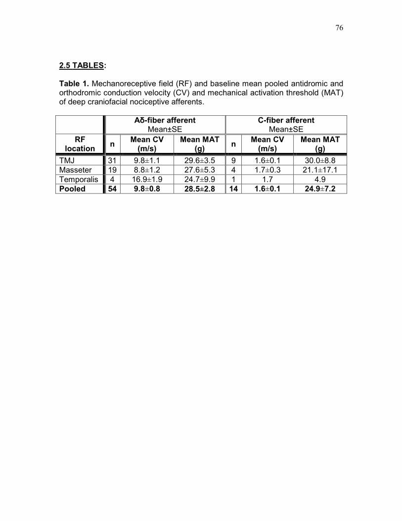
76
2.5 TABLES:
Table 1. Mechanoreceptive field (RF) and baseline mean pooled antidromic and orthodromic conduction velocity (CV) and mechanical activation threshold (MAT) of deep craniofacial nociceptive afferents.
Aδ-fiber afferent Mean±SE
C-fiber afferent Mean±SE
RF location
n Mean CV
(m/s) Mean MAT
(g) n
Mean CV (m/s)
Mean MAT (g)
TMJ 31 9.8±1.1 29.6±3.5 9 1.6±0.1 30.0±8.8
Masseter 19 8.8±1.2 27.6±5.3 4 1.7±0.3 21.1±17.1
Temporalis 4 16.9±1.9 24.7±9.9 1 1.7 4.9
Pooled 54 9.8±0.8 28.5±2.8 14 1.6±0.1 24.9±7.2
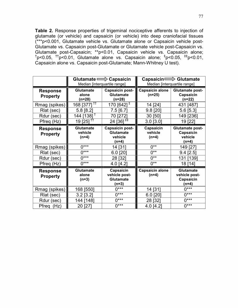
77
Table 2. Response properties of trigeminal nociceptive afferents to injection of glutamate (or vehicle) and capsaicin (or vehicle) into deep craniofacial tissues (***p<0.001, Glutamate vehicle vs. Glutamate alone or Capsaicin vehicle post-Glutamate vs. Capsaicin post-Glutamate or Glutamate vehicle post-Capsaicin vs. Glutamate post-Capsaicin; **p<0.01, Capsaicin vehicle vs. Capsaicin alone; †p<0.05, ††p<0.01, Glutamate alone vs. Capsaicin alone; ‡p<0.05, ‡‡p<0.01, Capsaicin alone vs. Capsaicin post-Glutamate; Mann-Whitney U test).
Glutamate Capsaicin Median [interquartile range]
Capsaicin Glutamate Median [interquartile range]
Response Property
Glutamate alone (n=28)
Capsaicin post-Glutamate
(n=28)
Capsaicin alone (n=25)
Glutamate post-Capsaicin
(n=22)
Rmag (spikes) 168 [377] †† 170 [642] ‡ 14 [24] 431 [487]
Rlat (sec) 5.8 [8.2] 7.5 [6.7] 9.8 [20] 5.6 [5.3]
Rdur (sec) 144 [138] † 70 [272] 30 [50] 149 [236]
Pfreq (Hz) 19 [25] †† 24 [36] ‡‡ 3.0 [3.0] 19 [22]
Response Property
Glutamate vehicle (n=4)
Capsaicin post-Glutamate
vehicle (n=4)
Capsaicin vehicle (n=4)
Glutamate post-Capsaicin
vehicle (n=4)
Rmag (spikes) 0*** 14 [31] 0** 149 [27]
Rlat (sec) 0*** 6.0 [20] 0** 9.4 [2.5]
Rdur (sec) 0*** 28 [32] 0** 131 [139]
Pfreq (Hz) 0*** 4.0 [4.2] 0** 18 [14]
Response Property
Glutamate alone (n=3)
Capsaicin vehicle post-
Glutamate (n=3)
Capsaicin alone (n=4)
Glutamate vehicle post-
Capsaicin (n=4)
Rmag (spikes) 168 [550] 0*** 14 [31] 0***
Rlat (sec) 3.2 [3.2] 0*** 6.0 [20] 0***
Rdur (sec) 144 [148] 0*** 28 [32] 0***
Pfreq (Hz) 20 [27] 0*** 4.0 [4.2] 0***

78
Table 3. Properties of responses of TMJ-responsive Vc/UCC nociceptive neurons to injection of glutamate (or vehicle) and capsaicin (or vehicle) into the TMJ (***p<0.001, Glutamate vehicle vs. Glutamate alone or Glutamate alone vs. Glutamate post-Capsaicin or Capsaicin vehicle post-Glutamate vs. Capsaicin post-Glutamate; †††p<0.001, Capsaicin vehicle vs. Capsaicin alone or Glutamate vehicle post-Capsaicin vs. Glutamate post-Capsaicin; ‡p<0.05, Capsaicin alone vs. Capsaicin post-Glutamate; Mann-Whitney U test).
Glutamate Capsaicin Median [interquartile range]
Capsaicin Glutamate Median [interquartile range]
Response Property
Glutamate alone (n=14)
Capsaicin post-Glutamate
(n=13)
Capsaicin alone (n=16)
Glutamate post-Capsaicin
(n=10)
Rmag (spikes) 833 [1186] 2915 [4079] ‡ 161 [942] 0***
Rlat (sec) 4.3 [2.6] 5.3 [2.5] ‡ 16.4 [10.5] 0***
Rdur (sec) 98.6 [49.1] 377 [283] ‡ 112 [201] 0***
Pfreq (Hz) 31.7 [39.2] 45.1 [43.7] ‡ 16.6 [35.6] 0***
Response Property
Glutamate vehicle (n=5)
Capsaicin post-Glutamate
vehicle (n=5)
Capsaicin vehicle (n=5)
Glutamate post-Capsaicin
vehicle (n=5)
Rmag (spikes) 0*** 131 [718] 0††† 643 [1396]
Rlat (sec) 0*** 15.1 [14.4] 0††† 4.3 [2.6]
Rdur (sec) 0*** 111 [153] 0††† 108 [50.6]
Pfreq (Hz) 0*** 13.3 [29.8] 0††† 33.5 [41]
Response Property
Glutamate alone (n=5)
Capsaicin vehicle post-
Glutamate (n=5)
Capsaicin alone (n=4)
Glutamate vehicle post-
Capsaicin (n=4)
Rmag (spikes) 472 [1253] 0*** 131 [718] 0†††
Rlat (sec) 3.6 [4.7] 0*** 15.1 [14.4] 0†††
Rdur (sec) 126 [103] 0*** 111 [153] 0†††
Pfreq (Hz) 21.7 [46.9] 0*** 13.3 [29.8] 0†††

79
Table 4. Comparison of response properties of WDR and NS TMJ-responsive Vc/UCC nociceptive neurons and of deep craniofacial nociceptive afferents to injection of glutamate and/or capsaicin into deep craniofacial tissues. In general, both glutamate and capsaicin-evoked responses were greater in Vc/UCC neurons compared to afferents (i.e. increased activation incidence, Rmag or Rdur).
Glutamate Capsaicin Capsaicin Glutamate
Glutamate alone Capsaicin post-
Glutamate Capsaicin alone
Glutamate post-Capsaicin
Response Property
Afferent Vc/UCC Afferent Vc/UCC Afferent Vc/UCC Afferent Vc/UCC
Activation + ++‡ ++† +++†‡ + ++‡ + -‡‡
Deep MAT + + +†† +†† + + + -
Cutaneous MAT N/A + N/A + N/A + N/A +
Cutaneous RF N/A + N/A + N/A + N/A +††
+ : Sensitized- increased excitability (↑Rmag, ↑Pfreq, ↑Rdur, ↓Rlat or ↓MAT)
- : Desensitized- decreased excitability (↓Rmag, ↓Pfreq, ↓Rdur, ↑Rlat or ↑MAT)
N/A: Not applicable †
: increased excitability compared to capsaicin alone ††
: decreased excitability compared to glutamate/capsaicin alone ‡: increased excitability compared to afferents tested
‡‡: glutamate-evoked activity abolished

80
2.6 FIGURES: Fig. 1A. Example of typical mechanoreceptive field and response properties of deep craniofacial nociceptive afferent to injection of glutamate vehicle followed by capsaicin (CV=2.8m/s, MAT=38 g, Aδ-fiber TMJ afferent). (a) Deep mechanoreceptive field (white circle) of nociceptive afferent involving the TMJ region indicated by arrow, (b) Afferent mechanical activation threshold determined with von Frey device, (c) No response in this afferent was evoked by injection of glutamate vehicle into the TMJ, (d) Afferent response evoked by injection of capsaicin following glutamate vehicle into the TMJ.
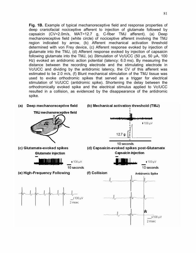
81
Fig. 1B. Example of typical mechanoreceptive field and response properties of deep craniofacial nociceptive afferent to injection of glutamate followed by capsaicin (CV=2.0m/s, MAT=12.7 g, C-fiber TMJ afferent). (a) Deep mechanoreceptive field (white circle) of nociceptive afferent involving the TMJ region indicated by arrow, (b) Afferent mechanical activation threshold determined with von Frey device, (c) Afferent response evoked by injection of glutamate into the TMJ, (d) Afferent response evoked by injection of capsaicin following glutamate into the TMJ, (e) Stimulation of Vc/UCC (50 µs, 50 µA, 100 Hz) evoked an antidromic action potential (latency: 6.0 ms). By measuring the distance between the recording electrode and the stimulating electrode in Vc/UCC and dividing by the antidromic latency, the CV of this afferent was estimated to be 2.0 m/s, (f) Blunt mechanical stimulation of the TMJ tissue was used to evoke orthodromic spikes that served as a trigger for electrical stimulation of Vc/UCC (antidromic spike). Shortening the delay between the orthodromically evoked spike and the electrical stimulus applied to Vc/UCC resulted in a collision, as evidenced by the disappearance of the antidromic spike.

82
Fig. 2. The unit recording sites reconstructed from histology were plotted on diagrams of the brainstem (Paxinos and Watson 1997). Coronal sections at 3 different rostrocaudal levels of subnucleus caudalis/upper cervical spinal cord (Vc/UCC). Filled symbols represent wide dynamic range (WDR) neurons; opened symbols nociceptive-specific (NS) neurons. The dot and triangle symbols represent the glutamate injection followed by capsaicin injection and capsaicin injection followed by glutamate injection subgroups, respectively. The labels 3A, 3B, and 4 represent the NS neurons shown in Figs. 3 and 4.
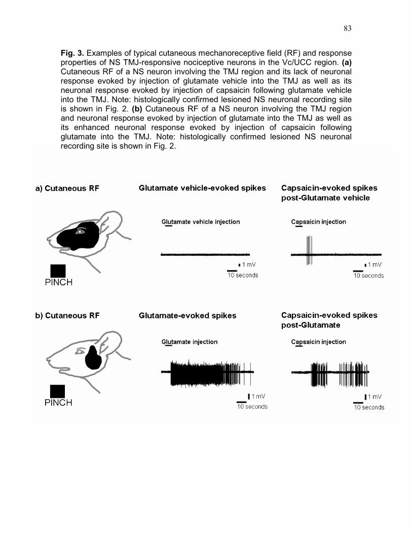
83
Fig. 3. Examples of typical cutaneous mechanoreceptive field (RF) and response properties of NS TMJ-responsive nociceptive neurons in the Vc/UCC region. (a) Cutaneous RF of a NS neuron involving the TMJ region and its lack of neuronal response evoked by injection of glutamate vehicle into the TMJ as well as its neuronal response evoked by injection of capsaicin following glutamate vehicle into the TMJ. Note: histologically confirmed lesioned NS neuronal recording site is shown in Fig. 2. (b) Cutaneous RF of a NS neuron involving the TMJ region and neuronal response evoked by injection of glutamate into the TMJ as well as its enhanced neuronal response evoked by injection of capsaicin following glutamate into the TMJ. Note: histologically confirmed lesioned NS neuronal recording site is shown in Fig. 2.

84
Fig. 4. Example of capsaicin and glutamate-induced central sensitization in a NS TMJ-responsive nociceptive neuron in the Vc/UCC region. (a) Neuronal activity at baseline and responses evoked by injection of capsaicin and glutamate into the TMJ, (b) Cutaneous mechanoreceptive field (RF) of NS neuron at baseline and following injection of capsaicin and glutamate into the TMJ, (c) Cutaneous mechanical activation threshold (MAT) of NS neuron at baseline and following injection of capsaicin and glutamate into the TMJ. Note: histologically confirmed lesioned NS neuronal recording site is shown in Fig. 2.

85
Fig. 5. The time courses of glutamate (GLU) and capsaicin (CAP)-induced mechanical activation threshold (MAT) in deep craniofacial afferents. Arrow indicates time point for injection of GLU (black) and CAP (grey) into deep craniofacial tissues. Circles indicate median normalized MAT following injection of GLU and CAP. Triangles indicate GLU or CAP-induced median normalized MAT before or after injection of vehicle controls for GLU or CAP. Raw MAT threshold values were normalized to the initial baseline pre-injection value of the first agonist. Lines: interquartile range. Note that injection of (a) GLU alone and (b) GLU following CAP into deep craniofacial tissues significantly reduced the MAT (*p<0.05, **p<0.01, ***p<0.001; RM ANOVA-on-ranks, Dunn’s Method).

86
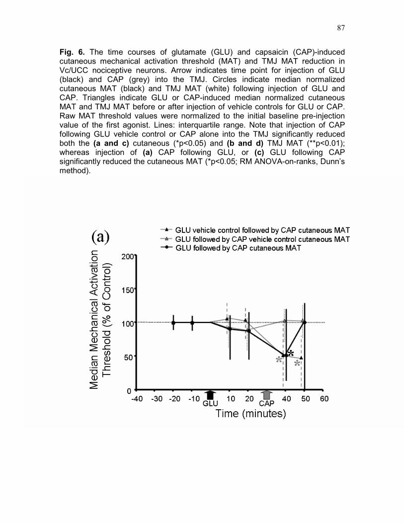
87
Fig. 6. The time courses of glutamate (GLU) and capsaicin (CAP)-induced cutaneous mechanical activation threshold (MAT) and TMJ MAT reduction in Vc/UCC nociceptive neurons. Arrow indicates time point for injection of GLU (black) and CAP (grey) into the TMJ. Circles indicate median normalized cutaneous MAT (black) and TMJ MAT (white) following injection of GLU and CAP. Triangles indicate GLU or CAP-induced median normalized cutaneous MAT and TMJ MAT before or after injection of vehicle controls for GLU or CAP. Raw MAT threshold values were normalized to the initial baseline pre-injection value of the first agonist. Lines: interquartile range. Note that injection of CAP following GLU vehicle control or CAP alone into the TMJ significantly reduced both the (a and c) cutaneous (*p<0.05) and (b and d) TMJ MAT (**p<0.01); whereas injection of (a) CAP following GLU, or (c) GLU following CAP significantly reduced the cutaneous MAT (*p<0.05; RM ANOVA-on-ranks, Dunn’s method).

88

89
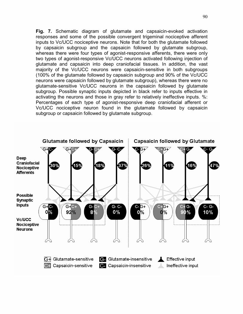
90
Fig. 7. Schematic diagram of glutamate and capsaicin-evoked activation responses and some of the possible convergent trigeminal nociceptive afferent inputs to Vc/UCC nociceptive neurons. Note that for both the glutamate followed by capsaicin subgroup and the capsaicin followed by glutamate subgroup, whereas there were four types of agonist-responsive afferents, there were only two types of agonist-responsive Vc/UCC neurons activated following injection of glutamate and capsaicin into deep craniofacial tissues. In addition, the vast majority of the Vc/UCC neurons were capsaicin-sensitive in both subgroups (100% of the glutamate followed by capsaicin subgroup and 90% of the Vc/UCC neurons were capsaicin followed by glutamate subgroup), whereas there were no glutamate-sensitive Vc/UCC neurons in the capsaicin followed by glutamate subgroup. Possible synaptic inputs depicted in black refer to inputs effective in activating the neurons and those in gray refer to relatively ineffective inputs. %: Percentages of each type of agonist-responsive deep craniofacial afferent or Vc/UCC nociceptive neuron found in the glutamate followed by capsaicin subgroup or capsaicin followed by glutamate subgroup.

Chapter 3
Peripheral NMDA receptor modulation of jaw muscle electromyographic activity induced by capsaicin injection into
the temporomandibular joint of rats 3.0 ABSTRACT:
We have previously documented that peripheral N-methyl-D-aspartate (NMDA)
receptor mechanisms are involved in nociceptive reflex increases in jaw muscle
activity to injection of mustard oil or glutamate into the rat temporomandibular
joint (TMJ). The aim of the present study was to determine whether peripheral
NMDA receptor mechanisms are also involved in the nociceptive reflex
responses in the jaw muscles evoked by injection of the inflammatory irritant and
algesic chemical capsaicin into the TMJ. The effects of peripheral injection of
NMDA receptor antagonists, MK-801 and APV, on the increases in
electromyographic (EMG) activities of digastric and masseter muscles reflexly
evoked by capsaicin injection into the TMJ were tested in halothane-anesthetized
male rats. The capsaicin injection following pre-injection of vehicle evoked
significant increases in EMG activity in both digastric and masseter muscles
whereas pre-injection of MK-801 or APV into the TMJ resulted in a significant
concentration-related reduction in the magnitude of capsaicin-evoked digastric
and masseter EMG activity (ANOVA-on-ranks, P<0.05). This finding indicates
that capsaicin-evoked digastric and masseter EMG activity can be attenuated by
pre-injection into the TMJ of NMDA receptor antagonists, and that the activation
of peripheral NMDA receptors may be important in the mechanisms whereby
capsaicin evokes nociceptive trigeminal responses.
91

92
3.1 INTRODUCTION:
The vanilloid type 1 receptor, TRPV1, is activated by noxious heat, protons or
the inflammatory irritant and small-fiber excitant capsaicin, and is found on small-
diameter afferent nerve fibers and dorsal root ganglion neurons (Caterina et al.
1997; Tominaga et al. 1998; Jordt et al. 2003). TRPV1 receptors have also
recently been described on trigeminal afferents and trigeminal ganglion neurons
innervating the rat temporomandibular joint (TMJ) (Ichikawa et al. 2004).
Furthermore, capsaicin injected into the rat TMJ reflexly evokes a dose-
dependent increase in jaw muscle electromyographic (EMG) activity (Tang et al.
2004), produces an inflammatory response (Hu et al. 2005a), and induces
activation and sensitization in brainstem nociceptive neurons (Chapter 2).
Intramuscular injection of capsaicin in humans also results in intense pain and
hyperalgesia (Witting et al. 2000b). Both trigeminal afferent (Chapter 2) and
brainstem (Chapter 2) nociceptive neuronal responses to capsaicin injected into
the TMJ can be significantly increased following glutamate injection into the TMJ.
These findings raise the possibility that peripheral NMDA receptors may
contribute to the mechanisms whereby capsaicin evokes nociceptive responses.
It has been previously shown that glutamate injection into the rat TMJ induces
a concentration-related reflex increase in jaw muscle activity that can be
significantly attenuated by co-injection of NMDA receptor antagonists (Cairns et
al. 1998). Similarly, glutamate injection into the human masseter muscle causes
pain that can be attenuated by co-injection of an NMDA receptor antagonist
(Cairns et al. 2001a, 2003ab, Svensson et al. 2003). Increases in jaw muscle
reflex activity as a result of mustard oil application to the TMJ are also attenuated
by TMJ pre-injection of an NMDA receptor antagonist (Yu et al. 1996). In order to
determine whether peripheral NMDA receptor mechanisms are involved in
nociceptive reflex responses evoked by capsaicin application to the TMJ, the
present study tested the possible effects of the peripheral (TMJ) application of
NMDA receptor antagonists, MK-801 and APV, on the increases in jaw muscle
EMG activity that could be reflexly evoked by capsaicin application to the TMJ.

93
A portion of this data has been previously presented in abstract form (Hu et al.
2003, Lam et al. 2003c).
3.2 MATERIALS AND METHODS:
ANIMAL PREPARATION. A total of 45 male Sprague Dawley rats (250–450 gm)
were prepared for acute in vivo recording of jaw muscle EMG activity, as
previously described (Hu et al. 1993; Yu et al. 1994, 1995, 1996). Briefly, under
surgical anesthesia (O2: 1 L/min; halothane: 1.5–2.5%), a tracheal cannula was
inserted and artificial ventilation initiated. The rat’s head was then placed in a
stereotaxic frame to facilitate placement of EMG recording electrodes. Bipolar
electrodes were fashioned out of 40 gauge Teflon-coated single-strand stainless
steel wire and inserted into the left digastric and masseter muscles.
After completion of all surgical procedures, the halothane level was titrated (1–
1.3%) until noxious pressure applied to the hindpaw could not induce a flexion
reflex of the hindlimb to ensure that an adequate level of anesthesia was
maintained for the duration of the experiment. Heart rate and body core
temperature were continuously monitored throughout the experiment and kept
within the physiological range of 330–430/min and 37–37.5°C, respectively. All
methods and experimental approaches were approved by the University of
Toronto Animal Care Committee in accordance with the regulations of the
Ontario Animal Research Act (Canada).
DRUG SOLUTIONS. For the TMJ administration of drug solutions, two 27-gauge
cannulae were first cemented side-by-side and connected to two Hamilton
syringes with polyethylene tubes. One cannula was filled with either the non-
competitive NMDA receptor antagonist (+)-MK-801 (0.001, 0.01 or 0.1M; 10 µL;
n=6 per group; Research Biochemicals International, Natick, MA), competitive
NMDA receptor antagonist (±)-D-2-amino-5-phosphonovalerate, APV (0.005 or
0.05M; 10 µL; n=6 per group Research Biochemicals International, Natick, MA),

94
or vehicle (sterile normal saline; 10 µL; n=6), and the other with 1% capsaicin
(10% capsaicin in ethanol:Tween-80: sterile normal saline in a 1:1:8 ratio by
volume; 10 µL; Calbiochem, La Jolla, CA). Both NMDA receptor antagonists
were dissolved in isotonic saline (pH 7.2-7.4). The concentrations of the NMDA
receptor antagonists and capsaicin were chosen on the basis of our previous
findings (Yu et al. 1996; Cairns et al. 1998; Tang et al. 2004; Hu et al. 2005a).
The two cannulae were then carefully inserted into the left TMJ, and capsaicin
and NMDA receptor antagonists or vehicle were injected into the left TMJ via the
needle and catheter. We have previously demonstrated that injection of capsaicin
vehicle or isotonic saline into the TMJ region does not evoke any significant
change in EMG activity in the digastric or masseter muscles (Yu et al. 1995,
1996; Tang et al. 2004).
STIMULATION AND RECORDING TECHNIQUES. Bipolar recordings were
made of the EMG activities of the left digastric (jaw-opening) and left masseter
(jaw-closing) muscles (Hu et al. 1993; Yu et al. 1995) before and after injections
of MK-801, APV, or vehicle control, and capsaicin. EMG activity was amplified
(gain, 500x; bandwidth, 30–1000 Hz) and fed into a computer equipped with a
CED 1401 Plus board and analysis software (Spike2; Cambridge Electronics;
signal sampling rate was 2000 Hz). Recorded EMG activity was stored
electronically and analyzed offline. The EMG activities with the jaw in resting
position were first recorded for 20 min to establish a baseline, and either MK-801,
APV or saline vehicle was administrated into the left TMJ. In another series of
experiments, MK-801 (0.1M, n=6) or APV (0.05M, n=3) was injected into the right
TMJ to control for possible systemic effects. Five minutes following the
administration, capsaicin was injected into the left TMJ and the resulting changes
in EMG activity were continuously recorded for another 30 min. All drug solutions
were slowly injected into the TMJ over ~5 sec.
DATA ANALYSIS AND STATISTICS. Recorded EMG data were rectified off-
line, and EMG area bins (microvolts per minute) were calculated. Baseline EMG

95
activity was calculated as a mean of EMG area bins recorded over the first 20
min before injection of agents into the TMJ. Relative EMG activity was calculated
by normalizing EMG area bins to the baseline EMG activity and was used to
illustrate the results of individual experiments. Agents applied to the TMJ were
considered to have evoked jaw muscle activity if the value of the first EMG bin
after TMJ application was 2 SD above the baseline (Yu et al. 1995). The value of
the baseline plus 2 SD was chosen as a signal-to-noise limit because it
represents an approximation of the 95% confidence interval for the mean
baseline activity. The relative area under the EMG response curve (AUC) was
calculated by summing the value of the first and all subsequent EMG area bins
greater than 2 SDs above the mean baseline (20 min) EMG activity and defined
as the overall response. ANOVA-on-ranks and post-hoc Dunnett’s or Dunn’s
tests were used as appropriate (p<0.05 considered to reflect statistically
significant differences).
3.3 RESULTS:
Consistent with our previous findings (Yu et al. 1994, 1995), none of the animals
revealed any variation in baseline EMG activities greater than two standard
deviations (e.g. Fig. 1) during the initial 20 min period prior to capsaicin injection
into the TMJ. Compared with baseline EMG activity, administration of either the
saline vehicle (n=6), MK-801 (0.001, 0.01 or 0.1M; n=6 per group), or APV (0.005
or 0.05M; n=6 per group) into the left TMJ did not evoke any significant change in
EMG activity (e.g. Fig. 1). In contrast, compared with the baseline EMG activity,
the capsaicin injection into the left TMJ following the pre-injection of saline
vehicle evoked significant increases in EMG activity in both digastric and
masseter muscles of all animals (6/6) tested that lasted up to 20 min (Figs. 1-3).

96
Effects of MK-801 on capsaicin-induced EMG responses
Compared with the EMG responses in the animals receiving pre-injection of
saline vehicle, the pre-injection of MK-801 at all 3 concentrations tested (0.001,
0.01, and 0.1M) into the left TMJ significantly reduced the AUC of the capsaicin-
induced EMG response in both digastric and masseter muscles; the reduction
was greater in the masseter muscle than in the digastric muscle and was most
pronounced with the highest concentration of MK-801 (Figs. 1 and 2). Thus,
compared with pre-injection of saline, pre-injection of MK-801 into the left TMJ
resulted in a significant concentration-related reduction in the magnitude of
capsaicin-evoked digastric and masseter EMG responses (ANOVA-on-ranks,
Dunnett’s test, p<0.05) (Figs. 1 and 2).
Experiments to rule out systemic effects of MK-801 were conducted by
determining if pre-injection of the highest concentration of MK-801 into the right
TMJ can attenuate the EMG responses induced by capsaicin injected into the left
TMJ via a systemic route. There was however no significant difference in
capsaicin-evoked EMG activity following the pre-injection of vehicle into the left
TMJ or 0.1M MK-801 into the right TMJ (ANOVA-on-ranks, Dunnett’s test,
p>0.05). In contrast, there was a significant difference in capsaicin-induced EMG
activity following the pre-injection of 0.1M MK-801 into the left TMJ when
compared to pre-injection of 0.1M MK-801 into the right TMJ (ANOVA-on-ranks,
Dunnett’s test, p<0.05) (Fig. 3).
Effects of APV on capsaicin-induced EMG responses
Compared with the EMG responses in the animals receiving pre-injection of
saline vehicle, the pre-injection of APV at a concentration of 0.005M into the left
TMJ did not significantly reduce the AUC of the capsaicin-induced EMG
response in the digastric muscle although it did significantly reduce the
capsaicin-induced EMG response in the masseter muscle. The pre-injection of
0.05M APV did significantly reduce the AUC of the capsaicin-induced EMG
response in both digastric and masseter muscles; the reduction was greater in
the masseter muscle than in the digastric muscle and was most pronounced with

97
the highest concentration of APV (Figs. 1 and 2). Thus, compared with pre-
injection of saline, pre-injection of APV into the left TMJ resulted in a significant
reduction in the magnitude of capsaicin-evoked digastric and masseter EMG
responses (ANOVA-on-ranks, Dunnett’s test, p<0.05) (Figs. 1 and 2).
There was no significant difference in capsaicin-evoked EMG activity following
the pre-injection of vehicle into the left TMJ or 0.05M APV into the right TMJ
(ANOVA-on-ranks, Dunn’s test, p>0.05). However, there was a significant
difference in capsaicin-induced EMG activity following the pre-injection of 0.05M
APV into the left TMJ when compared to pre-injection of 0.05M APV into the right
TMJ (ANOVA-on-ranks, Dunn’s test, p<0.05) (Fig. 3).
3.4 DISCUSSION:
Local application of the small fiber-excitant and inflammatory irritant capsaicin to
the left TMJ increased jaw muscle activity and these capsaicin-evoked increases
in EMG activity were attenuated by pre-injection of NMDA receptor antagonists
into the left TMJ. It may be argued that the attenuation in capsaicin-evoked
increases in EMG activity may be due to mechanisms unrelated to NMDA
receptors since although MK-801 non-competitively antagonizes excitation
mediated by the NMDA receptor, at higher concentrations (~100 microM) MK-
801 also blocks sodium channels (Rothman 1988; Halliwell et al. 1989). Another
concern is that MK-801 at lower concentrations (0.3-30 microM) also blocks
nicotinic acetylcholine channels (Halliwell et al. 1989; Galligan and North 1990;
Ramoa et al. 1990; Amador and Dani 1991 Briggs and McKenna 1996).
However, our finding that the competitive NMDA receptor antagonist APV also
similarly attenuates capsaicin-evoked EMG activity confirms the involvement of
NMDA receptors in the mechanism behind capsaicin-evoked nociceptive EMG
responses. It is unlikely that the effect of MK-801 or APV injected into the left
TMJ was produced by a systemic action since there was no significant effect of
either MK-801 or APV injection into the right TMJ on capsaicin-evoked EMG

98
activity. We have interpreted these results to suggest that the activation of
peripheral NMDA receptors contributes, in part, to the mechanism whereby
capsaicin injection into the TMJ evokes nociceptive jaw muscle responses.
Peripheral neural mechanisms underlying the reflex activation of jaw
muscles
We have previously reported that application of mustard oil or glutamate to the
rat TMJ region results in a characteristic increase in the EMG activity of both the
digastric and masseter muscles that involves a brainstem reflex circuit involving
trigeminal brainstem subnucleus caudalis (Yu et al. 1994, 1995, 1996; Bakke et
al. 1998; Cairns et al. 1998, 2001c; Hu et al. 1997; Tsai et al. 1999). It has also
been shown that injection of glutamate into the masseter muscle of humans
caused significantly higher levels of pain and pain spread than injection of
isotonic saline (Cairns et al. 2001a, 2003b, Svensson et al. 2003). Peripheral
NMDA receptors may play a role in these effects of glutamate since recent
evidence indicates that ketamine, an NMDA receptor antagonist, applied in
combination with glutamate, decreases glutamate-evoked muscle pain in
humans (Cairns et al. 2003a). The results of the present study indicate that
application of capsaicin to the rat TMJ evokes a similar co-activation of these jaw
muscles, and thus it is possible that capsaicin may activate the same putative
nociceptive reflex pathways as mustard oil, glutamate and other algesic
chemicals. The receptor mechanisms underlying the mustard oil or capsaicin-
evoked activation of TMJ afferents remain unclear. However, our present results
along with our earlier data that local application to the TMJ region of the NMDA
antagonist MK-801 blocks mustard oil-evoked jaw muscle activity (Yu et al. 1996)
indicate that peripheral NMDA receptors appear to play a role in mediating
mustard oil and capsaicin-evoked increases in jaw muscle activity.
Since TRPV1 receptors are activated by heat, protons, or capsaicin (Caterina
et al. 1997; Tominaga et al. 1998; Benham et al. 2003; Jordt et al. 2003), the
results of this study suggest there may be functional TRPV1 receptors located
within the TMJ region. Our findings are supported by recent findings of TRPV1

99
receptors in TMJ tissues of the rat (Ichikawa et al. 2004). However, the use of
specific receptor antagonists would be required to confirm the involvement of the
TRPV1 receptor. One possible mechanism to account for our finding that
antagonism of peripheral NMDA receptors contributes to capsaicin-evoked jaw
muscle activity is via autocrine and/or paracrine activation of peripheral NMDA
receptors (Carlton 2001; Hinoi et al. 2004). That is, activation of peripheral
TRPV1 receptors via noxious heat, protons or capsaicin may result in the release
of glutamate from neuronal terminals. The glutamate released could then further
activate NMDA receptors on the same neuronal terminal or adjacent surrounding
peripheral terminals. Activation of peripheral NMDA receptors could then lead to
the release of more glutamate in the peripheral tissues and might alter TRPV1
receptor responsiveness to enhance nociceptive responses. Another possible
mechanism, albeit a much slower process, is that blockade of calcium (Ca2+)
influx via peripheral NMDA receptors in nociceptive afferents may prevent the
potentiation of NMDA and/or TRPV1 activity (Petrenko et al. 2003; Geppetti and
Trevisani 2004). No studies to date have demonstrated the co-localization of
peripheral NMDA and TRPV1 receptors on the same trigeminal primary afferent
terminal but our recent evidence that peripherally applied glutamate and
capsaicin may activate the same TMJ or craniofacial muscle fiber (Chapter 2)
suggests that both receptors may be found on a single trigeminal primary
afferent. Since mustard oil excites sensory nerve fibers through activation of the
peripheral TRPA1 (ANKTM1) receptor (Jordt et al. 2004; Bandell et al. 2004),
also a member of the TRP family, the mechanisms proposed above for
capsaicin-evoked jaw muscle activity may also account for previous findings in
our laboratory that antagonism of peripheral NMDA receptors can reduce
mustard oil-evoked nociceptive responses (Yu et al. 1996). Thus, these findings
suggest that peripheral NMDA receptors may play a role in mediating the
nociceptive responses elicited by the activation of these TRP receptors.
It is possible that under conditions that are associated with deep tissue pain,
glutamate may be released from afferent fiber terminals and act on peripheral
EAA receptors to excite nociceptive fibers. Glutamate is present in and released

100
from the central terminals of small-diameter spinal cord and trigeminal afferents,
including TMJ afferents (Salt and Hill 1983; Wanaka et al. 1987; Battaglia and
Rustioni 1988; Kai-Kai 1989; Westlund et al. 1989; Clements and Beitz 1991;
Clements et al. 1991; Kai-Kai and Howe 1991; Azerad et al. 1992; Boucher et al.
1993; Bereiter and Benetti 1996). It is not known whether glutamate is also
released from the peripheral endings of trigeminal afferent fibers but capsaicin
application to the sciatic nerve has been shown to result in the neurogenic
release of glutamate from peripheral terminals (deGroot et al. 2000). In addition
to the cytosolic release of glutamate from affected neuronal terminals, non-
neuronal cells such as macrophages (Piani et al. 1991), blood serum (McAdoo et
al. 1997) or Schwann cells (Parpura et al. 1995) may also contribute to the
increase in peripheral levels of glutamate following activation of TRPV1
receptors. It has been demonstrated that application of inflammatory irritants
such as mustard oil and capsaicin to the TMJ region results in significant plasma
extravasation (Haas et al. 1992; Yu et al. 1995, 1996; Hu et al. 2005a). Plasma
extravasation can occur within a few seconds after a noxious stimulus is applied
(Raab 1992). The concentration of glutamate in plasma is ~300 µM which is
greater than the reported ED50 for activation of peripheral glutamate receptors
(Erdo 1991; Ault and Hildebrand 1993a,b). Therefore, plasma extravasation into
the TMJ region could rapidly elevate glutamate concentrations to a level that
could activate EAA receptors located within the TMJ region. Thus, changes in
peripheral glutamate levels through cytosolic release from tissue damage or
inflammation or through neurogenic release as a result of nociceptive activation
may all play a role in modulating the sensitivity of deep craniofacial tissues
through autocrine and/or paracrine regulation of ionotropic glutamate receptor
mechanisms.
Clinical relevance
Peripheral glutamate receptor mechanisms may have an important role in
craniofacial pain since peripheral glutamate levels are elevated during cutaneous
or deep tissue inflammation (Omote et al. 1998; Lawand et al. 2000; McNearny

101
et al. 2000). We have demonstrated that injection of glutamate into the rat
masseter muscle or TMJ can activate and induce peripheral sensitization in
masseter muscle and TMJ afferent fibers (Cairns et al. 2001a,b, 2002a; Chapter
2), activate and induce central sensitization in brainstem nociceptive neurons
(Chapter 2), and reflexly increase jaw EMG activity (Cairns et al. 1998, 2001c),
through the activation of peripheral glutamate receptors. It has also been shown
that injection of glutamate into the masseter muscle induces pain in humans
(Cairns et al. 2001a, 2003b, Svensson et al. 2003). These findings suggest that
activation of peripheral glutamate receptors, NMDA receptors in particular, may
excite nociceptors that contribute to pain responses. The glutamate-evoked
peripheral sensitization may contribute to the primary hyperalgesia or allodynic
states characteristic of craniofacial pain conditions such as temporomandibular
disorders (TMD) (see Carlsson and LeResche 1995, Stohler 1995,1999). The
glutamate-evoked central sensitization as reflected in receptive field expansion,
mechanical activation threshold reduction, and increases in responses to
suprathreshold stimuli and neuronal spontaneous activity may contribute to pain
spread and referral, allodynia, hyperalgesia, and pain at rest in TMD (see
Carlsson and LeResche 1995, Stohler 1995,1999). The finding in the present
study that increases in capsaicin-evoked jaw muscle reflex activity can be
attenuated by NMDA receptor antagonists indicates that peripheral NMDA
receptors are involved in the mechanisms whereby capsaicin evokes nociceptive
reflex responses.
The demonstration of a role for peripheral NMDA receptor mechanisms in
modulating nociceptive trigeminal responses evoked by the activation of various
peripheral nociceptive receptors is important in the targeting of treatment for
craniofacial pain disorders of peripheral origin. Our findings that nociceptive
responses evoked by the peripheral application of mustard oil, glutamate or
capsaicin may be attenuated by the peripheral application of NMDA receptor
antagonists suggest that it might prove more efficacious to target peripheral
NMDA receptors rather than each of those activated by the above receptor
agonists in the treatment of craniofacial disorders. The formulation of peripheral

102
NMDA receptor antagonists that do not cross the blood brain barrier may be of
potential benefit by reducing peripheral nociceptive excitability while avoiding
many harmful side effects that may be found with antagonism of central NMDA
receptors. Although we have demonstrated a novel antinociceptive role for
peripheral NMDA receptor antagonists in the animal and human craniofacial pain
studies discussed above, there has been a paucity of reports on the effects of
locally administered NMDA receptor antagonists in other human experimental
pain models and data from the few existing studies are not consistent. For
example, Warncke and colleagues (1997) showed, in a forearm burn injury
model, that the development of mechanical hyperalgesia is inhibited with
peripheral ketamine pretreatment and Pederson and colleagues (1998) showed,
using a similar burn injury model, that peripheral ketamine pretreatment reduced
spontaneous pain during burn injury induction and increased the heat pain
threshold. In contrast to the results of the burn injury models, Gottrup and
colleagues (2000, 2004), using a capsaicin model, failed to find an effect on pain
and hyperalgesia after peripheral ketamine pretreatment. Differences in pain
models, tissues (i.e. skin vs. deep craniofacial), dosage and timing of NMDA
receptor antagonists and injury type and extent may explain the discrepancy in
experimental results. Further studies are required to achieve a better
understanding of the role of peripheral NMDA receptors in the pathobiological
mechanisms underlying craniofacial pain conditions of peripheral origin.
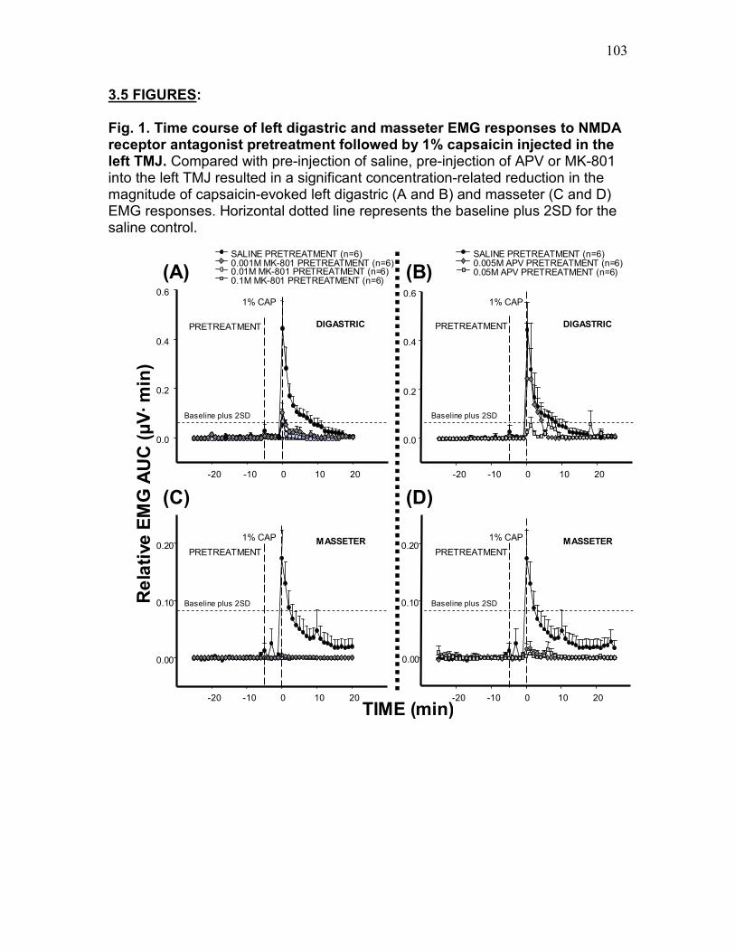
103
3.5 FIGURES:
Fig. 1. Time course of left digastric and masseter EMG responses to NMDA receptor antagonist pretreatment followed by 1% capsaicin injected in the left TMJ. Compared with pre-injection of saline, pre-injection of APV or MK-801 into the left TMJ resulted in a significant concentration-related reduction in the magnitude of capsaicin-evoked left digastric (A and B) and masseter (C and D) EMG responses. Horizontal dotted line represents the baseline plus 2SD for the saline control.
0.1M MK-801 PRETREATMENT (n=6)
1% CAP
1% CAP
-20 -10 0 10 20
0.0
0.2
0.4
0.6
PRETREATMENT
TIME (min)-20 -10 0 10 20
0.00
0.10
0.20
SALINE PRETREATMENT (n=6) 0.001M MK-801 PRETREATMENT (n=6)0.01M MK-801 PRETREATMENT (n=6)
PRETREATMENT
Re
lati
ve
EM
G A
UC
(µ
V·m
i n)
PRETREATMENT
1% CAP
-20 -10 0 10 20
0.0
0.2
0.4
0.6
PRETREATMENT
1% CAP
-20 -10 0 10 20
0.00
0.10
0.20
SALINE PRETREATMENT (n=6)0.005M APV PRETREATMENT (n=6) 0.05M APV PRETREATMENT (n=6)(A)
(C)
(B)
(D)
DIGASTRIC DIGASTRIC
MASSETERMASSETER
Baseline plus 2SD
Baseline plus 2SD
Baseline plus 2SD
Baseline plus 2SD

104
Fig. 2. Median EMG responses to NMDA receptor antagonist pretreatment followed by 1% capsaicin injected in the left TMJ. Compared with pre-injection of saline, pre-injection of APV or MK-801 into the left TMJ resulted in a significant reduction in the magnitude of capsaicin-evoked left digastric (A) and masseter (B) EMG responses (ANOVA-on-ranks, *p<0.05, **p<0.01, ***p<0.001). Median: transverse line within box; 75th percentile: top half box; 25th percentile: bottom half box.
0.0
0.4
0.8
1.2
(B)
SALINE 0.001M MK-801
0.01M MK-801
0.1M
MK-801
SALINE
MK-801
APV
*****
***
0.0
0.4
0.8
1.2
0.005MAPV
0.05M APV
***
SALINE 0.001M MK-801
0.01M MK-801
0.1M MK-801
0
1
2
3
4
***
***
Med
ian
EM
G A
UC
(µV
·m
in)
0.005MAPV
0.05M APV
0
1
2
3
4 *
(A)
SALINE
0
1
2
3
4
PRETREATMENT
DIGASTRIC MASSETER
**** **
*

105
Fig. 3. Comparison of left versus right NMDA receptor antagonist pretreatment effects on median digastric and masseter EMG responses to 1% capsaicin injected in the left TMJ. There was a significant difference in capsaicin-induced EMG activity following the pre-injection of APV or MK-801 into the left TMJ when compared to pre-injection of APV or MK-801 into the right TMJ (ANOVA-on-ranks, *p<0.05, **p<0.01, ***p<0.001). Median: transverse line within box; 75th percentile: top half box; 25th percentile: bottom half box.
SALINE SALINE
0
1
2
3
4
SALINE
MK-801
APV PRETREATMENT
DIGASTRIC MASSETER
Med
ian
EM
G A
UC
(µ
V·
min
)
Right
0.05M APV
Left
0.05M APV
Right
0.05M APV
Left
0.05M APV
*
*
*
**
Right
0.1M MK-801
Left
0.1M MK-801
Right
0.1M MK-801
Left
0.1M APV
0
1
2
3
**
***
**
***

Chapter 4
Surgical incision can alter capsaicin-induced central sensitization
in rat brainstem nociceptive neurons
4.0 ABSTRACT:
Surgical trauma can affect spinal neuronal excitability, but there have been no studies of
the effects of surgical cutaneous injury on central nociceptive processing of deep
afferent inputs evoked by noxious stimuli such as capsaicin. Thus our aim was to test
the effect of surgical cutaneous incision in influencing central sensitization induced by
capsaicin injection into the temporomandibular joint (TMJ). The activity of single
nociceptive neurons activated by noxious mechanical stimulation of the TMJ was
recorded in the trigeminal subnucleus caudalis/upper cervical cord of halothane-
anaesthetized rats. The cutaneous mechanoreceptive field (RF), cutaneous mechanical
activation threshold (MAT) and TMJ MAT of neurons before and after both surgical
cutaneous incision alone and capsaicin injection were compared with results of incision
and lidocaine pretreatment of the facial skin overlying the TMJ and capsaicin injection
into the TMJ. Incision itself induced a barrage of neuronal spikes and excitability
increases reflecting central sensitization (cutaneous RF expansion, cutaneous MAT
reduction) in most neurons tested whereas lidocaine pretreatment significantly
attenuated the barrage and central sensitization. Capsaicin injection into the TMJ
induced cutaneous RF expansion, cutaneous MAT reduction and TMJ MAT reduction
following lidocaine pretreatment of the cutaneous incision site whereas most neurons
tested with capsaicin following incision alone showed no capsaicin-induced excitability
increase. These findings suggest that central sensitization induced by capsaicin alone or
by cutaneous incision alone can readily occur in TMJ-responsive nociceptive neurons
and that following incision-induced excitability increases, nociceptive neurons may be
temporarily refractory to any further capsaicin-induced changes reflecting central
sensitization.
106

107
4.1 INTRODUCTION:
The trigeminal subnucleus caudalis/upper cervical spinal cord region (Vc/UCC) is an
important relay site of nociceptive information from the temporomandibular joint (TMJ)
and TMJ nociceptive afferent inputs can induce central sensitization in Vc/UCC
(Takeshita et al. 2001; Bereiter et al. 2005; Hu et al. 2005b). A peripheral receptor that
may be involved in craniofacial nociceptive mechanisms is the vanilloid TRPV1 receptor
which is activated by the inflammatory irritant capsaicin, protons or noxious heat
(Caterina et al. 1997; Dray 2005). We have documented that capsaicin injected into the
rat TMJ activates nociceptive afferents and Vc/UCC neurons (Chapter 2), reflexly
evokes increases in jaw muscle electromyographic activity (Tang et al. 2004; Chapter 3)
and induces inflammation within these tissues that can be blocked by TRPV1 receptor
antagonists (Hu et al. 2005a).
The intense, injury-induced afferent barrage caused by surgical trauma may in itself
produce central sensitization and enhance neuronal excitability. Indeed, rat hindlimb
(Brennan et al. 1996; Vandermeulen and Brennan,2000; Pogatzki et al. 2002a;
Kawamata et al. 2005a) and human forearm (Kawamata et al. 2002) postoperative pain
models have demonstrated central sensitization following surgical incision. Evidence
from the rat incision models suggests that incision-induced central sensitization effects
may vary depending on the injured skin type (hairy or glabrous) (Kawamata et al.
2005a). In addition, previous studies suggest that activation of cutaneous afferents is
much less effective than deep tissue afferents in inducing central sensitization in spinal
(Woolf and Wall 1986; Witting et al. 2000b; Sluka 2002) and trigeminal systems (Yu et
al. 1993). However, the effect of cutaneous incision on central nociceptive neurons with
deep afferent inputs has not been investigated in the above studies and furthermore, the
effect of incision of facial cutaneous tissues on Vc/UCC nociceptive neurons has not
been tested. Therefore, the aims of the present study were to determine: (1) if a surgical
incision of the facial skin and fascia overlying the TMJ can induce central sensitization in
nociceptive neurons receiving TMJ afferent input in the Vc/UCC region, (2) if injection of
capsaicin into the TMJ can induce central sensitization in these neurons, and (3) if the
surgical cutaneous incision can influence the central sensitization induced by the

108
subsequent injection of capsaicin into the TMJ in these neurons. A portion of this data
has been previously presented in abstract form (Lam et al., 2004c).
4.2 MATERIALS AND METHODS:
ANIMAL PREPARATION. The methods used for animal preparation and neuronal
recording and classification were similar to those described previously in detail (Hu
1990; Hu et al. 2005b) and so will only be briefly outlined. Ninety adult male (250-400 g)
Sprague-Dawley rats were prepared for acute recording of TMJ-responsive nociceptive
neurons in the Vc/UCC under surgical anaesthesia (O2: 1 L/min; halothane: 1.5–2.5%;
Cairns et al. 2001). A cannula was inserted into the trachea and artificial ventilation
initiated. The head of the rat was then stabilized in a stereotaxic frame, and surgery was
performed to allow placement of a recording microelectrode in contact with the left
Vc/UCC. Upon completion of surgery, the halothane level was slowly reduced to a level
(1–1.3%) that was just sufficient to produce reflex suppression of the hindlimb to noxious
pressure applied to the hindpaw to ensure that an adequate level of anaesthesia was
maintained for the duration of the experiment (Cairns et al. 2001). Heart rate and body
core temperature were continuously monitored and kept within the physiological range
of 330–430/min and 37–37.5°C, respectively. All methods and experimental approaches
were approved by the University of Toronto Animal Care Committee in accordance with
the regulations of the Ontario Animal Research Act (Canada).
RECORDING OF NEURONAL ACTIVITY. One hour after completion of surgical
procedures, a round dental burnisher (1-mm diameter) was applied as a noxious
mechanical search stimulus (~100 g) to the skin overlying the TMJ while an epoxy-resin-
coated tungsten microelectrode was lowered to identify a TMJ-responsive nociceptive
neuron. A nociceptive neuron was considered TMJ-responsive when the neuron was
found to respond to direct, blunt noxious mechanical stimulation of the TMJ region.
Baseline spontaneous neuronal activity (spikes/second) was observed for a 10-minute
period prior to mechanical and chemical stimulation. To classify each TMJ-responsive

109
nociceptive neuron as a wide dynamic range (WDR) or nociceptive-specific (NS)
neuron, mechanical stimuli (brush, pressure, and pinch) were applied to the cutaneous
tissues (Hu 1990). The cutaneous mechanoreceptive field (RF) and mechanical
activation threshold (MAT) of the nociceptive neurons were assessed at the time
intervals specified under the experimental paradigm (see below). A brush, blunt probe,
and a pair of nonserrated forceps were used to determine the extent of the neuronal
cutaneous RF which was outlined on a life-size drawing of the rat’s head (Hu 1990). The
neuronal MAT was determined with an electronic von Frey device (SENSELab Model
735, 1.0-mm diameter probe tip, Somedic Sales AB, Sweden) applied to the center of
the cutaneous RF or the TMJ region. The MAT of the neuron was defined as the force
(g) required to evoke the first spike, or a firing rate of greater than 2 standard deviations
above baseline when the neuron was spontaneously active, measured at the neuronal
RF site with a ramp of gradually increasing force (see example in Fig. 2).
DRUG ADMINISTRATION. The local anaesthetic Lidocaine (2% in 10 µL; AstraZeneca
Canada Inc., Mississauga, ON) was infiltrated subcutaneously in one experimental
group of rats to anaesthetize the skin overlying the left TMJ prior to incision of the skin.
One percent capsaicin (10% capsaicin in ethanol:Tween-80: sterile normal saline in a
1:1:8 ratio by volume; 10 µL; Calbiochem, La Jolla, CA) or vehicle control
(ethanol:Tween-80: sterile normal saline in a 1:1:8 ratio by volume; 10 µL) was then
injected into the left TMJ in both experimental groups of rats. The concentration of
capsaicin was chosen on the basis of its effectiveness in evoking inflammation and jaw
muscle electromyographic activity and we have previously documented that its injected
volume is localized to the TMJ region (Tang et al. 2004; Hu et al. 2005a).
EXPERIMENTAL PARADIGM. In each animal, only one TMJ-responsive nociceptive
neuron was tested with capsaicin after the surgical cutaneous incision with or without
lidocaine pretreatment. Experimental animals were divided into two groups according to
the presence (n=45) or absence (n=45) of lidocaine pretreatment prior to incision:
neuronal properties of rats with cutaneous incision alone and injection of capsaicin (or

110
vehicle) were compared with properties of neurons in the other group of rats with
cutaneous incision and lidocaine pretreatment and injection of capsaicin (or vehicle).
A similar experimental paradigm was applied to both groups: 10 minutes after a TMJ-
responsive nociceptive neuron was identified as a WDR or NS neuron, the cutaneous
RF and MAT were determined and served as baseline values. A short-acting local
anaesthetic (2% Lidocaine in 10 µL) was infiltrated subcutaneously 15 minutes prior to
incision of the facial skin overlying the left TMJ for one experimental group of rats
(Pogatzki et al. 2002a; Sun et al. 2004). The skin overlying the TMJ was then raised with
tissue forceps to avoid damage to the TMJ and deep tissues during the surgical incision
(<2 mm long, 30 seconds duration) made with a 16-gauge needle through skin and
fascia overlying the left TMJ. The cutaneous incision was made to confirm a deep tissue
afferent input by allowing direct mechanical stimulation of the TMJ, allow for the later
injection of capsaicin into the TMJ and allow also direct assessment of the MAT of the
TMJ before and after capsaicin injection.
A surgical incision-induced barrage in TMJ-responsive nociceptive neurons was
defined as the total number of evoked spikes, greater than 2 standard deviations above
baseline spontaneous neuronal activity, from completion of incision up to 10-minutes
post-incision or a 1-minute quiescent period. If no baseline activity was present before
incision, the total number of evoked spikes following incision was considered as the
incision-induced barrage. Changes in cutaneous RF, cutaneous MAT and TMJ MAT
were assessed 10 and 20-minutes post-incision. This 30-minute observation period for
incision-induced neuronal excitability central sensitization effects was limited to the
acute phase of injury and was based on previous studies demonstrating that incision-
induced neuronal excitability was significantly increased in spinal dorsal horn neurons
for up to 30 minutes after the incision and returned to baseline thereafter (Kawamata et
al. 2005b).
Thirty minutes after incision, the tip of a catheter (a 27-gauge needle connected by
polyethylene tubing to a Hamilton syringe, 100 µL) was inserted into the left TMJ
through the incision site. Baseline neuronal activity was recorded for 10 minutes and
then capsaicin or vehicle control was slowly injected into the left TMJ (over a 5-second
period), and any resulting neuronal responses were monitored for 10 minutes after the

111
injection. Response properties of TMJ-responsive nociceptive neurons to injection of
capsaicin into the TMJ were assessed as: (1) Response magnitude: the total number of
evoked spikes, or a firing rate of greater than 2 standard deviations above baseline
neuronal activity when the neuron was spontaneously active, following capsaicin or
vehicle injection, (2) Response latency: the total time (seconds) from capsaicin or
vehicle injection to the first spike following capsaicin injection, (3) Response duration:
the total time (seconds) from the first spike following capsaicin or vehicle injection to the
last spike, and (4) Peak frequency: the highest firing rate in a one second period (Hz)
during the Rdur. The above response properties were measured over the 10-minute
period post-injection of capsaicin. Changes in cutaneous RF, cutaneous MAT and TMJ
MAT were also assessed 10 and 20 minutes post-injection of capsaicin or vehicle
control. This 30-minute observation period for capsaicin-induced neuronal excitability
and central sensitization effects was limited to the acute phase of injury and was based
on our previous studies demonstrating that mustard oil injected into rat craniofacial
tissues induced central sensitization, as reflected in RF and MAT changes, that was
readily observed in Vc/UCC nociceptive neurons for up to 30 minutes post-injection but
dissipated by 40 minutes (Hu et al. 1992; Yu et al. 1993; Chiang et al. 1998, 2002) and
that capsaicin injection into the rat TMJ reflexly evokes increases in jaw muscle
electromyographic activity for up to 30 minutes (Tang et al. 2004; Chapter 3).
The cutaneous RF, cutaneous MAT and TMJ MAT of nociceptive neurons before and
after both cutaneous incision alone and injection of capsaicin were compared with
results with incision and lidocaine pretreatment and injection of capsaicin. Cutaneous
RF expansion was defined as expansion in RF size to include a predetermined point
lying 5 mm outside the perimeter of the baseline cutaneous RF. This methodology for
assessment of RF reduces the possibility of iatrogenically induced sensitization by
avoiding multiple repeated sites of noxious stimulation. In a small number of neurons,
the border of the cutaneous RF was mapped in detail for illustration purposes as in the
examples shown in Figs. 2 and 3. Cutaneous MAT reduction was defined as ≥ 50%
threshold reduction from baseline MAT score measured at the center of the cutaneous
RF site. TMJ MAT reduction was defined as ≥ 50% threshold reduction from baseline
MAT score measured at the TMJ RF site (note: this property could only be assessed

112
following surgical incision of skin overlying the TMJ in order to directly expose the TMJ).
Raw MAT threshold values measured were normalized to the initial baseline pre-incision
value to illustrate population responses.
In order to further assess the time course of the surgical incision-induced refractory
period to further capsaicin-induced central sensitization effects, capsaicin was injected
into the left TMJ with the above procedure at 60-90 minutes post-incision without
lidocaine pretreatment rather than the usual 30 minutes post-incision in a small number
of neurons (n=3),
TERMINAL PROCEDURES. At the completion of each experiment, electrolytic lesions
were made in the Vc/UCC recording site of rats by applying a monophasic current pulse
(10 µA, 10 seconds) for subsequent histological verification of the recording site. Rats
were then euthanized with the agent T61 (Hoechst, Canada). The unit recording sites
confirmed histologically were subsequently reconstructed and plotted on standardized
diagrams of the brainstem (Paxinos and Watson 1997). Postmortem dissection of the
TMJ region also confirmed the correct placement of the catheter tip (see above) in the
TMJ region, in accordance with our previous studies (Tang et al. 2004; Hu et al. 2005a).
DATA ANALYSIS. The activity of identified TMJ-responsive nociceptive neuronal
activity was stored electronically and analyzed off-line (Hu 1990). Experiments were
designed to record from only a single neuron in each animal and all neurons were
isolated and characterized for baseline properties. However, for some neurons, every
test stimulus could not be applied to each neuron; the number of neurons completing
each part of the experimental paradigm are reported. Population data are reported as
mean±SE or, if not normally distributed, as median values with interquartile ranges
indicated in square brackets; median [IQR]. Mann-Whitney U test, Fisher exact test and
RM ANOVA-on-ranks were used as appropriate (p<0.05 considered to reflect statistical
significance).

113
4.3 RESULTS:
The properties of 90 nociceptive neurons (59 NS, 31 WDR) that responded to noxious
mechanical stimulation of the TMJ region and that were recorded in the deep laminae
(III-V) of Vc/UCC were studied (Figs. 1-3). Less than 6% (5/90) of these neurons
displayed baseline spontaneous activity (0.29±0.14 spikes/sec) prior to mechanical and
chemical stimulation. All 59 NS neurons and 31 WDR neurons had at baseline (i.e.
before surgical cutaneous incision) an ipsilateral cutaneous RF that involved the facial
skin overlying the TMJ and included both the maxillary and mandibular divisions. There
were no differences in baseline spontaneous activity, laminae location (i.e. superficial
vs. deep), or response to surgical cutaneous incision and capsaicin-induced activation
and sensitization (i.e. Rmag, Rdur, Rlat and Pfreq; cutaneous RF expansion and
cutanteous/TMJ MAT reduction) between the WDR and NS TMJ-responsive neurons
(p>0.05, Mann-Whitney U test; p>0.05, Fisher exact test) and as a result, the data from
WDR and NS neurons were pooled together for analysis of incision and capsaicin-
induced activation and sensitization. Table 1 provides a summary of incision and
capsaicin-evoked effects in Vc/UCC nociceptive neurons. Examples of incision and
capsaicin-evoked effects in neurons with or without lidocaine pretreatment of the
cutaneous incision site are shown in Figs. 2 and 3.
Surgical incision effects:
NEURONAL ACTIVATION. The cutaneous RF of neurons tested in both groups [i.e.
with (n=34) or without (n=31) lidocaine pretreatment] included the facial skin overlying
the TMJ (i.e. responsive to mechanical stimulation at the cutaneous incision site).
Following lidocaine pretreatment of the incision site and immediately prior to the incision
of the skin overlying the TMJ, neurons tested (n=34) were still responsive to mechanical
stimulation of the underlying deep tissues (TMJ) but were not responsive to mechanical
stimulation of the skin overlying the TMJ. In the absence of lidocaine pretreatment,
incision of the skin overlying the TMJ produced a barrage of spikes in all neurons tested
(105 [571] spikes; n=31; e.g. Fig. 2). In contrast, following lidocaine pretreatment (15
minutes prior to incision), the incision-induced barrage was almost completely abolished

114
(0 [2] spikes; n=34; p<0.001, Mann-Whitney U test; e.g. Fig. 3). When neuronal
responses were assessed 10 minutes post-incision (25 minutes post-lidocaine
pretreatment), mechanical stimulation of the cutaneous (skin overlying the TMJ) RF site
as well as the deep (TMJ) RF site activated all neurons tested in both groups.
CUTANEOUS RF EXPANSION. In the absence of lidocaine pretreatment of the incision
site, incision of the facial skin overlying the TMJ induced cutaneous RF expansion in
50% (8/16; 2/8 NS, 6/8 WDR) of neurons tested (Fig. 2) whereas following lidocaine
pretreatment, the incision induced cutaneous RF expansion in only 7% (2/29; 1/20 NS,
1/9 WDR) of neurons (Fig. 3) tested (p<0.01, Fisher exact test).
CUTANEOUS MAT REDUCTION. There was no difference in baseline cutaneous MAT
(i.e. prior to local anaesthetic infiltration) between neurons tested in the absence or
presence of lidocaine pretreatment (p>0.05, Mann Whitney U test). The incidence of
cutaneous MAT reduction (≥ 50% threshold reduction from baseline MAT score) was
68% (19/28; 8/14 NS, 11/14 WDR) of neurons tested in the absence of lidocaine
pretreatment (Fig. 2) whereas following lidocaine pretreatment, the incidence of incision-
induced cutaneous MAT reduction was only 12% (3/26; 3/17 NS, 0/9 WDR) (Fig. 3)
(p<0.001, Fisher exact test). Similarly, in the absence of lidocaine pretreatment of the
incision site, incision of the facial skin overlying the TMJ induced a significant reduction
in median cutaneous MAT at 10 and 20 minutes post-incision relative to the pre-incision
baseline of 65.7 [46.3] g (p<0.01, RM ANOVA-on-ranks, Dunn’s method), whereas in
the presence of lidocaine pretreatment, incision did not induce a significant median
cutaneous MAT reduction from a baseline of 64.2 [70.6] g (p>0.05, RM ANOVA-on-
ranks) (Fig. 4).
Capsaicin effects:
NEURONAL ACTIVATION. The capsaicin-induced response properties were generally
similar between both groups: in the presence of lidocaine pretreatment of the incision
site prior to surgical cutaneous incision, capsaicin injected into the TMJ activated 88%
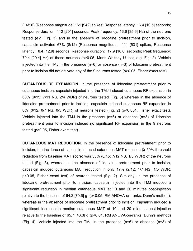
115
(14/16) (Response magnitude: 161 [942] spikes; Response latency: 16.4 [10.5] seconds;
Response duration: 112 [201] seconds; Peak frequency: 16.6 [35.6] Hz) of the neurons
tested (e.g. Fig. 3) and in the absence of lidocaine pretreatment prior to incision,
capsaicin activated 67% (8/12) (Response magnitude: 411 [531] spikes; Response
latency: 8.4 [12.9] seconds; Response duration: 17.9 [18.0] seconds; Peak frequency:
70.4 [29.4] Hz) of these neurons (p>0.05, Mann-Whitney U test; e.g. Fig. 2). Vehicle
injected into the TMJ in the presence (n=6) or absence (n=3) of lidocaine pretreatment
prior to incision did not activate any of the 9 neurons tested (p>0.05, Fisher exact test).
CUTANEOUS RF EXPANSION. In the presence of lidocaine pretreatment prior to
cutaneous incision, capsaicin injected into the TMJ induced cutaneous RF expansion in
60% (9/15; 7/11 NS, 2/4 WDR) of neurons tested (Fig. 3) whereas in the absence of
lidocaine pretreatment prior to incision, capsaicin induced cutaneous RF expansion in
0% (0/12; 0/7 NS, 0/5 WDR) of neurons tested (Fig. 2) (p<0.001, Fisher exact test).
Vehicle injected into the TMJ in the presence (n=6) or absence (n=3) of lidocaine
pretreatment prior to incision induced no significant RF expansion in the 9 neurons
tested (p>0.05, Fisher exact test).
CUTANEOUS MAT REDUCTION. In the presence of lidocaine pretreatment prior to
incision, the incidence of capsaicin-induced cutaneous MAT reduction (≥ 50% threshold
reduction from baseline MAT score) was 53% (8/15; 7/12 NS, 1/3 WDR) of the neurons
tested (Fig. 3), whereas in the absence of lidocaine pretreatment prior to incision,
capsaicin induced cutaneous MAT reduction in only 17% (2/12; 1/7 NS, 1/5 WDR;
p>0.05, Fisher exact test) of neurons tested (Fig. 2). Similarly, in the presence of
lidocaine pretreatment prior to incision, capsaicin injected into the TMJ induced a
significant reduction in median cutaneous MAT at 10 and 20 minutes post-injection
relative to the baseline of 64.2 [70.6] g (p<0.05, RM ANOVA-on-ranks, Dunn’s method),
whereas in the absence of lidocaine pretreatment prior to incision, capsaicin induced a
significant increase in median cutaneous MAT at 10 and 20 minutes post-injection
relative to the baseline of 65.7 [46.3] g (p<0.01, RM ANOVA-on-ranks, Dunn’s method)
(Fig. 4). Vehicle injected into the TMJ in the presence (n=6) or absence (n=3) of

116
lidocaine pretreatment prior to incision induced no significant cutaneous MAT reduction
in the 9 neurons tested (p>0.05, Fisher exact test).
TMJ MAT REDUCTION. There was no significant difference in baseline TMJ MAT (i.e.
after cutaneous incision to expose the TMJ for direct assessment) between neurons
tested in the absence or presence of lidocaine pretreatment (p>0.05, Mann Whitney U
test) (Fig. 4). In the presence of lidocaine pretreatment prior to incision, the incidence of
capsaicin-induced TMJ MAT reduction (≥ 50% threshold reduction from baseline MAT
score) was 64% (9/14; 8/11 NS, 1/3 WDR) of the neurons tested (Fig. 3), whereas in the
absence of lidocaine pretreatment prior to incision, capsaicin injection into the TMJ
induced TMJ MAT reduction in only 17% (2/12; 2/7 NS, 0/5 WDR; p<0.05, Fisher exact
test) of neurons tested (Fig. 2). Similarly, in the presence of lidocaine pretreatment prior
to incision, capsaicin injected into the TMJ induced a significant reduction in median
TMJ MAT at 20 minutes post-injection relative to the pre-injection baseline of 80.5 [63.1]
g (p<0.01, RM ANOVA-on-ranks, Dunn’s method), whereas in the absence of lidocaine
pretreatment prior to incision, capsaicin induced a significant increase in median TMJ
MAT at 10 minutes post-injection relative to the pre-injection baseline of 47.5 [49.3] g
(p<0.05, RM ANOVA-on-ranks, Dunn’s method) (Fig. 4). Vehicle injected into the TMJ in
the presence (n=6) or absence (n=3) of lidocaine pretreatment prior to incision induced
no significant TMJ MAT reduction in the 9 neurons tested (Fisher exact test, p>0.05).
The above findings of capsaicin-induced activation and central sensitization were
documented in experiments wherein capsaicin was injected into the TMJ 40 minutes
after the cutaneous incision. Such effects were also seen in 3 neurons tested with
capsaicin injected into the TMJ 60 min (n=2) or 90 min (n=1) post-incision; in the
absence of lidocaine pretreatment prior to incision, capsaicin induced activation and
cutaneous RF expansion, cutaneous MAT reduction, and TMJ MAT reduction in all 3
neurons tested.

117
4.4 DISCUSSION:
This is the first study to examine the influence of cutaneous incision injury on central
nociceptive processing of deep afferent inputs evoked by noxious stimuli such as
capsaicin. In addition, our findings have provided the first demonstration in the trigeminal
system that surgical incision of facial skin can readily activate and evoke central
sensitization, a process reflected in cutaneous RF expansion and MAT reduction, in
TMJ-responsive nociceptive neurons in the Vc/UCC and can render most of these
neurons temporarily refractory to sensitization induced by capsaicin injected into the
TMJ. However, local anaesthetic application to the skin prior to incision enables
capsaicin injected into the TMJ to induce central sensitization despite skin incision. Both
peripheral and/or central sensitization mechanisms may contribute to the TMJ MAT
reduction in this study. However, the observed cutaneous RF expansion and cutaneous
MAT reduction cannot be explained by peripheral sensitization and likely reflect central
sensitization since the cutaneous central sensitization is induced by capsaicin applied to
a deep tissue RF site (TMJ) remote from the cutaneous RF sites assessed. These
findings suggest that cutaneous injury may modify central nociceptive processing of
deep tissue afferent inputs.
Surgical incision-induced central sensitization
This is the first study to show that, in the absence of local anaesthetic pretreatment,
surgical incision of facial skin may induce a barrage and subsequent central
sensitization in trigeminal nociceptive neurons receiving deep afferent input. Incision-
induced cutaneous RF expansion and cutaneous MAT reduction may contribute to the
pain spread and referral and to the development of allodynia that have been
documented in postoperative pain states (see Melzack et al. 2001; Kawamata et al.
2002). Differences in surgical preparation, anaesthetic type and concentration,
experimental paradigm, time course and quantification of central sensitization effects
make it difficult to directly compare amongst findings in previous studies as well as with
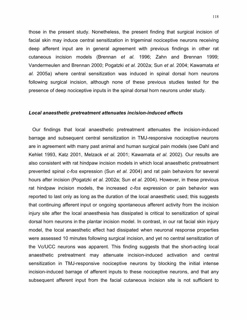
118
those in the present study. Nonetheless, the present finding that surgical incision of
facial skin may induce central sensitization in trigeminal nociceptive neurons receiving
deep afferent input are in general agreement with previous findings in other rat
cutaneous incision models (Brennan et al. 1996; Zahn and Brennan 1999;
Vandermeulen and Brennan 2000; Pogatzki et al. 2002a; Sun et al. 2004; Kawamata et
al. 2005a) where central sensitization was induced in spinal dorsal horn neurons
following surgical incision, although none of these previous studies tested for the
presence of deep nociceptive inputs in the spinal dorsal horn neurons under study.
Local anaesthetic pretreatment attenuates incision-induced effects
Our findings that local anaesthetic pretreatment attenuates the incision-induced
barrage and subsequent central sensitization in TMJ-responsive nociceptive neurons
are in agreement with many past animal and human surgical pain models (see Dahl and
Kehlet 1993, Katz 2001, Melzack et al. 2001; Kawamata et al. 2002). Our results are
also consistent with rat hindpaw incision models in which local anaesthetic pretreatment
prevented spinal c-fos expression (Sun et al. 2004) and rat pain behaviors for several
hours after incision (Pogatzki et al. 2002a; Sun et al. 2004). However, in these previous
rat hindpaw incision models, the increased c-fos expression or pain behavior was
reported to last only as long as the duration of the local anaesthetic used; this suggests
that continuing afferent input or ongoing spontaneous afferent activity from the incision
injury site after the local anaesthesia has dissipated is critical to sensitization of spinal
dorsal horn neurons in the plantar incision model. In contrast, in our rat facial skin injury
model, the local anaesthetic effect had dissipated when neuronal response properties
were assessed 10 minutes following surgical incision, and yet no central sensitization of
the Vc/UCC neurons was apparent. This finding suggests that the short-acting local
anaesthetic pretreatment may attenuate incision-induced activation and central
sensitization in TMJ-responsive nociceptive neurons by blocking the initial intense
incision-induced barrage of afferent inputs to these nociceptive neurons, and that any
subsequent afferent input from the facial cutaneous incision site is not sufficient to

119
induce Vc/UCC central sensitization. Although our observation period for incision-
induced Vc/UCC central sensitization was restricted to the acute phase of injury, our
results provide support for the use of preemptive regional analgesia in addition to
general anaesthesia prior to surgery in order to attenuate or prevent incision-induced
activation and central sensitization in nociceptive pathways both in experimental models
of pain as well as in clinical surgical cases.
Capsaicin-induced neuronal activation and central sensitization
Capsaicin activated and induced central sensitization in a large proportion of TMJ-
responsive nociceptive neurons and the effects were documented in neurons tested with
capsaicin injected into the TMJ at 40, 60 and 90 minutes following cutaneous incision.
The capsaicin-induced effects were especially apparent in the presence of local
anaesthetic pretreatment prior to surgical incision, and are in agreement with those
shown previously in the spinal and trigeminal systems. Capsaicin applied to skin has
been shown to activate and sensitize spinal afferents and dorsal horn neurons (Simone
et al. 1991; LaMotte et al. 1992) and evoke intense pain and secondary hyperalgesia in
humans (LaMotte et al. 1991, 1992; Witting et al. 2000a; Magerl and Treede 2004;
Poyhia and Vainio 2006). TRPV1-immunoreactive sensory neurons have been
demonstrated in both the rat (Helliwell et al. 1998; Guo et al. 1999; Ichikawa and
Sugimoto 2001, 2004) and human (Hou et al. 2002; Renton et al. 2003; Morgan et al.
2005) trigeminal ganglion. TRPV1 receptors have also been demonstrated on trigeminal
afferents innervating the rat TMJ (Ichikawa et al. 2004). Capsaicin has also been shown
to activate and sensitize both trigeminal afferents (Liu and Simon 1994, 1996, 2003;
Strassman et al., 1996; Chapter 2) and Vc nociceptive neurons (Carstens et al. 1998;
Zanotto et al. 2007; Chapter 2) and induce secondary hyperalgesia, allodynia and jaw
muscle pain in humans (Sohn et al. 2000, 2004; Wang et al. 2002; Gazerani and Arendt-
Nielsen 2005; Gazerani et al. 2006). Injection of capsaicin into the TMJ was also shown
to evoke an increase in jaw muscle electromyographic activity (Tang et al. 2004; Chapter
3) with a time course comparable with the Vc/UCC central sensitization changes
revealed in this study. More importantly, the time course of capsaicin-induced activation
120
and central sensitization effects in the present study correlate with findings in human
spinal and trigeminal studies where pain and mechanical sensitization effects were
almost immediate and reached their maximal levels over 15-30 minutes following
capsaicin injection (LaMotte et al. 1991; Simone et al. 1991; Wang et al. 2002; Sohn et
al. 2004; Gazerani and Arendt-Nielsen 2005; Gazerani et al. 2006). Taken together,
these findings suggest a role for peripheral TRPV1 receptor mechanisms in enhanced
pain responses such as allodynia, as reflected in MAT reduction, as well as in pain
spread in deep craniofacial tissues, as reflected in cutaneous RF expansion. However,
the use of specific TRPV1 receptor antagonists should be used to confirm the role of
TRPV1 in trigeminal nociceptive processing.
Incision effects on capsaicin-induced neuronal activation and central sensitization
Our findings suggest that surgical cutaneous incision-induced excitability increases in
TMJ-responsive nociceptive neurons may render most of these neurons temporarily
refractory to sensitization induced by capsaicin injected into the TMJ. Although the
mechanisms underlying noxious mechanical transduction are not fully understood,
several candidate high-threshold mechanotransducers such as TRPs, acid-sensing ion
channels (ASICs), and K+ channels (Hu et al. 2006; Woolf and Ma 2007) may be
activated following surgical cutaneous incision. Peripheral TRP receptors that may be
involved in incision-induced nociceptive mechanisms are the TRPV4 receptor which is
activated by mechanical stimuli and shear stress (Caterina et al. 1997; Nilius et al. 2004;
Woolf and Ma 2007) and the TRPA1 receptor which is activated by chemical (e.g.,
mustard oil), thermal or mechanical noxious stimuli (Kwan et al. 2006; Woolf and Ma
2007). Some of these TRP receptors are coupled to G protein-coupled receptors
(GPCRs) (Bautista et al. 2006; Woolf and Ma 2007) and surgical incision itself could
therefore conceivably activate afferent inputs to the Vc/UCC directly by activating these
GPCR-coupled receptors or indirectly by injury-induced release of inflammatory
mediators that activate GPCR and second messenger-mediated phosphorylation and
sensitization of central EAA receptors (Kress and Zeilhofer 1999; Wallace 2006). The
possible involvement of one or more of these TRP receptor mechanisms in incision-
121
induced nociceptive processing may modulate the effects of subsequent TRPV1
activation on TMJ-responsive nociceptive neurons.
However, a combination of incision-induced excitatory and inhibitory effects most
likely accounts for the TMJ-responsive nociceptive neurons being temporarily refractory
to further central sensitization induced by capsaicin. Surgical incision without local
anaesthetic pretreatment may increase brainstem EAAs and/or activate descending
facilitatory mechanisms (Dubner and Ren 2004) and impose a ceiling effect on
subsequent sensitizing effects of capsaicin by limiting any further capsaicin-evoked
brainstem increases in EAAs. That is, the neurons may have reached their maximal
increases in cutaneous RF expansion and MAT reduction following incision without local
anaesthetic pretreatment such that they could not be sensitized further by capsaicin 40
minutes post-incision. It has also been proposed that the large increase in EAAs in the
spinal cord as a result of noxious stimulation is followed (Sorkin et al. 1992) or
accompanied (Sluka and Westland 1992) by increased concentrations of inhibitory
amino acids (IAAs), which may reflect activation of endogenous inhibitory responses,
consistent with findings in the trigeminal system (Yu et al. 1994; Hu et al. 1997).
Increased concentrations of the IAA serine have been shown to parallel increased
concentrations of the EAAs glutamate and aspartate in the spinal cord for 10-30 minutes
after plantar incision (Zahn et al. 2002). We also have recent evidence that central
inhibitory effects may be activated following a capsaicin-induced afferent barrage and
that these inhibitory effects may contribute to the marked attenuation of glutamate-
evoked activation and central sensitization effects in Vc/UCC nociceptive neurons
following capsaicin injection 40 minutes prior to the injection of glutamate into the TMJ
(Chapter 2). Central sensitization in spinal dorsal horn nociceptive neurons with afferent
input from the cat knee is attenuated by a progressive enhancement of descending
inhibition during the development of inflammation induced by the intra-articular injection
of kaolin and carrageenan (Schaible et al. 1991b). Accordingly, the incision-induced
barrage without local anaesthetic pretreatment may also conceivably activate or
enhance descending inhibitory modulation of Vc/UCC nociceptive neurons from the
brainstem and suprabulbar areas (Chiang et al. 1991, 1994; Sessle et al. 1992; Fields
and Basbaum 1994; Dubner and Ren 2004) although in the case of incision-induced

122
central sensitization, peripheral or segmental inhibitory mechanisms may play a greater
role since it has been reported that incision-induced central sensitization effects on
spinal dorsal horn neurons are not modulated by descending influences from the brain
(Pogatzki et al. 2002b; Kawamata et al. 2005a).
Whether incision-induced central facilitatory or inhibitory actions predominate may
also depend on the magnitude and local distribution of intracellular Ca2+ (Kress and
Zeilhofer 1999; Swope et al. 1999; Rycroft and Gibb 2004). The importance of increased
Ca2+ influx in mediating spinal sensitization after incision has been demonstrated since a
spinally administered antagonist for Ca2+-permeable non-NMDA receptors reduced
central sensitization after incision (Pogatzki et al. 2003; see Pogatzki-Zahn 2006).
Increased intracellular Ca2+ may also activate the phosphatase calcineurin, which can
dephosphorylate and desensitize central EAA receptors (Swope et al. 1999; Rycroft
and Gibb 2004). Indeed, the finding that, in the absence of lidocaine pretreatment prior
to incision, capsaicin induced significant increases in both cutaneous MAT and TMJ
MAT provides further evidence for enhanced central inhibitory mechanisms. This finding
wherein cutaneous injury may modify central nociceptive processing of deep tissue
afferent inputs is consistent with the recent finding that cutaneous injury evoked by
mustard oil application to the rat hindpaw attenuated the subsequent electrically-evoked
increased excitability of spinal nociceptive neurons (Merrill et al. 2008).
Since incision-induced increases in EAAs and IAAs have been shown to return to
baseline 60 minutes post-incision (Zahn et al. 2002; see Pogatzki-Zahn 2006), it
provides an explanation for our present finding that capsaicin can readily induce central
sensitization when injected into the TMJ 60-90 minutes after an incision-induced barrage
without lidocaine pretreatment. Also, we have previously documented that mustard oil
injected into deep craniofacial tissues evokes not only Vc central sensitization and jaw
muscle electromyographic activity but also a subsequent opioid-related inhibitory effect
that persists 50-60 minutes following mustard oil injection into the TMJ (Yu et al. 1993,
1994; Hu et al. 1992, 1997). As a result, the 40-minute period post-incision, coupled with
our previous finding of a 50-60 minute opioid depressive period (Yu et al. 1994; Hu et al.
1997), suggests that the surgical incision-induced refractory period in the present study
may follow a time course on the order of 40-60 minutes post-incision.

123
4.5 TABLE:
Table 1. Summary of statistical comparisons for incision and capsaicin-evoked effects in TMJ-responsive nociceptive neurons in the subnucleus caudalis/upper cervical spinal cord (Vc/UCC) region. Although capsaicin-evoked neuronal activation was equivalent between both groups with and without lidocaine pretreatment of the cutaneous incision site, capsaicin failed to evoke significant central sensitization in TMJ-responsive nociceptive neurons following incision without lidocaine pretreatment.
No Lidocaine +*** +** + -
Lidocaine - - + +*
+ = effect induced by incision or capsaicin, - = no or reduced effect induced by incision or capsaicin
* = p<0.05, ** = p<0.01, *** = p<0.001, Mann-Whitney U-test or Fisher’s exact test as appropriate to
compare Lidocaine vs. No Lidocaine pretreatment groups
PRETREATMENT
OF INCISION SITE
Activation Activation Central
Sensitization
Central
Sensitization
SURGICAL INCISION CAPSAICIN
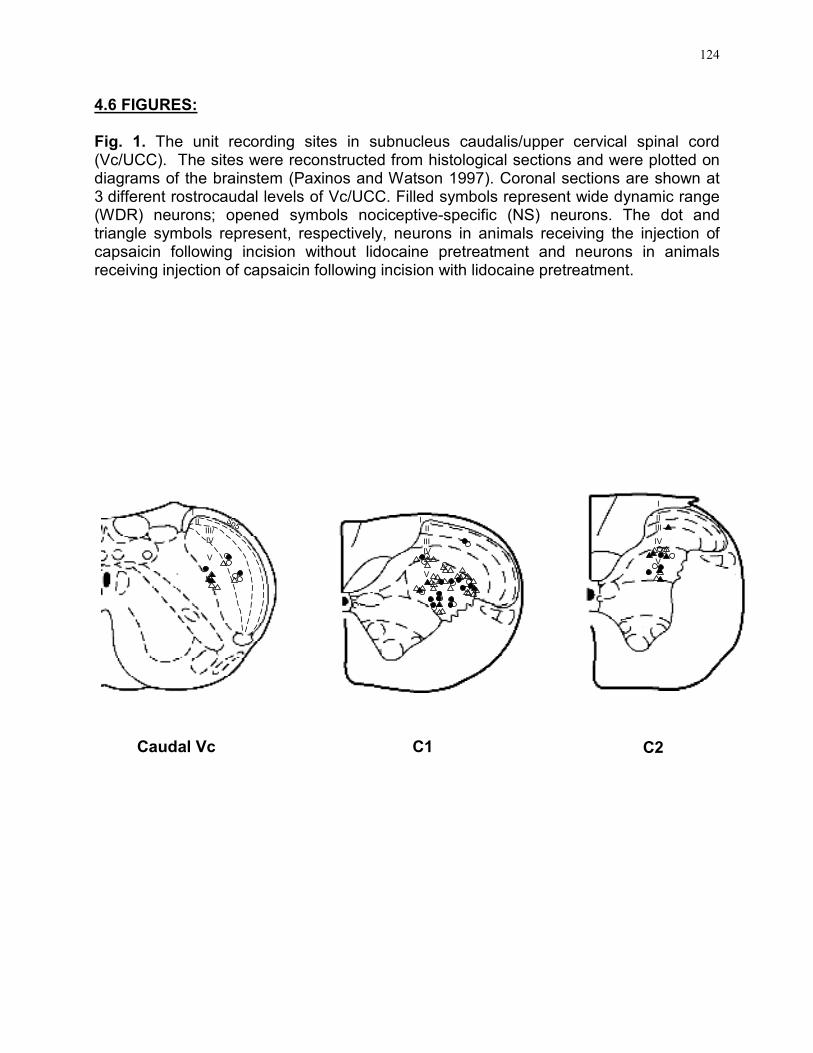
124
4.6 FIGURES:
Fig. 1. The unit recording sites in subnucleus caudalis/upper cervical spinal cord (Vc/UCC). The sites were reconstructed from histological sections and were plotted on diagrams of the brainstem (Paxinos and Watson 1997). Coronal sections are shown at 3 different rostrocaudal levels of Vc/UCC. Filled symbols represent wide dynamic range (WDR) neurons; opened symbols nociceptive-specific (NS) neurons. The dot and triangle symbols represent, respectively, neurons in animals receiving the injection of capsaicin following incision without lidocaine pretreatment and neurons in animals receiving injection of capsaicin following incision with lidocaine pretreatment.
I
II
III
IV
V
III
III
IV
V
Sp5
III
III/
IV
V
C1 C2Caudal Vc

125
Fig. 2. Example of incision and capsaicin-evoked effects in a TMJ-responsive nociceptive neuron in the Vc/UCC region without lidocaine pretreatment of the cutaneous incision site. The asterisk in (a) represents the lesioned neuronal recording site confirmed histologically; tissue stained by conventional hematoxylin and eosin techniques. Note that without lidocaine pretreatment, surgical cutaneous incision of the facial skin overlying the TMJ resulted in a large incision-induced neuronal activation (b), cutaneous mechanoreceptive field (RF) expansion (d) and cutaneous mechanical activation threshold (MAT) reduction (e.g. 100-g von Frey force was ineffective pre-incision but effective post-incision in activating the neuron) (e). In contrast, subsequent neuronal activation induced by capsaicin injection into the TMJ without lidocaine pretreatment (c) did not result in cutaneous RF expansion (d) or cutaneous MAT reduction (e).
PRE-INCISION POST-INCISION/ POST-CAPSAICIN
PRE-CAPSAICIN
RF expansion RF reduction
100 g
5 sec
Evoked
spikes
von
Frey
Force
PRE-INCISION POST-INCISION/ POST-CAPSAICIN
PRE-CAPSAICIN
MAT reduction MAT increase
INCISION
SITE
BRUSH
PINCH
(d) Cutaneous Mechanoreceptive Field (RF):
(e) Cutaneous Mechanical Activation
Threshold (MAT):
(c) TMJ capsaicin-evoked neuronal activation:
(b) Cutaneous incision-evoked neuronal activation:
200
100
0
0 50 100Time (sec)
Incision with no lidocaine pretreatment
100
50
0
Capsaicin injection with no lidocaine pretreatment
0 50 100Time (sec)
Impulse Frequency
(spik
es/s
ec)
Impulse Frequency
(spik
es/s
ec)
100 g100 g
*
(a) Neuronal recording site:

126
Fig. 3. Example of incision and capsaicin-evoked effects in a TMJ-responsive nociceptive neuron in the Vc/UCC region with lidocaine pretreatment of the cutaneous incision site. The asterisk in (a) represents the lesioned neuronal recording site confirmed histologically; tissue stained by conventional hematoxylin and eosin techniques. Note that with lidocaine pretreatment, surgical cutaneous incision of the facial skin overlying the TMJ resulted in minimal incision-induced neuronal activation (b), no cutaneous mechanoreceptive field (RF) expansion (d) or cutaneous mechanical activation threshold (MAT) reduction (e). In contrast, subsequent neuronal activation induced by capsaicin injection into the TMJ with lidocaine pretreatment (c) resulted in both cutaneous RF expansion (d) and cutaneous MAT reduction (e).
Evoked
spikes
von
Frey
Force
(d) Cutaneous Mechanoreceptive Field (RF):
(e) Cutaneous Mechanical Activation
Threshold (MAT):
(c) TMJ capsaicin-evoked neuronal activation:
(b) Cutaneous incision-evoked neuronal activation:
Impulse Frequency
(spik
es/s
ec)
Impulse Frequency
(spik
es/s
ec)
0
0 50 100Time (sec)
Incision with lidocaine pretreatment200
100
100
50
00 50 100
Time (sec)
Capsaicin injection with lidocaine pretreatment
BRUSH
PINCH
Lidocaine
PRE-INCISION POST-INCISION/ POST-CAPSAICIN
PRE-CAPSAICIN
RF no change RF expansion
INCISION
SITE
PRE-INCISION POST-INCISION/ POST-CAPSAICIN
PRE-CAPSAICIN
MAT no change MAT reduction
100 g
5 sec
100 g 100 g
*
(a) Neuronal recording site:

127
Fig. 4. The time course of surgical cutaneous incision and capsaicin-induced cutaneous mechanical activation threshold (MAT) and TMJ MAT reduction in TMJ-responsive nociceptive neurons with (black) or without (white) lidocaine pretreatment. Triangle indicates time point for incision (grey) and injection of capsaicin into the TMJ (black). Squares indicate median normalized cutaneous MAT and circles indicate median normalized TMJ MAT. Raw MAT threshold values were normalized to the initial baseline pre-incision value. Lines: interquartile range. Note that incision alone without lidocaine pretreatment significantly reduced the cutaneous MAT; injection of capsaicin following incision with lidocaine pretreatment significantly reduced both the cutaneous and TMJ MAT, whereas capsaicin injection following incision without lidocaine pretreatment significantly increased the cutaneous and TMJ MAT (*p<0.05, **p<0.01, ***p<0.001, RM ANOVA-on-ranks, Dunn’s method).
Time (minutes)
**
-15INCISION CAPSAICIN
0
50
100
150
200
250
50 6010 20
***
** *
**
**
*
**
Med
ian M
echa
nic
al A
ctivation
Thre
shold
(%
of
Contr
ol)
Cutaneous MAT without lidocaine pretreatment
Cutaneous MAT with lidocaine pretreatment
TMJ MAT with lidocaine pretreatment
TMJ MAT without lidocaine pretreatment
30
Chapter 5
General Discussion
5.0 GENERAL DISCUSSION:
The present doctoral study is the first to provide evidence in support of the
following hypotheses:
Hypothesis 1: Peripheral glutamatergic and capsaicin-sensitive
mechanisms modulate the properties of primary
afferents and brainstem neurons processing deep
craniofacial nociceptive information.
Hypothesis 2: Surgical cutaneous incision modulates the properties of
brainstem neurons processing deep craniofacial
nociceptive information.
Peripheral glutamatergic and capsaicin-sensitive mechanisms do indeed
influence both peripheral and central nociceptive processing in deep craniofacial
tissues and there are differences in peripheral compared to central nociceptive
processing of these peripheral receptor mechanisms. By using the same
experimental paradigm in both peripheral and central processing studies on
glutamate and capsaicin-evoked responses in nociceptive afferents supplying
deep craniofacial tissues, direct comparisons of peripheral versus central
nociceptive neural responses to peripherally applied glutamate and capsaicin
were possible. Both peripheral and/or central sensitization mechanisms may
contribute to enhanced responses to glutamate and capsaicin (e.g., increase in
Rmag, Rdur or Pfreq and/or a decrease in Rlat) and TMJ MAT reduction.
However, cutaneous RF expansion and cutaneous MAT reduction induced by
injection of glutamate and capsaicin likely reflect central sensitization since the
cutaneous central sensitization in TMJ-responsive nociceptive neurons in the
128

129
Vc/UCC region was induced by glutamate and capsaicin applied to a deep tissue
RF site remote from the cutaneous RF sites assessed. We have also shown for
the first time that the increased EMG activity in both jaw-opening and jaw-closing
muscles as a result of capsaicin injection into the TMJ can be attenuated in a
dose-dependent manner by TMJ pre-injection of an NMDA receptor antagonist.
Taken together, these novel findings support Hypothesis 1 and suggest that both
peripheral glutamate and capsaicin receptor mechanisms may be involved in the
nociceptive processing of deep craniofacial afferent inputs and may interact to
modulate both activation as well as peripheral and central sensitization evoked
from these tissues.
In addition, this is the first study to detail the effect of surgical cutaneous
incision on V nociceptive neuroplasticity. The present findings support
Hypothesis 2 and suggest that TMJ-responsive nociceptive neurons in the
Vc/UCC region can be activated by surgical incision of the skin overlying the TMJ
and this incision-induced afferent barrage can cause nociceptive neurons to be
refractory to further central sensitization effects mediated by capsaicin.
5.1 SPECIFIC HYPOTHESES AND STUDY AIMS:
5.1.1 Glutamate and capsaicin activate and induce peripheral sensitization
in nociceptive afferents supplying TMJ or associated musculature
and projecting to the caudal Vc/UCC
Studies addressing AIMS 1-1 to 1-3 have demonstrated that TMJ and muscle
nociceptive afferents projecting to the caudal Vc/UCC are activated and
modulated by peripheral glutamatergic and capsaicin-sensitive mechanisms. The
present study appears to be the first to document the capsaicin-induced
activation and sensitization of V nociceptive afferents with deep craniofacial RFs.
These findings are consistent with previous data that capsaicin evokes dose-
dependent increases in jaw muscle EMG activity when injected into the rat TMJ
(Tang et al. 2004) and induces an inflammatory process within these tissues (Hu
130
et al. 2005a). The present findings are also consistent with studies in humans
that have shown capsaicin-induced craniofacial pain (e.g. Sohn et al. 2000, 2004;
Wang et al. 2002; Gazerani and Arendt-Nielsen 2005; Gazerani et al. 2006), and
with findings that capsaicin can activate and sensitize V afferents (Liu and Simon
1994, 1996, 2003; Strassman et al. 1996) and spinal afferents (Baumann et al.
1991; Peterson and LaMotte 1991; LaMotte et al. 1992; Marchettini 1996; Simone
et al. 1997; Dray 2005; Gold 2005).
Although glutamate-evoked activation and sensitization have not been
demonstrated in spinal primary afferents, the findings in the present study in
deep craniofacial afferents (masseter and temporalis muscle; TMJ) are
consistent with previous evidence of glutamate-evoked activation and
sensitization of masseter nociceptive afferents, jaw muscle EMG activity and pain
in human masseter and trapezius muscles (Cairns et al. 1998, 2001ab, 2002a,
2003ab, 2006; Svensson et al. 2003; Ge et al. 2005).
TMJ and muscle nociceptive afferents projecting to the caudal Vc/UCC are not
only activated and modulated by peripheral glutamatergic and capsaicin-sensitive
mechanisms but there are also interactions between these peripheral
mechanisms that influence the properties of these nociceptive afferents. This is
the first study to document that a large proportion of V nociceptive afferents
innervating deep craniofacial tissues may be activated and sensitized by the
peripheral application of glutamate, capsaicin, or both. These changes in
response properties reflect peripheral sensitization as shown in enhanced
activation to agonist injection (e.g., increase in Rmag and Pfreq) and/or a MAT
reduction in V nociceptive afferents. In addition, glutamate sensitized afferent
responses to subsequent noxious stimulation of the deep craniofacial tissues by
capsaicin, whereas capsaicin neither sensitized nor desensitized afferent
responses to subsequent noxious stimulation by glutamate. These findings
suggest that both peripheral glutamatergic and capsaicin-sensitive mechanisms
may be involved in the activation and sensitization of deep craniofacial
nociceptive afferents and may interact to modulate the activation and peripheral
sensitization in some nociceptive afferents supplying deep craniofacial tissues.
131
5.1.2 Glutamate and capsaicin activate and induce central sensitization in
TMJ-responsive nociceptive neurons in the caudal Vc/UCC
Studies addressing AIMS 1-4 to 1-6 have demonstrated that TMJ-responsive
nociceptive neurons in the caudal Vc/UCC are not only activated and modulated
by peripheral glutamatergic and capsaicin-sensitive mechanisms but there are
also interactions between these peripheral receptor mechanisms that influence
the properties of nociceptive neurons in the CNS. This is the first study to
document that a large proportion of TMJ-responsive nociceptive neurons in the
caudal Vc/UCC can be activated and sensitized by the peripheral application to
the TMJ of glutamate, capsaicin or both, and that peripheral glutamate and
capsaicin receptor mechanisms interact to modulate activation and central
sensitization in deep craniofacial tissues. Glutamate applied to the TMJ may
sensitize TMJ-responsive nociceptive neurons, and thereby produce more
immediate, larger and more prolonged responses to noxious stimulation of the
TMJ by capsaicin. In contrast, capsaicin applied to the TMJ may desensitize
these neurons to subsequent noxious stimulation of the TMJ by glutamate.
The present findings of a role for peripheral glutamatergic and capsaicin-
sensitive mechanisms in central processing of nociceptive afferent inputs to
Vc/UCC from deep craniofacial tissues are in agreement with previous findings of
an important role for peripheral EAA and TRPV1 receptors in central
sensitization, and are consistent with both V (Carstens et al. 1998; Sohn et al.
2000, 2004; Wang et al. 2002; Gazerani and Arendt-Nielsen 2005; Gazerani et
al. 2006; Zanotto et al. 2006) and spinal (LaMotte et al. 1991,1992; Simone et al.
1991; Raja et al. 1999; Gottrup et al. 2000, 2004; Witting et al. 2000ab; Sluka
2002; Gazerani et al. 2006; Poyhia and Vainio 2006) evidence in animals and
humans that suggest a role for central sensitization (e.g., secondary
hyperalgesia) in mediating capsaicin-evoked pain in peripheral tissues. Similarly,
this project’s findings for Vc/UCC nociceptive neurons are consistent with
previous evidence of glutamate-evoked jaw muscle EMG activity that could be
attenuated by NMDA receptor antagonist application to Vc and of pain evoked by

132
glutamate in human masseter and trapezius muscles (Cairns et al. 1998, 2001a,
2003ab, 2006; Svensson et al. 2003; Ge et al. 2005). The present findings are
also consistent with previous spinal evidence of thermal and mechanical
hyperalgesia induced by subcutaneous or intra-articular (knee) glutamate
injection (for review, see Carlton 2001; Carlton et al. 2003). In addition,
peripheral application of a selective EAA receptor antagonist has been shown to
attenuate irritant chemical-induced hyperalgesia and dorsal horn c-fos
expression in animals (Jackson et al. 1995; Davidson et al. 1997; Lawand et al.
1997; Wang et al. 1997; Davidson and Carlton 1998; Carlton 2001; Carlton et al.
2003) and inhibit the development of secondary hyperalgesia in humans
(Warncke et al. 1997).
5.1.3 Differences in peripheral and central nociceptive processes evoked
by the peripheral application of glutamate and capsaicin to deep
craniofacial tissues
The same experimental paradigm was employed in the studies of V primary
nociceptive afferents (AIMS 1-1 to 1-3) and caudal Vc/UCC neurons (AIMS 1-4 to
1-6) to test for possible modulating effects of glutamate and capsaicin in order to
allow direct comparisons between peripheral and central nociceptive responses
to the peripheral application of glutamate and capsaicin. Our findings that the
glutamate-evoked responses were similar to those evoked by capsaicin in TMJ-
responsive nociceptive neurons contrast with our V nociceptive primary afferent
results where glutamate-evoked responses in the nociceptive afferents were
significantly greater than capsaicin-evoked responses. These findings suggest
the involvement of convergent nociceptive signaling pathways may contribute to
the markedly enhanced incidence of activation and response properties evoked
by glutamate and capsaicin in Vc/UCC neurons.
The finding that both V nociceptive afferent and brainstem nociceptive
neuronal responses to capsaicin injection were significantly increased following
glutamate injection suggests that the activation of peripheral glutamate receptors
by glutamate may sensitize both primary afferents and Vc/UCC nociceptive

133
neurons supplying deep craniofacial tissues to subsequent stimuli. Peripheral
NMDA receptors, in particular, may play a role in mediating the glutamate-
induced peripheral and/or central sensitization as evidenced by our finding that
jaw muscle activities reflexly evoked by capsaicin injection into the rat TMJ can
be attenuated by preinjection into the TMJ of NMDA receptor antagonists. These
results suggest that NMDA and possibly capsaicin receptors are located within
the TMJ and the activation of NMDA receptors contributes, in part, to the
mechanism whereby capsaicin evokes nociceptive responses. One possible
mechanism involving autocrine and/or paracrine regulation of nociceptive
excitability via ionotropic glutamate receptors may contribute to the mechanisms
whereby capsaicin evokes nociceptive jaw muscle activity. Similar peripheral
NMDA receptor mechanisms may also play an important role in the nociceptive
responses evoked by mustard oil. Jaw muscle activity evoked by mustard oil
injected into the TMJ (Yu et al. 1996) as well as nocifensive behaviour (Ro 2003),
oedema formation (Ro 2003) and c-fos expression in the V brainstem nuclei (Ro
et al. 2004) evoked by mustard oil injected into the masseter are also similarly
attenuated by local TMJ or muscle MK-801 pretreatment. In addition to possible
interactions between ionotropic receptors, there is evidence of a major coupling
between G-protein-coupled receptors and some TRP channels in the membrane
such as TRPA1 and TRPV1 (Sikand and Premkumar 2007; Woolf and Ma 2007).
This type of coupling may also exist between TRPV1 and other G-protein-
coupled receptors (Woolf and Ma 2007), such as the metabotropic EAA
receptors, and provides a means for potential interactions between peripheral
EAA and TRPV1 receptors.
The interactions between peripheral glutamate and capsaicin receptors on
deep craniofacial afferent inputs to Vc/UCC nociceptive neurons suggest a
convergent nociceptive signaling pathway (see Fig. 1). That is, the nociceptive
signaling pathways are not separate; the glutamate and capsaicin-activated deep
craniofacial afferents converge on Vc/UCC neurons. In contrast to the glutamate-
induced sensitizing effects on deep craniofacial afferent responses to
subsequent noxious stimulation by capsaicin, there was a lack of capsaicin-

134
induced modulation of glutamate-evoked afferent excitability. Indeed many of the
afferents remained responsive to glutamate following capsaicin injection.
However, the glutamate-evoked activation responses and incidence of cutaneous
RF expansion in the TMJ-responsive nociceptive Vc/UCC neurons following
capsaicin injection were abolished or significantly reduced. These findings
suggest that capsaicin may desensitize Vc/UCC neurons to subsequent noxious
stimuli (e.g. to glutamate) which is consistent with previous evidence showing
that capsaicin may desensitize Vc neurons to subsequent activation by various
irritant chemicals applied to craniofacial tissues (Carstens et al. 1998).
It is possible that there may be differences in the types of convergent presynaptic
receptors or the neurotransmitters released from the central terminals of
capsaicin-sensitive and glutamate-sensitive afferents that may account for the
differential sensitizing or desensitizing effects on Vc/UCC neurons (Woolf and
Salter 2000; Dubner 2005; Woolf and Ma 2007). The variety of these
neurotransmitters may result in considerable spatial and temporal summation
capabilities that are subject to both excitatory and inhibitory influences. The
capsaicin-induced desensitizing effects on Vc/UCC neurons may involve
increased inhibitory GABAergic synaptic transmission and alterations in cation-
chloride cotransporters such as NKCC1 or KCC2 (Galan and Cervero 2005;
Karlsson et al. 2005; Price et al. 2005, 2006; Garcia-Nicas et al. 2006; Pitcher et
al. 2007). The present findings are also consistent with recent evidence of other
peripheral TRP receptors such as TRPA1 participating in the activation of central
inhibitory circuits (Kosugi et al. 2007; Merrill et al. 2008). Thus it is possible that
the capsaicin-induced desensitization in Vc/UCC neurons in the present study
may occur via the activation of similar central inhibitory circuits.
5.1.4 Targeting peripheral glutamate and capsaicin receptor mechanisms
The glutamate-evoked sensitization of primary afferent fibers in the present
study may contribute to the primary hyperalgesia or allodynic states found in
craniofacial pain conditions such as TMD (for review, see Carlsson and
LeResche 1995, Stohler 1995,1999; Sessle 1999, 2005; Svensson and Sessle
135
2004; Lam et al. 2005). Glutamate injection into the TMJ also evoked activity and
central sensitization in Vc/UCC nociceptive neurons. This glutamate-evoked
central sensitization, reflected as RF expansion, MAT reduction, and increase in
neuronal responses to subsequent noxious stimuli, may be central mechanisms
contributing to pain spread and referral, allodynia, pain at rest, and hyperalgesia
in craniofacial pain conditions. However, further studies are required to achieve a
better understanding of the role of peripheral glutamate receptors in the
pathobiological mechanisms underlying craniofacial pain conditions. These
should involve the use in female as well as male subjects of specific ionotropic
and metabotropic EAA receptor agonists, antagonists and gene knockouts in
both acute and chronic craniofacial pain models studying peripheral (primary
afferent) and central (Vc, thalamic, somatosensory cortex) nociceptive
processing. Moreover, since the present study suggests that peripheral
glutamate receptors may modulate the activity of other peripheral nociceptors
(i.e., TRPV1 receptor), future studies could examine the modulatory role of
peripheral EAA receptors on other peripheral nociceptors (e.g., P2X receptors).
The demonstration of a relationship between peripheral glutamate receptor
mechanisms and craniofacial pain may lead to the development of novel
diagnostic and therapeutic approaches for TMD and other craniofacial pain
conditions of peripheral origin. Thus, peripheral glutamate receptors may be
potential novel targets for the treatment of craniofacial pain conditions and may
provide a non-opioid rationale for pain therapy. The formulation of specific
peripheral ionotropic glutamate receptor antagonists that do not cross the blood
brain barrier may be of potential benefit by reducing peripheral nociceptive
excitability while avoiding any harmful central side effects associated with central
glutamate receptor antagonism.
Currently, TRPV1 receptors have been targeted successfully in the treatment of
some neuropathic pain and chronic pain syndromes such as postherpetic
neuralgia, postmastectomy neuroma, reflex sympathetic dystrophy syndrome,
diabetic neuropathy, and rheumatoid arthritis (Rumsfield and West 1991;
Sawynok 2005). However, further studies are needed to elucidate the role of

136
TRPV1 receptors in deep craniofacial pain. These include some approaches
analogous to those suggested above to elucidate glutamate receptor
mechanisms (e.g., sex differences), plus the use of specific TRPV1 receptor
agonists, antagonists and gene knockouts in both acute and chronic craniofacial
pain models studying peripheral (primary afferent) and central (Vc, thalamic,
somatosensory cortex) nociceptive processing. Moreover, the role of other
members of the TRP family (e.g., TRPA1, see Jordt et al. 2004) and any sex
differences in these receptor properties in craniofacial pain should also be
investigated with receptor-specific agonists and antagonists. The demonstration
of a relationship between peripheral capsaicin receptor mechanisms and
craniofacial pain may lead to the further development of diagnostic and
therapeutic approaches for TMD and other craniofacial pain conditions of
peripheral origin. However, since the present study suggests that peripheral
glutamate receptors, NMDA in particular, may modulate TRPV1 receptor activity,
the targeting of peripheral glutamate receptors may be a novel approach to
indirectly target TRPV1 receptors.
5.1.5 Surgical incision-induced excitability
Studies addressing AIMS 2-1 to 2-2 are the first to demonstrate that surgical
cutaneous incision can readily activate and evoke central sensitization in TMJ-
responsive nociceptive neurons in the Vc/UCC and can render most of these
neurons temporarily refractory to sensitization induced by capsaicin injected into
the TMJ. Our findings that local anaesthetic pretreatment attenuates the incision-
induced barrage and subsequent central sensitization in TMJ-responsive
nociceptive neurons are in general agreement with many past animal and human
surgical pain models (for review, see Dahl and Kehlet 1993, Abram 1996, Katz
2001, Melzack et al. 2001; Kawamata et al. 2002; Pogatzki et al. 2002a; Sun et
al. 2004). However, there are some interesting differences demonstrated
between incision-induced effects on V compared to spinal nociceptive
processing. Previous studies have shown minimal to no changes in cutaneous
and deep (muscle) tissue incision-induced cutaneous MAT reduction

137
(Vandermeulen and Brennan 2000) or cutaneous RF size (Kawamata et al.
2005a), whereas cutaneous incision-induced cutaneous MAT reduction and
cutaneous RF expansion were markedly robust in Vc/UCC nociceptive neurons.
This difference suggests that the V system may be more readily sensitized to
peripheral injury than the spinal system. However, the effect of cutaneous
incision on spinal nociceptive neurons with deep afferent inputs has not been
investigated since the above studies did not test for the presence of a deep
afferent RF in the spinal dorsal horn neurons tested. Incision-induced cutaneous
RF expansion and cutaneous MAT reduction may contribute to the pain spread
and referral and to the development of allodynia that have been documented in
postoperative pain states (for review, see Melzack et al. 2001; Kawamata et al.
2002).
Particularly interesting is that surgical incision can induce central sensitization
in TMJ-responsive nociceptive neurons and can render most of these neurons
temporarily refractory to sensitization induced by capsaicin injected into the TMJ.
This finding suggests that cutaneous injury may modify central nociceptive
processing of deep tissue afferent inputs. Possible explanations for the observed
effects may be due to incision-induced activation of TRP channels coupled to G
protein-coupled receptors, competing peak excitatory and inhibitory responses in
the brainstem, or descending inhibitory modulation.
The present results provide support for the use of preemptive regional
analgesia in addition to general anaesthesia prior to surgery in order to attenuate
or prevent incision-induced activation and central sensitization in nociceptive
pathways both in experimental models of pain as well as in clinical surgical
cases. Furthermore, given that this is the first study to suggest that there may be
differences in sensitivity to surgical incision between V and spinal systems,
further studies of incision-induced injury on V processing are warranted. These
include some approaches analogous to those suggested above to elucidate
glutamate and capsaicin receptor mechanisms, plus studies on the possible
incision-induced central excitatory and inhibitory mechanisms that may contribute
to the subsequent refractory period in VC/UCC nociceptive neurons. Future

138
studies may also investigate potential differences between superficial and deep
tissue injury as well as acute and chronic injury on peripheral (primary afferent)
and central (Vc, thalamic, somatosensory cortex) craniofacial nociceptive
processing. Moreover, in addition to studies with local anaesthetics, further
investigations of the role of peripheral receptor antagonists (e.g., EAA and
TRPV1 receptor antagonists) in incision-induced craniofacial nociceptive
processing may provide novel targets for the treatment and management of
traumatic tissue injuries and surgical patients.
5.2 STUDY LIMITATIONS:
5.2.1 Identification of nociceptive neurons supplying deep craniofacial
tissues
The accurate identification of deep RFs in these studies is complicated by two
factors. First, when pressure, chemical or electrical stimuli are applied to deep
structures, they can activate afferent fibers remote from the stimulus source.
Second, extensive dissection is required with a risk of damaging the afferent
terminal/receptor itself (Bove and Light 1995). In spite of these limitations, we
have employed a novel methodology to study identified afferent fibers that project
from the TMJ or masticatory muscles to the caudal Vc/UCC (see Cairns et al.
2001ab, 2002a) as well as TMJ-responsive nociceptive neurons in the caudal
Vc/UCC. We avoided damaging the tissue and associated afferent terminals
while studying the afferent responses to glutamate and capsaicin by delaying the
verification of the afferent’s RF until the end of the experiment where skin and
connective tissue overlying the RF was removed and mechanical and electrical
stimuli reapplied. Similarly, in the study of TMJ-responsive Vc/UCC neurons, our
conservative surgical preparation allowed the craniofacial tissues within the
neuronal RF being studied to remain intact throughout the experiment with the
exception of the small (<2mm long) incision in the experiments involving local
anaesthetic pretreatment of skin overlying the TMJ. We have demonstrated that
the local anaesthetic pretreatment of skin overlying the TMJ serves to attenuate

139
the surgical incision-induced activation and central sensitization in TMJ-
responsive nociceptive neurons. As a result, the more conservative surgical
preparation in the present study has allowed us to study nociceptive neurons
closer to their resting state with minimal prior excitability.
5.2.2 Assessment of neuronal sensitization
The use of inhalational anaesthesia may potentially confound the assessment
of neuronal excitability since halothane may modulate thermal responses of
cutaneous nociceptive afferents (Campbell et al. 1984) and the MAT of spinal
nociceptive neurons (Herrero and Headley 1995; Yamamori et al. 1995).
However, the concentrations employed in these studies were different than that
utilized in the present study and the effect of halothane on the response of deep
afferents to chemical stimuli has yet to be evaluated. Other studies have not
shown a difference in sensitization of rat dorsal horn neurons between 1%
halothane anaesthesia, pentobarbital anaesthesia or decerebrate preparations
(Laird and Cervero 1989). Thus, although unlikely, the anaesthetic regimen could
have lead to iatrogenically enhanced primary afferent or central neuronal
responses.
A further limitation of the study of nociceptive processing is that despite the
more conservative surgical measures instituted, the surgery required to expose
the V ganglion and/or the caudal Vc/UCC may excite and/or sensitize the afferent
or neuron under study. Moreover, the determination of sensitization relies on
repeated mechanical stimuli - which can, in itself, result in sensitization. In order
to prevent or limit this iatrogenic sensitization, we instituted a novel protocol that
limited the stimulation magnitude, duration, frequency and extent required to
determine sensitization. The cutaneous RF expansion in the present study was
defined as expansion in cutaneous RF size to include a predetermined point lying
5 mm outside the perimeter of the baseline RF. However, this approach does not
measure the full extent of RF expansion and the RF may not expand in the
direction of the predetermined point; as a result, the true incidence of RF
expansion may actually be underrepresented with this methodology. In addition,

140
most of these Vc/UCC neurons tested for central sensitization were located in the
deep laminae of the Vc/UCC. Thus further investigations of the effects of
glutamate, capsaicin and surgical incision on neurons in the superficial laminae
are warranted in future studies, given the importance of this region in central
nociceptive processing.
5.2.3 Evidence for peripheral EAA and TRPV1 receptor mechanisms in deep
craniofacial nociception
Studies from this thesis project have demonstrated that peripheral
glutamatergic and capsaicin-sensitive mechanisms do indeed influence deep
craniofacial nociceptive processing. However, with the exception of a role for
peripheral NMDA receptors in capsaicin-evoked EMG activity (see Chapter 2),
there is a lack of direct evidence for NMDA, non-NMDA and TRPV1 receptor
mechanisms in V nociception in the present studies. A role for both NMDA and
non-NMDA receptors in deep craniofacial nociceptive processing has been
previously demonstrated in animal (Cairns et al. 1998; Dong et al. 2006, 2007)
and human craniofacial pain models (Cairns et al. 2001a, 2003ab, 2006;
Svensson et al. 2003, 2005). Thus, the results of this thesis project may be
consistent with the involvement of functional NMDA and non-NMDA receptor
mechanisms in deep craniofacial nociception shown previously.
In the craniofacial region, TRPV1 receptors have also been demonstrated on
V afferents innervating the rat TMJ (Ichikawa et al. 2004; Ioi et al. 2006).
Capsaicin injection into the rat TMJ reflexly evokes a dose-dependent increase in
jaw muscle EMG activity (Tang et al. 2004) and produces an inflammatory
response within these tissues that can be blocked by TRPV1 receptor
antagonists (Hu et al. 2005a). Since TRPV1 receptors are activated by capsaicin
(Caterina et al. 1997; Tominaga et al. 1998; Benham et al. 2003; Jordt et al.
2003), the results of this study suggest there may be functional TRPV1 receptors
located within the TMJ region. However, the use of specific receptor antagonists
would be required to confirm the involvement of the TRPV1 receptor.

141
5.2.4 Sex differences in deep craniofacial nociception
It was beyond the scope of the present study to investigate sex-related
differences in V nociceptive processing. However, the prevalence of TMD-related
pain is higher among women of reproductive age compared to postmenopausal
women or to men (Carlsson and LeResche 1995, LeResche et al. 1997; for
review, see Fillingim and Ness 2000). Estrogen receptors are found in muscle
(Dalberg 1982; Meyer and Raap 1985), TMJ (Aufdemorte et al. 1986; Milam et
al. 1987), and dorsal root ganglion neurons (Sohrabji et al. 1994), indicating that
deep tissues as well as peripheral ganglia are potential targets for sex steroids to
modulate sensory functions. Also, in view of the finding of Vc estrogen receptors
(e.g., Amandusson et al. 1996; Bereiter et al. 2005b), central actions of estrogen
on nociception cannot be ruled out. The present study used only male rats and
consequently could not investigate sex-related differences in V nociceptive
processing of glutamatergic and capsaicin-sensitive mechanisms. Previous
studies in animal (Cairns et al. 2001ab, 2002b; Dong et al. 2007) and human
(Cairns et al. 2001a, 2003a,b, 2006; Svensson et al. 2003; Castrillon et al. 2007)
craniofacial pain models support the view that females may exhibit an enhanced
neural responsiveness to glutamate injected into the deep craniofacial tissues,
which may be due, in part, to a sex-related increase in the excitability of V
primary afferents as well as central nociceptive neurons. However, it is not
known whether there are any sex differences with respect to peripheral TRPV1
receptor mechanisms nor if there are any sex-related interactions between
peripheral EAA and TRPV1 receptor mechanisms. As noted above, this would be
a fruitful future direction.
5.2.5 Translation of experimental animal models to human pain states
The findings in the present rat TMJ injury model suggest that peripheral
glutamatergic and capsaicin-sensitive mechanisms may contribute to craniofacial
pain states in humans. However, one may question the applicability of translating
the results of experimental animal models to human pain states due to genetic
and species differences. In this context, there is a strong genetic influence on rat

142
responses to stress and susceptibility to inflammatory conditions (Cizza and
Sternberg 1994). Similarly, genetics influence morphine effects on mice (Mogil et
al. 1996; Belknap et al. 1998; Elmer et al. 1998) as well as mice responses to
various nociceptive tests (Mogil 1999, see 2005). There is also remarkable
variability among humans in terms of susceptibility to chronic orofacial pain and
its characteristics, which suggests that these syndromes are complex heritable
traits controlled by alleles of certain polymorphic genes that interact with the
environment (Seltzer et al. 2004).
In many respects, the organization of the somatosensory system of the rat is
comparable to that of other animals including humans (Kunc 1970; Afshar and
Dykes 1984; Dallel et al. 1989; Tracey and Waite 1995; Brown 1997; Usunoff et
al. 1997; Pajot et al. 2000; Jawahar et al. 2001; Paxinos 2004). Taken together,
data from rats, cats and monkey suggest similar organizational and functional
features of primary afferents and spinal and medullary dorsal horns (Grant 1995;
Paxinos 2004). Although there are some differences with higher-order structures,
e.g., unlike carnivores and primates, no interneurons or GABAergic cells are
present in the VPM of rats (Barbaresi et al. 1986; Williams and Faull 1987, Waite
and Tracey 1995; Amadeo et al. 2001), and the representation in the CNS of the
face and vibrissae in the rat occupies a disproportionately larger area in the
primary somatosensory cortex than in many higher mammals (Welker 1971;
Waite and Tracey 1995; Brett-Green et al. 2001; Frostig 2006). In addition,
despite the mandibular condyle extending anterior-posteriorly in rats as opposed
to the lateromedial direction in humans, the rat TMJ articular apparatus is very
similar to that of humans in that they both possess a reciprocally fitting
condylomeniscal joint and meniscotemporal joint (Bermejo et al. 1993; Fletcher
et al. 2004). More importantly, the demonstration of a novel nociceptive role for
peripheral glutamatergic and capsaicin-sensitive mechanisms in our animal
models agrees with findings in humans (for review, see Chapter 1).
Although the same limitations as above apply to the incision study, our
findings that local anaesthetic pretreatment attenuates the surgical cutaneous
incision-induced barrage and subsequent central sensitization in TMJ-responsive

143
nociceptive neurons are in agreement with past animal and human surgical pain
models (see Dahl and Kehlet, 1993, Abram, 1996, Katz, 2001, Melzack et al.,
2001; Kawamata et al., 2002). Moreover, although our observation period for
incision-induced central sensitization was only during the acute phase of injury,
our findings provide support for the use of preemptive regional analgesia in order
to attenuate or prevent incision-induced activation and central sensitization in
nociceptive pathways in clinical surgical cases.

144
5.3 FIGURE:
Fig. 1. Schematic diagram of glutamate and capsaicin-evoked activation responses and the possible convergent trigeminal nociceptive afferent inputs to Vc/UCC nociceptive neurons. Note that for both the glutamate followed by capsaicin subgroup and the capsaicin followed by glutamate subgroup, whereas there were four types of agonist-responsive afferents, there were only two types of agonist-responsive Vc/UCC neurons activated following injection of glutamate and capsaicin into deep craniofacial tissues. In addition, the vast majority of the Vc/UCC neurons were capsaicin-sensitive in both subgroups (100% of the glutamate followed by capsaicin subgroup and 90% of the Vc/UCC neurons were capsaicin followed by glutamate subgroup), whereas there were no glutamate-sensitive Vc/UCC neurons in the capsaicin followed by glutamate subgroup. Possible synaptic inputs depicted in black refer to inputs effective in activating the neurons and those in gray refer to relatively ineffective inputs. %: Percentages of each type of agonist-responsive deep craniofacial afferent or Vc/UCC nociceptive neuron found in the glutamate followed by capsaicin subgroup or capsaicin followed by glutamate subgroup.
Glutamate followed by Capsaicin
92% 8%
15%30% 18% 37%
Deep
Craniofacial
Nociceptive
Afferents
Possible
Synaptic
Inputs
Vc/UCC
Nociceptive
Neurons
G+ C- G+C+ G- C+ G- C-
G+ C- G+C+ G- C+ G- C-
Capsaicin followed by Glutamate
90%
16% 47%
10%
11%26%
C- G+ C+G+ C+ G- C- G-
C- G+ C+G+ C+ G- C- G-
0% 0% 0% 0%
G+ Glutamate-sensitive
C+ Capsaicin-sensitive
G- Glutamate-insensitive
C- Capsaicin-insensitive
Effective input
Ineffective input
REFERENCES:
Abram SE. Importance of preincisional analgesia. Reg Anesth 21:117-121,
1996.
Afrah AW, Stiller CO, Olgart L, Brodin E, and Gustafsson H. Involvement of
spinal N-methyl-D-aspartate receptors in capsaicin-induced in vivo release of
substance P in the rat dorsal horn. Neurosci Lett 316:83-86, 2001.
Afshar F and Dykes E. Computer-generated three-dimensional visualization of
the trigeminal nuclear complex. Surg Neurol 22:189-196, 1984.
Ahn DK, Kim KH, Jung CY, Choi HS, Lim EJ, Youn DH and Bae YC. Role of
peripheral group I and II metabotropic glutamate receptors in IL-1beta-induced
mechanical allodynia in the orofacial area of conscious rats. Pain 118:53-60,
2005.
Alfredson H and Lorentzon RJ. Chronic tendon pain: no signs of chemical
inflammation but high concentrations of the neurotransmitter glutamate
Implications for treatment? Curr Drug Targets 3:43–54, 2002.
Alfredson H, Forsgren S, Thorsen K, and Lorentzon RJ. In vivo microdialysis
and immunohistochemical analyses of tendon tissue demonstrated high
amounts of free glutamate and glutamate NMDAR1 receptors, but no signs of
inflammation, in Jumper’s knee. Orthop Res 19:881–886, 2001.
Alfredson H, Ljung BO, Thorsen K, and Lorentzon RJ. In vivo investigation of
ECRB tendons with microdialysis technique – no signs of inflammation but
high amounts of glutamate in tennis elbow. Acta Orthop Scand 71:475–479,
2000.
Amadeo A, Ortino B, Frassoni C. Parvalbumin and GABA in the developing
somatosensory thalamus of the rat: an immunocytochemical ultrastructural
correlation. Anat Embryol (Berl) 203:109-119, 2001.
145

146
Amador M, and Dani JA. MK-801 inhibition of nicotinic acetylcholine receptor
channels. Synapse 7:207-15, 1991.
Amandusson A, Hermanson O and Blomqvist A. Colocalization of oestrogen
receptor immunoreactivity and preproenkephalin mRNA expression to
neurons in the superficial laminae of the spinal and medullary dorsal horn of
rats. Eur J Neurosci 8:2440-2445, 1996.
Ambalavanar R, Moritani M, Dessem D. Trigeminal P2X3 receptor expression
differs from dorsal root ganglion and is modulated by deep tissue
inflammation. Pain 117:280-291, 2005.
American Pain Society. Principles of Analgesic Use in the Treatment of Acute
Pain and Cancer Pain. Fifth Edition. Glenview: American Pain Society 2003.
Arendt-Nielsen L and Svensson P. Referred muscle pain: basic and clinical
findings. Clin. J. Pain 17:11-19, 2001.
Aufdemorte TB, Van Sickels JE, Dolwick MF, Sheridan PJ, Holt GR, Aragon
SB and Gates GA. Estrogen receptors in the temporomandibular joint of the
baboon (Papio cynocephalus): an autoradiographic study. Oral. Surg Oral
Med Oral Pathol 61:307-314, 1986.
Ault B, Hildebrand LM. Activation of nociceptive reflexes by peripheral kainate
receptors. J Phamacol Exp Ther 265:927–932, 1993a.
Ault B, Hildebrand LM. L-Glutamate activates peripheral nociceptors. Agents
Actions 39:142–144, 1993b.
Australian and New Zealand College of Anaesthestists (ANZCA) and
Faculty of Pain Medicine. Acute Pain Management: Scientific Evidence,
Second Edition 2005.
Azerad J, Boucher Y and Pollin B. Demonstration of glutamate in primary
sensory trigeminal neurons innervating dental pulp in rats. C R Acad Sci III
314:469-475, 1992.
Bakke M, Hu JW and Sessle BJ. Involvement of NK-1 and NK-2 tachykinin
receptor mechanisms in jaw muscle activity reflexly evoked by inflammatory
irritant application to the rat temporomandibular joint. Pain. 75:219–227, 1998.
147
Bandell M, Story GM, Hwang SW, Viswanath V, Eid SR, Petrus MJ, Earley
TJ, and Patapoutian A. Noxious cold ion channel TRPA1 is activated by
pungent compounds and bradykinin. Neuron 41:849-857, 2004.
Bar KJ, Natura G, Telleria-Diaz A, Teschner P, Vogel R, Vasquez E, Schaible
HG, Ebersberger A. Changes in the effect of spinal prostaglandin E2 during
inflammation: prostaglandin E (EP1-EP4) receptors in spinal nociceptive
processing of input from the normal or inflamed knee joint. J Neurosci 24:642-
651, 2004.
Barbaresi P, Spreafico R, Frassoni C and Rustioni A. GABAergic neurons are
present in the dorsal column nuclei but not in the ventroposterior complex of
rats. Brain Res 382:305-325, 1986.
Basbaum AI and Jessell TM. The Perception of Pain. In: Priniciples of
Neuroscience, edited by Kandel ER, Schwartz JH and Jessell TM. Toronto:
McGraw-Hill, p. 472-491, 2000.
Battaglia G and Rustioni A. Coexistence of glutamate and substance P in
dorsal root ganglion neurons of the rat and monkey. J Comp Neurol 277:302-
312, 1988.
Baumann TK, Simone DA, Shain CN, and LaMotte RH. Neurogenic
hyperalgesia: the search for the primary cutaneous afferent fibers that
contribute to capsaicin-induced pain and hyperalgesia. J Neurophysiol 66:212-
227, 1991.
Bautista DM, Jordt SE, Nikai T, Tsuruda PR, Read AJ, Poblete J, Yamoah
EN, Basbaum AI, and Julius D. TRPA1 mediates the inflammatory actions of
environmental irritants and proalgesic agents. Cell 124:1269-1282, 2006.
Belknap JK, Riggan J, Cross S, Young ER, Gallaher EJ and Crabbe JC.
Genetic determinants of morphine activity and thermal in 15 inbred mouse
strains. Pharmacol Biochem Behav 59:353–360, 1998.
Belmonte C and Cervero F. Neurobiology of nociceptors. New York, NY: Oxford
University Press, 1996.
Benham CD, Gunthorpe MJ, and Davis JB. TRPV channels as temperature
sensors. Cell Calcium 33:479-487, 2003.

148
Bennett RM. Fibromyalgia: the commonest cause of widespread pain. Compr
Ther 21:269–275, 1995.
Bereiter DA, Okamoto K, Bereiter DF. Effect of persistent monoarthritis of the
temporomandibular joint region on acute mustard oil-induced excitation of
trigeminal subnucleus caudalis neurons in male and female rats. Pain 117:58-
67, 2005a.
Bereiter DA, Cioffi JL, Bereiter DF. Oestrogen receptor-immunoreactive
neurons in the trigeminal sensory system of male and cycling female rats.
Arch Oral Biol 50:971-979, 2005b.
Bereiter DA. Sex differences in brainstem neural activation after injury to the
TMJ region. Cells, Tissues, Organs 169:226-237, 2001.
Bereiter DA and Bereiter DF. Morphine and NMDA receptor antagonism reduce
c-fos expression in spinal trigeminal nucleus produced by acute injury to the
TMJ region. Pain 85:65-77, 2000.
Bereiter DA, Shen S, Benetti AP. Sex differences in amino acid release from
rostral trigeminal subnucleus caudalis after acute injury to the TMJ region.
Pain 98:89-99, 2002.
Bereiter DA, Hiraba H, Hu JW. Trigeminal subnucleus caudalis beyond
homologies with the spinal dorsal horn. Pain 88:221-224, 2000.
Bereiter DA and Benetti AP. Excitatory amino release within spinal trigeminal
nucleus after mustard oil injection into the temporomandibular joint region of
the rat. Pain 67:451-459, 1996.
Bermejo A, Gonzalez O and Gonzalez JM. The pig as an animal model for
experimentation on the temporomandibular articular complex. Oral Surg Oral
Med Oral Pathol 75:18-23, 1993.
Bevan S and Szolcsanyi J. Sensory neuron-specific actions of capsaicin:
mechanisms and applications. Trends Neurosci 11:330-333, 1990.
Bockaert J, Pin J and Fagni L. Metabotropic glutamate receptors: an original
family of G protein-coupled receptors. Fundam Clin Pharmacol 7:473-85,
1993.

149
Borsook D, DaSilva AF, Ploghaus A and Becerra L. Specific and somatotopic
functional magnetic resonance imaging activation in the trigeminal ganglion by
brush and noxious heat. J Neurosci 23:7897-7903, 2003.
Boucher Y, Pollin B and Azerad J. Microinfusions of excitatory amino acid
antagonists into the trigeminal sensory complex antagonize the jaw opening
reflex in freely moving rats. Brain Res 614:155-163, 1993.
Bove GM and Light AR. Unmyelinated nociceptors of rat paraspinal tissues. J
Neurophysiol 73:1752-1762, 1995.
Brennan TJ, Vandermeulen EP, and Gebhart GF. Characterization of a rat
model of incisional pain. Pain 64:493-501, 1996.
Brett-Green BA, Chen-Bee CH, Frostig RD. Comparing the functional
representations of central and border whiskers in rat primary somatosensory
cortex. J Neurosci 21:9944-9954, 2001.
Briggs CA and McKenna DG. Effect of MK-801 at the human alpha 7 nicotinic
acetylcholine receptor. Neuropharmacology 35:407-414, 1996.
Broton JG, Hu JW, and Sessle BJ. Effects of temporomandibular joint
stimulation on nociceptive and nonnociceptive neurons of the cat’s trigeminal
subnucleus caudalis (medullary dorsal horn). J Neurophysiol 59:1575–1589,
1988.
Brown AG, Fyffe RE, Rose PK and Snow PJ. Spinal cord collaterals from
axons of type II slowly adapting units in the cat. J Physiol 316:469-480, 1981.
Brown JA. The trigeminal complex: anatomy and physiology. Neurosurg Clin
North Am 8:1-10, 1997.
Burgess PR and Perl ER. Cutaneous mechanoreceptors and nociceptors,
Handbook of Sensory Physiology, Somatosensory System, First edition,
edited by Iggo A. Heidelberg: Springer-Verlag. p. 29-78, 1973.
Bushnell MC. Brain Imaging of Pain: A Thirty-Year Perspective. In: The Paths of
Pain 1975-2005, edited by Merskey H, Loeser JD, and Dubner R. Seattle, WA:
IASP. p. 285–297, 2005.
150
Byers MR and Narhi MV. Dental injury models: experimental tools for
understanding neuroinflammatory interactions and polymodal nociceptor
functions. Crit Rev Oral Biol Med 10:4-39, 1999.
Cai BB, Cairns BE, Sessle BJ and Hu JW. Sex-related suppression of reflex
jaw muscle activity by peripheral morphine but not GABA. Neuroreport
12:3457-3460, 2001.
Cairns BE, Dong X, Mann MK, Svensson P, Sessle BJ, Arendt-Nielsen L, and
McErlane KM. Systemic administration of monosodium glutamate elevates
intramuscular glutamate levels and sensitizes rat masseter muscle afferent
fibers. Pain 132:33-41, 2007.
Cairns BE, Svensson P, Wang K, Castrillon E, Hupfeld S, Sessle BJ and
Arendt-Nielsen L. Ketamine attenuates glutamate-induced mechanical
sensitization of the masseter muscle in human males. Exp Brain Res 169:467-
472, 2006.
Cairns BE. Nociceptors in the orofacial region- TMJ and muscle. In: Pain
Encyclopedia, edited by Schmidt RF, Willis WD. Heidelberg: Springer-Verlag,
in press, 2005.
Cairns BE, Svensson P, Wang K, Hupfeld S, Graven-Nielsen T, Sessle BJ,
Berde CB, and Arendt-Nielsen L. Activation of peripheral NMDA receptors
contributes to human pain and rat afferent discharges evoked by injection of
glutamate into the masseter muscle. J Neurophys 90:2098-2105, 2003a.
Cairns BE, Wang K, Hu JW, Sessle BJ, Arendt-Nielsen L, and Svensson P.
The effect of glutamate-evoked masseter muscle pain on the human jaw-
stretch reflex differs in men and women. J Orofac Pain 17:317-325, 2003b.
Cairns BE, Gambarota G, Svensson P, Arendt-Nielson L, and Berde CB.
Glutamate-induced sensitization of rat masseter muscle fibers. Neurosci
109:389-399, 2002a.
Cairns BE, Sim Y, Bereiter DA, Sessle BJ and Hu JW. Influence of sex on
reflex jaw muscle activity evoked from the rat temporomandibular joint. Brain
Res 957:338-344, 2002b.
151
Cairns BE, Hu JW, Arendt-Nielsen L, Sessle BJ, and Svensson P. Sex-
related differences in human pain and rat afferent discharge evoked by
injection of glutamate into the masseter muscle. J Neurophysiol 86:782-791,
2001a.
Cairns BE, Sessle BJ, and Hu JW. Characteristics of glutamate-evoked
temporomandibular joint afferent activity in the rat. J Neurophysiol 85:2446-
2454, 2001b.
Cairns BE, Sessle BJ, and Hu JW. Temporomandibular-evoked jaw muscle
reflex: role of brain stem NMDA and non-NMDA receptors. Neuroreport
12:1875-1878, 2001c.
Cairns BE, Sessle BJ, and Hu JW. Evidence that excitatory amino acid
receptors within the temporomandibular joint region are involved in the reflex
activation of the jaw muscles. J Neurosci 18, 8056-8064, 1998.
Cairns BE, Fragoso MC, and Soja PJ. Active-sleep-related suppression of
feline trigeminal sensory neurons: evidence implicating presynaptic inhibition
via a process of primary afferent depolarization. J Neurophysiol 75:1152-1162,
1996.
Capra NF and Dessem D. Central connections of trigeminal primary afferent
neurons: topographical and functional considerations. Crit Rev Oral Biol Med
4:1-52, 1992.
Capra NF. Localization and central projections of primary afferent neurons that
innervate the temporomandibular joint in cats. Somatosens Res 4:201-13,
1987.
Carlson RC, Reid KI, Curran SL, Studts J, Okeson JP, Falace D, Nitz A, and
Bertrand PM. Psychological and physiological parameters of masticatory
muscle pain. Pain 76:297–307, 1998.
Carlsson GE and LeResche L. Epidemiology of temporomandibular disorders.
In: Temporomandibular disorders and related pain conditions, edited by
Sessle BJ, Bryant PS, and Dionne RA. Progress in Pain Research and
Management, Vol. 4. Seattle: IASP Press, p. 211-226, 1995.
152
Carlton SM, McNearney T, and Cairns BE. Peripheral glutamate receptors:
Novel targets for Analgesics? In: Proceedings of the 10th World Congress on
Pain, edited by Dostrovsky JO, Carr DB, and Koltzenburg M. Seattle, WA:
IASP. p. 125–139, 2003.
Carlton SM. Peripheral excitatory amino acids. Curr Opinion in Pharmacol 1:52-
56, 2001.
Carlton SM and Coggeshall RE. Inflammation-induced changes in peripheral
glutamate receptor populations. Brain Res 820:63-70, 1999.
Carlton SM, Zhou S, and Coggeshall RE. Evidence for the interaction of
glutamate and NK1 receptors in the periphery. Brain Res 790:160-169, 1998.
Carlton SM, Hargett GL, and Coggeshall RE. Localization and activation of
glutamate receptors in unmyelinated axons of rat glabrous skin. Neurosci Lett
197:25-28, 1995.
Carstens E, Kuenzler N, and Handwerker HO. Activation of neurons in rat
trigeminal subnucleus caudalis by different irritant chemicals applied to oral or
ocular mucosa. J Neurophysiol 80:465-492, 1998.
Casatti CA, Frigo L, and Bauer JA. Origin of sensory and autonomic
innervation of the rat temporomandibular joint: a retrograde axonal tracing
study with the fluorescent dye Fast Blue. J Dent Res 78:776-783, 1999.
Castrillon EE, Cairns BE, Ernberg M, Wang K, Sessle BJ, Arendt-Nielsen L,
and Svensson P. Effect of a peripheral NMDA receptor antagonist on
glutamate-evoked masseter muscle pain and mechanical sensitization in
women. J Orofac Pain 21:216-224, 2007.
Caterina MJ and Julius D. Sense and specificity: a molecular identity for
nociceptors. Curr Opin Neurobiol 9:525-530, 1999.
Caterina MJ, Schumacher MA, Tominaga M, Rosen TA, Levine JD, and
Julius D. The capsaicin receptor: a heat-activated ion channel in the pain
pathway. Nature 389:816-824, 1997.
Cervero F, Laird JM, García-Nicas E. Secondary hyperalgesia and presynaptic
inhibition: an update. Eur J Pain 7:345-351, 2003.
153
Chang PF, Arendt-Nielsen L, Graven-Nielsen T, Svensson P, and Chen AC.
Comparative EEG activation to skin pain and muscle pain induced by
capsaicin injection. Int J Psychophysiol 51:117-126, 2004.
Chiang CY, Wang J, Xie YF, Zhang S, Hu JW, Dostrovsky JO, Sessle BJ.
Astroglial glutamate-glutamine shuttle is involved in central sensitization of
nociceptive neurons in rat medullary dorsal horn. J Neurosci 27:9068-9076,
2007.
Chiang CY, Zhang S, Xie YF, Hu JW, Dostrovsky JO, Salter MW, and Sessle
BJ. Endogenous ATP involvement in mustard oil-induced central sensitization
in trigeminal subnucleus caudalis (medullary dorsal horn). J Neurophysiol In
press, 2005.
Chiang CY, Hu B, Park SJ, Zhang S, Kwan CL, Hu JW, Dostrovsky JO and
Sessle BJ. Purinergic and NMDA-receptor mechanisms underlying tooth pulp
stimulation-induced central sensitization in trigeminal nociceptive neurons. In:
Dostrovsky JO, Carr DB, Koltzenburg M editors. Proceedings of the 10th
World Congress on Pain, Progress in Pain Research and Management, Vol.
24; 2002 Aug 17-22; San Diego, USA. Seattle: IASP Press. p. 345-354, 2003.
Chiang CY, Hu B, Hu JW, Dostrovsky JO, Sessle BJ. Central sensitization of
nociceptive neurons in trigeminal subnucleus oralis depends on integrity of
subnucleus caudalis. J Neurophysiol 88:256-264, 2002.
Chiang CY, Kwan CL, Hu JW, and Sessle BJ. Effects of GABA receptor
antagonist on trigeminal caudalis nociceptive neurons in normal and
neonatally capsaicin-treated rats. J Neurophysiol 82: 2154–2162, 1999
Chiang CY, Park SJ, Kwan CL, Hu JW, and Sessle BJ. NMDA receptor
mechanisms contribute to neuroplasticity induced in caudalis nociceptive
neurons by tooth pulp stimulation. J Neurophysiol 80: 2621–2631, 1998.
Chiang CY, Hu JW, and Sessle BJ. NMDA receptor involvement in neuroplastic
changes induced by neonatal capsaicin treatment in trigeminal nociceptive
neurons. J Neurophysiol 78:2799-2803, 1997.
Chiang CY, Hu JW, and Sessle BJ. Parabrachial area and nucleus raphe
magnus-induced modulation of nociceptive and nonnociceptive trigeminal

154
subnucleus caudalis neurons activated by cutaneous or deep inputs. J
Neurophysiol 71:2430-2445, 1994.
Chiang CY, Dostrovsky DO, and Sessle BJ. Periaqueductal gray matter and
nucleus raphe magnus involvement in anterior pretectal nucleus-induced
inhibition of jaw-opening reflex in rats. Brain Res 544:71-78, 1991.
Chu CJ, Huang SM, De Petrocellis L, Bisogno T, Ewing SA, Miller JD, Zipkin
RE, Daddario N, Appendino G, Di Marzo V, and Walker JM. N-
oleoyldopamine, a novel endogenous capsaicin-like lipid that produces
hyperalgesia. J Biol Chem 278:13633-13639, 2003.
Chuang HH, Prescott ED, Kong H, Shields S, Jordt SE, Basbaum AI, Chao
MV and Julius D. Bradykinin and nerve growth factor release the capsaicin
receptor from PtdIns(4,5)P2-mediated inhibition. Nature 411:957-962, 2001.
Cizza G and SternbergEM. The role of the hypothalamic-pituitary-adrenal axis in
susceptibility to autoimmune/inflammatory disease. Immunomethods 5:73–78,
1994.
Clauw DJ and Crofford LJ. Chronic widespread pain and fibromyalgia: what we
know, and what we need to know. Best Pract Res Clin Rheumatol 17:685-
701, 2003.
Clements JR and Beitz AJ. An electron microscopic description of glutamate-
like immunoreactive axon terminals in the rat principal sensory and spinal
trigeminal nuclei. J Comp Neurol 309:271-280, 1991.
Clements JR, Magnusson KR, Hautman J, and Beitz AJ. Rat tooth pulp
projections to spinal trigeminal subnucleus caudalis are glutamate-like
immunoreactive. J Comp Neurol 309:281-288, 1991.
Coderre TJ and Katz J. Peripheral and central hyperexcitability: differential
signs and symptoms in persistent nociception. Brain Res-Brain Res Rev 20:
404-419, 1997.
Coderre TJ, Fisher K and Fundytus ME. The role of ionotropic and metabotropic
glutamate receptors in persistent nociception. In: Proceedings of the 8th World
Congress on Pain, edited by Jensen TS, Turner JA and Wiesenfeld-Hallin Z.
Seattle: IASP Press, p. 259-275, 1997.
155
Coderre TJ, Katz J, Vaccarino AL and Melzack R. Contribution of central
neuroplasticity to pathological pain: review of clinical and experimental
evidence. Pain 52:259-285, 1993.
Coggeshall RE and Carlton SM. Ultrastructural analysis of NMDA, AMPA, and
kainate receptors on unmyelinated and myelinated axons in the periphery. J
Comp Neurol 391:78-86, 1998.
Coimbra F and Coimbra A. Dental noxious input reaches the subnucleus
caudalis of the trigeminal complex in the rat, as shown by c-fos expression
upon thermal or mechanical stimulation. Neurosci Lett 173:201-204, 1994.
Collingridge GL and Lester RA. Excitatory amino acid receptors in the
vertebrate central nervous system. Pharmacol Rev. 41:143-210, 1989.
Cook AJ, Woolf CJ, Wall PD, and McMahon SB. Dynamic receptive field
plasticity in rat spinal cord dorsal horn following C-primary afferent input.
Nature 325:151-153, 1987.
Cook SP and McCleskey EW. Cell damage excites nociceptors through release
of cytosolic ATP. Pain 95:41-47, 2002.
Cooper B. Nociceptors in the orofacial region- skin/mucosa. In: Schmidt RF and
Willis WD (eds). Encyclopedia of Pain. Heidelberg: Springer-Verlag, in press,
2006.
Cooper B, Loughner B, Friedman RM, Heft MW, LaBanc J and Fonte A.
Parallels between properties of high-threshold mechanoreceptors of the goat
oral mucosa and human pain report. Exp Brain Res 94:323-335, 1993.
Cooper BY and Sessle BJ. Anatomy, physiology, and pathophysiology of the
trigeminal system and its relationship to the development and maintenance of
paresthesias, dysesthesias, and chronic pain. In: Clinics of North America,
edited by Grigg J and LaBanc J.New York: W.B. Saunders, p. 297-332, 1992.
Craft RM and Porreca F. Treatment parameters of desensitization to capsaicin.
Life Sci 51:1767-1775, 1992.
Craig AD. Distribution of trigeminothalamic and spinothalamic lamina I
terminations in the macaque monkey. J Comp Neurol 477:119-148, 2004.
156
Craig AD and Dostrovsky JO. Differential projections of thermoreceptive and
nociceptive lamina I trigeminothalamic and spinothalamic neurons in the cat. J
Neurophysiol 86:856-870, 2001.
Crandall M, Kwash J, Yu W, and White G. Activation of protein kinase C
sensitizes human VR1 to capsaicin and to moderate decreases in pH at
physiological temperatures in Xenopus oocytes. Pain 98:109-117, 2002.
Dahl JB and Kehlet H. The value of pre-emptive analgesia in the treatment of
postoperative pain. Br J Anaesth 70:434-439, 1993.
Dalberg E. Characterization of the cytosolic estrogen receptor in rat skeletal
muscle. Biochimica et Biophysica Acta 717: 65-75, 1982.
Dalle C and Eisenach JC. Peripheral block of the hyperpolarization-activated
cation current (Ih) reduces mechanical allodynia in animal models of
postoperative and neuropathic pain. Reg Anesth Pain Med 30:243-248, 2005.
Dallel R, Clavelou P, Woda A. Effects of tractotomy on nociceptive reactions
induced by tooth pulp stimulation in the rat. Exp Neurol 106:78-84, 1989.
Dao TT and LeResche L. Gender differences in pain. J Orofac Pain 14:169-184,
2000.
Darian-Smith I. Neural mechanisms of facial sensation. Int Rev Neurobiol 9:301-
395, 1966.
Darian-Smith I. Neurone activity in the cat's trigeminal main sensory nucleus
elicited by graded afferent stimulation. J Physiol 153:52-73, 1960.
DaSilva AF, Becerra L, Makris N, Strassman AM, Gonzalez RG, Geatrakis N
and Borsook D. Somatotopic activation in the human trigeminal pain
pathway. J Neurosci 22:8183-8192, 2002.
Davidson EM and Carlton SM. Intraplantar injection of dextrorphan, ketamine or
memantine attenuates formalin-induced behaviors. Brain Res 785:136-142,
1998.
Davidson EM, Coggeshall RE, and Carlton SM. Peripheral NMDA and non-
NMDA glutamate receptors contribute to nociceptive behaviors in the rat
formalin test. Neuroreport 8:941-946, 1997.

157
Davidson JA, Metzinger SE, Tufaro AP, Dellon AL. Clinical implications of the
innervation of the temporomandibular joint. J Craniofac Surg 14:235-239,
2003.
Davis AJ and Perkins MN. Substance P and capsaicin-induced mechanical
hyperalgesia in the rat knee joint; the involvement of bradykinin B1 and B2
receptors. Br J Pharmacol 118:2206-2212, 1996.
deGroot JF, Zhou S, and Carlton SM. Peripheral glutamate release in the
hindpaw following low and high intensity sciatic stimulation. NeuroReport
11:497-502, 2000.
Denny-Brown D and Yanagisawa N. The function of the descending root of the
fifth nerve. Brain 96: 783-814, 1973.
Di Marzo V, Bisogno T and De Petrocellis L. Anandamide: some like it hot.
Trends Pharmacol Sci 22:346-349, 2001.
Docherty RJ, Yeats JC, Bevan S, and Boddeke HW. Inhibition of calcineurin
inhibits the desensitization of capsaicin-evoked currents in cultured dorsal root
ganglion neurones from adult rats. Pflugers Arch 431:828-837, 1996.
Dong XD, Mann MK, Kumar U, Svensson P, Arendt-Nielsen L, Hu JW,
Sessle BJ, and Cairns BE. Sex-related differences in NMDA-evoked rat
masseter muscle afferent discharge result from estrogen-mediated modulation
of peripheral NMDA receptor activity. Neuroscience 146:822-832, 2007.
Dong XD, Mann MK, Sessle BJ, Arendt-Nielsen L, Svensson P, and Cairns
BE. Sensitivity of rat temporalis muscle afferent fibers to peripheral N-methyl-
D-aspartate receptor activation. Neuroscience 141:939-945, 2006.
Dostrovsky JO and Craig AD. Ascending projection systems. In: Textbook of
Pain, 5th Edition. McMahon S and Koltzenburg M (eds). Philadelphia:Lea &
Fibiger p.187-203, 2006.
Dougherty PM and Willis WD. Enhanced responses of spinothalamic tract
neurons to excitatory amino acids accompany capsaicin-induced sensitization
in the monkey J. Neurosci 12:883-894, 1992.
Dougherty PM, Sluka KA, Sorkin LS, Westlund KN, and Willis WD. Neural
change in acute arthritis in monkeys. I. Parallel enhancement of responses of

158
spinothalamic tract neurons to mechanical stimulation and excitatory amino
acids. Brain Res Brain Res Rev 17:1-13, 1992.
Dray A. Pharmacology of inflammatory pain. In: The Paths of Pain 1975-2005,
edited by Merskey H, Loeser JD, and Dubner R. Seattle, WA: IASP, p. 177–
190, 2005.
Dray A. Future pharmacologic management of neuropathic pain. J Orofac Pain
18:381-385, 2004.
Dray A. Neuropharmacological mechanisms of capsaicin and related
substances. Biochem Pharmacol 44:611-615, 1992.
Du J, Zhou S, Carlton SM. Kainate-induced excitation and sensitization of
nociceptors in normal and inflamed rat glabrous skin. Neuroscience 137:999-
1013, 2006.
Du J, Koltzenburg M and Carlton SM. Glutamate-induced excitation and
sensitization of nociceptors in rat glabrous skin. Pain. 89:187-198, 2000.
Duale C, Luccarini P, Cadet R and Woda A. Effects of morphine
microinjections into the trigeminal sensory complex on the formalin test in the
rat. Exp Neurol 142:331-339, 1996.
Dubner R. Plasticity in central nociceptive pathways. In: The Paths of Pain 1975-
2005, edited by Merskey H, Loeser JD, and Dubner R. Seattle, IASP Press,
p.101-115, 2005.
Dubner R and Ren K. Brainstem mechanisms of persistent pain following injury.
J Orofac Pain 18:299-305, 2004.
Dubner R and Basbaum AI. Spinal dorsal horn plasticity following tissue or
nerve injury. In: Textbook of Pain (3rd ed.), edited by Wall PD and Melzack R.
New York: Churchill Livingstone, p. 225-241, 1994.
Dubner R and Bennett GJ. Spinal and trigeminal mechanisms of nociception.
Ann Rev Neurosci 6:381-418, 1983.
Dubner R, Sessle BJ, Storey AT. The Neural Basis of Oral and Facial Function.
New York: Plenum Press, 1978.
159
Dworkin SF. Psychosocial issues. In: Lund JP, Lavigne GJ, Dubner R, Sessle
BJ editors. Orofacial Pain. From Basic Science to Clinical Management.
Chicago: Quintessence. p.115-127, 2001.
Ebersberger A, Grubb BD, Willingale HL, Gardiner NJ, Nebe J and Schaible
HG. The intraspinal release of prostaglandin E2 in a model of acute arthritis is
accompanied by an upregulation of cyclooxygenase-2 in the rat spinal cord.
Neuroscience 93: 775-781, 1999.
Ebersberger A, Anton F, Tolle TR and Zieglgansberger W. Morphine, 5-HT2
and 5-HT3 receptor antagonists reduce c-fos expression in the trigeminal
nuclear complex following noxious chemical stimulation of the rat nasal
mucosa. Brain Res 676:336-342, 1995.
Elmer GI, Pieper JO, Negus SS, and Woods JH. Genetic variance in
nociception and its relationship to the potency of morphine-induced analgesia
in thermal and chemical tests. Pain 75:129–140, 1998.
Erdo SL. Excitatory amino acid receptors in the mammalian periphery.
Trends Pharmacol Sci 12:426–429, 1991.
Falls WM and Alban MM. Morphological features of identified
trigeminocerebellar projection neurons in the border zone of rat trigeminal
nucleus oralis. Somatosens Res 4:1-12, 1986.
Feinmann C and Newton-John T. Psychiatric and psychological management
considerations associated with nerve damage and neuropathic trigeminal pain.
J Orofac Pain 18:360-365, 2004.
Fernandes PR, de Vasconsellos HA, Okeson JP, Bastos RL and Maia ML.
The anatomical relationship between the position of the auriculotemporal
nerve and mandibular condyle. Cranio 21:165-171, 2003.
Fields HL. Pain. New York, NY: McGraw-Hill, 1987.
Fields HL and Basbaum AI. Central nervous system mechanisms of pain
modulation. In: Textbook of pain. 3rd ed, edited by Wall PD, Melzack R.
London: Churchill-Livingstone, p. 243-257, 1994.

160
Fiorentino PM, Cairns BE and Hu JW. Development of inflammation after
application of mustard oil or glutamate to the rat temporomandibular joint. Arch
Oral Biol 44:27-32, 1999.
Fletcher MC, Piecuch JF and Lieblich SE. Anatomy and pathophysiology ot
the temporomandibular joint. In: Peterson’s Principles of Oral and Maxillofacial
Surgery. Miloro, M., Ghali, G.E., Larsen, P.E., and Waite, P.D. (eds). 2nd Ed.
B.C. Decker, Hamilton, p.933-947, 2004.
Fromm GH and Sessle BJ: Trigeminal Neuralgia: Current Concepts Regarding
Pathogenesis and Treatment. Boston: Butterworth- Heinemann, 1991.
Frommer J and Monroe CW. The morphology and distribution of nerve fibers
and endings associated with the mandibular joint of the mouse. J Dent Res
45:1762–1766, 1966.
Frostig RD. Functional organization and plasticity in the adult rat barrel cortex:
moving out-of-the-box.Curr Opin Neurobiol 16:445-450, 2006.
Fujita T, Kamisaki Y and Yonehara N. Nitric oxide-induced increase of
excitatory amino acid levels in the trigeminal nucleus caudalis of the rat with
tactile hypersensitivity evoked by the loose-ligation of the inferior alveolar
nerves. J Neurochem 91:558-567, 2004.
Galan A and Cervero F. Painful stimuli induce in vivo phosphorylation and
membrane mobilization of mouse spinal cord NKCC1 co-transporter.
Neuroscience 133:245-252, 2005.
Galligan JJ and North RA. MK-801 blocks nicotinic depolarizations of guinea
pig myenteric neurons. Neurosci Lett 108:105-109, 1990.
Gambarota G, Philippens M, Cairns BE, Dong XD, Renema WKJ and
Heerschap A. MRS assessment of glutamate clearance in a novel
masticatory muscle pain model. NMR Biomed 18:345-351, 2005.
Garcia-Nicas E, Laird JM, and Cervero F. GABAA-Receptor blockade reverses
the injury-induced sensitization of nociceptor-specific (NS) neurons in the
spinal dorsal horn of the rat. J Neurophysiol 96:661-670, 2006.

161
Gardner EP, Martin JH and Jessell TM. The Bodily Senses. In: Priniciples of
Neuroscience, edited by Kandel ER, Schwartz JH and Jessell TM. Toronto:
McGraw-Hill, p. 430-450, 2000.
Gazerani P, Staahl C, Drewes AM and Arendt-Nielsen L. The effects of
Botulinum Toxin type A on capsaicin-evoked pain, flare, and secondary
hyperalgesia in an experimental human model of trigeminal sensitization. Pain
122:315-325, 2006.
Gazerani P and Arendt-Nielsen L. The impact of ethnic differences in response
to capsaicin-induced trigeminal sensitization. Pain 117:223-229, 2005.
Ge HY, Madeleine P, and Arendt-Nielsen L. Gender differences in pain
modulation evoked by repeated injections of glutamate into the human
trapezius muscle. Pain 113:134-140, 2005.
Ge W and Khalsa PS. Encoding of compressive stress during indentation by
group III and IV muscle mechano-nociceptors in rat gracilis muscle. J
Neurophysiol 89:785–792, 2003.
Geppetti P and Trevisani M. Activation and sensitisation of the vanilloid
receptor: role in gastrointestinal inflammation and function. Br J Pharmacol
141:1313-1320, 2004.
Gobel S, Hockfield S, Ruda MA. Anatomical similarities between medullary and
spinal dorsal horns. In: Oral-Facial Sensory and Motor Functions, edited by
Kawamura Y and Dubner R. Tokyo: Quintessence, p. 211-223, 1981.
Gojyo F, Sugiyo S, Kuroda R, Kawabata A, Varathan V, Shigenaga Y and
Takemura M. Effects of somatosensory cortical stimulation on expression of
c-Fos in rat medullary dorsal horn in response to formalin-induced noxious
stimulation. J Neurosci Res 68:479-488, 2002.
Gold MS. Molecular basis of receptors. In: The Paths of Pain 1975-2005, edited
by Merskey H, Loeser JD, and Dubner R. Seattle, WA: IASP, p. 49–67, 2005.
Goldman B. Acute Pain. In Jovey R, editor: Managing Pain, The Canadian
Healthcare Professional’s Reference. Toronto:Healthcare & Financial
Publishing, 2002.

162
Gottrup H, Bach FW and Jensen TS. Differential effects of peripheral ketamine
and lidocaine on skin flux and hyperalgesia induced by intradermal capsaicin
in humans. Clin Physiol Funct Imaging 24:103-108, 2004.
Gottrup H, Bach FW, Arendt-Nielsen L and Jensen TS. Peripheral Lidocaine
but not ketamine inhibits capsaicin-induced hyperalgesia in humans. Br J
Anaesth 85:520-528, 2000.
Graham TE, Sgro V, Friars D and Gibala MJ. Glutamate ingestion: the plasma
and muscle free amino acid pools of resting humans. Am J Physiol Endocrinol
Metab 278:E83-E89, 2000.
Grant G. Primary Afferent Projections to the Spinal Cord. In: The Rat Nervous
System, edited by Paxinos G. Toronto: Academic Press, p.61-66, 1995.
Grape S and Tramèr MR. Do we need preemptive analgesia for the treatment of
postoperative pain? Best Pract Res Clin Anaesthesiol 21:51-63, 2007.
Graven-Nielsen T, Curatolo M and Mense S. Central Sensitization, Referred
Pain, and Deep Tissue Hyperalgesia in Musculoskeletal Pain. In: Proceedings
of the 11th World Congress on Pain, edited by Flor H, Kalso E and Dostrovsky
JO. Seattle: IASP Press, p. 217-230, 2006.
Graven-Nielsen T, Mense S, and Arendt-Nielsen L. Painful and non-painful
pressure sensations from human skeletal muscle. Exp Brain Res 159:273-283,
2004.
Graven-Nielsen T and Arendt-Nielsen L. Induction and assessment of muscle
pain, referred pain, and muscular hyperalgesia. Curr Pain Headache Rep
7:443-451, 2003.
Graven-Nielsen T and Arendt-Nielsen L. Peripheral and central sensitization in
musculoskeletal pain disorders - an experimental approach. Curr Rheumatol
Rep 4:313-321, 2002.
Graven-Nielsen T and Mense S. The peripheral apparatus of muscle pain:
evidence from animal and human studies. Clin J Pain 17:2-10, 2001.
Greffrath W, Binzen U, Schwarz ST, Saaler-Reinhardt S and Treede RD. Co-
expression of heat sensitive vanilloid receptor subtypes in rat dorsal root
ganglion neurons. Neuroreport 14:2251-2255, 2003.

163
Guo W, Wang H, Watanabe M, Shimizu K, Zou S, LaGraize SC, Wei F,
Dubner R, and Ren K. Glial-cytokine-neuronal interactions underlying the
mechanisms of persistent pain. J Neurosci 27:6006-6018, 2007.
Guo A, Vulchanova L, Wang J, Li X, and Elde R. Immunocytochemical
localization of the vanilloid receptor 1 (VR1): relationship to neuropeptides, the
P2X3 purinoceptor and IB4 binding sites. Eur J Neurosci 11:946-958, 1999.
Guy N, Chalus M, Dallel R, Voisin DL. Both oral and caudal parts of the spinal
trigeminal nucleus project to the somatosensory thalamus in the rat. Eur J
Neurosci 21:741-754, 2005.
Haas DA, Nakanishi O, MacMillan RE, Jordan RC and Hu JW. Development
of an orofacial model of acute inflammation. Arch Oral Biol 37:417– 422, 1992.
Halliwell RF, Peters JA and Lambert JJ. The mechanism of action and
pharmacological specificity of the anticonvulsant NMDA antagonist MK-801: a
voltage clamp study on neuronal cells in culture. Br J Pharmacol 96:480-494,
1989.
Han JS, Bird GC, Li W, Jones J, and Neugebauer V. Computerized analysis of
audible and ultrasonic vocalizations of rats as a standardized measure of pain-
related behavior. J Neurosci Methods 141:261-269, 2005.
Handwerker HO. Electrophysiological mechanisms in inflammatory pain. Agents
Actions Suppl 32:91-99, 1991.
Hannam AG and Sessle BJ. Temporomandibular neurosensory and
neuromuscular physiology, In: Temporomandibular Joint and Masticatory
Muscle Disorders, edited by Zarb GA, Carlsson GE, Sessle BJ, Mohl N.
Munksgaard:Copenhagen, p. 67-100, 1994.
Hargreaves KM, Roszkowski MT, Jackson DL and Swift JQ. Orofacial pain.
Peripheral mechanisms. In: Orofacial pain and temporomandibular disorders,
edited by Fricton JR and Dubner R. New York: Raven Press, p 33-42, 1995.
Harriott AM, Dessem D and Gold MS. Inflammation increases the excitability of
masseter muscle afferents. Neuroscience 141:433-442, 2006.
164
Hathaway CB, Hu JW, and Bereiter DA. Distribution of Fos-like
immunoreactivity in the caudal brainstem of the rat following noxious chemical
stimulation of the temporomandibular joint. J Comp Neurol 356:444-456, 1995.
Hayashi H, Sumino R and Sessle BJ. Functional organization of trigeminal
subnucleus interpolaris: nociceptive and innocuous afferent inputs, projections
to thalamus, cerebellum, and spinal cord, and descending modulation from
periaqueductal gray. J Neurophysiol 51:890-905, 1984.
Helliwell RJ, McLatchie LM, Clarke M, Winter J, Bevan S, McIntyre P.
Capsaicin sensitivity is associated with the expression of the vanilloid
(capsaicin) receptor (VR1) mRNA in adult rat sensory ganglia. Neurosci Lett
250:177-180, 1998.
Henriksson KG. Fibromyalgia--from syndrome to disease. Overview of
pathogenetic mechanisms. J Rehabil Med 41 Suppl:89-94, 2003.
Henry JL. Molecular events of chronic pain: from neuron to whole animal in an
animal model of osteoarthritis. Novartis Found Symp 260:139-145, 2004.
Heppelmann B and Pawlak M. Peripheral application of cyclo-somatostatin, a
somatostatin antagonist, increases the mechanosensitivity of the knee joint
afferents. Neurosci. Lett 259: 62-64, 1999.
Herrero JF and Headley PM. Cutaneous responsiveness of lumbar spinal
neurons in awake and halothane-anesthetized sheep. J Neurophysiol
74:1549-1562, 1995.
Heym C, Common B, Klimaschewski L, Preissler U and Kummer W.
Immunohistochemical evidence from co-localization and denervation studies
for four types of substance P-containing nervous structures in the rat superior
cervical ganglion. Anat Embryol (Berl) 187:485-492, 1993a.
Heym C, Common B, Yin S, Klimaschewski L, Couraud JY and Bachmann
S. Neurochemistry, connectivity and plasticity of small intensely fluorescent
(SIF) cells in the rat superior cervical ganglion. Ann Anat 175:309-319, 1993b.
Heym C, Liu N, Gleich A, Oberst P and Kummer W. Immunohistochemical
evidence for different pathways immunoreactive to substance P and calcitonin

165
gene-related peptide (CGRP) in the guinea-pig stellate ganglion. Cell Tissue
Res 272:563-574, 1993c.
Hinoi E, Takarada T, Ueshima T, Tsuchihashi Y and Yoneda Y. Glutamate
signaling in peripheral tissues. Eur J Biochem 271:1-13, 2004.
Hoheisel U. and Mense S. Long-term changes in discharge behaviour of cat
dorsal horn neurones following noxious stimulation of deep tissues. Pain
36:239-247, 1989.
Hoheisel U, Unger T and Mense S.Excitatory and modulatory effects of
inflammatory cytokines and neurotrophins on mechanosensitive group IV
muscle afferents in the rat. Pain 114:168-176, 2005.
Hoheisel U, Reinohl J, Unger T and Mense S. Acidic pH and capsaicin activate
mechanosensitive group IV muscle receptors in the rat. Pain 110:149-157,
2004.
Hou M, Uddman R, Tajti J, Kanje M, and Edvinsson L. Capsaicin receptor
immunoreactivity in the human trigeminal ganglion. Neurosci Lett 330:223-
226, 2002.
Hu B, Chiang CY, Hu JW, Dostrovsky JO, and Sessle BJ. P2X receptors in
trigeminal subnucleus caudalis modulate central sensitization in trigeminal
subnucleus oralis. J Neurophysiol 88:1614-1624, 2002.
Hu J, Milenkovic N and Lewin GR. The high threshold mechanotransducer: a
status report. Pain 120:3-7, 2006.
Hu JW and Sessle BJ. Properties of functionally identified nociceptive and
nonnociceptive facial primary afferents and presynaptic excitability changes
induced in their brain stem endings by raphe and orofacial stimuli in cat. Exp.
Neurol 101:85-399, 1988.
Hu JW and Woda A. Trigeminal brainstem nuclear complex, physiology. In:
Schmidt RF and Willis WD (eds). Encyclopedia of Pain. Heidelberg: Springer-
Verlag, in press, 2006.
Hu JW, Fiorentino PM, Cairns BE, and Sessle BJ. TRPV1 Receptor
Mechanisms Involved in Capsaicin-Induced Oedema in the
Temporomandibular Joint Region. Oral Bio & Med 2:241-248, 2005a.
166
Hu JW, Sun KQ, Vernon H and Sessle BJ. Craniofacial inputs to upper cervical
dorsal horn: implications for somatosensory information processing. Brain Res
1044:93-106, 2005b.
Hu JW, Lam DK, Cairns BE and Sessle BJ. Peripheral NMDA Receptor
Modulation of Capsaicin-evoked Jaw Muscle Activity. (Abst) Program No.
175.3. 2003 Abstract Viewer/Itinerary Planner. Washington, DC: Society for
Neuroscience. Online.
Hu JW, Tsai C-M, Bakke M, Seo K, Tambeli CH, Vernon H, Bereiter DA and
Sessle BJ. Deep craniofacial pain: involvement of trigeminal subnucleus
caudalis and its modulation. In: Jensen TS, Turner JA, Wiesenfeld-Hallin,
editors. Progress in pain research and management. Seattle: IASP, p 497–
506, 1997.
Hu JW, Yu X-M, Vernon H and Sessle BJ. Excitatory effects on neck and jaw
muscle activity of inflammatory irritant applied to cervical paraspinal tissues.
Pain 55:243–250, 1993.
Hu JW, Sessle BJ, Raboisson P, Dallel R, and Woda A. Stimulation of
craniofacial muscle afferents induces prolonged facilitatory effects in
trigeminal nociceptive brain-stem neurones. Pain 48:53–60, 1992.
Hu JW. Response properties of nociceptive and non-nociceptive neurons in the
rat’s trigeminal subnucleus caudalis (medullary dorsal horn) related to
cutaneous and deep craniofacial afferent stimulation and modulation by diffuse
noxious inhibitory controls. Pain 41: 331–345, 1990.
Hu JW, Dostrovsky JO, and Sessle BJ. Functional properties of neurons in cat
trigeminal subnucleus caudalis (medullary dorsal horn). I. Responses to oral-
facial noxious and nonnoxious stimuli and projections to thalamus and
subnucleus oralis. J Neurophysiol 45:173-192, 1981.
Huang SM, Bisogno T, Trevisani M, Al-Hayani A, De Petrocellis L, Fezza F,
Tognetto M, Petros TJ, Krey JF, Chu CJ, Miller JD, Davies SN, Geppetti P,
Walker JM, and Di Marzo V. An endogenous capsaicin-like substance with
high potency at recombinant and native vanilloid VR1 receptors. Proc Natl
Acad Sci USA 99:8400-8405, 2002.

167
Hucho T and Levine JD. Signaling pathways in sensitization: toward a
nociceptor cell biology. Neuron 55:365-376, 2007.
Huettner JE. Glutamate receptor channels in rat DRG neurons: activation by
kainate and quisqualate and blockade of desensitization by con A. Neuron
5:255-266, 1990.
Hwang SW, Cho H, Kwak J, Lee SY, Kang CJ, Jung J, Cho S, Min KH, Suh
YG, Kim D, and Oh U. Direct activation of capsaicin receptors by products of
lipoxygenases: endogenous capsaicin-like substances. Proc Natl Acad Sci
USA 97:6155-6160, 2000.
Hylden JL, Nahin RL, Traub RJ and Dubner R. Expansion of receptive fields of
spinal lamina I projection neurons in rats with unilateral adjuvant-induced
inflammation: the contribution of dorsal horn mechanisms Pain 37:229-243,
1989.
IASP Task Force on Taxonomy. Classification of Chronic Pain, Second Edition,
edited by Merskey H. and Bogduk, N. Seattle: IASP Press, p. 209-214, 1994.
Ichikawa H, Fukunaga T, Jin HW, Fujita M, Takano-Yamamoto T, and
Sugimoto T. VR1-, VRL-1- and P2X3 receptor-immunoreactive innervation of
the rat temporomandibular joint. Brain Res 1008:131-136, 2004.
Ichikawa H and Sugimoto T. The co-expression of P2X3 receptor with VR1 and
VRL-1 in the rat trigeminal ganglion. Brain Res 998:130-135, 2004.
Ichikawa H and Sugimoto T. VR1-immunoreactive primary sensory neurons in
the rat trigeminal ganglion. Brain Res 890:184-188, 2001.
Ichikawa H, Matsuo S, Wakisaka S and Akai M. Fine structure of calcitonin
gene-related fibres in the rat temporomandibular joint. Arch Oral Biol 35:727-
730, 1990.
Ichikawa H, Wakisaka S, Matsuo S and Akai M. Peptidergic innervation of the
temporomandibular disk in the rat. Experientia 45:303-304, 1989.
Ikeda H, Tokita Y, and Suda H. Capsaicin-sensitive A delta fibers in cat tooth
pulp. J Dent Res 76:1341-1349, 1997.
Imbe H, Iwata K, Zhou QQ, Zou S, Dubner R and Ren K.Orofacial deep and
cutaneous tissue inflammation and trigeminal neuronal activation. Implications

168
for persistent temporomandibular pain. Cells Tissues Organs 169:238-247,
2001.
Ioi H, Kido MA, Zhang JQ, Yamaza T, Nakata S, Nakasima A and Tanaka T.
Capsaicin receptor expression in the rat temporomandibular joint. Cell Tissue
Res 325:47-54, 2006.
Iwata K, Tashiro A, Tsuboi Y, Imai T, Sumino R, Morimoto T, Dubner R, and
Ren K. Medullary dorsal horn neuronal activity in rats with persistent
temporomandibular joint and perioral inflammation. J Neurophysiol 82:1244–
1253, 1999.
Jackson DL and Hargreaves KM. Activation of excitatory amino acid receptors
in bovine dental pulp evokes the release of iCGRP. J Dent Res 78:54-60,
1999.
Jackson DL, Graff CB, Richardson JD, and Hargreaves KM. Glutamate
participates in the peripheral modulation of thermal hyperalgesia in rats. Eur J
Pharmacol 284:321-325, 1995.
Jacquin MF and Rhoades RW. Cell structure and response properties in the
trigeminal subnucleus oralis. Somatosens Mot Res 7:265-288, 1990.
Jacquin MF, Renehan WE, Rhoades RW and Panneton WM. Morphology and
topography of identified primary afferents in trigeminal subnuclei principalis
and oralis. J Neurophysiol 70:1911-1936, 1993.
Jacquin MF, Renehan WE, Mooney RD and Rhoades RW. Structure-function
relationships in rat medullary and cervical dorsal horns. I. Trigeminal primary
afferents. J Neurophysiol 55:1153-1186, 1986.
Jacquin MF, Semba K, Egger MD and Rhoades RW. Organization of HRP-
labeled trigeminal mandibular primary afferent neurons in the rat. J Comp
Neurol 215:397-420, 1983.
Jang JH, Kim DW, Sang Nam T, Se Paik K and Leem JW. Peripheral
glutamate receptors contribute to mechanical hyperalgesia in a neuropathic
pain model of the rat. Neuroscience 128:169-176, 2004.
169
Jawahar A, Kondziolka D, Kanal E, Bissonette DJ, Lunsford LD. Imaging the
trigeminal nerve and pons before and after surgical intervention for trigeminal
neuralgia. Neurosurgery 48:101-106, 2001.
Jessel TM and Iversen LL. Opiate analgesics inhibit substance P release from
rat trigeminal nucleus. Nature 268:549–551, 1977.
Ji RR, Samad TA, Jin SX, Schmoll R and Woolf CJ. p38 MAPK activation by
NGF in primary sensory neurons after inflammation increases TRPV1 levels
and maintains heat hyperalgesia. Neuron 36:57-68, 2002.
Johansson AS, Isberg A and Isacsson G. A radiographic and histologic study
of the topographic relations in the temporomandibular joint region: implications
for a nerve entrapment mechanism. J Oral Maxillofac Surg 48:953-961, 1990.
Johansson AS, Isacsson G, Isberg A and Granholm AC. Distribution of
substance P-like immunoreactive nerve fibers in temporomandibular joint soft
tissues of monkey. Scand J Dent Res 94:225-232, 1986.
Jordt SE, Bautista DM, Chuang H, McKemy DD, Zygmunt PM, Hogestatt ED,
Meng ID and Julius D. Mustard oils and cannabinoids excite sensory nerve
fibres through the TRP channel ANKTM1. Nature 427:260-265, 2004.
Jordt SE, McKemy DD, and Julius D. Lessons from peppers and peppermint:
the molecular logic of thermosensation. Curr Opin Neurobiol 13:487-492,
2003.
Julius D. The molecular biology of thermosensation. In: Dostrovsky JO, Carr DB,
Koltzenburg M editors. Proceedings of the 10th World Congress on Pain,
Progress in Pain Research and Management, Vol. 24; 2002 Aug 17-22; San
Diego, USA. Seattle: IASP Press. p. 345-354, 2003.
Julius D and Basbaum AI. Molecular mechanisms of nociception. Nature
413:203-210, 2001.
Jung J, Shin JS, Lee SY, Hwang SW, Koo J, Cho H, and Oh U.
Phosphorylation of vanilloid receptor 1 by Ca2+/calmodulin-dependent kinase
II regulates its vanilloid binding. J Biol Chem 279:7048-7054, 2004.

170
Kai-Kai MA and Howe R. Glutamate-immunoreactivity in the trigeminal and
dorsal root ganglia, and intraspinal neurons and fibres in the dorsal horn of the
rat. Histochem J 23:171-179, 1991.
Kai-Kai MA. Cytochemistry of the trigeminal and dorsal root ganglia and spinal
cord of the rat. Comp Biochem Physiol A 93:183-193, 1989.
Kalso E. Prevention of chronicity. In Jensen T, Turner J, Wiesenfeld-Hallin Z,
editors: Proceedings of the 8th World Congress on Pain, vol 8, Seattle: IASP
Press, 1997.
Kanpolat Y, Savas A, Ugur HC and Bozkurt M. The trigeminal tract and
nucleus procedures in treatment of atypical facial pain. Surg Neurol 64:Suppl
2:S96-100; 2005.
Karlsson U, Sundgren-Andersson AK, Johansson S, and Krupp JJ.
Capsaicin augments synaptic transmission in the rat medial preoptic nucleus.
Brain Res 1043:1-11, 2005.
Karim F, Bhave G and Gereau RW. Metabotropic glutamate receptors on
peripheral sensory neuron terminals as targets for the development of novel
analgesics. Mol Psychiatry 6:615-617, 2001.
Kato S, Aihara E, Nakamura A, Xin H, Matsui H, Kohama K, Takeuchi K.
Expression of vanilloid receptors in rat gastric epithelial cells: role in cellular
protection. Biochem Pharmacol 66:1115 1121, 2003.
Katz J. Pre-emptive analgesia: importance of timing. Can J Anaesth 48:105-114,
2001.
Katz J. Perioperative predictors of long-term pain following surgery. In Jensen T,
Turner J, Wiesenfeld-Hallin, Z, editors: Proceedings of the 8th World Congress
on Pain, vol 8, Seattle:IASP Press, 1997.
Kaufman E, Epstein JB, Gorsky M, Jackson DL, and Kadari A. Preemptive
analgesia and local anesthesia as a supplement to general anesthesia: a
review. Anesth Prog 52:29-38, 2005.
Kawamata M, Koshizaki M, Shimada SG, Narimatsu E, Kozuka Y, Takahashi
T, Namiki A, and Collins JG. Changes in response properties and receptive

171
fields of spinal dorsal horn neurons in rats after surgical incision in hairy skin.
Anesthesiology 102:141-151, 2005a.
Kawamata M, Narimatsu E, Kozuka Y, Takahashi T, Sugino S, Niiya T, and
Namiki A. Effects of halothane and isoflurane on hyperexcitability of spinal
dorsal horn neurons after incision in the rat. Anesthesiology 102:165-74,
2005b.
Kawamata M, Watanabe H, Nishikawa K, Takahashi T, Kozuka Y, Kawamata
T, Omote K, and Namiki A. Different mechanisms of development and
maintenance of experimental incision-induced hyperalgesia in human skin.
Anesthesiology 97:550-559, 2002.
Keast JR and Stephensen TM. Glutamate and aspartate immunoreactivity in
dorsal root ganglion cells supplying visceral and somatic targets and evidence
for peripheral axonal transport. J Comp Neurol 424:577-587, 2000.
Keele C and Armstrong D. Substances producing pain and itch. Baltimore, MD:
Williams and Wilkins, 1964.
Keller JH and Moffett BC Jr. Nerve endings in the temporomandibular joint of
the Rhesus Macaque. Anat Rec 160:587-594, 1968.
Kellgren JH. Observations on referred pain arising from muscle. Clin Sci 3:175-
190, 1938.
Kenshalo DR Jr, Leonard RB, Chung JM, and Willis WD. Facilitation of the
responses of primate spinothalamic cells to cold and mechanical stimuli by
noxious heating of the skin. Pain 12: 141-152, 1982.
Kidd BL, Morris VH and Urban L. Pathophysiology of joint pain. Ann
Rheumatoid Dis 55:276-283, 1996.
Kido MA, Zhang JQ, Muroya H, Yamaza T, Terada Y and Tanaka T.
Topography and distribution of sympathetic nerve fibers in the rat
temporomandibular joint: immunocytochemistry and ultrastructure. Anat
Embryol (Berl) 203:357-366, 2001.
Kido MA, Kiyoshima T, Ibuki T, Shimizu S, Kondo T, Terada Y and Tanaka
T. A topographical and ultrastructural study of sensory trigeminal nerve
endings in the rat temporomandibular joint as demonstrated by anterograde
172
transport of wheat germ agglutinin-horseradish peroxidase (WGA-HRP). J
Dent Res 74:1353-1359, 1995.
Kido MA, Kiyoshima T, Kondo T, Ayasaka N, Moroi R, Terada Y, and Tanaka
T. Distribution of substance P and calcitonin gene-related peptide-like
immunoreative nerve fibers in the rat temporomandibular joint. J Dent Res
72:592-598, 1993.
Kido MA, Kondo T, Ayasaka N, Terada Y and Tanaka T. The peripheral
distribution of trigeminal nerve fibres in the rat temporomandibular joint studied
by an anterograde axonal transport method with wheat germ agglutinin-
horseradish peroxidase. Arch Oral Biol 36:397-400, 1991.
Kishimoto H, Cairns BE, Sessle BJ and Hu JW. Application of glutamate to the
temporomandibular joint (TMJ) region evokes C-fos expression in the rat
caudal trigeminal (V) brain stem complex. Soc Neurosci Abstr 25: 1681, 1999.
Klineberg I. Structure and function of temporomandibular joint innervation.
Ann R Coll Surg Engl 49:268-288, 1971.
Ko MC, Tuchman JE, Johnson MD, Wiesenauer K, and Woods JH. Local
administration of mu or kappa opioid agonists attenuates capsaicin-induced
thermal hyperalgesia via peripheral opioid receptors in rats. Psychopharmacol
148:180-185, 2000.
Kojima Y. Convergence patterns of afferent information from the
temporomandibular joint and masseter muscle in the trigeminal subnucleus
caudalis. Brain Res Bull 24: 609–616, 1990.
Koltzenburg M, Lundberg LE and Torebjork HE. Dynamic and static
components of mechanical hyperalgesia in human hairy skin Pain 51: 207-219,
1992.
Kopp S. Neuroendocrine, immune, and local responses related to
temporomandibular disorders. J Orofac Pain 15:9-28, 2001.
Kosugi M, Nakatsuka T, Fujita T, Kuroda Y, and Kumamoto E. Activation of
TRPA1 channel facilitates excitatory synaptic transmission in substantia
gelatinosa neurons of the adult rat spinal cord. J Neurosci 27:4443-4451,
2007.

173
Kress M and Zeilhofer HU. Capsaicin, protons and heat: new excitement about
nociceptors. Trends Pharmacol Sci 20:112-118, 1999.
Kruger L and Michel F. Reinterpretation of the representation of pain based on
physiological excitation of single neurons in the trigeminal sensory complex.
Exp Neurol 5:157–178, 1962.
Kunc Z. Significant factors pertaining to the results of trigeminal tractotomy. In:
Trigeminal neuralgia, pathogenesis and pathophysiology, edited by Hassler R
and Walker AE. Philadelphia: WB Saunders Company, p.90-100, 1970.
Kwan KY, Allchorne AJ, Vollrath MA, Christensen AP, Zhang DS, Woolf CJ,
Corey DP. TRPA1 contributes to cold, mechanical, and chemical nociception
but is not essential for hair-cell transduction. Neuron 50:277-289, 2006.
Laird JM, Roza C, De Felipe C, Hunt SP, Cervero F. Role of central and
peripheral tachykinin NK1 receptors in capsaicin-induced pain and
hyperalgesia in mice. Pain 90:97-103, 2001.
Laird JMA and Cervero FA. A comparative study of the changes in receptive-
field properties of multireceptive and nocireceptive rat dorsal horn neurons
following noxious mechanical stimulation. J Neurophysiol 62:854-863, 1989.
Lam DK, Sessle BJ, Cairns BE, and Hu JW. Neural mechanisms of
temporomandibular joint and masticatory muscle pain: a possible role for
peripheral glutamate receptor mechanisms. Pain Res Manag 10:145-152,
2005.
Lam DK, Sessle BJ, and Hu JW. Glutamate and capsaicin-evoked activity in
deep craniofacial trigeminal nociceptive afferents. Program No. 3817. 2004
Abstract Viewer/Itinerary Planner. International Association for Dental
Research, 2004a.
Lam DK, Sessle BJ, and Hu JW. Glutamate and capsaicin-induced activation
and peripheral sensitisation in deep craniofacial trigeminal nociceptive primary
afferents. Program No. 294.6. 2004 Abstract Viewer/Itinerary Planner.
Washington, DC: Society for Neuroscience. Online, 2004b.
Lam DK, Sessle BJ, and Hu JW. Local anaesthetic pretreatment reduces
surgically induced activation and central sensitisation of rat brainstem

174
nociceptive neurones receiving temporomandibular joint afferent input. J Pain
5:34, 2004c.
Lam DK, Sessle BJ, and Hu JW. Trigeminal Nociceptive Neuronal Activity
Modulated by Glutamate and Capsaicin Application to Rat TMJ. Program No.
1178. 2003 Abstract Viewer/Itinerary Planner. International Association for
Dental Research, 2003a.
Lam DK, Teramoto K, Sessle BJ, and Hu JW. Trigeminal Nociceptive Neuronal
Central Sensitization Induced by Glutamate and Capsaicin Application to
Temporomandibular Joint (TMJ) in Male and Female Rats. Program No.
175.2. 2003 Abstract Viewer/Itinerary Planner. Washington, DC: Society for
Neuroscience. Online, 2003b.
Lam DK, Sessle BJ, Cairns BE and Hu JW. NMDA Receptor Antagonist
Application to Rat TMJ Attenuates Capsaicin-Evoked Jaw Muscle Activity.
(Abst) Program No. 1112. 2003 CD-ROM of Abstracts. J Dent Res 82(A),
2003c.
Lam DK, Sessle BJ, and Hu JW. Surgical incision can alter capsaicin (CAP)-
induced central sensitization in rat brainstem nociceptive neurons. Program
No. 157.7. 2002 Abstract Viewer/Itinerary Planner. Washington, DC: Society
for Neuroscience. Online, 2002.
LaMotte RH, Lundberg LE, and Torebjork HE. Pain, hyperalgesia and activity
in nociceptive C units in humans after intradermal injection of capsaicin. J
Physiol 448:749-764, 1992.
LaMotte R, Shain CN, Simone DA, and Tsai EF. Neurogenic hyperalgesia:
psychophysical studies of underlying mechanisms. J Neurophysiol 66:190-
211, 1991.
Lawand NB, McNearney T and Westlund KN. Amino acid release into the knee
joint: key role in nociception and inflammation. Pain 86:69-74, 2000.
Lawand NB, Willis WD, and Westlund KN. Excitatory amino acid receptor
involvement in peripheral nociceptive transmission in rats. Eur J Pharmacol
324:169-177, 1997.

175
Lee JS and Ro JY. Peripheral metabotropic glutamate receptor 5 mediates
mechanical hypersensitivity in craniofacial muscle via protein kinase C
dependent mechanisms. Neuroscience 146:375-383, 2007a.
Lee J and Ro JY. Differential regulation of glutamate receptors in trigeminal
ganglia following masseter inflammation. Neurosci Lett 421:91-95, 2007b.
Lee KS, Kim J, Yoon YW, Lee MG, Hong SK, and Han HC. The peripheral role
of group I metabotropic glutamate receptors on nociceptive behaviors in rats
with knee joint inflammation. Neurosci Lett 416:123-127, 2007.
Lee HJ, Choi HS, Ju JS, Bae YC, Kim SK, Yoon YW, and Ahn DK. Peripheral
mGluR5 antagonist attenuated craniofacial muscle pain and inflammation but
not mGluR1 antagonist in lightly anesthetized rats. Brain Res Bull 70:378-385,
2006.
Leem JW, Hwang JH, Hwang SJ, Park H, Kim MK, and Choi Y. The role of
peripheral N-methyl-D-aspartate receptors in Freund's complete adjuvant
induced mechanical hyperalgesia in rats. Neurosci Lett 297:155-158, 2001.
Leem JW, Willis WD, Weller SC, Chung JM. Differential activation and
classification of cutaneous afferents in the rat. J Neurophysiol 70:2411-2424,
1993.
Leiser SC and Moxon KA. Relationship between physiological response type
(RA and SA) and vibrissal receptive field of neurons within the rat trigeminal
ganglion. J Neurophysiol 95:3129-3145, 2006.
LeResche L. Epidemiology of orofacial pain. In: Orofacial Pain: From Basic
Science to Clinical Management, edited by Lund JP, Lavigne GJ, Dubner R,
and Sessle BJ. Chicago: Quintessence, p15-25, 2001.
LeResche L, Saunders K, Von Korff MR, Barlow W and Dworkin SF. Use of
exogenous hormones and risk of temporomandibular disorder pain. Pain
69:153–160, 1997.
Levine JD and Taiwo Y. Inflammatory pain. In: Textbook of pain, edited by Wall
PD and Melzack R. Edinburgh: Churchill Livingstone, p. 45-56. 1994.

176
Li T, Ghishan FK, and Bai L. Molecular physiology of vesicular glutamate
transporters in the digestive system. World J Gastroenterol 11:1731-1736,
2005.
Liu L and Simon SA. Modulation of IA currents by capsaicin in rat trigeminal
ganglion neurons. J Neurophysiol 89:1387-1401, 2003.
Liu L and Simon SA. Capsaicin-induced currents with distinct desensitization
and Ca2+ dependence in rat trigeminal ganglion cells. J Neurophysiol
75:1503-1514, 1996.
Liu L and Simon SA. A rapid capsaicin-activated current in rat trigeminal
ganglion neurons. Proc Natl Acad Sci USA 91:738-741, 1994.
Loughner B, Miller J, Broumand V and Cooper B. The development of strains,
forces and nociceptor activity in retrodiscal tissues of the temporomandibular
joint of male and female goats. Exp Brain Res 113:311-326, 1997.
Lu J and Bereiter DA. Acute injection of adrenal steroids reduces cornea-
evoked expression of c-fos within the spinal trigeminal nucleus of
adrenalectomized rats. Neuroscience 66:933-941, 1995.
Lund JP. Mastication and its control by the brainstem. Crit Rev Oral Biol Med
2:33-64, 1991.
Lund JP, Sessle BJ. Neurophysiological mechanisms related to chronic pain
disorders of the temporomandibular joint and masticatory muscles. In: Zarb
GA, Carlsson GE, Sessle BJ, Mohl ND editors. Temporomandibular Joint and
Masticatory Muscle Disorders. Copenhagen: Munksgaard. p.188-207, 1994.
Lundeberg T, Alstergren P, Appelgren A, Appelgren B, Carleson J, Kopp S
and Theodorsson E. A model for experimentally induced
temporomandibular joint arthritis in rats: effects of carrageenan on
neuropeptide-like immunoreactivity. Neuropeptides 30:37-41, 1996.
Magerl W and Treede RD. Secondary tactile hypoesthesia: a novel type of pain-
induced somatosensory plasticity in human subjects. Neurosci Lett 361:136-
139, 2004.

177
Maixner W, Fillingim R, Sigurdsson A, Kincaid S and Silva S. Sensitivity of
patients with temporomandibular disorders to experimentally evoked pain:
evidence for altered temporal summation of pain. Pain 76: 71-81, 1998.
Maixner W, Fillingim R, Booker D and Sigurdsson A. Sensitivity of patients
with painful temporomandibular disorders to experimentally evoked pain. Pain
63:341-351, 1995.
Marchettini P, Simone DA, Caputi G, and Ochoa JL. Pain from excitation of
identified muscle nociceptors in human. Brain Res 740:109-116, 1996.
Marchettini P. Muscle pain - animal and human experimental and clinical
studies. Muscle Nerve 16:1033-1039, 1993.
Marfurt CF and Turner DF. The central projections of tooth pulp afferent
neurons in the rat as determined by the transganglionic transport of
horseradish peroxidase. J Comp Neurol 223:535-547, 1984.
Marfurt CF and Turner DF. Sensory nerve endings in the rat oro-facial region
labeled by the anterograde and transganglionic transport of horseradish
peroxidase: a new method for tracing peripheral nerve fibers. Brain Res 261:1-
12, 1983.
Martinez S and Belmonte C. C-Fos expression in trigeminal nucleus neurons
after chemical irritation of the cornea: reduction by selective blockade of
nociceptor chemosensitivity. Exp Brain Res 109:56-62, 1996.
Matthews B, Sessle BJ. Peripheral mechanisms of orofacial pain. In: Lund JP,
Lavigne GJ, Dubner R, Sessle BJ, editors. Orofacial Pain. From Basic Science
to Clinical Management. Chicago: Quintessence. p.37-46, 2001.
McAdoo DJ, Hughes M, Xu G-Y, Robak G, and DeCastro R. Microdialysis
studies of the role of chemical agents in secondary damage upon spinal cord
injury. J Neurotrauma 14:507-515, 1997.
McMahon SB and Wood JN. Increasingly irritable and close to tears: TRPA1 in
inflammatory pain. Cell 124:1123-1125, 2006.
McNearney T, Baethge BA, Cao S, Alam R, Lisse JR, and Westlund KN.
Excitatory amino acids, TNF-alpha, and chemokine levels in synovial fluids of
patients with active arthropathies. Clin Exp Immunol 137:621-627, 2004.

178
McNearney T, Speegle D, Lawand N, Lisse J and Westlund KN. Excitatory
amino acid profiles of synovial fluid from patients with arthritis. J Rheumatol
27:739-745, 2000.
McNeill DL, Chandler MJ, Fu QG and Foreman RD. Projection of nodose
ganglion cells to the rat upper cervical spinal cord in the rat. Brain Res Bull
27:151-155, 1991.
Meddings JB, Hogaboam CM, Tran K, Reynolds JD, and Wallace JL.
Capsaicin effects on non-neuronal plasma membranes. Biochim Biophys Acta
1070:43-50, 1991.
Melzack R, Coderre TJ, Katz J, and Vaccarino AL. Central Neuroplasticity and
Pathological Pain. Ann N Y Acad Sci 933:157-174, 2001.
Mense S. Group III and IV receptors in skeletal muscle: Are they specific or
polymodal? Prog. in Brain Res 113:83-100, 1996.
Mense S. Nociception from skeletal muscle in relation to clinical muscle pain.
Pain 54:241-289, 1993.
Mense S, Simons DG and Russell IJ. Muscle Pain: Understanding Its Nature,
Diagnosis and Treatment. First Edition. Lippincott Williams & Wilkins, 2000.
Mense S and Stahnke M. Responses in muscle afferent fibres of slow
conduction velocity to contractions and ischaemia in the cat. J Physiol
342:383-397, 1983.
Merrill AW, Cuellar JM, Judd JH, Carstens MI, and Carstens E. Effects of
TRPA1 Agonists Mustard Oil and Cinnamaldehyde on Lumbar Spinal Wide-
Dynamic Range Neuronal Responses to Innocuous and Noxious Cutaneous
Stimuli in Rats. J Neurophysiol 99:415-425, 2008.
Meyer HH and Raap M. Estrogen receptor in bovine skeletal muscle. J Ani Sci
60:294-300, 1985.
Meyer RA and Campbell JN. Myelinated nociceptive afferents account for the
hyperalgesia that follows a burn to the hand. Science 213:1527-1529, 1981.
Meyer RA, Campbell JN and Raja SN. Peripheral neural mechanisms of
nociception. In: Textbook of pain, edited by Wall PB and Melzack R. New
York: Churchill Livingstone, p.13-44, 1994.

179
Milam SB, Aufdemorte TB, Sheridan PJ, Triplett RG, Van Sickels JE and
Holt GR. Sexual dimorphism in the distribution of estrogen receptors in the
temporomandibular joint complex of the baboon. Oral Surg Oral Med Oral
Pathol 64:527-532, 1987.
Miles TS, Nauntofte B and Svensson P (eds.) Clinical Oral Physiology,
Copenhagen: Quintessence, 2004.
Min SS, Han JS, Kim YI, Na HS, Yoon YW, Hong SK and Han HC. A novel
method for convenient assessment of arthritic pain in voluntarily walking rats
Neurosci Lett 308:95-98, 2001.
Mitsikostas DD and Sanchez del Rio M. Receptor systems mediating c-fos
expression within trigeminal nucleus caudalis in animal models of migraine.
Brain Res Brain Res Rev 35:20-35, 2001.
Mitsikostas DD, Sanchez del Rio M, Waeber C, Moskowitz MA, Cutrer FM.
The NMDA receptor antagonist MK-801 reduces capsaicin-induced c-fos
expression within rat trigeminal nucleus caudalis. Pain 76:239-248, 1998.
Mogil JS. The study of the Genetics of Pain in Humans and Animals. In: The
Paths of Pain 1975-2005, edited by Merskey H, Loeser JD, and Dubner R.
Seattle, IASP Press, p69-84, 2005.
Mogil JS. The genetic mediation of individual differences in sensitivity to pain
and its inhibition. Proc Natl Acad Sci USA 96:7744–7751, 1999.
Mogil JS, Kest B, Sadowski B, and Belknap JK. Differential genetic mediation
of sensitivity to morphine in genetic models of opiate antinociception:
influence of nociceptive assay. J Pharmacol Exp Ther 276:532–544, 1996.
Moldofsky H and Merskey H. From Fibrositis and Psychogenic Rheumatism to
Fibromyalgia. In: The Paths of Pain 1975-2005, edited by Merskey H, Loeser
JD, and Dubner R. Seattle, IASP Press, p391-401, 2005.
Monaghan DT, Bridges RJ and Cotman CW. The excitatory amino acid
receptors: their classes, pharmacology, and distinct properties in the function
of the central nervous system. Annu Rev Pharmacol Toxicol 29:365-402,
1989.

180
Mørch CD, Hu JW, Arendt-Nielsen L, Sessle BJ. Convergence of cutaneous,
musculoskeletal, dural and visceral afferents onto nociceptive neurons in the
first cervical dorsal horn. Eur J Neurosci 26:142-154, 2007.
Morgan CR, Rodd HD, Clayton N, Davis JB, and Boissonade FM. Vanilloid
receptor 1 expression in human tooth pulp in relation to caries and pain.
J Orofac Pain 19:248-260, 2005.
Narhi M, Jyvasjarvi E, Virtanen A, Huopaniemi T, Ngassapa D and Hirvonen
T. Role of intradental A- and C-type nerve fibres in dental pain mechanisms.
Proc Finn Dent Soc 88 Suppl 1:507-516, 1992.
Neubert JK, Rossi HL, Malphurs W, Vierck CJ Jr and Caudle RM.
Differentiation between capsaicin-induced allodynia and hyperalgesia using a
thermal operant assay. Behav Brain Res 170:308-315, 2006.
Neugebauer V. Metabotropic glutamate receptors--important modulators of
nociception and pain behavior. Pain 98:1-8, 2002.
Neugebauer V. Peripheral metabotropic glutamate receptors: fight the pain
where it hurts. Trends Neurosci 24:550-552, 2001.
Neugebauer V, Lucke T and Schaible HG. Requirement of metabotropic
glutamate receptors for the generation of inflammation-evoked
hyperexcitability in rat spinal cord neurons. Eur J Neurosci 6:1179-1186,
1994.
Neugebauer V and Schaible HG. Evidence for a central component in the
sensitization of spinal neurons with joint input during development of acute
arthritis in cat´s knee. J Neurophysiol 64:299-311, 1990.
Nie H; Arendt-Nielsen L; Andersen H; and Graven-Nielsen T. Temporal
summation of pain evoked by mechanical stimulation in deep and superficial
tissue. J Pain 6:348-355, 2005.
Nilius B, Vriens J, Prenen J, Droogmans G and Voets T. TRPV4 calcium
entry channel: a paradigm for gating diversity. Am J Physiol Cell Physiol 286:
C195-205, 2004.

181
Nishimori T, Sera M, Suemune S, Yoshida A, Tsuru K, Tsuiki Y, Akisaka T,
Okamoto T, Dateoka Y, and Shigenaga Y. The distribution of muscle
primary afferents from the masseter nerve to the trigeminal sensory nuclei.
Brain Res 372:372-381, 1986.
Nord SG. Receptor field characteristics of single cells in the rat spinal trigeminal
complex. Exp Neurol 21:236-243, 1968.
Nordlind, K., Johansson, O., Liden, S., and Hökfelt, T. Glutamate- and
aspartate-like immunoreactivities in human normal and inflamed skin. Cell Path
Mol Path 64:75-82, 1993.
Nozaki K, Boccalini P and Moskowitz MA. Expression of c-fos-like
immunoreactivity in brainstem after meningeal irritation by blood in the
subarachnoid space. Neuroscience 49:669-680, 1992.
Ochoa J and Torebjork E. Sensations by intraneuronal microstimulation of
single mechanoreceptor units innervating the human hand. J Physiol 73:633-
654, 1983.
Okamoto K, Kimura A, Donishi T, Imbe H, Senba E, and Tamai Y. Central
serotonin 3 receptors play an important role in the modulation of nociceptive
neural activity of trigeminal subnucleus caudalis and nocifensive orofacial
behavior in rats with persistent temporomandibular joint inflammation.
Neuroscience 135:569-581, 2005a.
Okamoto K, Tashiro A, Hirata H and Bereiter DA. Differential modulation of
TMJ neurons in superficial laminae of trigeminal subnucleus caudalis/upper
cervical cord junction region of male and cycling female rats by morphine. Pain
114:203-211, 2005b.
Okamoto K, Hirata H, Takeshita S, and Bereiter DA. Response properties of
TMJ units in superficial laminae at the spinomedullary junction of female rats
vary over the estrous cycle. J Neurophysiol 89:1467-1477, 2003.
Olah Z, Karai L, and Iadarola MJ. Anandamide activates vanilloid receptor 1
(VR1) at acidic pH in dorsal root ganglia neurons and cells ectopically
expressing VR1. J Biol Chem 276:31163-31170, 2001.

182
Omote K, Kawamata T, Nakayama Y, Kawamata M, Hazama K, and Namiki
A. The effects of peripheral administration of a novel selective antagonist for
prostaglandin E receptor subtype EP(1), ONO-8711, in a rat model of
postoperative pain. Anesth Analg 92:233-238, 2001.
Omote K, Kawamata T, Kawamata M, and Namiki A. Formalin-induced release
of excitatory amino acids in the skin of the rat hindpaw. Brain Res 787:161-164,
1998.
O'Neill S, Manniche C, Graven-Nielsen T, and Arendt-Nielsen L. Generalized
deep-tissue hyperalgesia in patients with chronic low-back pain. Eur J Pain
11:415-420. 2007.
Pajot J, Pelissier T, Sierralta F, Raboisson P, Dallel R. Differential effects of
trigeminal tractotomy on Adelta- and C-fiber-mediated nociceptive responses.
Brain Res 863:289-292, 2000.
Palazzo E, de Novellis V, Marabese I, Cuomo D, Rossi F, Berrino L, Rossi F,
Maione S. Interaction between vanilloid and glutamate receptors in the central
modulation of nociception. Eur J Pharmacol 439:69-75, 2002.
Paoletti P, and Neyton J. NMDA receptor subunits: function and pharmacology.
Curr Opin Pharmacol 7:39-47, 2007.
Papka RE, Srinivasan B, Miller KE and Hayashi S. Localization of estrogen
receptor protein and estrogen receptor messenger RMA in peripheral
autonomic and sensory neurons. Neuroscience 79:1153-1163, 1997.
Park SJ, Zhang S, Chiang CY, Hu JW, Dostrovsky JO, and Sessle BJ.
Central sensitization induced in thalamic nociceptive neurons by tooth pulp
stimulation is dependent on the functional integrity of trigeminal brainstem
subnucleus caudalis but not subnucleus oralis. Brain Res 1112:134-145, 2006.
Park SJ, Chiang CY, Hu JW, and Sessle BJ. Neuroplasticity induced by tooth
pulp stimulation of nociceptive neurons in trigeminal subnucleus oralis involves
NMDA receptor mechanisms. J Neurophysiol 85:1836–1846, 2001.
183
Parpura V, Lui F, Jeftinija K, Haydon PG, and Jeftinija S. Neuroligand evoked
calcium-dependent release of excitatory amino acids from Schwann Cells. J
Neurosci 15:5831-5839, 1995.
Paxinos G. The Rat Nervous System, edited by Paxinos G. Toronto: Academic
Press, 2004.
Paxinos G and Watson C. The rat brain in stereotaxic coordinates, compact
third edition. Toronto: Academic Press, 1997.
Pedersen JL, Galle TS and Kehlet H. Peripheral analgesic effect of ketamine in
acute inflammatory pain. Anesthesiology 89:58-66, 1998.
Pelkey KA and Marshall KC. Actions of excitatory amino acids on
mesencephalic trigeminal neurons. Can J Physiol Pharmacol 76:900-908,
1998.
Pelissier T, Pajot J, and Dallel R. The orofacial capsaicin test in rats: effects of
different capsaicin concentrations and morphine. Pain 96:81-87, 2002.
Petersen M and LaMotte RH. Relationships between capsaicin sensitivity of
mammalian sensory neurons, cell size and type of voltage gated Ca-currents.
Brain Res 561:20-26, 1991.
Petrenko AB, Yamakura T, Baba H and Shimoji K. The role of N-methyl-D-
aspartate (NMDA) receptors in pain: a review. Anesth Analg 97:1108-1116,
2003.
Petruska JC, Cooper BY, Johnson RD and Gu JG. Distribution patterns of
different P2x receptor phenotypes in acutely dissociated dorsal root ganglion
neurons of adult rats. Exp Brain Res 134:126-132, 2000.
Pfaller K and Arvidsson J. Central distribution of trigeminal and upper cervical
primary afferents in the rat studied by anterograde transport of horseradish
peroxidase conjugated to wheat germ agglutinin. J Comp Neurol 268:91-108,
1988.
Piani D, Frei K, Do KQ, Cuénod M, and Fontana A. Murine brain macrophages
induce NMDA receptor mediated neurotoxicity in vitro by secreting glutamate.
Neurosci Lett 133:159-162, 1991.

184
Pitcher MH, Price TJ, Entrena JM, and Cervero F. Spinal NKCC1 blockade
inhibits TRPV1-dependent referred allodynia. Mol Pain 3:17, 2007.
Pogatzki-Zahn EM and Zahn PK. From preemptive to preventive analgesia.
Curr Opin Anaesthesiol 19:551-555, 2006.
Pogatzki-Zahn EM. Nick Model of Cutaneous Pain and Hyperalgesia. In:
Schmidt RF and Willis WD (eds). Encyclopedia of Pain. Heidelberg: Springer-
Verlag, in press, 2006.
Pogatzki EM, Niemeier JS, Sorkin LS, and Brennan TJ. Spinal glutamate
receptor antagonists differentiate primary and secondary mechanical
hyperalgesia caused by incision. Pain 105:97–107, 2003.
Pogatzki E, Vandermeulen E, and Brennan T. Effect of plantar local
anaesthetic injection on dorsal horn neuron activity and pain behaviours
caused by incision. Pain 97:151-161, 2002a.
Pogatzki EM, Urban MO, Brennan TJ, Gebhart GF. Role of the rostral medial
medulla in the development of primary and secondary hyperalgesia after
incision in the rat. Anesthesiology 96:1153-1160, 2002b.
Poyhia R and Vainio A. Topically administered ketamine reduces capsaicin-
evoked mechanical hyperalgesia. Clin J Pain 22:32-36, 2006.
Premkumar LS and Ahern GP. Induction of vanilloid receptor channel activity
by protein kinase C. Nature 408:985-990, 2000.
Price DD and Dubner R. Mechanisms of first and second pain in the peripheral
and central nervous systems. J Invest Dermatol 69:167-171, 1977.
Price DD, Greenspan JD and Dubner R. Neurons involved in the exteroceptive
function of pain. Pain 106:215-219, 2003.
Price DD, Hu JW, Dubner R and Gracely RH. Peripheral suppression of first
pain and central summation of second pain evoked by noxious heat pulses.
Pain 3: 57–68, 1977.
Price DD, Dubner R, and Hu JW. Trigeminothalamic neurons in nucleus
caudalis responsive to tactile, thermal, and nociceptive stimulation of
monkey's face. J Neurophysiol 39:936-953, 1976.

185
Price TJ, Hargreaves KM, and Cervero F. Protein expression and mRNA
cellular distribution of the NKCC1 cotransporter in the dorsal root and
trigeminal ganglia of the rat. Brain Res 1112:146-158, 2006.
Price TJ, Cervero F, and de Koninck Y. Role of cation-chloride-cotransporters
(CCC) in pain and hyperalgesia. Curr Top Med Chem 5:547-555, 2005.
Puil E and Spigelman I. Electrophysiological responses of trigeminal root
ganglion neurons in vitro. Neuroscience 24:635-646, 1988.
Raab W.H-M. Temperature related changes in pulpal microcirculation. Proc Finn
Dent Soc 88:469–479, 1992.
Raja SN, Meyer RA, Ringkamp M, and Campbell JN. Peripheral neural
mechanisms of nociception. In: Textbook of Pain, Fourth Edition, edited Wall
P, and Melzack R. Toronto: Churchill Livingstone, p. 11-57, 1999.
Ramoa AS, Alkondon M, Aracava Y, Irons J, Lunt GG, Deshpande SS,
Wonnacott S, Aronstam RS and Albuquerque EX. The anticonvulsant MK-
801 interacts with peripheral and central nicotinic acetylcholine receptor ion
channels. J Pharmacol Exp Ther 254:71-82, 1990.
Ramsey IS, Delling M, and Clapham DE. An introduction to TRP channels.
AnnuRev Physiol 68:619-647, 2006.
Reinohl J, Hoheisel U, Unger T and Mense S. Adenosine triphosphate as a
stimulant for nociceptive and non-nociceptive muscle group IV receptors in the
rat. Neurosci Lett 338:25-28, 2003.
Ren K and Dubner R. Central nervous system plasticity and persistent pain. J
Orofac Pain 13:155-163, 1999.
Renehan WE, Jacquin MF, Mooney RD and Rhoades RW. Structure-function
relationships in rat medullary and cervical dorsal horns. II. Medullary dorsal
horn cells. J Neurophysiol 55:1187-1201, 1986.
Renton T, Yiangou Y, Baecker PA, Ford AP, and Anand P. Capsaicin receptor
VR1 and ATP purinoceptor P2X3 in painful and nonpainful human tooth pulp.
J Orofac Pain 17:245-250, 2003.

186
Riveros N, Fiedler J, Lagos N, Munoz C and Orrego F. Glutamate in rat brain
cortex synaptic vesicles: influence of the vesicle isolation procedure. Brain
Res 386:405–408, 1986.
Rizvi SI, Luqman S. Capsaicin-induced activation of erythrocyte membrane
sodium/potassium and calcium adenosine triphosphatases. Cell Mol Biol Lett
8:919-925, 2003.
Ro JY, Capra NF, and Masri R. Contribution of peripheral N-methyl-D-aspartate
receptors to c-fos expression in the trigeminal spinal nucleus following acute
masseteric inflammation. Neurosci 123:213-219, 2004.
Ro JY. Contribution of peripheral NMDA receptors in craniofacial muscle
nociception and edema formation. Brain Res 979:78-84, 2003.
Robinson JP, Ricketts D, and Hanscom D. Musculoskeletal Pain. In: The
Paths of Pain 1975-2005, edited by Merskey H, Loeser JD, and Dubner R.
Seattle, IASP Press, p353-366, 2005.
Robinson PP, Boissonade FM, Loescher AR, Smith KG, Yates JM, Elcock C,
Bird EV, Davies SL, Smith PL and Vora AR. Peripheral mechanisms for the
initiation of pain following trigeminal nerve injury. J Orofac Pain 18:287-92,
2004.
Rosendal L, Larsson B, Kristiansen J, Peolsson M, Søgaard K, Kjær M,
Sørensen J, and Gerdle B. Increase in muscle nociceptive substances and
anaerobic metabolism in patients with trapezius myalgia: microdialysis in rest
and during exercise. Pain 112:324–334, 2004.
Rothman S. Noncompetitive N-methyl-D-aspartate antagonists affect
multiple ionic currents. J Pharmacol Exp Ther 246:137-142, 1988.
Rumsfield JA and West DP. Topical capsaicin in dermatologic and peripheral
pain disorders. DICP 25:381-387, 1991.
Rycroft BK, and Gibb AJ. Inhibitory interactions of calcineurin (phosphatase
2B) and calmodulin on rat hippocampal NMDA receptors. Neuropharmacology
47:505-514, 2004.

187
Sahara Y, Noro N, Iida Y, Soma K and Nakamura Y. Glutamate receptor
subunits GluR5 and KA-2 are co-expressed in rat trigeminal ganglion neurons.
J Neurosci 17:6611-6625, 1997.
Salt TE and Hill RG. Neurotransmitter candidates of somatosensory primary
afferent fibres. Neuroscience 10:1083–1103, 1983.
Salter MW. Cellular neuroplasticity mechanisms mediating pain persistence. J
Orofac Pain 18:318-324, 2004.
Sarkar S, Aziz Q, Woolf CJ, Hobson AR and Thompson DG. Contribution of
central sensitisation to the development of non-cardiac chest pain. Lancet:
356:1154-1159, 2000.
Sato K, Kiyama H, Park H and Tohyama M. AMPA, KA and NMDA receptors
are expressed in the rat DRG neurones. NeuroReport 4:1263-1265, 1993.
Sawynok J. Topical analgesics in neuropathic pain. Curr Pharm Des 11:2995-
3004, 2005.
Schaible HG and Grubb BD. Afferent and spinal mechanisms of joint pain. Pain
55: 5-54, 1993.
Schaible HG and Schmidt RF. Neurophysiology of chronic inflammatory pain:
electrophysiological recordings from spinal cord neurons in rats with prolonged
acute and chronic unilateral inflammation at the ankle. Prog Brain Res
110:167-176, 1996.
Schaible HG, Ebersberger A, and Von Banchet GS. Mechanisms of pain in
arthritis. Ann N Y Acad Sci 966:343-354, 2002.
Schaible HG, Grubb BD, Neugebauer V and Oppmann M. The effects of
NMDA antagonists on neuronal activity in cat spinal cord evoked by acute
inflammation in the knee joint. Eur. J. Neurosci 3:981-991, 1991a.
Schaible HG, Neugebauer V, Cervero F, and Schmidt RF. Changes in tonic
descending inhibition of spinal neurons with articular input during the
development of acute arthritis in the cat. J Neurophysiol 66: 1021 – 1032,
1991b.
188
Schaible HG, Schmidt RF and Willis WD. Enhancement of the responses of
ascending tract cells in the cat spinal cord by acute inflammation of the knee
joint. Exp Brain Res 66:489-499, 1987.
Sharif Naeini R, Cahill CM, Ribeiro-da-Silva A, Ménard HA, Henry JL.
Remodelling of spinal nociceptive mechanisms in an animal model of
monoarthritis. Eur J Neurosci 22:2005-2015, 2005.
Schaumburg HH, Byck R, Gerstl R and Mashman JH. Monosodium L-
glutamate: its pharmacology and role in the Chinese restaurant syndrome.
Science 163:826-828, 1969.
Schmidt R, Schmelz M, Torebjork HE, and Handwerker HO. Mechano-
insensitive nociceptors encode pain evoked by tonic pressure to human skin.
Neuroscience 98:793-800, 2000.
Schmidt RF, Schaible H-G, Meblinger K, Heppelmann B, Hanesch U and
Pawlak, M. Silent and active nociceptors: structure, functions and clinical
implications. In: Proceedings of the 7th World Congress on Pain, edited by
Gebhart GF, Hammond DL and Jensen TS. Seattle: IASP, p. 213-250, 1994.
Schoepp DD and Conn PJ. Metabotropic glutamate receptors in brain function
and pathology. Trends Pharmacol Sci 14:13-20, 1993.
Schott E, Berge OG, Angeby-Moller K, Hammarstrom G, Dalsgaard CJ and
Brodin E. Weight bearing as an objective measure of arthritic pain in the rat. J
Pharmacol Toxicol Meth 31:79-83, 1994.
Segond von Banchet G, Petrow PK, Bräuer R and Schaible HG.
Monoarticular antigen-induced arthritis leads to pronounced bilateral
upregulation of the expression of neurokinin 1 and bradykinin 2 receptors in
dorsal root ganglion neurons of rats. Arthritis Res 2: 424-427, 2000.
Seltzer Z and Dorfman R. Identifying genetic and environmental risk factors for
chronic orofacial pain syndromes: human models. J Orofac Pain 18:311-317,
2004.
Serra J, Campero M, Bostock H, and Ochoa J. Two types of C nociceptors in
human skin and their behaviour in areas of capsaicin-induced secondary
hyperalgesia. J Neurophysiol 91: 2770-2781, 2004.
189
Sessle BJ. Orofacial Pain. In: The Paths of Pain 1975-2005, edited by Merskey
H, Loeser JD, and Dubner R. Seattle, IASP Press, p131-150, 2005.
Sessle BJ. Acute and chronic craniofacial pain: brainstem mechanisms of
nociceptive transmission and neuroplasticity, and their clinical correlates. Crit
Rev Oral Biol Med 11:57-91, 2000.
Sessle BJ. The neural basis of temporomandibular joint and masticatory muscle
pain. J Orofacial Pain 13:238–245, 1999.
Sessle BJ. Neurophysiological mechanisms related to craniofacial and cervical
pain. Topics in Clinical Chiropractic 5: 36-38, 1998.
Sessle BJ. Mechanisms of trigeminal and occipital pain. Pain Rev 3: 91-116,
1996.
Sessle BJ. The neurobiology of orofacial and dental pain. J Dent Res 66:962-
981, 1987.
Sessle BJ, Chiang CY, and Dostrovsky JO. Interrelationships between
sensorimotor cortex, anterior pretectal nucleus and periaqueductal gray in
modulation of trigeminal sensorimotor function in the rat. In: Processing and
inhibition of nociceptive information, edited by Inoki R, Shigenaga Y,
Tohyama, M. Amsterdam: Elsevier, p.77-82, 1992.
Sessle BJ and Hu JW. Mechanisms of pain arising from articular tissues. Can J
Physiol Pharmacol 69:617-626, 1991.
Sessle BJ and Iwata K. Central nociceptive pathways. In: Lund JP, Lavigne GJ,
Dubner R, Sessle BJ editors. Orofacial Pain. From Basic Science to Clinical
Management. Chicago: Quintessence; p. 47-58, 2001.
Shigenaga Y, Hirose Y, Yoshida A, Fukami H, Honma S and Bae YC.
Quantitative ultrastructure of physiologically identified premotoneuron
terminals in the trigeminal motor nucleus in the cat. J Comp Neurol 426:13-30,
2000.
Shigenaga Y, Mitsuhiro Y, Shirana Y and Tsuru H. Two types of jaw-muscle
spindle afferents in the cat as demonstrated by intra-axonal staining with HRP.
Brain Res 514:219-237, 1990.

190
Shigenaga Y, Sera M, Nishimori T, Suemune S, Nishimura M, Yoshida A.
and Tsura K. The central projection of masticatory afferent fibers to the
trigeminal sensory nuclear complex and upper cervical spinal cord. J Comp
Neurol 268:489-507, 1988.
Shinoda M, Ozaki N, Asai H, Nagamine K and Sugiura Y. Changes in P2X3
receptor expression in the trigeminal ganglion following monoarthritis of the
temporomandibular joint in rats. Pain 116:42-51, 2005.
Shinoda M, Honda T, Ozaki N, Hattori H, Mizutani H, Ueda M and Sugiura Y.
Nerve terminals extend into the temporomandibular joint of adjuvant arthritic
rats. Eur J Pain 7:493-505, 2003.
Sikand P and Premkumar LS. Potentiation of glutamatergic synaptic
transmission by protein kinase C-mediated sensitization of TRPV1 at the first
sensory synapse. J Physiol 581:631-647, 2007.
Simone DA and Kajander KC. Responses of cutaneous A-fiber nociceptors to
noxious cold. J Neurophysiol 77:2049-2060, 1997.
Simone DA, Marchettini P, and Ochoa JL. Primary afferent nerve fibers that
contribute to muscle pain sensation in human. Pain Forum 6:207-212, 1997.
Simone DA, Marchettini P, Caputi G and Ochoa JL. Identification of muscle
afferents subserving sensation of deep pain in humans. J Neurophysiol 72:883-
889, 1994.
Simone DA, Sorkin LA, Oh U, Chung JM, Owens C, LaMotte RH, and Willis
WD. Neurogenic hyperalgesia: central correlates in responses of
spinothalamic tract neurons. J Neurophysiol 66:228–246, 1991.
Simone DA, Baumann TK and LaMotte RH. Dose-dependent pain and
mechanical hyperalgesia in humans after intradermal injection of capsaicin.
Pain: 38:99-107, 1989.
Singer E and Dionne R. A controlled evaluation of ibuprofen and diazepam for
chronic orofacial muscle pain. J Orofac Pain 11:139-46, 1997.
Sjöqvist O. Studies on pain conduction in the trigeminal nerve. Acta Psychiatry
Neurol Scand (Suppl17):1–139, 1938.
191
Skerry TM, Genever PG. Glutamate signalling in non-neuronal tissues. Trends
Pharmacol Sci 22: 174-181, 2001.
Skilling SR, Smullin DH and Larson AA. Extracellular amino acid
concentrations in the dorsal spinal cord of freely moving rats following
veratridine and nociceptive stimulation. J Neurochem 51:127-132, 1988.
Sluka KA. Stimulation of deep somatic tissue with capsaicin produces long-
lasting mechanical allodynia and heat hypoalgesia that depends on early
activation of the cAMP pathway. J Neurosci 22:5687-5693, 2002.
Sluka KA and Westlund KN. Spinal cord amino acid release and content in an
arthritis model - the effects of pretreament with non-NMDA, NMDA, and NKA
receptor antagonists. Brain Res 627:89-103, 1993a.
Sluka KA and Westlund KN. Behavioral and immunohistochemical changes in
an experimental arthritis model in rats. Pain 55:367-377, 1993b.
Sluka KA and Westlund KN. An experimental arthritis model in rats: dorsal horn
aspartate and glutamate increases. Neurosci Lett 145:141-144, 1992.
Snider WD and McMahon SB. Tackling pain at the source: new ideas about
nociceptors. Neuron 20:629–632, 1998.
Sohn MK, Graven-Nielsen T, Arendt-Nielsen L, and Svensson P. Effects of
experimental muscle pain on mechanical properties of single motor units in
human masseter. Clin Neurophysiol 115:76-84, 2004.
Sohn MK, Graven-Nielsen T, Arendt-Nielsen L, and Svensson P. Inhibition of
motor unit firing during experimental muscle pain in humans. Muscle Nerve
23:1219–1226, 2000.
Sohrabji F, Miranda RC and Toran-Allerand CD. Estrogen differentially
regulates estrogen and nerve growth factor receptor mRMAs in adut sensory
neurons. J Neurosci 14:459-471, 1994.
Sorkin LS, Westlund KN, Sluka KA, Dougherty PM, and Willis WD. Neural
changes in acute arthritis in monkeys. IV. Time-course of amino acid release
into the lumbar dorsal horn. Brain Res Rev 17:39-50, 1992.

192
Staikopoulos V, Sessle BJ, Furness JB, and Jennings EA. Localization of
P2X2 and P2X3 receptors in rat trigeminal ganglion neurons. Neuroscience
144:208-216, 2007.
Stenholm E, Bongenhielm U, Ahlquist M, and Fried K. VRl- and VRL-l-like
immunoreactivity in normal and injured trigeminal dental primary sensory
neurons of the rat. Acta Odontol Scand 60:72-79, 2002.
Stohler CS. Muscle-Related Temporomandibular Disorders. J Orofac Pain
13:273-284, 1999.
Stohler CS. Clinical perspectives on masticatory and related muscle disorders.
In: Temporomandibular disorders and related pain conditions, edited by
Sessle BJ, Bryant PS, and Dionne RA. Seattle: Progress in Pain Research
and Management, Vol 4. IASP Press. p. 3-29, 1995.
Strassman AM, Raymond SA, and Burstein R. Sensitization of meningeal
sensory neurons and the origin of headaches. Nature 384:560-564, 1996.
Strassman AM, Mineta Y and Vos BP. Distribution of fos-like immunoreactivity
in the medullary and upper cervical dorsal horn produced by stimulation of
dural blood vessels in the rat. J Neurosci 14:3725-3735, 1994.
Strassman AM and Vos BP. Somatotopic and laminar organization of Fos-like
immunoreactivity in the medullary and cervical dorsal horn induced by
noxious facial stimulation in the rat. J Comp Neurol 331: 495–516, 1993.
Suarez-Roca H, Maixner W. Activation of kappa opioid receptors by U50488H
and morphine enhances the release of substance P from rat trigeminal
nucleus slices. J Pharmacol Exp Ther 264:648-653, 1993.
Sugiyo S, Takemura M, Dubner R and Ren K. Trigeminal transition
zone/rostral ventromedial medulla connections and facilitation of orofacial
hyperalgesia after masseter inflammation in rats. J Comp Neurol 493:510-523,
2005.
Suh YG, and Oh U. Activation and activators of TRPV1 and their pharmaceutical
implication. Curr Pharm Des 11:2687-98, 2005.

193
Sun X, Yokoyama M, Mizobuchi S, Kaku R, Nakatsuka H, Takahashi T, and
Morita K. The Effects of Pretreatment with Lidocaine or Bupivacaine on the
Spatial and Temporal Expression of c-Fos Protein in the Spinal Cord Caused
by Plantar Incision in the Rat. Anesth Analg 98:1093-1098, 2004.
Sunakawa M, Chiang CY, Sessle BJ, and Hu JW. Jaw electromyographic
activity induced by the application of algesic chemicals to the rat tooth pulp.
Pain 80:493-501, 1999.
Svensson P, Wang K, Arendt-Nielsen L, Cairns BE, and Sessle BJ. Pain
effects of glutamate injections into human jaw or neck muscles. J Orofac Pain
19:109-118, 2005.
Svensson P and Sessle BJ. Orofacial pain. In: Clinical Oral Physiology, edited
by Miles TS, Nauntofte B, and Svensson P. Copenhagen: Quintessence, pp
93-139, 2004.
Svensson P, Cairns BE, Wang K, Hu JW, Graven-Nielsen T, Arendt-Nielsen
L, and Sessle BJ. Glutamate-evoked pain and mechanical allodynia in the
human masseter muscle. Pain 101:221-227, 2003.
Svensson P, Beydoun A, Morrow TJ, Casey KL. Human intramuscular and
cutaneous pain - psychophysical comparisons. Exp Brain Res 114:390-392,
1997.
Swope SL, Moss SJ, Raymond LA, and Huganir RL. Regulation of ligand-
gated ion channels by protein phosphorylation. Adv Second Messenger
Phosphoprotein Res 33:49-78, 1999.
Szolcsanyi J. Forty years in capsaicin research for sensory pharmacology and
physiology. Neuropeptides 38:377-384, 2004.
Szolcsanyi J. Anandamide and the question of its functional role for activation of
capsaicin receptors. Trends Pharmacol Sci 6:203-204, 2000.
Szolcsanyi J, Jancso-Gabor A and JOO F. Functional and fine structural
characteristics of the sensory neuron blocking effect of capsaicin. Naunyn
Schmiedebergs Arch Pharmacol 287:157-169, 1975.
Tai C, Zhu S, and Zhou N. TRPA1: the central molecule for chemical sensing in
pain pathway? J Neurosci 28:1019-1021, 2008.

194
Takemura M, Sugimoto T and Shigenaga Y. Difference in central projection of
primary afferents innervating facial and intraoral structures in the rat. Exp
Neurol 111:324-331, 1991.
Takeshita S, Hirata H, and Bereiter DA. Intensity coding by TMJ-responsive
neurons in superficial laminae of caudal medullary dorsal horn of the rat. J
Neurophysiol 86:2393-2404, 2001.
Tang ML, Haas DA, and Hu JW. Capsaicin-induced joint inflammation is not
blocked by local anesthesia. Anesth Prog 51:2-9, 2004.
Tashiro A, Okamoto K, Milam SB, and Bereiter DA. Differential Effects of
Estradiol on Encoding Properties of TMJ Units in Laminae I and V at the
Spinomedullary Junction in Female Rats. J Neurophysiol 98:3242-3253,
2007.
Teramoto K, Lam DK, Sessle BJ, and Hu JW. Responses of trigeminal (V)
nociceptive neurones to glutamate (GLU) and capsaicin (CAP) application to
temporomandibular joint (TMJ) in male and female rats. Program No. 175.1.
2003 Abstract Viewer/Itinerary Planner. Washington, DC: Society for
Neuroscience. Online, 2003.
Tominaga M. and Julius D. Capsaicin receptor in the pain pathway. Jpn J
Pharmacol 83:20-24, 2000.
Tominaga M, and Tominaga T. Structure and function of TRPV1. Pflugers Arch
451:143-150, 2005.
Tominaga M, Caterina MJ, Malmberg AB, Rosen, TA, Gilbert H, Skinner K,
Raumann BE, Basbaum AI, and Julius D. The cloned capsaicin receptor
integrates multiple pain-modulating stimuli. Neuron 21:531-543, 1998.
Tonussi CR and Ferreira SH. Rat knee-joint carrageenan incapacitation test -
an objective screen for central and peripheral analgesics. Pain 48:421-427,
1992.
Tracey I and Mantyh PW. The cerebral signature for pain perception and its
modulation. Neuron 55:377-391, 2007.
Tracey DJ and Waite PME. Somatosensory System. In: The Rat Nervous
System, edited by Paxinos G. Toronto: Academic Press, p.689-704, 1995.
195
Treede RD, Klein T and Magerl W. Pain Memory and Central Sensitization in
Humans. In: Proceedings of the 11th World Congress on Pain, edited by Flor
H, Kalso E and Dostrovsky JO. Seattle: IASP Press, p. 251-267, 2006.
Treede RD, Meyer RA, Raja SN and Campbell JN. Evidence for two different
heat transduction mechanisms in nociceptive primary afferents innervating
monkey skin. J Physiol 483:747-758, 1995.
Tsai CM, Chiang CY, Yu X-M, and Sessle BJ. Involvement of trigeminal
subnucleus caudalis (medullary dorsal horn) in craniofacial nociceptive reflex
activity. Pain 81:115–128, 1999.
Uddman R, Grunditz T, Kato J, and Sundler F. Distribution and origin of nerve
fibers in the rat temporomandibular joint capsule. Anat Embryol 197:273-282,
1998.
Usunoff KG, Marani E and Schoen JH. The trigeminal system in man. Adv
Anat Embryol Cell Biol 136:1-126, 1997.
van der Stelt M, Trevisani M, Vellani V, De Petrocellis L, Schiano Moriello A,
Campi B, McNaughton P, Geppetti P and Di Marzo V. Anandamide acts as
an intracellular messenger amplifying Ca2+ influx via TRPV1 channels. EMBO
J 24:3026-3037, 2005.
Vandermeulen EP, and Brennan TJ. Alterations in ascending dorsal horn
neurons by a surgical incision in the rat foot. Anesthesiology 93:1294–1302,
2000.
Varney MA and Gereau RW. Metabotropic glutamate receptor involvement in
models of acute and persistent pain: prospects for the development of novel
analgesics. Curr Drug Target CNS Neurol Disord 1:283-296, 2002.
Von Korff M and LeResche L. Epidemiology of Pain. In: The Paths of Pain
1975-2005, edited by Merskey H, Loeser JD, and Dubner R. Seattle, IASP
Press, p339-352, 2005.
Waite PME and Tracey DJ. Trigeminal Sensory System. In: The Rat Nervous
System, edited by Paxinos G. Toronto: Academic Press, p.705-724, 1995.

196
Wakisaka S, Sasaki Y, Ichikawa H and Matsuo S. Increase in c-fos-like
immunoreactivity in the trigeminal nucleus complex after dental treatment.
Proc Finn Dent Soc 88:551-555, 1992.
Waldmann R and Lazdunski M. H(1)-gated cation channels: neuronal acid
sensors in the NaC/DEG family of ion channels. Curr Opin Neurobiol 8:418–
424, 1998.
Wallace VCJ. Cytokines, Effects on Nociceptors. In: Schmidt RF and Willis
WD (eds). Encyclopedia of Pain. Heidelberg: Springer-Verlag, in press, 2006.
Wanaka A, Shiotani Y, Kiyama H, Matsuyama T, Kamada T, Shiosaka S and
Tohyama M. Glutamate-like immunoreactive structures in primary sensory
neurons in the rat detected by a specific antiserum against glutamate. Exp
Brain Res 65:691-694, 1987.
Wang H, Wei F, Dubner R and Ren K. Selective distribution and function of
primary afferent nociceptive inputs from deep muscle tissue to the brainstem
trigeminal transition zone. J Comp Neurol 498:390-402, 2006.
Wang K, Arendt-Nielsen L, and Svensson P. Capsaicin-induced muscle pain
alters the excitability of the human jaw-stretch reflex. J Dent Res 81:650-654,
2002.
Wang H, Liu RJ, Zhang R-X, and Qiao J-T. Peripheral NMDA receptors
contribute to the activation of nociceptors: a c-fos expression study in rats.
Neurosci Lett 221:101-104, 1997.
Warncke T, Jørum E, and Stubhaug A. Local treatment with N-methyl-D-
aspartate receptor antagonist ketamine inhibits development of secondary
hyperalgesia in man by a peripheral action. Neurosci Lett 227:1-4, 1997.
Watkins LR and Maier SF. Glia and Pain: Past, Present, and Future. In: The
Paths of Pain 1975-2005, edited by Merskey H, Loeser JD, and Dubner R.
Seattle, IASP Press, p165-175, 2005.
Watt-Watson J, Stevens B, Katz J, Costello J, Reid GJ and David T. Impact
of preoperative education on pain outcomes after coronary artery bypass graft
surgery. Pain 109:73-85, 2004a.

197
Watt-Watson J, Chung F, Chan VW and McGillion M. Pain management
following discharge after ambulatory same-day surgery. J Nurs Manag 12:153-
161, 2004b.
Welker C. Microelectrode delineation of fine grain somatotopic organization of
SmI cerebral neocortex in albino rat. Brain Res 26:259-275, 1971.
Westlund KN, Sun YC. Sluka KA, Dougherty PM, Sorkin LS, and Willis WD.
Neural changes in acute arthritis in monkeys. II. Increased glutamate
immunoreactivity in the medial articular nerve. Brain Res Rev 17:15-27, 1992.
Westlund KN, McNeill DL, and Coggeshall RE. Glutamate immunoreactivity in
rat dorsal roots. Neurosci Lett 96:13-17, 1989.
Widenfalk B and Wiberg M. Origin of sympathetic and sensory innervation of
the temporomandibular joint. A retrograde axonal tracing study in the rat.
Neurosci Lett 109:30-35, 1990.
Williams MN and Faull RLM. The distribution and morphology of identified
thalamocortical projection neurons and glial cells with reference to the
question of interneurons in the ventrolateral nucleus of the rat thalamus.
Neuroscience 21:767-780.
Willis WD. Physiology and Anatomy of the Spinal Cord Pain System. In: The
Paths of Pain 1975-2005, edited by Merskey H, Loeser JD, and Dubner R.
Seattle, IASP Press, p85-100, 2005.
Willis WD. Long-term potentiation in spinothalamic neurons. Brain Res Brain
Res Rev 40:202-214, 2002.
Willis WD. Mechanical allodynia: A role for sensitized nociceptive tract cells with
convergent input from mechanoreceptors and nociceptors? APS J 2:23-33,
1993.
Willis WD and Coggeshall RE. Sensory Mechanisms of the Spinal Cord:
Primary Afferent Neurons and the Spinal Dorsal Horn, 3rd ed. New York:
Kluwer Academic/Plenum Publishers, 2004.
Willis WD, Westlund KN and Carlton SM. Pain. In: The Rat Nervous System,
edited by Paxinos G. Toronto: Academic Press, p.725-750, 1995.

198
Witting N, Svensson P, Arendt-Nielsen L, and Jensen TS. Repetitive
intradermal capsaicin: differential effect on pain and areas of allodynia and
punctate hyperalgesia. Somatosens Mot Res 17:5-12, 2000a.
Witting N, Svensson P, Gottrup H, Arendt-Nielsen L, and Jensen TS.
Intramuscular and intradermal injection of capsaicin: a comparison of local and
referred pain. Pain 84:407-412, 2000b.
Woda A. Pain in the trigeminal system: from orofacial nociception to neural
network modeling. J Dent Res 82:764-768, 2003.
Woda A. Mechanisms of neuropathic pain. In: Lund JP, Lavigne GJ, Dubner R,
Sessle BJ, editors. Orofacial Pain. From Basic Science to Clinical
Management. Chicago: Quintessence. p. 67-78, 2001.
Wolfe F, Ross K, Anderson J and Russell IJ. Aspects of fibromyalgia in the
general population: sex, pain threshold, and fibromyalgia symptoms. Journal
of Rheumatology 22:151-156, 1995.
Woolf CJ. The dorsal horn: State-dependent sensory processing and the
generation of pain. In: Textbook of Pain (3rd edition), edited by Wall PD and
Melzack R. Edinburgh: Churchill Livingston, p. 101-112, 1994.
Woolf CJ. Evidence for a central component of post-injury pain hypersensitivity.
Nature. 306:686-688, 1983.
Woolf CJ and Ma Q. Nociceptors--noxious stimulus detectors. Neuron 55:353-
364, 2007.
Woolf CJ and Salter MW. Neuronal plasticity: increasing the gain in pain.
Science 288:1765-1769, 2000.
Woolf CJ, Shortland P, Sivilotti LG. Sensitization of high mechanothreshold
superficial dorsal and flexor motor neurones following chemosensitive primary
afferent activation. Pain 58:141-155, 1994.
Woolf CJ and Wall PD. The relative effectiveness of C primary afferent fibres of
different origins in evoking a prolonged facilitation of the flexor reflex in the rat.
J Neurosci 6:1433-1443, 1986.

199
Xie YF, Zhang S, Chiang CY, Hu JW, Dostrovsky JO, Sessle BJ. Involvement
of glia in central sensitization in trigeminal subnucleus caudalis (medullary
dorsal horn). Brain Behav Immun 21:634-641, 2007.
Xin H, Tanaka H, Yamaguchi M, Takemori S, Nakamura A, Kohama K.
Vanilloid receptor expressed in the sarcoplasmic reticulum of rat skeletal
muscle. Biochem Biophys Res Commun 332:756-762, 2005.
Xing J and Li J. TRPV1 receptor mediates glutamatergic synaptic input to
dorsolateral periaqueductal gray (dl-PAG) neurons. J Neurophysiol 97:503-
511, 2006.
Yaksh TL and Malmberg AB. Central pharmacology of nociceptive
transmission. In: Textbook of pain. 3rd ed, edited by Wall PD and Melzack R.
London: Churchill-Livingstone, p.165-200, 1994.
Yamakura T, and Shimoji K. Subunit- and site-specific pharmacology of the
NMDA receptor channel. Prog Neurobiol 59:279-298, 1999.
Yamamori Y, Kishikawa K and Collins JG. Halothane effects on low-threshold
receptive field size of rat spinal dorsal horn neurons appear to be independent
of supraspinal modulatory systems. Brain Res 702:162-168, 1995.
Yang LC, Marsala M and Yaksh TL. Characterization of time course of spinal
amino acids, citrulline and PGE2 release after carrageenan/kaolin-induced knee
joint inflammation - a chronic microdialysis study. Pain 67:345-354, 1996.
Yoshida A, Yamamoto M, Moritani M, Fukami H, Bae YC, Chang Z, Sugiyo
S, Takemura M, Park KP and Shigenaga Y. Bilateral projection of functionally
characterized trigeminal oralis neurons to trigeminal motoneurons in cats. Brain
Res 1036:208-212, 2005.
Yu XM, Sessle BJ, Haas DA, Izzo A, Vernon H and Hu JW. Involvement of
NMDA receptor mechanisms in jaw electromyographic activity and plasma
extravasation induced by inflammatory irritant application to
temporomandibular joint region of rats. Pain 68:169-178, 1996.

200
Yu XM, Sessle BJ, Vernon H and Hu JW. Effects of inflammatory irritant
application to the rat temporomandibular joint on jaw and neck muscle activity.
Pain 60:143-149, 1995.
Yu XM, Sessle BJ, Vernon H, and Hu JW. Administration of opiate antagonist
naloxone induces recurrence of increased jaw muscle activities related to
inflammatory irritant application to rat temporomandibular joint region. J
Neurophysiol 72:1430-1433, 1994.
Yu XM, Sessle BJ, and Hu JW. Differential effects of cutaneous and deep
application of inflammatory irritant on mechanoreceptive field properties of
trigeminal brain stem nociceptive neurons. J Neurophysiol 70:1704-1707,
1993.
Zahn PK, Sluka KA, and Brennan TJ. Excitatory amino acid release in the
spinal cord caused by plantar incision in the rat. Pain 100:65-76, 2002.
Zahn PK, Brennan TJ. Incision-induced changes in receptive field properties of
rat dorsal horn neurons. Anesthesiology 91:772-785, 1999.
Zanotto K, Merrill AW, Carstens MI and Carstens E. Neurons in superficial
trigeminal subnucleus caudalis responsive to oral cooling, menthol and other
irritant stimuli. J Neurophysiol 97:966-978, 2007.
Zarb GA, Carlsson GE, Sessle BJ and Mohl ND. Temporomandibular joint and
masticary muscle disorders, 2nd edn. Copenhagen: Blackwell Munksgaard,
1994.
Zhang S, Chiang CY, Xie YF, Park SJ, Lu Y, Hu JW, Dostrovsky JO, and
Sessle BJ. Central sensitization in thalamic nociceptive neurons induced by
mustard oil application to rat molar tooth pulp. Neuroscience 142:833-842,
2006.
Zhou Q, Imbe H, Dubner R, and Ren K. Persistent Fos protein expression after
orofacial deep or cutaneous tissue inflammation in rats: implications for
persistent orofacial pain. J Comp Neurol 412: 276–291, 1999.
Zhou S, Bonasera L, and Carlton SM. Peripheral administration of NMDA,
AMPA or KA results in pain behaviors in rats. NeuroReport 7:895-900, 1996.