neural induction: toward a unifying mechanism induction... · bmps that permit surrounding cells to...
TRANSCRIPT

Neural tissue is derived from the embryonic ectoderm, which isalso the source of the epidermis (skin). The progression fromnaive ectodermal cells to postmitotic neurons involves severaldistinct steps (Table 1) and requires the integration of a numberof signaling pathways. Here we use the term neural induction todenote the step whereby embryonic ectodermal cells are exposedto signals that will instruct the cells to become neural stem orprecursor cells unless exposed to signals that divert them to alter-native fates. Thus, neural induction is defined as the step whenectodermal cells become ‘specified’ as neural stem or precursorcells (Table 1). Later in development, these specified cells will nolonger respond to signals that induce alternative fates, and havethus ‘committed’ to a neural fate (Table 1). Ultimately, these cellswill differentiate into neurons (Table 1).
The first insight into the mechanism of neural induction camefrom transplantation studies done in amphibian embryos in themid 1920s. These studies identified a morphologically distinctgroup of mesodermal cells called the ‘organizer,’ formed duringgastrulation in vertebrate embryos as a source of neural induc-ing signals1,2. Over the past decade, a considerable effort has beeninvested in identifying organizer-secreted molecular signals thatcould induce neural character in ectodermal cells. These studies,performed mainly in the frog Xenopus laevis, led to the idea thatubiquitously expressed bone morphogenetic protein (BMP) sig-nals (Table 2) normally prevent embryonic ectoderm from exe-cuting its natural ‘default’ tendency to differentiate into neuraltissue and instead instruct cells to form epidermis. During gas-trulation, as the organizer forms, it emits diffusible inhibitors ofBMPs that permit surrounding cells to execute their naturaldefault tendency to generate neural tissue3. Collectively, thesestudies propose that neural induction is initiated during gastru-lation when the organizer is formed, and depends on BMP antag-onists secreted by cells in the organizer. This model is referred toas the default model of neural induction.
In contrast, more recent studies in amniote embryos (a termused to describe animals that form an amnion, such as humans,rodents and birds, but not amphibians or fish) provide evidencethat the organizer is not required for neural induction, and thatneural induction occurs much earlier in development, at the blas-tula stage before the organizer region has formed. In addition,
Neural induction: toward a unifyingmechanism
Sara I. Wilson and Thomas Edlund
Department of Molecular Biology, Umeå University, S-901 87 Umeå, Sweden
Correspondence should be addressed to T.E. ([email protected])
Published online: 29 October 2001, DOI: 10.1038/nn747
Neural induction constitutes the initial step in the generation of the vertebrate nervous system. In attempting to understand the principles that underlie this process, two key issues need to beresolved. When is neural induction initiated, and what is the cellular source and molecular nature ofthe neural inducing signal(s)? Currently, these aspects of neural induction seem to be very differentin amphibian and amniote embryos. Here we highlight the similarities and the differences, and wepropose a possible unifying mechanism.
these studies also provide evidence that BMP antagonists are nei-ther sufficient nor required for neural induction4–11 and that theselection of neural and epidermal fate depends on a cascade ofsignaling events that is regulated by Wnt signals (Table 2)11.
Thus, key aspects of neural induction seem to differ betweenamphibians and amniotes. These observations raise a number ofquestions. Why is the specification of neural cells initiated at dif-ferent developmental stages in amphibian and amniote embryos?Why are the cellular sources and the molecular nature of neuralinducing signals different in amphibian and amniote embryos? Piv-otally, have anamniote and amniote embryos developed differentmechanisms of neural induction, or do these results reflect differ-ences in experimental approaches or conditions? Here we first brieflysummarize the key aspects of the model derived from studies inXenopus as has been discussed extensively in several reviews3,12–15,and then discuss recent findings derived from studies in amnioteembryos in more detail. We highlight the similarities and the dif-ferences between these models of neural induction and we suggestsome possible explanations for the apparent discrepancies.
Neural induction in amphibiansIn amphibians, neural tissue forms on the dorsal side of theembryo (dorsal ectoderm), whereas the epidermis forms on theventral side of the embryo (ventral ectoderm). Early studies ofneural induction were conducted in amphibian embryos. About75 years ago, it was discovered that when the dorsal lip of theblastopore of gastrula stage newt embryos (later called the orga-nizer) was transplanted to the region that normally forms theepidermis (prospective epidermis) of another gastrula stageembryo, organizer cells followed their normal path of differen-tiation and generated primarily axial mesoderm1,2. Strikingly,however, recipient ectoderm cells surrounding the site of trans-plantation cells were recruited to form an entire embryo with afully developed secondary nervous system. The equivalent struc-ture (node, embryonic shield) in other species including amnioteembryos is formed on the dorsal side adjacent to the neural ter-ritory and was later found to have similar inductive proper-ties16–23. Collectively, these findings led to the idea that theorganizer region is a local source of inductive signals that imposeneural fate on the surrounding dorsal ectoderm at gastrula stages.
review
nature neuroscience supplement • volume 4 • november 2001 1161
©20
01 N
atu
re P
ub
lish
ing
Gro
up
h
ttp
://n
euro
sci.n
atu
re.c
om
© 2001 Nature Publishing Group http://neurosci.nature.com

1162 nature neuroscience supplement • volume 4 • november 2001
In Xenopus, the mechanism by which the organizer regioninduces neural fate in amphibians was studied using both wholeembryos and by in vitro culture of explants3,12. Primarily, theexplants used for such studies encompass a large piece of ecto-derm (called an animal cap) excised from the embryonic part(animal hemisphere) of late blastula or early gastrula embryos(Fig. 1a)24–28. Animal caps from blastula stage embryos gener-ate cells of epidermal character when cultured in vitro (Fig. 1a).However, dissociation of these explants into single cells gener-ates cells of neural character29, which raised the possibility thatsignaling between early ectodermal cells, possibly mediated bya secreted protein, normally suppresses neural differentiation(Fig. 1a).
For more than half a century, the neuralizing signals ema-nating from cells in the organizer region remained elusive. Thefirst clue came from experiments aimed at studying the mecha-nism of mesoderm formation. Overexpression of a dominantnegative form of the activin receptor in Xenopus blastula stageectoderm was found to promote neural differentiation25. Activinis a member of a large class of secreted proteins called trans-forming growth factor beta (TGFβ). Interestingly, a dominantnegative form of a receptor that binds BMP30, another memberof the TGFβ family, was soon found to have similar neuralizingproperties31,32. These results fueled the idea that normally in theXenopus embryo, BMP signaling prevents cells from adoptingtheir default tendency to become neural. Consistent with thisidea, it was shown that normally in the Xenopus embryo, BmpmRNA is initially expressed ubiquitously, but as gastrulationproceeds, Bmp mRNA is cleared from the neural territory (theneural plate) as the organizer begins to form28,33. In addition,Bmp mRNA expression is maintained in epidermal and repressedin neural cells. Blockade of BMP signals represses Bmp mRNAexpression, indicating that Bmp mRNA is autoregulated in botha positive and a negative feedback loop33,34. Finally, the neural-ization of dissociated animal cap cells is suppressed if the cellsare exposed to BMP4 protein28 or if the animal cap has beenobtained from embryos ectopically expressing an effector of BMPsignaling30 such as Smad1 (ref. 35) or Msx1 (ref. 36). Theseresults triggered the idea that inhibitors of BMP signaling secret-ed from the organizer act as neural inducing signals in Xenopus.Consistent with this idea, BMP inhibitors including Follistatin,Noggin and Chordin are expressed in the organizer region ofXenopus embryos and can induce neural markers in blastulastage animal cap explants (Fig. 1a)24,26,27. Collectively, theseresults imply that the absence of BMP signaling is sufficient toinduce neural differentiation and that naive ectoderm has a nat-ural ‘default’ tendency to differentiate into neural tissue unless it
is instructed by BMP to become epidermis (Fig. 1b). Thus, themodel of neural induction in amphibians, in its simplest form,suggests that ectodermal cells acquire neural fate during gastru-lation in response to BMP antagonists secreted by the cells inthe organizer region (Fig. 1b).
Are inhibitory signals derived from the organizer the entirebasis for neural induction? A number of observations disputethis idea. First, evidence suggests that at least two additional sig-naling pathways are involved in selecting neural and epidermalfate in Xenopus. FGFs are a large class of secreted diffusible gly-coproteins that bind to four classes of extracellular receptors tomediate their effects (called FGFR 1–4)37. These transmembranereceptors consist of an extracellular FGF ligand binding domain,a transmembrane domain and an intracellular signalingdomain37. Different FGFs show high affinity to different FGFreceptors37. It has been suggested that intact FGF signaling isrequired for neural induction38,39. The evidence for this comeslargely from overexpression of dominant negative forms of FGFreceptors, which contain the FGF-binding domain but lack theintracellular domain, and therefore allow fewer FGF ligands tointeract with the natural receptor. Under these conditions, thegeneration of neural tissue may be blocked. Moreover, expres-sion of these dominant negative FGF receptors in animal cap cellsblocks the ability of Noggin or Chordin to induce neural cells39,40.Finally, FGF alone may also induce a neural character in animalcap cells41,42. However, several studies from Xenopus contradictthe proposed requirement of FGF signaling in neural induc-tion43–45. One possible explanation for the contradictory resultsmay be FGF receptor specificity. In a study that compared the‘neural inhibiting’ properties of dominant negative FGF recep-tors 1 and 4, the dominant negative form of FGFR4 was consid-erably more effective at inhibiting a neural fate38. Thus, althoughthere is evidence to suggest that FGF is involved in neural induc-tion in Xenopus, this issue remains contentious, and the preciserole of FGF signaling in this process in amphibians and the inter-action with BMP signaling remain to be determined.
Wnt signaling has also been implicated in the selection ofneural or epidermal fate in Xenopus. The acquisition of neuralfate is a direct consequence of the establishment of the dorsoven-tral axis of the embryo46. This axis is defined during the first cellcycle by cytoplasmic rearrangements that result in the activationof the Wnt signaling pathway on the dorsal side of the cell47,48.Wnts are a large class of secreted glycoproteins, which can bedivided into two functionally distinct groups49. Introduction ofmRNA encoding Wnts or their effectors into the animal hemi-sphere of one-cell embryos generates an ‘over-dorsalized’ embryowith ectopic neural tissue46. Furthermore, animal cap explantsexcised from such embryos undergo neural differentiation, indi-cating that early Wnt signaling is important in dorsoventral pat-terning of the Xenopus embryo and consequently in thegeneration of neural cells46.
Conversely, there is evidence to suggest that later in develop-ment, Wnt signaling may suppress the generation of neural cells.If Wnts are overexpressed in Xenopus embryos at blastula stages,the resulting embryos are ‘over-ventralized’ and the generationof neural tissue is inhibited46,50. Furthermore, expression of sev-eral different inhibitors of Wnt signaling induces neural mark-ers in animal cap cells51–53. It is difficult to assess whether excessWnt signaling at the one-cell stage directly or indirectly affectsthe generation of neural cells at a much later stage. Nevertheless,although in Xenopus the role of Wnt signaling in neural induc-tion is unclear and the relationship between Wnt and FGF sig-naling remains to be determined, results suggest that Wnt
review
Table 1. Major steps in neural differentiation.
Competence: Cells have the ability to become neuralprecursors if they are exposed to the rightcombination of signals.
Specification: Cells have received the signals to become neuralprecursors cells but will still respond to signalsthat repress a neural character.
Commitment: Cells have received the signals to become neuralprecursors cells and will progress to becomeneurons even in the presence of signals thatrepress a neural character.
Differentiation: Neural precursors cells exit the cell cycle tobecome post-mitotic neurons.
Differentiation from pluripotient stem cell to postmitotic neuron canbe dissected into at least four major steps.
©20
01 N
atu
re P
ub
lish
ing
Gro
up
h
ttp
://n
euro
sci.n
atu
re.c
om
© 2001 Nature Publishing Group http://neurosci.nature.com

signaling is involved in the selection of neural and epidermalfate39,41,45,46,54,55.
The idea that BMP antagonists are sufficient as neural induc-ing signals is based on the assertion that blastula ectoderm ofXenopus embryos used to isolate animal cap explants is a homo-geneous ventral tissue that is specified to generate epidermis.However, a number of observations contradict this idea. At mid-blastula stages, the ectoderm appears to exhibit both ventral (theregion from where the epidermis forms) and dorsal (the regionfrom where neural territory forms) character, as it is specified toexpress markers that are later either selectively expressed in epi-dermal or neural ectoderm56. The early neural markers Sox3,SoxD and Geminin, which result in overt neural differentiationif ectopically expressed, are already expressed in the ectodermbefore gastrulation in late-blastula embryos, and Sox3 expressionbecomes restricted to dorsal ectoderm before the onset of gas-trulation57–59. In addition, there is evidence to suggest that bylate-blastula stages in Xenopus, the dorsal ectoderm (the regionfrom where neural territory forms) may be predisposed to neur-al differentiation56. Consistent with this idea, the border betweenthe future neural and epidermal cells seems also to be establishedbefore onset of gastrulation60. Moreover, by using an antibodythat recognizes the activated (phosphorylated) form of the BMPeffectors SMAD1, 5 and 8, which are indicators of ongoing BMPsignaling (Table 2), it was shown that BMP signaling starts to berestricted ventrally by late blastula, before the organizer hasformed61. Finally, a recent study demonstrated that neural induc-tion can occur in the absence of mesoderm55. Collectively, thesestudies indicate that blastula stages animal caps contain bothprospective neural and epidermal cells. One possible explanationfor the fact that experiments using animal caps explants do notdetect the predisposition to neural differentiation may be as aresult of BMPs derived from prospective epidermal cells in theexplant suppressing the fate of prospective neural cells in thesame explant. Thus, embryonic ectoderm cells may be exposedto signals that specify neural fate before the generation of theorganizer. In this view, BMP antagonists would not induce neur-al character but rather prevent the suppression of a previouslyspecified neural fate.
The organizer is not required for neural inductionLike the amphibian organizer, the chick and mouse node/orga-nizer can induce ectopic neural cells19,20. However, although suchtransplantation studies demonstrate that the node is sufficientto induce ectopic neural cells, they do not address whether theorganizer region is required during the normal process of neur-al induction. The requirement for the node/organizer in neuralinduction has been addressed in mouse, by analyzing mutantsthat fail to generate a node/organizer or its derivatives. In theabsence of a functional transcription factor HNF3β or the Arkadia protein, which acts upstream of HNF3β, mouse embryosfail to generate the node and node derivatives4,5,7. However, theseembryos develop a neural plate with an initial rostrocaudal pat-tern, providing genetic evidence that the generation of neuralcells in mouse embryos does not require a functional node ornode derivatives4,5,7. Consistent with these results, a neural plateis formed when the gastrula organizer is surgically removed inchick, frog, zebrafish and mouse embryos21,62–64. Collectively,these findings indicate that the necessary neural inducing signalsderive from tissues other or in addition to the node/organizer.
Neural induction is initiated before gastrulationThe lack of requirement of the node for the generation of neur-al cells in the mouse leaves open the possibility that the specifi-cation of neural cells is initiated before the formation of the node.The stage at which ectodermal cells become specified as neuralcells has been addressed in chick9,10. At blastula stages, before theonset of gastrulation and formation of the node, chick embryosare flat and disc-shaped. However, already at blastula stages, theembryo is patterned along the mediolateral axis as revealed bythe expression of the transcription factors Dlx5 mRNA andGATA2 mRNA in lateral but not in medial primitive ectoder-mal/epiblast cells (Fig. 2a)65,66. During gastrulation, Dlx5 mRNAand GATA2 mRNA are expressed in epidermal ectoderm andexcluded from the neural plate65,66. Lateral epiblast cells isolat-ed from blastula embryos generate epidermal cells, and medialepiblast cells generate neural cells when grown as explants in vitro9, and using this experimental protocol, a specificationmap of the blastula chick epiblast has been established (Fig. 2a)9.This map shows that the medial part of the embryo constitutesa neurogenic region, whereas cells in the lateral region are spec-ified as cells of epidermal character (Fig. 2a). The map closelyresembles the early mediolateral patterning of the epiblast asdepicted by the patterns of expression of Dlx5 mRNA65 and
review
nature neuroscience supplement • volume 4 • november 2001 1163
Fig. 1. Neural induction in Xenopus. (a) Position of explants isolated forthe animal cap assay. White, animal part of the embryo; gray, vegetal partof the embryo; purple, position of the prospective cement gland (theborder between neural and non-neural territories)60. D, dorsal; V, ven-tral; line bisecting circle, dorsal–ventral axis. Animal caps matured in vitrodifferentiate into epidermal tissue. If the animal caps are excised, disso-ciated for a suitable length of time, reassociated and allowed to maturein vitro, they differentiate into neural cells29. However, under these con-ditions, if the dissociated cells are exposed to BMP4 protein, they differ-entiate into epidermal tissue28. If animal caps are allowed to mature inthe presence of BMP inhibitors, or if BMP inhibitors are ectopicallyexpressed in animal cap cells, then the explants differentiate into neuraltissue3. (b) The default model of neural induction. At blastula stages, theectoderm secretes BMP signals (green), which promote epidermal andsuppress neural fate. During gastrulation, the organizer (blue) forms andcells in the organizer express BMP inhibitors such as Noggin, Chordin,Follistatin and Xnr3. As a result, BMP signals are blocked, which allowsthe ectoderm to execute its natural default tendency to differentiateinto neural tissue (red).
a
b
©20
01 N
atu
re P
ub
lish
ing
Gro
up
h
ttp
://n
euro
sci.n
atu
re.c
om
© 2001 Nature Publishing Group http://neurosci.nature.com

1164 nature neuroscience supplement • volume 4 • november 2001
GATA2 mRNA66. These findings are also consistent with thegenetic evidence in mouse, which shows that the node/organiz-er is not required for neural induction. Thus, collectively theseresults provide evidence that the specification of neural cells inamniote embryos is independent of signals provided by the nodeand is initiated before the onset of gastrulation.
Previously, neural induction in chick was considered to occurduring gastrulation, which is initiated when the primitive streakforms in the midline of the embryo. As discussed above, morerecent results provide evidence that ectodermal cells become spec-ified as either neural or epidermal cells before the onset of gas-trulation. This implies that at this stage, the character of thesecells is still flexible and can be changed if exposed to appropri-ate signals; during gastrulation, some of the medial cells maymigrate to the primitive streak and be re-instructed (re-speci-fied) to become either mesodermal or endodermal progenitorcells. Consistent with this idea, ectopic neural tissue is generat-ed in mouse and zebrafish mutants that fail to induce meso-derm67,68. Chick embryos at defined stages of development arereadily available, making it feasible to isolate prospective neuralcells at different stages of development to monitor their responseto signals that block neural differentiation and induce alterna-tive fates. Using this approach, it has been shown that at the endof gastrulation, neural precursor cells no longer respond to signalsthat induce alternative fates, and have thus committed to neuraldifferentiation (Table 1, Fig. 1c)9,69,70.
BMP antagonists are not required for neural inductionThe ability of BMP signals to block neural and promote epider-mal fate in early embryonic cells is conserved among anamnioteand amniote embryos9,28. In contrast, the sufficiency and require-ment of BMP inhibitors to induce neural character have not sofar gained support from studies in chick and mouse4–9. In mouse,genetic studies provide evidence that a neural plate is formed inembryos lacking functional Follistatin, Noggin and Chordin, orboth Noggin and Chordin6,71,72. However, these studies do notexclude functional redundancy or yet undiscovered BMPinhibitors. Moreover, mouse embryonic stem (ES) cells acquireneural character when cultivated on stromal feeder cells, but BMPantagonists cannot substitute for the feeder cells. However, underthese conditions, the acquisition of neural character can be pre-vented by BMP signals73. Together, these findings indicate thatin amniote embryos, although BMP can promote an epidermalcell fate at the expense of a neural cell fate, BMP inhibition is notsufficient to induce a neural fate.
In chick, the node can induce neural tissue when transplant-ed to recipient embryos16–19. However, the temporal patterns ofexpression of noggin, chordin and follistatin mRNA do not coin-cide with the ability of the node to induce ectopic neuralcells8,19,74. In addition, BMP inhibitors are unable to induce neur-al character in epidermal or extra-embryonic ectoderm of both orblastula and gastrula stage chick embryos8,9,19,74,75. Thus, inamniotes, BMP antagonists are neither sufficient nor required toinduce neural cells and may instead maintain or stabilize neur-al character, indicating that signals distinct from BMP antago-nists are required for induction of neural fate.
In chick, two studies have indicated that cells in the borderregion between the prospective neural and epidermal ectodermare the only cells that acquire neural character in response to inhi-bition of BMP signaling70,76. Exposure of these cells to BMPinhibitors leads to a widening of the neural plate. In contrast,BMP inhibitors do not induce neural character in the prospec-tive epidermal ectoderm or in non-embryonic ectoderm8,11.These results indicate that the cells at the border region are simultaneously exposed to signals that promote neural and epi-dermal character fate and that under these conditions BMPantagonists are sufficient to promote neural fate. This situation
review
Fig. 2. Neural induction in chick. (a) Patterns of expression of the epi-dermal markers Dlx5 and GATA2 in blastula-stage chick embryos(adapted from refs. 65 and 66). Specification map of the blastula stagechick embryos; the neurogenic (red) region is located in the medial partand the epidermal (green) region in the lateral part of the embryo9. Themedial neurogenic region and the region expressing Dlx5 are comple-mentary. The positions of medial (M) and lateral (L) explants used (b)are indicated. (b) If medial explants are matured in vitro, they differenti-ate into neural tissue unless exposed to either BMP protein, FGF signal-ing antagonist or Wnt protein9,11. If lateral explants are matured in vitro,they differentiate into epidermal tissue even in the presence of BMPinhibitors, FGF or a combination of both9,11. However, if lateral explantsare matured in the presence of a Wnt inhibitor, they differentiateinstead into neural tissue. Under these conditions, ongoing FGF signal-ing is required. (c) Mid- and late-gastrula stage chick embryo; theprospective neural plate is shown in red and the primitive streak in themidline of the embryo in gray. Hensen’s node (blue) is indicated at thetop of the primitive streak of the late-gastrula embryo. Medial explantsfrom both mid- and late-gastrula chick embryos differentiate into neuraltissue when matured in vitro9. However, if these explants are exposed toBMP4 protein, medial explant from mid-gastrula embryos generates epi-dermal tissue, whereas medial explants from the late-gastrula embryosstill generate neural tissue, indicating that prospective neural cells havecommitted to neural differentiation by the late-gastrula stage9.
a
b
c
©20
01 N
atu
re P
ub
lish
ing
Gro
up
h
ttp
://n
euro
sci.n
atu
re.c
om
© 2001 Nature Publishing Group http://neurosci.nature.com

may be mirrored by observations in zebrafish where genetic evi-dence has demonstrated that BMP signaling is involved in regu-lating the size of the neural plate. Mutations in the ortholog ofthe chordin gene (chordinio)77 or in bozozok78, which is requiredfor the expression of chordino mRNA, generate mutant embryoswith a neural plate reduced in size. Consistent with this, ectopicexpression of Bmp4 mRNA phenocopies the chordino mutantphenotype. Conversely, mutations in the Bmp2b gene (swirl)79,Bmp7 (snailhouse)80 and Smad5 (somitabun)81, a mediator ofBMP2b (swirl) activity, are associated with a neural plate that isexpanded to different degrees, at the expense of neural crest andepidermal ectoderm. Collectively, these results are consistent with the idea that BMP antagonists are involved in maintainingneural fate.
A dual role for FGF signaling in neural inductionAs in Xenopus, Bmp mRNA is expressed ubiquitously at low lev-els in the epiblast of chick blastula embryos8,9. During gastru-lation, the expression of Bmp mRNA is extinguished in medialprospective neural cells but maintained in lateral prospectiveepidermal cells8. Thus, the exclusion of Bmp mRNA expressionfrom the prospective neural plate seems to be a common themeamong vertebrate embryos and may represent a common initialstep in the specification of neural precursor cells. Fgf3 mRNA isexpressed in medial epiblast cells of blastula chick embryos; whenthe FGF signaling in medial epiblast cells is inhibited by FGFreceptor antagonists, Bmp mRNA expression is maintained,neural fate is blocked, and cells acquire epidermal fate (Fig. 2b)9.Attenuation of FGF signaling also blocks the generation of cellsof neural character in pregastrula stage chick whole embryo cul-tures10. Together, these studies indicate that in chick, intact FGFsignaling is required for neural induction to proceed. BMPantagonists can restore neural fate in medial epiblast cellsexposed to low but not to high concentrations of FGF receptorantagonists11. These findings indicate that FGF signaling pro-motes neural differentiation through the activation of two trans-duction pathways. The first is the repression of Bmp mRNAexpression, which requires a high level of FGF signaling9. There-fore, blocking FGF signaling at a low level (resulting in lowerFGF signaling) can be reversed by BMP inhibitors9. The secondpathway is independent of repression of Bmp mRNA expressionand requires a lower level of FGF signaling9 (Fig. 3). Thus, theseresults provide evidence that FGF signals expressed by theprospective neural cells in chick are required for neural inductionby repressing Bmp mRNA expression cells and by promotingneural fate (Fig. 3).
Wnt signaling regulates neural or epidermal fateFgf3 mRNA is also expressed in lateral epiblast cells of blastula-stage chick embryos, which maintain Bmp expression andacquire epidermal fate. In these cells, exogenous FGF signalsare unable to induce neural character9. Collectively, these
findings may indicate that lateral epiblast cells are exposed tosignals that prevent them from responding to FGF signals9;recent results provide evidence that exposure to Wnt signalscauses this effect11.
Wnt3A mRNA and Wnt8C mRNA are expressed in lateralbut not in medial epiblast cells of blastula chick embryos, andWnts block neural and induce epidermal character in medialepiblast cells both in vitro and in vivo in chick embryos11. Inthe presence of Wnt signals, medial epiblast cells maintain BmpmRNA expression; an identical situation is observed when thesecells are exposed to FGF receptor antagonists11. Wnts alsomimic the concentration-dependent action of inhibitors of FGFsignaling. At low concentrations of Wnts, BMP antagonistsrestore neural fate, whereas at high concentrations of Wnts,medial epiblast cells maintain epidermal character in the pres-ence of BMP antagonists. Moreover, Wnts block the ability ofadded FGF to promote neural fate in medial epiblast cells11.Collectively, these results suggest Wnts induce epidermal fateand repress neural fate by attenuating the response of epiblastcells to FGF signaling (Fig. 3). It remains to be determined howthe how the initial medial–lateral distribution of Wnt activityis established.
Consistent with this idea, a truncated soluble fragment of themouse Wnt receptor Frizzled 8 (mFrz8CRD-IgG), which antag-onizes Wnt signaling82, induces neural character and blocks epi-dermal character in prospective epidermal cells both in vitro andin intact chick embryos (Fig. 2b)11. Under these conditions, FGFsignaling is required to repress Bmp mRNA expression and toinduce neural fate, as BMP signals restore epidermal fate (Fig. 2b, Fig. 3). Thus, blockade of Wnt signaling in lateral epi-blast cells initiates a program of neural differentiation that resem-bles the pathway normally followed by medial epiblast cells.Presumably, lateral epiblast cells are exposed to Wnts at concen-trations sufficient to block both FGF signaling pathways, whichprovides an explanation for why BMP antagonists alone or incombination with FGF are insufficient to induce neural charac-ter in lateral epiblast cells. In support of this idea, a reduction inthe level of Wnt signaling in later epiblast cells achieved by addi-tion of a concentration of mFrz8CRD-IgG threefold lower thanthe threshold for neural induction permits both Noggin and FGFto induce neural fate11.
Thus, these results provide evidence that the status of Wntsignaling regulates the selection of neural or epidermal fate inthe chick blastula, proposing a model for how interactionsbetween Wnt, FGF and BMP signals expressed in the embryobefore the onset of gastrulation generate cells of neural and epi-dermal fate. The lack of exposure of medial epiblast cells to Wntspermits FGF signaling both to repress Bmp mRNA expressionand to activate an independent pathway necessary for progres-sion to neural fate (Fig. 3). High-level Wnt signaling in lateralepiblast cells inhibits both FGF transduction pathways and per-mits Bmp mRNA expression and BMP signaling to direct cells toan epidermal fate11. The challenge now is to resolve how Wntactivity becomes spatially restricted along the mediolateral axisof the epiblast in the absence of underlying mesoderm and endo-derm and how Wnt activity blocks the ability of ectodermal cellsto respond to FGF signals (Fig. 3).
Cells in the organizer or in organizer-derivatives express Wntantagonists52,83,84 in addition to FGFs85 and BMP antago-nists24,26,27,86, which may provide an explanation for why thesetissues but neither BMP antagonists nor FGF signals alone or incombination are sufficient to induce neural character in chickepidermal or extra-embryonic ectoderm8–11.
review
nature neuroscience supplement • volume 4 • november 2001 1165
Table 2. Major signaling pathways involved in neuralinduction.
Signaling pathway Intracellular effectorsBMP SMAD30
Msx30
FGF MAPK37
RAS37
Wnts β-catenin49
©20
01 N
atu
re P
ub
lish
ing
Gro
up
h
ttp
://n
euro
sci.n
atu
re.c
om
© 2001 Nature Publishing Group http://neurosci.nature.com
1166 nature neuroscience supplement • volume 4 • november 2001
Neural cells exhibit an initial rostral forebrain characterBased on the assumption that neural cells are induced at gastru-la stages, several models have been proposed that involve sepa-rate head- and trunk-inducing signals derived from the organizeror organizer derivatives87,88. However, the finding that neuralcells become specified before the onset of gastrulation and theformation of the organizer allows a reassessment of the mecha-nism of rostrocaudal patterning of neural plate cells.
In chick, medial epiblast cells from blastula embryos gener-ate neural cells that express Sox2, Sox3, Otx2 and Pax6, a com-bination of neural markers characteristic of the forebrain.However, these cells do not express En1/2, Krox20 or Hoxb8,markers characteristic of cells in the midbrain, hindbrain andspinal cord9 (S.W. and T.E., unpublished observations). Thesefindings support the hypothesis that neural progenitor cells ini-tially possess a rostral ‘forebrain-like’ character and that cellsof caudal character (such as midbrain, hindbrain and spinalcord) are generated by reprogramming of rostral cells89. Morerecent molecular studies have supported this idea69,90,91. Neur-al cells induced by the blockade of Wnt signals in the lateralepiblast express the same combination of neural markers11.Thus, in chick, neural cells are initially specified as cells char-acteristic of the rostral forebrain. However, these prospectiveneural cells acquire midbrain, hindbrain and spinal cord char-acter at gastrula stages in response to caudalizing signals derivedin part from the paraxial mesoderm92 before the node-derivedaxial mesoderm of the notochord starts to be generated. Con-sistent with this observation, the notochord lacks caudalizingactivity92–94. Previously isolated ‘head inducers’ expressed bycells in the organizer or in organizer derivatives act as Wntinhibitors87. However, whereas Wnt inhibitors can induce neur-al cells of forebrain character when misexpressed in earlyembryos, putative head inducers expressed in the organizer orin organizer-derivatives are likely to maintain rather than induceforebrain character.
The extra-embryonic anterior visceral endoderm (AVE) inmouse has been implicated in the induction of anterior neuralfate95. Recent results, however, indicate that signals derived fromthe AVE, rather than inducing forebrain cells, protect prospec-tive forebrain cells from caudalizing signals88,96. Thus, specifica-tion of neural cells in mouse may also be initiated before theprimitive streak is formed and before the AVE is positioned adja-cent to the prospective forebrain.
A conserved mechanism of neural induction?This overview brings us back to the pivotal question raised in thebeginning. Are the apparent differences in the time and in themechanism of specification of neural cells in anamniote andamniote embryos genuine? At least two key aspects of the mech-anism of specification of neural and epidermal cells are conservedin amphibian and amniote embryos. First, BMP signals blockneural and promote epidermal fate. Second, Bmp mRNA expres-sion is excluded from prospective neural cells and present inprospective epidermal cells. However, the time at which cellsreceive signals that initiate the exclusion of Bmp expression fromprospective neural cells and the molecular nature of these signalsseem to be different in amphibian and chick embryos.
At the blastula stage, the chick embryo is relatively large andalready patterned along the mediolateral axis, which makes it fea-sible to identify and separately isolate medial and lateral explantsthat contain prospective neural and epidermal cells9. In addition,prospective neural and epidermal cells can be isolated fromembryos at different stages of development and their response toextracellular signals can be monitored. Using blastula and gas-trula stage explants, the results provide evidence that the speci-fication of neural cells is initiated at the blastula stage andprospective neural cells are committed to neural differentiation bythe late gastrula stage. These studies also show that in the absenceof expression of Wnt signals, FGF signals expressed by medialprospective neural cells both repress Bmp mRNA expression andactivate an independent pathway necessary for the progressionto a neural fate (Fig. 3). Wnt signals prevent lateral epiblast cellsfrom responding to FGF signaling, which in turn allows BmpmRNA expression and causes cells to exhibit epidermal character.Thus, in the presence of a high level of Wnt signaling, neitherBMP antagonists nor FGF signals alone or in combination areable to induce neural fate in prospective epidermal cells11. How-ever, when Wnt signals are partially blocked in prospective epi-dermal cells, both BMP antagonists and FGF signals can induceneural character in these cells11.
In Xenopus, the prevailing model in its simplest form sug-gests that BMP antagonists derived from cells in the organizerregion inhibit a positive autoregularory loop of Bmp expressionin prospective neural cells, and that the blockade of BMP sig-nals is sufficient for cells to acquire neural fate (Fig. 1b). FGFand Wnt signaling are also implicated in the selection of neur-al and epidermal fate in Xenopus, although the precise rolesremain to be determined38,39,41,44,46,52,53. However, as discussedabove, recent evidence suggests that Xenopus blastula stage ecto-derm is composed of cells that are specified as either neural orepidermal cells56. Moreover, neural induction in Xenopus canoccur in the absence of mesoderm55 and both Wnt and FGF sig-naling seem to be ongoing in the blastula ectoderm39,51–53. These
review
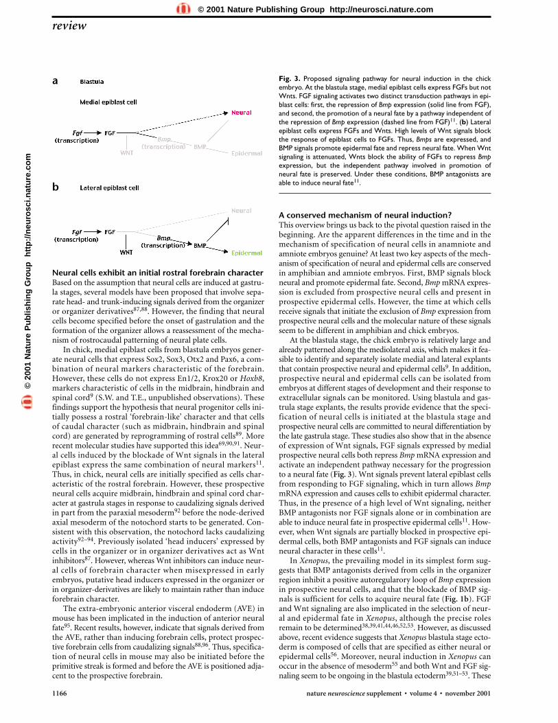
Fig. 3. Proposed signaling pathway for neural induction in the chickembryo. At the blastula stage, medial epiblast cells express FGFs but notWnts. FGF signaling activates two distinct transduction pathways in epi-blast cells: first, the repression of Bmp expression (solid line from FGF),and second, the promotion of a neural fate by a pathway independent ofthe repression of Bmp expression (dashed line from FGF)11. (b) Lateralepiblast cells express FGFs and Wnts. High levels of Wnt signals blockthe response of epiblast cells to FGFs. Thus, Bmps are expressed, andBMP signals promote epidermal fate and repress neural fate. When Wntsignaling is attenuated, Wnts block the ability of FGFs to repress Bmpexpression, but the independent pathway involved in promotion ofneural fate is preserved. Under these conditions, BMP antagonists areable to induce neural fate11.
a
b
©20
01 N
atu
re P
ub
lish
ing
Gro
up
h
ttp
://n
euro
sci.n
atu
re.c
om
© 2001 Nature Publishing Group http://neurosci.nature.com

observations raise the possibility that in blastula-stage animalcap explants, low-level Wnt signaling attenuates FGF signaling,which allows Bmp mRNA expression, causing cells to acquireepidermal character. Under these conditions, both BMP antag-onists and FGF signals would promote neural fate, which maymirror the condition when Wnt signaling is partially inhibitedin chick prospective epidermal cells. Thus, neural induction inanamniotes and amniotes may be more similar than would seemat a first glance.
In Drosophila, neural cells are derived from a neurogenicregion that is generated during the initial dorsoventral patterningof the embryonic ectoderm. Like in vertebrate embryos, the gen-eration of neurogenic domain in Drosophila depends on theexclusion of Dpp, the fly homolog of BMPs, from prospectiveneural cells. Dpp is expressed in dorsal ectoderm, which differ-entiates into epidermis. The generation of the lateral neurogenicregion in the fly embryo is initiated before the onset of gastrula-tion, depends on maternal effect genes, and requires signalsderived from non-embryonic cells97,98. The mediolateral axis ofthe blastula stage chick epiblast defines the future dorsoventralaxis of the ectoderm. Thus, the specification of the chick epiblastinto a medial neurogenic and a lateral epidermal region beforethe onset of gastrulation has certain parallels with the generationof neurogenic and epidermal ectoderm in Drosophila. In con-trast, the prevailing model of neural induction in amphibianssuggests that the specification of neural cells is initiated at a muchlater stage in development in response to signals derived fromthe organizer. It seems unlikely, however, that amphibians havedeveloped an independent mechanism of specification of neuralcells that requires gastrulation and the formation of the organiz-er. Consistent with this idea, recent evidence suggests that inXenopus, the dorsal ectoderm has already been exposed to sig-nals that specify neural fate at the blastula stage56 and that neur-al induction can occur in the absence of mesoderm55. Thus, thedevelopmental stage at which exclusion of Bmp mRNA expres-sion and specification of neural cells is initiated may be conservedbetween fly, amphibian and amniote embryos.
ACKNOWLEDGEMENTST.E. is supported by the Swedish Medical Research Council and by the Foundation
for Strategic Research. We thank members of the Edlund lab for helpful discussions
and we are grateful to T.M. Jessell for comments, suggestions and ideas.
RECEIVED 5 JULY; ACCEPTED 27 AUGUST 2001
1. Spemann, H. & Mangold, H. Uber Induktion von Embryonanlagen durchImplantation artfremder Organisatoren. Roux. Arch. Entw. Mech. Organ. 100,599–538 (1924).
2. Spemann, H. & Mangold, H. Induction of embryonic primordia byimplantation of organizers from a different species. 1923. Int. J. Dev. Biol. 45,13–38 (2001).
3. Hemmati-Brivanlou, A. & Melton, D. Vertebrate neural induction. Annu.Rev. Neurosci. 20, 43–60 (1997).
4. Ang, S. L. & Rossant, J. HNF-3 beta is essential for node and notochordformation in mouse development. Cell 78, 561–574 (1994).
5. Klingensmith, J., Ang, S. L., Bachiller, D. & Rossant, J. Neural induction andpatterning in the mouse in the absence of the node and its derivatives. Dev.Biol. 216, 535–549 (1999).
6. Bachiller, D. et al. The organizer factors Chordin and Noggin are required formouse forebrain development. Nature 403, 658–661 (2000).
7. Episkopou, V. et al. Induction of the mammalian node requires Arkadiafunction in the extraembryonic lineages. Nature 410, 825–830 (2001).
8. Streit, A. et al. Chordin regulates primitive streak development and thestability of induced neural cells, but is not sufficient for neural induction inthe chick embryo. Development 125, 507–519 (1998).
9. Wilson, S. I., Graziano, E., Harland, R., Jessell, T. M. & Edlund, T. An earlyrequirement for FGF signalling in the acquisition of neural cell fate in thechick embryo. Curr. Biol. 10, 421–429 (2000).
10. Streit, A., Berliner, A. J., Papanayotou, C., Sirulnik, A. & Stern, C. D.Initiation of neural induction by FGF signalling before gastrulation. Nature406, 74–78 (2000).
11. Wilson, S. I. et al. The status of Wnt signalling regulates neural and epidermalfates in the chick embryo. Nature 411, 325–330 (2001).
12. Harland, R. Neural induction. Curr. Opin. Genet. Dev. 10, 357–362 (2000).13. Streit, A. & Stern, C. D. Neural induction. A bird’s eye view. Trends. Genet. 15,
20–24 (1999).14. Wilson, P. A. & Hemmati-Brivanlou, A. Vertebrate neural induction:
inducers, inhibitors, and a new synthesis. Neuron 18, 699–710 (1997).15. Sasai, Y. & De Robertis, E. M. Ectodermal patterning in vertebrate embryos.
Dev. Biol. 182, 5–20 (1997).16. Waddington, C. H. Experiments on the development of chick and duck
embryos, cultivated in vitro. Phil. Trans. R. Soc. Lond. B Biol. Sci. 221, 179–230(1932).
17. Waddington, C. H. Induction of the primitive streak and its derivatives in thechick. J. Exp. Biol. 10, 38–46 (1933).
18. Kintner, C. R. & Dodd, J. Hensen’s node induces neural tissue in Xenopusectoderm. Implications for the action of the organizer in neural induction.Development 113, 1495–1505 (1991).
19. Storey, K. G., Crossley, J. M., De Robertis, E. M., Norris, W. E. & Stern, C. D.Neural induction and regionalisation in the chick embryo. Development 114,729–741 (1992).
20. Beddington, R. S. Induction of a second neural axis by the mouse node.Development 120, 613–620 (1994).
21. Shih, J. & Fraser, S. E. Characterizing the zebrafish organizer: microsurgicalanalysis at the early-shield stage. Development 122, 1313–1322 (1996).
22. Waddington, C. H. Experiments on determination in the rabbit embryo.Arch. Biol. 48, 273–290 (1937).
23. Knoetgen, H., Teichmann, U., Wittler, L., Viebahn, C. & Kessel, M. Anteriorneural induction by nodes from rabbits and mice. Dev. Biol. 225, 370–380 (2000).
24. Sasai, Y., Lu, B., Steinbeisser, H. & De Robertis, E. M. Regulation of neuralinduction by the Chd and Bmp-4 antagonistic patterning signals in Xenopus.Nature 376, 333–336 (1995).
25. Hemmati-Brivanlou, A. & Melton, D. A. A truncated activin receptor inhibitsmesoderm induction and formation of axial structures in Xenopus embryos.Nature 359, 609–614 (1992).
26. Hemmati-Brivanlou, A., Kelly, O. G. & Melton, D. A. Follistatin, anantagonist of activin, is expressed in the Spemann organizer and displaysdirect neuralizing activity. Cell 77, 283–295 (1994).
27. Lamb, T. M. et al. Neural induction by the secreted polypeptide noggin.Science 262, 713–718 (1993).
28. Wilson, P. A. & Hemmati-Brivanlou, A. Induction of epidermis andinhibition of neural fate by Bmp-4. Nature 376, 331–333 (1995).
29. Grunz, H. & Tacke, L. Neural differentiation of Xenopus laevis ectoderm takesplace after disaggregation and delayed reaggregation without inducer. CellDiffer. Dev. 28, 211–217 (1989).
30. Dale, L. & Jones, C. M. BMP signalling in early Xenopus development.Bioessays 21, 751–760 (1999).
31. Hawley, S. H. et al. Disruption of BMP signals in embryonic Xenopusectoderm leads to direct neural induction. Genes Dev. 9, 2923–2935 (1995).
32. Xu, R. H. et al. A dominant negative bone morphogenetic protein 4 receptorcauses neuralization in Xenopus ectoderm. Biochem. Biophys. Res. Commun.212, 212–219 (1995).
33. Fainsod, A., Steinbeisser, H. & De Robertis, E. M. On the function of BMP-4in patterning the marginal zone of the Xenopus embryo. EMBO J. 13,5015–5025 (1994).
34. Hemmati-Brivanlou, A. & Thomsen, G. H. Ventral mesodermal patterning inXenopus embryos: expression patterns and activities of BMP-2 and BMP-4.Dev. Genet. 17, 78–89 (1995).
35. Wilson, P. A., Lagna, G., Suzuki, A. & Hemmati-Brivanlou, A.Concentration-dependent patterning of the Xenopus ectoderm by BMP4 andits signal transducer Smad1. Development 124, 3177–3184 (1997).
36. Suzuki, A., Ueno, N. & Hemmati-Brivanlou, A. Xenopus msx1 mediatesepidermal induction and neural inhibition by BMP4. Development 124,3037–3044 (1997).
37. Powers, C. J., McLeskey, S. W. & Wellstein, A. Fibroblast growth factors, theirreceptors and signaling. Endocr. Relat. Cancer 7, 165–197 (2000).
38. Hongo, I., Kengaku, M. & Okamoto, H. FGF signaling and the anteriorneural induction in Xenopus. Dev. Biol. 216, 561–581 (1999).
39. Launay, C., Fromentoux, V., Shi, D. L. & Boucaut, J. C. A truncated FGFreceptor blocks neural induction by endogenous Xenopus inducers.Development 122, 869–880 (1996).
40. Sasai, Y., Lu, B., Piccolo, S. & De Robertis, E. M. Endoderm induction by theorganizer-secreted factors chordin and noggin in Xenopus animal caps.EMBO J. 15, 4547–4555 (1996).
41. Lamb, T. M. & Harland, R. M. Fibroblast growth factor is a direct neuralinducer, which combined with noggin generates anterior-posterior neuralpattern. Development 121, 3627–3636 (1995).
42. Kengaku, M. & Okamoto, H. bFGF as a possible morphogen for theanteroposterior axis of the central nervous system in Xenopus. Development121, 3121–3130 (1995).
43. Kroll, K. L. & Amaya, E. Transgenic Xenopus embryos from sperm nucleartransplantations reveal FGF signaling requirements during gastrulation.Development 122, 3173–3183 (1996).
review
nature neuroscience supplement • volume 4 • november 2001 1167
©20
01 N
atu
re P
ub
lish
ing
Gro
up
h
ttp
://n
euro
sci.n
atu
re.c
om
© 2001 Nature Publishing Group http://neurosci.nature.com

1168 nature neuroscience supplement • volume 4 • november 2001
44. Holowacz, T. & Sokol, S. FGF is required for posterior neural patterning butnot for neural induction. Dev. Biol. 205, 296–308 (1999).
45. Ribisi, S. Jr. et al. Ras-mediated FGF signaling is required for the formation ofposterior but not anterior neural tissue in Xenopus laevis. Dev. Biol. 227,183–196 (2000).
46. Baker, J. C., Beddington, R. S. & Harland, R. M. Wnt signaling in Xenopusembryos inhibits bmp4 expression and activates neural development. GenesDev 13, 3149–3159 (1999).
47. Vincent, J. P., Oster, G. F. & Gerhart, J. C. Kinematics of gray crescentformation in Xenopus eggs: the displacement of subcortical cytoplasmrelative to the egg surface. Dev. Biol. 113, 484–500 (1986).
48. Miller, J. R. et al. Establishment of the dorsal-ventral axis in Xenopus embryoscoincides with the dorsal enrichment of dishevelled that is dependent oncortical rotation. J. Cell Biol. 146, 427–437 (1999).
49. Moon, R. T., Brown, J. D. & Torres, M. WNTs modulate cell fate and behaviorduring vertebrate development. Trends Genet. 13, 157–162 (1997).
50. Christian, J. L. & Moon, R. T. Interactions between Xwnt-8 and Spemannorganizer signaling pathways generate dorsoventral pattern in the embryonicmesoderm of Xenopus. Genes Dev. 7, 13–28 (1993).
51. Glinka, A., Wu, W., Onichtchouk, D., Blumenstock, C. & Niehrs, C. Headinduction by simultaneous repression of Bmp and Wnt signalling in Xenopus.Nature 389, 517–519 (1997).
52. Glinka, A. et al. Dickkopf-1 is a member of a new family of secreted proteinsand functions in head induction. Nature 391, 357–362 (1998).
53. Itoh, K., Tang, T. L., Neel, B. G. & Sokol, S. Y. Specific modulation ofectodermal cell fates in Xenopus embryos by glycogen synthase kinase.Development 121, 3979–3988 (1995).
54. Umbhauer, M., Penzo-Mendez, A., Clavilier, L., Boucaut, J. & Riou, J.Signaling specificities of fibroblast growth factor receptors in early Xenopusembryo. J. Cell Sci. 113, 2865–2875 (2000).
55. Wessely, O., Agius, E., Oelgeschlager, M., Pera, E. M. & De Robertis, E. M.Neural induction in the absence of mesoderm: beta-catenin-dependentexpression of secreted BMP antagonists at the blastula stage in Xenopus. Dev.Biol. 234, 161–173 (2001).
56. Gamse, J. T. & Sive, H. Early anteroposterior division of the presumptiveneurectoderm in Xenopus. Mech. Dev. 104, 21–36 (2001).
57. Penzel, R., Oschwald, R., Chen, Y., Tacke, L. & Grunz, H. Characterizationand early embryonic expression of a neural specific transcription factorxSOX3 in Xenopus laevis. Int. J. Dev. Biol. 41, 667–677 (1997).
58. Mizuseki, K., Kishi, M., Shiota, K., Nakanishi, S. & Sasai, Y. SoxD: an essentialmediator of induction of anterior neural tissues in Xenopus embryos. Neuron21, 77–85 (1998).
59. Kroll, K. L., Salic, A. N., Evans, L. M. & Kirschner, M. W. Geminin, aneuralizing molecule that demarcates the future neural plate at the onset ofgastrulation. Development 125, 3247–3258 (1998).
60. Zhang, J. & Jacobson, A. G. Evidence that the border of the neural plate maybe positioned by the interaction between signals that induce ventral anddorsal mesoderm. Dev. Dyn. 196, 79–90 (1993).
61. Kurata, T., Nakabayashi, J., Yamamoto, T. S., Mochii, M. & Ueno, N.Visualization of endogenous BMP signaling during Xenopus development.Differentiation 67, 33–40 (2001).
62. Smith, J. L. & Schoenwolf, G. C. Notochordal induction of cell wedging in thechick neural plate and its role in neural tube formation. J. Exp. Zool. 250,49–62 (1989).
63. Sater, A. K. & Jacobson, A. G. The role of the dorsal lip in the induction ofheart mesoderm in Xenopus laevis. Development 108, 461–470 (1990).
64. Davidson, B. P., Kinder, S. J., Steiner, K., Schoenwolf, G. C. & Tam, P. P.Impact of node ablation on the morphogenesis of the body axis and thelateral asymmetry of the mouse embryo during early organogenesis. Dev.Biol. 211, 11–26 (1999).
65. Pera, E., Stein, S. & Kessel, M. Ectodermal patterning in the avian embryo:epidermis versus neural plate. Development 126, 63–73 (1999).
66. Sheng, G. & Stern, C. D. Gata2 and Gata3: novel markers for early embryonicpolarity and for non-neural ectoderm in the chick embryo. Mech. Dev. 87,213–216 (1999).
67. Schier, A. F. & Talbot, W. S. Nodal signaling and the zebrafish organizer. Int. J.Dev. Biol. 45, 289–297 (2001).
68. Yamaguchi, T. P., Takada, S., Yoshikawa, Y., Wu, N. & McMahon, A. P. T(Brachyury) is a direct target of Wnt3a during paraxial mesodermspecification. Genes Dev. 13, 3185–3190 (1999).
69. Muhr, J., Graziano, E., Wilson, S., Jessell, T. M. & Edlund, T. Convergentinductive signals specify midbrain, hindbrain, and spinal cord identity ingastrula stage chick embryos. Neuron 23, 689–702 (1999).
70. Streit, A. & Stern, C. D. Establishment and maintenance of the border of theneural plate in the chick: involvement of FGF and BMP activity. Mech. Dev.82, 51–66 (1999).
71. Matzuk, M. M. et al. Multiple defects and perinatal death in mice deficient infollistatin. Nature 374, 360–363 (1995).
72. McMahon, J. A. et al. Noggin-mediated antagonism of BMP signaling isrequired for growth and patterning of the neural tube and somite. Genes Dev.12, 1438–1452 (1998).
73. Kawasaki, H. et al. Induction of midbrain dopaminergic neurons from EScells by stromal cell-derived inducing activity. Neuron 28, 31–40 (2000).
74. Levin, M. The roles of activin and follistatin signaling in chick gastrulation.Int. J. Dev. Biol. 42, 553–559 (1998).
75. Connolly, D. J., Patel, K. & Cooke, J. Chick noggin is expressed in theorganizer and neural plate during axial development, but offers no evidenceof involvement in primary axis formation. Int. J. Dev. Biol. 41, 389–396(1997).
76. Knoetgen, H., Viebahn, C. & Kessel, M. Head induction in the chick byprimitive endoderm of mammalian, but not avian origin. Development 126,815–825 (1999).
77. Hammerschmidt, M., Serbedzija, G. N. & McMahon, A. P. Genetic analysis ofdorsoventral pattern formation in the zebrafish: requirement of a BMP-likeventralizing activity and its dorsal repressor. Genes Dev. 10, 2452–2461(1996).
78. Fekany-Lee, K., Gonzalez, E., Miller-Bertoglio, V. & Solnica-Krezel, L. Thehomeobox gene bozozok promotes anterior neuroectoderm formation inzebrafish through negative regulation of BMP2/4 and Wnt pathways.Development 127, 2333–2345 (2000).
79. Nguyen, V. H. et al. Ventral and lateral regions of the zebrafish gastrula,including the neural crest progenitors, are established by a bmp2b/swirlpathway of genes. Dev. Biol. 199, 93–110 (1998).
80. Dick, A. et al. Essential role of Bmp7 (snailhouse) and its prodomain indorsoventral patterning of the zebrafish embryo. Development 127, 343–354(2000).
81. Hild, M. et al. The smad5 mutation somitabun blocks Bmp2b signalingduring early dorsoventral patterning of the zebrafish embryo. Development126, 2149–2159 (1999).
82. Hsieh, J. C., Rattner, A., Smallwood, P. M. & Nathans, J. Biochemicalcharacterization of Wnt-frizzled interactions using a soluble, biologicallyactive vertebrate Wnt protein. Proc. Natl. Acad. Sci. USA 96, 3546–3551(1999).
83. Pera, E. M. & De Robertis, E. M. A direct screen for secreted proteins inXenopus embryos identifies distinct activities for the Wnt antagonistsCrescent and Frzb-1. Mech. Dev. 96, 183–195 (2000).
84. Shibata, M., Ono, H., Hikasa, H., Shinga, J. & Taira, M. Xenopus crescentencoding a Frizzled-like domain is expressed in the Spemann organizer andpronephros. Mech. Dev. 96, 243–246 (2000).
85. Isaacs, H. V., Pownall, M. E. & Slack, J. M. eFGF is expressed in the dorsalmidline of Xenopus laevis. Int. J. Dev. Biol. 39, 575–579 (1995).
86. Hansen, C. S., Marion, C. D., Steele, K., George, S. & Smith, W. C. Directneural induction and selective inhibition of mesoderm and epidermisinducers by Xnr3. Development 124, 483–492 (1997).
87. Niehrs, C. Head in the WNT: the molecular nature of Spemann’s headorganizer. Trends Genet. 15, 314–319 (1999).
88. Stern, C. D. Initial patterning of the central nervous system: how manyorganizers? Nat. Rev. Neurosci. 2, 92–98 (2001).
89. Nieuwkoop, P. D. et al. Activation and organization of the central nervoussystem in amphibians. J. Exp. Zool. 120, 1–108 (1952).
90. Foley, A. C., Skromne, I. & Stern, C. D. Reconciling different models offorebrain induction and patterning: a dual role for the hypoblast.Development 127, 3839–3854 (2000).
91. Kimura, C. et al. Visceral endoderm mediates forebrain development bysuppressing posteriorizing signals. Dev. Biol. 225, 304–321 (2000).
92. Muhr, J., Jessell, T. M. & Edlund, T. Assignment of early caudal identity toneural plate cells by a signal from caudal paraxial mesoderm. Neuron 19,487–502 (1997).
93. Fukushima, M., Nakamura, M., Ohta, K., Okamura, R. & Negi, A. Regionalspecification of motoneurons along the anterior-posterior axis isindependent of the notochord. Development 122, 905–914 (1996).
94. Dale, J. K. et al. Cooperation of BMP7 and SHH in the induction offorebrain ventral midline cells by prechordal mesoderm. Cell 90, 257–269(1997).
95. Thomas, P. & Beddington, R. Anterior primitive endoderm may beresponsible for patterning the anterior neural plate in the mouse embryo.Curr. Biol. 6, 1487–1496 (1996).
96. Martinez Barbera, J. P. et al. The homeobox gene Hex is required in definitiveendodermal tissues for normal forebrain, liver and thyroid formation.Development 127, 2433–2445 (2000).
97. Roth, S., Stein, D. & Nusslein-Volhard, C. A gradient of nuclear localizationof the dorsal protein determines dorsoventral pattern in the Drosophilaembryo. Cell 59, 1189–1202 (1989).
98. St Johnston, R. D. & Gelbart, W. M. Decapentaplegic transcripts are localizedalong the dorsal-ventral axis of the Drosophila embryo. EMBO J. 6,2785–2791 (1987).
review©
2001
Nat
ure
Pu
blis
hin
g G
rou
p
htt
p:/
/neu
rosc
i.nat
ure
.co
m
© 2001 Nature Publishing Group http://neurosci.nature.com