the response of early neural genes to fgf signaling or inhibition
TRANSCRIPT

RESEARCH ARTICLE Open Access
The response of early neural genes to FGFsignaling or inhibition of BMP indicate theabsence of a conserved neural induction moduleCrystal D Rogers, George S Ferzli and Elena S Casey*
Abstract
Background: The molecular mechanism that initiates the formation of the vertebrate central nervous system haslong been debated. Studies in Xenopus and mouse demonstrate that inhibition of BMP signaling is sufficient toinduce neural tissue in explants or ES cells respectively, whereas studies in chick argue that instructive FGFsignaling is also required for the expression of neural genes. Although additional signals may be involved in neuralinduction and patterning, here we focus on the roles of BMP inhibition and FGF8a.
Results: To address the question of necessity and sufficiency of BMP inhibition and FGF signaling, we comparedthe temporal expression of the five earliest genes expressed in the neuroectoderm and determined theirrequirements for induction at the onset of neural plate formation in Xenopus. Our results demonstrate that theonset and peak of expression of the genes vary and that they have different regulatory requirements and aretherefore unlikely to share a conserved neural induction regulatory module. Even though all require inhibition ofBMP for expression, some also require FGF signaling; expression of the early-onset pan-neural genes sox2 andfoxd5a requires FGF signaling while other early genes, sox3, geminin and zicr1 are induced by BMP inhibition alone.
Conclusions: We demonstrate that BMP inhibition and FGF signaling induce neural genes independently of eachother. Together our data indicate that although the spatiotemporal expression patterns of early neural genes aresimilar, the mechanisms involved in their expression are distinct and there are different signaling requirements forthe expression of each gene.
BackgroundDevelopment of the vertebrate central nervous system(CNS) is initiated during gastrulation when dorsal ecto-dermal cells are converted to the neural fate. There aretwo prevailing models for the induction of the CNS.The first, the neural default model, arose from experi-ments demonstrating that in the absence of bone mor-phogenetic protein (BMP) signaling, amphibianectodermal explants form neural tissue instead of epi-dermis [1,2]. Formation of the nervous system by defaultis highly conserved. In the protostome Drosophila mela-nogaster, neural tissue forms as a result of inhibition ofthe BMP homolog Decapentaplegic (Dpp) by the Chor-din ortholog Sog [3]. Furthermore, the Xenopus BMPantagonist Noggin is sufficient to inhibit Dpp and
induce neuroectoderm in fruit flies [4], and overexpres-sion of Sog induces a secondary axis in Xenopusembryos [5]. The second model, the instructive signalingmodel, arose from studies of chick embryonic develop-ment and indicated that inhibition of BMP signaling isnot sufficient to induce neural tissue, and that instruc-tion from another signaling pathway such as FGF (fibro-blast growth factor), is required. There is evidencesupporting both models in multiple vertebrates [6,7],thus feeding the controversy over which signals arenecessary and sufficient during vertebrate CNSinduction.Experiments in ES cells, mouse and zebrafish embryos
support the model that the vertebrate CNS is formed bydefault. Like Xenopus ectodermal explants [8], mouse EScells [9], and human ES [10-12] and induced pluripotentstem cells [11] are converted to rostral neural tissuewhen BMP signaling is inhibited. In the mouse epiblast,* Correspondence: [email protected]
Department of Biology, Georgetown University, Washington DC, USA
Rogers et al. BMC Developmental Biology 2011, 11:74http://www.biomedcentral.com/1471-213X/11/74
© 2011 Rogers et al; licensee BioMed Central Ltd. This is an Open Access article distributed under the terms of the Creative CommonsAttribution License (http://creativecommons.org/licenses/by/2.0), which permits unrestricted use, distribution, and reproduction inany medium, provided the original work is properly cited.

BMP2/4 signaling maintains pluripotency and preventsthe acquisition of a neural fate, whereas inhibition ofBMP signaling induces neural tissue independent ofFGF signaling [13]. Similarly, in zebrafish, BMP inhibi-tion is sufficient for the induction of anterior neuralgenes and FGF signaling is not required for inductionbut rather for posteriorization of the induced tissue [14].Although BMP inhibition clearly plays a role in neural
specification in many organisms, alone it does not effec-tively induce neural tissue formation in Xenopus ventralectoderm [15-17] or outside of the chick dorsal ecto-derm [18], which indicates that an instructive signal isrequired. The leading candidate for this instructive sig-nal is FGF. In both chick and frog, overexpression ofFGF2, FGF4, or FGF8 induces the expression of poster-ior neural genes [18-21] and the activation of FGF sig-naling in combination with BMP antagonism inducesthe expression of pan-neural genes in non-neural ecto-dermal territories [17,19]. Loss of function studies inmESCs, chick and Xenopus embryos also suggest a rolefor instructive signaling by FGF in neural induction. Forexample, inhibition of FGF receptors or Erk1/2 by expo-sure to pharmacological inhibitors eliminated differen-tiation of mESCs into neurons [22] and resulted in aloss of neural tissue in frog [15], zebrafish [23] andchick [24]. Furthermore, overexpression of the dominantnegative FGF receptor 4a reduced the expression of theneural progenitor marker, sox2, in tailbud-stage Xenopusembryos [15] and the formation of neural tissue in ecto-dermal explants in response to Noggin [25].It has been difficult to dissect out an independent role
for FGF in neural induction because FGF signaling hassignificant roles in mesodermal development [26] andneural anterior-posterior patterning [27], induces neuraltissue via interference with BMP transcription andtransduction [26-28] and has been proposed to maintainrather than induce a neural progenitor population[29-32]. Specifically, studies suggest that neural specifi-cation in response to FGF is not instructive or indepen-dent from BMP inhibition but rather, is the result ofinterference with BMP signaling via inhibition of Smad1activity [28] or the transcription of BMP [29,30]. It hasalso been proposed that FGF signaling is dispensable forinduction and instead is required for the maintenance ofneural progenitors. This is supported by studies in: (1)Xenopus ectodermal explants in which sox2 and sox3expression is not maintained when FGF signalingthrough FGFR1 and 2 is inhibited [31]; (2) the mouseolfactory bulb and retina in which a proliferating pro-genitor population is decreased in the absence of FGFsignaling [32,33] and; (3) hESCs in which exogenousFGF maintains cells in an undifferentiated state [34].Furthermore, the approaches and techniques used toinvestigate the role of FGF have been called to question.
It has been argued that incorrect markers were analyzedat the wrong developmental stages and in the wrong tis-sues, and that doses of pharmacological inhibitors werelethal or detrimental to development [8]. With this con-flicting data and the variables added by the use of manydifferent model organisms, stages, neural markers andFGF inhibitors (small molecules, dominant negativereceptors, morpholinos), it remains unclear if FGF sig-naling is required in addition to, or independent of,BMP inhibition for the induction of neural genes.This study compares the regulatory requirements for
the onset and maintenance of multiple early neuralgenes in Xenopus embryos. Using multiple genes in oneorganism has allowed us to determine whether FGF sig-naling is required for the induction of multiple earlyneural genes independent of BMP inhibition, and also todetermine the role of FGF8a in neural development.Using gain and loss of function assays, we show thatsox2 and foxd5a require FGF signaling for neural induc-tion and that sox3 and geminin require FGF signalingfor maintenance of expression. Ultimately, we show thatdepending on the gene of interest, the ability of FGF toinduce expression can be dependent on the absence ofBMP signaling and may be indirect via the induction ofmesoderm.
ResultsResponse of early neural genes to BMP inhibition andFGF signalingThe early neural genes sox2, sox3, geminin, foxD5a, soxDand zicr1 are expressed broadly in the neuroectoderm atthe time of neural induction in response to neural indu-cing signals [31,35-39]. We compared their expressionsat stage 8 (mid-blastula transition, 7+ hpf), through theonset of gastrulation and neural induction (stage 10.5,10 hpf) and in neurula embryos (stage 17, 24 hpf) (Fig-ure 1A). The maternal genes, soxD, sox3 and gemininare expressed strongly at 7 hpf whereas foxD5a, which isalso maternally expressed, is expressed at low levels at 7hpf with levels increasing at 8 hpf and peaking at 10hpf. The expression levels of the zygotically expressedgene zicr1 are fairly constant whereas sox2 levelsincrease from 7 to 24 hpf. Both FGF8 and the BMPantagonist, noggin are expressed at 7 hpf precedingneural induction. All of the genes are expressed prior tothe onset of gastrulation with zygotic FGF8 expressedprior to foxd5a and sox2 expression indicating that itmay play a role in the induction of these two genes butnot the others.To determine if the early neural genes respond to
BMP antagonism and/or FGF signaling, we overex-pressed Noggin, FGF8a or a combination of both andanalyzed the expression of the early neural genes andthe proneural gene neurogenin (ngnr1). We used FGF8a
Rogers et al. BMC Developmental Biology 2011, 11:74http://www.biomedcentral.com/1471-213X/11/74
Page 2 of 13
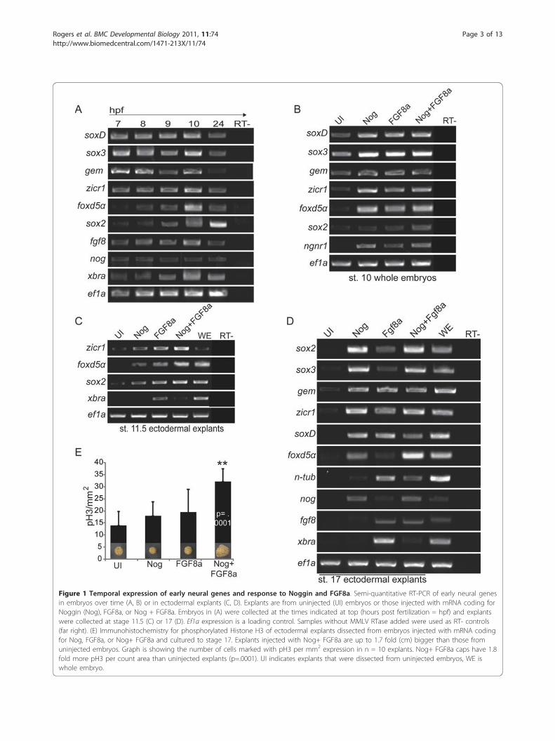
Figure 1 Temporal expression of early neural genes and response to Noggin and FGF8a. Semi-quantitative RT-PCR of early neural genesin embryos over time (A, B) or in ectodermal explants (C, D). Explants are from uninjected (UI) embryos or those injected with mRNA coding forNoggin (Nog), FGF8a, or Nog + FGF8a. Embryos in (A) were collected at the times indicated at top (hours post fertilization = hpf) and explantswere collected at stage 11.5 (C) or 17 (D). Ef1a expression is a loading control. Samples without MMLV RTase added were used as RT- controls(far right). (E) Immunohistochemistry for phosphorylated Histone H3 of ectodermal explants dissected from embryos injected with mRNA codingfor Nog, FGF8a, or Nog+ FGF8a and cultured to stage 17. Explants injected with Nog+ FGF8a are up to 1.7 fold (cm) bigger than those fromuninjected embryos. Graph is showing the number of cells marked with pH3 per mm2 expression in n = 10 explants. Nog+ FGF8a caps have 1.8fold more pH3 per count area than uninjected explants (p=.0001). UI indicates explants that were dissected from uninjected embryos, WE iswhole embryo.
Rogers et al. BMC Developmental Biology 2011, 11:74http://www.biomedcentral.com/1471-213X/11/74
Page 3 of 13

because its misexpression was reported to induce neuraltissue in explants without inducing mesoderm [21], andbecause FGF8a overexpression does not induce Xbraexpression in whole embryos (Additional File 1, Fig.S1A). Embryos were injected at the 1-cell stage, col-lected at stage 10 and assayed by RT-PCR. Expression ofsoxD, sox3, zicr1, foxd5a and ngnr1 was enhanced byoverexpression of Noggin or FGF (Figure 1B). However,expression of gem and sox2 was unchanged (Figure 1B)indicating that BMP antagonism and FGF8 are not suffi-cient to enhance their expression at this stage.To determine if FGF8a signaling is sufficient to acti-
vate or maintain the expression of the early neuralgenes, we analyzed their expression in gastrula (st.11.5) and neurula (st. 17) stage ectodermal explants(Figure 1C and 1D) in response to Noggin, FGF8aand Nog+FGF8a. Genes expressed at high maternallevels (sox3, soxD and gem) were not examined atstage 11.5. As shown in prior studies, BMP inhibitioninduced the expression of neural progenitor markers(sox2, sox3 and geminin) and the other early neuralmarkers, (soxD, zicr1, foxd5a) by st. 17. At stage 11.5,FGF8a induced zicr1, foxd5a and sox2 expression andby stage 17 induced neuron formation; however, italso induced the expression of the pan-mesodermalmarker xbra (Figure 1C, D) and the dorsal mesodermmarker and BMP antagonist, noggin (Figure 1D).Therefore, it is possible that FGF8a alters neural geneexpression indirectly via signals from the dorsalmesoderm.Explants from embryos co-injected with Noggin and
FGF8a are comprised of neural progenitors (sox2+, sox3+ and gem+) and neurons (n-tub+) (Figure 1D) andhave a distinct morphology; by stage 17, they are ~ 2-fold larger than UI, Noggin or FGF8a caps indicatingeither increased cell size or proliferation (Figure 1E). Todetermine if the increase in explant size was due toincreased proliferation, we performed immunohisto-chemistry to detect phosphorylated Histone H3 (Figure1E), an indicator of mitosis. Explants from embryosinjected with Nog and FGF8a mRNA had an average of1.8 fold more proliferating cells per square millimeterthan uninjected explants (Figure 1E, p = .0001, Student’sT-Test, n = 10). To verify that the size difference wasnot due to the presence of large migratory neural crestcells with extended processes, we assayed for the expres-sion of the neural crest marker, slug. These explants didnot express slug (data not shown), but did express theepidermal and mesodermal marker vent2, the neuronalmarker n-tub and the proliferating progenitor markerssox2, sox3 and gem (Figure 1D). One possible explana-tion for the mixed cell population in these large explantsis that their fate varies with the level of Noggin or FGFreceived.
Ectoderm is competent to respond to BMP inhibition andFGF signaling prior to the onset of gastrulationTo determine when ectodermal cells are competent torespond to FGF8a signaling or BMP inhibition by Nog-gin, we assayed for the expression of the early neuralgenes, zicr1, foxd5a and sox2 prior to MBT (6 hpf) anduntil the onset of gastrulation and neural induction (10hpf). Embryos were injected with mRNA coding forNoggin, FGF8a or Noggin+ FGF8a in 1 of 2-cells andcollected at 6 hpf and every hour after until stage 10.5(10 hpf). We performed whole-mount in situ hybridiza-tion (WISH) and semi-quantitative RT-PCR to detectthe expression of zicr1, sox2 and foxd5a (Figure 2A-D),which were expressed between 7 and 10 hpf (Figure 1A)in embryos and induced by both Noggin and FGF8a bystage 11.5 in ectodermal explants (Figure 1C). Endogen-ous expression of zicr1 was first detected by WISH at 8hpf (Figure 2A) and by RT-PCR at 7 hpf. BMP inhibi-tion by Noggin enhanced expression of zicr1 at 8 hpf(Figure 2A red arrows, Figure 2D) and by a truncatedBMPR (tBR) at 7 hpf (Additional File 2, Figure S2A, C).FGF8a also increased zicr1 expression by 8-9 hpf (Figure2A, D, Additional File 2, Figure S2C). Low levels ofzicr1 were detected at all stages tested, thus neitherBMP inhibition nor FGF signalling induced expressionprematurely. In contrast, foxd5a expression was inducedby Noggin and FGF8a by 8 and 7 hpf (Figure 2B redarrows, 2D), respectively, whereas endogenous dorsal-specific expression was first detected at 9 hpf. Sox2expression was also induced prematurely at 8-9 hpf (Fig-ure 2C, D), and expanded by FGF8a, Noggin and tBR at10 hpf (stage 10.5) (Figure 2C, Additional File 2, FigureS2B). In summary, foxd5a and zicr1 are induced andexpanded, respectively, in the ectoderm in response toBMP inhibition and FGF8a signalling by 8 hpf, whereasSox2 expression is not significantly altered in responseto FGF8a signaling until much later after the onset ofendogenous expression (10 hpf). These experimentsindicate that early neural genes are induced by Nogginby 8 hpf and respond to FGF8a at different times.
FGF signaling is required for the induction of sox2 andfoxd5a expressionIn Xenopus, the induction of neural tissue occurs at theonset of gastrulation in response to BMP inhibition, andthis induction may be dependent on instructive FGF sig-naling. To determine if BMP inhibition and/or FGF sig-naling are required for neural tissue formation, weanalyzed the expression of the early neural genes inresponse to constitutively active BMP signaling and theloss of FGF signaling. In support of the neural defaultmodel, overexpression of a constitutively active BMPreceptor (Alk3) inhibited the expression of the six earlyneural genes at the onset of neural induction
Rogers et al. BMC Developmental Biology 2011, 11:74http://www.biomedcentral.com/1471-213X/11/74
Page 4 of 13

(Additional File 3, Fig. S3) and Noggin expanded theirexpression (Figure 3A-D and data not shown), suggest-ing that the expression of all early neural genes requiresBMP inhibition. In contrast, each of the early neuralgenes responded differently to loss of FGF signaling. Tointerfere with FGF signaling, we used dominant negativeFGFR4a (Δ4a) because it is the receptor through whichFGF8 stimulates neuron formation [40] and was deemedmore effective at blocking neural development thandominant negative FGFR1 (XFD) [25,41]. Surprisingly,although FGF8a induced soxD expression in explants,Δ4a overexpression had no effect on soxD expression inembryos (data not shown). However, Δ4a expressioninhibited sox2 (n = 42/62) and foxd5a (n = 23/25)expression and reduced zicr1 expression (n = 20/30) atstage 10.5, and sox3 and gem expression (sox3, n = 56/63 and gem 12/12) at stage 12 (Figure 3A-D). Since FGFsignaling inhibits BMP signaling via phosphorylation ofSmad1 [28], we wanted to determine if the Δ4a pheno-types could be rescued by Noggin, or in other words,
were due to increased BMP signaling. Zicr1, sox3 andgem expression were rescued by overexpression of Nog-gin (Figure 3B, n = 26/26, 3D, n = 17/21, n = 15/20)and therefore not dependent on FGF signaling. How-ever, sox2 and foxd5a expression were not rescued byoverexpression of Noggin (Figure 3A, C) indicating anindependent role for FGF signaling. These data indicatethat FGF signaling is required for the expression of sox2and foxd5a.We next tested whether FGF signaling through FGFR4ais required for the expression of early neural genes inexplants in which BMP signaling is inhibited. We over-expressed Δ4a in ectodermal explants neuralized byNoggin and performed semi-quantitative RT-PCR. As inembryos, FGF signaling was required for the expressionof sox2 and foxd5a in neuralized mid-gastrula or neur-ula explants (Figure 3E). Although, zicr1 and soxDexpression were reduced by Δ4a in noggin-injected stage10.5 embryos (Figure 3A, data not shown), their expres-sion was greatly reduced or inhibited by Δ4a expression
Figure 2 BMP inhibition and FGF signaling prematurely induce the expression of early neural genes. (A-C) WISH for zicr1, foxd5a andsox2 of embryos injected with mRNA coding for Nog, FGF8a or Nog + FGF8a and lacZ mRNA (cyan) and collected at stage 8 (t = 6 hpf) andeach subsequent hour after until stage 10.5 (t = 10 hpf) when cultured at room temperature. Red arrows indicate earliest onset of expression. Allimages are animal pole view with dorsal to the top. (D) RT-PCR of whole embryos dissected from uninjected embryos (UI) or embryos injectedwith Nog, FGF8a or Nog+FGF8a. Embryos were collected as in A-C. Genes analysed are indicated on left side, treatment on top, time ofcollection below panel. ODC used for loading control. All images are animal pole view with dorsal to the top.
Rogers et al. BMC Developmental Biology 2011, 11:74http://www.biomedcentral.com/1471-213X/11/74
Page 5 of 13

in noggin-injected explants (Figure 3E). In fact, by neur-ula stage, the expression of all of the early neural geneswas greatly reduced or lost in explants co-injected withNoggin and Δ4a. XFD inhibited sox2 and foxd5a expres-sion and by st. 17 also reduced soxD and gem
expression. In summary, the explant RT-PCR data andWISH embryo data (Figure 3A-D) indicate that soxD,sox2, zicr1 and foxd5a require FGF signaling for robustexpression at the gastrula stage, whereas sox3 and gemrequire FGF signaling for the maintenance of their
Figure 3 FGF signaling is required for the induction of sox2 and foxd5a expression in the neural plate and the maintenance of otherearly neural genes. WISH for (A) sox2, (B) zicr1, (C) foxd5a, and (D) sox3 and gem of embryos that were either uninjected (UI) or injected withNoggin (Nog), dominant negative FGFR4a (Δ4a), or Nog + Δ4a and were collected at stages 10.5 and 11.5 (A-C) or 12.5 (D). All embryos aredorsal view with anterior to the top. The dashed line separates the domain of gem expression (right) from that inhibited by Δ4a (top left). (E) RT-PCR of ectodermal explants for genes indicated on the left side of panel. Explants were dissected from embryos that were either uninjected (UI)or injected with mRNA coding for Nog, Nog + dominant negative FGFR1 (XFD), Nog+Δ4a, or Nog+XFD+Δ4a.
Rogers et al. BMC Developmental Biology 2011, 11:74http://www.biomedcentral.com/1471-213X/11/74
Page 6 of 13

expression in explants after induction by BMPinhibition.
FGF8a induces neurons in the presence of BMP signalingand epidermis formationFGF8a induces neural gene expression in explants (Fig-ure 1) and expands their expression in embryos (Figure2), and FGF signaling is required for the expression ofthe neural progenitor markers sox2 and foxd5a in theneural plate (Figure 3) [8,17]. To determine if FGFinduces early neural gene expression by repressing BMPsignaling, we tested the effect of FGF8a on the expres-sion of BMP, its targets, and epidermis formation. First,we determined the temporal expression of BMP targetgenes prior to and at the onset of neural induction inembryos (Figure 4A). Embryos were collected as for Fig-ure 1. Bmp4 expression was detectable by RT-PCR at 7hpf, and was increased after the onset of zygotic tran-scription at 8 hpf (Figure 4A). The direct target ofBMP4, vent2 [42], was also expressed robustly at 7 hpf,and the expression of vent1, a direct Vent2 target [43],followed at 8 hpf. Msx-1, a BMP target that is eventuallyrestricted to the neural plate border, was first detectedat 8 hpf and the definitive epidermal marker epi-k wasnot expressed in any stages prior to stage 10.5 but isexpressed at stage 17 (24 hpf, Figure 4A).To determine the effect of FGF signaling on BMP sig-
naling and epidermal development, we injected mRNAcoding for Noggin, FGF8a, or Nog and FGF8a together,dissected ectodermal explants, cultured them until stage17 and performed RT-PCR for markers of BMP signal-ing and epidermal development. In Noggin-expressingexplants, the expression of BMP4 and its targets weredecreased significantly (Figure 4B). In contrast, FGF8aenhanced the expression of bmp4, BMP target geneexpression and epidermal formation (Figure 4B) eventhough it also induced neurons and mesoderm inexplants (Figure 1D). In embryos, overexpression ofFGF8a inhibited vent2 expression at 8 hpf, but expres-sion recovered one hour later (Additional File 1, Fig.S1B). When FGF8a and Noggin were injected together,the expression of all markers except vent2 was lost (Fig-ure 4B).These data indicate that FGF signaling is required for
the induction of both sox2 and foxd5a and for thecontinued expression of sox3 and gem (Figure 3). Addi-tionally, overexpression of FGF8a in explants leads tothe expression of markers of neural progenitors, neu-rons and mesoderm and has no overt effect on epider-mal development in embryos. To determine the fate ofFGF8a- and Nog/FGF8a -injected cells in embryos, weinjected mRNA coding for Noggin, FGF8a, or Noggin+FGF8a into 1 of 2 cell embryos, cultured the embryosuntil neurula stage, and performed WISH for sox3
(neural progenitors), foxd5a (early neural), ngnr-1(proneural), n-tub (neurons), msx-1(border cells), vent2(non-neural ectoderm), and epi-k (epidermis). Overex-pression of Noggin or FGF8a expanded the neural pro-genitor marker, sox3 (Figure 4C, Nog, n = 20/22;FGF8a, n = 19/21) and together, dramatically expandedits expression throughout the entire injected side (n =20/20). Alone, neither Noggin nor FGF8a affected theexpression of foxd5a (Nog, n = 13/13; FGF8a, n = 6/9), but together increased foxd5a expression (Figure4D, n = 11/13). Noggin had no effect on proneural(ngnr-1, Figure 4E, n = 14/16) or neuronal (n-tub, Fig-ure 4F, n = 21/21) gene expression whereas FGF8a(ngnr-1, n = 14/14; n-tub, n = 20/20) and Nog+ FGF8a(ngnr-1, n = 14/14; n-tub, n = 20/20) induced ectopicexpression of ngnr-1 and n-tub in lateral and ventralectoderm (Figure 4E, F). These data confirm previousobservations that BMP inhibition is sufficient to induceneural progenitors, but only weakly induces the forma-tion of neurons [44]. Furthermore, FGF8 only weaklyexpands the neural progenitor population but greatlyexpands the population of neurons [40]. These dataindicate that after induction of neural tissue by BMPantagonists, an additional signal is required to cue pro-genitors to differentiate into neurons. FGF8a effectivelyserves as this signal.Experiments in explants show that FGF8a does not
induce the expression of neural genes by inhibitingBMP signaling (Figure 4B). To determine if this is thecase in embryos, we injected FGF8a with and withoutNoggin and assayed for the expression of non-neuralectodermal markers. Noggin repressed the expression ofvent2 (Figure 4H, Additional File 1, Fig. S1B, n = 21/21)and epi-k (Figure 4F, n = 12/12) and reduced but dis-persed msx-1 expression (Figure 4G, G’, n = 12/12) inneurula stage embryos. In contrast, even though FGF8ainduced ectopic neurons in embryos, it did not repressthe expression of non-neural ectodermal or differen-tiated epidermal markers (Figure 4G-I, vent2, n = 27/29;msx-1, n = 14/14; epi-k, n = 14/22). In fact, ectopic n-tub (Figure 4F) expression overlapped with that of epi-kand ectopic msx-1 in the superficial ectodermal layer(Figure 4 G, I and Additional File 4, Fig. S4). With theaddition of Noggin, the ectopic neurons were excludedfrom the deep layer and did not overlap with epidermis,which is repressed by Noggin (Additional File 4, Fig. S4).In summary, FGF8a expands proneural, neuronal, and
non-neural domains such that msx1, n-tub and epi-k areexpressed in the same cells but segregated from prolifer-ating progenitors. Noggin represses epidermal develop-ment to induce neural markers whereas FGF8a doesnot, suggesting that BMP antagonism and FGF signalingdo not use the same mechanisms to induce the expres-sion of neural genes.
Rogers et al. BMC Developmental Biology 2011, 11:74http://www.biomedcentral.com/1471-213X/11/74
Page 7 of 13

Figure 4 FGF signaling induces neuron formation despite the presence of BMP signaling. (A) RT-PCR of embryos collected at the hpfindicated at the top. Primers are indicated on the left side of the panel. Ef1a was used as a loading control and samples without MMLV RTaseadded were used as RT- controls (far right). (B) RT-PCR of stage 17 ectodermal explants dissected from uninjected embryos (UI) or embryosinjected with mRNA coding for Nog, FGF8a, or Nog+ FGF8a. WE is whole embryo control. Primers for markers of the neural plate border (slug,msx-1), epidermis (bmp4, vent2, vent1, epi-k) and mesoderm (xbra) and are shown on the left side of the panel. (C-I) WISH for (C) sox3, (D) foxd5a,(E) ngnr-1, (F) n-tub, (G, G’) msx-1, (H) vent2 and (H) epi-k using embryos that were either uninjected (UI) or injected with either Nog, FGF8a orNog+ FGF8a and lacZ mRNA. All stage 17 embryos are a dorsal view with anterior to the top (C-G, I) and G’ embryos are a lateral view ofembryos in G with anterior to the left. Embryos in H are stage 10.5 animal pole view with dorsal to the top. Asterisk indicates expansion in thedeep layer of the ectoderm; white arrow indicates expansion in both the deep and superficial ectoderm and red arrow indicates expansion inthe superficial layer only.
Rogers et al. BMC Developmental Biology 2011, 11:74http://www.biomedcentral.com/1471-213X/11/74
Page 8 of 13

DiscussionThe long-standing debate over the signals required forneural induction led us to investigate the requirementsfor the induction and maintenance of expression of thefirst five genes expressed in the neural plate, which wedesignate as early neural genes. We have determined thatBMP inhibition is necessary but not sufficient for theonset of expression of all of the early neural genes. BMPinhibition is both necessary and sufficient to induce theexpression of zicr1, but expression of sox2 and foxd5a inthe neural plate also requires signaling through FGFR4a.Additionally, FGF8a signaling is sufficient to prematurelyinduce or expand expression of zicr1, foxd5a and sox2 inunspecified ectoderm, and to expand the neural tube andinduce ectopic neurons at later stages. However, FGF8ahas no effect on epidermal development indicating that itdoes not exert its effect via BMP inhibition. Our resultsadd to the prior known roles of FGF signaling and BMPinhibition in neural gene expression by showing that theyare involved in the expression of different early neuralgenes, and that they act independently of each other.
BMP signaling is sufficient to inhibit neural geneexpression at the onset of neural inductionMany studies have shown that inhibition of BMP signal-ing by antagonists such as Chordin [45], Noggin [46]and Follistatin [47] induces neural tissue. Additionally,studies have shown that overexpression of known BMPtargets such as Msx-1 [48,49], Xvent1 [50] and Xvent2[51] ventralize Xenopus embryos. Although BMP signal-ing is known to repress neural development and induceepidermal genes [52], studies have yet to show if BMPsignaling inhibits the onset of expression of neural spe-cification genes (early neural genes) in dorsal ectodermor if the induction of their expression is dependent onother signals. We overexpressed a constitutively activeBMP receptor (CaBMPR) and showed that active BMPsignaling repressed the onset of zygotic expression ofsox2, zicr1, sox3, gem, and foxd5a. CaBMPR alsorepressed the expression of the neural inducer, soxD, bythe end of gastrulation (Additional File 3, Fig. S3). Sincesox3 and gem are expressed pan-ectodermally until stage11.5, and thus initially overlap with bmp4 and vent2expression, it is surprising that CaBMPR repressed theirexpression. Why aren’t they repressed by BMP signalingin ventral cells in early gastrulae? One possibility is thatrepression requires both Vent1 and Vent2, which arenot expressed together until mid-gastrula stages unlessprematurely activated by overexpression of CaBMPR.
Overexpression of FGF and BMP antagonists induce zicr1and foxd5a expression prematurelyPrior experiments led to the conclusion that BMPantagonism is not sufficient for the induction of neural
tissue in chick epiblast or ventral ectoderm of frogs[16,18]. We argue that BMP inhibition is sufficient forthe induction of some early neural genes in unspecifiedectoderm. Through our analysis of early neural geneexpression, we find that the initiation of sox2 andfoxd5a expression requires FGF signaling in addition toinhibition of BMP signalling. This supports the conclu-sions of previous studies that demonstrated that anFGF4 morpholino inhibited the expression of sox2 inectoderm in response to the dominant negative R-Smad,Smad5-sbn [17], but contradicts recent studies thatdemonstrated that FGF is only required for expressionof sox2 in the circumblastoporal region [8]. However,we also show that Noggin is sufficient to induce theexpression of foxd5a prematurely in unspecified ecto-derm indicating that FGF is present in the ectoderm atthis time (Figure 2A, B). This is in contrast to studiesin: (1) chick embryos in which inhibition of BMP signal-ing is not sufficient to induce the expression of sox2 innon-neural tissue even in the presence of FGF8 [18]and; (2) frog embryos in which BMP inhibition by over-expression of Smad6 was insufficient to induce sox2 inventral ectoderm unless TGF-b signaling was also inhib-ited [15,16] or eFGF added [15]. We argue that differ-ences in developmental timing, tissue competence andexperimental approaches may account for the differentconclusions drawn. Specifically, our experiments differfrom past studies in that we assay for the expression ofneural genes prior to and contemporaneous with neuralectoderm specification whereas previous studies testedthe ability of BMP inhibition to convert the fate of epi-blast or epidermal cells permanently by testing geneexpression in later stage embryos. In past studies, it ispossible that BMP inhibition allowed for transientexpression of neural genes at the onset of neural induc-tion, but this expression was not maintained and there-fore not detected. Our studies considered with thesestudies indicate that inhibition of BMP induces neuralgenes in unspecified ectoderm of early gastrula embryosbut this induction is transient and a second signal suchas a TGFb inhibition [16] or FGF signaling [15] foundonly in dorsal ectoderm is required to maintain theirexpression.
FGF signaling is required for the induction of sox2 andfoxd5a expression in the neural plateFGF8a signaling is sufficient to expand sox3 expressionand induce ectopic neurons in embryos (Figure 1 andAdditional File 4, Figure S4). Furthermore, FGF signal-ing is required for the expression of sox2 and foxd5a.These two genes are reported to be essential for the dif-ferentiation of neural ectoderm [53,54] indicating thatFGF signaling is also essential for neural development.The loss of expression of these genes is not rescued by
Rogers et al. BMC Developmental Biology 2011, 11:74http://www.biomedcentral.com/1471-213X/11/74
Page 9 of 13

antagonism of BMP signaling by Noggin indicating thatFGF signaling is instructive or permissive and does notinduce sox2 or foxd5a expression by inhibiting BMP sig-naling. However, the requirements for FGF are differentfor other early neural genes. For example, although mis-expression of dominant negative FGFR4a reduces zicr1expression at st. 10.5, this loss is rescued by Noggin.Therefore, FGF signaling induces zicr1 expression byeither inhibiting BMP signaling [28], or sensitizing theectoderm to BMP inhibition. The latter hypothesis issupported by our in vivo experiments (Additional File 4,Figure S4), which show that subsequent to FGF8a over-expression, the ectoderm is competent to express bothneural and epidermal markers in the same tissue. Con-trary to this theory, recent studies in mouse embryonicstem cells suggest that unlike mFGF2, which promotesstem-like renewal of multipotent epiblast cells, mFGF8creates a population of specified transient neural pro-genitors [55]. However, in these studies the neural pro-genitors were unable to differentiate into neurons as doFGF8+ Xenopus ectodermal cells (Additional File 4, Fig-ure S4) suggesting that xFGF8a has additional functionsin development [55].Sox3 and gem are unaffected by knock down of FGF
signaling via Δ4a at early gastrula stages, but theirexpression is lost by the end of gastrulation indicatingthat although these genes do not require FGF signalingfor induction, FGF is necessary for maintenance of theirexpression (Figure 3D). This is supported by the loss oftheir expression in stage 17 but not stage 11.5 explantsin response to Δ4a. Furthermore, Sox3 directly activatesthe expression of gem [56], and Sox3 and Geminin mayboth activate expression of themselves forgoing thenecessity for an instructive signal such as FGF.Finally, soxD expression has different regulatory
requirements than all of the other early neural genes.Although it is induced by FGF8a overexpression inexplants (Figure 1D), knock down of FGF signaling viaΔ4a has no effect on soxD expression in embryos (datanot shown) and past studies showed that knock down ofFGF13 signaling also had no effect [57]. Furthermore,soxD is induced by BMP inhibition (Figure 1D) [37], butconstitutive BMP signaling has no effect on the induc-tion of soxD at stage 10.5, only on its maintenance atstage 12 (Additional File 3, Fig. S3).
FGF8a induces and expands neural genes in the presenceof mesoderm without inhibiting BMP signaling andepidermal developmentBMP inhibition by misexpression of Noggin predomi-nantly induces neural progenitors at the expense ofepidermis (Figure 1D, 4B). In contrast, FGF8a overex-pression induces the formation of neurons (Figure 1A)without inhibiting epidermal formation (Figure 4 and
Additional File 4, Fig. S4), suggesting that FGF8a actsin a pathway independent of BMP inhibition to induceneural gene expression and neuron formation. How-ever, induction of mesoderm by FGF8a is dependenton BMP signaling as it is blocked with the co-injectionof noggin mRNA.FGF8a signaling induces the premature expression of
neural genes in ectoderm and ectodermal explants in cellsthat are also expressing epidermal genes (Figure 4 andAdditional File 4, Figure S4). Although previous studiesshowed that the superficial layer of the ectoderm is lesscompetent to respond to neuronal inducing signals [58],we found that FGF8a induces n-tub positive neurons inboth the superficial and deep ectodermal layers that over-lap with epi-k expressing cells (Additional File 4, FigureS4). One interpretation is that FGF8a maintains the com-petency of the superficial layer to undergo neurogenesis. Itis also possible that FGF8a induces neurons withoutincreasing the progenitor pool, but this is hard to envisionwithout a loss of epidermal gene expression. Another pos-sibility is that FGF8a overexpression induces mosaic cellfates in explants based on the levels of protein bound byeach cell. Our data support the hypothesis that overex-pression of FGF8a maintains cellular competence sincethe FGF8 injected cells form many tissue types indicatedby markers of epidermis, neurons and neural progenitors.
ConclusionsIn toto, this research shows that FGF signaling inducestwo early neural genes, sox2 and foxd5a, independent ofepidermal development. These data refute prior studiesbecause they show that both BMP inhibition and FGFsignaling are sufficient to induce neural tissue as markedby early neural genes in Xenopus ectoderm. Finally, wehave shown that the regulation of early neural genes isunique even within gene families (e.g. soxB1) and there-fore, conclusions about the requirement for FGF signal-ing in neural induction may be gene specific. Althoughprevious models for neural induction stated that eitherBMP inhibition was sufficient or FGF signaling wasrequired for neural induction, here we show that inXenopus, there is no neural induction regulatory modulethat can explain the induction of all early neural genes;sox2 and foxd5a require FGF signaling for expressionbut sox3, geminin and zicr1 do not.
MethodsEmbryo culturing and manipulationsXenopus laevis embryos were obtained using standardmethods [59] and staged according to Nieuwkoop andFaber (1994). Animal ectodermal explants were isolatedfrom stage 8-9 embryos, cultured in 0.75 × NormalAmphibian Medium (NAM), and were collectedbetween stages 11.5 and 17 based on sibling embryos.
Rogers et al. BMC Developmental Biology 2011, 11:74http://www.biomedcentral.com/1471-213X/11/74
Page 10 of 13

mRNA Synthesis and MicroinjectionSynthetic capped mRNA was made by in vitro transcrip-tion using mMessage mMachine kits (Ambion). Forexplant and gain of function assays, 25 pg noggin mRNA[60,61], 0.5ng of tBR [62] mRNA and 0.3 ng of lacZ or0.3 ng GFP mRNA was injected into the animal pole ofa 1 or 2-cell embryo with or without 0.5 ng of dominantnegative Xfgfr1 mRNA (XFD) [63], 0.5-1.5 ng of domi-nant negative XFGFR4a mRNA (Δ4a) [25], or 0.2 ng ofFGF8a mRNA. Embryos were cultured until stages 8-17and analyzed by WISH or reverse transcription- poly-merase chain reaction (RT-PCR).
RT-PCRSemi quantitative RT-PCR was performed as described[64] with some modifications. Prior to reverse transcrip-tion, 1 μl of isolated RNA was used for PCR with pri-mers for ef1a (XMMR) to determine if there was DNAcontamination. To make cDNA, 10 μl of isolated RNAwas mixed with 1 μl random hexamers and heated to65° for five minutes then incubated at 42° with MMLVfor 1 hour. RT minus samples underwent the sametreatment minus MMLV reverse transcriptase. Primersused: sox2, sox3, geminin, zicr1, soxD, foxd5a, vent2,vent1, msx1, bmp4, epi-k, nog, fgf8, xbra. RNA wasextracted from a minimum of two whole embryos or 10explants per stage/treatment.
WISH and b-galactosidase assayWhole mount in situ hybridization (WISH) was per-formed as described [65,66] and with the following modi-fications: embryos were not treated with proteinase K,triethanolamine, or acetic anhydride, and pre-hybridiza-tion was shortened to one hour. After an overnight hybri-dization, embryos were washed in 1× maleic acid buffer(MAB) and then incubated in digoxigenin antibody atroom temperature for four hours followed by three times15 minute washed with 1× MAB, and an overnight incu-bation at 4° in 1× MAB. Finally, embryos were eitherfixed in 4% formaldehyde with 0.5% acetic acid, and 2×SSC, or Bouin’s fixative. For lineage tracing, b-galactosi-dase activity was visualized with X-gal (Research Organ-ics). We generated digoxigenin labeled mRNA probes forsox2 [36], sox3 [67], vent1 [50], vent2 [51,68-70], geminin[39], or GFP [71], zicr1 [36], soxD [37], foxd5a [35], msx-1 [72], n-tub [73], and epi-keratin [74].
Additional material
Additional file 1: Fig. S1. FGF8a does not induce Xbra expressionand transiently inhibits vent2 expression. (A) WISH for xbra usingembryos that were uninjected (UI) or injected with mRNA coding forFGF8a or Nog+ FGF8a with lacZ (cyan) as a tracer and collected at stage8 and every hour after until stage 10. Embryos were cultured at roomtemperature. There is no ectopic induction of Xbra expression in the
injected cells at any stage. (B) WISH for vent2 using embryos that wereuninjected (UI) or injected with mRNA coding for Nog, FGF8a or Nog+FGF8a with lacZ (cyan) as a tracer and collected at stage 8 (6 hpf) andevery hour after until stage 10.5 (10 hpf). Embryos were cultured at roomtemperature. At 8 hpf vent2 expression is decreased in FGF8a injectedembryos but expression rebounded by 9 hpf.
Additional file 2: Fig. S2. BMP inhibition and FGF signaling inducethe expression of early neural genes. (A-B) WISH for zicr1 and sox2 ofembryos injected with mRNA coding for tBR, FGF8a or tBR + FGF8a andlacZ mRNA (cyan) and collected at stage 8 (t = 6 hpf) and eachsubsequent hour after until stage 10.5 (t = 10 hpf) when cultured atroom temperature. (C) RT-PCR of ectodermal explants dissected fromuninjected embryos (UI) or embryos injected with tBR, FGF8a or tBR+FGF8a. Genes analysed are indicated on left side, treatment on top,time of collection below panel. ODC used for loading control. All imagesare animal pole view with dorsal to the top.
Additional file 3: Fig. S3. CaBMPR is sufficient to repress theexpression of early neural genes in gastrulae. (A-F) WISH of embryosstages 10.5 (A-E) or 12 (F) for genes as indicated next to each panel.Embryos were either uninjected (UI) or injected with constitutively activeBMP receptor Alk3 (CaBMPR) and lacZ mRNA. Arrowhead points to site ofinjection. Pictures shown are representative of majority phenotype: sox2(st. 10.5, n = 35/56; st. 12.5, n = 22/30), zicr1 (st. 10.5, n = 27/27; st. 12.5,n = 26/26), soxD (st. 10.5, n = 18/19; st. 12.5, n = 27/27), sox3 (st. 10.5, n= 62/70; st. 12.5, n = 39/58), geminin (st. 10.5, n = 14/16; st. 12.5, n = 22/26), and , foxd5a (st. 10.5, n = 25/30; st. 12.5, n = 14/20).
Additional file 4: Fig. S4. FGF8a induces n-tub positive neurons inthe same tissue layer as epi-k positive epidermis. Bisections of stage17 whole embryos show that Nog expands sox3 expression in the deeplayer (marked by asterisk) and represses epidermal gene expression n-tubexpression. FGF8a overexpression can expand n-tub expression in boththe deep and superficial layer where epidermal genes (epi-k) areexpressed (white arrow). Nog+ FGF8a only expands sox3 and n-tub inthe superficial layer.
AcknowledgementsWe would like to thank Hitoshi Okamoto and Richard Harland for DNAplasmids. This work was funded by grants from the NIH (NSO48918) to ESC.
Authors’ contributionsCDR performed the experiments, participated in the study design anddrafted the manuscript with ESC. GSF performed experiments and embryophenotype analysis and photography. ESC conceived of the study,participated in its design and coordination and helped to draft themanuscript. All authors read and approved the final manuscript.
Received: 28 June 2011 Accepted: 15 December 2011Published: 15 December 2011
References1. Hemmati-Brivanlou A, Wright DA, Melton DA: Embryonic expression and
functional analysis of a Xenopus activin receptor. Dev Dyn 1992, 194:1-11.2. Hemmati-Brivanlou A, Melton DA: Inhibition of activin receptor signaling
promotes neuralization in Xenopus. Cell 1994, 77:273-81.3. Padgett RW, St Johnston RD, Gelbart WM: A transcript from a Drosophila
pattern gene predicts a protein homologous to the transforminggrowth factor-beta family. Nature 1987, 325:81-4.
4. Holley SA, Neul JL, Attisano L, Wrana JL, Sasai Y, O’Connor MB, DeRobertis EM, Ferguson EL: The Xenopus dorsalizing factor nogginventralizes Drosophila embryos by preventing DPP from activating itsreceptor. Cell 1996, 86:607-17.
5. Schmidt J, Francois V, Bier E, Kimelman D: Drosophila short gastrulationinduces an ectopic axis in Xenopus: evidence for conservedmechanisms of dorsal-ventral patterning. Development 1995, 121:4319-28.
6. Munoz-Sanjuan I, Brivanlou AH: Neural induction, the default model andembryonic stem cells. Nat Rev Neurosci 2002, 3:271-80.
Rogers et al. BMC Developmental Biology 2011, 11:74http://www.biomedcentral.com/1471-213X/11/74
Page 11 of 13

7. Stern CD: Neural induction: 10 years on since the ‘default model’. CurrOpin Cell Biol 2006, 18:692-7.
8. Wills AE, Choi VM, Bennett MJ, Khokha MK, Harland RM: BMP antagonistsand FGF signaling contribute to different domains of the neural plate inXenopus. Dev Biol 2010, 337:335-50.
9. Tropepe V, Hitoshi S, Sirard C, Mak TW, Rossant J, van der Kooy D: Directneural fate specification from embryonic stem cells: a primitivemammalian neural stem cell stage acquired through a defaultmechanism. Neuron 2001, 30:65-78.
10. Chambers SM, Fasano CA, Papapetrou EP, Tomishima M, Sadelain M,Studer L: Highly efficient neural conversion of human ES and iPS cells bydual inhibition of SMAD signaling. Nat Biotechnol 2009, 27:275-80.
11. Hutson MR, Zeng XL, Kim AJ, Antoon E, Harward S, Kirby ML: Arterial poleprogenitors interpret opposing FGF/BMP signals to proliferate ordifferentiate. Development 2010, 137:3001-11.
12. Pera MF, Andrade J, Houssami S, Reubinoff B, Trounson A, Stanley EG,Ward-van Oostwaard D, Mummery C: Regulation of human embryonicstem cell differentiation by BMP-2 and its antagonist noggin. J Cell Sci2004, 117:1269-80.
13. Di-Gregorio A, Sancho M, Stuckey DW, Crompton LA, Godwin J, Mishina Y,Rodriguez TA: BMP signalling inhibits premature neural differentiation inthe mouse embryo. Development 2007, 134:3359-69.
14. Rentzsch F, Bakkers J, Kramer C, Hammerschmidt M: Fgf signaling inducesposterior neuroectoderm independently of Bmp signaling inhibition. DevDyn 2004, 231:750-7.
15. Delaune E, Lemaire P, Kodjabachian L: Neural induction in Xenopusrequires early FGF signalling in addition to BMP inhibition. Development2005, 132:299-310.
16. Chang C, Harland RM: Neural induction requires continued suppressionof both Smad1 and Smad2 signals during gastrulation. Development2007, 134:3861-72.
17. Marchal L, Luxardi G, Thome V, Kodjabachian L: BMP inhibition initiatesneural induction via FGF signaling and Zic genes. Proc Natl Acad Sci USA2009, 106:17437-42.
18. Linker C, Stern CD: Neural induction requires BMP inhibition only as alate step, and involves signals other than FGF and Wnt antagonists.Development 2004, 131:5671-81.
19. Albazerchi A, Stern CD: A role for the hypoblast (AVE) in the initiation ofneural induction, independent of its ability to position the primitivestreak. Dev Biol 2007, 301:489-503.
20. Lamb TM, Harland RM: Fibroblast growth factor is a direct neural inducer,which combined with noggin generates anterior-posterior neuralpattern. Development 1995, 121:3627-36.
21. Fletcher RB, Baker JC, Harland RM: FGF8 spliceforms mediate earlymesoderm and posterior neural tissue formation in Xenopus.Development 2006, 133:1703-14.
22. Stavridis MP, Lunn JS, Collins BJ, Storey KG: A discrete period of FGF-induced Erk1/2 signalling is required for vertebrate neural specification.Development 2007, 134:2889-94.
23. Sheng G, dos Reis M, Stern CD: Churchill, a zinc finger transcriptionalactivator, regulates the transition between gastrulation and neurulation.Cell 2003, 115:603-13.
24. Streit A, Berliner AJ, Papanayotou C, Sirulnik A, Stern CD: Initiation ofneural induction by FGF signalling before gastrulation. Nature 2000,406:74-8.
25. Hongo I, Kengaku M, Okamoto H: FGF signaling and the anterior neuralinduction in Xenopus. Dev Biol 1999, 216:561-81.
26. Smith JC: Mesoderm induction and mesoderm-inducing factors in earlyamphibian development. Development 1989, 105:665-77.
27. Kengaku M, Okamoto H: bFGF as a possible morphogen for theanteroposterior axis of the central nervous system in Xenopus.Development 1995, 121:3121-30.
28. Pera EM, Ikeda A, Eivers E, De Robertis EM: Integration of IGF, FGF, andanti-BMP signals via Smad1 phosphorylation in neural induction. GenesDev 2003, 17:3023-8.
29. Baker JC, Beddington RS, Harland RM: Wnt signaling in Xenopus embryosinhibits bmp4 expression and activates neural development. Genes Dev1999, 13:3149-59.
30. Wilson SI, Graziano E, Harland R, Jessell TM, Edlund T: An early requirementfor FGF signalling in the acquisition of neural cell fate in the chickembryo. Curr Biol 2000, 10:421-9.
31. Rogers CD, Archer TC, Cunningham DD, Grammer TC, Casey EM: Sox3expression is maintained by FGF signaling and restricted to the neuralplate by Vent proteins in the Xenopus embryo. Dev Biol 2008, 313:307-19.
32. Tucker AS, Al Khamis A, Sharpe PT: Interactions between Bmp-4 and Msx-1 act to restrict gene expression to odontogenic mesenchyme. Dev Dyn1998, 212:533-9.
33. Ishii M, Morigiwa K, Takao M, Nakanishi S, Fukuda Y, Mimura O,Tsukamoto Y: Ectopic synaptic ribbons in dendrites of mouse retinal ON-and OFF-bipolar cells. Cell Tissue Res 2009, 338:355-75.
34. Lanner F, Rossant J: The role of FGF/Erk signaling in pluripotent cells.Development 2010, 137:3351-60.
35. Sullivan SA, Akers L, Moody SA: foxD5a, a Xenopus winged helix gene,maintains an immature neural ectoderm via transcriptional repressionthat is dependent on the C-terminal domain. Dev Biol 2001, 232:439-57.
36. Mizuseki K, Kishi M, Matsui M, Nakanishi S, Sasai Y: Xenopus Zic-related-1and Sox-2, two factors induced by chordin, have distinct activities in theinitiation of neural induction. Development 1998, 125:579-87.
37. Mizuseki K, Kishi M, Shiota K, Nakanishi S, Sasai Y: SoxD: an essentialmediator of induction of anterior neural tissues in Xenopus embryos.Neuron 1998, 21:77-85.
38. Tropepe V, Li S, Dickinson A, Gamse JT, Sive HL: Identification of a BMPinhibitor-responsive promoter module required for expression of theearly neural gene zic1. Dev Biol 2006, 289:517-29.
39. Kroll KL, Salic AN, Evans LM, Kirschner MW: Geminin, a neuralizingmolecule that demarcates the future neural plate at the onset ofgastrulation. Development 1998, 125:3247-58.
40. Hardcastle Z, Chalmers AD, Papalopulu N: FGF-8 stimulates neuronaldifferentiation through FGFR-4a and interferes with mesoderm inductionin Xenopus embryos. Curr Biol 2000, 10:1511-4.
41. Launay C, Fromentoux V, Shi DL, Boucaut JC: A truncated FGF receptorblocks neural induction by endogenous Xenopus inducers. Development1996, 122:869-80.
42. Rastegar S, Friedle H, Frommer G, Knochel W: Transcriptional regulation ofXvent homeobox genes. Mech Dev 1999, 81:139-49.
43. Friedle H, Knochel W: Cooperative interaction of Xvent-2 and GATA-2 inthe activation of the ventral homeobox gene Xvent-1B. J Biol Chem 2002,277:23872-81.
44. Lamb TM, Knecht AK, Smith WC, Stachel SE, Economides AN, Stahl N,Yancopolous GD, Harland RM: Neural induction by the secretedpolypeptide noggin. Science 1993, 262:713-8.
45. Sasai Y, Lu B, Steinbeisser H, Geissert D, Gont LK, De Robertis EM: Xenopuschordin: a novel dorsalizing factor activated by organizer-specifichomeobox genes. Cell 1994, 79:779-90.
46. Smith WC, Harland RM: Expression cloning of noggin, a new dorsalizingfactor localized to the Spemann organizer in Xenopus embryos. Cell1992, 70:829-40.
47. Hemmati-Brivanlou A, Kelly OG, Melton DA: Follistatin, an antagonist ofactivin, is expressed in the Spemann organizer and displays directneuralizing activity. Cell 1994, 77:283-95.
48. Takeda M, Saito Y, Sekine R, Onitsuka I, Maeda R, Maeno M: Xenopus msx-1regulates dorso-ventral axis formation by suppressing the expression oforganizer genes. Comp Biochem Physiol B Biochem Mol Biol 2000,126:157-68.
49. Yamamoto TS, Takagi C, Ueno N: Requirement of Xmsx-1 in the BMP-triggered ventralization of Xenopus embryos. Mech Dev 2000, 91:131-41.
50. Gawantka V, Delius H, Hirschfeld K, Blumenstock C, Niehrs C: Antagonizingthe Spemann organizer: role of the homeobox gene Xvent-1. Embo J1995, 14:6268-79.
51. Onichtchouk D, Gawantka V, Dosch R, Delius H, Hirschfeld K,Blumenstock C, Niehrs C: The Xvent-2 homeobox gene is part of theBMP-4 signalling pathway controlling [correction of controling]dorsoventral patterning of Xenopus mesoderm. Development 1996,122:3045-53.
52. Suzuki A, Ueno N, Hemmati-Brivanlou A: Xenopus msx1 mediatesepidermal induction and neural inhibition by BMP4. Development 1997,124:3037-3044.
53. Kishi M, Mizuseki K, Sasai N, Yamazaki H, Shiota K, Nakanishi S, Sasai Y:Requirement of Sox2-mediated signaling for differentiation of earlyXenopus neuroectoderm. Development 2000, 127:791-800.
Rogers et al. BMC Developmental Biology 2011, 11:74http://www.biomedcentral.com/1471-213X/11/74
Page 12 of 13

54. Yan B, Neilson KM, Moody SA: foxD5 plays a critical upstream role inregulating neural ectodermal fate and the onset of neuraldifferentiation. Dev Biol 2009.
55. Sterneckert J, Stehling M, Bernemann C, Arauzo-Bravo MJ, Greber B,Gentile L, Ortmeier C, Sinn M, Wu G, Ruau D, et al: Neural inductionintermediates exhibit distinct roles of Fgf signaling. Stem Cells 2010,28:1772-81.
56. Rogers CD, Harafuji N, Archer T, Cunningham DD, Casey ES: Xenopus Sox3activates sox2 and geminin and indirectly represses Xvent2 expressionto induce neural progenitor formation at the expense of non-neuralectodermal derivatives. Mech Dev 2009, 126:42-55.
57. Nishimoto S, Nishida E: Fibroblast growth factor 13 is essential for neuraldifferentiation in Xenopus early embryonic development. J Biol Chem2007, 282:24255-61.
58. Chalmers AD, Welchman D, Papalopulu N: Intrinsic differences betweenthe superficial and deep layers of the Xenopus ectoderm controlprimary neuronal differentiation. Dev Cell 2002, 2:171-82.
59. Sive HL, Grainger RM, Harland RM: Early Development of Xenopus laevis:A Laboratory Manual. Cold Spring Harbor: Cold Spring Harbor LaboratoryPress; 2000.
60. Knecht AK, Good PJ, Dawid IB, Harland RM: Dorsal-ventral patterning anddifferentiation of noggin-induced neural tissue in the absence ofmesoderm. Development 1995, 121:1927-35.
61. Geng X, Xiao L, Tao Q, Hu R, Rupp RA, Ding X: The Xenopus nogginpromoter drives roof-plate specific transcription. Neuroreport 2003,14:2163-6.
62. Suzuki A, Thies RS, Yamaji N, Song JJ, Wozney JM, Murakami K, Ueno N: Atruncated bone morphogenetic protein receptor affects dorsal-ventralpatterning in the early Xenopus embryo. Proc Natl Acad Sci USA 1994,91:10255-9.
63. Amaya E, Stein PA, Musci TJ, Kirschner MW: FGF signalling in the earlyspecification of mesoderm in Xenopus. Development 1993, 118:477-87.
64. Wilson PA, Hemmati-Brivanlou A: Induction of epidermis and inhibition ofneural fate by Bmp-4. Nature 1995, 376:331-3.
65. Hemmati-Brivanlou A, Frank D, Bolce ME, Brown BD, Sive HL, Harland RM:Localization of specific mRNAs in Xenopus embryos by whole-mount insitu hybridization. Development 1990, 110:325-30.
66. Harland RM: In situ hybridization: an improved whole-mount method forXenopus embryos. Methods Cell Biol 1991, 36:685-95.
67. Penzel R, Oschwald R, Chen Y, Tacke L, Grunz H: Characterization andearly embryonic expression of a neural specific transcription factorxSOX3 in Xenopus laevis. Int J Dev Biol 1997, 41:667-77.
68. Ladher R, Mohun TJ, Smith JC, Snape AM: Xom: a Xenopus homeoboxgene that mediates the early effects of BMP-4. Development 1996,122:2385-94.
69. Papalopulu N, Kintner C: A Xenopus gene, Xbr-1, defines a novel class ofhomeobox genes and is expressed in the dorsal ciliary margin of theeye. Dev Biol 1996, 174:104-14.
70. Schmidt JE, von Dassow G, Kimelman D: Regulation of dorsal-ventralpatterning: the ventralizing effects of the novel Xenopus homeoboxgene Vox. Development 1996, 122:1711-21.
71. Kroll KL, Gerhart JC: Transgenic X. laevis embryos from eggs transplantedwith nuclei of transfected cultured cells. Science 1994, 266:650-3.
72. Maeda R, Kobayashi A, Sekine R, Lin JJ, Kung H, Maeno M: Xmsx-1 modifiesmesodermal tissue pattern along dorsoventral axis in Xenopus laevisembryo. Development 1997, 124:2553-60.
73. Moody SA, Miller V, Spanos A, Frankfurter A: Developmental expression ofa neuron-specific beta-tubulin in frog (Xenopus laevis): a marker forgrowing axons during the embryonic period. J Comp Neurol 1996,364:219-30.
74. Jonas E, Sargent TD, Dawid IB: Epidermal keratin gene expressed inembryos of Xenopus laevis. Proc Natl Acad Sci USA 1985, 82:5413-7.
doi:10.1186/1471-213X-11-74Cite this article as: Rogers et al.: The response of early neural genes toFGF signaling or inhibition of BMP indicate the absence of a conservedneural induction module. BMC Developmental Biology 2011 11:74.
Submit your next manuscript to BioMed Centraland take full advantage of:
• Convenient online submission
• Thorough peer review
• No space constraints or color figure charges
• Immediate publication on acceptance
• Inclusion in PubMed, CAS, Scopus and Google Scholar
• Research which is freely available for redistribution
Submit your manuscript at www.biomedcentral.com/submit
Rogers et al. BMC Developmental Biology 2011, 11:74http://www.biomedcentral.com/1471-213X/11/74
Page 13 of 13