nerve repair: how to avoid complications · nerve repair: how to avoid complications mark rekantmd...
TRANSCRIPT

1
Nerve Repair: How to Avoid Complications
Nerve Repair: How to Avoid Complications
Mark Rekant MDAssociate Professor
Philadelphia Hand to Shoulder CenterThomas Jefferson University
Mark Rekant MDAssociate Professor
Philadelphia Hand to Shoulder CenterThomas Jefferson University
Avoiding Pitfalls
• Proper Diagnosis
• Treatment Decisions
• Nerve Preparation
• Nerve Repair Strategies
• Post-Operative Management
Preoperative Considerations
• Patient expectations– Patient characteristics
– Injury characteristics
– 50-80% good/functional recovery
• M3/S3 or better

2
Discuss treatment options– Repair/reconstruct/transfer
– Graft options
Preoperative Considerations
Sensorimotor deficits that correlate with laceration injury
– Proceed with exploration
Office Evaluation
Wound assessment– Contamination
– Soft tissue defects
Associated injuries– Tendons
– Vascular
– Fractures
Global Injury Assessment
3
Requirements for Success
1. Healthy proximal and distal nerve stumps• MOST IMPORTANT
2. Proper alignment of proximal and distal stumps
3. Potential Nerve Gap Management
4. Tension-free neurorraphy (repair)
5. Atraumatic and secure coaptation
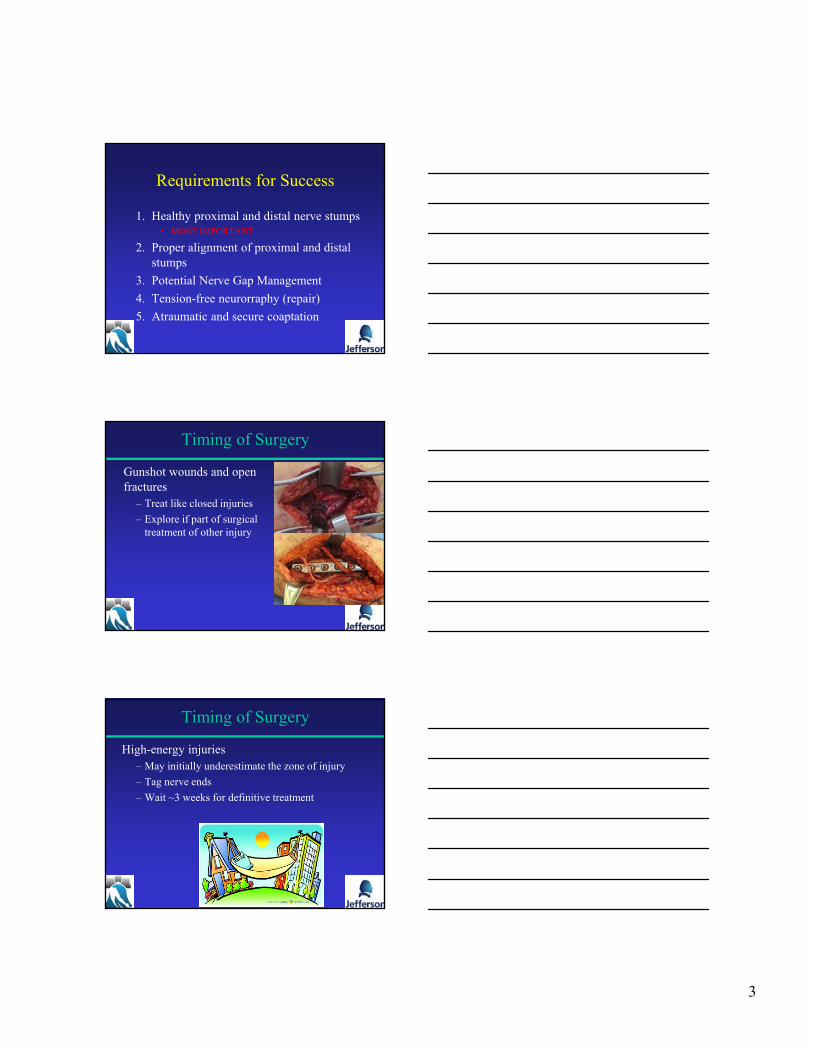
Gunshot wounds and open fractures
– Treat like closed injuries
– Explore if part of surgical treatment of other injury
Timing of Surgery
High-energy injuries – May initially underestimate the zone of injury
– Tag nerve ends
– Wait ~3 weeks for definitive treatment
Timing of Surgery

4
Patient positioning– Stable access to site
Microscope available
Intraoperative Considerations
Instruments
• Specialized microsurgical instruments– Preferably at least 10 cm long
– Fine spring loaded needle holders and scissors
• Surgical loupes – (see wealthy relative or win lotto)
• Operative microscope– (at least 20X)
Dissection

5
This is the MOST important step
Healthy Nerve Stumps
• Appreciate the zone of injury
• Need to remove damaged tissue both proximally and distally
• Do resection BEFORE determining final repair technique
Resection Matters
• Suturing Scarred Nerve Provides Limited Value
• Scar Inhibits Revascularization, Axonal Regeneration and Schwann Cell Migration
• Proximal Stump should have at least 75% preserved neural elements(Wolfe et al., 2013)
Limited Viable Neural
Elements
Berocal et al. 2013
Nerve Debridement
• Morphologic features of healthy nerve ends:
• Normal fascicular architecture
• Pliability / tactile feel of nerve
• Appearance of “pouting” fascicles
• Punctate endoneurial bleeding
• Tourniquet?
6
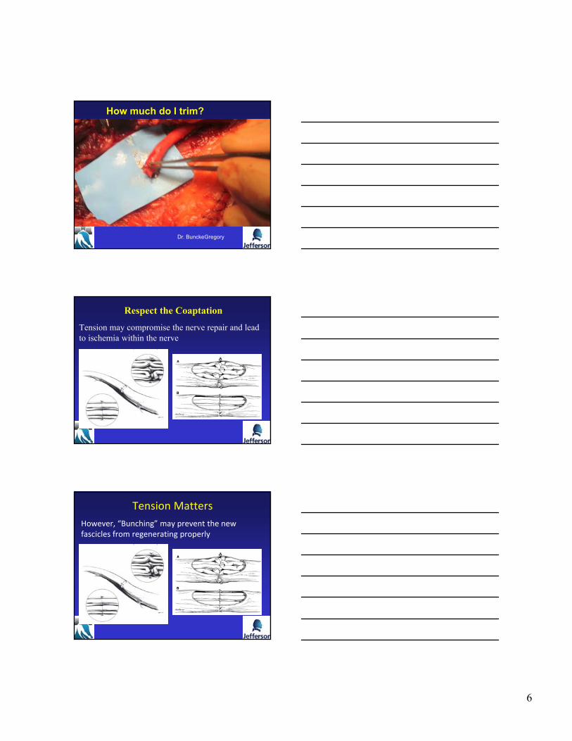
How much do I trim?
Dr. BunckeGregory
Respect the Coaptation
Tension may compromise the nerve repair and lead to ischemia within the nerve
Tension Matters
However, “Bunching” may prevent the new fascicles from regenerating properly
7
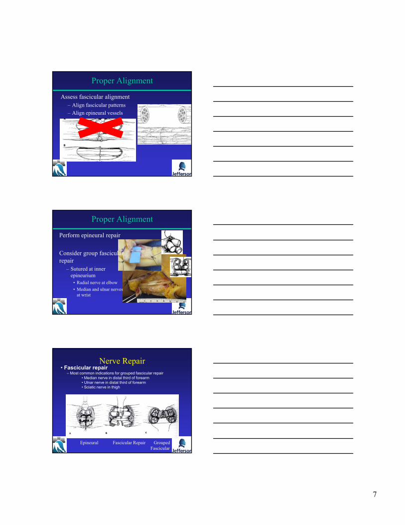
Assess fascicular alignment– Align fascicular patterns
– Align epineural vessels
Proper Alignment
Proper Alignment
Perform epineural repair
Consider group fascicular repair
– Sutured at inner epineurium
• Radial nerve at elbow
• Median and ulnar nerves at wrist
Nerve Repair
Epineural Fascicular Repair GroupedFascicular
• Fascicular repair– Most common indications for grouped fascicular repair
• Median nerve in distal third of forearm• Ulnar nerve in distal third of forearm• Sciatic nerve in thigh

8
The Coaptation
Assessing tension– Epineural sutures with 9-0 nylon
– Nerve ends “kissing” / “just touching”
– No gapping with extremity ROM
Giddins 1989 JHSBr.
Tension-Free Neurorraphy
Neutral posture of adjacent joints limits:
– Joint contractures
– Pain from tension during rehab
– Failure of repair
Tension-Free Neurorraphy
Nerve transposition/Mobilization ?– Not much gain, increased devascularization

9
• Appropriate number of sutures
• Conduit/connecter-assisted
• Fibrin glue– Decreases gapping
– Does not improve strength
Isaacs 2010 JHSAm. Isaacs 2008 JHSAm.
Atraumatic and Secure Coaptation
Tension Causes Restriction in the Blood Supply
With increased elongation, first signs of tissue injury are observed along with ~50% reduction in blood flow.
Excessive elongation can cause complete blockage of blood vessels.
At minimal elongation, there is slight scarring but blood vessels are not constricted.
References:Lundborg and Rydevik., 1973, J Bone Joint Surg.Clark, Trumble, et al., 1992 J Hand Surg Am.
Tension at the repair site: 8% stretch impedes venous return
16% stretch causes arterial ischemia
If attempt to repair gap creates tension
primary repair results in suboptimal outcome
Yi, et al Am J Surgery 193(1):e1-e6

10
What About the Gap
• Conduits
Autologous Vein Graft Entubulation
Commercially available Hollow Tube Conduits
Integra Lifesciences ‐ NeuraGen™ and NeuraWrap™ (bovine collagen)
Synovis ‐ Neurotube® (PGA)
Polyganics ‐ Neurolac® (poly‐ester)
AxoGuard Nerve Connector/Protector (porcine submucosa)
• Processed Nerve Allografts
Avance® Nerve Graft
• Autologous Nerve Grafts (Autograft)
Role of Tube Conduits• Tubes play an important role in modern nerve repair
• Consideration should be given to:– The function of the nerve
• Typically reserved for non-critical sensory nerves
– The length of the gap• Less than 20mm
– The diameter of the nerve• The larger the diameter, the shorter the gap should be
• Less than 6 mm in mixed nerves
• Resurgence as an aid to direct repair with very short gap (<5mm)
11
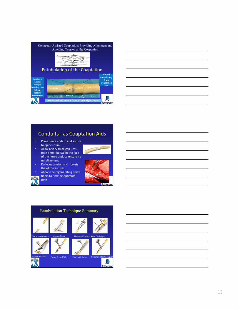
Connector Assisted Coaptation: Providing Alignment and Avoiding Tension at the Coaptation
Sutures moved away
from Coaptation
Site
No forced mismatch from overly tight repair
Barrier to Axonal Escape,
Scarring and Inflam-matory
Infiltration
Entubulation of the Coaptation
Conduits– as Coaptation Aids• Place nerve ends in and suture
to epineurium• Allow a very small gap (less
than 5mm) between the face of the nerve ends to ensure no misalignment.
• Reduces tension and fibrosis the of the sutures
• Allows the regenerating nerve fibers to find the optimum path
Entubulation Technique Summary
Trim to healthy nerve Horizontal Mattress Suture TechniqueMeasure Nerve
Flush with Saline Suture Second Side Flush with Saline Completed Nerve Repair

12
Increasing Gap Length
Fibrin cable is robust enough to allow regeneration at short gaps.Fibrin cable is robust enough to allow regeneration at short gaps.
Thinning restricts the regenerative space at longer gaps.
Thinning restricts the regenerative space at longer gaps.
Decre
asing Efficacy The cable does not form when
length limits are exceeded. This can result in no regeneration or a neuroma.
The cable does not form when length limits are exceeded. This can result in no regeneration or a neuroma.
References:Zhao, et al., 1993, Res Neurol and Neurosci.Whitlock et al., 2009 Muscle and Nerve
Length Limitations of Conduits
Tube Assisted Coaptation Clinical Outcomes
Gaps <6 mm Literature
StudyNerve Injury Types
Test Article Gap Outcomes*
Lundborg 1998 Mixed Silicone Coaptation Aid vs. Suture
<5mmComparable to Suture, better sensation
Weber et al., 2000 SensoryPGA Coaptation Aid vs. Suture
<5mm
91% Coaptation Aid, 49% Suture
Farole et al. 2009 SensoryWrapping vs. Non-Wrapping
<2mm<Pain with Wrapping
Boeckstyns et al. 2013 Sensory NervesCollagen Coaptation Aid vs. Suture
< 6mmComparable to Suture at 2 years
* As reported, based on individual study parameters for positive outcomes
Conduits Limitations
Liodaki et al., Journal Recon Micro. 2013
Isaacs et al., Major Peripheral Nerve Repair. Hand Clinics 2014
• Length • Impacts reliability of Outcomes
• Schmauss et al J. Recon Micro 2014, Lohmeyer et al., J. Recon Micro 2009
• Diameter• Proper size match is essential >1mm larger
decrease efficacy. • Isaacs et al JBJS 2014, Moore et al., Hand 2010
• Response to Materials• Extrusion.
• Rinker et al., JHS 2010, Weber J. Plas Recon 2000, Chiriac JHS EU 2011
• Encapsulation/Scar.• Moore et al., Hand 2010, Liodaki J Recon Micro 2013
Chiriac et al., JHS-EU 2011

13
Reports from Published Literature:▪ Lundborg et al. (199?)
▪ 5mm gap
▪ Silicone conduit equivalent to direct repair
▪ Weber et al. (2000)▪ <5mm 100% recovered Static 2PD
▪ ≥5mm 66% recovered Static 2PD
▪ Battiston et al. (2005)▪ Only 4/19 good/excellent results
▪ 3/4 for 3‐4cm gaps poor
▪ Lohmeyer et al. (2009)▪ Greater than 15mm no recovery
Limitations to Gap size?
Future of Tube Conduits
• ECM Components
• Internal Architecture
• Lumenal Fillers
• Neurotrophic Factors
• Neurtropic Factors
• Cell Delivery
• Electro-conductive
Bellamkonda et al., Biomaterials 2011
Deleterious effects of tension– Ischemia, Fibrosis
Limitations of nerve stretch:– 8% causes transient ischemia
– 10% may be acceptable in pliable nerves
– 15% leads to irreversible ischemia
Isaacs 2010 JHSAm. Isaacs 2008 JHSAm.
Tension-Free Neurorraphy

14
Nerve Repairs
•Direct muscular neurotization•insert proximal nerve stump into affected muscle belly•results in less than normal function but is indicated in certain cases
•Epineural Repair• Primary repair of the epineurium in a tension free fashion• First resect proximal neuroma and distal glioma• It is critical to properly align nerve ends during repair to maximize potential of recovery
Nerve Repairs
•Nerve grafting• Autologous graft
•remains the gold standard of repair for segmental defects > 5cm is autologous nerve grafting•digital nerve defects
•Wrist to common digital nerve bifurcation -use sural nerve•MCP to DIP level –
•Use LABC, AIN, PIN or MABC• Collagen conduit
•defects up to 1.5 cm•quality of nerve recovery drops with gaps >5mm
• Allograft•off-the-shelf option for defects up to 7cm
• 9-0 nylon for digital nerve repairs
• 8-0 nylon for median and ulnar nerve repair (wrist and above)
• 8-0 nylon for synthetic nerve conduits
What do I use ?

15
Splint extremity for soft tissue rest
Benefit of tension-free neurorraphy:• Begin hand therapy according to associated
injuries• Flexor tendons• Fractures
• Isolated nerve injuries• Initiate ROM 5-10 days following repair• Initiate ROM 2-3 weeks following nerve transfers
Postoperative Care
- Patient Age
- Type of Nerve Injured
- Mechanism of Injury
- Extent of concomitant injuries affecting tissue bed
- Location of injury
- Degree of injury
- Patient Co-morbidities
- Trimming back to healthy nerve tissue
- Alignment of nerve ends
- Leave a small gap ( < 5 mm) between nerve ends
- Tension free coaptations
- Barriers to Control Ingrowth and Axonal Escape
- Wrap vs. Connector vs. Allograft vs. Autograft
Summary Successful nerve repair will depend:
Case Example
42YO o/w healthy RDM male– Numbness L thumb and index
– Thumb weakness
– Minimal/no pain
– Irritating, does not interfere w/ ADL

16
History
• Started 1 ½ years ago– Diagnosed w/ CTS (w/ NCS)
– Endoscopic CTR• Uneventful according to op report
– Immed. post op: • Patient reports worsening pain/numbness and inability to abduct thumb
• Surgeon’s notes: sens intact to LT and motor fx normal– Sees patient regularly for 9 months
» Inconsistent exam, symptoms improving
» Subjective complaints noted
History continued
• Undergoes 3 separate NCS and ultrasound
– Increased latency and decreased amplitude
– EMG c/w denervation of APB
– U/S: c/w nerve constriction one cm distal to crease
• Patient told to “exercise” thumb
• After 9 months scheduled for revision CTR
– Case postponed when surgeon in car accident
Exam
• Thenar atrophy
• Inability to abduct thumb
• No sharp/dull discrimination thumb, index, radial half of middle finger
• Strong but non‐focal Tinel’s over carpal tunnel

17
Now what?
• More exercises?
• More time?
• More studies?
• Live with it?
• Surgery?
18
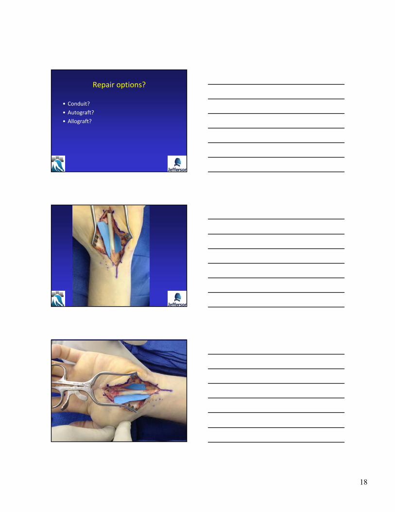
Repair options?
• Conduit?
• Autograft?
• Allograft?

19
Thank You
Case 2
• 71 yo taken to the OR emergently by trauma service for accidental self inflicted shot gun blast to left medial brachium (+ETOH)
– Emergent vein grafting to brachial artery
– Hand team (not me) identified median nerve which was intact
– Ulnar nerve not explored

20
• Sent to me 3 mos out with no median or ulnar nerve function
– Supported by NCS/EMG
• Now what?
– Wait it out?
– Nerve transfers?
– Tendon transfers?
– Explore
• How to repair?
Surgical exploration
• 4 mos post injury
• Nerves intact
– Median N. feels fibrotic.. Multiple areas of “neuroma‐in‐continuity”
– Ulnar nerve not as bad.. But not normal
• Now what?
– Intra‐operative nerve studies did not reveal any regenerating axons
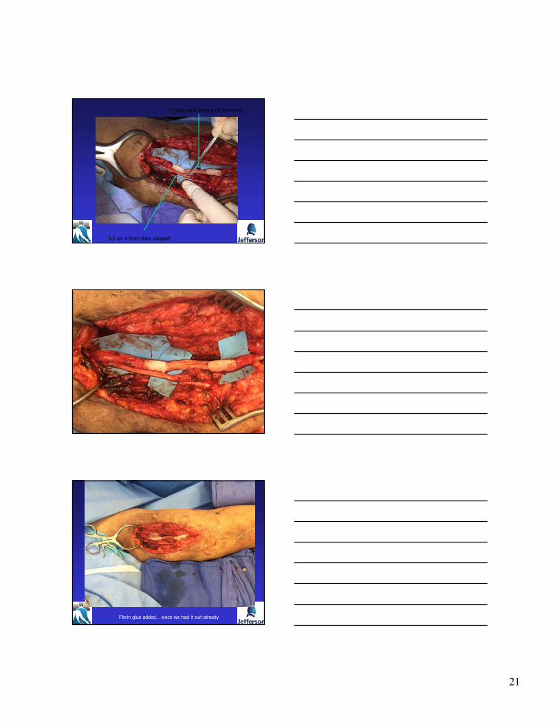
Median N…. 7-8 cm defect
Ulnar N… 5.5cm defect
Shotgun
pelletsfound
in both nerves
Now what?.....
21
4 cable sural nerve graft 8cm long
5.5 cm 4-5mm diam allograft
Fibrin glue added… since we had it out already

22
Nerve Injury Classification& Intervention Timing
Grabb & Smith’s Plastic Surgery, 2007